Development and multicenter validation of a CT-based ...
12
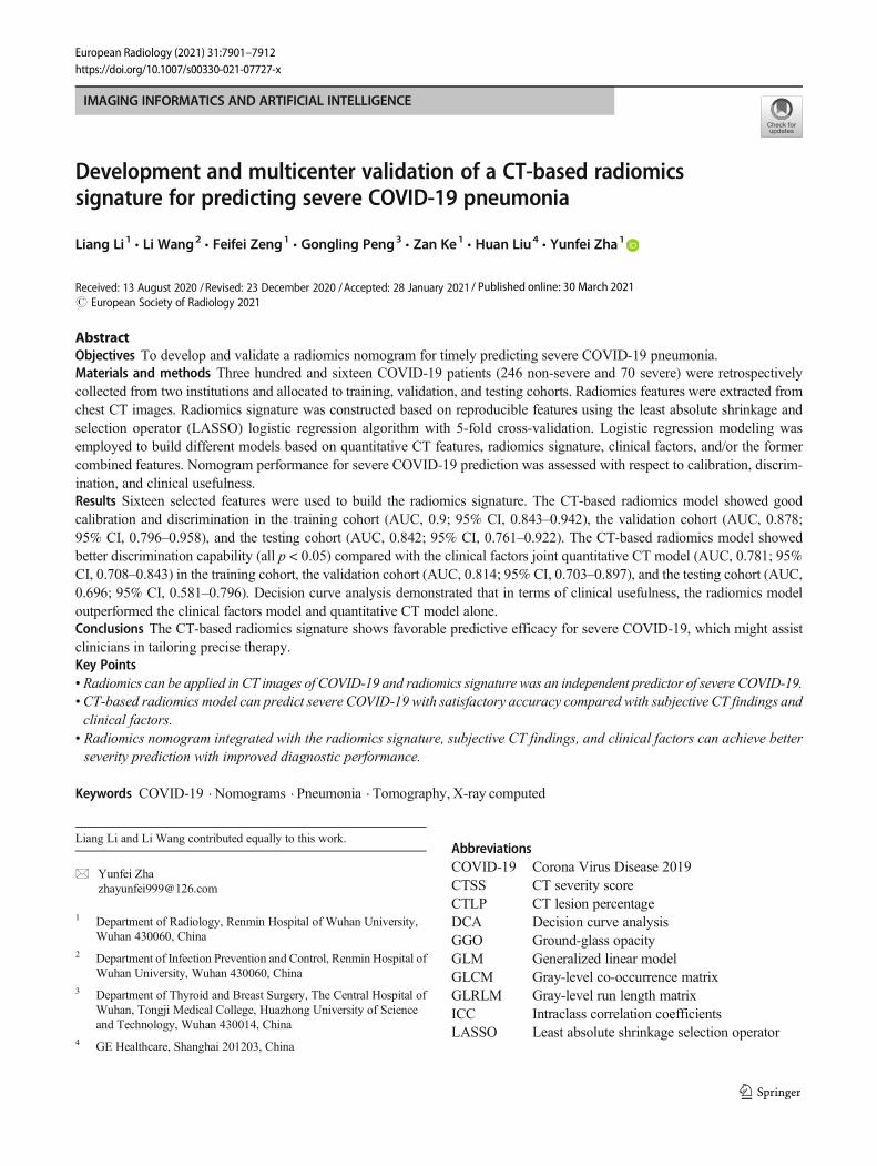
European Radiology (2021) 31:7901–7912 IMAGING INFORMATICS AND ARTIFICIAL INTELLIGENCE Development and multicenter validation of a CT-based radiomics signature for predicting severe COVID-19 pneumonia Liang Li 1 & Li Wang 2 & Feifei Zeng 1 & Gongling Peng 3 & Zan Ke 1 & Huan Liu 4 & Yunfei Zha 1 Received: 13 August 2020 /Revised: 23 December 2020 /Accepted: 28 January 2021 # European Society of Radiology 2021 Abstract Objectives To develop and validate a radiomics nomogram for timely predicting severe COVID-19 pneumonia. Materials and methods Three hundred and sixteen COVID-19 patients (246 non-severe and 70 severe) were retrospectively collected from two institutions and allocated to training, validation, and testing cohorts. Radiomics features were extracted from chest CT images. Radiomics signature was constructed based on reproducible features using the least absolute shrinkage and selection operator (LASSO) logistic regression algorithm with 5-fold cross-validation. Logistic regression modeling was employed to build different models based on quantitative CT features, radiomics signature, clinical factors, and/or the former combined features. Nomogram performance for severe COVID-19 prediction was assessed with respect to calibration, discrim- ination, and clinical usefulness. Results Sixteen selected features were used to build the radiomics signature. The CT-based radiomics model showed good calibration and discrimination in the training cohort (AUC, 0.9; 95% CI, 0.843–0.942), the validation cohort (AUC, 0.878; 95% CI, 0.796–0.958), and the testing cohort (AUC, 0.842; 95% CI, 0.761–0.922). The CT-based radiomics model showed better discrimination capability (all p < 0.05) compared with the clinical factors joint quantitative CT model (AUC, 0.781; 95% CI, 0.708–0.843) in the training cohort, the validation cohort (AUC, 0.814; 95% CI, 0.703–0.897), and the testing cohort (AUC, 0.696; 95% CI, 0.581–0.796). Decision curve analysis demonstrated that in terms of clinical usefulness, the radiomics model outperformed the clinical factors model and quantitative CT model alone. Conclusions The CT-based radiomics signature shows favorable predictive efficacy for severe COVID-19, which might assist clinicians in tailoring precise therapy. Key Points • Radiomics can be applied in CT images of COVID-19 and radiomics signature was an independent predictor of severe COVID-19. • CT-based radiomics model can predict severe COVID-19 with satisfactory accuracy compared with subjective CT findings and clinical factors. • Radiomics nomogram integrated with the radiomics signature, subjective CT findings, and clinical factors can achieve better severity prediction with improved diagnostic performance. Keywords COVID-19 . Nomograms . Pneumonia . Tomography, X-ray computed Abbreviations COVID-19 Corona Virus Disease 2019 CTSS CT severity score CTLP CT lesion percentage DCA Decision curve analysis GGO Ground-glass opacity GLM Generalized linear model GLCM Gray-level co-occurrence matrix GLRLM Gray-level run length matrix ICC Intraclass correlation coefficients LASSO Least absolute shrinkage selection operator Liang Li and Li Wang contributed equally to this work. * Yunfei Zha [email protected] 1 Department of Radiology, Renmin Hospital of Wuhan University, Wuhan 430060, China 2 Department of Infection Prevention and Control, Renmin Hospital of Wuhan University, Wuhan 430060, China 3 Department of Thyroid and Breast Surgery, The Central Hospital of Wuhan, Tongji Medical College, Huazhong University of Science and Technology, Wuhan 430014, China 4 GE Healthcare, Shanghai 201203, China https://doi.org/10.1007/s00330-021-07727-x / Published online: 30 March 2021
Transcript of Development and multicenter validation of a CT-based ...
European Radiology (2021) 31:7901–7912
IMAGING INFORMATICS AND ARTIFICIAL INTELLIGENCE
Development and multicenter validation of a CT-based radiomicssignature for predicting severe COVID-19 pneumonia
Liang Li1 & Li Wang2& Feifei Zeng1
& Gongling Peng3& Zan Ke1
& Huan Liu4& Yunfei Zha1
Received: 13 August 2020 /Revised: 23 December 2020 /Accepted: 28 January 2021# European Society of Radiology 2021
AbstractObjectives To develop and validate a radiomics nomogram for timely predicting severe COVID-19 pneumonia.Materials and methods Three hundred and sixteen COVID-19 patients (246 non-severe and 70 severe) were retrospectivelycollected from two institutions and allocated to training, validation, and testing cohorts. Radiomics features were extracted fromchest CT images. Radiomics signature was constructed based on reproducible features using the least absolute shrinkage andselection operator (LASSO) logistic regression algorithm with 5-fold cross-validation. Logistic regression modeling wasemployed to build different models based on quantitative CT features, radiomics signature, clinical factors, and/or the formercombined features. Nomogram performance for severe COVID-19 prediction was assessed with respect to calibration, discrim-ination, and clinical usefulness.Results Sixteen selected features were used to build the radiomics signature. The CT-based radiomics model showed goodcalibration and discrimination in the training cohort (AUC, 0.9; 95% CI, 0.843–0.942), the validation cohort (AUC, 0.878;95% CI, 0.796–0.958), and the testing cohort (AUC, 0.842; 95% CI, 0.761–0.922). The CT-based radiomics model showedbetter discrimination capability (all p < 0.05) compared with the clinical factors joint quantitative CT model (AUC, 0.781; 95%CI, 0.708–0.843) in the training cohort, the validation cohort (AUC, 0.814; 95% CI, 0.703–0.897), and the testing cohort (AUC,0.696; 95% CI, 0.581–0.796). Decision curve analysis demonstrated that in terms of clinical usefulness, the radiomics modeloutperformed the clinical factors model and quantitative CT model alone.Conclusions The CT-based radiomics signature shows favorable predictive efficacy for severe COVID-19, which might assistclinicians in tailoring precise therapy.Key Points• Radiomics can be applied in CT images of COVID-19 and radiomics signature was an independent predictor of severe COVID-19.•CT-based radiomics model can predict severe COVID-19 with satisfactory accuracy compared with subjective CT findings andclinical factors.
• Radiomics nomogram integrated with the radiomics signature, subjective CT findings, and clinical factors can achieve betterseverity prediction with improved diagnostic performance.
Keywords COVID-19 . Nomograms . Pneumonia . Tomography, X-ray computed
AbbreviationsCOVID-19 Corona Virus Disease 2019CTSS CT severity scoreCTLP CT lesion percentageDCA Decision curve analysisGGO Ground-glass opacityGLM Generalized linear modelGLCM Gray-level co-occurrence matrixGLRLM Gray-level run length matrixICC Intraclass correlation coefficientsLASSO Least absolute shrinkage selection operator
Liang Li and Li Wang contributed equally to this work.
* Yunfei [email protected]
1 Department of Radiology, Renmin Hospital of Wuhan University,Wuhan 430060, China
2 Department of Infection Prevention and Control, Renmin Hospital ofWuhan University, Wuhan 430060, China
3 Department of Thyroid and Breast Surgery, The Central Hospital ofWuhan, Tongji Medical College, Huazhong University of Scienceand Technology, Wuhan 430014, China
4 GE Healthcare, Shanghai 201203, China
https://doi.org/10.1007/s00330-021-07727-x
/ Published online: 30 March 2021

Eur Radiol (2021) 31:7901–7912
NGTDM Neighboring gray tone difference matrixNSD Non-severe diseaseROC Receiver operating characteristicSD Severe disease
Introduction
Corona Virus Disease 2019 (COVID-19) has spread out in theworld, posing a critical threat to public health [1]. Initial reportof the disease indicated that from November 16, 2019, toNovember 5, 2020, there were 47,596,852 confirmed casesof COVID-19, including 1,216,357 deaths, with a global fa-tality of approximately 2.56% [2].
According to the “Diagnosis and Treatment Program ofPneumonia of New Coronavirus Infection (Trial 7th Edition)”recommended by China’s National Health Commission, severeCOVID-19 patients were more likely to develop poor clinicaloutcomes including acute respiratory distress syndrome(ARDS), acute cardiac/kidney injury, andmultiple organ failure[3]. Current epidemiological data also suggests that the mortal-ity rate of severe COVID-19 is about 20 times higher than thatof non-severe COVID-19 [4]. Therefore, it is very crucial forearly identification of severe cases, which prevent disease pro-gression and reduce the mortality.
Computed tomography (CT) is widely used to diagnose,evaluate, and monitor COVID-19 pneumonia in high-risk areas[5]. However, the evaluation of these conventional CT featuresdepends heavily on the radiologist’s experience and is non-ob-jective. Furthermore, some scholars have tried using quantitativeCT method to assess the severity of lung injury in COVID-19pneumonia, such as visual CT severity score (CTSS) [6] and theCT lesion percentage (CTLP) [7] in each lung lobe, which werealternative ways to stratify COVID-2019 cases. The radiomicsapproach has drawn increased attention in recent years, whichmay help the detection, diagnosis, monitoring, and prognosticassessment of lung disease [8]. To our knowledge, there hasbeen rarely radiomics study for the accurate prediction of theclinical severity of COVID-19 pneumonia to date.
Therefore, the aim of our study is to (1) investigate the roleof radiomics features for predicting severe patients withCOVID-19 pneumonia, and (2) examine whether the additionof quantitative CT characteristics and/or clinical factors canimprove the performance of the predictive model.
Materials and methods
Patient population and groups
Our institutional review board approved this retrospectivestudy, and the requirement for informed consent was waived.
We searched the electronic database at two institutions ((I)Renmin Hospital of Wuhan University, (II) Central HospitalofWuhan optical valley) and retrospectively reviewed recordsfor patients between January 30, 2020, and April 30, 2020,and identified 449 patients infected with COVID-19. All pa-tients with COVID-19 were proven using real-time reversetranscriptase–polymerase chain reaction. The whole pipelineof our study was shown in Fig. 1.
According to the “Diagnosis and Treatment Program ofPneumonia of New Coronavirus Infection (Trial 7th
Edition)” recommended by China’s National HealthCommission, all patients are classified as having minimal,ordinary, severe, and critical type [9]. In our study, ordinarycases were included in the non-severe disease (NSD) group,while severe and critical cases were merged into the severedisease (SD) group. All patients in the SD group should meetany of the following criteria: (1) respiratory rate ≥ 30 breathsper minute; (2) finger of oxygen saturation ≤ 93% in a restingstate; (3) arterial oxygen tension (PaO2)/inspiratory oxygenfraction (FiO2) ≤ 300 mmHg (1 mmHg = 0.133 kPa); (4)respiratory failure occurred and mechanical ventilation re-quired; (5) shock; (6) other organ failure needing intensivecare unit (ICU) monitoring treatment.
Image acquisition and lesion segmentation
Non-enhanced chest CT scans of 316 patients were carried outfrom the lung apex to the lung base using multi-detector CT(MDCT) scanners (Brightspeed CT or Optima 680 CT, GEHealthcare) at the end of inspiration. Breath-hold training wascarried out before each examination. Parameters for chest CTscanning were listed as follows: field of view (FOV), 36 cm;tube voltage, 120 kV; tube current adjusted automatically; noiseindex, 13; section thickness, 5 mm; slice interval, 5 mm; pitch,1.375; collimation 64 × 0.625 mm; gantry rotation speed, 0.7 s;matrix, 512 × 512; the mediastinal window: window width of200 HU with a window level of 35 HU, and the lung window:window width of 1500 HU with a window level of - 700 HU.
All images were segmented on the commercial segmentationsoftware (Lung Intelligence Kit 2.1, LK 2.1, GE Healthcare)[10]. First, pre-processing was executed and included the fol-lowing steps: resampling adjust the x-spacing, y-spacing, z-spac-ing size (spatial resolution = 1 mm × 1 mm × 1 mm). Gaussianfilter with a standard deviation of 0.5 was applied for signalsmoothing. Then, the lung was automatically segmented intofive lobes; the volumes of interest (VOIs) were automaticallycontoured for each lobe. The segmentation results are manuallycorrected by a radiologist (Z.K., 2 years of experience in radiol-ogy) and then confirmed by another radiologist (F.Z., 5 years ofexperience in radiology). CTLP was defined as the volume ofthe lesions (including ground-glass opacity (GGO), consolida-tion, and reticulation) divided by the volume of the entire lung.CTSS was used to estimate pulmonary involvement of all
7902

Eur Radiol (2021) 31:7901–7912
abnormalities on the basis of the area involved [11]. Each of thefive lung lobes was visually scored from 0 to 5 as follows: 0, noinvolvement; 1, less than 5% involvement; 2, less than 25%involvement; 3, 26–49% involvement; 4, 50–75% involvement;or 5, more than 75% involvement [12].
Collection of clinical data and evaluation of CTradiological features
Clinical data were recorded, including the following 10characteristics: age, sex, duration of onset, comorbidity,clinical type, treatments, respiratory support strategies(RSS), ICU admission, length of ICU stay, and length ofhospital stay. We also recorded other clinical parameters,such as oxygen saturation and temperature, but whichwere not used in the clinic’s model, because the formerwas one of the known diagnostic criteria for severeCOVID-19, and the latter was a variable parameter dueto uncertain medical history. Comorbidities included hy-pertension, diabetes, chronic obstructive pulmonary dis-ease (COPD), chronic kidney disease (CKD), malignanttumors, and surgery history (on any part of the body inthe last 10 years). The number of comorbidities was from
0 to 4; 0 means no complications, and 4 means there are 4kinds of diseases. Treatments included antiviral therapy,antibiotic therapy, glucocorticoid therapy, immunoglobu-lin therapy, and Chinese medicine therapy. RSS includednasal catheter, high-flow nasal cannula oxygen therapy,non-invasive mechanical ventilation, invasive mechanicalventilation, and extracorporeal membrane oxygenation(ECMO).
All CT images were evaluated by 2 radiologists (Z.K. andL.L.) who were blinded to each subject’s clinical data. Fordisagreement between the two primary radiologist interpreta-tions, a third experienced thoracic radiologist with 25 years ofexperience (Y.Z.) adjudicated a final decision. Ten CT radio-logical features were assessed, namely GGO, consolidation,reticular pattern, interlobular septal thickening, airbronchogram sign, lesion location, distribution, involved lobe,thickening of pleura, and pleural effusion.
Radiomics feature extraction
A total of 851 radiomics features were extracted from the VOIssegmented based on the L. K software, including first-order sta-tistics parameters (n = 18), morphological parameters (n = 14),
316 patients with confirmed COVID-19 from two institutions
Training dataset
(Institution I, n=159)
Spearman analysis
Testing dataset
(Institution II, n=87)
Mann-Whitney U test
Lasso Logistic Regression
Radiomics signature
Multivariate Logistic Regression
analysis
Building a radiomics nomogram
Clinical
characteristic
Internal validation
Validation dataset
(Institution I, n=70)
External validation
CTSS and CTLP
quantification
Fig. 1 Flowchart of the study
7903

Eur Radiol (2021) 31:7901–7912
gray-level co-occurrence matrix (GLCM) parameters (n = 24),gray-level run length matrix (GLRLM) parameters (n = 16),gray-level size zone matrix (GLSZM) parameters (n = 16),gray-level dependence matrix (GLDM) parameters (n = 14),neighboring gray tone difference matrix (NGTDM) parameters(n = 5), and wavelet parameters (n = 744). All the featuresdefined were in compliance with feature definitions as describedby the Imaging Biomarker Standardization Initiative (IBSI) [13].The detailed workflow of radiomics analysis can be found inFig. 2. Intra- and interclass correlation coefficients (ICC) wereused to assess the intra- and inter-observer reproducibility ofradiomics feature extraction.
Radiomics features selection and radiomics signatureconstruction
The outlier values were replaced by the median value of theparticular variance vector once the values were beyond therange of the mean and standard deviation. And standardizationwas performed to scale the data in a specific interval.Spearman correlation, generalized linear model (GLM), andleast absolute shrinkage and selection operator (LASSO) wereused to reduce the redundancy or selection bias of the features,thereby removing a high correlation. A radiomics score (Rad-score) was calculated for each patient via a linear combinationof selected features that were weighted by their respectivecoefficients.
Development of predictive models
The most significant features were investigated to constructradiomics model based on logistic regression. The likelihoodratio test with backward step-down selection was applied tothe multivariate logistic regression model. We grouped theselected features into seven models—the radiomics model
(radiomics features), the CTSS model (semi-quantitativeCTSS), CTLP model (quantitative CTLP), the clinical model(clinical features), the integrated A model (CTSS + CTLP +clinical features), the integrated B model (clinical features +radiomics features), and the integrated C model (radiomicsfeatures + CTSS + CTLP + clinical features). The calibrationcurves were used to investigate the performance characteris-tics of the nomograms.
Statistical analysis
Statistical analyses were performedwith the Institute of PrecisionMedicine Statistics (IPMs, version 1.1, GE Healthcare). The dif-ferences in all variables between NSD and SD groups wereassessed using theMann-WhitneyU test or independent samplest test for continuous variables, and the chi-square test or Fisher’sexact test for categorical variables. Univariate analysis was usedto estimate the relationship between clinical factors and the iden-tification of the two subtypes. The performances of the sevenmodels were assessed by area under the receiver operating char-acteristic curve (AUC), specificity, and sensitivity. The optimalcut-off points to predict the severity of COVID-19 were deter-mined by Youden’s index. The DeLong test was used forpairwise comparisons among the seven models using the R soft-ware (v. 3.6.0; http://www.Rproject.org). A two-sided p < 0.05was considered statistically significant throughout the study.
Results
Clinical and CT radiological features
The detailed distribution of clinical characteristics in the NSD andSD group was summarized in Table 1. Clinical factors (genderand duration of onset) were found not significantly different
Fig. 2 Radiomics framework of predicting the severe patients with COVID-19
7904
Eur Radiol (2021) 31:7901–7912
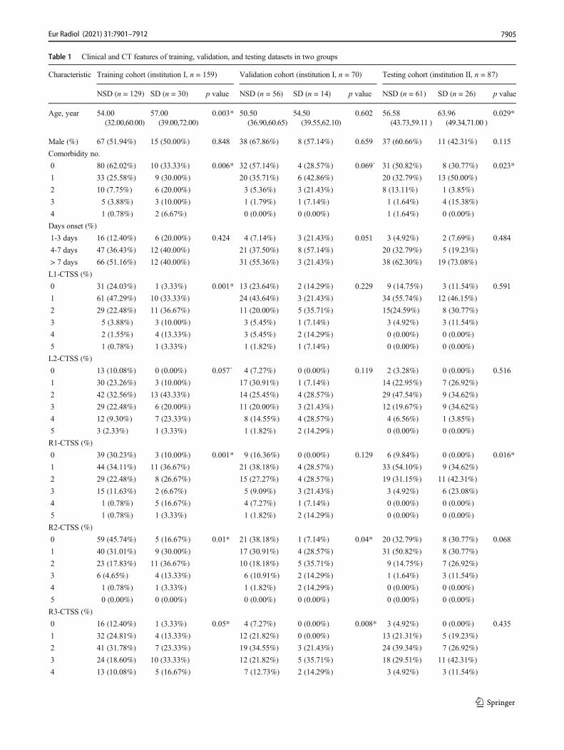
Table 1 Clinical and CT features of training, validation, and testing datasets in two groups
Characteristic Training cohort (institution I, n = 159) Validation cohort (institution I, n = 70) Testing cohort (institution II, n = 87)
NSD (n = 129) SD (n = 30) p value NSD (n = 56) SD (n = 14) p value NSD (n = 61) SD (n = 26) p value
Age, year 54.00(32.00,60.00)
57.00(39.00,72.00)
0.003* 50.50(36.90,60.65)
54.50(39.55,62.10)
0.602 56.58(43.73,59.11 )
63.96(49.34,71.00 )
0.029*
Male (%) 67 (51.94%) 15 (50.00%) 0.848 38 (67.86%) 8 (57.14%) 0.659 37 (60.66%) 11 (42.31%) 0.115
Comorbidity no.
0 80 (62.02%) 10 (33.33%) 0.006* 32 (57.14%) 4 (28.57%) 0.069` 31 (50.82%) 8 (30.77%) 0.023*
1 33 (25.58%) 9 (30.00%) 20 (35.71%) 6 (42.86%) 20 (32.79%) 13 (50.00%)
2 10 (7.75%) 6 (20.00%) 3 (5.36%) 3 (21.43%) 8 (13.11%) 1 (3.85%)
3 5 (3.88%) 3 (10.00%) 1 (1.79%) 1 (7.14%) 1 (1.64%) 4 (15.38%)
4 1 (0.78%) 2 (6.67%) 0 (0.00%) 0 (0.00%) 1 (1.64%) 0 (0.00%)
Days onset (%)
1-3 days 16 (12.40%) 6 (20.00%) 0.424 4 (7.14%) 3 (21.43%) 0.051 3 (4.92%) 2 (7.69%) 0.484
4-7 days 47 (36.43%) 12 (40.00%) 21 (37.50%) 8 (57.14%) 20 (32.79%) 5 (19.23%)
> 7 days 66 (51.16%) 12 (40.00%) 31 (55.36%) 3 (21.43%) 38 (62.30%) 19 (73.08%)
L1-CTSS (%)
0 31 (24.03%) 1 (3.33%) 0.001* 13 (23.64%) 2 (14.29%) 0.229 9 (14.75%) 3 (11.54%) 0.591
1 61 (47.29%) 10 (33.33%) 24 (43.64%) 3 (21.43%) 34 (55.74%) 12 (46.15%)
2 29 (22.48%) 11 (36.67%) 11 (20.00%) 5 (35.71%) 15(24.59%) 8 (30.77%)
3 5 (3.88%) 3 (10.00%) 3 (5.45%) 1 (7.14%) 3 (4.92%) 3 (11.54%)
4 2 (1.55%) 4 (13.33%) 3 (5.45%) 2 (14.29%) 0 (0.00%) 0 (0.00%)
5 1 (0.78%) 1 (3.33%) 1 (1.82%) 1 (7.14%) 0 (0.00%) 0 (0.00%)
L2-CTSS (%)
0 13 (10.08%) 0 (0.00%) 0.057` 4 (7.27%) 0 (0.00%) 0.119 2 (3.28%) 0 (0.00%) 0.516
1 30 (23.26%) 3 (10.00%) 17 (30.91%) 1 (7.14%) 14 (22.95%) 7 (26.92%)
2 42 (32.56%) 13 (43.33%) 14 (25.45%) 4 (28.57%) 29 (47.54%) 9 (34.62%)
3 29 (22.48%) 6 (20.00%) 11 (20.00%) 3 (21.43%) 12 (19.67%) 9 (34.62%)
4 12 (9.30%) 7 (23.33%) 8 (14.55%) 4 (28.57%) 4 (6.56%) 1 (3.85%)
5 3 (2.33%) 1 (3.33%) 1 (1.82%) 2 (14.29%) 0 (0.00%) 0 (0.00%)
R1-CTSS (%)
0 39 (30.23%) 3 (10.00%) 0.001* 9 (16.36%) 0 (0.00%) 0.129 6 (9.84%) 0 (0.00%) 0.016*
1 44 (34.11%) 11 (36.67%) 21 (38.18%) 4 (28.57%) 33 (54.10%) 9 (34.62%)
2 29 (22.48%) 8 (26.67%) 15 (27.27%) 4 (28.57%) 19 (31.15%) 11 (42.31%)
3 15 (11.63%) 2 (6.67%) 5 (9.09%) 3 (21.43%) 3 (4.92%) 6 (23.08%)
4 1 (0.78%) 5 (16.67%) 4 (7.27%) 1 (7.14%) 0 (0.00%) 0 (0.00%)
5 1 (0.78%) 1 (3.33%) 1 (1.82%) 2 (14.29%) 0 (0.00%) 0 (0.00%)
R2-CTSS (%)
0 59 (45.74%) 5 (16.67%) 0.01* 21 (38.18%) 1 (7.14%) 0.04* 20 (32.79%) 8 (30.77%) 0.068
1 40 (31.01%) 9 (30.00%) 17 (30.91%) 4 (28.57%) 31 (50.82%) 8 (30.77%)
2 23 (17.83%) 11 (36.67%) 10 (18.18%) 5 (35.71%) 9 (14.75%) 7 (26.92%)
3 6 (4.65%) 4 (13.33%) 6 (10.91%) 2 (14.29%) 1 (1.64%) 3 (11.54%)
4 1 (0.78%) 1 (3.33%) 1 (1.82%) 2 (14.29%) 0 (0.00%) 0 (0.00%)
5 0 (0.00%) 0 (0.00%) 0 (0.00%) 0 (0.00%) 0 (0.00%) 0 (0.00%)
R3-CTSS (%)
0 16 (12.40%) 1 (3.33%) 0.05* 4 (7.27%) 0 (0.00%) 0.008* 3 (4.92%) 0 (0.00%) 0.435
1 32 (24.81%) 4 (13.33%) 12 (21.82%) 0 (0.00%) 13 (21.31%) 5 (19.23%)
2 41 (31.78%) 7 (23.33%) 19 (34.55%) 3 (21.43%) 24 (39.34%) 7 (26.92%)
3 24 (18.60%) 10 (33.33%) 12 (21.82%) 5 (35.71%) 18 (29.51%) 11 (42.31%)
4 13 (10.08%) 5 (16.67%) 7 (12.73%) 2 (14.29%) 3 (4.92%) 3 (11.54%)
7905
Eur Radiol (2021) 31:7901–7912
between the two groups in all cohorts, while the comorbiditiesand age were the only similarity between the two groups in thevalidation cohort.
Three hundred sixteen patients (NSD, 246 vs. SD, 70)from two hospitals were included in the study. The patientsin both groups showed GGO with mainly peripheral distri-bution; there was no statistical difference (78% vs. 84%,
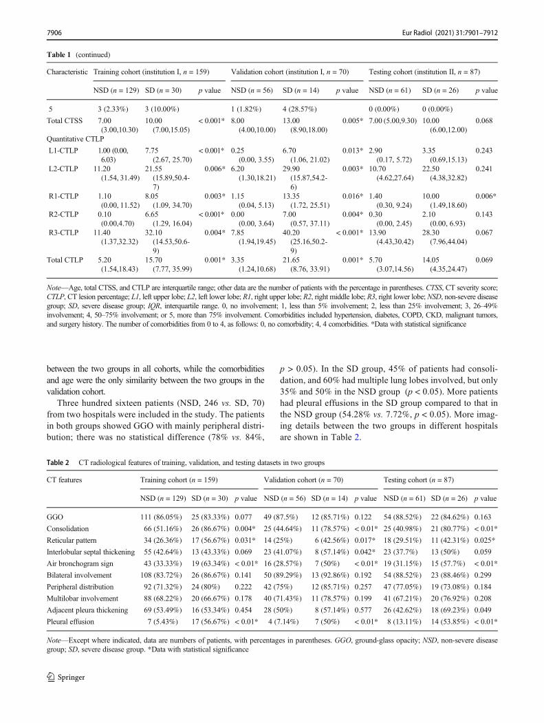
p > 0.05). In the SD group, 45% of patients had consoli-dation, and 60% had multiple lung lobes involved, but only35% and 50% in the NSD group (p < 0.05). More patientshad pleural effusions in the SD group compared to that inthe NSD group (54.28% vs. 7.72%, p < 0.05). More imag-ing details between the two groups in different hospitalsare shown in Table 2.
Table 1 (continued)
Characteristic Training cohort (institution I, n = 159) Validation cohort (institution I, n = 70) Testing cohort (institution II, n = 87)
NSD (n = 129) SD (n = 30) p value NSD (n = 56) SD (n = 14) p value NSD (n = 61) SD (n = 26) p value
5 3 (2.33%) 3 (10.00%) 1 (1.82%) 4 (28.57%) 0 (0.00%) 0 (0.00%)
Total CTSS 7.00(3.00,10.30)
10.00(7.00,15.05)
< 0.001* 8.00(4.00,10.00)
13.00(8.90,18.00)
0.005* 7.00 (5.00,9.30) 10.00(6.00,12.00)
0.068
Quantitative CTLP
L1-CTLP 1.00 (0.00,6.03)
7.75(2.67, 25.70)
< 0.001* 0.25(0.00, 3.55)
6.70(1.06, 21.02)
0.013* 2.90(0.17, 5.72)
3.35(0.69,15.13)
0.243
L2-CTLP 11.20(1.54, 31.49)
21.55(15.89,50.4-7)
0.006* 6.20(1.30,18.21)
29.90(15.87,54.2-6)
0.003* 10.70(4.62,27.64)
22.50(4.38,32.82)
0.241
R1-CTLP 1.10(0.00, 11.52)
8.05(1.09, 34.70)
0.003* 1.15(0.04, 5.13)
13.35(1.72, 25.51)
0.016* 1.40(0.30, 9.24)
10.00(1.49,18.60)
0.006*
R2-CTLP 0.10(0.00,4.70)
6.65(1.29, 16.04)
< 0.001* 0.00(0.00, 3.64)
7.00(0.57, 37.11)
0.004* 0.30(0.00, 2.45)
2.10(0.00, 6.93)
0.143
R3-CTLP 11.40(1.37,32.32)
32.10(14.53,50.6-9)
0.004* 7.85(1.94,19.45)
40.20(25.16,50.2-9)
< 0.001* 13.90(4.43,30.42)
28.30(7.96,44.04)
0.067
Total CTLP 5.20(1.54,18.43)
15.70(7.77, 35.99)
0.001* 3.35(1.24,10.68)
21.65(8.76, 33.91)
0.001* 5.70(3.07,14.56)
14.05(4.35,24.47)
0.069
Note—Age, total CTSS, and CTLP are interquartile range; other data are the number of patients with the percentage in parentheses. CTSS, CT severity score;CTLP, CT lesion percentage; L1, left upper lobe; L2, left lower lobe; R1, right upper lobe; R2, right middle lobe; R3, right lower lobe; NSD, non-severe diseasegroup; SD, severe disease group; IQR, interquartile range. 0, no involvement; 1, less than 5% involvement; 2, less than 25% involvement; 3, 26–49%involvement; 4, 50–75% involvement; or 5, more than 75% involvement. Comorbidities included hypertension, diabetes, COPD, CKD, malignant tumors,and surgery history. The number of comorbidities from 0 to 4, as follows: 0, no comorbidity; 4, 4 comorbidities. *Data with statistical significance
Table 2 CT radiological features of training, validation, and testing datasets in two groups
CT features Training cohort (n = 159) Validation cohort (n = 70) Testing cohort (n = 87)
NSD (n = 129) SD (n = 30) p value NSD (n = 56) SD (n = 14) p value NSD (n = 61) SD (n = 26) p value
GGO 111 (86.05%) 25 (83.33%) 0.077 49 (87.5%) 12 (85.71%) 0.122 54 (88.52%) 22 (84.62%) 0.163
Consolidation 66 (51.16%) 26 (86.67%) 0.004* 25 (44.64%) 11 (78.57%) < 0.01* 25 (40.98%) 21 (80.77%) < 0.01*
Reticular pattern 34 (26.36%) 17 (56.67%) 0.031* 14 (25%) 6 (42.56%) 0.017* 18 (29.51%) 11 (42.31%) 0.025*
Interlobular septal thickening 55 (42.64%) 13 (43.33%) 0.069 23 (41.07%) 8 (57.14%) 0.042* 23 (37.7%) 13 (50%) 0.059
Air bronchogram sign 43 (33.33%) 19 (63.34%) < 0.01* 16 (28.57%) 7 (50%) < 0.01* 19 (31.15%) 15 (57.7%) < 0.01*
Bilateral involvement 108 (83.72%) 26 (86.67%) 0.141 50 (89.29%) 13 (92.86%) 0.192 54 (88.52%) 23 (88.46%) 0.299
Peripheral distribution 92 (71.32%) 24 (80%) 0.222 42 (75%) 12 (85.71%) 0.257 47 (77.05%) 19 (73.08%) 0.184
Multilobar involvement 88 (68.22%) 20 (66.67%) 0.178 40 (71.43%) 11 (78.57%) 0.199 41 (67.21%) 20 (76.92%) 0.208
Adjacent pleura thickening 69 (53.49%) 16 (53.34%) 0.454 28 (50%) 8 (57.14%) 0.577 26 (42.62%) 18 (69.23%) 0.049
Pleural effusion 7 (5.43%) 17 (56.67%) < 0.01* 4 (7.14%) 7 (50%) < 0.01* 8 (13.11%) 14 (53.85%) < 0.01*
Note—Except where indicated, data are numbers of patients, with percentages in parentheses. GGO, ground-glass opacity; NSD, non-severe diseasegroup; SD, severe disease group. *Data with statistical significance
7906

Eur Radiol (2021) 31:7901–7912
Radiomics features selection and radiomics signaturebuilding
In total, 99 features with statistical significance (p < 0.05)between the NSD and SD groups were selected in the train-ing dataset. A radiomics signature was further constructedbased on sixteen features with respective non-zero coeffi-cients selected from these 99 features. One first-order fea-ture and fifteen wavelet-based features were used to repre-sent the radiomics signature. Details of the procedure forthe construction of the radiomics signature are showed inFig. 3a, b. The lists of the selected features and their asso-ciated coefficients in the logistic regression model are il-lustrated in Fig. 3c. The intra-observer ICCs ranged from0.822 to 0.957 and the inter-observer ICCs ranged from0.769 to 0.936, indicating favorable intra- and inter-observer feature extraction reproducibility. Figure 4 showsrepresentative images and lesion segmentation result ofsevere COVID-19
The utility of severity prediction using developedradiomics signature
The developed initial CT-based radiomics signature modelshowed a favorable result in predicting the severity (NSD vs.SD) that produced an AUC of 0.9 in the training set (95% CI,0.843 to 0.942), 0.878 in the internal validation set (95% CI,0.796 to 0.958), and 0.842 in the testing set (95% CI, 0.761 to0.922).
As shown in Fig. 5, predictive nomogram and correlationcoefficients were built by combining Rad-score, age, comor-bidity, L1-CTSS, L1-CTLP, R1-CTLP, and R2-CTLP. Thepredictive nomogram had the best differentiation ability ofthe severe cases with an AUC of 0.918 (95% CI, 0.864–0.956) in the training set, an AUC of 0.934 (95% CI, 0.848–0.979) in the validation set, and an AUC of 0.854 (95% CI,0.762–0.92) in the testing set, as shown in Fig. 6a–c. Thecalibration curves of the nomogram also showed that the pre-dictions agreed well with the observations in Fig. 6d–f.
Fig. 3 Texture feature selection using the least absolute shrinkage andselection operator (LASSO) binary logistic regression model. a LASSOcoefficient profiles of the radiomics features. Vertical line was drawn atthe value selected using 5-fold cross-validation in the ln(alpha) sequence,and 16 non-zero coefficients are indicated. b The tuning parameter λ
selection in the LASSO model used 5-fold cross-validation via the min-imum criteria. Mean square error was plotted vs. log (λ). The dottedvertical lines were drawn at the optimal values using theminimum criteriaand the 1-SE criteria. c Multivariate logistic of the predictive radiomicsfeatures. OR, odds ratio
7907

Eur Radiol (2021) 31:7901–7912
Evaluation of models and comparison of predictivemodel performance
The diagnostic performance of each model is shown inTable 3 and the results of ROC curve analysis areshown in Fig. 6. In all cohorts, the integrated C modelachieved the best performance and radiomics modeloutperformed clinic’s model for predicting severe
COVID-19. Moreover, the AUC of the integrated Bmodel was better than CTSS model and CTLP model(all p < 0.05). In the training set, although the integrat-ed C model showed the highest AUC (0.918) among theseven predictive models, no statistical difference wasfound between any of the two models using theDeLong test (p = 0.832 for CTSS vs. CTLP; p =0.165 for CTSS vs. clinics; p = 0.323 for CTLP vs.
Fig. 4 A case of confirmed severeCOVID-19. A 62-year-old femalepresented with a 7-day history offever and cough. First CT imag-ing revealed diffuse pure GGOwith mainly peripheral distribu-tion in the bilateral lobes (a). Thearea of the lesions was delineatedon the axial, coronal, and recon-structed three-dimensional im-ages (b, c, d). CTSS = total 10scores, predicted probability forsevere COVID-19 = 90.1%
Variable Coefficient P Value
Age -0.094 0.047
Comorbidities 1.940 0.010
L1-CTSS 1.141 0.253
L1-CTLP -0.076 0.315
R2-CTLP 0.110 0.077
R1-CTLP -0.053 0.344
Radscore 0.669 0.003
Constant 2.225 0.262
a b
Fig. 5 Radiomics-based nomogram (a) and their correlation coefficients (b) were developed in the training set, including the Rad-score, age,comorbidities, CTSS, and CTLP
7908

Eur Radiol (2021) 31:7901–7912
clinics). No statistical difference in AUC was also foundbetween any of the two models (p = 0.09 for CTSS vs.integrated A; p = 0.137 for CTLP vs. integrated A; p =0.751 for clinics vs. integrated A), but a statistical
difference in AUC was found between the radiomicsmodel and the integrated A model (p = 0.017), andbetween radiomics model and clinics model (p =0.00075)
Fig. 6 The ROC curves of the seven prediction models that indicatesevere COVID-19 cases in the training cohort (a), validation cohort (b),and testing cohort (c). Calibration curves of the combined nomogram inthe training cohort (d), internal validation cohort (e), and testing cohort
(f). Calibration curves depict the calibration of the nomogram in terms ofagreement between the predicted risk and actual probability for severeCOVID-19
Table 3 Comparison of predictive model performance for identifying severe COVID-19 pneumonia
Model Training cohort (n = 159) Validation cohort (n = 70) Testing cohort (n = 87)
AUC (95% CI) Sensitivity Specificity AUC (95% CI) Sensitivity Specificity AUC (95% CI) Sensitivity Specificity
Radiomics model 0.90 (0.84–0.94) 0.80 0.85 0.88 (0.80–0.96) 1.00 0.70 0.84 (0.76–0.92) 0.92 0.71
CTSS model 0.77 (0.70–0.83) 0.93 0.54 0.82 (0.71–0.90) 0.79 0.82 0.67 (0.56–0.77) 0.73 0.56
CTLP model 0.71 (0.63–0.78) 0.68 0.77 0.84 (0.74–0.92) 0.71 0.91 0.68 (0.57–0.77) 0.88 0.41
Clinical model 0.72 (0.64–0.78) 0.50 0.84 0.78 (0.66–0.87) 0.79 0.79 0.67 (0.54–0.76) 0.52 0.87
Integrated A model 0.78 (0.71–0.84) 0.73 0.77 0.82 (0.70–0.90) 0.79 0.82 0.70 (0.58–0.80) 0.70 0.67
Integrated B model 0.91 (0.85–0.95) 0.97 0.69 0.93 (0.84–0.98) 0.93 0.82 0.84 (0.75–0.91) 0.92 0.71
Integrated C model 0.92 (0.86–0.96) 0.73 0.95 0.93 (0.85–0.98) 0.79 0.95 0.84 (0.76–0.92) 0.92 0.72
Note—The integrated A model contained CTSS, CTLP, and clinical features. The integrated B model contained the selected radiomics features andclinical features. The integrated C model contained the selected radiomics features, CTSS, CTLP, and clinical features. CTSS, CT severity score; CTLP,CT lesion percentage; AUC, area under the receiver operating characteristic curve
7909
Eur Radiol (2021) 31:7901–7912
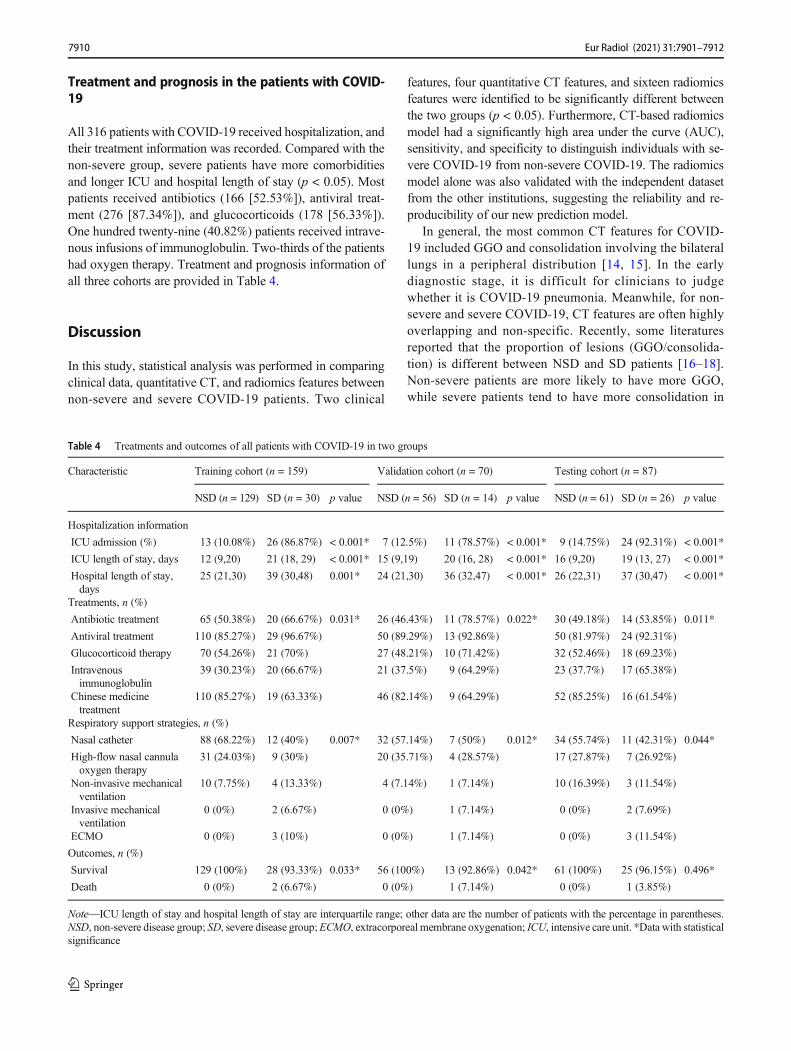
Treatment and prognosis in the patients with COVID-19
All 316 patients with COVID-19 received hospitalization, andtheir treatment information was recorded. Compared with thenon-severe group, severe patients have more comorbiditiesand longer ICU and hospital length of stay (p < 0.05). Mostpatients received antibiotics (166 [52.53%]), antiviral treat-ment (276 [87.34%]), and glucocorticoids (178 [56.33%]).One hundred twenty-nine (40.82%) patients received intrave-nous infusions of immunoglobulin. Two-thirds of the patientshad oxygen therapy. Treatment and prognosis information ofall three cohorts are provided in Table 4.
Discussion
In this study, statistical analysis was performed in comparingclinical data, quantitative CT, and radiomics features betweennon-severe and severe COVID-19 patients. Two clinical
features, four quantitative CT features, and sixteen radiomicsfeatures were identified to be significantly different betweenthe two groups (p < 0.05). Furthermore, CT-based radiomicsmodel had a significantly high area under the curve (AUC),sensitivity, and specificity to distinguish individuals with se-vere COVID-19 from non-severe COVID-19. The radiomicsmodel alone was also validated with the independent datasetfrom the other institutions, suggesting the reliability and re-producibility of our new prediction model.
In general, the most common CT features for COVID-19 included GGO and consolidation involving the bilaterallungs in a peripheral distribution [14, 15]. In the earlydiagnostic stage, it is difficult for clinicians to judgewhether it is COVID-19 pneumonia. Meanwhile, for non-severe and severe COVID-19, CT features are often highlyoverlapping and non-specific. Recently, some literaturesreported that the proportion of lesions (GGO/consolida-tion) is different between NSD and SD patients [16–18].Non-severe patients are more likely to have more GGO,while severe patients tend to have more consolidation in
Table 4 Treatments and outcomes of all patients with COVID-19 in two groups
Characteristic Training cohort (n = 159) Validation cohort (n = 70) Testing cohort (n = 87)
NSD (n = 129) SD (n = 30) p value NSD (n = 56) SD (n = 14) p value NSD (n = 61) SD (n = 26) p value
Hospitalization information
ICU admission (%) 13 (10.08%) 26 (86.87%) < 0.001* 7 (12.5%) 11 (78.57%) < 0.001* 9 (14.75%) 24 (92.31%) < 0.001*
ICU length of stay, days 12 (9,20) 21 (18, 29) < 0.001* 15 (9,19) 20 (16, 28) < 0.001* 16 (9,20) 19 (13, 27) < 0.001*
Hospital length of stay,days
25 (21,30) 39 (30,48) 0.001* 24 (21,30) 36 (32,47) < 0.001* 26 (22,31) 37 (30,47) < 0.001*
Treatments, n (%)
Antibiotic treatment 65 (50.38%) 20 (66.67%) 0.031* 26 (46.43%) 11 (78.57%) 0.022* 30 (49.18%) 14 (53.85%) 0.011*
Antiviral treatment 110 (85.27%) 29 (96.67%) 50 (89.29%) 13 (92.86%) 50 (81.97%) 24 (92.31%)
Glucocorticoid therapy 70 (54.26%) 21 (70%) 27 (48.21%) 10 (71.42%) 32 (52.46%) 18 (69.23%)
Intravenousimmunoglobulin
39 (30.23%) 20 (66.67%) 21 (37.5%) 9 (64.29%) 23 (37.7%) 17 (65.38%)
Chinese medicinetreatment
110 (85.27%) 19 (63.33%) 46 (82.14%) 9 (64.29%) 52 (85.25%) 16 (61.54%)
Respiratory support strategies, n (%)
Nasal catheter 88 (68.22%) 12 (40%) 0.007* 32 (57.14%) 7 (50%) 0.012* 34 (55.74%) 11 (42.31%) 0.044*
High-flow nasal cannulaoxygen therapy
31 (24.03%) 9 (30%) 20 (35.71%) 4 (28.57%) 17 (27.87%) 7 (26.92%)
Non-invasive mechanicalventilation
10 (7.75%) 4 (13.33%) 4 (7.14%) 1 (7.14%) 10 (16.39%) 3 (11.54%)
Invasive mechanicalventilation
0 (0%) 2 (6.67%) 0 (0%) 1 (7.14%) 0 (0%) 2 (7.69%)
ECMO 0 (0%) 3 (10%) 0 (0%) 1 (7.14%) 0 (0%) 3 (11.54%)
Outcomes, n (%)
Survival 129 (100%) 28 (93.33%) 0.033* 56 (100%) 13 (92.86%) 0.042* 61 (100%) 25 (96.15%) 0.496*
Death 0 (0%) 2 (6.67%) 0 (0%) 1 (7.14%) 0 (0%) 1 (3.85%)
Note—ICU length of stay and hospital length of stay are interquartile range; other data are the number of patients with the percentage in parentheses.NSD, non-severe disease group; SD, severe disease group;ECMO, extracorporeal membrane oxygenation; ICU, intensive care unit. *Data with statisticalsignificance
7910

Eur Radiol (2021) 31:7901–7912
CT imaging. Although we have also observed this phe-nomenon, no statistical difference was found (p > 0.05).Therefore, it is also very difficult for the radiologist tojudge whether it is severe pneumonia [19, 20].
Radiomics has great potential to capture useful medical in-formation and to enhance the accuracy of clinical differentialdiagnosis. In this study, 851 candidate radiomics features wereextracted from CT images and were reduced to only 16 poten-tial predictors by using a LASSO regression model to developthe radiomics signatures. The selected radiomics features weredivided into two types (first-order and wavelet features) andvaried significantly between NSD and SD groups. These fea-tures reflect intrinsical information from the distribution of pixelintensity and the texture morphology that cannot be detected byradiologists [21]. For example, first-order features mainly re-flect the internal texture of lesions. Wavelet features mainlyreflect the change of time domain and frequency domain insidethe lesion. Among the selected radiomics features in this study,Wave l e t -HHL_g l cm_ Imc2 and Wave l e t -LHL_glrlm_ShortRunLowGrayLevelEmphasis were the most signif-icant and robust features associated with severe cases, whichreflect lesion’s intensity and textural features within the high-intensity CT voxels.
Several studies have reported and analyzed the value oftraditional CT characteristics and clinical features in the diag-nosis of COVID-19. Chen et al [22] developed and validated adiagnostic model for COVID-19 based on CT imaging andclinical manifestations. Kang et al [23] introduced an AI-assisted model based on the radiological and clinical featuresto estimate the clinical progression to critical illness. In addi-tion, in order to quantify the severity of COVID-19, CT quan-tification of pneumonia lesions is also used in clinical practice.The CTSS and CTLP are the two most common CT quantifi-cation methods, but the differences among observers are stillinevitable [7, 16, 22]. In our model, L1-CTSS, L1-CTLP, R1-CTLP, and R2-CTLP were selected as independent risk fac-tors for severe COVID-19 cases. The possible explanation isthat the SARS-CoV-2mainly invades the bilateral lower lobesin patients with COVID-19, so the difference in the distribu-tion of lesions in the upper or middle lobe is often more sta-tistically different among different patients with COVID-19.
The integrated Cmodel composed of radiomics, quantitativeCT, and clinical features had the highest AUC (0.92, 0.93, 0.84)in all three cohorts. The model with radiomics features alonereached an AUC of 0.9, 0.878, and 0.842, which is not inferiorto the integrated B or Cmodel (all p > 0.05). In other words, theaddition of quantitative CT and clinical features to the integrat-ed model did not increase the model’s efficacy, suggesting thestrong efficacy of radiomics as a tool to predict the severity ofCOVID-19. Furthermore, the predictive power of the radiomicsmodel also outperformed that of integrated A model (clinics +quantitative CT) (p < 0.05). In fact, radiomics features andclinical or quantitative CT features were highly correlated.
This result was in accordance with that in other similar studiesin gliomas [24], pancreatic neuroendocrine tumor (PNET) [23],lung adenocarcinoma [21], and breast cancer [25].
Several studies reported single or multiple risk factors of lunginjury for severe COVID-19. Dong et al [26] found that under-lying comorbidity, older age, higher LDH, and lower lympho-cyte count were independent high-risk factors associated withCOVID-19 progression in a multicenter study. Guan et al [27]reported that increasing age and comorbidity were associatedwith the disease. Zhou et al [28] showed that older age, highSOFA score, and D-dimer value are potential risk factors toidentify patients with poor prognosis at an early stage. Chenet al [29] proposed the MuLBSTA score as an early warningindicator for predicting 90-day mortality in patients withCOVID-19. Among the clinical features in our study, age andcomorbidity were the most powerful factors to predict severeCOVID-19. A possible reason is that poor immunosenescenceof older patients with comorbidities could have higher levels ofangiotensin-converting enzyme 2 in their alveoli [30], whichwas regarded as a receptor for novel coronaviruses [31].
Some limitations in this study should also be acknowl-edged. The biggest limitation is that no blood samples or someother clinical parameters are considered in the risk model. Webelieve that a reliable and robust multi-modality predictionmodel should be further developed to address this issue. Inaddition, the sample size was relatively small in order to buildup a stable predicting model and the cases outside of Hubeiprovince are not included. However, in this study, we tried toadopt a multicenter research method, including internal andexternal validation sets, subjective and objective CT evalua-tion, all of which ensure that the conclusions are reasonable.Furthermore, with the retrospective design, there may be po-tential biases in identifying and recruiting participants.Increasing a large number of different regional populationsand imaging-pathologic control studies is needed in the futurestudy.
In conclusion, initial CT-based radiomics features providean excellent performance for the prediction of severe COVID-19. Sixteen features were significantly different between thetwo groups. This prediction model based on the radiomicsfeatures was developed and validated on the training, valida-tion, and testing cohorts, which may have broad clinicalapplications.
Funding This study has received funding from the National NaturalScience Foundation of China (Nos. 81871332, 81601461), NovelCoronavirus Pneumonia Emergency Key Project of Science andTechnology of Hubei Province (No. 2020FCA015), and theFundamental Research Funds for the Central Universities (No.2042020kfxg10).
Compliance with ethical standards
Guarantor The scientific guarantor of this publication is Yunfei Zha.
7911

Eur Radiol (2021) 31:7901–7912
Statistics and biometry Statistician Huan Liu kindly provided all statis-tical work for this study.
Informed consent Written informed consent was waived by theInstitutional Review Board.
Ethical approval Institutional Review Board approval was obtained.
Conflict of interest One of the authors of this manuscript (Huan Liu) isan employee of GE Healthcare. The remaining authors declare no rela-tionships with any companies whose products or services may be relatedto the subject matter of the article.
Methodology• retrospective• diagnostic or prognostic study• multicenter study
References
1. Zhu N, Zhang D, Wang W et al (2020) A novel coronavirus frompatients with pneumonia in China, 2019. N Engl JMed 382:727–733
2. World Health Organization. Coronavirus disease (COVID-19) pan-demic on 5 November 2020 [EB/OL]. https://www.whoint/emergencies/diseases/novel-coronavirus-2019. Accessed 5Nov 2020
3. Huang C, Wang Y, Li X et al (2020) Clinical features of patientsinfected with 2019 novel coronavirus in Wuhan, China. Lancet395:497–506
4. Henry BM, de Oliveira MHS, Benoit S, Plebani M, Lippi G (2020)Hematologic, biochemical and immune biomarker abnormalitiesassociated with severe illness and mortality in coronavirus disease2019 (COVID-19): a meta-analysis. Clin Chem Lab Med. https://doi.org/10.1515/cclm-2020-0369
5. XiongY, Sun D, Liu Y et al (2020) Clinical and high-resolution CTfeatures of the COVID-19 infection: comparison of the initial andfollow-up changes. Invest Radiol 55:332–339
6. Ding X, Xu J, Zhou J, Long Q (2020) Chest CT findings ofCOVID-19 pneumonia by duration of symptoms. Eur J Radiol127:109009
7. Shen C, Yu N, Cai S et al (2020) Quantitative computed tomogra-phy analysis for stratifying the severity of Coronavirus Disease2019. J Pharm Anal. https://doi.org/10.1016/j.jpha.2020.03.004
8. Lambin P, Rios-Velazquez E, Leijenaar R et al (2012) Radiomics:extracting more information from medical images using advancedfeature analysis. Eur J Cancer 48:441–446
9. The National HealthCommission of the People’s Republic of China.Diagnosis and treatment protocols of COVID-19 infection (trial ver-sion 7) [EB/OL]. http://www.nhc.gov.cn/yzygj/s7653p/202002/8334a8326dd94d329df351d7da8aefc2s.html. Accessed 3 Mar 2020
10. WeiW, HuXW, ChengQ, Zhao YM,GeYQ (2020) Identification ofcommon and severe COVID-19: the value of CT texture analysis andcorrelation with clinical characteristics. Eur Radiol 30:6788–6796
11. Pan F, Ye T, Sun P et al (2020) Time course of lung changes onchest CT during recovery from 2019 novel coronavirus (COVID-19) pneumonia. Radiology. https://doi.org/10.1148/radiol.2020200370:200370
12. Li K, Wu J, Wu F et al (2020) The clinical and chest CT featuresassociated with severe and critical COVID-19 pneumonia. InvestRadiol 55:327–331
13. Zwanenburg A, Vallieres M, Abdalah MA et al (2020) The imagebiomarker standardization initiative: standardized quantitative
radiomics for high-throughput image-based phenotyping.Radiology 295:328–338
14. Bao C, Liu X, Zhang H, Li Y, Liu J (2020) Coronavirus Disease2019 (COVID-19) CT findings: a systematic review andmeta-anal-ysis. J Am Coll Radiol. https://doi.org/10.1016/j.jacr.2020.03.006
15. Zhou S, Wang Y, Zhu T, Xia L (2020) CT features of coronavirusdisease 2019 (COVID-19) pneumonia in 62 patients inWuhan, China.AJR Am J Roentgenol. https://doi.org/10.2214/AJR.20.22975:1-8
16. Li K, Fang Y, Li W et al (2020) CT image visual quantitativeevaluation and clinical classification of coronavirus disease(COVID-19). Eur Radiol 30:4407–4416
17. Pan F, Ye T, Sun P et al (2020) Time course of lung changes atchest CT during recovery from coronavirus disease 2019 (COVID-19). Radiology 295:715–721
18. Wang Y, Dong C, Hu Y et al (2020) Temporal changes of CTfindings in 90 patients with COVID-19 pneumonia: a longitudinalstudy. Radiology 296:E55–E64
19. Lyu P, Liu X, Zhang R, Shi L, Gao J (2020) The performance ofchest CT in evaluating the clinical severity of COVID-19 pneumo-nia: identifying critical cases based on CT characteristics. InvestRadiol. https://doi.org/10.1097/RLI.0000000000000689
20. Wu J, Wu X, Zeng W et al (2020) Chest CT findings in patientswith coronavirus disease 2019 and its relationship with clinicalfeatures. Invest Radiol 55:257–261
21. Song L, Zhu Z, Mao L et al (2020) Clinical, conventional CT andradiomic feature-based machine learning models for predictingALK rearrangement status in lung adenocarcinoma patients. FrontOncol 10:369
22. Chen X, Tang Y, Mo Y et al (2020) A diagnostic model for coro-navirus disease 2019 (COVID-19) based on radiological semanticand clinical features: a multi-center study. Eur Radiol. https://doi.org/10.1007/s00330-020-06829-2
23. Zhang K, Liu XH, Shen J et al (2020) Clinically applicable AIsystem for accurate diagnosis, quantitative measurements and prog-nosis of COVID-19 pneumonia using computed tomography. Cell.https://doi.org/10.1016/j.cell.2020.04.045
24. Wang Y, Wei W, Liu Z et al (2020) Predicting the type of tumor-related epilepsy in patients with low-grade gliomas: a radiomicsstudy. Front Oncol 10:235
25. Liu Z, Li Z, Qu J et al (2019) Radiomics ofmultiparametricMRI forpretreatment prediction of pathologic complete response to neoad-juvant chemotherapy in breast cancer: a multicenter study. ClinCancer Res 25:3538–3547
26. Ji D, Zhang D, Xu J et al (2020) Prediction for progression risk inpatients with COVID-19 pneumonia: the CALL score. Clin InfectDis. https://doi.org/10.1093/cid/ciaa414
27. Guan WJ, Ni ZY, Hu Y et al (2020) Clinical characteristics ofcoronavirus disease 2019 in China. N Engl J Med. https://doi.org/10.1056/NEJMoa2002032
28. Zhou F, Yu T, Du R et al (2020) Clinical course and risk factors formortality of adult inpatients with COVID-19 in Wuhan, China: aretrospective cohort study. Lancet 395:1054–1062
29. Guo L, Wei D, Zhang X et al (2019) Clinical features predictingmortality risk in patients with viral pneumonia: the MuLBSTAscore. Front Microbiol 10:2752
30. Pera A, Campos C, Lopez N et al (2015) Immunosenescence: im-plications for response to infection and vaccination in older people.Maturitas 82:50–55
31. Reynolds HR, Adhikari S, Pulgarin C et al (2020) Renin-angiotensin-aldosterone system inhibitors and risk of Covid-19. NEngl J Med. https://doi.org/10.1056/NEJMoa2008975
Publisher’s note Springer Nature remains neutral with regard to jurisdic-tional claims in published maps and institutional affiliations.
7912