Development and characterization of oil-in-water ... · Development and Characterization of...
114
e University of Toledo e University of Toledo Digital Repository eses and Dissertations 2011 Development and characterization of oil-in-water nanoemulsions from self-microemulsifying mixtures Ishan Shah e University of Toledo Follow this and additional works at: hp://utdr.utoledo.edu/theses-dissertations is esis is brought to you for free and open access by e University of Toledo Digital Repository. It has been accepted for inclusion in eses and Dissertations by an authorized administrator of e University of Toledo Digital Repository. For more information, please see the repository's About page. Recommended Citation Shah, Ishan, "Development and characterization of oil-in-water nanoemulsions from self-microemulsifying mixtures" (2011). eses and Dissertations. 715. hp://utdr.utoledo.edu/theses-dissertations/715
Transcript of Development and characterization of oil-in-water ... · Development and Characterization of...

The University of ToledoThe University of Toledo Digital Repository
Theses and Dissertations
2011
Development and characterization of oil-in-waternanoemulsions from self-microemulsifyingmixturesIshan ShahThe University of Toledo
Follow this and additional works at: http://utdr.utoledo.edu/theses-dissertations
This Thesis is brought to you for free and open access by The University of Toledo Digital Repository. It has been accepted for inclusion in Theses andDissertations by an authorized administrator of The University of Toledo Digital Repository. For more information, please see the repository's Aboutpage.
Recommended CitationShah, Ishan, "Development and characterization of oil-in-water nanoemulsions from self-microemulsifying mixtures" (2011). Thesesand Dissertations. 715.http://utdr.utoledo.edu/theses-dissertations/715

A Thesis
entitled
Development and Characterization of Oil-in-Water Nanoemulsions from
Self-Microemulsifying Mixtures
by
Ishan Shah
Submitted to the Graduate Faculty as partial fulfillment of the requirements for
the Master of Science Degree in Pharmaceutical Sciences
with Industrial Pharmacy option
______________________________________
Jerry Nesamony, Ph.D., Committee Chair
______________________________________
Kenneth Alexander, Ph.D., Committee Member
______________________________________
Curtis D. Black, Pharm.D., Committee Member
______________________________________
Sai Hanuman Sagar Boddu, Ph.D.,
Committee Member
____________________________________
Patricia R. Komuniecki, Dean
College of Graduate Studies
The University of Toledo
May 2011

Copyright 2011, Ishan Shah
This document is copyrighted material. Under copyright law, no parts of this document
may be reproduced without the expressed permission of the author.

iii
An Abstract of
Development and Characterization of Oil-in-Water Nanoemulsions from
Self-Microemulsifying Mixtures
by
Ishan Shah
As partial fulfillment of the requirements of the
Master of Science Degree in Pharmaceutical Sciences with Industrial Pharmacy Option
The University of Toledo
May 2011
Self-Microemulsifying Drug Delivery Systems (SMEDDS) are isotropic mixtures of oil,
surfactant and/or cosurfactants, and a drug that spontaneously forms an oil-in-water
nanoemulsion upon gentle agitation with water. When dispersed in the gastrointestinal
(GI) tract, the motility of stomach provides necessary agitation for emulsification.
SMEDDS incorporated with a poorly water soluble drug demonstrates improved drug
absorption since it maintains the drug in a solubilized state in the GIT tract. The purpose
of this research was to screen lipid excipients for their self-microemulsification efficiency
and to develop SMEDDS based formulation in liquid and solid forms using ibuprofen as
a model drug. Excipients evaluated for SMEDDS were Tween 80 and Cremophor RH 40
as surfactants, Transcutol P, Capyrol 90 and PEG 400 as cosurfactants and Labrafac
Lipophile WL 1349 (a medium chain triglyceride) as the oil. Self Microemulsifying
(SME) mixtures containing various proportions of these components were tested for their
self-microemulsification ability and were characterized by ternary phase diagrams. Based
on these results, a particular mixture containing Tween 80-PEG 400-LL WL 1349 was
selected and optimized for drug delivery purpose. Liquid SMEDDS was formulated by

iv
dissolving ibuprofen in the SME mixture. Solid SMEDDS was formulated by adsorbing
Liquid SMEDDS onto an inert carrier Neusilin US2 by physical mixing. SMEDDS were
analyzed for their droplet size and zeta potential. Solid state characterization of Solid
SMEDDS was performed using scanning electron microscopy, differential scanning
calorimetry and powder x-ray diffractometry. Finally, in vitro drug release studies were
performed on Liquid and Solid SMEDDS and the results compared to plain ibuprofen
dissolution. Ibuprofen was found to be physically and chemically stable in the SMEDDS
and did not precipitate upon aqueous dilution. Solid and Liquid SMEDDS showed a
droplet size of less than 50 nm and possessed a neutral zeta potential. Solid state
characterization of Solid SMEDDS confirmed the presence of ibuprofen in a molecularly
dissolved state in the formulation. In vitro drug release studies showed that 75 % of drug
was released from Solid and Liquid SMEDDS within first five minutes of the dissolution
time. A SMEDDS based dosage form was successfully developed and shows potential for
application in the delivery of poorly water soluble drugs.

v
Acknowledgements
I would like to thank Dr. Jerry Nesamony for providing me an opportunity to be a part of
his research group. His advice and suggestions were very helpful to me throughout the
two years of graduate study. He made all the possible resources available for me to
succeed in the research project. I thank Dr. Kenneth Alexander for being a part of my
thesis committee and for giving me a wonderful TA experience in his lab.
I also thank Drs. Black and Boddu, members on my thesis committee for their support
and guidance. I thank Dr. Joseph Lawrence at CMSC, UT for his help with the DSC and
SEM. I thank Dr. Burckel for her assistance with PXRD. I thank Dr. Surya Nauli and
Maki for their help with optical microscopy. I thank my fellow labmates Bivash, Ermias,
Sriramya, Meghavi, Praytusha, Priti, Sanjeev, Ashish, Heather, Vishak and Sharath. We
have had some wonderful times over the past two years.
Finally, I thank my dad, mom, sister and uncle for their encouragement and support. I
thank my dear friend Kinjal for her support throughout these two years.

vi
Contents
Abstract………………………………………………………………………………...…iii
Acknowledgements………………………………………………………………………..v
Contents………….……………………………………………………………...………..vi
List of Tables……………………………………………………………………..............ix
List of Figures……………………………………………………………………..............x
1. Self-Emulsifying Drug Delivery Systems (SEDDS)/Self-Microemulsifying
Drug Delivery Systems (SMEDDS)………………..….………………..………………...1
1.1 Lipid based drug delivery………………………………………….…………2
1.2 Self-Emulsifying / Microemulsifying drug delivery systems…………………7
1.3 Excipient selection for lipid based formulations……………………………...8
1.3.1 Oils………………………………………………...………..............9
1.3.2 Surfactants………………………………………………………….11
1.3.3 Cosolvents……………………………………………………….....13
1.4 Role of SEDDS/SMEDDS in improvement of oral absorption……...............13
1.5 Formulation of SEDDS/SMEDDS…………………………..........................17
1.6 Mechanism of Self-emulsification……………………………………...……19
1.7 Conversion of Liquid SEDDS to Solid SEDDS……………………..............19

vii
1.8 Dosage form development of SEDDS/SMEDDS…………………………....22
2. Instrumentation……………………………………………………………………..26
2.1 Dynamic Light Scattering…………………………………………………....26
2.2 Electrophoretic Light Scattering………………………………………….….29
2.3 Scanning Electron Microscopy………………………………….…………...31
2.4 Differential Scanning Calorimetry……………………………..………….…33
2.5 Powder X –ray Diffraction……………………………………..………….…37
2.6 UV/Vis spectrophotometry……………………………………..…………....41
3. Methods and Materials…..………………………………………………..…………...45
3.1 Materials. ………………………………………………………...............….45
3.1.1 Tween 80………………………………………………………...…45
3.1.2 Cremophor RH 40……………………………………………….....46
3.1.3 Labrafac Lipophile WL 1349………………………………………48
3.1.4 Polyethylene Glycol 400…………………………………………...49
3.1.5 Capyrol 90……………………………………………….................50
3.1.6 Transcutol P……………………………………………..................51
3.1.7 Ibuprofen……………………………………………….…………..52
3.1.8 Neusilin US2…………………………………………….................53
3.2 Methods……………………………………………………………………...54
3.2.1 Screening of lipid excipients for SMEDDS………………………..54
3.2.2 Test for emulsification……………………………………………..55
3.2.3 Drug Solubility………………………………………………….…56
3.2.4 Preparation of Liquid SMEDDS………………………………...…56

viii
3.2.5 Preparation of Solid SMEDDS…………………………………….57
3.2.6 Droplet size and Zeta Potential of SMEDDS……………………..57
3.2.7 Morphology of Solid SMEDDS…………………………………..58
3.2.8 Differential Scanning Calorimetry of Solid SMEDDS…………….58
3.2.9 Powder X-ray Diffraction of Solid SMEDDS……………………..58
3.2.10 In vitro drug release from SMEDDS…………………………….59
4. Results and Discussions…………………………………………………………….…60
4.1 Screening of lipid excipients for SMEDDS ………………………………....60
4.2 Ternary phase diagrams……………………………………………………...61
4.3 Drug solubility studies………………………………………………….……68
4.4 Liquid and Solid SMEDDS………………………………………………….69
4.5 Characterization of Liquid and Solid SMEDDS……………………………..71
4.5.1 Droplet size analysis………………………………………….……71
4.5.2 Zeta potential…………………………………………………....…73
4.5.3 Morphology of SMEDDS………………………………………….74
4.5.4 Differential Scanning Calorimetry of Solid SMEDDS…………….77
4.5.5 Powder X ray diffraction of Solid SMEDDS……………………...78
4.5.6 In vitro drug release studies………………………………………..79
5 Conclusions……………………………………………………………….……………84
References………………………………………………………….…………………….86

ix
List of Tables
1.1 Lipid Formulation Classification System (LFCS) showing typical
proportions of lipid formulations……………………………..…………………..3
1.2 Characteristics, advantages and disadvantages of lipid formulations……….…….4
1.3 Examples of Type III lipid based formulations…………………………………...6
3.1 Lipid excipients used in SMEDDS……………………………………………....54
3.2 Composition of an optimized Solid SMEDDS…………………………………..57
4.1 Time taken by various SME mixtures to completely self-microemulsify……….64
4.2 Solubility of ibuprofen in various excipients…………………………………….68
4.3 Cumulative percentage drug release for liquid SMEDDS, Solid SMEDDS
and plain ibuprofen……………………………………………….……………..80

x
List of Figures
1-1 Mechanism of drug partitioning from S(M)EDDS.……………………………….8
1-2 Pathways of drug absorption from lipid based formulations………………….....14
2-1 Schematics of Dynamic Light Scattering (DLS) instrument…………………….27
2-2 Schematics of Electrophoretic Light Scattering (ELS) instrument……………....30
2-3 Schematics of Scanning Electron Microscopy (SEM) instrument……………....32
2-4 A typical thermogram obtained from Differential Scanning Calorimetry……….34
2-5 Power compensated DSC………………………………………………………...35
2-6 Heat Flux DSC…………………………………………………………………...36
2-7 Diffraction of X rays by crystalline material…………………………………….38
2-8 Schematics of Powder X ray Diffractometer…………………………………….39
2-9 Energy diagram showing various energy transitions………………………...…42
2-10 Schematics of Single beam spectrophotometer………………………………….43
2-11 Schematics of Double beam spectrophotometer………………………………....43
3-1 Chemical structure of Tween 80……………………………………………...….46
3-2 Chemical structure of Hydrogenated castor oil………………………………. 47
3-3 Chemical structure of Caprylic/Capric triglyceride……………………………...48
3-4 Chemical structure of Polyethylene glycol 400………………………………….49

xi
3-5 Chemical structure of Capyrol 90………………………………………………..50
3-6 Chemical structure of Transcutol P……………………………………………....51
3-7 Chemical structure of Ibuprofen…………………………………………………52
4-1 Results from test of emuslification. (a) milky emulsion (b) slightly turbid
emulsion (c) clear nanoemulsion………………………………………………...63
4-2 Ternary phase diagrams of SME mixture comprising of
Tween 80, Capyrol 90, LL WL1349…………………....……………………..…65
4-3 Ternary phase diagrams of SME mixture comprising of
Cremophor RH 40, Capyrol 90, LL WL 1349…………….……….…….………65
4-4 Ternary phase diagrams of SME mixture comprising of
Tween 80, Transcutol P, LL WL 1349………………………….………….……66
4-5 Ternary phase diagrams of SME mixture comprising of
Cremophor RH 40, Transcutol P, LL WL 1349…………………..……………..66
4-6 Ternary phase diagrams of SME mixture comprising of
Cremophor RH 40, PEG 400, LL WL 1349…………………….……………….67
4-7 Ternary phase diagrams of SME mixture comprising of
Tween 80, PEG 400, LL WL 1349………………………….……….……...…...67
4-8 Droplet size distribution of nanoemulsion obtained from Liquid SMEDDS……72
4-9 Droplet size distribution of nanoemulsion obtained from Solid SMEDDS……...72
4-10 Zeta potential distribution of nanoemulsion obtained from Liquid SMEDDS.….74
4-11 Scanning Electron Microscopy images of: (a) Neusilin US2 (b) Solid SMEDDS
(c) Ibuprofen ….....................................................................................................75

xii
4-12 Differential Scanning Calorimetry (DSC) of Ibuprofen, Physical mixture
(Ibuprofen and Neusilin US2), Neusilin US2 and Solid SMEDDS…….………...77
4-13 Powder X-ray Diffraction (PXRD) of Ibuprofen, Physical mixture
(Ibuprofen and Neusilin US2), Neusilin US2 and Solid SMEDDS……………....78
4-14 Calibration of Ibuprofen in PBS pH 7.2…………………………………………79
4-15 Cumulative percentage drug release from Liquid SMEDDS,
plain Ibuprofen and Solid SMEDDS…………………………………………….81

1
Chapter 1
Self-Emulsifying Drug Delivery Systems (SEDDS)/Self-Microemulsifying Drug
Delivery Systems (SMEDDS)
Approximately one third of the drugs emerging from drug discovery programs are poorly
water soluble, presenting the pharmaceutical scientist with several problems when
developing formulations for such active pharmaceutical ingredients (API). Conventional
oral dosage forms for poorly water soluble drugs present the drug in a solid form to the
gastrointestinal tract which means the drug has to dissolve in the GI fluids before it can
be absorbed. Thus, their rate and extent of absorption is largely dependent on the rate of
dissolution. The formulation technique plays an important role in overcoming this
shortcoming of poorly water soluble drugs. According to the Biopharmaceutical
Classification System (BCS) classification, two classes of drugs show poor aqueous
solubility namely BCS II and BCS IV. BCS II drugs possess poor aqueous solubility but
have good permeation properties. BCS class IV drugs are poorly water soluble and
poorly permeable. Developing a formulation for a class IV drug is nearly impossible

2
unless the dose necessary is very small. Most of the times, such drugs are withdrawn at
its lead optimization stage of drug discovery and reworked to improve its physico-
chemical properties. Developing a formulation for a drug belonging to BCS II is often
challenging as it requires improved dissolution characteristics. Popular formulation
techniques used for delivering a poorly water soluble drug include: (a) micronized
crystalline solids (b) amorphous formulation or solid solutions and (c) lipid based
formulations. When particles of drug are milled to smaller particle sizes, there is an
increase in surface area resulting in an increased dissolution of the drug. Micronization
using an air jet mill will yield particles in the size range of 2-5 µm. Using a new
technique NanoCrystal®, which employs high-speed attrition process, particles can be
reduced in nanometer ranges. Such powders can be processed into tablets and capsules
[1]. Solid dispersions can be defined as a “dispersion of one or more active ingredient in
an inert excipient or matrix” wherein the active ingredient exists in a finely crystalline,
solubilized or amorphous state [2]. When solid dispersions are exposed to aqueous media,
the matrix dissolves and releases the drug as very fine colloidal particles. This results in
increased dissolution of the drug. This research project involves development of lipid
based formulations and hence I will elaborate more on this topic below.
1.1 Lipid based drug delivery
Lipid based drug delivery consists of delivering a drug dissolved in a mixture of one or
more excipients which may be a mono, di and tri-glyceride, lipophilic and hydrophilic
surfactants and a cosurfactant. When a drug is delivered through lipid formulations, it
remains in the dissolved state throughout its transit in the GI tract. The absorption of the

3
drug when presented in a solubilized form within a colloidal dispersion is enhanced since
the drug dissolution step is partially evaded.
In the year 2000, Pouton classified lipid based formulations into three categories based on
their composition and properties. In 2006, this classification was updated by adding one
more class (Type IV) as shown in Table 1.1 [3].
Table 1.1: Lipid formulation classification system (LFCS) showing typical proportions of
lipid formulations
Excipient in formulation
Content of formulation (%w/w)
Type
I
Type
II
Type
III A
Type
III B
Type
IV
Oils: triglyceride or mixed mono and
diglyceride 100 40-80 40-80 <20 -
Water insoluble surfactants (HLB*<12) - 20-60 - - 0-20
Water soluble surfactants (HLB*>12) - - 20-40 20-50 30-80
Hydrophilic cosolvents (eg. PEG, propylene
glycol, transcutol) - - 0-40 20-50 0-50
*HLB: Hydrophilic Lipophilic Balance
Type I formulations are simply oil based, type II systems are water-insoluble self
emulsifying drug delivery systems (SEDDS), type III systems are SEDDS or self-
microemulsifying drug delivery systems (SMEDDS) and type IV systems are oil-free
formulations. One common requirement for all lipid formulation types is that they should
be able to keep the drug in the solubilized form. If by any chance the drug precipitates,
the advantage of lipid formulations is nullified. Each lipid formulation type has specific
features as described in Table 1.2 by Pouton [3].

4
Table 1.2: Characteristics, advantages and disadvantages of lipid formulations
LCFS type Characteristics Advantages Disadvantages
Type I Non-dispersing ,
require digestion
GRAS status,
simple, excellent
capsule
compatibility
Poor solvent
capacity unless the
drug is highly
lipophilic
Type II
SEDDS without
water soluble
components
Unlikely to lose
solvent capacity on
dispersion
Turbid o/w emulsion
(0.25-2 µm)
Type IIIA
SEDDS/SMEDDS
with water soluble
components
Clear or almost
clear dispersion,
drug absorption
without digestion
Possible loss of
solvent capacity on
dispersion, less
easily digested
Type IIIB
SMEDDS with
water-soluble
components and low
oil content
Clear dispersion,
drug absorption
without digestion
Likely loss of
solvent capacity on
dispersion
Type IV
Oil-free formulation
based on surfactant
and cosolvents
Good solvent
capacity for many
drugs, disperses to
micellar solution
Loss of solvent
capacity on
dispersion, may not
be digestible
Formulation of Type I systems
Type I systems are comprised of only one excipient which is a triglyceride or a mixture
of a triglyceride with a mono or diglyceride. They have no or limited solubility in water
and used only if the drug is sufficiently soluble in the glyceride mixture. Highly
lipophilic drugs with logP >5 can be delivered by this formulation. To improve solvent
capacity of the drug, triglycerides may be blended with mono or diglycerides. These
formulations are very safe. These can be easily administered via oral route resulting in
complete intestinal absorption of the drug molecules. They are poorly dispersible since
they do not have any surfactant. Upon digestion in intestine the digested products are

5
solubilized in mixed micelles forming a colloidal dispersion of drug from which the drug
partitions out [4]. Examples of commercially available type I formulations (in soft gelatin
capsules) are Progesterone dissolved in peanut oil (Prometrium®), testosterone
undecanoate dissolved in oleic acid (Restandol®) and valproic acid dissolved in corn oil
(Depakene®
).
Formulation of Type II formulations
These formulations contain a lipophilic surfactant (HLB<12) in addition to the
triglycerides. Surfactants aid in the emulsification of these systems as well as provide
solvent capacity for the drug. If the surfactant is not sufficiently hydrophilic, it exists as a
dispersed phase, either within or separated from the oily components. Such formulations
do retain solvent capacity after dispersion in aqueous media. They require at least 25% of
surfactant to self-disperse. If surfactant concentration exceeds 65%w/w, emulsification
progress is affected by formation of a liquid crystalline phase at the oil-water interface
[5]. A SEDDS comprising of a medium chain triglyceride and a polyoxyethylene-(25)-
glyceryl trioleate (Tagat TO) is an example of type II system [6].
Formulation of Type III systems
Addition of a hydrophilic surfactant (HLB>12) and /or water soluble cosolvent to the
triglyceride makes the formulation self-emulsifying. Addition of cosolvent has a double
effect: (a) along with the surfactant it is able to form a very fine dispersion with droplet
size of less than 100 nm and (b) it increases the solvent capacity of the formulation since
it can dissolve large quantities of a drug. Only disadvantage of using cosolvents is loss of

6
solvent capacity upon dispersion. The cosolvent upon dispersion will separate from oily
components and dissolve in the aqueous phase causing partial drug precipitation. Thus, it
is essential to consider this factor while formulating such systems. Type III formulations
are further classified into two categories based on the amount of hydrophilic components.
Type III B systems, also referred to as SMEDDS have large quantities of cosolvents but
contain lesser amount of oil. Such formulations possess highest risk of precipitation.
Most of the marketed lipid formulations belong to class III. Table 1.3 shows few
examples of such formulations
Table 1.3: Examples of Type III lipid based formulations [2]
Drug/Trade
name/Manufacturer Glyceride Surfactant Cosolvent
Cyclosporine/Neoral®/
Novartis Partial glycerides Cremophor RH 40
Ethanol,
propylene glycol
Lopinavir and
Ritonavrir/Kaletra®/Abbott Oleic acid Cremophor EL Propylene glycol
Formulation of Type IV system
These systems are comprised of just surfactants or a mixture of surfactant and a
cosolvent. If the drug is formulated in a pure solvent, there are chances of drug being
precipitated as amorphous or fine crystalline particles. If the drug is formulated in pure
surfactants, there will be less chance of precipitation but owing to formation of a liquid
crystalline state at the oil-water interface, surfactants will take more time to disperse in
water. Moreover, use of surfactants in high concentrations may cause gastric irritation
and local damage [4]. Commercially examples of such a system is Amprenavir

7
(Agenerase®, GSK) which is formulated in a blend of tocopheryl polyethylene glycol
1000 succinate (TPGS), PEG 400, and propylene glycol.
1.2 Self-Emulsifying / Microemulsifying drug delivery systems
Self-Emulsifying / Microemulsifying drug delivery systems (S(M)EDDS) are isotropic
mixtures of oil, hydrophilic surfactant and/or a cosurfactant, and a solubilized drug. They
can be encapsulated in hard or soft gelatin capsules or can be converted to solid state
(Solid SEDDS/SMEDDS). These formulations spontaneously form a fine oil-in-water
emulsion in case of SEDDS and a nanoemulsion in the case of SMEDDS upon dilution
with water. In the GI tract, they are readily dispersed, where the motility of the stomach
and small intestine provides the gentle agitation necessary for emulsification. SEDDS
produces coarse emulsions while SMEDDS produces droplets of size less than 100 nm.
This property of S(M)EDDS makes them a natural choice for delivery of hydrophobic
drugs that have adequate solubility in oil-surfactant blends. S(M)EDDS improves the rate
and extent of absorption of hydrophobic drugs, whose absorption is considered to be
dissolution rate-limited. Upon aqueous dilution the drug remains in the oil droplets or as
a micellar solution since the surfactant concentration is very high in such formulations
[4]. The drug in the oil droplet may partition out in the intestinal fluid as shown in figure
1-1

8
Figure 1-1: Mechanism of drug partitioning in S(M)EDDS
1.3 Excipient selection for lipid based formulations
Chemically, lipids are considered as one of the most versatile excipient classes available
today. There are various subcategories of lipids available and there is a constant influx of
new lipid based excipients in the market. This provides flexibility to the formulator in
terms of selecting a suitable excipient, but at the same time the formulator should be
cautious while selecting a particular excipient. Pouton et al. described few factors that
should be considered while selecting a lipid excipient. They are: (a) regulatory issues-
irritancy, toxicity (b) solvent capacity (c) miscibility (d) morphology at room temperature
(e) self-dispersibility (f) digestibility and fate of digested products (g) capsule
compatibility (h) purity, chemical stability and (i) cost [4]. Apart from these factors,
lipids have been shown to increase the bioavailability of drug by other means, as
described in the section 1.5.
The following description on lipid based excipients is in relation to the S(M)EDDS.

9
1.3.1 Oils
Oils play a critical role in S(M)EDDS because it is responsible for solubilization of the
hydrophobic drug, aiding in self-emulsification and moreover contributes to the intestinal
lymphatic transport of the drug. The emulsification property of the oil is said to be
dependent on the molecular structure of the oil [7]. Oils used in self-dispersing systems
can be classified into three categories.
Triglyceride vegetable oils: They are easily ingested, digested and absorbed presenting
no safety issues. Depending on the vegetable source, they can have different proportions
of long chain triglycerides (LCT) and medium chain triglycerides (MCT). Generally
vegetable oils are rich in unsaturated LCT with the exception of coconut oil and palm
kernel oil which are rich in saturated MCT. They are highly lipophilic and their effective
concentration of ester group determines its solvent capacity. MCT‟s are preferred over
LCT‟s in lipid based drug delivery owing to its good solvent capacity and resistance to
oxidation [4]. Vegetable oils are not widely used in SEDDS because of their poor
solubility for the hydrophobic drug and due to poor self dispersing property.
Vegetable oils derivatives: Popular vegetable oil derivatives are hydrogenated
vegetable oil, mixed glycerides, polyoxylglycerides, ethoxylated glycerides and esters of
fatty acids with various alcohols. Hydrogenated vegetable oils are produced by
hydrogenation of the unsaturated bonds present in the oil. Usually vegetable oils are
hydrogenated before they are transformed into their derivatives since hydrogenation
increases chemical stability. Examples of such oils are hydrogenated cottonseed oil

10
(Lubritab), hydrogenated palm oil (Dynasan), hydrogenated castor oil (Cutina HR) and
hydrogenated soybean oil (Lipo) [4].
Mixed Partial Glycerides: They are formed by partial hydrolysis of triglycerides
present in the vegetable oil resulting in a mixture of mono-,di- and tri-glycerides. The
physical state, melt characteristics, and the HLB of the partial glycerides depend on the
nature of the fatty acid present and the degree of esterification. Glycerides with medium
chain or unsaturated fatty acids are used for improving bioavailability, while ones with
saturated long chain fatty acids are used for sustained-release purposes [8]. Examples of
glycerides with medium chain fatty acids are glyceryl monocaprylocaprate (Capmul
MCM) and ones with long chain fatty acids are glyceryl monoleate (Peceol) and
glyceryl monolinoleate (Maisine 35-1).
Polyoxylglycerides / Macrogolglycerides: They are formed by polyglycolysis of
vegetable oil (hydrogenated or not hydrogenated) with polyethylene glycols (PEG) of a
particular molecular weight. It has a fixed composition of a mixture of mono-, di- and
triglycerides and mono and diesters of PEG. They are readily dispersible in water making
them a good choice for SEDDS. Like glycerides, they may be composed of unsaturated
long chain fatty acids such as oleyl polyoxylglycerides (Labrafil 1944CS) and linoleyl
polyoxylglycerides (Labrafil M 2125CS) or medium chain fatty acids such as
caprylocaproyl polyoxylglycerides (Labrasol) and lauroyl polyoxylglycerides (Gelucire
44/14).
Ethoxylated glycerides: They are formed from ethoxylation (etherification) of ricinoleic
acid (present in glyceride) of castor oil. This reaction makes the oil hydrophilic.

11
Examples of such glycerides are ethoxylated castor oil (Cremphor EL) and ethoxylated
hydrogenated castor oil (Cremophor RH40 and Cremophor RH 60). Because of its
amphiphilic nature, Cremophor‟s are widely used as surfactants in the formulation of
SEDDS. Moreover, they can dissolve large quantities of drugs, have good self-
emuslification property, and their degradation products are similar to those obtained from
intestinal digestion [9, 10].
Polyalcohol esters of fatty acids: These are newer oil derivatives that possess surfactant
properties because of its amphiphilic nature and are effective in replacing conventionally
used oils [10]. Their composition is based on nature of alcohol used. They can be
polyglycerol (Plurol Oleique CC 497), and propylene glycol (Capryol), and
polyoxyethylene glycol (Mirj).
1.3.2 Surfactants
Surfactants are surface active molecules which concentrate at the oil-water interface and
stabilize the internal phase in an emulsion. Surfactants are critical components of
S(M)EDDS systems since they are responsible for forming a stable emulsion upon
aqueous dilution. Nonionic surfactants are commonly used in this type of formulation.
Proper selection of the surfactant is based on its Hydrophilic Lipophilic Balance (HLB)
value and safety considerations. Nonionic surfactants with high hydrophilicity are
required for SEDDS. A surfactant with an HLB value of more than 12 is necessary in
SMEDDS to spontaneously form a fine oil-in-water nanoemulsion when dispersed in the
GI tract fluids. Surfactants used in lipid based drug delivery are usually polyethoxylated
lipid derivatives [11]. These lipids can be fatty acids, alcohols or glycerides which are

12
linked to a certain number of repeating polyexthylene oxide units through ester linkage
(fatty acids and glycerides) and ether linkage (alcohols). The polyethylene groups
provide hydrophilic characteristics to the surfactant. Examples of such surfactants are
polyethoxylated fatty acid ester (Myrj and Solutol HS 15), polyethoxylated alkyl
ethers(Brij), polyethoxylated sorbitan esters(Tweens), and polyethoxylated glycerides
(Cremphors, Labrasol) [11]. The most commonly used surfactants in SMEDDS are
Tweens, Cremophors, and Labrasols. Block copolymers such as Pluronics have also
been used in SEDDS [12]. Emulsifiers of natural origin are preferred due to safety
considerations but are not widely used because of their poor self emulsification property
[10]. Nonionic surfactants are less toxic and possess good emulsion stability over wider
range of ionic strength and pH than ionic surfactants [13], but may cause changes in
intestinal lumen permeability [14]. The surfactant concentration necessary to form a
stable S(M)EDDS ranges from 30%w/w to 60%w/w [15]. The least possible surfactant
concentration should be used so as to prevent gastric irritation. Extremely small droplet
size produced in case of SMEDDS promotes rapid gastric emptying and low local
concentration of surfactant, thereby reducing the gastric irritation [16]. The surfactant
concentration is shown to have varied effects on emulsion droplet size. Increase in
surfactant concentration causes a decrease in droplet size associated with stabilization of
surfactant molecules at the oil-water interface [17], while the reverse is possible due to
enhanced water penetration into oil droplets leading to breakdown of oil droplets [18].
The surfactants being amphiphilic can dissolve large quantities of the hydrophobic drug.
They can contribute to the total solubility of the drug in S(M)EDDS, thus preventing

13
drug precipitation upon aqueous dilution and keep the drug in solubilized state in GI
tract for further absorption [17].
1.3.3 Cosolvents
Water soluble cosolvents are widely used in lipid based dosage forms. Ethanol,
polyethylene glycol (PEG), propylene glycol, and glycerol are examples of cosolvents
used. Their role is: (a) to increase the solvent capacity of the drugs which are freely
soluble in them. But this is associated with the risk of drug precipitation when
S(M)EDDS are dispersed in water, (b) to dissolve large quantities of the hydrophilic
surfactant in the oil. S(M)EDDS requires use of high concentration of surfactants to
ensure proper dispersion of the formulation, (c) to increase the stability of nanoemulsion
by wedging themselves between surfactant molecules [19]. There are several key issues
that have to be considered before using a particular cosolvent. The cosolvents are
miscible with the oil only up to a certain limit. There are some incompatibilities of using
alcohol since it may penetrate into soft and hard gelatin shell causing precipitation of the
drug.
1.4 Role of SEDDS/SMEDDS in improvement of oral absorption
S(M)EDDS partially avoids the additional drug dissolution step prior to absorption in the
GI tract. They increase the amount of solubilized drug in the intestinal fluids resulting in
good drug absorption. Apart from this, absorption of the drug may also be enhanced by
using lipid based excipients in the formulation. There are several mechanisms through

14
which increased absorption can be achieved; the following schematic diagram describes
these mechanisms.
Figure 1-2: Pathways for drug absorption from lipid based formulations [20]
Retardation of gastric emptying time: Surfactants are believed to play a role in
retardation of gastric transit time, thereby increasing the time available for the drug to
dissolve and get absorbed. Surfactants may slow down gastric emptying for a period of
time by formation of viscous mass in the gastric and intestinal lumen. Labrasol (a
caprylocaproyl macrogolglyceride) was shown to improve bioavailability of an
investigational compound by retarding gastric emptying time [21].
Increase in effective drug solubility in lumen: When exogenous lipid excipients are
encountered in the gastric environment, they are digested by gastric lipases. Triglycerides

15
are digested to di-glycerides and fatty acids. The duodenum secretes bile salts (BS),
phosphatidylcholine (PL) and cholesterol (Ch) from the gall bladder and pancreatic
lipases from pancreas. These agents in combination with lipid digestion products get
adsorbed to the surface of emulsion droplet and transform into small, stable droplets.
They also produce a series of colloidal particles such as micelles, mixed micelles, and
vesicles as shown in figure 1.2. The drug contained in the oil droplet partitions into these
micellar structures making them a drug reservoir at the absorption site. This results in an
increased solubilization capacity of the drug in the GI tract. This capacity is dependent on
the type (medium chain or long chain triglycerides) and quantity of the lipids, presence of
additional lipid excipients such as surfactants and cosurfactants, and the level of
endogenous BS and PL present [22]. The micelles and nanoemulsions can be absorbed
through following mechanisms: pinocytosis, diffusion, or endocytosis [9]. The partition
of the drug from the oil droplets depends on their size and polarity. Nano sized droplets
will result in faster partitioning since the drug can diffuse faster from smaller droplets
[23]. In case of SMEDDS, it has been shown that digestion of the resultant nanoemulsion
acts independently of bile salts [24] and the polarity of the oil droplets is not significant
because the drug reaches the capillaries within the oil droplets [19].
Lymphatic transport of the drug: Most of the drugs delivered using S(M)EDDS are
absorbed systematically via portal vein except for certain type of drugs. Lymphatic
transport of the drug occurs when the drug is highly lipophilic (logP >5) and shows high
solubility in triglycerides (>50mg/ml) [8]. Such drugs are absorbed via lymph vessels in
the intestine which are responsible for absorption of lipids. Since the drug is cleared by
the lymph vessels, they bypass the liver metabolism. This results in an increased

16
bioavailability of these drugs. The bioavailability of Ontazolast, an extensively first-pass
metabolized drug was improved when delivered in a lipid based formulation. The drug
was absorbed via lymphatic pathway and thus bypassed first-pass metabolism [25].
Enterocyte based drug transport: Few endogenous lipid transporters have been
identified which are responsible for intestinal passage of lipophilic drugs. At low lipid
concentrations drugs are actively transported, while at high lipid concentrations drugs are
passively permeated. P-glycoprotein (P-gp) is an efflux transporter present in enterocytes
that acts as a substrate for many lipophilic drugs. Surfactants are reported to inhibit these
P-gp efflux transporters resulting in an increase in permeability of poorly permeated
drugs [20]. Labrasol was identified as the most effective surfactant in inhibiting the P-gp.
Increasing membrane permeability: Lipids are responsible for causing fluidization of
intestinal cell membrane and opening of tight junctions resulting in increased membrane
permeability. Labrasol has a dual property of increasing membrane permeability by both
the mechanisms, while Cremphor EL and Tween 80 act by opening the tight junction
barrier [8]. Surfactants also penetrate into the intestinal cell membrane and disrupt the
structural organization of the membrane leading to an increased permeability [17].

17
1.5 Formulation of SEDDS/SMEDDS
Formulation Composition
S(M)EDDS are composed of oil, hydrophilic surfactant, and a cosolvent. The process of
self-emulsification is only specific to certain combinations of pharmaceutical excipients.
It depends on the type of oil and surfactant pair, their ratios, the surfactant concentration
and the temperature at which self-emulsification occurs. The primary step during
formulation of a S(M)EDDS is the identification of these specific combinations of
excipients and construct a phase diagram which shows various concentrations of
excipients that possess self-emulsification. Mutual miscibility of these excipients is also
important for producing a stable liquid formulation. Long chain triglycerides (LCT) are
usually immiscible with hydrophilic surfactants and cosolvents. Polar oils such as mixed
glycerides show an affinity towards hydrophilic surfactants and thus are miscible with
the surfactant and also aids in self-dispersion of the formulation. The diversity of
chemical nature of lipids used may lead to immiscibility on long-term storage, so it is
essential to perform physical stability tests on the formulation. If a waxy excipient is
used, they should be melted before weighing and then mixed with other liquid excipients
[4].
Drug incorporation
Poorly water soluble drugs are often a choice for S(M)EDDS based dosage form. It is
essential that the therapeutic dose of the drug be soluble in an acceptable volume of self-
emulsifying mixture. The use of newer synthetic oils that are amphiphilic in nature can

18
dissolve large quantities of the drug when compared to conventionally used pure
vegetable oils or its derivatives. Surfactants also provide good solvency for the drug.
Although, the cosolvent is capable of dissolving a large quantity of the drug, they may
cause drug precipitation on aqueous dilution due to loss of solvent capacity. This
necessitates performing equilibrium solubility measurements of the drug in the
excipients under use. The drug may affect the self-emulsification efficiency by changing
optimal oil/surfactant ratio. It may interact with the Liquid Crystalline (LC) phase of
some of the mixture components causing blockage of charge movement through the
system [26] or may penetrate the surfactant monolayer [27]. The incorporated drug may
increase or decrease the self-emulsifying efficiency or may not affect it at all [28, 29].
Hence S(M)EDDS should also be evaluated for its self-emuslification efficiency in the
presence of the drug. SMEDDS are known to be more sensitive towards any changes in
the ratio of excipients [30]. Because of these reasons, pre-formulation solubility and
phase diagrams should be thoroughly evaluated when choosing the optimized
formulation.
Capsule compatibility
Liquid S(M)EDDS filled in hard and soft gelatin capsules are more acceptable as dosage
forms. Presence of hygroscopic material in the liquid formulation may cause dehydration
of capsule shell or polar molecules such as polyethylene glycol or alcohol may penetrate
into the capsule shell. Thus it is necessary to investigate such effects at an early stage of
development [10]. Solid S(M)EDDS possess an advantage in this regard due to lack of
contact of liquid material with the capsule shell.

19
1.6 Mechanism of Self-emulsification
Conventional emulsions are formed by mixing two immiscible liquids namely water and
oil stabilized by an emulsifying agent. When an emulsion is formed surface area
expansion is created between the two phases. The emulsion is stabilized by the surfactant
molecules that form a film around the internal phase droplet. In conventional emulsion
formation, the excess surface free energy is dependent on the droplet size and the
interfacial tension. If the emulsion is not stabilized using surfactants, the two phases will
separate reducing the interfacial tension and the free energy [31]. In case of S(M)EDDS,
the free energy of formation is very low and positive or even negative which results in
thermodynamic spontaneous emulsification. It has been suggested that self emulsification
occurs due to penetration of water into the Liquid Crystalline (LC) phase that is formed at
the oil/surfactant-water interface into which water can penetrate assisted by gentle
agitation during self-emulsification. After water penetrates to a certain extent, there is
disruption of the interface and a droplet formation. This LC phase is considered to be
responsible for the high stability of the resulting nanoemulsion against coalescence [32,
33].
1.7 Conversion of Liquid SEDDS to Solid SEDDS
Liquid SEDDS can be filled in soft or hard gelatin capsule. Recently, there have been
efforts by research groups working on SEDDS to convert liquid SEDDS to solid state
SEDDS. These Solid SEDDS can be made into tablets or be encapsulated. The primary
reason to formulate SEDDS in a solid form is to consolidate the advantages of Liquid
SEDDS with convenience of solid oral dosage forms. Oral solid dosage forms have the

20
following advantages [34]: (a) low production cost (b) convenience of process control (c)
high stability and reproducibility and (d) better patient compliance. Generally, the
formulated S(M)EDDS are liquid in state, but sometimes it could be in a semisolid state
depending on the physical state of excipients used. Researchers have adopted various
techniques to obtain this conversion. Solid SMEDDS also offers added versatility in
terms of possible dosage forms. The following description elaborates various Liquid to
Solid SMEDDS conversion techniques.
Spray drying: Spray drying is the most widely used technique to convert Liquid SEDDS
into solid state. In this method the Liquid SEDDS is mixed with a solid carrier in a
suitable solvent. The solvent is then atomized into a spray of fine droplets. These droplets
are introduced into a drying chamber, where the solvent gets evaporated forming dry
particles under a controlled temperature and airflow conditions [34]. The process
parameters required to be controlled are inlet and outlet temperature, feed rate of solvent,
and aspiration and drying air flow rate. The dry particles can then be either filled into
capsules or made into tablets after addition of suitable excipients. Various solid carriers
that have been used for this purpose are: Aerosil 200 suspended in ethanol [35] and
aqueous solution of Dextran 40 [36].
Adsorption to solid carriers: The Liquid SEDDS can be made to adsorb onto free
flowing powders that possess very large surface area and are capable of adsorbing high
quantities of oil material. The adsorption can be done either by mixing Liquid SEDDS
and the adsorbent in a blender or by simple physical mixing. The resulting powders can
be either filled into capsules or can be made into tablets after addition of appropriate
21
excipients. The adsorbents are capable of adsorbing Liquid SEDDS up to 70 %w/w of its
own weight. Solid carriers used for this purpose can be microporous inorganic
substances, high surface area colloidal inorganic substances or cross-linked polymers
[34]. Categories of solid adsorbents used are: silicates, magnesium trisilicate, talcum,
crospovidone, cross-linked sodium carboxymethyl cellulose and cross-linked polymethyl
methacrylate [37]. Oral solid heparin and gentamicin SMEDDS were prepared using
three kinds of adsorbents: microporous calcium silicate (Florite RE), magnesium
aluminometa silicate (Neusilin US2) and silicon dioxide (Sylysia 320) [38, 39].
Encapsulation of Liquid and Semisolid SEDDS: It is one of the simplest techniques for
conversion of Liquid SEDDS to solid oral dosage form. Liquid SEDDS can be simply
filled in capsules, sealed using a microspray or a banding process. For a semisolid
SEDDS, it is a four step process: (1) heating the semisolid excipients to at least 20°C
above its melting point; (2) adding the drug in the molten mixture while stirring; (3)
filling the drug loaded molten mixture into the capsule shell and (4) cooling the product
to room temperature. The compatibility of the excipients used with the capsule shell
should be well investigated. Lipid excipients compatible with the capsule shell are
described in the work by Cole et al [40]. Capsule filling of SEDDS is suitable for low
dose highly potent drugs and allows high drug incorporation [34].
Extrusion Spheronization: This is a solvent free technique that converts Liquid SEDDS
into pellets using extrusion and spheronization processes. In this method the Liquid
SEDDS is first mixed with a binder, followed by addition of water until the mass is
suitable for extrusion. The extruded mass is then spheronized to form uniform sized

22
pellets. The pellets are then dried and size separated. The relative quantity of water and
Liquid SEDDS used in the process has an effect on size distribution, extrusion force,
surface roughness of pellets, and disintegration time [15]. High drug incorporation can be
achieved by using this technique. Abdalla et al. used microcrystalline cellulose (MCC) as
a binder in preparation of progesterone self-emulsifying pellets [41]. A mixture of silicon
dioxide, glyceryl behenate, pregelatinized starch, sodium croscarmellose, and MCC were
used by Setthacheewakul et al. in the preparation curcumin loaded SMEDDS pellets [42].
Melt Granulation: Melt Granulation is another solvent free technique for converting
Liquid SEDDS. In this method, Liquid SEDDS is mixed with a binder that melts or
softens at relatively low temperature. This melted mixture can be granulated. This
technique is advantageous since it does not require addition of a liquid binder and
subsequent drying unlike conventional wet granulation. The variables to be controlled in
this process are impeller speed, mixing time, binder particle size, and the viscosity of the
binder [34]. A mixture of mono-, di- and triglycerides and esters of polyethylene glycol
(PEG) called as Gelucire are used as binders to prepare immediate release pellets by melt
granulation and as a self-emulsifying drug delivery system by capsule moulding or as
powder obtained by cryogenic grinding [43].
1.8 Dosage form development of Solid SEDDS
Dry Emulsions: Dry emulsions are powdered solid dosage forms which spontaneously
emulsify with the addition of water. Dry emulsions could be obtained by emulsifiable
glass system, freeze dying, and spray drying. Lipid based surfactant free emulsifiable
glass system was developed by Myers et al [44]. In this method a poorly water soluble

23
drug dissolved in a vegetable oil is mixed with aqueous solution of sucrose. The mixture
is then evaporated under vacuum producing dry foam. This dry foam produces an
emulsion when added to water. Attempts have been made to deliver cyclosporin A by via
method [45]. Freeze drying of oil-in-water emulsions using amorphous cryoprotectants
was described by Bamba et al [46]. Vyas et al. prepared dry emulsion of griseofulvin
using mannitol as the cryoprotectant [47]. Corveleyn et al. studied parameters affecting
the preparation of lyophilized dry emulsion tablets [48]. Dry emulsion of Amlodipine
was produced by spray drying of an emulsion using dextrin as a carrier [49]. An enteric
coated dry emulsion for the delivery of peptides and proteins was developed by
Toorisaka et al [50].
Capsules: Solid S(M)EDDS prepared by various techniques mentioned above can be
filled into capsule shells. This prevents physical incompatibility of Liquid S(M)EDDS
with the capsule shell. If semi-solid excipients are used in the formulation, they are first
melted and then filled into capsules. Contents of the capsule then solidify at room
temperature.
Tablets: Nazzal et al. [51] formulated eutectic based self-emulsifying tablets in which
irreversible precipitation of the drug within the formulation was inhibited. A eutectic
forming combination of a drug and suitable semi-solid oil was used in the formulation.
Using the melting point depression method the oil phase containing the drug melts at
body temperature producing emulsion droplets in the nanometer size range. During
preparation of such tablets maltodextrin, modified povidone, and microcrystalline

24
cellulose (MCC) were used as additional excipients. The drug release from such tablets
can be sustained by modulating the particle size of MCC.
Pellets: Pellets are convenient multiple unit dosage forms, which are made by
extrusion/spheronization technique mentioned previously.
Solid dispersions: Availability of self-dispersing waxy semi-solid excipients have
reduced the manufacturing and stability problems associated with solid dispersions.
Excipients such as Gelucire 44/14 and Gelucire 50/02 are used for this purpose. These are
semisolid excipients which can be directly filled into capsules in a molten state.
Gelucire‟s have high surface activity which enhances dissolution of poorly water soluble
drugs. Absorption of drug is also improved when Gelucire is used as a carrier in solid
dispersions [2]. The bioavailability of an investigational compound was reported to have
enhanced using Gelucire 44/14 relative to its conventional PEG based formulation [52].
Beads: Patil et al. used porous polystyrene beads for delivering self-emulsifying
formulations. The formulation is incorporated into microchannels of the bead through
capillary action. The beads were prepared by copolymerizing styrene and divinyl benzene
[53].
Microspheres: Sustained release microspheres of Zedoary turmeric oil (traditional
Chinese medicine) were prepared by a quasi-emulsion-solvent-diffusion method. The
microspheres reported in this research work were made using hydroxypropyl methyl
cellulose acetate succinate and Aerosil 200 [54].

25
Nanoparticles: Self-emulsifying nanoparticles can be formulated using solvent injection
technique, wherein the excipients and drug are melted together and injected into a non-
solvent solution. Resulting nanoparticles can be separated by centrifugation and
lyophilization. Self-emulsifying nanoparticles of drugs were prepared using goat fat and
Tween 65 using this method [55]. Glyceryl monooleate (GMO) which has self-
emulsifying property was used along with chitosan for preparation of paclitaxel
nanoparticles. Chitosan was responsible for bioadhesion of nanoparticles, while 100%
drug incorporation was achieved because of self-emulsifying property of GMO [56].
Implants: Self-emulsified 1,3-bis(2-chloroethyl)-1-nitrosourea (carmustine, BCNU) was
incorporated into PLGA wafer and used as an implant. The SEDDS formulation retarded
the exposure of BCNU from the aqueous media and thus improved its stability and shelf-
life. The formulation comprised of tributyrin, cremophor RH 40, Labrafil 1944 and
BCNU [57].
Suppositories: Glycerrhizin self-emulsifying suppositories were formulated using C6-C18
fatty acid glycerol ester and C6-C8 fatty acid macrogol ester. The formulation
demonstrated good drug absorption as indicated by high plasma drug levels when
delivered via rectal/vaginal route [58].

26
Chapter 2
Instrumentation
2.1 Dynamic Light Scattering
Dynamic Light Scattering (DLS) technique is the most frequently used technique for
determining the size of submicron particles. DLS is also called as Photon Correlation
Spectroscopy (PCS) or Quasi-Elastic Light Scattering. It can determine size of colloidal
suspensions and solutions (microemulsions, micelles).
Principle [59]: Brownian motion of particles is utilized by the instrument to measure the
particle size. Brownian motion is the random thermal, translational, and rotational motion
(diffusion) of the particles in the solution. When these particles come in the path of laser
light, they scatter the light. Because of the constant motion of the particles, there is a
temporal variation in the intensity of the scattered light. These variations are recorded and
detected by the detectors in the form of intensity versus time profile. The autocorrelator
of the instrument creates a correlation function, which is an exponentially decaying time
function. The decay constant of this function is related to the diffusion coefficient in the
Stokes-Einstein equation (Equation 2.1).

27
/ 3πηr ………………………………..Equation 2.1
where D is the diffusion coefficient, r is the hydrodynamic diameter of the particle, k is
the Boltzman constant, T is the temperature in Kelvin scale and η is the viscosity of the
medium. DLS can provide three types of particle size information: intensity based (Z
average diameter), volume based, and number based.
Instrumentation: The primary components of a DLS instrument are a laser source, laser
delivering optics, sample holder, scattered light collecting optics, detector, and an
autocorrelator (Figure 2-1).
Figure 2-1: Schematics of Dynamic Light Scattering instrument [60]

28
A monochromatic, vertically polarized, and coherent laser light emitted from a He-Ne
laser is used as a light source. Argon ion laser and diode lasers are the other less
commonly used laser light sources. Delivering optics consists of apertures and
collimators that focus the light in a small area of the sample. The sample is placed in a
glass cuvette which is placed in the sample holder. The scattered light collecting optics
consists of lenses and apertures which collect the light at a specific angle. Generally these
are placed at 90° to the source, but multi angle collecting optics is also available.
Commonly used detectors are photomultiplier tubes (PMT) and photodiodes (PD).
Sample Preparation: Various parameters such as solvent viscosity, refractive index, and
sample temperature are to be considered before measurements [61]. The instrument
software uses this information when calculating the particle size. Samples should be
dilute to minimize inter-particle interactions. Samples should also be free from
contaminants and air bubbles. Dry solid particulate samples should be suspended in a
liquid medium before measurements.
Applications: DLS is the most popular technique used for measuring particle size and
distribution of colloidal dispersions, emulsions, microemulsions, polymers, micelles, and
proteins. Rapid measurements, easy sample preparation, and low sample volume
requirement are few advantages associated with DLS [62].

29
2.2 Electrophoretic Light Scattering
Electrophoretic Light Scattering (ELS) is used to characterize the zeta potential or surface
charge of colloids in solution. Zeta potential is useful in evaluating the charge stability of
the colloidal dispersions. A particle in solution can acquire charge either by adsorption of
ions present in solution, by ionization of its surface groups or due to difference in
dielectric constant between particle and dispersing medium. Depending on the surface
charge, there will be two layers: a stern layer comprising of tightly bound ions
surrounding the charged particles and a diffuse layer containing less firmly associated
ions. These two layers form an electric double layer and the potential difference between
the double layer and the electro-neutral region of solution is called the zeta potential. The
double layer acts as the true surface of the moving particle dictating its stability.
Principle: ELS measures the electrophoretic mobility of particles moving in a liquid
medium under the effect of electric field. Charged particles move towards anode or
cathode with a mobility proportional to their zeta potential. When these particles in
motion are illuminated by laser light, they cause light scattering. The mobility of particles
can be measured from the shift in frequency of the incident laser beam caused by moving
particles [59]. Zeta potential can be calculated from the particle mobility using
Smoluchowski equation (Equation 2.2)
µ = ζ ε / η ……………………………..(Equation 2.2)
where µ is the electrophoretic mobility of particles, ζ is the zeta potential, ε is the electric
permittivity and η is the viscosity of the solution.

30
Instrumentation: The instrumentation for ELS is similar to that of DLS since they
operate on same principles and detect similar signals. Main components of ELS are the
laser source, delivering optics (beam splitter, focusing lens), sample holder, collecting
optics, detectors (Photomultiplier tube), and an auto-correlator (Figure 2-2). A
monochromatic coherent laser is divided into two beams of equal intensity. These two
beams cross each other at a point called as measurement volume. The beam are scattered
by moving particles at the measurement volume and the scattered light reaches the
detector [63].
Figure 2-2: Schematics of Electrophoretic Light Scattering [64]
Sample Preparation: Sample requirements and preparation is similar to that of DLS.
Applications: Zeta Potential is an indicator of the magnitude of repulsive forces existing
between particles and hence provides information about colloid stability in solution [65].

31
2.3 Scanning Electron Microscopy
Scanning Electron Microscopy (SEM) is widely used to visualize the surface topography
and chemical composition of material of various types.
Principle: SEM uses high energy electrons for creating an image of the sample. A
focused beam of electrons falls on a sample, the interactions between the electrons and
the sample atoms leads to generation of various signals. The interactions can be classified
into elastic and non elastic interactions. Both types of interactions occur when incident
electrons interact with atomic nucleus or electrons of the sample. This leads to generation
of back scattered electrons (BSE) when incident electrons are elastically scattered at an
angle of more than 90°. High energy BSE‟s provides deep seated information about the
sample. Inelastic interactions involve transfer of energy from incident electrons to the
sample atom leading to generations of secondary electrons (SE). The signal produced
from SE‟s provides information about surface texture and roughness with high resolution.
Since they have low energy, they are only emitted from the surface of the sample. In
addition to these, Auger electrons, x rays and cathodeluminescence (visible/UV/IR)
signals are also produced. The X-ray signals are particularly useful to probe the chemical
composition of a sample [66].
Instrumentation: Principal components of an SEM include an electron gun, lenses,
sample stage, detectors, data output device, and a vacuum system (Figure 2-3). The
electron gun produces a stable electron beam which rasters across the specimen surface.
Electromagnetic lenses control the size of the beam and focus it on the sample. Scanning
coils around the electron beam correct the astigmatism and contribute to improvement in

32
the resolution of the acquired image. The incident beam of electrons after interacting with
the sample generates signals which are detected and amplified. The amplified signals are
processed and displayed in the output device. The entire optical system is maintained in a
vacuum in order to prevent electron-air molecule interactions [67].
Figure 2-3: Schematics of Scanning Electron Microscopy [68]
Sample preparation: Sample preparation depends on the nature of the sample and the
type of image desired. If the sample is conductive, then it can be directly mounted on a
carbon or copper tape for imaging. For non conductive material (organics, polymers and
biological samples), a thin layer of conductive material (Gold, Palladium, or Platinum)
should be coated on the sample surface to make the surface conductive. The coating
further prevents excessive charging of the sample surface. Non conductive and non

33
coated materials can be visualized by Environmental SEM (ESEM). This method allows
the sample to remain in a moist state unlike conventional SEM where samples are
required to be completely dry [69].
Applications: SEM has been used to analyze shape and surface topography of
pharmaceutical solids. It is widely used for characterization of excipients and powder
mixtures, granulations, pellets, tablets, coatings, and spray and freeze dried products [65,
70]. Nanometer size colloids are visualized in three dimensions using SEM. However its
applicability is limited to solid samples that are stable in vacuum.
2.4 Differential Scanning Calorimetry
Differential Scanning Calorimetry (DSC) is a thermo analytical technique used for
analyzing thermal transitions involving thermal energy with a great sensitivity.
Principle: DSC measures the heat energy necessary to maintain a zero temperature
difference between a sample and an inert reference material when both the sample and
reference are exposed to identical temperatures. In other words, DSC measures the
difference in the heat flow rate between the sample and the reference, when both are
subjected to identical controlled temperature program [71]. It is essential to understand
the response of drugs and their formulations to thermal stress in order to develop a stable
product. DSC provides details about thermodynamic properties of the sample. Whenever
a material undergoes any physical or chemical change it is associated with an exothermic

34
or an endothermic event. DSC provides accurate information about such events occurring
in the material [72].
Instrumentation: A typical thermogram obtained from a DSC experiment is shown in
the figure 2-4. When there is no thermal event occurring in the sample a baseline
thermogram is recorded. With the occurrence of an enthalpic event, a peak is displayed in
the thermogram at the temperature corresponding to the temperature at which event
occurred. The peak area related to this enthalpic event is proportional to the change in
heat capacity.
Figure 2-4: A typical thermogram obtained from DSC [73]
There are two common types of DSC systems commercially available: (1) Power
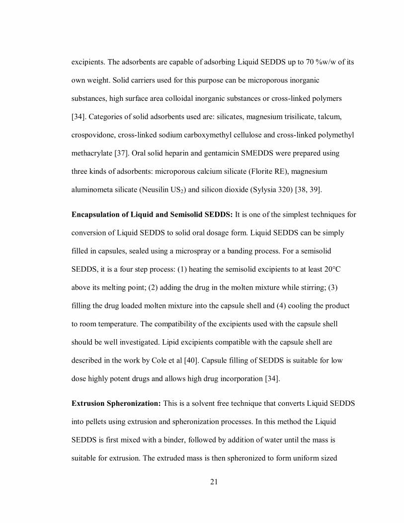
compensated DSC and (2) Heat flux DSC
Power compensated DSC (Figure 2-5): In a power compensated DSC, the sample and
the reference pan are kept in isolated furnaces (that is both of them are heated by separate
35
heat sources). The power required to maintain a zero temperature differential between the
sample and reference is measured. At the onset of any enthalpic event
(exothermic/endothermic), the sample temperature would change relative to the
reference; hence in order to maintain isothermal conditions energy is supplied to one or
the other pan depending on the nature of the enthalpic event. During an exothermic
reaction, heat is supplied to the reference, while heat is supplied to the sample during an
endothermic reaction [72]. A change in power signal results in a peak in the thermogram.
Figure 2-5: Power compensated DSC [74]
Heat flux DSC (Figure 2-6): In heat flux DSC, both the sample and reference pans are
enclosed in a single furnace and are heated by a thermoelectric disk. A thermocouple
placed below the pans is used to measure the temperature differential which is

36
transformed into heat flow using mathematical equations.
Figure 2-6: Heat flux DSC [74]
Sample preparation: The samples are placed in metal pans that are inserted into the
sample holders of the instrument. Material that possesses high thermal conductivity and
compatibility with sample material are used to fabricate sample pans. The pans may be
open, sealed, covered, or pin-holed depending on the nature of the sample used. A
reference pan of identical to the sample pan is placed in reference sample holder. It may
be filled with an inert material or remain empty. The sample size is ideally 3-5 mg. Low
density powders should be lightly pressed into the bottom of the pan to ensure good
thermal contact. For calculation of heat capacity or heat of fusion, the samples should be
accurately weighed and sample weight should be entered into instrument software.
Applications: DSC is used in pharmaceutical industry for characterization of active
pharmaceutical ingredients and excipients. Any material that exhibits a transition in its
physical state or reacts chemically with associated heat exchange can be evaluated using

37
DSC. DSC may be used for single material characterization or multi-components
investigations. Some pharmaceutical applications of DSC include studies involving:
Melting point, crystallization,
sublimation, dehydration,
desolvation, and glass transition
[75].
Measurement of heat of reaction
and heat capacities [76]
Purity of materials
Degree of crystallinity
Polymorphism [77]
Decomposition, degradation and
stability kinetics [78]
Protein unfolding [79]
Freeze drying [80]
Drug-excipient compatibility
studies [81-83]
Drug-Polymer interactions in
polymeric drug delivery [84]
Solid dispersions [85-87]
2.5 Powder X-ray Diffraction
X-rays are electromagnetic radiations having a wavelength of about 1 °A, which is
approximately the size of an atom (7). It is used for analysis of crystalline solids at an
atomic level.
Principle [88, 89]: The atoms in a crystalline solid are arranged in discrete parallel
planes separated by a distance d. Such planes exist in a number of different orientations,
each with its own specific d. When a monochromatic X ray beam of wavelength of λ is
incident on the parallel planes at an angle of θ, diffraction only occurs if the distance
travelled by reflected rays from successive planes differs by a complete n number of
wavelengths (Figure 2-7). When this occurs, the reflected rays constructively interfere
according to the Bragg‟s law. Bragg‟s law (Equation 2.3) states that if the wavelengths of

38
reflected X rays differ by an integer multiple n of wavelength λ, then they constructively
interfere and the angle of diffraction θ will be equal to the angle of incidence θ.
n. λ = 2d sinθ ………………………..……………….(Equation 2.3)
When the angle θ is changed, Bragg law‟s conditions are satisfied at various d spacing‟s
of crystalline material resulting in diffracted X rays. A plot of intensities of the diffracted
X ray peak‟s versus 2θ (angular positions) gives a pattern that is characterist ic of
crystalline material, also called as “fingerprint” of the material.
Figure 2-7: Diffraction of X ray by a crystalline material [90]
Instrumentation [88, 89]: The PXRD instrument (Figure 2-8) essentially consists of an
X-ray tube, sample holder, and an X-ray detector. X rays of suitable wavelength and
intensity are produced by the x ray tube.

39
Figure 2-8: Schematics X ray diffractometer [91]
The X-ray tube consists of an anode and a cathode. A tungsten filament cathode is heated
to produce electrons. These electrons are bombarded on a metal anode (target) which
results in the generation of X-ray spectra. A commonly used target is copper, although
molybdenum, chromium, silver, and rhodium can also be used. The X-ray spectra consist
of Kα and Kβ components with a certain wavelength characteristic to the type of the target
used. Copper produces CuKα rays with a wavelength of 1.5418 °A. Monochromatic X-
rays are produced by filtering the X-ray spectra using foils or a crystal monochromator.
These monochromatic X-rays are collimated on the sample at an angle of θ. The
goniometer in the instrument simultaneously moves the sample holder and the detector in
such a way that incident X-ray beam hits the sample and the reflected X-ray is detected at
an angle of 2θ. At a certain angle of θ, where Bragg‟s law is obeyed constructive
interference is detected by the detector. The detector processes the signal and converts it

40
into a count rate which is displayed as a function of intensity verses 2θ (diffracting
angle).
Sample Preparation: The sample quantity analyzed depends on the capacity of sample
holder used. Coarse samples should be grounded to about 200 mesh. A flat compact bed
of sample powder should be made on the sample holder using a glass slide or a razor
blade. The upper surface of the sample should be flat to ensure random distribution of
lattice orientation.
Applications: PXRD is a characeterization technique used by material scientists working
in pharmaceuticals, geologicals, engineering, and environmental sciences areas. The
advantages associated with this technique are: non-destructive technique, rapid output of
results, minimal sample preapartion, and easy spectra interpretation [92]. PXRD is used
to assess solid state properties in general. In pharmaceuticals, PXRD finds its use in the
following ways:
Identification of drugs in pharmaceutical dosage forms [93]
Studying solid-solid interactions and properties of different polymorphs, solvate
forms and their phase transformations [94, 95]
Drug-excipient compatibility [96]
Alterations in crystallinity of materials [97]

41
2.6 UV/ Visible Spectroscopy
UV/Visible spectroscopy uses UV (200-400 nm) and visible (400-800 nm) radiations to
analyze samples. It is one of the simplest and most widely used spectroscopic techniques
in quantitative determination of organic, inorganic and biological samples.
Principle: With the absorption of UV/ Visible light, valence electrons of the molecule
are excited and undergo an electronic transition from their highest occupied molecular
orbital (HOMO) to lowest unoccupied molecular orbital (LUMO). In UV/Visible
spectroscopy (Figure- 2.9), (n to π*) and (π to π*) transitions are observed because the
energy required for this transitions fall in the UV/Visible range. Such transitions are
possible in alkenes, aromatics, and conjugated dienes and trienes because of the presence
of unsaturated double bonds. Molecules that have these functional groups are capable of
absorbing UV/Visible light are called as chromophores. The vibrational and rotational
spectra also gets superimposed on the electronic spectra of the chromophore (or the
molecule), which makes the spectra to look like a broad band. The Beer‟s-Lambert law
(Equation 2.4) states that the absorbance of a solution is directly proportional to the
concentration of the absorbing species present in the sample. This law can be used to
measure concentration of absorbing species in the solution.
A = -log (I/Io) = ε . c. L……………………(Equation 2.4)
where A is the measured absorbance, Io is the intensity of incident light, I is the intensity
of transmitted light, L is the pathlength of light through the sample, c is the concentration
of the absorbing species and ε is the molar absorptivity or extinction coefficient of the

42
species. ε is characteristic of a species under specific conditions of solvent, wavelength,
and temperature.
Figure 2-9: Energy diagram showing various energy transitions [98]
Instrumentation: A typical UV/Visible spectrophotometer consists of a light source
(deuterium and tungsten lamps), wavelength selector (prism, gratings monochromator,
interferometer), sample holder and sample cells (cuvette, 96 well plate), detector
(photomultipier tube, photodiode array, CCD camera), and data acquisition system
(computer). There are two kinds of spectrophotometers: (1) Single beam and (2) Double
beam spectrophotometer. In a single beam spectrophotometer (Figure 2-10), the
transmittance of the sample and reference (blank) is to be measured separately (first
blank and then sample), while a double beam spectrophotometer (Figure 2-11) measures
both the transmittances simultaneously by splitting the light beam into two halves [73,
99].

43
Figure 2-10: Schematics of a single beam spectrophotometer [100]
Figure 2-11: Schematics of a double beam spectrophotometer
[100]
Sample Preparation: Samples for UV-Visible spectroscopy are generally solutions.
Samples are placed in a cuvette which is made of UV transparent material such as fused
silica, quartz glass, or plastic. Commonly used solvents with their wavelength cutoff for
UV/Visible measurements are acetonitrile (190 nm), water (191 nm), cyclohexane (195
nm), methanol (201nm), ethanol (204 nm), ether (215 nm), and methylene chloride
(220nm).

44
Applications: Qualitative applications of UV/Vis spectroscopy is limited to the extent of
identification of functional groups in the absorbing species based on the absorption peaks
observed in the spectra. Results obtained from UV are often used in conjunction with
other techniques such as Nuclear Magnetic Resonance (NMR), Infra red (IR), and Mass
Sprectroscopy (MS) for complete chemical structure elucidation. Quantitative estimation
of organic and inorganic species can be made with good accuracy and a detection limit of
10-4
to 10-5
M is possible. Derivative spectroscopy is used to measure concentration of an
analyte in a mixture of two or more interfering analytes with ease. Derivative
spectroscopy yields a plot of derivative of absorbance with respect to wavelength as a
function of wavelength rather than a conventional absorbance verses wavelength plot [73,
101]. Difference spectroscopy is used for detecting two compounds having a very close
absorption maxima are present in a sample or where there is a matrix (solvent)
interference in the sample [65].

45
Chapter 3
Materials and Methods
3.1 Materials
3.1.1 Tween 80
Source: Spectrum Chemicals, Lot # C136790, CAS 156-27-1
Chemical name: Polyoxyethylene 20 sorbitan monoleate
Molecular Formula: C64H124O26
Molecular weight: 1310 gram/mole
Specific gravity: 1.08
Physical state: Yellow oily liquid
Soluble in alcohol, methanol, toluene, insoluble in mineral oil

46
O
O (CH2CH2O)bHOH(OH2CH2C)a
CHCH2O-C-(CH2)7CH
O(CH2CH2O)CH
CH(CH2)7CH3
Figure 3-1: Chemical structure of Tween 80
Tween 80 is the partial fatty acid ester of sorbitol and it forms anhydrides as a result of
copolymerization of approximately 20 moles of ethylene oxide for each mole of sorbitol.
They are hydrophilic nonionic surfactants (HLB-15) and are used as emulsifiers in the
preparation of oil-in-water emulsions, oral and injectable suspensions and solutions
[102], and as solubilizing agents for oil soluble vitamins [103].
Tween 80 has been reported to protect proteins from surface induced denaturation during
freeze drying [104]. It is one of the most widely used surfactant in the preparation of self
dispersing type of formulations [9]. It is approved by the FDA for oral use. Peroxide
impurities present in Tween 80 causes protein denaturation when such products are stored
for extended periods of time.
3.1.2 Cremophor RH 40
Source: BASF Chemicals, Lot #72809647G0, CAS #61788-85-0
Chemical name: Polyethoxylated castor oil
Other names: PEG 40 Hydrogenated Castor oil, polyoxythylene 40 castor oil

47
Soluble in water, ethanol, chloroform
Physical state: white, semisolid paste at 20°C
CH2 O C (CH2)10 CH3 (CH2)5CH3
O
O C (CH2)10 CH (CH2)5CH3
O
O C (CH2)10 CH (CH2)5CH3
O
CH2 O C (CH2)10 CH (CH2)5CH3
O
OH
OH
OH
CH2
Figure 3-2: Chemical structure of Hydrogenated castor oil
It is a polyoxyethylene of derivative castor oil containing 70% of components which are
hydrophobic in nature with an HLB of 14-16. Cremophor RH 40 contains fatty acid
esters of glycerol polyethylene glycol and fatty acid esters of polyethylene glycol. It aids
in improving aqueous solubility of propellant in water based aerosol vehicles [103].
It is used as a solubilizing agent for various hydrophobic Active Pharmaceutical
Ingredient‟s (API), fat soluble vitamins, and essential oils; and as an emulsifier in the
preparation of pharmaceutical emulsions and SEDDS. It aids in solubilization of
Lopinavir and Ritonavir in Kaletra® oral solution and Cyclosporine in Neoral
® oral
microemulsions [105].

48
3.1.3 Labrafac Lipophile WL 1349
OH
O-R
OH
O
O
R:
or
Figure 3-3: Chemical structure of Caprylic /Capric Triglyceride
Source: Gattefosse, Batch # 123717
Chemical name: Caprylic /Capric Triglyceride
Physical state: Colorless viscous liquid
Boiling point: >150°C
Specific gravity: 0.93-0.96
Soluble in ethanol, chloroform, methylene chloride, vegetable oils, insoluble in water.
Labrafac Lipophile WL 1349 is a medium chain triglyceride of fractionated vegetable C8
and C10 fatty acids (mainly fractionated coconut oil or palm kernel oil) with an HLB of 1.
It is a non rancidable fluid used as a vehicle in oral and topical preparations, emulsions,
self-emulsifying drug delivery systems, suspensions, ointments, suppositories, and
creams. It can be used as filler in capsules and as an anti adherent in tablets. In
combination with long chain triglycerides, it serves as a total parenteral nutrition (TPN)

49
component. They possess excellent spreadability, skin penetration, and solvent properties
when compared to long-chain triglycerides.
3.1.4 Polyethylene Glycol 400
Source: Spectrum Chemicals, Lot # YW0048, CAS 25322-68-3
Chemical name: α-Hydroxy-ω-hydroxy-poly(oxy-1,2-ethanediyl)
Chemical formula: C2nH4n+2On+1
Molecular weight: 380-420 (average)
O
OHH
n
n=8.2-9.1
Figure 3-4: Chemical structure of PEG 400
Physical state: Colorless viscous liquid
Specific gravity: 1.1254
Soluble in water, acetone, alcohol, glycerin, benzene
Polyethylene glycols (PEG) have a wide range of applications including topical, oral,
parenteral, ophthalmic and rectal delivery. Liquid grade PEG‟s are used as a water
miscible co-solvents which possess good solvent properties for poorly water soluble

50
drugs. Due to this property, they are widely used in lipid based drug delivery systems
such as solid dispersions and self-emulsifying mixtures. When used in soft gelatin
capsules, they are known to cause hardening of capsule shells by absorption of moisture
from gelatin in the shell [106].
3.1.5 Capyrol 90
O
OH
Fig 3-5: Chemical structure of Capyrol 90
Source: Gattefosse, Batch # 118148, CAS # 85883-73-4
Chemical name: Propylene glycol monocaprylate
Chemical formula: C11H22O3
Molecular weight: 202.29
Physical state: Colorless oily liquid
Specific gravity: 0.935-0.955
Soluble in ethanol, chloroform, methylene chloride, and vegetable oils, insoluble in water
Capryol 90 contains more than 90% monoester of C8 fatty acid (caprylic acid). It is used
as an emulsifier in oil-in-water emulsions and self emulsifying drug delivery systems. It

51
is reported to possess bioavailability enhancing properties due to its inhibitory action on
CYP3A4 enzyme [106].
3.1.6 Transcutol P
O
O
OH
Figure 3-6: Chemical structure of Transcutol P
Source: Gattefosse, Batch # 450844010, CAS # 111-90-0
Chemical name: Diethylene glycol monoethyl ether
Chemical formula: C6H14O3
Molecular weight: 134.17
Physical state: Colorless limpid liquid
Specific gravity: 0.985-0.991
Soluble in water, and ethanol, insoluble in mineral oils
Transcutol P has good solvent properties for poorly water soluble drugs. It enhances drug
penetration, permeation, and produces a drug depot effect [103]. It is used as a co-solvent
in the formulation of SEDDS.

52
3.1.7 Ibuprofen
OH
H3C
CH3
CH3
O
Figure 3-7: Chemical structure of Ibuprofen
Source: PCCA, Lot # C136790, CAS # 15687-27-1
Chemical name: 2-(4-isobutylphenyl) propionic acid
Chemical formula: C13H18O2
Molecular weight: 206.29
Physical state: White color powder
Slightly soluble in water, soluble in alcohol and other organic solvents
Ibuprofen is a non steroidal anti inflammatory drug (NSAID), used for management of
pain and fever. It belongs to the class of propionic acid derivatives possessing anti-
inflammatory activity [107]. Ibuprofen is a nonselective cyclooxygenase inhibitor (COX-
I, II). These COX enzymes are responsible for producing prostaglandins that are
considered to be pain and inflammatory mediators in the body. The analgesic, anti-
inflammatory, and antipyretic activity of ibuprofen is manifested by action of COX-I

53
enzyme, while action on COX-II causes unwanted actions like GI irritation and platelet
aggregation [108]. Recently, it has been shown that ibuprofen inhibits up-regulation of
pro-inflammatory genes like tumor necrosis factor (TNF-α) and interleukin-1β (IL-1β) in
the nucleus of cell [109]. Ibuprofen is used in conditions such as rheumatoid arthritis,
osteoarthritis, body pain, dysmenorrhea and dental pain. It is absorbed rapidly, binds
extensively to plasma proteins, undergoes hepatic metabolism, and is excreted through
urine [107].
Commercial dosage forms and dose
Marketed products: Motrin®, Advil
®, Nuprin
®, Rufen
®, Medipren
®
Dosage forms: Tablets, Caplets, Sofgels, Suspensions, Capsules, Gelcaps
Dose: 200-400 mg every 4-6 hours for analgesic purpose, 300-400 mg every 6-8 hours
for rheumatoid arthritis and osteoarthritis.
3.1.8 Neusilin US2
Source: Fuji Chemicals, Lot # 901001, CAS 12511-31-8
Chemical Name: Magnesium Aluminometasilicate
Chemical Formula: Al203.MgO.1.7SiO2.xH2O
Physical State: White powder
Specific gravity: 2.0-2.2
Insoluble in water and ethanol

54
Neusilin US2 is a very fine powder of amorphous magnesium aluminosilicate. It
possesses very large surface area enabling it to adsorb oils up to three times of its weight.
It has good flowability and compressibility and can be directly compressed into tablets. It
has been used as an adsorbent for oil-emulsifier mixtures [39] in SEDDS and for melt
granulation in solid dispersion technology [110]. Upon co-grinding with a crystalline
drug, it converts the drug into an amorphous form [111].
3. 2 Methods
3.2.1 Screening of lipid excipients for SMEDDS
A SMEDDS is an isotropic mixture of oil, surfactant, and cosurfactant. The property of
self-microemulsification is only exhibited by certain combination of these components.
In this study, excipients shown in Table 3.1 were evaluated for potential self-
emulsification property.
Table 3.1: Lipid excipients used in SMEDDS
Oil Surfactant Cosurfactant
Labrafac Lipophile WL 1349 Tween 80 Capyrol 90
Cremophor RH 40 Transcutol P
PEG 400
A total of six self emulsifying mixtures comprising of components shown in Table 3.1
were evaluated.
1. Tween 80-PEG 400-LL WL 1349

55
2. Tween 80-Capyrol 90-LL WL 1349
3. Tween 80-Transcutol P-LL WL 1349
4. Cremophor RH 40-PEG 400-LL WL 1349
5. Cremophor RH 40-Capyrol 90-LL WL 1349
6. Cremophor RH 40-Transcutol P-LL WL 1349
All six combinations were prepared with the ratios of Oil:(Surfactant/Cosurfactant) as
1:1, 1:2, 1:3, 1:4, 1:5, 1:6 and 1:7. The Surfactant/Cosurfactant ratio‟s (km) of 1:1, 2:1
and 3:1 were evaluated. Surfactant/cosurfactant mixtures possessing various km ratio‟s
were prepared by weighing appropriate amount of surfactant and cosurfactant and were
vortexed for 30 min to produce a homogenous mixture. Mixtures with 12-50% of the oil,
25-66% of the surfactant and 12-44% of cosurfactant were evaluated for their self-
emulsifying properties. These mixtures were then mixed with the oil phase to form an
isotropic self microemulsifying (SME) mixture. Thus, each combination had a total of 21
samples with different proportions of oil, surfactants and cosurfactants.
3.2.2 Test for Self Emulsification
A test for self emulsification was performed on all the six SME combinations described
in section 3.2.1 according to the method suggested by Craig et al [26].
0.6 ml of each of the 21 SME samples was placed in 400 ml of water and contents were
agitated via a magnetic stirrer. The spontaneity of emulsification, clarity of dispersion,
and apparent stability were evaluated. The emulsion so formed was either a visually clear
nanoemulsion, or a slightly turbid emulsion, or a milky emulsion that immediately phase

56
separated. The formulations that were emulsified into a clear, transparent nanoemulsion
and showed no signs of instability for 24 hours were delineated in ternary phase diagrams
using Sigmaplot® software.
3.2.3 Drug Solubility
Excess amount of Ibuprofen was added to 2ml of each excipient placed in microtubes and
the mixture was vortexed, heated to 40°C in a water bath to facilitate drug solubilization.
The mixture was finally kept at ambient room temperature (25°C) under continuous
shaking for 48 hours to attain equilibrium. The mixtures were then centrifuged at 3000
rpm for 15 min. Aliquots of supernatant were diluted with methanol, and the drug content
was quantified using a UV spectroscopic method. The solubility of drug was determined
from a calibration curve of Ibuprofen in methanol.
3.2.4 Preparation of Liquid SMEDDS
After careful evaluation of phase diagrams, Tween 80-Labrafac Lipophile WL 1349-PEG
400 were selected as a SME mixture for drug delivery. Liquid SMEDDS formulation was
prepared by dissolving 200 mg of Ibuprofen in the optimized SME mixture consisting of
Tween 80(50%w/w), Labrafac Lipophile WL 1349(25%) and PEG 400(25%). Drug
containing SME mixture was vortexed until a clear solution was obtained. These
mixtures were observed for any signs of turbidity or phase separation for a period of 48
hours.

57
3.2.5 Preparation of Solid SMEDDS
The Liquid SMEDDS described in section 3.2.4 was adsorbed onto Neusilin US2 by
physical mixing in a small mortar and pestle. The resulting Solid SMEDDS was a free
flowing powder that was subsequently subjected to solid state characterization and
dissolution studies. The formula of the optimized Solid SMEDDS is shown in table 3.2.
Table 3.2: Composition of an optimized Solid SMEDDS
Formula components Proportions
Ibuprofen 200 mg
Tween 80 750 mg
Labrafac Lipophile WL 1349 375 mg
PEG 400 375 mg
Neusilin US2 500 mg
3.2.6 Droplet size and Zeta potential of nanoemulsions
Liquid SMEDDS and Solid SMEDDS were dispersed in 400 ml of water to obtain to a
nanoemulsion. The droplet size and zeta potential of the resultant nanoemulsion was
measured using Dynamic Light Scattering (Nicomp 380 ZLS, Particle Sizing Systems,
CA). The nanoemulsion samples were taken in disposable Durex borosilicate glass
culture tubes (VWR Scientific products) and volume weighted diameter was determined
by placing the sample in the path of a Helium Neon laser of wavelength 658 nm at a
scattering angle of 90°C and a temperature of 23°C.

58
3.2.7 Morphological Analysis of Solid SMEDDS
The surface morphology of Solid SMEDDS, Ibuprofen, and Neusilin US2 was analyzed
in an FEI Quanta 3D FEG Dual Beam Electron Microscope. The samples were fixed on
an aluminum stub using a double sided carbon adhesive tape and were made electrically
conductive by coating with palladium under vacuum. An accelerating voltage of 5 kV
was used to visualize the samples.
3.2.8 Differential Scanning Calorimetry of Solid SMEDDS
The physical state of Ibuprofen in Solid SMEDDS was characterized by differential
scanning calorimetry (Diamond DSC, PerkinElmer) equipped with an Intercooler 1P.
Samples of Ibuprofen powder, Solid SMEDDS, Neusilin US2 and physical mixture
(Ibuprofen+Neusilin US2) were run on DSC. The samples (about 3-6 mg) were placed in
standard 20µl aluminum pans using nitrogen as effluent gas at 20 ml/min. Samples were
scanned at a temperature ramp speed of 10°C/min from 0 to 140°C. The acquired data
was analyzed using Pyris Manager (v 1.3) software.
3.2.9 Powder X-ray Diffraction of Solid SMEDDS
The physical state of ibuprofen in Solid SMEDDS was characterized by X ray powder
scattering (PXRD) measurements using X ray diffractometer (X:Pert PRO with X-Pert
Data Collecter, PAN analytical). The measurements were performed at room temperature
using monochromatic CuKα-radiation at 40mA and at 40kV over a 2Ө range of 7 to 80°
with a continuous scanning speed of 4°/min. The analyzed samples were compactly
packed in the cavity of an aluminum sample holder using a glass slide. PXRD was

59
performed on samples of Ibuprofen powder, Solid SMEDDS, Neusilin US2, and a
physical mixture of Ibuprofen and Neusilin US2.
3.2.10 In vitro drug release studies
In vitro drug release studies from Solid SMEDDS were performed using USP Type II
dissolution apparatus (Vanderkamp® 600 six spindle dissolution tester) with number of
paddle rotations set to 50 rpm. The dissolution medium consisted of 900 ml of phosphate
buffer pH 7.2 maintained at 37 ±0.5°C. Solid SMEDDS containing 200 mg of Ibuprofen
was introduced into the dissolution medium. At predetermined time intervals 5ml of
aliquot was withdrawn, filtered using 0.45µm syringe filter and an equivalent volume of
fresh dissolution medium was immediately added. The amount of drug released was
estimated by measuring absorbance at 264 nm using a single beam spectrophotometer
(Agilent UV spectrophotometer 8453). Dissolution of Liquid SMEDDS and plain
ibuprofen powder was also determined in identical manner. The calibration curve of
ibuprofen was made in phosphate buffer pH 7.2 and at 264 nm. Release data was
statistically analyzed using Sigma Plot software employing a one way ANOVA followed
by a Tukey‟s test to determine group differences. Statistical significance was set at
p<0.001.

60
Chapter 4
Results and Discussion
4.1 Screening of lipid excipients
Successful formulation of lipid based drug delivery systems is dependent on proper
selection of lipid excipients. With a variety of lipid based excipients currently available
commercially, a formulator has more options for selecting an excipient than was possible
in the past. It is essential that the selected excipients should possess properties required
for the desired type of lipid based drug delivery. The choice of selecting excipients for
developing Self Micro-Emulsifying Drug Delivery System (SMEDDS) is based on
following criteria
1. The self micro-emulsifying (SME) mixture should instantly self-microemulsify
forming a fine oil-in-water nanoemulsion upon aqueous dilution in the GIT.
2. The SME mixture should be capable of solubilizing the entire drug dose in a
volume that is acceptable for unit oral administration.
3. The drug should remain physically and chemically stable in the SME mixture.
4. The excipients should not produce any systemic toxicity.

61
Six SME mixtures listed below were evaluated for self micro-emulsifying performance
and one of them was selected for drug delivery based on its self-microemulsifying
efficiency, drug solubility, and drug stability studies. All the excipients selected for the
study were FDA „Generally regarded as safe‟ (GRAS) material.
1. Tween 80-PEG 400-LL WL 1349
2. Tween 80-Capyrol 90-LL WL 1349
3. Tween 80-Transcutol P-LL WL 1349
4. Cremophor RH 40-PEG 400-LL WL 1349
5. Cremophor RH 40-Capyrol 90-LL WL 1349
6. Cremophor RH 40-Transcutol P-LL WL 1349
4.2 Ternary Phase diagrams
Self micro-emulsifying performance of SME mixture was assessed from their ternary
phase diagrams and time taken to produce a fine nanoemulsion. Only certain
combinations of oil, surfactant and a cosurfactant in a certain composition range will
produce a fine nanoemulsion upon aqueous dilution. To check emulsification efficiency
of SME mixtures, test for emulsification was performed on all six combinations and the
resultant dispersions were visually assessed. Resulting dispersions either formed a clear
nanoemulsion, a slightly turbid emulsion or a milky emulsion which immediately phase
separated as shown in Figure 4-1. Table 4.1 shows the time taken by SME combinations
to form a clear nanoemulsion. It also shows corresponding concentrations (%w/w) of
mixture components based on the ratio of surfactant to cosurfactant (km) under test.
Ternary phase diagrams provide a lucid representation of the concentration range of the

62
three SME components (Figures 4-2~4-7). The area bound by the points in the phase
diagram displays the concentration range of SME mixture components that resulted in a
clear nanoemulsion out of all the trial concentrations. All the combinations under test
formed a nanoemulsion in certain concentrations, but the combination with wider SME
region is considered to be a better combination in terms of self-microemulsification
efficiency. Figures 4-2~4-7, clearly indicate that Tween 80-PEG 400-LL WL 1349,
Cremphor RH 40-PEG 400-LL WL 1349, and Cremophor RH 40-Transcutol P-LL WL
1349 have the largest SME region in the ternary phase diagram. The imaginary line on
which all the points lie represents the km ratio chosen. The other factor taken into
consideration for selection of best SME mixture is the time taken for complete
emulsification. All the time readings were taken in triplicates. No substantial
disimmilarity was observed between the time readings. Results from table 4.1 shows that
SME mixtures containig Cremophor RH 40 took more time to emulsify than the ones
with Tween 80. This may be attributed to the semi-solid nature of Cremophor RH 40. At
a fixed km ratio, an increase in surfactant concentration resulted in faster emulsification
of the SME mixture. This is possibly due to spontaneous stabilization of oil droplets by
surfactant monolayers owing to their high concentration. The Tween 80-PEG 400-LL
WL 1349 mixture possessed the largest SME region in the phase diagrams and took the
least time to micro-emulsify. They were completely emulsified within a minute and the
dispersion was clear and transparent. It has been reported that drug incorporation into
SMEDDS can affect the SME performance [10]. This can be due to the drug interacting
with the liquid crystalline phase of the SME mixure causing blockage of drug charge
movement in the system [26] or due to drug penetration into the surfactant monolayer

63
producing perturbations at the interface [10, 27].To verify this, ibuprofen loaded SME
mixtures (10%w/w) were subjected to the test for emulsification. No differences in self-
emulsification region in the phase diagram and time taken for complete emulsification
were observed between the drug loaded and blank SME mixtures. This suggests that the
presence of ibuprofen does not affect self micro-emuslifying property of the SME
mixture.
Figure 4-1: Results from test of emuslification. (a) milky emulsion (b) slightly turbid
emulsion (c) clear nanoemulsion.

64
Table 4.1: Time taken various SME mixtures*(I-VI) to completely self-microemulsify
Km
ratio
Oil (%w/w
)
Surf
acta
nt
(%w
/w)
Cosur
factan
t
(%w/
w)
Time
(sec)
I
Time
(sec)
II
Time
(sec)
III
Time
(sec)
IV
Time
(sec)
V
Time
(sec)
VI
1:1 50 25 25 No ME No ME No ME No ME No ME No ME
33.3 33.3 33.3 No ME No ME No ME 60 No ME No ME
25 37.5 37.5 20 33 No ME 32 No ME 20
20 40 40 26 23 No ME 33 No ME 15
16.6 41.6 41.6 19 20 23 35 +300 12
14.2 42.8 42.8 15 29 26 30 +300 11
12.5 43.7 43.7 18 30 15 30 +300 10
2:1 50 33.3 16.6 No ME No ME No ME No ME +300 No ME
33.3 44 22 35 No ME No ME 48 +300 No ME
25 50 25 24 24 No ME 68 +300 150
20 53.3 26.6 17 18 28 60 +300 150
16.6 55.5 27.7 16 21 26 44 +300 165
14.2 57.1 28.5 14 22 23 36 +300 150
12.5 58.3 29.16 17 28 20 32 +300 120
3:1 50 37.5 12.5 No ME No ME No ME No ME +300 No ME
33.3 49.5 16.5 No ME 140 No ME No ME +300 No ME
25 56.2 18.7 No ME 63 No ME No ME +300 No ME
20 60 20 24 14 No ME No ME +300 210
16.6 62.5 20.8 41 20 No ME 80 +300 210
14.2 64.2 21.4 42 18 No ME 67 250 180
12.5 65.6 21.8 27 90 No ME 40 200 170
* I. Tween 80-PEG 400-oil
II. Tween-Capyrol 90-oil
III. Tween 80-Transcutol P-oil
IV. Cremphor RH 40-PEG 400-oil
V. Cremophor RH 40-Capyrol 90-oil
VI. Cremophor RH 40-Transcutol P-oil

65
Figure 4-2: Ternary phase diagrams of SME mixture with Tween 80,Capyrol 90, and LL
WL 1349
Figure 4-3: Ternary phase diagrams of SME mixture with Cremphor RH 40, Capyrol 90
and LL WL 1349

66
Figure 4-4: Ternary phase diagrams of SME mixture with Cremophor RH 40, Transcutol
P, and LL WL 1349
Figure 4-5: Ternary phase diagrams of SME mixture withTween 80, Transcutol P,
and LL WL 1349

67
Figure 4-6: Ternary phase diagrams of SME mixture with Cremophor RH 40, PEG 400,
and LL WL 1349
Figure 4-7: Ternary phase diagrams of SME mixture with Tween 80, PEG 400 and LL
WL 1349

68
4.3 Drug Solubility studies
Table 4.2: Solubility of Ibuprofen in various excipients
For a successful delivery of drugs via SMEDDS, the entire dose of the drug should be
soluble in an acceptable volume of SME mixture. If the drug solubility is inadequate
there are chances of drug precipitation upon aqueous dilution. Equilibrium solubility
measurements of the drug in SME mixture excipients should be carried out to negate the
risk of drug precipitation. Thus the solubility of the drug in the excipients is an important
criterion for selection apart from the excipient‟s self-microemulsifying tendency.
Equilibrium solubility measurements of Ibuprofen were carried out in all lipid excipients
tested. Ibuprofen, a poorly water soluble drug showed good solubility in all the excipients
(Table 4.2). Ibuprofen showed highest solubility in Tween 80 and Transcutol P among
surfactants and cosurfactants respectively. There are more chances of drug precipitation
when cosurfactants contribute greatly to the overall drug solubility. Cosurfactants such as
polyethylene glycols, propylene glycol, and alcohols help to solubilize large quantities of
a drug in the oil. Upon aqueous dilution, the cosurfactants will separate from oil
components forming a micellar dispersion producing a reduction in solvent capacity for
Excipient Solubility
(mg/ml )
Tween 80 182.56 ± 23.39
LL WL 1349 77.39 ± 6.82
Capyrol 90 208.11 ± 8.95
PEG 400 207.20 ± 19.69
Cremophor RH 40 145.35 ± 3.29
Transcutol P 319.02 ± 23.21
69
the drug [5]. Thus it necessary for the drug to have good solubility in the oil as well as the
surfactant. Ibuprofen showed good solubility in Labrafac Lipophile WL 1349. Ibuprofen
is reported to have good solubility in medium chain triglycerides and polysorbates
(Tweens) [112]. All the evaluated excipients demonstrated good solubility for Ibuprofen
suggesting that their use in drug delivery would be appropriate. Based on the results of
ternary phase diagrams and drug solubility studies, Tween 80(50%w/w)-PEG
400(25%w/w)-LL WL 1349(25%w/w) was selected as an optimized SME mixture or
blank formulation for drug delivery. The concentrations of excipients selected are within
the usual range as recommended by the classification of lipid based formulations reported
by Pouton [5] and used by other research groups [2].
4.4 Liquid and Solid SMEDDS
Ibuprofen is available as an over the counter product in a 200 mg strength dosage form.
Hence, 200 mg of ibuprofen was incorporated in Liquid and Solid SMEDDS. It is
necessary that the dose of Ibuprofen(200mg) should be soluble in an accepatable volume
of Tween 80(50%w/w)-PEG 400(25%w/w)-LL WL 1349(25%w/w) so as to limit the
excess use of surfactant and cosurfactant in the formulation. To evaluate this criterion
200 mg of Ibuprofen was dissolved in various quantites of blank formulation and
observed for any signs of phase separation or precipitation of the drug. The minimum
amount of Tween 80-PEG 400-LL WL 1349 SME mixture required was found to be 1.5
gm. The drug was soluble in this mixture and exhibited no signs of phase separation,
discoloration, and precipitation for a period of 48 hours. This suggests that the drug is
physically stable in the formulation. Test for emulsification was performed on the

70
formulated liquid SME mixture. It showed a progression of emulsification similar to that
of a blank formulation and formed a clear nanoemulsion. The nanoemulsion showed no
signs of drug precipitation over a period of 24 h which suggests that the drug was present
in the oil droplets and the surfactant portion of the nanoemulsion. Also, Ibuprofen‟s
appreciable solubility in the cosurfactant did not cause loss of solvent capacity upon
aqueous dilution. To convert Ibuprofen loaded Liquid SMEDDS to Solid SMEDDS, it
was adsorbed onto 500 mg Neusilin US2 using a glass mortor pestle. On physical mixing,
a free flowing powder with adsorbed liquid SME mixture was obtained. It was suitable
for further solid state characterization studies. Neusilin US2 has high oil adsorbing
capacity (about 3.2 mg/ml) because of its porous amorphous structure [113]. Few
researchers have reported coversion of Liquid SEDDS to solid intermediates using
Neusilin US2 [38, 39]. Solid SMEDDS developed in this project were also tested for
emulsification test and the results were similar to that obtained from Liquid SMEDDS.
Liquid SMEDDS took about 30 seconds to completely emulsify, whereas Solid SMEEDS
took about 38 seconds. These observations confirmed the presence of Ibuprofen in a
solubilized form in the solid formulation. The physical state of ibuprofen in the Solid
SMEDDS was further verified using DSC and PXRD.

71
4.5 Characterization of Liquid and Solid SMEDDS
4.5.1 Droplet size analysis.
The droplet size of the nanoemulsion is important since it determines the rate and extent
of drug release and absorption.The drug can diffuse faster from smaller droplets into the
aqueous phase, thereby increasing the drug dissolution [23]. Smaller droplet size presents
large surface area for drug absorption. Reduction in droplet size improved bioavailibility
of cyclosporin emulsion when compared to a coarse emulsion [114]. Increase in
surfactant concentration decreases the droplet size upto a certain size but thereafter
anymore increase in surfactant concentration results in an increase in droplet size
[115].The reduction in droplet size can be attributed to the stablization of oil droplets due
to localization of surfactant monolayers at the oil-water interface [17]. Increase in
surfactant concentration causes enhanced water penetration into oil droplets leading to
breakdown of oil droplets and resultant bigger droplets [18]. The droplet size of the
nanoemulsion was measured using dynamic light scattering. The Ibuprofen loaded
SMEDDS displayed a Gaussian distribution of droplet sizes. The mean droplet size
(volume weighted) obtained from liquid SME mixture was 18.70 ± 0.82 nm (Figure 4-8),
while the mean droplet size obtained from Solid SMEDDS was 19.17 ± 2.29 nm (Figure
4-9). The Solid SMEDDS showed a bimodal (Nicomp®) distribution due to the presence
of Neusilin US2 particles in the sample. For Solid SMEDDS samples, the large Neusilin
US2 particles were allowed to sediment and then the sample of nanoemulsion was tested
for droplet size. In their bi-modal distribution, more than 90% of the particles by volume

72
constitute the nanoemulsion oil droplet, while the rest shows the size of the Neusilin US2
particles.
Figure 4-8: Droplet size distribution of the nanoemulsion obtained from Liquid SMEDDS
Figure 4-9: Droplet size distribution of nanoemulsion obtained from Solid SMEDDS

73
4.5.2 Zeta potential measurements
The surface charge (zeta potential) of the nanoemulsion formed from SMEDDS is
believed to play a role in its bioavailability. Because of the presence of fatty acids in the
structure of the excipients used, generally the surface charge of the droplet is negative.
The zeta potential of the nanoemulsion formulated from dispersion of Liquid SMEDDS
in water was found to be neutral. Surface charge of the oil droplets present in the
nanoemulsion may affect its interaction with the luminal intestinal mucosal cells. These
cells are negatively charged with respect to mucosal solution in lumen [116]. If the
surface charge of the droplet is positive, then there may be an electrostatic interaction
between the mucosal cell surface and the droplets. This leads to an increased absorption
of the administered drug and hence its bioavailability. It was observed that if a cationic
lipid like oleyl amine (upto 3% concentration) is used as one of the components of the
SME mixture, positively charged droplets were obtained. Oleyl amine localizes at the oil-
water interface and renders a positive charge to the oil droplets [117]. It was reported that
such positively charged emulsions get electrostatically attracted to the mucosal cell
surface resulting in an improved bioavailability of the drug. Such observations were only
found true in case of the coarse emulsions, wheras finer emulsions or nanoemulsions
showed a higher bioavalibility than positively charged coarse emulsions irrespective of
its surface charge [118, 119].

74
Figure 4-10: Zeta potential distribution of nanoemulsion obtained from Liquid SMEDDS
4.5.3 Morpholoy of SMEDDS.
For converting a Liquid SMEDDS into the solid state, a highy porous powder with good
oil adsorbing capacity is required. Such powders can adsorb oil components of the Liquid
SMEDDS and convert them into a free flowing powder. Neusilin US2 has a highly porous
structure which is capable of adsorbing upto three times its weight of oil [113, 120].
Scanning electron microscopy reveals the morphology of solid SMEDDS. From the
figure 4-11(c), ibuprofen appeared to be made of smooth rectangular crystalline
structures. Similar observations about ibuprofen micrographs were made by other
researchers [121]. Neusilin US2 appears to be spherical porous particles of size of
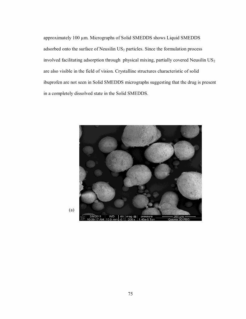
75
approximately 100 µm. Micrographs of Solid SMEDDS shows Liquid SMEDDS
adsorbed onto the surface of Neusilin US2 particles. Since the formulation process
involved facilitating adsorption through physical mixing, partially covered Neusilin US2
are also visible in the field of vision. Crystalline structures characteristic of solid
ibuprofen are not seen in Solid SMEDDS micrographs suggesting that the drug is present
in a completely dissolved state in the Solid SMEDDS.
(a)

76
(b)
(c)
Figure 4-11: Scanning Electron Microscopy images of: (a) Neusilin US2 (b) Solid
SMEDDS (c) Ibuprofen

77
4.5.4 Differential Scanning Calorimetry
The physical state of the drug present in solid SMEDDS was confirmed from DSC
studies. The physical state of a drug in the dosage form affects the in vitro and in vivo
release characteristics of the drug [35]. Ibuprofen shows a sharp endothermic peak at
69.92°C with heat of enthalpy of 93.89 joule/gm. Neusilin US2 did not show any peak in
the temperature range studied which conforms to data reported by Vadher et al [122]. The
physical mixture comprising equal amounts of Neusilin US2 and Ibuprofen showed a less
intense melting point peak at 67.89°C due to presence of Ibuprofen. The Solid SMEDDS
did not show any peaks under the studied temperature range indicating that Ibuprofen in
Solid SMEDDS was present either in an amorphous form or in a disordered crystalline
form of a molecular dispersion [36].
Figure 4-12: Differential Scanning Calorimetry (DSC) of Ibuprofen, Physical mixture
(Ibuprofen and Neusilin US2), Neusilin US2 and Solid SMEDDS.

78
4.5.5 Powder X ray Diffraction (PXRD)
PXRD further verified the physical state of the drug in the Solid SMEDDS. In figure 4-13
the presence of sharp peaks is indicative of the presence of Ibuprofen in a highly
crystalline form. Neusilin US2 is in amorphous state as demonstrated by the absence of
sharp diffraction patterns. The physical mixture (1:1) of Ibuprofen and Neusilin US2
showed some crystalline peaks due to the presence of Ibuprofen in the mixture. In
contrast to the physical mixture of Ibuprofen and Neusilin US2, the Solid SMEDDS did
not show significant crystalline peaks, which further confirms the molecularly dispersed
state of Ibuprofen in the formulation.
Figure 4-13: Powder X-ray Diffraction (PXRD) of Ibuprofen, Physical mixture
(Ibuprofen and Neusilin US2), Neusilin US2 and Solid SMEDDS.

79
4.6.6 Drug release studies
The in vitro dissolution of the Liquid and Solid SMEDDS was performed in phosphate
buffer (PBS) of pH 7.2 using a USP Type II dissolution apparatus with a paddle speed of
50 RPM. Liquid SMEDDS, Solid SMEDDS, and plain ibuprofen were placed in the
dissolution medium and were sampled at time intervals of 5, 10, 15, 20, 30, 40, 50 and 60
minutes. The amount of drug released was calculated from the calibration curve of
ibuprofen (figure 4-14) dissolved in PBS of pH 7.2.
Figure 4-14: Calibration of Ibuprofen in phosphate buffer (PBS) of pH 7.2 (n=3)

80
For both the formulations, it was observed that 80 % of the drug was released within 30
minutes dissolution time with about 75 % of the drug released within first five minutes of
the dissolution (Table 4.3).
Table 4.3: Cumulative percentage drug release (expressed as Mean±S.D.) from Liquid
SMEDDS, Solid SMEDDS and plain Ibuprofen (n=3)
However, the total amount of drug released from the Liquid SMEDDS was 88 %, while
that from Solid SMEDDS was about 85 % (Table 4.3). The dissolution rate was
compared with that of plain Ibuprofen. Ibuprofen shows a pH dependent solubility with
highest solubility shown at pH 7.2 [123]. The initial release of the drug from the
formulations was faster as compared to the plain drug dissolution (Figure 4-15).
Time (minutes) Liquid SMEDDS
(%)
Ibuprofen
(%)
Solid SMEDDS
(%)
0 0.00 0.00 0.00
5 75.60±3.48 38.73±4.27 76.44±1.45
10 76.15±2.46 65.39±0.34 79.34±0.69
15 76.90±2.45 86.40±3.01 80.96±2.06
20 78.95±1.52 93.96±4.25 82.17±1.81
30 80.45±1.39 94.81±4.24 83.04±2.26
40 83.26±1.52 95.59±3.95 83.96±1.10
50 83.10±6.36 97.78±2.54 84.47±2.27
60 87.65±0.67 99.06±1.40 84.83±1.66

81
Figure 4-15: Cumulative drug release profile of Liquid SMEDDS, Plain Ibuprofen and
Solid SMEDDS (n=3)
One way ANOVA followed by Tukey‟s multiple comparison test showed that the drug
release from Liquid SMEDDS and Solid SMEDDS significantly differed from that of
plain drug (p<0.001). There was no significant difference observed between Solid and
Liquid SMEDDS (p=0.086). The rapid release of the drug from the oil droplets suggests

82
that the polarity of the oil and the logP value of Ibuprofen was appropriate, thus enabling
the drug to partition out from the oil droplet. This establishes that SMEDDS can
effectively increase the drug dissolution rate of poorly water soluble drugs and can be
formulated as an immediate release dosage form for poorly water soluble drugs.
Nanoemulsion drug delivery has been successfully utilized for the delivery of anti-cancer
agents, anti-retroviral drugs, nutraceuticals, and cholesterol reducing statins. There is a
concomitant increase in bioavailability of these drugs when delivered through via
nanoemulsions because of a large increase in surface to volume ratio of these
nanoemulsions [124-126].
Results of one way ANOVA followed by Tukey‟s test as obtained from Sigma Plot®
One Way Analysis of Variance
Data source: Data 1 in Notebook1
Normality Test (Shapiro-Wilk) Passed (P = 0.330)
Equal Variance Test: Passed (P = 0.453)
Group Name N Missing Mean Std Dev SEM
Liquid SMEDDS 3 0 76.150 2.457 1.419
Solid SMEDDS 3 0 79.348 0.690 0.398
Plain Ibuprofen 3 0 65.396 0.338 0.195
Source of Variation DF SS MS F P

83
Between Groups 2 320.560 160.280 72.547 <0.001
Residual 6 13.256 2.209
Total 8 333.816
The differences in the mean values among the treatment groups are greater than would be
expected by chance; there is a statistically significant difference (P = <0.001).
Power of performed test with alpha = 0.050: 1.000
All Pairwise Multiple Comparison Procedures (Tukey Test):
Comparisons for factor:
Comparison Diff of Mean p q P P<0.050
Solid SMEDDS vs. Plain Ibuprofen 13.952 3 16.258 <0.001 Yes
Solid SMEDDS vs. Liquid SMEDDS 3.198 3 3.726 0.086 No
Liquid SMEDDS vs Plain Ibuprofen 10.755 3 12.532 <0.001 Yes

84
Conclusions
In this study, self-microemulsifying (SME) mixtures containing surfactant, cosurfactant
and a medium chain triglyceride were prepared and their tendency to efficiently emulsify
was evaluated. Upon aqueous dilution, such mixtures spontaneously emulsified forming
an oil-in-water nanoemulsion. This property was dependent on the composition of the
excipients as well as their individual concentration in the mixture. Excipients evaluated
for self-microemulsification were Tween 80 and Cremophor RH 40 as surfactants,
Transcutol P, Capyrol 90 and PEG 400 as cosurfactants and Labrafac Lipophile (LL) WL
1349 (a medium chain triglyceride) as an oil. All the excipients showed a tendency to
form a nanoemulsion with varying degrees of efficiency. A particular SME mixture
comprising of Tween 80-PEG 400-LL WL 1349 was selected and optimized for the
purpose for delivering a model drug, ibuprofen. Self-microemulsifying drug delivery
system (SMEDDS) is known to improve dissolution characteristics of a poorly water
soluble drug since they maintain the drug in a solubilized state in the GI tract. Using the
optimized SME mixture, ibuprofen loaded Liquid and Solid SMEDDS were prepared,
evaluated for their self-microemulsification tendency and characterized. The resulting
nanoemulsions from the trial formulations showed a droplet size of approximately 20 nm
and a neutral zeta potential. Differential Scanning Calorimetry and Powder X-Ray
Diffractometry confirmed the presence of ibuprofen in a molecularly dissolved state in

85
the Solid SMEDDS. In vitro drug release studies showed a 70% drug release within first
five minutes of dissolution time from the trial formulations. Our studies indicated that
SMEDDS can be potentially used for delivering a poorly water soluble drug. Ongoing
studies related to this project include in vitro drug permeability testing using Caco-2 cell
cultures.

86
References
1. Merisko-Liversidge, E., G.G. Liversidge, and E.R. Cooper, Nanosizing: a
formulation approach for poorly-water-soluble compounds. EUROPEAN
JOURNAL OF PHARMACEUTICAL SCIENCES, 2003. 18(2): p. 113-120.
2. Hauss, D., Oral lipid-based formulations. Advanced Drug Delivery Reviews,
2007. 59(7): p. 667-676.
3. Pouton, C., Formulation of poorly water-soluble drugs for oral administration:
physicochemical and physiological issues and the lipid formulation classification
system. EUROPEAN JOURNAL OF PHARMACEUTICAL SCIENCES, 2006.
29(3-4): p. 278-287.
4. Pouton, C.W. and C.J.H. Porter, Formulation of lipid-based delivery systems for
oral administration: materials, methods and strategies. Advanced Drug Delivery
Reviews, 2008. 60(6): p. 625-637.
5. Pouton, C., Lipid formulations for oral administration of drugs: non-emulsifying,
self-emulsifying and [] self-microemulsifying'drug delivery systems. EUROPEAN
JOURNAL OF PHARMACEUTICAL SCIENCES, 2000. v. 11: p. p. S93.

87
6. Wakerly, M., et al. Self-emulsification of vegetable oil-non-ionic surfactant
mixtures. 1986: ACS Publications.
7. Kimura, M., et al., Relationship between the molecular structures and
emulsification properties of edible oils. Bioscience, Biotechnology, and
Biochemistry (Japan), 1994.
8. Jannin, V., J. Musakhanian, and D. Marchaud, Approaches for the development of
solid and semi-solid lipid-based formulations. Advanced Drug Delivery Reviews,
2008. 60(6): p. 734-746.
9. Gershanik, T. and S. Benita, Self-dispersing lipid formulations for improving oral
absorption of lipophilic drugs. European Journal of Pharmaceutics and
Biopharmaceutics, 2000. 50(1): p. 179.
10. Constantinides, P., Lipid microemulsions for improving drug dissolution and oral
absorption: physical and biopharmaceutical aspects. Pharmaceutical research,
1995. 12(11): p. 1561-1572.
11. W.Pouton, C., Properties and Uses of Common Formulation Lipids, Surfactants
and Cosolvents, in AAPS Workshop. March 2007.
12. Singh, B., et al., Self-Emulsifying Drug Delivery Systems (SEDDS): Formulation
Development, Characterization, and Applications. Critical reviews in therapeutic
drug carrier systems, 2009. 26(5): p. 427-521.
13. Tenjarla, S., Microemulsions: an overview and pharmaceutical applications.
Critical reviews in therapeutic drug carrier systems, 1999. 16(5): p. 461.

88
14. Swenson, E.S., W.B. Milisen, and W. Curatolo, Intestinal permeability
enhancement: efficacy, acute local toxicity, and reversibility. Pharmaceutical
research, 1994. 11(8): p. 1132-1142.
15. Fanun, M., Colloids in Drug Delivery. 2010, CRC Press.
16. Charman, S., et al., Self-emulsifying drug delivery systems: formulation and
biopharmaceutic evaluation of an investigational lipophilic compound.
Pharmaceutical research, 1992. 9(1): p. 87-93.
17. Neslihan Gursoy, R. and S. Benita, Self-emulsifying drug delivery systems
(SEDDS) for improved oral delivery of lipophilic drugs. Biomedecine &
Pharmacotherapy, 2004. 58(3): p. 173-182.
18. Pouton, C., Formulation of self-emulsifying drug delivery systems. Advanced
Drug Delivery Reviews, 1997. 25(1): p. 47-58.
19. Benita, S., Microencapsulation: methods and industrial applications. 2006:
Informa Healthcare.
20. Porter, C.J.H., N.L. Trevaskis, and W.N. Charman, Lipids and lipid-based
formulations: optimizing the oral delivery of lipophilic drugs. Nature Reviews
Drug Discovery, 2007. 6(3): p. 231-248.
21. Chang, R.K. and A.H. Shojaei, Effect of a lipoidic excipient on the absorption
profile of compound UK 81252 in dogs after oral administration. J Pharm Pharm
Sci, 2004. 7(1): p. 8-12.
22. Porter, C.J.H., et al., Enhancing intestinal drug solubilisation using lipid-based
delivery systems. Advanced Drug Delivery Reviews, 2008. 60(6): p. 673-691.

89
23. Shah, N., et al., Self-emulsifying drug delivery systems (SEDDS) with
polyglycolyzed glycerides for improving in vitro dissolution and oral absorption
of lipophilic drugs. International journal of pharmaceutics, 1994. 106(1): p. 15-23.
24. Trull, A., et al., Absorption of cyclosporin from conventional and new
microemulsion oral formulations in liver transplant recipients with external
biliary diversion. British journal of clinical pharmacology, 1995. 39(6): p. 627.
25. Hauss, D.J., et al., Lipid based delivery systems for improving the bioavailability
and lymphatic transport of a poorly water soluble LTB4 inhibitor. Journal of
Pharmaceutical Sciences, 1998. 87(2): p. 164-169.
26. Craig, D., et al., An investigation into the physico-chemical properties of self-
emulsifying systems using low frequency dielectric spectroscopy, surface tension
measurements and particle size analysis. International journal of pharmaceutics,
1993. 96(1-3): p. 147-155.
27. Malcolmson, C. and M. Lawrence, A comparison of the incorporation of model
steroids into non-ionic micellar and microemulsion systems. The Journal of
pharmacy and pharmacology, 1993. 45(2): p. 141.
28. Pouton, C., Effects of the inclusion of a model drug on the performance of
selfemulsifying formulations. J. Pharm. Pharmacol, 1985. 37: p. 1P.
29. Charman, S.A., et al., Self-emulsifying drug delivery systems: formulation and
biopharmaceutic evaluation of an investigational lipophilic compound.
Pharmaceutical research, 1992. 9(1): p. 87-93.

90
30. Meinzer, A., Microemulsion-A Suitable Galenical Approach for the Absorption
Enhancement of Poorly Soluble Compounds. BULLETIN TECHNIQUE-
GATTEFOSSE, 1995: p. 21-26.
31. Craig, D., et al., An investigation into the mechanisms of self-emulsification using
particle size analysis and low frequency dielectric spectroscopy. International
journal of pharmaceutics, 1995. 114(1): p. 103-110.
32. Groves, M. and D. De Galindez, The self-emulsifying action of mixed surfactants
in oil. Acta pharmaceutica Suecica, 1976. 13(4): p. 361.
33. Wakerly, M.G., et al. Self-emulsification of vegetable oil-non-ionic surfactant
mixtures. 1986: ACS Publications.
34. Tang, B., et al., Development of solid self-emulsifying drug delivery systems:
preparation techniques and dosage forms. Drug Discovery Today, 2008. 13(13-
14): p. 606-612.
35. Balakrishnan, P., et al., Enhanced oral bioavailability of dexibuprofen by a novel
solid Self-emulsifying drug delivery system (SEDDS). European Journal of
Pharmaceutics and Biopharmaceutics, 2009. 72(3): p. 539-545.
36. Yi, T., et al., A new solid self-microemulsifying formulation prepared by spray-
drying to improve the oral bioavailability of poorly water soluble drugs.
European Journal of Pharmaceutics and Biopharmaceutics, 2008. 70(2): p. 439-
444.
37. Carli, F. and E. Chiellini, Pharmaceutical composition comprising a
water/oil/water double microemulsion incorporated in a solid support. 2004,
Google Patents.

91
38. Ito, Y., et al., Preparation and evaluation of oral solid heparin using emulsifier
and adsorbent for in vitro and in vivo studies. International journal of
pharmaceutics, 2006. 317(2): p. 114-119.
39. Ito, Y., et al., Oral solid gentamicin preparation using emulsifier and adsorbent.
Journal of Controlled Release, 2005. 105(1-2): p. 23-31.
40. Cole, E.T., D. Cadé, and H. Benameur, Challenges and opportunities in the
encapsulation of liquid and semi-solid formulations into capsules for oral
administration. Advanced Drug Delivery Reviews, 2008. 60(6): p. 747-756.
41. Abdalla, A., S. Klein, and K. Mäder, A new self-emulsifying drug delivery system
(SEDDS) for poorly soluble drugs: Characterization, dissolution, in vitro
digestion and incorporation into solid pellets. EUROPEAN JOURNAL OF
PHARMACEUTICAL SCIENCES, 2008. 35(5): p. 457-464.
42. Setthacheewakul, S., et al., Development and evaluation of self-microemulsifying
liquid and pellet formulations of curcumin, and absorption studies in rats.
European Journal of Pharmaceutics and Biopharmaceutics, 2010.
43. Chambin, O., et al., Influence of cryogenic grinding on properties of a self-
emulsifying formulation. International journal of pharmaceutics, 2004. 278(1): p.
79-89.
44. Myers, S.L. and M.L. Shively, Preparation and characterization of emulsifiable
glasses: Oil-in-water and water-in-oil-in-water emulsions. Journal of colloid and
interface science, 1992. 149(1): p. 271-278.

92
45. Porter, C.J.H., et al., Evaluation of emulsifiable glasses for the oral
administration of cyclosporin in beagle dogs. International journal of
pharmaceutics, 1996. 141(1-2): p. 227-237.
46. Bamba, J., et al., Cryoprotection of emulsions in freeze-drying: freezing process
analysis. Drug development and industrial pharmacy, 1995. 21(15): p. 1749-1760.
47. Vyas, S., et al., Preparation and characterization of griseofulvin dry emulsion.
Pharmazie, 1992. 47(6): p. 463-464.
48. Corveleyn, S. and J.P. Remon, Formulation of a lyophilized dry emulsion tablet
for the delivery of poorly soluble drugs. International journal of pharmaceutics,
1998. 166(1): p. 65-74.
49. Jang, D.J., et al., Improvement of bioavailability and photostability of amlodipine
using redispersible dry emulsion. EUROPEAN JOURNAL OF
PHARMACEUTICAL SCIENCES, 2006. 28(5): p. 405-411.
50. Toorisaka, E., et al., An enteric-coated dry emulsion formulation for oral insulin
delivery. Journal of Controlled Release, 2005. 107(1): p. 91-96.
51. Nazzal, S., et al., Preparation and in vitro characterization of a eutectic based
semisolid self-nanoemulsified drug delivery system (SNEDDS) of ubiquinone:
mechanism and progress of emulsion formation. International journal of
pharmaceutics, 2002. 235(1-2): p. 247-265.
52. Serajuddin, A.T.M., et al., Preformulation study of a poorly water-soluble drug, -
pentyl-3-(2-quinolinylmethoxy) benzenemethanol: Selection of the base for
dosage form design. Journal of Pharmaceutical Sciences, 1986. 75(5): p. 492-496.

93
53. Patil, P. and A. Paradkar, Porous polystyrene beads as carriers for self-
emulsifying system containing loratadine. AAPS PharmSciTech, 2006. 7(1): p.
199-205.
54. You, J., et al., Study of the preparation of sustained-release microspheres
containing zedoary turmeric oil by the emulsion-solvent-diffusion method and
evaluation of the self-emulsification and bioavailability of the oil. Colloids and
Surfaces B: Biointerfaces, 2006. 48(1): p. 35-41.
55. Attama, A.A. and M.O. Nkemnele, In vitro evaluation of drug release from self
micro-emulsifying drug delivery systems using a biodegradable homolipid from
Capra hircus. International journal of pharmaceutics, 2005. 304(1-2): p. 4-10.
56. Trickler, W., A. Nagvekar, and A. Dash, A novel nanoparticle formulation for
sustained paclitaxel delivery. AAPS PharmSciTech, 2008. 9(2): p. 486-493.
57. Chae, G.S., et al., Enhancement of the stability of BCNU using self-emulsifying
drug delivery systems (SEDDS) and in vitro antitumor activity of self-emulsified
BCNU-loaded PLGA wafer. International journal of pharmaceutics, 2005. 301(1-
2): p. 6-14.
58. Takada, K.K., JP), Murakami, Masahiro (Fukuchiyama, JP), Glycyrrhizin
preparations for transmucosal absorption. 2005, Amato Pharmaceutical Products,
Ltd. (Kyoto, JP): United States.
59. Xu, R., Particle characterization: light scattering methods. 2001: Springer.
60. Instruments, M. [cited 2011 03/07]; Available from:
http://www.malvern.com/LabEng/technology/dynamic_light_scattering/classical_
90_degree_scattering.htm.

94
61. Schärtl, W., Light scattering from polymer solutions and nanoparticle
dispersions. 2007: Springer Verlag.
62. Dynamic Light Scattering. [cited 2011 03/07]; Available from:
http://www.malvern.com/LabEng/technology/dynamic_light_scattering/dynamic_
light_scattering.htm.
63.
Laser Doppler Anemometry (LDA). [cited 2011 03/07]; Available from:
http://www.tut.fi/units/me/ener/laitteistot/EFD/LDA.html.
64. Laser Doppler: Theory. [cited 2011 03/07]; Available from:
http://www.efunda.com/designstandards/sensors/laser_doppler/laser_doppler_flo
w_theory.cfm.
65. Swarbrick, J. and J.C. Boylan, Encyclopedia of pharmaceutical technology. 2000:
Informa Healthcare.
66. Zhou, W. and Z.L. Wang, Scanning microscopy for nanotechnology: techniques
and applications. 2007: Springer.
67. Goldstein, J., et al., Scanning electron microscopy and X-ray microanalysis. 2003:
Springer Us.
68. Scanning Electron Microscopy. [cited 2011 03/07]; Available from:
http://www.purdue.edu/rem/rs/sem.htm.
69. Stokes, D.J., Recent advances in electron imaging, image interpretation and
applications: environmental scanning electron microscopy. Philosophical
Transactions of the Royal Society of London. Series A: Mathematical, Physical
and Engineering Sciences, 2003. 361(1813): p. 2771.

95
70. Shangraw, R., J. Wallace, and F. Bowers, Morphology and functionality in tablet
excipients for direct compression. Part 2. Pharmaceutical Technology, 1981.
5(Oct).
71. Höhne, G., W. Hemminger, and H.J. Flammersheim, Differential scanning
calorimetry. 2003: Springer Verlag.
72. Coleman, N.J. and D.Q.M. Craig, Modulated temperature differential scanning
calorimetry: a novel approach to pharmaceutical thermal analysis. International
journal of pharmaceutics, 1996. 135(1-2): p. 13-29.
73. Skoog, D.A., F.J. Holler, and T.A. Nieman, Principles of instrumental analysis.
1980: Saunders College New York.
74. Differential Scanning Calorimetry. [cited 2011 03/15]; Available from:
http://www.chtf.stuba.sk/kach/lab_538.php.
75. Clas, S.D., C.R. Dalton, and B.C. Hancock, Differential scanning calorimetry:
applications in drug development. Pharmaceutical Science & Technology Today,
1999. 2(8): p. 311-320.
76. Plato, C. and A.R. Glasgow, Differential scanning calorimetry as a general
method for determining the purity and heat of fusion of high-purity organic
chemicals. Application to 95 compounds. Analytical chemistry, 1969. 41(2): p.
330-336.
77. Hilfiker, R., Polymorphism in the pharmaceutical industry. 2006: Vch
Verlagsgesellschaft Mbh.
78. Rogers, R. and L. Smith, Application of scanning calorimetry to the study of
chemical kinetics. Thermochimica Acta, 1970. 1(1): p. 1-9.

96
79. Robertson, A.D. and K.P. Murphy, Protein structure and the energetics of protein
stability. Chem. Rev, 1997. 97(5): p. 1251-1268.
80. Hatley, R. and F. Franks, Applications of DSC in the development of improved
freeze-drying processes for labile biologicals. Journal of Thermal Analysis and
Calorimetry, 1991. 37(8): p. 1905-1914.
81. Tomassetti, M., et al., Thermal analysis study of the interactions between
acetaminophen and excipients in solid dosage forms and in some binary mixtures.
Journal of pharmaceutical and biomedical analysis, 2005. 37(5): p. 949-955.
82. Mura, P., et al., Compatibility study between ibuproxam and pharmaceutical
excipients using differential scanning calorimetry, hot-stage microscopy and
scanning electron microscopy1. Journal of pharmaceutical and biomedical
analysis, 1998. 18(1-2): p. 151-163.
83. Balestrieri, F., et al., Application of differential scanning calorimetry to the study
of drug-excipient compatibility. Thermochimica Acta, 1996. 285(2): p. 337-345.
84. Thassu, D., M. Deleers, and Y. Pathak, Nanoparticulate drug delivery systems.
2007: Informa Healthcare.
85. Ford, J.L. and P. Timmins, Pharmaceutical thermal analysis: techniques and
applications. 1989: Halsted Press.
86. Jung, J.Y., et al., Enhanced solubility and dissolution rate of itraconazole by a
solid dispersion technique. International journal of pharmaceutics, 1999. 187(2):
p. 209-218.

97
87. Kim, K.H., M.J. Frank, and N.L. Henderson, Application of differential scanning
calorimetry to the study of solid drug dispersions. Journal of Pharmaceutical
Sciences, 1985. 74(3): p. 283-289.
88. Suryanarayana, C. and M.G. Norton, X-ray diffraction: a practical approach.
1998: Plenum Publishing Corporation.
89. Pecharsky, V.K. and P.Y. Zavalij, Fundamentals of powder diffraction and
structural characterization of materials. 2009: Springer Verlag.
90. X ray Diffraction. [cited 2011 03/07]; Available from:
http://pubs.usgs.gov/of/2001/of01-041/htmldocs/images/beam.jpg.
91. Introduction to X-ray Powder Diffraction. [cited 2011 03/07]; Available from:
http://epswww.unm.edu/xrd/xrdclass/01-XRD-Intro.pdf.
92. Cullity, B.D. and S.R. Stock, Elements of X-ray Diffraction. Vol. 2. 1978:
Addison-Wesley Reading, MA.
93. Phadnis, N.V., R.K. Cavatur, and R. Suryanarayanan, Identification of drugs in
pharmaceutical dosage forms by X-ray powder diffractometry. Journal of
pharmaceutical and biomedical analysis, 1997. 15(7): p. 929-943.
94. Byrn, S., et al., Pharmaceutical solids: a strategic approach to regulatory
considerations. Pharmaceutical research, 1995. 12(7): p. 945-954.
95. Byrn, S., et al., Solid-state pharmaceutical chemistry. Chemistry of Materials,
1994. 6(8): p. 1148-1158.
96. Serajuddin, A., et al., Selection of solid dosage form composition through drug–
excipient compatibility testing. Journal of Pharmaceutical Sciences, 1999. 88(7):
p. 696-704.

98
97. Cheetham, N.W.H. and L. Tao, Variation in crystalline type with amylose content
in maize starch granules: an X-ray powder diffraction study. Carbohydrate
polymers, 1998. 36(4): p. 277-284.
98. Visible and Ultraviolet Spectroscopy. [cited 2011 03/07]; Available from:
http://www2.chemistry.msu.edu/faculty/reusch/VirtTxtJml/Spectrpy/UV-
Vis/spectrum.htm.
99. Harris, D.C., Quantitative chemical analysis. 2003: WH Freeman.
100. Ultraviolet and Visible Absorption Spectroscopy. [cited 2011 03/07]; Available
from: http://www.files.chem.vt.edu/chem-ed/spec/uv-vis/singlebeam.html.
101. De Micalizzi, Y., N. Pappano, and N. Debattista, First and second order
derivative spectrophotometric determination of benzyl alcohol and diclofenac in
pharmaceutical forms. Talanta, 1998. 47(3): p. 525-530.
102. Nielloud, F. and G. Marti-Mestres, Pharmaceutical emulsions and suspensions.
2000: Informa HealthCare.
103. Kibbe, A., Handbook of pharmaceutical excipients. 2000: Amer Pharmacists
Assn.
104. Chang, B., B. Kendrick, and J. Carpenter, Surface induced denaturation of
proteins during freezing and its inhibition by surfactants. Journal of
Pharmaceutical Sciences, 1996. 85(12): p. 1325-1330.
105. Strickley, R., Solubilizing excipients in oral and injectable formulations.
Pharmaceutical research, 2004. 21(2): p. 201-230.
106. Knepp, V., et al., Identification of antioxidants for prevention of peroxide-
mediated oxidation of recombinant human ciliary neurotrophic factor and

99
recombinant human nerve growth factor. PDA journal of pharmaceutical science
and technology/PDA. 50(3): p. 163.
107. Goodman, L., Goodman and Gilman's the pharmacological basis of therapeutics.
2006: McGraw-Hill New York:.
108. Kakuta, H., et al., Cyclooxygenase-1-selective inhibitors are attractive candidates
for analgesics that do not cause gastric damage. design and in vitro/in vivo
evaluation of a benzamide-type cyclooxygenase-1 selective inhibitor. Journal of
medicinal chemistry, 2008. 51(8): p. 2400-2411.
109. Stuhlmeier, K., H. Li, and J. Kao, Ibuprofen: new explanation for an old
phenomenon. Biochemical pharmacology, 1999. 57(3): p. 313-320.
110. Gupta, M., et al., Enhanced drug dissolution and bulk properties of solid
dispersions granulated with a surface adsorbent. Pharmaceutical Development
and Technology, 2001. 6(4): p. 563-572.
111. Gupta, M., A. Vanwert, and R. Bogner, Formation of physically stable
amorphous drugs by milling with Neusilin. Journal of Pharmaceutical Sciences,
2003. 92(3): p. 536-551.
112. Hu, L., et al., Preparation and evaluation of ibuprofen-loaded microemulsion for
improvement of oral bioavailability. Drug Delivery, 2011(00): p. 1-6.
113. Neusilin in Technical Newsletter. 2007, Fuji Chemical Industry Co Ltd.
114. Tarr, B. and S. Yalkowsky, Enhanced intestinal absorption of cyclosporine in rats
through the reduction of emulsion droplet size. Pharmaceutical research, 1989.
6(1): p. 40-43.

100
115. Yoo, J., et al., Novel self-nanoemulsifying drug delivery system for enhanced
solubility and dissolution of lutein. Archives of pharmacal research, 2010. 33(3):
p. 417-426.
116. Barry, R. and J. Eggenton, Membrane potentials of epithelial cells in rat small
intestine. The Journal of Physiology, 1972. 227(1): p. 201.
117. Gershanik, T. and S. Benita, Positively charged self-emulsifying oil formulation
for improving oral bioavailability of progesterone. Pharmaceutical Development
and Technology, 1996. 1(2): p. 147-157.
118. Gershanik, T., S. Benzeno, and S. Benita, Interaction of a self-emulsifying lipid
drug delivery system with the everted rat intestinal mucosa as a function of
droplet size and surface charge. Pharmaceutical research, 1998. 15(6): p. 863-
869.
119. Gershanik, T., et al., Charge-dependent interaction of self-emulsifying oil
formulations with Caco-2 cells monolayers: binding, effects on barrier function
and cytotoxicity. International journal of pharmaceutics, 2000. 211(1-2): p. 29-36.
120. Agarwal, V., et al., Dissolution and powder flow characterization of solid self-
emulsified drug delivery system (SEDDS). International journal of pharmaceutics,
2009. 366(1-2): p. 44-52.
121. Newa, M., et al., Preparation, characterization and in vivo evaluation of
ibuprofen binary solid dispersions with poloxamer 188. International journal of
pharmaceutics, 2007. 343(1-2): p. 228-237.

101
122. Vadher, A., et al., Preparation and Characterization of Co-Grinded Mixtures of
Aceclofenac and Neusilin US 2 for Dissolution Enhancement of Aceclofenac.
AAPS PharmSciTech, 2009. 10(2): p. 606-614.
123. Potthast, H., et al., Biowaiver monographs for immediate release solid oral
dosage forms: Ibuprofen. Journal of Pharmaceutical Sciences, 2005. 94(10): p.
2121-2131.
124. Kakumanu, S., et al., A nanoemulsion formulation of dacarbazine reduces tumor
size in a xenograft mouse epidermoid carcinoma model compared to dacarbazine
suspension. Nanomedicine: Nanotechnology, Biology and Medicine, 2011.
125. Kotyla, T., et al., Increased bioavailability of a transdermal application of a
nano-sized emulsion preparation. International journal of pharmaceutics, 2008.
347(1-2): p. 144-148.
126. Tagne, J.B., et al., A nanoemulsion formulation of tamoxifen increases its efficacy
in a breast cancer cell line. Molecular Pharmaceutics, 2008. 5(2): p. 280-286.