Development 140, 0000-0000 (2013) …uoneuro.uoregon.edu/doelab/pdfs2/carney13.pdf · STEM CELLS...
10
STEM CELLS AND REGENERATION RESEARCH ARTICLE 1 Development 140, 0000-0000 (2013) doi:10.1242/dev.093781 © 2013. Published by The Company of Biologists Ltd INTRODUCTION A defining characteristic of stem cells is the ability to produce daughters that retain stem cell fate (self-renew) as well as daughters that begin the process of differentiation. The robust adoption of these distinct fates is crucial both for the maintenance of stem cell pool size and for the production of differentiated progeny. In the Drosophila CNS, neuroblasts divide in a manner that is asymmetric in both progeny size and fate. The majority of neuroblasts – termed ‘type I’ neuroblasts – divide to generate a self-renewed neuroblast and a smaller ganglion mother cell (GMC), which divides only once more to produce neurons or glia. Additionally, there are eight bilateral ‘type II’ neuroblasts in the brain that repeatedly divide to self-renew and generate smaller intermediate neural progenitors (INPs), which each undergo a series of molecularly asymmetric divisions (similar to type I neuroblast divisions) to self-renew and generate a series of four to six GMCs (Bayraktar et al., 2010; Bello et al., 2008; Boone and Doe, 2008; Bowman et al., 2008; Izergina et al., 2009). Type I and II neuroblasts have emerged as an important model for studying stem cell self-renewal and differentiation. Type I neuroblast asymmetric division results in the segregation of cell fate determinants into the GMC. These fate determinants inhibit neuroblast self-renewal, direct cell cycle exit, promote neuronal differentiation and prevent tumor formation (Doe, 2008; Knoblich, 2010). Type II neuroblast lineages contain INPs that are particularly susceptible to dedifferentiation. Loss of function of earmuff (erm), barricade (barc), brain tumor (brat) or misexpression of activated Notch all lead to failure in neuronal differentiation and an expansion of type II neuroblast or INP fates (Bowman et al., 2008; Neumüller et al., 2011; Weng et al., 2010). One of the crucial differentiation factors is the transcription factor Prospero (Pros). In type I neuroblasts, Pros protein and mRNA are asymmetrically segregated into the GMC. In the GMC, pros mRNA is translated and Pros protein is imported into the nucleus (Broadus et al., 1998; Knoblich et al., 1995; Spana and Doe, 1995), where it represses cell cycle genes and promotes differentiation (Choksi et al., 2006; Li and Vaessin, 2000). Therefore, it is essential that GMCs inherit Pros from the neuroblast; in a Pros loss-of-function mutant, GMCs fail to exit the cell cycle, derepress neuroblast fate genes and can form tumorous overgrowths (Bello et al., 2006; Betschinger et al., 2006; Choksi et al., 2006; Lee et al., 2006). In the embryo, Pros protein can be detected in the GMC and transiently in newly born embryonic neurons (Srinivasan et al., 1998). In the larval CNS, Pros is detected in nearly all postmitotic neurons. In contrast to its tumor suppressor function in the GMC, the function of Pros in postmitotic larval neurons is unknown. Here we identify midlife crisis (mdlc; CG4973 – FlyBase) as a gene required to maintain Pros expression and neuronal differentiation in Drosophila larvae. mdlc encodes a conserved protein containing both a RING domain and a CCCH-type zinc finger. The yeast and human orthologs of Mdlc have been reported to be components of the spliceosome (Bessonov et al., 2008; Goldfeder and Oliveira, 2008). Clonal analysis of larval neuroblast lineages demonstrates that loss of mdlc function results in the loss of neuronal Pros expression followed by loss of the neuronal marker Embryonic lethal abnormal vision (Elav) and ectopic expression of the neuroblast transcription factors Asense (Ase) and Deadpan (Dpn). This results in single neuroblast clones containing multiple Dpn + Ase + Elav – Pros – cells, which are more like neuroblasts than neurons in terms of molecular marker expression, indicating that Mdlc promotes the maintenance of neuron fate gene expression in larval neurons and inhibits neuronal dedifferentiation. Mdlc also functions in neuroblasts to promote their characteristically rapid (~2 hour) cell cycle. Surprisingly, these roles for Mdlc do not require the RING domain, whereas the CCCH-type zinc finger is essential for all known Mdlc CNS functions. Institute of Molecular Biology, Institute of Neuroscience, Howard Hughes Medical Institute, University of Oregon, Eugene, OR 97403, USA. *Author for correspondence ([email protected]) Accepted 1 August 2013 SUMMARY Stem cells generate progeny that undergo terminal differentiation. The initiation and maintenance of the differentiated status is crucial for tissue development, function and homeostasis. Drosophila neural stem cells (neuroblasts) are a model for stem cell self- renewal and differentiation; they divide asymmetrically to self-renew and generate the neurons and glia of the CNS. Here we report the identification of midlife crisis (mdlc; CG4973) as a gene required for the maintenance of neuronal differentiation and for neuroblast proliferation in Drosophila. mdlc encodes a ubiquitously expressed zinc-finger-containing protein with conserved orthologs from yeast to humans that are reported to have a role in RNA splicing. Using clonal analysis, we demonstrate that mdlc mutant neurons initiate but fail to complete differentiation, as judged by the loss of the pro-differentiation transcription factor Prospero, followed by derepression of the neuroblast factors Deadpan, Asense and Cyclin E. RNA-seq shows that loss of Mdlc decreases pros transcript levels and results in aberrant pros splicing. Importantly, misexpression of the full-length human ortholog, RNF113A, completely rescues all CNS defects in mdlc mutants. We conclude that Mdlc plays an essential role in maintaining neuronal differentiation, raising the possibility that RNF113A regulates neuronal differentiation in the human CNS. KEY WORDS: Neuroblast, Neuron, Prospero, Self-renewal, Splicing, Stem cell midlife crisis encodes a conserved zinc-finger protein required to maintain neuronal differentiation in Drosophila Travis D. Carney, Adam J. Struck and Chris Q. Doe* Development Development ePress. Posted online 11 September 2013
Transcript of Development 140, 0000-0000 (2013) …uoneuro.uoregon.edu/doelab/pdfs2/carney13.pdf · STEM CELLS...

STEM CELLS AND REGENERATION RESEARCH ARTICLE 1
Development 140, 0000-0000 (2013) doi:10.1242/dev.093781© 2013. Published by The Company of Biologists Ltd
INTRODUCTIONA defining characteristic of stem cells is the ability to producedaughters that retain stem cell fate (self-renew) as well as daughtersthat begin the process of differentiation. The robust adoption ofthese distinct fates is crucial both for the maintenance of stem cellpool size and for the production of differentiated progeny. In theDrosophila CNS, neuroblasts divide in a manner that is asymmetricin both progeny size and fate. The majority of neuroblasts – termed‘type I’ neuroblasts – divide to generate a self-renewed neuroblastand a smaller ganglion mother cell (GMC), which divides only oncemore to produce neurons or glia. Additionally, there are eightbilateral ‘type II’ neuroblasts in the brain that repeatedly divide toself-renew and generate smaller intermediate neural progenitors(INPs), which each undergo a series of molecularly asymmetricdivisions (similar to type I neuroblast divisions) to self-renew andgenerate a series of four to six GMCs (Bayraktar et al., 2010; Belloet al., 2008; Boone and Doe, 2008; Bowman et al., 2008; Izerginaet al., 2009). Type I and II neuroblasts have emerged as an importantmodel for studying stem cell self-renewal and differentiation.
Type I neuroblast asymmetric division results in the segregationof cell fate determinants into the GMC. These fate determinantsinhibit neuroblast self-renewal, direct cell cycle exit, promoteneuronal differentiation and prevent tumor formation (Doe, 2008;Knoblich, 2010). Type II neuroblast lineages contain INPs that areparticularly susceptible to dedifferentiation. Loss of function ofearmuff (erm), barricade (barc), brain tumor (brat) ormisexpression of activated Notch all lead to failure in neuronaldifferentiation and an expansion of type II neuroblast or INP fates(Bowman et al., 2008; Neumüller et al., 2011; Weng et al., 2010).
One of the crucial differentiation factors is the transcription factorProspero (Pros). In type I neuroblasts, Pros protein and mRNA areasymmetrically segregated into the GMC. In the GMC, pros mRNAis translated and Pros protein is imported into the nucleus (Broaduset al., 1998; Knoblich et al., 1995; Spana and Doe, 1995), where itrepresses cell cycle genes and promotes differentiation (Choksi etal., 2006; Li and Vaessin, 2000). Therefore, it is essential that GMCsinherit Pros from the neuroblast; in a Pros loss-of-function mutant,GMCs fail to exit the cell cycle, derepress neuroblast fate genes andcan form tumorous overgrowths (Bello et al., 2006; Betschinger etal., 2006; Choksi et al., 2006; Lee et al., 2006). In the embryo, Prosprotein can be detected in the GMC and transiently in newly bornembryonic neurons (Srinivasan et al., 1998). In the larval CNS, Prosis detected in nearly all postmitotic neurons. In contrast to its tumorsuppressor function in the GMC, the function of Pros in postmitoticlarval neurons is unknown.
Here we identify midlife crisis (mdlc; CG4973 – FlyBase) as agene required to maintain Pros expression and neuronaldifferentiation in Drosophila larvae. mdlc encodes a conservedprotein containing both a RING domain and a CCCH-type zincfinger. The yeast and human orthologs of Mdlc have been reportedto be components of the spliceosome (Bessonov et al., 2008;Goldfeder and Oliveira, 2008). Clonal analysis of larval neuroblastlineages demonstrates that loss of mdlc function results in the lossof neuronal Pros expression followed by loss of the neuronal markerEmbryonic lethal abnormal vision (Elav) and ectopic expression ofthe neuroblast transcription factors Asense (Ase) and Deadpan(Dpn). This results in single neuroblast clones containing multipleDpn+ Ase+ Elav– Pros– cells, which are more like neuroblasts thanneurons in terms of molecular marker expression, indicating thatMdlc promotes the maintenance of neuron fate gene expression inlarval neurons and inhibits neuronal dedifferentiation. Mdlc alsofunctions in neuroblasts to promote their characteristically rapid (~2hour) cell cycle. Surprisingly, these roles for Mdlc do not require theRING domain, whereas the CCCH-type zinc finger is essential forall known Mdlc CNS functions.
Institute of Molecular Biology, Institute of Neuroscience, Howard Hughes MedicalInstitute, University of Oregon, Eugene, OR 97403, USA.
*Author for correspondence ([email protected])
Accepted 1 August 2013
SUMMARYStem cells generate progeny that undergo terminal differentiation. The initiation and maintenance of the differentiated status iscrucial for tissue development, function and homeostasis. Drosophila neural stem cells (neuroblasts) are a model for stem cell self-renewal and differentiation; they divide asymmetrically to self-renew and generate the neurons and glia of the CNS. Here we reportthe identification of midlife crisis (mdlc; CG4973) as a gene required for the maintenance of neuronal differentiation and forneuroblast proliferation in Drosophila. mdlc encodes a ubiquitously expressed zinc-finger-containing protein with conserved orthologsfrom yeast to humans that are reported to have a role in RNA splicing. Using clonal analysis, we demonstrate that mdlc mutantneurons initiate but fail to complete differentiation, as judged by the loss of the pro-differentiation transcription factor Prospero,followed by derepression of the neuroblast factors Deadpan, Asense and Cyclin E. RNA-seq shows that loss of Mdlc decreases prostranscript levels and results in aberrant pros splicing. Importantly, misexpression of the full-length human ortholog, RNF113A,completely rescues all CNS defects in mdlc mutants. We conclude that Mdlc plays an essential role in maintaining neuronaldifferentiation, raising the possibility that RNF113A regulates neuronal differentiation in the human CNS.
KEY WORDS: Neuroblast, Neuron, Prospero, Self-renewal, Splicing, Stem cell
midlife crisis encodes a conserved zinc-finger proteinrequired to maintain neuronal differentiation in DrosophilaTravis D. Carney, Adam J. Struck and Chris Q. Doe*
Dev
elop
men
t
Development ePress. Posted online 11 September 2013

2
MATERIALS AND METHODSFly stocksFly stocks used were UAS-Dicer2 (Dcr2), inscuteable-Gal4, tubulin-Gal4,UAS-mCD8:GFP, elav-Gal4; UAS-Dcr2, acj6-Gal4, atonal-Gal4 andw1118; Df(3R)ED6027 from the Bloomington Drosophila Stock Center(BDSC); w1118; P[GD11492]v42015 and w1118; P[ KK101588]VIE-260Bfrom the Vienna Drosophila RNAi Center (VDRC); mdlcc04701 from theExelixis collection at Harvard Medical School; worniu-Gal4 (Albertson etal., 2004); UAS-Dcr2; worniu-Gal4 asense-Gal80; UAS-mCD8:GFP(Neumüller et al., 2011). MARCM clones were generated using hs-flp70;tub-Gal4 UAS-mCD8:GFP; FRT82B tub-Gal80; this was crossed toFRT82B to generate control clones and FRT82B mdlcc04701 to generatemutant clones.
Immunostaining and confocal microscopyAntibodies used were rat anti-Dpn (1:50; C.Q.D. lab), guinea pig anti-Miranda (Mira) (1:1000 or 1:2000; C.Q.D. lab), chicken anti-GFP (1:2000;Aves Labs), rabbit anti-Ase (1:2000) (Brand et al., 1993), mouse anti-Pros[1:1000; Developmental Studies Hybridoma Bank (DSHB) MR1A], ratanti-Elav (1:50; DSHB 7E8A10), mouse anti-Repo (1:4; DSHB), andguinea pig anti-Mdlc (1:100; this study). Secondary antibodies (MolecularProbes or Jackson ImmunoResearch) were used at 1:500. Antibody stainingwas performed as described (Carney et al., 2012) with the following change:the block used was 2.5% normal goat serum plus 2.5% normal donkeyserum. Microscopy images were captured using either a Zeiss 700 or 710confocal microscope.
EdU incorporationEdU (Life Technologies) was delivered at 100 μg/ml in larval food.Detection was performed according to the manufacturer’s instructions.
RNA interference (RNAi) and mosaic analysis with a repressiblecell marker (MARCM)RNAi was performed at 30°C. MARCM clones were induced at 24±4 hoursafter larval hatching (ALH) by heat shock at 37°C for 20 minutes. Larvaewere then raised at 23°C until late third instar.
Molecular biology and antibody generationUAS-mdlc and UAS-RNF113A constructs were generated by PCR-mediatedmutagenesis (where applicable) and cloned into a pUAST-attB vector(Bischof et al., 2007) using XhoI (5�) and XbaI (3�) overhangs. Forwardprimers included a Kozak sequence (CAAC) immediately upstream of thestart codon, as well as sequence coding for an N-terminal hemagglutinin(HA) epitope tag. All transgenes were site-specifically inserted via ΦC31integrase-mediated transgenesis at the VK37 site on chromosome 2L(GenetiVision Corporation, Houston, TX, USA).
For the generation of the anti-Mdlc antibody, sequence coding for the N-terminal 165 residues of Mdlc was PCR amplified and cloned into a pET15bvector (Novagen) using NdeI (5�) and XhoI (3�) overhangs. The vector hasbeen modified to encode an N-terminal penta-histidine epitope tag and hasa Tobacco Etch Virus (TEV) cleavage site between the His tag and peptide-of-interest sequence. Protein was expressed in BL21 E. coli cells andpurified by adsorption to Ni-NTA agarose (Qiagen), followed by elution viacleavage with TEV protease. Antibody was raised in guinea pig (AlphaDiagnostics, San Antonio, TX, USA) and affinity purified using theImmunoLink Plus Immobilization Kit (Thermo Scientific) according to themanufacturer’s instructions.
To verify the presence of the transposon insertion c04701, we performedPCR on genomic DNA from a single c04701 fly using primers: 5�-TACCATCACTAGCCGGGAAG-3�, which recognizes the mdlc 5� UTRupstream of the transposon insertion site; and 5�-CCTCGATATACA -GACCGATAAAACACATG-3�, which recognizes a site near the 3� end ofthe piggyBac ‘PB’ transposon (Thibault et al., 2004).
RNA-seqGenotypes used were elav-Gal4; UAS-Dcr2/+ and elav-Gal4; UAS-Dcr2/UAS-mdlc RNAi. We performed two biological replicates for bothtreatments. Larvae were raised at 25°C for 48 hours and then transferred to
30°C for the remainder of their development. Total RNA was isolated frombrains of wandering third instar larvae as described (Miller et al., 2009).Poly(A) RNA was purified using the Poly(A)Purist Kit (Ambion),fragmented to generate 250-400 base fragments using the NEBNextMagnesium RNA Fragmentation Module (New England BioLabs), andprepared for Illumina sequencing using the ScriptSeq v2 RNA-seq LibraryPreparation Kit (Epicentre Biotechnologies, Madison, WI, USA).Sequencing resulted in 16.5-29.3 million single-end 100 bp reads from eachbarcoded library. The reads were aligned against the Drosophilamelanogaster release 5.69 genome sequence (Ensembl) using GSNAP (Wuand Nacu, 2010) (http://research-pub.gene.com/gmap/) allowing for up toseven mismatches and set to look for novel splice junctions.
Detecting differentially expressed genesThe number of reads mapping to the exons of each gene was quantifiedusing the HTseq-count Python script in ‘union’ mode (http://www-huber.embl.de/users/anders/HTSeq/doc/index.html). The BAM alignmentfiles for each sample and a downloaded Ensembl GTF file were used asinputs. Differentially expressed genes were called using the DEseq package(Anders and Huber, 2010) following the developer’s recommendedworkflow in R (http://www.R-project.org).
Detection of differential intron retention (DIR) using MISOThe software package MISO (Katz et al., 2010) (http://genes.mit.edu/burgelab/miso/docs/) was used to detect differentially regulated intronsacross samples. An ‘exon-centric’ MISO analysis was performedaccording to the developer’s recommended workflow using single-endreads. The BAM alignment files produced by GSNAP and a custom set ofpros annotations as well as the alternative events file provided by MISOwere used as inputs. To identify highly reliable mdlc-associated DIRevents, we required the following stringent criteria to be met: (1) theabsolute value of the difference (ΔѰ) >0.2; (2) the sum of inclusion andexclusion reads is greater than 10 (≥1 inclusion read and ≥1 exclusionread); (3) the Bayes factor >1000; (4) these criteria must be met for allcomparisons between wild-type (wt) and mdlc RNAi samples; and (5) theevent did not appear differentially regulated between wt biologicalreplicates. Alternative events were visualized using the includedsashimi_plot software from the MISO package (http://genes.mit.edu/burgelab/miso/docs/sashimi.html).
RESULTSmdlc RNAi results in ectopic Dpn+ neuroblast-likecellsWe initially observed ectopic Dpn+ cells in the mdlc RNAi centralbrain lobes in an RNAi screen (Carney et al., 2012). The screen usedthe neuroblast-specific worniu-Gal4 [wor-Gal4 (Albertson et al.,2004)] driving expression of UAS-Dcr2 to increase RNAi efficacy(Dietzl et al., 2007) and single UAS-RNAi transgenes targetingtranscripts enriched in neuroblasts. Wt type I neuroblast lineagescontain a single large Dpn+ neuroblast (Fig. 1A), whereasknockdown of mdlc results in small ectopic Dpn+ cells at a distancefrom the large parental neuroblast (Fig. 1B). This indicates that theectopic Dpn+ cells are not the result of symmetric neuroblastdivisions, which always result in adjacent Dpn+ neuroblasts(Cabernard and Doe, 2009). To determine whether the ectopic Dpn+
cells might be dedifferentiating neurons, we stained for Pros, whichmarks GMCs and young neurons in wt lineages (Fig. 1A). Indeed,we observed loss of Pros in mdlc knockdown brains (Fig. 1B). Weconclude that mdlc RNAi leads to ectopic Dpn and loss of Pros intype I neuroblast progeny.
We next determined the mdlc knockdown phenotype in type IIneuroblasts, which generate small Dpn+ INPs, each of whichundergoes asymmetric cell division to generate small Dpn– Pros+
GMCs and neurons (Bayraktar et al., 2010; Bello et al., 2008; Booneand Doe, 2008; Bowman et al., 2008; Izergina et al., 2009). We
RESEARCH ARTICLE Development 140 (20)
Dev
elop
men
t
performed mdlc RNAi knockdown using UAS-Dcr2; wor-Gal4 ase-Gal80; UAS-mCD8:GFP, which results in Gal4-induced geneexpression specifically in type II neuroblasts (Neumüller et al.,2011). In wt, each type II neuroblast lineage contains a single largeDpn+ Ase– neuroblast and several adjacent smaller Dpn+ Ase+ INPs(Fig. 1C). By contrast, mdlc knockdown resulted in many ectopicsmall Dpn+ Ase+ cells, as well as many Dpn+ Ase– cells (Fig. 1D).Thus, the ectopic Dpn+ cells could have either neuroblast or INPidentity. We also observed a strong loss of Pros in the mdlcknockdown type II lineage (compare Fig. 1C,D), similar to the lossof Pros phenotype following mdlc knockdown in type I lineages(Fig. 1A,B). In addition, we observed the loss of some type IIneuroblasts, fewer cells per lineage and enlarged cell size (data notshown); these phenotypes will be explored below. We conclude thatmdlc knockdown causes ectopic Dpn+ neuroblast-like (or INP-like)cells and loss of Pros+ cells in both type I and type II neuroblastlineages.
mdlc RNAi results in a failure to maintain Pros inpostmitotic neuronsThe loss of Pros expression upon mdlc RNAi knockdown appearedmore penetrant than the ectopic Dpn+ cells, suggesting that it mightbe the earliest detectable phenotype and might directly result in theectopic Dpn expression. Here, we address whether mdlc knockdownleads to a failure to properly establish Pros expression in newly bornGMCs or a failure to maintain Pros in mature postmitotic neurons.In subsequent sections we will address the timing of the Pros versusDpn phenotypes and whether loss of Pros is sufficient to derepressDpn in neurons.
To determine whether mdlc knockdown results in a failure toestablish or to maintain Pros expression, we used EdU labeling toidentify newly born GMCs or mature postmitotic neurons derivedfrom type I neuroblasts. To unambiguously identify newly bornGMCs, we fed larvae at 48 hours ALH with EdU for 4 hours andimmediately fixed and stained brains; using this approach, onlythe neuroblast and newly born GMCs are EdU+. We found thatboth wt and mdlc knockdown brains have Pros present in all newlyborn GMCs (Fig. 2A,B, quantified in 2C). Similar results wereobserved for type II neuroblast lineages (Fig. 2G,H). Of course,this could be due to a lag in mdlc RNAi knockdown of Mdlcprotein, but RNAi knockdown begins over 36 hours prior toassaying GMC gene expression and we detected no Mdlc proteinover background levels (supplementary material Fig. S1). Weconclude that Mdlc is not required to establish Pros expression innewly born GMCs.
To unambiguously identify mature postmitotic neurons, we fedlarvae at 48 hours ALH with EdU for 4 hours and then ‘chased’ for36 hours with EdU-negative food; this approach ensures that onlypostmitotic neurons are EdU+ (neuroblasts dilute out all EdUlabeling, and newly born GMCs arise during the EdU-negativechase period). We found that some wt neurons lose Pros protein, asexpected due to downregulation of Pros in the most mature neuronsin the lineage (Fig. 2D), but significantly more mdlc knockdownneurons do not have detectable Pros protein (Fig. 2E, quantified in2F). Similar results were observed for type II neuroblast lineages(Fig. 2I,J). We conclude that mdlc RNAi results in a failure tomaintain Pros levels in ‘middle-aged’ neurons, leading to our choiceof gene name (midlife crisis).
3RESEARCH ARTICLEMdlc promotes neuronal differentiation
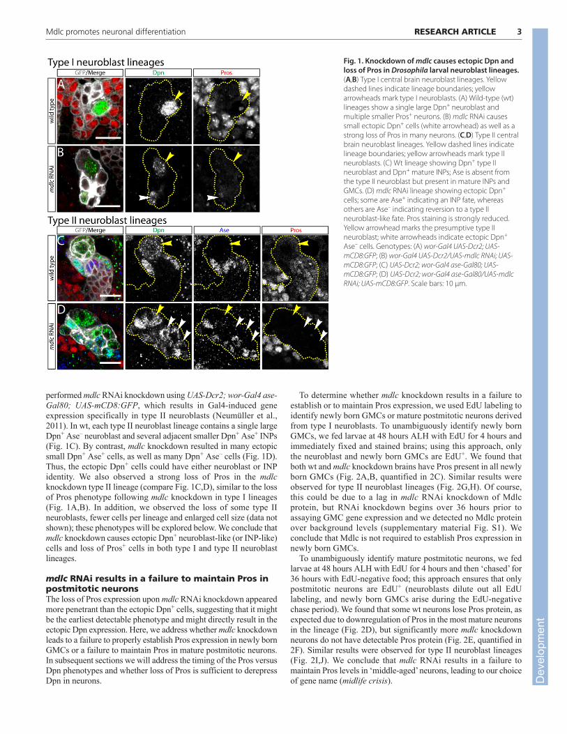
Fig. 1. Knockdown of mdlc causes ectopic Dpn andloss of Pros in Drosophila larval neuroblast lineages.(A,B) Type I central brain neuroblast lineages. Yellowdashed lines indicate lineage boundaries; yellowarrowheads mark type I neuroblasts. (A) Wild-type (wt)lineages show a single large Dpn+ neuroblast andmultiple smaller Pros+ neurons. (B) mdlc RNAi causessmall ectopic Dpn+ cells (white arrowhead) as well as astrong loss of Pros in many neurons. (C,D) Type II centralbrain neuroblast lineages. Yellow dashed lines indicatelineage boundaries; yellow arrowheads mark type IIneuroblasts. (C) Wt lineage showing Dpn+ type IIneuroblast and Dpn+ mature INPs; Ase is absent fromthe type II neuroblast but present in mature INPs andGMCs. (D) mdlc RNAi lineage showing ectopic Dpn+
cells; some are Ase+ indicating an INP fate, whereasothers are Ase– indicating reversion to a type IIneuroblast-like fate. Pros staining is strongly reduced.Yellow arrowhead marks the presumptive type IIneuroblast; white arrowheads indicate ectopic Dpn+
Ase– cells. Genotypes: (A) wor-Gal4 UAS-Dcr2; UAS-mCD8:GFP; (B) wor-Gal4 UAS-Dcr2/UAS-mdlc RNAi; UAS-mCD8:GFP; (C) UAS-Dcr2; wor-Gal4 ase-Gal80; UAS-mCD8:GFP; (D) UAS-Dcr2; wor-Gal4 ase-Gal80/UAS-mdlcRNAi; UAS-mCD8:GFP. Scale bars: 10 μm.
Dev
elop
men
t
4
mdlc mutants fail to maintain Pros and Elav inpostmitotic neuronsTo confirm that the RNAi phenotype is due to loss of function ofmdlc (and not an off-target RNAi effect) we analyzed a geneticlesion in the mdlc gene. mdlc is on chromosome 3R and resideswithin an intron of CG4390 on the opposite strand (Fig. 3A). Weacquired a mutant allele at the mdlc locus: c04701, a piggyBactransposon from the Exelixis collection (Thibault et al., 2004). Itsannotated insertion site is in the 5� UTR, 10 bp upstream of the mdlctranslation start site (Fig. 3A). We used PCR analysis to confirm the
presence of c04701, which is homozygous lethal (see Materials andmethods), renamed the insertion c04701 as mdlcc04701, and used itfor subsequent genetic analysis of mdlc function.
We analyzed mdlcc04701 phenotypes using the MARCM method(Lee and Luo, 2001). Wt type I neuroblast clones possess a singleneuroblast that is Dpn+, Ase+ and Cyclin E (CycE)+, and all neuronsin the clone are Pros+ with the exception of the oldest neurons atthe most distal tip of the clone (Fig. 3B; supplementary materialMovie 1; data not shown). By contrast, mdlcc04701 clones frequentlyhave cells in the middle of the clone that have lost Pros expression(Fig. 3C), which is very rarely observed in wt neuroblast clones.This phenotype is less penetrant but remarkably similar to the mdlcRNAi phenotype (Figs 1, 2). We conclude that both mdlc RNAi andmdlc mutants lead to a failure to maintain Pros levels in middle-aged neurons.
We next aimed to determine the timing of Pros loss and Dpnderepression, as well as examining a second neuroblast marker, Ase,and a second neuronal differentiation marker, Elav, within mdlcmutant clones. First, we co-stained for Pros, Dpn and Ase. GMCslying adjacent to the parental neuroblast typically co-express Prosand Ase and sometimes have weak Dpn due to perdurance from theneuroblast; therefore, they were not considered to be ectopicallyexpressing cells and were excluded from our counts. We observedsome clones in which the Pros– cells do not show detectable Dpn orAse (Fig. 3C), whereas some have derepressed Ase but are Dpn–
(Fig. 3D) and others are both Dpn+ and Ase+ (Fig. 3E, quantified in3H,I; supplementary material Movie 2). Similar results wereobserved for the well-characterized neuronal differentiation markerElav: it is present in all wt neurons, but absent from most (but notall) of the Pros– neurons in mdlc mutants (Fig. 3F,G). We concludethat that loss of the neuronal differentiation factor Pros precedes theloss of the neuronal marker Elav and the ectopic expression of theneuroblast markers Dpn and Ase in mdlc mutant neurons (Fig. 3J).
mdlc mutant neuroblasts have a longer cell cycleand reduced clone sizeIn addition to the neuronal dedifferentiation phenotype shown bymdlc RNAi and mutant clones, we noted that mdlc mutantneuroblast lineages have fewer cells than comparable wt lineages.We quantified this phenotype by counting total cells in both mutantand wt clones generated in 96 hours (slightly longer or shorterintervals were normalized to 96 hours). Wt clones averaged 89±35cells, which is significantly more than mdlc mutant clones at 43±19cells (P<10−5; Fig. 4A). Decrease in clone size was not due toapoptosis, as there was no increase in the apoptotic marker Caspase3 in mdlc mutant clones (Fig. 4B). Instead, we found that the cellcycle is extended: wt neuroblast clones generate 14±4 EdU+ cellsover 8 hours of EdU labeling, whereas mdlc mutants only generate4±6 EdU+ cells during the same span (P<10−5; Fig. 4C). Thevariability stems from the fact that some mutant neuroblastsgenerate near wt numbers of progeny whereas others fail entirely toundergo S phase during the EdU incorporation period (not shown).We conclude that Mdlc promotes larval neuroblast cell cycleprogression.
Mdlc is a conserved zinc-finger-containing proteinwith broad expressionmdlc encodes a well-conserved protein that is 70% similar (58%identical) to the human ortholog RNF113A. Both proteins have twoconserved zinc-finger domains: a CCCH zinc finger, which iscommonly found in RNA-binding proteins involved in splicing; anda RING domain, which is frequently found in E3 ubiquitin ligases
RESEARCH ARTICLE Development 140 (20)
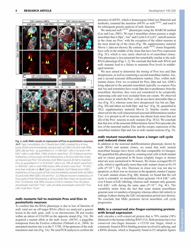
Fig. 2. mdlc RNAi results in repression of Pros in postmitotic neurons.(A-F) Type I neuroblasts. (A-C) Newly born GMCs marked by a 4-hourpulse of EdU and immediately stained; both wt GMCs (A) and mdlc RNAiGMCs (B) are Pros+, as quantified in C. n=304 EdU+ cells in wt and n=99EdU+ cells in mdlc RNAi, 100% of which were Pros+. (D-F) Mature neuronsmarked by a 4-hour pulse of EdU followed by a 36-hour EdU-free chase;wt neurons are Pros+ (D) whereas mdlc RNAi neurons (E) fail to maintainPros, as quantified in F. n=1016 EdU+ cells in wt, 79% of which were Pros+;n=438 EdU+ cells in mdlc RNAi brains, 44% of which were Pros+. *P<0.01;error bars indicate s.d. (G-J) Type II neuroblasts. (G,H) Newly born GMCsmarked by a 4-hour pulse of EdU and immediately stained; both wt GMCs(G) and mdlc RNAi GMCs (H) are Pros+. (I,J) Mature neurons marked by a 4-hour pulse of EdU followed by a 36-hour EdU-free chase; wt neurons arePros+ (I) whereas mdlc RNAi neurons (J) fail to maintain Pros. All yellowarrowheads mark EdU+ Pros+ cells; all white arrowheads mark EdU+ Pros–
cells. Scale bars: 10 μm.
Dev
elop
men
t
(Fig. 5A). In order to analyze the expression patterns and subcellularlocalization of Mdlc, we generated an antibody against the N-terminal 165 amino acids. Immunofluorescent staining with thisantibody revealed that Mdlc is a nuclear protein that is presentthroughout the larval CNS (Fig. 5B).
mdlc knockdown using inscuteable-Gal4 (insc-Gal4) to driveUAS-mdlc RNAi caused a marked decrease in Mdlc immunostainingin the larval central brain (but not in glia or optic lobe; Fig. 5C),
validating the efficacy of mdlc RNAi and the specificity of the Mdlcantibody. Furthermore, mosaic clones of homozygous mdlcc04701
cells showed strongly reduced Mdlc levels compared with thesurrounding wt cells, indicating that mdlcc04701 is a strong loss-of-function allele (Fig. 5D). Upon close examination of larval brains,we found that Mdlc is expressed ubiquitously, including NBs,neurons and glia, as judged by staining with antibodies against Mira,Elav and Reversed polarity (Repo), respectively (Fig. 5E,E�). Mdlc
5RESEARCH ARTICLEMdlc promotes neuronal differentiation
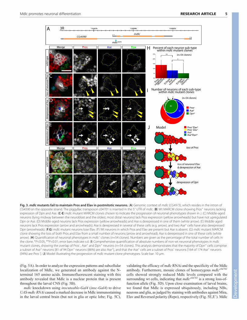
Fig. 3. mdlc mutants fail to maintain Pros and Elav in postmitotic neurons. (A) Genomic context of mdlc (CG4973), which resides in the intron ofCG4390 on the opposite strand. The piggyBac transposon c04701 is inserted in the 5� UTR of mdlc. (B) Wt MARCM clone showing Pros+ neurons lackingexpression of Dpn and Ase. (C-E) mdlc mutant MARCM clones chosen to indicate the progression of neuronal phenotypes shown in J. (C) Middle-agedneurons (lying midway between the neuroblast and the oldest, most distal neurons) lack Pros expression (yellow arrowheads) but have not upregulatedDpn or Ase. (D) Middle-aged neurons lack Pros expression (yellow arrowheads) and Ase is derepressed in one of them (white arrow). (E) Middle-agedneurons lack Pros expression (arrow and arrowheads); Ase is derepressed in several of these cells (e.g. arrow), and two Ase+ cells have also derepressedDpn (arrowheads). (F,G) mdlc mutant neurons lose Elav. (F) Wt neurons in which Pros and Elav are present but Ase is absent. (G) mdlc mutant MARCMclone showing the loss of both Pros and Elav from a small number of neurons (arrow and arrowhead); Ase is derepressed in one of these cells (whitearrow). (H) Quantification of neuronal phenotypes in mdlc– clones (n=54 clones). Numbers are given as the percentage of the total number of cells inthe clone. *P<0.05, **P<0.01; error bars indicate s.d. (I) Comprehensive quantification of absolute numbers of non-wt neuronal phenotypes in mdlcmutant clones, showing the overlap of Pros–, Ase+ and Dpn+ neurons (n=54 clones). This analysis demonstrates that the majority of Dpn+ cells comprisea subset of Ase+ neurons [81 of 94 Dpn+ neurons (86%) are also Ase+], and that the Ase+ cells are a subset of Pros– neurons [164 of 174 Ase+ neurons(94%) are Pros–]. (J) Model illustrating the progression of mdlc mutant clone phenotypes. Scale bar: 10 μm.
Dev
elop
men
t

6
is also broadly expressed in larval imaginal discs (Fig. 5F), in theembryo (Fig. 5G) and in the adult ovary (Fig. 5H).
Mdlc zinc-finger deletions reveal differentrequirements for the CCCH and RING domainsWe have shown that Mdlc has context-dependent functions: it isrequired for neuroblast cell cycle progression as well as in middle-aged neurons to prevent dedifferentiation. Here we asked whetherthe CCCH and RING zinc-finger domains (which are proposed toregulate RNA splicing and ubiquitylation, respectively) havespecific roles in either of these two phenotypes. We generated flylines expressing different Mdlc domains under UAS control: full-
length Mdlc (MdlcFL); Mdlc lacking the CCCH zinc finger(MdlcΔCCCH); Mdlc lacking the RING domain (MdlcΔRING); Mdlcwith RING domain isoleucine 266 mutated to alanine (MdlcI266A),which is reported to interfere with E2/E3 interactions in RINGdomain-containing E3 ubiquitin ligases (Mulder et al., 2007); andMdlc lacking both zinc fingers (MdlcΔCCCH+RING). In addition, wegenerated a transgene expressing the full-length human ortholog ofMdlc (RNF113A). Each construct included a hemagglutinin (HA)epitope tag at the N-terminus of the protein (Fig. 6A).
We assayed the ability of each construct to rescue mutantphenotypes by monitoring both the most penetrant neuronalphenotype (loss of Pros) and the most severe phenotype (ectopic Dpnexpression), as well as neuroblast lineage size and extent of EdUincorporation. Misexpression of MdlcFL, or the full-length humanortholog RNF113A, in mdlc mutant clones significantly rescued clonesize (Fig. 6B) and completely rescued the loss of Pros/ectopic Dpn+
phenotypes (Fig. 6C). In addition, ubiquitous expression of eitherprotein using tubulin-Gal4 rescued mdlcc04701/Df(3R)ED6027hemizygotes to viability (Table 1). These results indicate that theepitope-tagged full-length version of Mdlc is functional and that Mdlcand its human ortholog have conserved functions.
Next we tested the ability of Mdlc proteins lacking RING domainfunction (MdlcΔRING or MdlcI266A) to rescue mdlc mutantphenotypes. Expression of MdlcΔRING or MdlcI266A fully rescuedmdlcc04701/Df(3R)ED6027 hemizygotes to viability (Table 1), aswell as fully rescuing the mdlc mutant neuroblast lineage size andcell cycle phenotypes and preventing the dedifferentiationphenotypes of neurons (Fig. 6B,C; data not shown). We concludethat the RING domain, and its presumptive function inubiquitylation, is not required for any known aspect of Mdlc neuralfunction; the role of the conserved Mdlc RING domain thereforeremains to be elucidated.
RESEARCH ARTICLE Development 140 (20)
Fig. 4. mdlc mutant neuroblasts have an extended cell cycle.(A) Comparison of wt and mdlc– clone sizes. The time between cloneinduction and dissection was 96 hours. (B) Number of Caspase 3+ cellsper clone in wt and mdlc– clones. (C) Comparison of wt and mdlc– EdUincorporation rates. The EdU incorporation period was 8 hours prior todissection. ***P<10−5; NS, not significantly different; error bars indicate s.d.
Fig. 5. Mdlc is a zinc-finger protein with broad expression inthe CNS and other tissues. (A) Mdlc protein and the humanortholog RNF113A have a conserved CCCH zinc finger and a C-terminal RING domain. Numbers indicate amino acids. (B-D) Anti-Mdlc shows ubiquitous nuclear localization of Mdlc,which is lost upon mdlc loss of function. (B) Wt Mdlc is expressedin larval brain neuroblasts (arrowhead) and glia (arrow). (C) mdlcRNAi in central brain neuroblasts and their immediate progenyreduces Mdlc protein staining in the central brain, particularly inneuroblasts (arrowhead), as identified by the presence of Dpn(not shown). Mdlc expression is still visible in glia (arrow) and inthe optic lobe (OL), where the RNAi transgene was notexpressed. (D) Homozygous mutant mdlcc04701 clones (yellowdashed lines) show a strong reduction in Mdlc protein. (E,E�) In the wt larval brain, Mdlc protein is detected inneuroblasts (wide arrowheads), neurons (narrow arrowheads)and glia (arrows), which can be identified based on staining withantibodies against Mira, Elav and Repo, respectively. (F-H) Mdlcis ubiquitously expressed in all tissues examined. (F)Representative imaginal disc. (G) Mdlc expression in the embryo,including in neuroblasts (arrowheads) identified by Miraexpression (not shown). (H) Adult ovariole. Scale bars: 100 μm inB-D,F-H; 10 μm in E.
Dev
elop
men
t
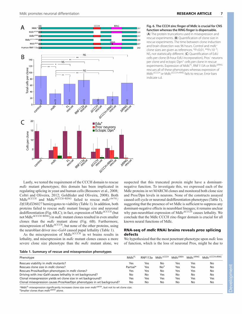
Lastly, we tested the requirement of the CCCH domain to rescuemdlc mutant phenotypes; this domain has been implicated inregulating splicing in yeast and human cells (Bessonov et al., 2008;Coltri and Oliveira, 2012; Goldfeder and Oliveira, 2008). BothMdlcΔCCCH and MdlcΔCCCH+RING failed to rescue mdlcc04701/Df(3R)ED6027 hemizygotes to viability (Table 1). In addition, bothproteins failed to rescue mdlc mutant lineage size and neuronaldedifferentiation (Fig. 6B,C); in fact, expression of MdlcΔCCCH (butnot MdlcΔCCCH+RING) in mdlc mutant clones resulted in even smallerclones than the mdlc mutant alone (Fig. 6B). Furthermore,misexpression of MdlcΔCCCH, but none of the other proteins, usingthe neuroblast driver insc-Gal4 caused pupal lethality (Table 1).
As the misexpression of MdlcΔCCCH in wt brains results inlethality, and misexpression in mdlc mutant clones causes a moresevere clone size phenotype than the mdlc mutant alone, we
suspected that this truncated protein might have a dominant-negative function. To investigate this, we expressed each of theMdlc proteins in wt MARCM clones and monitored both clone sizeand Pros/Dpn levels in neurons. None of the constructs assayedcaused cell cycle or neuronal dedifferentiation phenotypes (Table 1),suggesting that the presence of wt Mdlc is sufficient to suppress anydominant-negative effects in neuroblast lineages; it remains unclearwhy pan-neuroblast expression of MdlcΔCCCH causes lethality. Weconclude that the Mdlc CCCH zinc-finger domain is crucial for allknown neural functions of Mdlc.
RNA-seq of mdlc RNAi brains reveals pros splicingdefectsWe hypothesized that the most penetrant phenotype upon mdlc lossof function, which is the loss of neuronal Pros, might be due to
7RESEARCH ARTICLEMdlc promotes neuronal differentiation
Fig. 6. The CCCH zinc finger of Mdlc is crucial for CNSfunction whereas the RING finger is dispensable.(A) The protein truncations used in misexpression andrescue experiments. (B) Quantification of clone size inrescue experiments. The time between clone inductionand brain dissection was 96 hours. Control and mdlc–
clone sizes are given as references. *P<0.01, **P<10−3;NS, not statistically different. (C) Quantification of EdUcells per clone (8-hour EdU incorporation), Pros– neuronsper clone and ectopic Dpn+ cells per clone in rescueexperiments. Expression of MdlcFL, RNF113A or MdlcΔRING
rescues all of these phenotypes whereas expression ofMdlcΔCCCH or MdlcΔCCCH+RING fails to rescue. Error barsindicate s.d.
Table 1. Summary of rescue and misexpression phenotypes
Phenotype MdlcFL RNF113a MdlcΔCCCH MdlcI266A MdlcΔRING MdlcΔCCCH+RING
Rescues viability in mdlc mutants? Yes Yes No Yes Yes NoRescues clone size in mdlc clones? Partial* Yes No‡ Yes Yes NoRescues Pros/Ase/Dpn phenotypes in mdlc clones? Yes Yes No Yes Yes NoDriving with insc-Gal4 causes lethality in wt background? No No Yes No No NoClonal misexpression yields wt clone size in wt background? Yes Yes Yes Yes Yes YesClonal misexpression causes Pros/Ase/Dpn phenotypes in wt background? No No No No No No
*MdlcFL misexpression significantly increases clone size over mdlcc04701, but not to wt clone size.‡Smaller clones than mdlcc04701 alone. D
evel
opm
ent
8
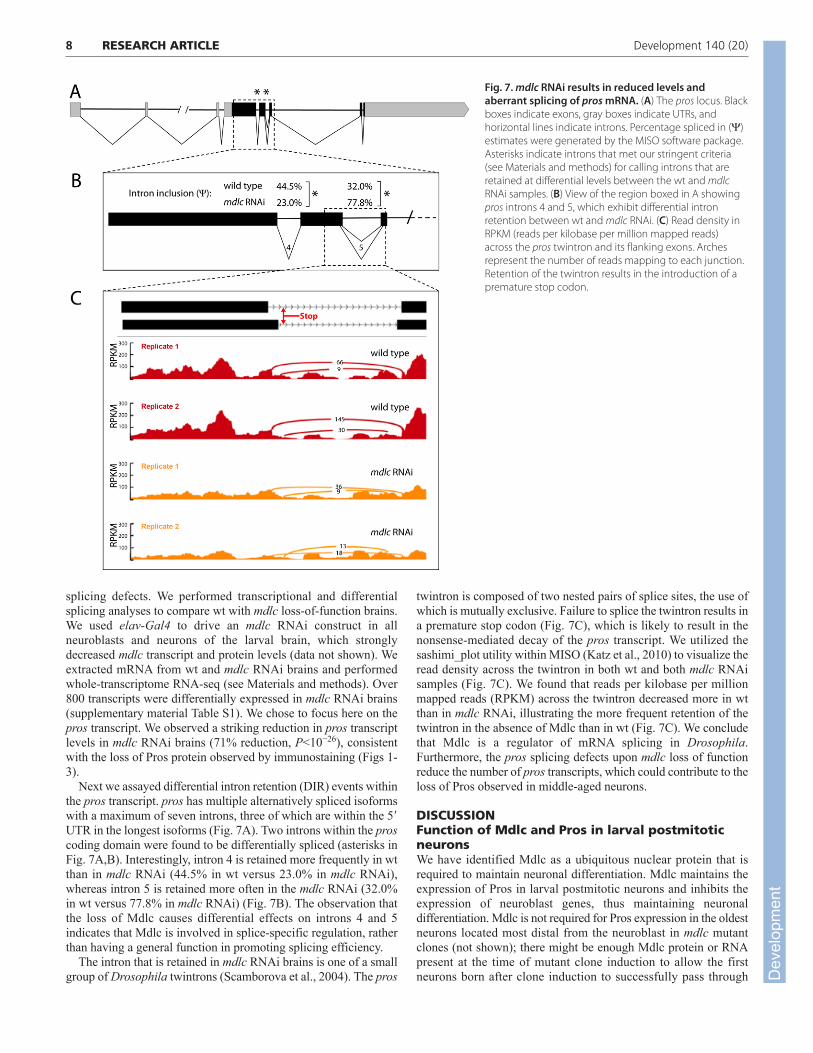
splicing defects. We performed transcriptional and differentialsplicing analyses to compare wt with mdlc loss-of-function brains.We used elav-Gal4 to drive an mdlc RNAi construct in allneuroblasts and neurons of the larval brain, which stronglydecreased mdlc transcript and protein levels (data not shown). Weextracted mRNA from wt and mdlc RNAi brains and performedwhole-transcriptome RNA-seq (see Materials and methods). Over800 transcripts were differentially expressed in mdlc RNAi brains(supplementary material Table S1). We chose to focus here on thepros transcript. We observed a striking reduction in pros transcriptlevels in mdlc RNAi brains (71% reduction, P<10−26), consistentwith the loss of Pros protein observed by immunostaining (Figs 1-3).
Next we assayed differential intron retention (DIR) events withinthe pros transcript. pros has multiple alternatively spliced isoformswith a maximum of seven introns, three of which are within the 5�UTR in the longest isoforms (Fig. 7A). Two introns within the proscoding domain were found to be differentially spliced (asterisks inFig. 7A,B). Interestingly, intron 4 is retained more frequently in wtthan in mdlc RNAi (44.5% in wt versus 23.0% in mdlc RNAi),whereas intron 5 is retained more often in the mdlc RNAi (32.0%in wt versus 77.8% in mdlc RNAi) (Fig. 7B). The observation thatthe loss of Mdlc causes differential effects on introns 4 and 5indicates that Mdlc is involved in splice-specific regulation, ratherthan having a general function in promoting splicing efficiency.
The intron that is retained in mdlc RNAi brains is one of a smallgroup of Drosophila twintrons (Scamborova et al., 2004). The pros
twintron is composed of two nested pairs of splice sites, the use ofwhich is mutually exclusive. Failure to splice the twintron results ina premature stop codon (Fig. 7C), which is likely to result in thenonsense-mediated decay of the pros transcript. We utilized thesashimi_plot utility within MISO (Katz et al., 2010) to visualize theread density across the twintron in both wt and both mdlc RNAisamples (Fig. 7C). We found that reads per kilobase per millionmapped reads (RPKM) across the twintron decreased more in wtthan in mdlc RNAi, illustrating the more frequent retention of thetwintron in the absence of Mdlc than in wt (Fig. 7C). We concludethat Mdlc is a regulator of mRNA splicing in Drosophila.Furthermore, the pros splicing defects upon mdlc loss of functionreduce the number of pros transcripts, which could contribute to theloss of Pros observed in middle-aged neurons.
DISCUSSIONFunction of Mdlc and Pros in larval postmitoticneuronsWe have identified Mdlc as a ubiquitous nuclear protein that isrequired to maintain neuronal differentiation. Mdlc maintains theexpression of Pros in larval postmitotic neurons and inhibits theexpression of neuroblast genes, thus maintaining neuronaldifferentiation. Mdlc is not required for Pros expression in the oldestneurons located most distal from the neuroblast in mdlc mutantclones (not shown); there might be enough Mdlc protein or RNApresent at the time of mutant clone induction to allow the firstneurons born after clone induction to successfully pass through
RESEARCH ARTICLE Development 140 (20)
Fig. 7. mdlc RNAi results in reduced levels andaberrant splicing of pros mRNA. (A) The pros locus. Blackboxes indicate exons, gray boxes indicate UTRs, andhorizontal lines indicate introns. Percentage spliced in (Y)estimates were generated by the MISO software package.Asterisks indicate introns that met our stringent criteria(see Materials and methods) for calling introns that areretained at differential levels between the wt and mdlcRNAi samples. (B) View of the region boxed in A showingpros introns 4 and 5, which exhibit differential intronretention between wt and mdlc RNAi. (C) Read density inRPKM (reads per kilobase per million mapped reads)across the pros twintron and its flanking exons. Archesrepresent the number of reads mapping to each junction.Retention of the twintron results in the introduction of apremature stop codon.
Dev
elop
men
t

middle-age without dedifferentiation. This suggests that after acertain age, neurons do not require Mdlc to maintain neuronaldifferentiation. Alternatively, neurons born at early larval stagesmight not have the same requirement for Mdlc as neurons born atlater larval stages.
Interestingly, mdlc mutant clones never show EdU incorporationin ectopic Dpn+ cells, raising the question of whether they are trueneuroblasts or have a mixed neuroblast/neuronal fate. We note thatloss of Mdlc results in cell cycle delay in parental neuroblasts, so itis perhaps unsurprising that the ectopic Dpn+ cells do not proliferate.Nevertheless, our data show that Mdlc is not required to suppresscell cycle entry in postmitotic neurons. Similarly, our data show thatPros is not required to suppress cell cycle entry in postmitoticneurons, as pros RNAi specifically within postmitotic neuronsremoves all detectable Pros protein (supplementary materialFig. S2) but does not trigger entry into the cell cycle (data notshown). This is comparable to the situation in wt embryonicneurons, which rapidly lose Pros but never re-enter the cell cycle,and contrasts with the role of Pros in GMCs, where it is required torepress neuroblast genes and promote cell cycle exit (Choksi et al.,2006). The maturation step that is taken by neurons to make themincapable of re-entering the cell cycle in the absence of Pros is notwell understood.
Pros is known to bind the dpn, ase and CycE loci (Choksi et al.,2006; Southall and Brand, 2009), and is known to keep theexpression of these genes low in embryos (Li and Vaessin, 2000).Does Pros directly or indirectly maintain repression of the dpn, aseand CycE neuroblast genes in larval neurons? To attempt to addressthis question, we used RNAi to ablate either Pros or Mdlc inpostmitotic neurons, assaying for derepression of the neuroblastfactor Dpn. Driving UAS-pros RNAi with either atonal-Gal4 oracj6-Gal4 specifically eliminated Pros protein in multiple clustersof postmitotic neurons, yet did not lead to the derepression of Dpn(supplementary material Fig. S2). However, RNAi knockdown ofmdlc using the same Gal4 lines also did not cause Dpn derepression(data not shown). Several factors may account for this unexpectedresult. First, the lineages expressing atonal-Gal4 and acj6-Gal4might not require Mdlc, consistent with our finding that somemdlcc04701 mutant clones had no Pros loss, Dpn/Ase derepressionor EdU incorporation phenotypes. Alternatively, or in addition, thepostmitotic Gal4 lines might have eliminated Mdlc and Pros inneurons after they passed through the susceptible middle-aged stage.However, we note that acj6-Gal4 appears to drive expression in onelineage in all ages of neurons (only excluding the neuroblast andGMCs; supplementary material Fig. S2C,D). This indicates eitherthat this lineage does not require Pros or Mdlc to maintain Dpnrepression or that Mdlc is required at the GMC level in this lineage.We lack appropriate drivers to distinguish between thesepossibilities. Given these caveats, we are unable to determinewhether neuronal loss of Pros alone is sufficient for derepression ofneuroblast genes. We think it likely that Pros does have some rolein the neuronal repression of neuroblast genes, however, becausewe nearly always observe derepression of Dpn/Ase in neurons thathave already lost Pros (Fig. 3I).
The role of Mdlc in splicing regulationWe found that the CCCH zinc-finger domain, which is implicatedin RNA-binding in other proteins, is essential for Mdlc function inthe nervous system and for organismal viability. The S. cerevisiaeand human orthologs have roles in splicing (Bessonov et al., 2008;Chan et al., 2003; Coltri and Oliveira, 2012; Goldfeder and Oliveira,2008; Ohi et al., 2002). In yeast, Cwc24p is reported to be a splicing
efficiency factor primarily affecting primary transcripts withatypical branchpoints. For example, splicing of the transcriptssnR17A and B, which encode the U3 snoRNAs, was stronglyaffected, resulting in defects in the processing of pre-rRNA (Coltriand Oliveira, 2012; Goldfeder and Oliveira, 2008). Our observationthat loss of Mdlc causes specific splicing defects (both increasedand decreased intron retention) in the pros transcript, together withour finding that RNF113A can rescue the mdlc loss-of-functionphenotypes, suggest that the fly and human proteins might have amore complex role in regulating splicing than that of the yeastgeneral splicing factor Cwc24p.
What might be the CNS splicing targets of Mdlc, in addition topros? In mammals, alternatively spliced transcripts resulting inprotein isoforms that influence stemness or differentiation areknown, including within the nervous system (Lipscombe, 2005;Nelles and Yeo, 2010). Work from our laboratory has recentlyidentified splice isoforms that are differentially regulated by theneuroblast transcription factor Wor (Lai et al., 2012). The analysisof genome-wide changes in splicing in mdlc RNAi brains is inprogress but beyond the scope of this paper.
Relevance of alternative splicing of the prostwintronThe pros twintron undergoes developmentally regulated alternativesplicing (Scamborova et al., 2004) to generate protein isoforms thatdiffer by 29 amino acids at the homeodomain N-terminus (Chu-Lagraff et al., 1991). The two nested pairs of splice sites in the prostwintron are utilized mutually exclusively by two separatespliceosomes: U2 and U12 (Scamborova et al., 2004). Loss of Mdlcspecifically reduces pros U12 splicing (Fig. 7), so we examinedmost of the other introns in Drosophila that are known to utilize theU12 spliceosome. The pros intron was the sole example ofdifferential retention (data not shown). This indicates that Mdlc doesnot preferentially affect the U12 spliceosome.
A role for Mdlc as a ubiquitin ligase?The RING-type zinc-finger proteins constitute one of the largestprotein families, with over 600 members (Deshaies and Joazeiro,2009). Many of these proteins have been shown to function as E3ubiquitin ligases, and the presence of a RING domain is oftensufficient for such an annotation. The Mdlc human orthologRNF113A is no exception and is thought to function as an E3 ligase;consistent with this assumption, RNF113A was found to physicallyinteract with one of the human E2 proteins, UBE2U (Li et al., 2008;van Wijk et al., 2009). Moreover, the Mdlc RING domain is verywell conserved from yeast to humans, suggesting its functionalimportance. We were therefore surprised to find that the RINGdomain was completely dispensable not only for CNS function butalso for organismal viability, since the ubiquitous misexpression ofa version of Mdlc lacking the RING domain was able to substitutefor the full-length protein.
AcknowledgementsWe thank Bruce Bowerman, Judith Eisen and Tasha Joy for comments on themanuscript and Leslie Gay for assistance generating the Mdlc antibody. Wethank the BDSC and VDRC for fly stocks and DSHB for antibodies.
FundingThis work was supported by the Howard Hughes Medical Institute, whereC.Q.D. is an Investigator. Deposited in PMC for release after 6 months.
Competing interests statementThe authors declare no competing financial interests.
9RESEARCH ARTICLEMdlc promotes neuronal differentiation
Dev
elop
men
t

10
Author contributionsT.D.C. and C.Q.D. conceived all experiments. T.D.C. and A.J.S. performed theRNA-seq experiments; T.D.C. performed all other experiments. C.Q.D. andT.D.C. wrote the manuscript. C.Q.D. was the principal investigator for thiswork.
Supplementary materialSupplementary material available online athttp://dev.biologists.org/lookup/suppl/doi:10.1242/dev.093781/-/DC1
ReferencesAlbertson, R., Chabu, C., Sheehan, A. and Doe, C. Q. (2004). Scribble protein
domain mapping reveals a multistep localization mechanism and domainsnecessary for establishing cortical polarity. J. Cell Sci. 117, 6061-6070.
Anders, S. and Huber, W. (2010). Differential expression analysis for sequencecount data. Genome Biol. 11, R106.
Bayraktar, O. A., Boone, J. Q., Drummond, M. L. and Doe, C. Q. (2010).Drosophila type II neuroblast lineages keep Prospero levels low to generatelarge clones that contribute to the adult brain central complex. Neural Dev. 5, 26.
Bello, B., Reichert, H. and Hirth, F. (2006). The brain tumor gene negativelyregulates neural progenitor cell proliferation in the larval central brain ofDrosophila. Development 133, 2639-2648.
Bello, B. C., Izergina, N., Caussinus, E. and Reichert, H. (2008). Amplification ofneural stem cell proliferation by intermediate progenitor cells in Drosophilabrain development. Neural Dev. 3, 5.
Bessonov, S., Anokhina, M., Will, C. L., Urlaub, H. and Lührmann, R. (2008).Isolation of an active step I spliceosome and composition of its RNP core.Nature 452, 846-850.
Betschinger, J., Mechtler, K. and Knoblich, J. A. (2006). Asymmetricsegregation of the tumor suppressor brat regulates self-renewal in Drosophilaneural stem cells. Cell 124, 1241-1253.
Bischof, J., Maeda, R. K., Hediger, M., Karch, F. and Basler, K. (2007). Anoptimized transgenesis system for Drosophila using germ-line-specific phiC31integrases. Proc. Natl. Acad. Sci. USA 104, 3312-3317.
Boone, J. Q. and Doe, C. Q. (2008). Identification of Drosophila type IIneuroblast lineages containing transit amplifying ganglion mother cells. Dev.Neurobiol. 68, 1185-1195.
Bowman, S. K., Rolland, V., Betschinger, J., Kinsey, K. A., Emery, G. andKnoblich, J. A. (2008). The tumor suppressors Brat and Numb regulate transit-amplifying neuroblast lineages in Drosophila. Dev. Cell 14, 535-546.
Brand, M., Jarman, A. P., Jan, L. Y. and Jan, Y. N. (1993). asense is a Drosophilaneural precursor gene and is capable of initiating sense organ formation.Development 119, 1-17.
Broadus, J., Fuerstenberg, S. and Doe, C. Q. (1998). Staufen-dependentlocalization of prospero mRNA contributes to neuroblast daughter-cell fate.Nature 391, 792-795.
Cabernard, C. and Doe, C. Q. (2009). Apical/basal spindle orientation is requiredfor neuroblast homeostasis and neuronal differentiation in Drosophila. Dev.Cell 17, 134-141.
Carney, T. D., Miller, M. R., Robinson, K. J., Bayraktar, O. A., Osterhout, J. A.and Doe, C. Q. (2012). Functional genomics identifies neural stem cell sub-type expression profiles and genes regulating neuroblast homeostasis. Dev.Biol. 361, 137-146.
Chan, S. P., Kao, D. I., Tsai, W. Y. and Cheng, S. C. (2003). The Prp19p-associated complex in spliceosome activation. Science 302, 279-282.
Choksi, S. P., Southall, T. D., Bossing, T., Edoff, K., de Wit, E., Fischer, B. E.,van Steensel, B., Micklem, G. and Brand, A. H. (2006). Prospero acts as abinary switch between self-renewal and differentiation in Drosophila neuralstem cells. Dev. Cell 11, 775-789.
Chu-Lagraff, Q., Wright, D. M., McNeil, L. K. and Doe, C. Q. (1991). Theprospero gene encodes a divergent homeodomain protein that controlsneuronal identity in Drosophila. Development 2 Suppl. 2, 79-85.
Coltri, P. P. and Oliveira, C. C. (2012). Cwc24p is a general Saccharomycescerevisiae splicing factor required for the stable U2 snRNP binding to primarytranscripts. PLoS ONE 7, e45678.
Deshaies, R. J. and Joazeiro, C. A. (2009). RING domain E3 ubiquitin ligases.Annu. Rev. Biochem. 78, 399-434.
Dietzl, G., Chen, D., Schnorrer, F., Su, K. C., Barinova, Y., Fellner, M., Gasser,B., Kinsey, K., Oppel, S., Scheiblauer, S. et al. (2007). A genome-widetransgenic RNAi library for conditional gene inactivation in Drosophila. Nature448, 151-156.
Doe, C. Q. (2008). Neural stem cells: balancing self-renewal with differentiation.Development 135, 1575-1587.
Goldfeder, M. B. and Oliveira, C. C. (2008). Cwc24p, a novel Saccharomycescerevisiae nuclear ring finger protein, affects pre-snoRNA U3 splicing. J. Biol.Chem. 283, 2644-2653.
Izergina, N., Balmer, J., Bello, B. and Reichert, H. (2009). Postembryonicdevelopment of transit amplifying neuroblast lineages in the Drosophila brain.Neural Dev. 4, 44.
Katz, Y., Wang, E. T., Airoldi, E. M. and Burge, C. B. (2010). Analysis and designof RNA sequencing experiments for identifying isoform regulation. Nat.Methods 7, 1009-1015.
Knoblich, J. A. (2010). Asymmetric cell division: recent developments and theirimplications for tumour biology. Nat. Rev. Mol. Cell Biol. 11, 849-860.
Knoblich, J. A., Jan, L. Y. and Jan, Y. N. (1995). Asymmetric segregation ofNumb and Prospero during cell division. Nature 377, 624-627.
Lai, S. L., Miller, M. R., Robinson, K. J. and Doe, C. Q. (2012). The Snail familymember Worniu is continuously required in neuroblasts to prevent Elav-induced premature differentiation. Dev. Cell 23, 849-857.
Lee, T. and Luo, L. (2001). Mosaic analysis with a repressible cell marker(MARCM) for Drosophila neural development. Trends Neurosci. 24, 251-254.
Lee, C. Y., Wilkinson, B. D., Siegrist, S. E., Wharton, R. P. and Doe, C. Q. (2006).Brat is a Miranda cargo protein that promotes neuronal differentiation andinhibits neuroblast self-renewal. Dev. Cell 10, 441-449.
Li, L. and Vaessin, H. (2000). Pan-neural Prospero terminates cell proliferationduring Drosophila neurogenesis. Genes Dev. 14, 147-151.
Li, W., Bengtson, M. H., Ulbrich, A., Matsuda, A., Reddy, V. A., Orth, A.,Chanda, S. K., Batalov, S. and Joazeiro, C. A. (2008). Genome-wide andfunctional annotation of human E3 ubiquitin ligases identifies MULAN, amitochondrial E3 that regulates the organelle’s dynamics and signaling. PLoSONE 3, e1487.
Lipscombe, D. (2005). Neuronal proteins custom designed by alternativesplicing. Curr. Opin. Neurobiol. 15, 358-363.
Miller, M. R., Robinson, K. J., Cleary, M. D. and Doe, C. Q. (2009). TU-tagging:cell type-specific RNA isolation from intact complex tissues. Nat. Methods 6,439-441.
Mulder, K. W., Inagaki, A., Cameroni, E., Mousson, F., Winkler, G. S., DeVirgilio, C., Collart, M. A. and Timmers, H. T. (2007). Modulation ofUbc4p/Ubc5p-mediated stress responses by the RING-finger-dependentubiquitin-protein ligase Not4p in Saccharomyces cerevisiae. Genetics 176, 181-192.
Nelles, D. A. and Yeo, G. W. (2010). Alternative splicing in stem cell self-renewaland differentiation. Adv. Exp. Med. Biol. 695, 92-104.
Neumüller, R. A., Richter, C., Fischer, A., Novatchkova, M., Neumüller, K. G.and Knoblich, J. A. (2011). Genome-wide analysis of self-renewal inDrosophila neural stem cells by transgenic RNAi. Cell Stem Cell 8, 580-593.
Ohi, M. D., Link, A. J., Ren, L., Jennings, J. L., McDonald, W. H. and Gould, K.L. (2002). Proteomics analysis reveals stable multiprotein complexes in bothfission and budding yeasts containing Myb-related Cdc5p/Cef1p, novel pre-mRNA splicing factors, and snRNAs. Mol. Cell. Biol. 22, 2011-2024.
Scamborova, P., Wong, A. and Steitz, J. A. (2004). An intronic enhancerregulates splicing of the twintron of Drosophila melanogaster prospero pre-mRNA by two different spliceosomes. Mol. Cell. Biol. 24, 1855-1869.
Southall, T. D. and Brand, A. H. (2009). Neural stem cell transcriptionalnetworks highlight genes essential for nervous system development. EMBO J.28, 3799-3807.
Spana, E. P. and Doe, C. Q. (1995). The prospero transcription factor isasymmetrically localized to the cell cortex during neuroblast mitosis inDrosophila. Development 121, 3187-3195.
Srinivasan, S., Peng, C. Y., Nair, S., Skeath, J. B., Spana, E. P. and Doe, C. Q.(1998). Biochemical analysis of ++Prospero protein during asymmetric celldivision: cortical Prospero is highly phosphorylated relative to nuclearProspero. Dev. Biol. 204, 478-487.
Thibault, S. T., Singer, M. A., Miyazaki, W. Y., Milash, B., Dompe, N. A., Singh,C. M., Buchholz, R., Demsky, M., Fawcett, R., Francis-Lang, H. L. et al.(2004). A complementary transposon tool kit for Drosophila melanogasterusing P and piggyBac. Nat. Genet. 36, 283-287.
van Wijk, S. J., de Vries, S. J., Kemmeren, P., Huang, A., Boelens, R., Bonvin,A. M. and Timmers, H. T. (2009). A comprehensive framework of E2-RING E3interactions of the human ubiquitin-proteasome system. Mol. Syst. Biol. 5, 295.
Weng, M., Golden, K. L. and Lee, C. Y. (2010). dFezf/Earmuff maintains therestricted developmental potential of intermediate neural progenitors inDrosophila. Dev. Cell 18, 126-135.
Wu, T. D. and Nacu, S. (2010). Fast and SNP-tolerant detection of complexvariants and splicing in short reads. Bioinformatics 26, 873-881.
RESEARCH ARTICLE Development 140 (20)
Dev
elop
men
t