Determination of Helicobacter Spp in Swine Stomach from ... · as positive or negative for...
6
Academia Journal of Biotechnology 6(7): 203-208, July 2018 DOI: 10.15413/ajb.2018.0123 ISSN 2315-7747 ©2018 Academia Publishing Research Paper Determination of Helicobacter Spp in Swine Stomach from Río Cuarto Zone, Córdoba, Argentina Accepted 31 st July, 2018 ABSTRACT Helicobacter spp is a gram-negative micro-organism that is lodged in the gastric mucosa and is considered the main cause of peptic ulcer. Studies confirmed the implication of Helicobacter spp in gastric pathologies of man and other animal species and one of them is pig. These bacteria could be pathogens responsible for zoonosis. Pigs develop naturally and frequently gastroesophageal ulcers, ulcers colonized by Helicobacter spp. Investigations carried out in Colombia established a prevalence of 34.8% porcine gastric ulcers in slaughter animals slaughtered in relation to Helicobacter spp in the production of gastric lesions. However, there are few reports of Helicobacter spp in gastric mucosa of pigs in Argentina. In the present study, the presence of Helicobacter spp was determined in gastric samples obtained from swine slaughter of Río Cuarto area. Samples (n=54) were classified as positive or negative for gastritis, and also as chronic or acute gastritis by histopathology using hematoxylin/eosin stain. The presence of Helicobacter spp was determined by Giemsa and Warthin Starry stain and immunohistochemistry method. Immunohistochemistry was performed with anti-Helicobacter pilory commercial antibody. In this study, a higher proportion of chronic than acute lesions was observed. The Giemsa and Warthin Starry stains revealed two species of Helicobacter in the gastric samples. The results indicate that 48% of the samples were Helicobacter (+) and gastritis (+), 35% were Helicobacter (-) and gastritis (+) and the remaining 17% were Helicobacter (-) and gastritis (-). This finding provides a basic knowledge for future studies of the role of Helicobacter in the species under study. Key words: Estomach, Helicobacter SPP, immunohistochemistry, swine, Warthin Starry. INTRODUCTION Helicobacter spp is a gram-negative micro-organism that is lodged in the gastric mucosa and is considered the main cause of peptic ulcer (Cárdenas et al., 2006; Lopez Brea et al., 2006; Sleinsinger et al., 2000). It was first observed by the pathologist, Robin Warren in early June, 1979. Its detection in histopathological studies continued for a couple of years associating the presence of the micro- organism with gastric diseases in humans. During this time, there were many attempts to isolate the bacteria but without any success (Tytgort, 1994; McColl et al., 1997; Warren, 1997; Thibaut et al., 2007; Morales, 2008). In 1981, the gastroenterologist, Barry Marshall joined Warren's research and confirmed the results of the latter. However, it was not until 1982 that Helicobacter pylori could be isolated for the first time. In 1984, the association of H. pylori with chronic gastritis was published in the journal Lancet and for the first time it was suggested that an infectious etiology could be the responsible cause of peptic ulcer. In 1985, Marshall demonstrated the bacteria's ability to cause the disease (Hunt, 1996; Bondi, 2006; V.H. Mac Loughlin 1 , O.E. Navarro 1 , R.A. Martínez 1 , M.C. Grosso 1 *, F. Savino 1 , L. Ritta1, Leandro 1 , M.P. Van Deer Veen 1 , SR Giménez 1 , M.E. Maine Soria 1 , A. Cristofolini 2 and C.I. Merkis 2 1 Histology, Department of Animal Anatomy, Agronomy and Veterinary Faculty, National University of Río Cuarto, RutaNacional 36, Km 601, Río Cuarto (5800) Córdoba, Argentina. 2 Electronic Microscopy, Department of Animal Pathology, Agronomy and Veterinary Faculty, National University of Río Cuarto, RutaNacional 36, Km 601, Río Cuarto (5800) Córdoba, Argentina. *Corresponding author. E-mail: [email protected].
Transcript of Determination of Helicobacter Spp in Swine Stomach from ... · as positive or negative for...
Academia Journal of Biotechnology 6(7): 203-208, July 2018 DOI: 10.15413/ajb.2018.0123 ISSN 2315-7747 ©2018 Academia Publishing
Research Paper
Determination of Helicobacter Spp in Swine Stomach from Río Cuarto Zone, Córdoba, Argentina
Accepted 31st July, 2018 ABSTRACT Helicobacter spp is a gram-negative micro-organism that is lodged in the gastric mucosa and is considered the main cause of peptic ulcer. Studies confirmed the implication of Helicobacter spp in gastric pathologies of man and other animal species and one of them is pig. These bacteria could be pathogens responsible for zoonosis. Pigs develop naturally and frequently gastroesophageal ulcers, ulcers colonized by Helicobacter spp. Investigations carried out in Colombia established a prevalence of 34.8% porcine gastric ulcers in slaughter animals slaughtered in relation to Helicobacter spp in the production of gastric lesions. However, there are few reports of Helicobacter spp in gastric mucosa of pigs in Argentina. In the present study, the presence of Helicobacter spp was determined in gastric samples obtained from swine slaughter of Río Cuarto area. Samples (n=54) were classified as positive or negative for gastritis, and also as chronic or acute gastritis by histopathology using hematoxylin/eosin stain. The presence of Helicobacter spp was determined by Giemsa and Warthin Starry stain and immunohistochemistry method. Immunohistochemistry was performed with anti-Helicobacter pilory commercial antibody. In this study, a higher proportion of chronic than acute lesions was observed. The Giemsa and Warthin Starry stains revealed two species of Helicobacter in the gastric samples. The results indicate that 48% of the samples were Helicobacter (+) and gastritis (+), 35% were Helicobacter (-) and gastritis (+) and the remaining 17% were Helicobacter (-) and gastritis (-). This finding provides a basic knowledge for future studies of the role of Helicobacter in the species under study. Key words: Estomach, Helicobacter SPP, immunohistochemistry, swine, Warthin Starry.
INTRODUCTION
Helicobacter spp is a gram-negative micro-organism that is lodged in the gastric mucosa and is considered the main cause of peptic ulcer (Cárdenas et al., 2006; Lopez Brea et al., 2006; Sleinsinger et al., 2000). It was first observed by the pathologist, Robin Warren in early June, 1979. Its detection in histopathological studies continued for a couple of years associating the presence of the micro-organism with gastric diseases in humans. During this time, there were many attempts to isolate the bacteria but without any success (Tytgort, 1994; McColl et al., 1997;
Warren, 1997; Thibaut et al., 2007; Morales, 2008). In 1981, the gastroenterologist, Barry Marshall joined
Warren's research and confirmed the results of the latter. However, it was not until 1982 that Helicobacter pylori could be isolated for the first time. In 1984, the association of H. pylori with chronic gastritis was published in the journal Lancet and for the first time it was suggested that an infectious etiology could be the responsible cause of peptic ulcer. In 1985, Marshall demonstrated the bacteria's ability to cause the disease (Hunt, 1996; Bondi, 2006;
V.H. Mac Loughlin1, O.E. Navarro1, R.A. Martínez1, M.C. Grosso1*, F. Savino1, L. Ritta1, Leandro1, M.P. Van Deer Veen1, SR Giménez1, M.E. Maine Soria1, A. Cristofolini2 and C.I. Merkis2 1Histology, Department of Animal Anatomy, Agronomy and Veterinary Faculty, National University of Río Cuarto, RutaNacional 36, Km 601, Río Cuarto (5800) Córdoba, Argentina. 2Electronic Microscopy, Department of Animal Pathology, Agronomy and Veterinary Faculty, National University of Río Cuarto, RutaNacional 36, Km 601, Río Cuarto (5800) Córdoba, Argentina. *Corresponding author. E-mail: [email protected].
Academia Journal of Biotechnology; Loughlin et al. 204 Cárdenas et al., 2006). In 2005, Dr. Warren and Marshall were awarded the Nobel Prize in Medicine and Physiology for the discovery of H. pylori and its role in the diagnosis, prevention and treatment of chronic gastritis, gastroduodenal ulcers, gastric lymphoma and in some gastric cancer caused by this micro-organism (Moreno, 2005; Tytgort, 1994).
Studies confirmed the implication of Helicobacter spp in gastric pathologies of humans and other animal species and one of them is pig (Eaton et al., 1997; Odriozola, 2011). The transmission of Helicobacter from animals to humans is not known, but is most likely to occur through direct contact. Living in proximity to cats, dogs and especially pigs, is a risk factor for these infections. The intensity of contact with animals is also important as a higher incidence of these conditions was observed in pig farmers and slaughterhouse staffs (Haesebrouck et al., 2009; Liang et al., 2013).
In human gastric conditions, H. pylori are the most frequently found micro-organism, although the presence of H. suis has also been reported in this species (Haesebrouck et al., 2009). That is why these bacteria could be pathogens responsible for zoonoses (Vale and Vitor, 2010). Pigs develop naturally and frequently gastroesophageal ulcers (GER); there have been observed ulcers colonized by Helicobacter spp. Investigations performed in Colombia established a prevalence of porcine gastric ulcers of 34.8% in slaughtered animals in slaughterhouse, relating to Helicobacteria with the production of gastric lesions (Rodriguez et al., 2008, 2009).
On the other hand, several studies have established that H. suis, associated with stomach ulcers in pigs, can be a cause of sudden death, thus, causing an unfavorable impact on production. In Argentina, there are few publications of Helicobacter spp in gastric mucosa of pigs. Due to the zoonotic importance of Helicobacter spp and the lack of antecedents on the presence of this bacterium and other species of the bacterium in our country, the objective of the present work was to determine the presence of Helicobacter spp in gastric samples of pigs coming from hatcheries in the southern zone of the province of Córdoba (Argentina).
Therefore, it is estimated that the present study would provide knowledge not only to the basic sciences, but also to those related to Animal Production and Public Health, due to the disciplinary and interdisciplinary link established respectively and to summoning of common aspects related to pathologies which affect humans and animal species. MATERIALS AND METHODS This work is endorsed by the UNRC ethics committee. Stomach samples of mongrel pigs of different ages and sexes were collected from different slaughterhouses in the Río Cuarto area, Argentina, considered free of disease,
according to clinical and post-mortem examination. Fifty-four (54) gastric biopsies were processed mainly from the antral region, which were washed with Hank's saline solution (SSH) (Gibco) and then immediately fixed with buffered saline formalin. Once fixed, they were subjected to the conventional histological technique, dehydrated with batteries of increasing graduation alcohols and included in paraffin. Histological sections of approximately 4 μm were performed. Parts of the sections were stained with hematoxylin-eosin for the diagnosis of gastritis, and with Giemsa and Warthin Starry for detection of Helicobacter spp. The other sections were subjected to immunohistochemical studies for the detection of H. pylori using commercial antibodies (anti-Helicobacterpilory 215 A 76 Cell Marque Corporation, USA Santa Cruz BiotechnologyInc, USA, Mac) (Loughlin et al., 2014). Once the histopathological diagnosis was made, the gastric biopsies were divided into different groups: Group A: pigs G / H +: Pigs with acute gastritis with presence of Helicobacter spp; Group B: pigs G / H-: Pigs with acute gastritis without presence of Helicobacter spp; Group E: pigs G / H +: Pigs with chronic gastritis with presence of Helicobacter spp; Group C: G / H pigs: Pigs with chronic gastritis without presence of Helicobacter spp; Group D: Pigs with normal gastric mucous. The images were taken using a Powershot G6 digital camera, 7.1 Megapixels (Canon Inc, Japan) attached to the optical microscope.
For the morphometric study of Helicobacter spp the staining of Warthin Starry is used; the variables analyzed were length and width of the micro-organisms. The images were processed through the software AxioVision Vs40 V 4.6.3.0 (Carl Zeiss, Germany). 100 micro-organisms were counted per sample at an augment of 1000 x. RESULTS In the histopathological evaluation of the gastric mucosa of samples diagnosed with acute gastritis, abundant leukocyte infiltrate was observed in the lamina propria (Figure 1). Figure 2 shows a gastric biopsy stained with Giemsa with diagnosis of acute gastritis, where the presence of bacteria with bacillary form corresponds to Helicobacter spp. With the staining of Warthin Starry in a gastric sample from the antral region of pigs with diagnosis of acute gastritis H (+), the presence of Helicobacter spp is observed (Figure 3).
Figure 4 shows the immunolabeling of Helicobacter spp with anti-H. pilory antibody. Different morphometric determinations were performed on the Helicobacter found in gastric swine samples (Figure 5) through the software AxioVisionAxio Vs40 V 4.6.3.0 (Carl Zeiss, Germany). The

Academia Journal of Biotechnology; Loughlin et al. 205
Figure 1: Optical microscopy of gastric sample, antral region with diagnosis of gastritis. LP: Lamina propria. G: Gastric gland. IL: Leukocyte infiltrate. H / E staining (400x).
Figure 2: Optical microscopy of gastric sample, antral region with diagnosis of H (+) gastritis. Helicobacter spp. (arrow). Giemsa staining (1000 x).

Academia Journal of Biotechnology; Loughlin et al. 206
Figure 3: Microphotograph of gastric sample antral region with Warthin-Starry staining where Helicobacterspp (arrow), (1000x) are observed.
Figure 4: Microphotographs of gastric samples of pigs from the antral region. Immunomarcation of Helicobacter spp (arrow) 1000x are observed.
Academia Journal of Biotechnology; Loughlin et al. 207
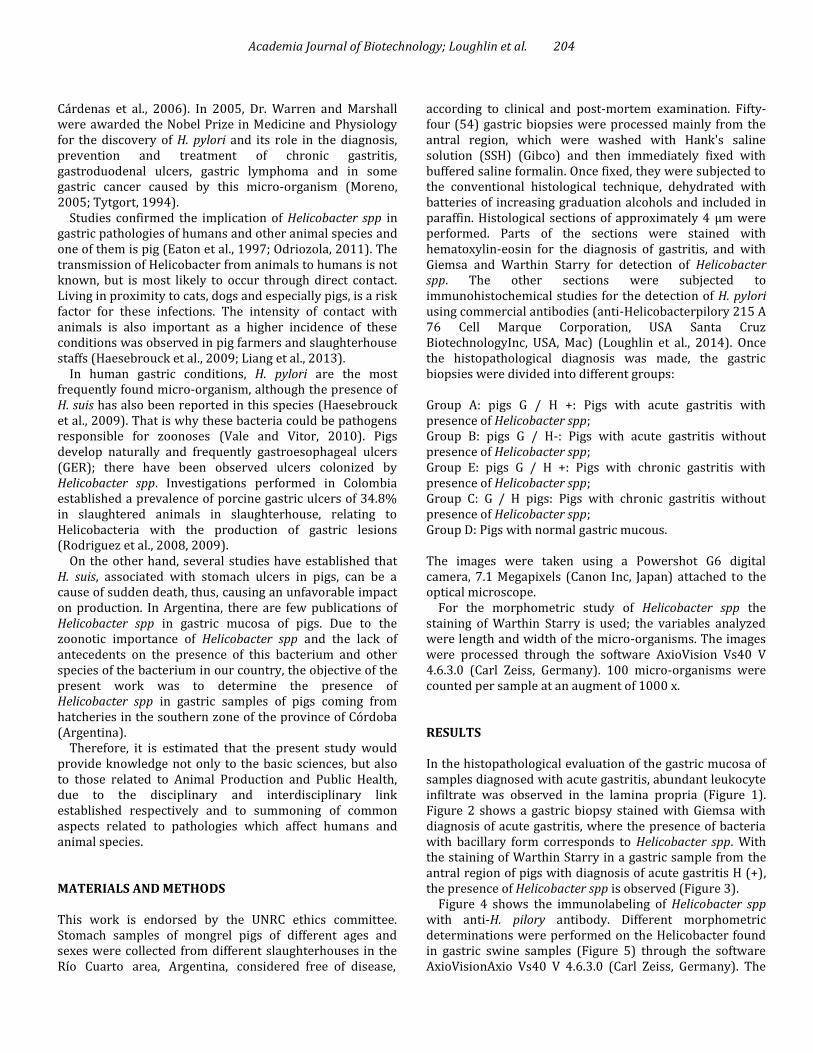
Figure 5: Microphotograph of gastric sample antral region with Warthin Starry staining where Helicobacter spp was observed, with an italic "S" shape (red), spiral form and larger size (blue) (1000x) are observed.
variables analyzed were length and width of the micro-organisms. The micro-organisms found were between 1 to 6 μm in length and between 0.4 to 1 μm in width, respectively. According to this morphological criterion, 52.6% of the micro-organisms presented characteristics compatible with H. pylori; the remaining 47.40%, features were compatible with H. heilmannii.
Of the fifty-four (54) samples observed were 17% samples belonging to normal gastric mucosa samples; 5% were diagnosed with acute gastritis without the presence of Helicobacter (-), 9% samples had acute gastritis Helicobacter (+), while 30% samples had chronic gastritis Helicobacter (-) and 39% chronic gastritis Helicobacter (+). DISCUSSION Histopathological results in gastric samples from pigs allowed detecting leukocyte infiltration and lesions compatible with acute and chronic gastritis, coinciding with data published by other researchers (Rodriguez et al., 2008, 2009).
With respect to the presence of gastric ulcers, the morphological pattern and the inflammatory changes found in pigs are similar to those observed in humans, except for
their location, because in pigs they predominate in the parsoesophagea of the stomach, whereas in humans they appear in the duodenal bulb and the pre-pyloric mucous membrane (Fawcett et al., 1999; Rodriguez et al., 2008). The difference in the localization of ulcers between the two species could be explained by the presence of a non-secretory epithelium in the parsoesophagea of the stomach of the pig, which would explain the absence of mucus secretion that acts as a buffer mechanism to neutralize gastric acidity. The histopathological lesions found in the pig stomachs in our study agree with the investigations of Rodriguez et al. (2008, 2009), who found a greater proportion of them in chronic lesions.
For the determination of Helicobacter spp in the gastric samples of pigs, the histological technique used revealed the presence of spiral bacteria that are in agreement with the previous descriptions of Helicobacter spp. However, staining with Giemsa allowed specific recognition of the pathogen (Shukla et al., 2001; Delgado et al., 2007). In line with the report of Rodríguez et al. (2008), Helicobacter was detected in the epithelium at the level of gypsum and gastric glands.
This finding confirms what other investigators had published, who observed the presence of Helicobacter spp in the stomach of pigs, most frequently in the fundic and

Academia Journal of Biotechnology; Loughlin et al. 208
pyloric mucosa, associating pyloric gastritis with infection of this bacterium (Eaton et al., 1997; Rodriguez et al., 2008, 2009).
The histological evaluation with the Warthin Starry coloration in the studied species allowed observing micro-organisms with the morphological characteristics of Helicobacter spp. With this staining, we could observe at least two types of Helicobacter spp: one of them of greater size and intensely spiral, and the other of smaller size with "S" shape, as described for H. heilmannii and H. pylori. Considering that some researchers suggest that H. heilmannii an H pylori and H. suis can naturally colonize the pig's stomach, and that the same bacterial genus could assume different morphological patterns due to inherent causes of the particular gastric environment of the host; there is need for new studies to typify the species of the pigs and to accurately identify the morphological types found in the present study (Fawcett et al., 1999; Rodriguez et al., 2009; Liang et al., 2013).
From the morphometric analysis of the micro-organisms we can infer that there is more than one species of Helicobacter in the stomach of the pigs, which could correspond with H. pylori and H. heilmannii (Rodriguez et al., 2008, 2009).
Researchers report that because 13.9 and 78.5% of human infections with Helicobacter no-pylori are produced by H. suis and that the transmission to humans could occur from the consumption of porcine meat, we are deepening our lines of research.
The diagnosis of the different types of gastritis in gastric swine specimens was obtained through the conventional histological hematoxylin / eosin technique. We could observe a higher proportion of chronic than acute lesions.
The differential staining of Giemsa and Warthin Starry in the gastric samples allowed determining the presence of Helicobacter spp. With the staining of Warthin Starry, we were able to visualize two species of the genus Helicobacter. In the species studied, the immunodetection of Helicobacter spp was expressed in the epithelium, gastric phasus and gastric gland. However, the greatest colonization by Helicobacter was detected in the antral region.
The presence of Helicobacter spp in the stomach of pigs indicates the ability of the micro-organism to naturally infect this animal species. In the samples studied we found more than one species of Helicobacter. ACKNOWLEDGMENTS This work was supported by Secretaria de Ciencia y Tecnología de la Universidad Nacional de Río Cuarto (Argentina). We are grateful to Technician Graciela Sagripanti for preparing the histological sections and also Professor Pascual Dauria from Universidad Nacional de Río Cuarto for his contributions and collaboration in this study.
REFERENCES Bondi J (2006). Aparato digestivo. Helicobacter pylori. URL:
http/www.bondisalud.com.ar/34html. Cárdenas Y, Castro V, Nevett A, Patiño P, Undaneta G (2006). Apoptosis
and Helicobacter pylori: a new model for infectious oncogénesis. VITAE Academia Biomédica Digital: 29.
Delgado F, Brihuega B, Venzano A, Funes D, Blanco Viera F, Auteri C, Romero G, Capellino F, Sarmiento L (2007). Adaptation of an immunohistochemistry protocol for the detection of Leptospira spp. in samples of formaldehyde-fixed tissue. Rev.Cubana Med. Trop. 59(1) on-line.
Eaton KA, Cover TL, Tummuru MKR, Blaser MJ, Krakowka S (1997) Role of vacuolatingcytotoxin in gastritis due to Helicobacter pylori in gnotobioticpiglets. Infec. Imm. 65:3462-3464.
Fawcett P, Gibney K, Vinette K (1999). Helicobacter pylori can be induced to assume the morphology of Helicobacter heilmanii. J.Clin. Mictobiol. 37:1045-1048.
Haesebrouck F, Pasmans F, Flahou B, Chiers K, Baele M, Meyns T, Decostere A, Ducatelle R (2009). Gastric Helicobacters in domestic animals and nonhuman primates and their significance for human health.Clin. Microbiol. Rev. 22(2):202-223.
Hunt RH (1996). The role of Helicobacter pylori in pathogenesis: the spectrum of clinical outcomes. Scand. J.Gastroenterol. Suppl. 220:3-9.
Liang J, Ducatelle R, Pasman F, Annemieke S, Haesebrouck F, Flahou B (2013). Sequence typing of porcine and human gastric pathogen Helicobacter suis. Int. J.Syst. Evol. Microbiol. 51(3):920-936.
Lopez Brea M, Alarcón T, Domingo D, Perez- Perez G, Correa P, Shirrow M (2006). Retos para el Siglo XXI..On line. Citado en www.helicobacterspain.com.
Mac Loughlin V, Merkis C, Dauria P, Cristofolini A, Bonino F, Sagripanti G, Sanchis G, Ritta L, Grosso C, Savino F (2014). Relación entre Helicobacterspp y células productoras de gastrina en estómago de cerdo. Morfovirtual. Online www.morfovirtual2014.sld.cu/index
McColl K, El Omar R, Guillen D (1997). The role of Helicobacter pylori in the pathophysiology of duodenal ulcer and gastrin cancer. Semin. Gastrointest.Disease. 8:142-155.
Morales A,Bermudez V (2008). Modelos Animales para el estudio de la infección por el género Helicobacter en humanos. Rev. Soc. Med. Hosp. Emerg. Perez de Leon. 39(1):30-33.
Moreno R (2005) La bacteria que destrono al stress. Diario El País, España. Odriozola V (2011). Helicobacter gástricos en caninos y felinos.Asoc. Arg.
Med. Felina.http:/www.aamefe.org/helicobacter.htm-2011 Rodriguez BJ, Arananzazu D, Ortiz L (2009). Association of gastric ulcer
and Helicobacter spp in pigs in Antioquia, Colombia. Rev.Colomb. Cienc. Pecu.22:54-60.
Rodriguez BJ, Aranzazu D, Giraldo GE, Alvarez LC, Lopera JA, Valencia F (2008). Determinación de Helicobacterspp en cerdos en el departamento de Antioquia, Colombia. Rev. Colomb. Cienc. Pecu. 21:210-218.
Shukla S, Pujani M, Agarwal A, Pujani M and Rohtagi A (2001). Correlation of serology with morphological changes in gastric biopsy in Helicobacter pylori infection and evaluation of inmunochemistry for H pylori identification. Arabia. J. Gastroenterol.18(6): 369-374.
Sleinsinger MH, Mark Feldman MD, Bruce MD, ScharschMidt K, Samuel Klein MB (2000). Enfermedades gastroduodenales y hepáticas. Fisiopatología, diagnóstico y tratamiento. 6º edición (eds) Panamericana, Madrid. pp. 649-661.
Thibaut J, Paz V, Paredes E y Ernest S (2007). Determinación de la presencia de Helicobacterspp en perros mediante biopsia gástrica obtenida por endoscopía. Rev. Cient. (Maracaibo) Venezuela. 17:3.
Tytgort G (1994). Long-term consequences of Helicobacter pylorieradication. Scand. J. Gastroenterol. 205: 38-44.
Vale F, Vitor JM (2010). Transmission pathway of Helicobacter pylori does food play a role on rural and urban areas? Int. J. FoodMicrobiol.38:1-12.
Warren JR (1997). Thediscovery and pathology of Helicobacter pylory. In Xth International Workshop on Gastroduodenal Pathology and Helicobacter pylori: The Logyc of Erradication, Lisboa-Portugal. pp. 7-13.


![CLASSIFICATION OF HEMATOXYLIN AND EOSIN …...and used for classification of Hematoxylin and Eosin (H&E) stained images. In SIFT [19], identical key points are extracted from im-ages](https://static.fdocuments.in/doc/165x107/5f0252f37e708231d403b4f8/classification-of-hematoxylin-and-eosin-and-used-for-classiication-of-hematoxylin.jpg)