Detection ofSerotype-Specific Antibodies …jcm.asm.org/content/28/2/312.full.pdf314 INZANA ET AL.-a...
7
JOURNAL OF CLINICAL MICROBIOLOGY, Feb. 1990, p. 312-318 0095-1137/90/020312-07$02.00/0 Copyright © 1990, American Society for Microbiology Detection of Serotype-Specific Antibodies or Capsular Antigen of Actinobacillus pleuropneumoniae by a Double-Label Radioimmunoassay THOMAS J. INZANA,* GARY F. CLARK,t AND JEANNE TODD Department of Pathobiology, Virginia-Maryland Regional College of Veterinary Medicine, Virginia Polytechnic Institute and State University, Blacksburg, Virginia 24061 Received 25 July 1989/Accepted 17 October 1989 Diagnostic tests for Actinobacillus pleuropneumoniae have been problematic because current tests do not use a purified antigen and in most cases measure either antibody or antigen, but not both. We describe a Farr-type double-label radioimmunoassay that utilizes purified, serotype-specific, 3H-capsule to measure antibody to capsule directly or that can measure capsule in a sample indirectly by inhibition of antibody binding. The assay could detect about 1 ng of serotype-specific antibody in serum or at least 100 pg of capsule in a sample. Due to the sensitivity of the assay, false-positive results were common with neat sera (probably due to cross-reacting antibodies to unrelated antigens), but the specificity was improved when the sera were diluted 1:100. The radioimmunoassay should prove to be a useful reference method for research and diagnostic testing and for comparison of new assays for detection of capsule or antibodies to capsule. Actinobacillus (Haemophilus) pleuropneumoniae is the etiologic agent of swine pleuropneumonia. Infection of pigs may result in acute, subacute, or chronic disease. Severe outbreaks of the disease occur when an immune, subclinical, chronic carrier is introduced into a nonimmune herd (21). Antibodies to A. pleuropneumoniae are commonly found in a high proportion of pig herds (5, 20). Current diagnostic tests for pleuropneumonia involve culture and serology. A wide variety of serologic tests have been developed to detect antibodies to any of the 12 serotypes of A. pleuropneumo- niae or for serotyping (14-17). However, only a few sero- types are commonly isolated in a given geographic region. In the United States, serotypes 1 and 5 account for 80 to 89% of reported infections (16, 19). Detection and epidemiology of berotypes that predominate in a given region can be improved when serologic tests are serotype specific. Serotype specificity, in turn, is determined by the capsular polymer of À. pleuropneumoniae (10). However, the most common tests for detection of antibody to A. pleuropneumoniae (complement fixation) or for sero- typing (slide agglutination) utilize whole cells or a crude extract or use antiserum to whole cells, respectively. So- matic antigens of different serotypes of A. pleuropneumo- niae (10), and possibly of different species of related bacteria (17, 18), are capable of cross-reacting among themselves. As more sensitive diagnostic tests are developed for serologic testing of A. pleuropneumoniae, serotype specificity may decrease and false-positive tests may increase. Coagglutina- tion has been reported to be useful for serotyping A. pleu- ropneumoniae (14), and enzyme-linked immunosorbent as- say (ELISA) and dot blot ELISA have been used for sensitive detection of antibodies to capsule (10) or somatic antigens. Although both tests are currently being used suc- cessfully, the coagglutination test is not necessarily specific for capsule, and the ELISA requires that the purified antigen adhere reproducibly to a solid phase. In addition, none of the * Corresponding author. t Present address: Department of Biochemistry, Eastern Virginia Medical School, Norfolk VA 23501. described tests have the flexibility to be easily used for detection of antibodies or antigen. Radioimmunoassay (RIA) is an extremely sensitive method for detection of antibodies or antigen. A double-label RIA is the reference method for detection of antibodies to Haemophilus influenza type b and can be used as an inhibition assay to detect picogram amounts of capsular antigen (3). We have developed a modification of the double- label RIA for detection of capsular antigen, or antibodies to A. pleuropneumoniae. The RIA is extremely sensitive for detection of serotype-specific antibodies. In addition, it can be used to detect and quantitate capsular antigen in respira- tory specimens or associated with the bacteria. The disad- vantage of the assay is that its high sensitivity results in detection of cross-reacting antibodies to common antigens unless the serum sample is diluted at least 1:100. (This work was presented, in part, at the 10th Interna- tional Pig Veterinary Society Meeting, Rio de Janeiro, Brazil [Abstr. Annu. Meet. Int. Pig Vet. Soc. 1988, p. 79].) MATERIALS AND METHODS Bacterial strains and growth medium. Serotype 1 strain 4045 and serotype 5 strains K17, K17-C (noncapsulated mutant of K17), J45, and 178 were grown in Casamino Acids-yeast extract medium supplemented with 5 ,ug of nicotinamide adenine dinucleotide per ml (CY medium). The source of the strains, medium preparation, and the growth conditions have been described previously (9, 10). Intrinsic radiolabeling and purification of A. pleuropneumo- niae capsule. A. pleuropneumoniae K17 was intrinsically labeled with D-[6-3H]glucose (18 Ci/mmol) (ICN Radiochem- icals, Irvine, Calif.) by modification of the procedure de- scribed by Anderson (3). Two procedures were used to radiolabel and purify capsule. In procedure A, 5 ml of bacteria was grown in CY medium to early stationary phase, the cells were washed three times with CY medium, and the pellet was suspended in 250 ,ul of CY medium. A 50-,ul portion of this culture was transferred to a sterile tube (10 by 75 mm) containing 25 ,ug of dried [3H]glucose (total activity, 2.5 mCi). Chloramphenicol (0.04 ,ug) was added, and the 312 Vol. 28, No. 2 on July 17, 2018 by guest http://jcm.asm.org/ Downloaded from
Transcript of Detection ofSerotype-Specific Antibodies …jcm.asm.org/content/28/2/312.full.pdf314 INZANA ET AL.-a...
JOURNAL OF CLINICAL MICROBIOLOGY, Feb. 1990, p. 312-3180095-1137/90/020312-07$02.00/0Copyright © 1990, American Society for Microbiology
Detection of Serotype-Specific Antibodies or Capsular Antigenof Actinobacillus pleuropneumoniae by a
Double-Label RadioimmunoassayTHOMAS J. INZANA,* GARY F. CLARK,t AND JEANNE TODD
Department of Pathobiology, Virginia-Maryland Regional College of Veterinary Medicine,Virginia Polytechnic Institute and State University, Blacksburg, Virginia 24061
Received 25 July 1989/Accepted 17 October 1989
Diagnostic tests for Actinobacillus pleuropneumoniae have been problematic because current tests do not usea purified antigen and in most cases measure either antibody or antigen, but not both. We describe a Farr-typedouble-label radioimmunoassay that utilizes purified, serotype-specific, 3H-capsule to measure antibody tocapsule directly or that can measure capsule in a sample indirectly by inhibition of antibody binding. The assaycould detect about 1 ng of serotype-specific antibody in serum or at least 100 pg of capsule in a sample. Dueto the sensitivity of the assay, false-positive results were common with neat sera (probably due to cross-reactingantibodies to unrelated antigens), but the specificity was improved when the sera were diluted 1:100. Theradioimmunoassay should prove to be a useful reference method for research and diagnostic testing and forcomparison of new assays for detection of capsule or antibodies to capsule.
Actinobacillus (Haemophilus) pleuropneumoniae is theetiologic agent of swine pleuropneumonia. Infection of pigsmay result in acute, subacute, or chronic disease. Severeoutbreaks of the disease occur when an immune, subclinical,chronic carrier is introduced into a nonimmune herd (21).Antibodies to A. pleuropneumoniae are commonly found ina high proportion of pig herds (5, 20). Current diagnostictests for pleuropneumonia involve culture and serology. Awide variety of serologic tests have been developed to detectantibodies to any of the 12 serotypes of A. pleuropneumo-niae or for serotyping (14-17). However, only a few sero-types are commonly isolated in a given geographic region. Inthe United States, serotypes 1 and 5 account for 80 to 89% ofreported infections (16, 19).
Detection and epidemiology of berotypes that predominatein a given region can be improved when serologic tests areserotype specific. Serotype specificity, in turn, is determinedby the capsular polymer of À. pleuropneumoniae (10).However, the most common tests for detection of antibodyto A. pleuropneumoniae (complement fixation) or for sero-typing (slide agglutination) utilize whole cells or a crudeextract or use antiserum to whole cells, respectively. So-matic antigens of different serotypes of A. pleuropneumo-niae (10), and possibly of different species of related bacteria(17, 18), are capable of cross-reacting among themselves. Asmore sensitive diagnostic tests are developed for serologictesting of A. pleuropneumoniae, serotype specificity maydecrease and false-positive tests may increase. Coagglutina-tion has been reported to be useful for serotyping A. pleu-ropneumoniae (14), and enzyme-linked immunosorbent as-say (ELISA) and dot blot ELISA have been used forsensitive detection of antibodies to capsule (10) or somaticantigens. Although both tests are currently being used suc-cessfully, the coagglutination test is not necessarily specificfor capsule, and the ELISA requires that the purified antigenadhere reproducibly to a solid phase. In addition, none of the
* Corresponding author.t Present address: Department of Biochemistry, Eastern Virginia
Medical School, Norfolk VA 23501.
described tests have the flexibility to be easily used fordetection of antibodies or antigen.Radioimmunoassay (RIA) is an extremely sensitive
method for detection of antibodies or antigen. A double-labelRIA is the reference method for detection of antibodies toHaemophilus influenza type b and can be used as aninhibition assay to detect picogram amounts of capsularantigen (3). We have developed a modification of the double-label RIA for detection of capsular antigen, or antibodies toA. pleuropneumoniae. The RIA is extremely sensitive fordetection of serotype-specific antibodies. In addition, it canbe used to detect and quantitate capsular antigen in respira-tory specimens or associated with the bacteria. The disad-vantage of the assay is that its high sensitivity results indetection of cross-reacting antibodies to common antigensunless the serum sample is diluted at least 1:100.
(This work was presented, in part, at the 10th Interna-tional Pig Veterinary Society Meeting, Rio de Janeiro, Brazil[Abstr. Annu. Meet. Int. Pig Vet. Soc. 1988, p. 79].)
MATERIALS AND METHODS
Bacterial strains and growth medium. Serotype 1 strain4045 and serotype 5 strains K17, K17-C (noncapsulatedmutant of K17), J45, and 178 were grown in CasaminoAcids-yeast extract medium supplemented with 5 ,ug ofnicotinamide adenine dinucleotide per ml (CY medium). Thesource of the strains, medium preparation, and the growthconditions have been described previously (9, 10).
Intrinsic radiolabeling and purification ofA. pleuropneumo-niae capsule. A. pleuropneumoniae K17 was intrinsicallylabeled with D-[6-3H]glucose (18 Ci/mmol) (ICN Radiochem-icals, Irvine, Calif.) by modification of the procedure de-scribed by Anderson (3). Two procedures were used toradiolabel and purify capsule. In procedure A, 5 ml ofbacteria was grown in CY medium to early stationary phase,the cells were washed three times with CY medium, and thepellet was suspended in 250 ,ul of CY medium. A 50-,ulportion of this culture was transferred to a sterile tube (10 by75 mm) containing 25 ,ug of dried [3H]glucose (total activity,2.5 mCi). Chloramphenicol (0.04 ,ug) was added, and the
312
Vol. 28, No. 2
on July 17, 2018 by guesthttp://jcm
.asm.org/
Dow
nloaded from

RIA TO DETECT A. PLEUROPNEUMONIAE ANTIBODY OR CAPSULE 313
culture was shaken gently overnight at 37°C. The tube wasopened in a radioisotope fume hood, and the culture wasdiluted with 0.8 ml of 4°C 0.1 M phosphate buffer, pH 7.0.The culture was transferred to a Microfuge tube, the bacteriawere sedimented at 10,000 x g for 15 min, and the superna-tant was transferred to a clean Microfuge tube. Hexadecyl-trimethylammonium bromide (Cetavlon; Eastman KodakCo., Rochester, N.Y.) was added to a final concentration of0.005 M, and the mixture was incubated at 4°C for 2 h. Themixture was centrifuged at 10,000 x g for 10 min, the pelletwas washed twice by vigorous vortexing with 1 ml of cold0.005 M Cetavlon in phosphate buffer, and the invisibleprecipitate was extracted twice with 0.15 ml of cold 0.4 MNaCI. The supernatant was transferred to a clean Microfugetube, 3 volumes of -20°C ethanol (95%) were added, and themixture was incubated at -20°C overnight. The mixture wascentrifuged for 30 min at 10,000 x g and extracted twice with0.3 ml of distilled water. The pooled aqueous phases wereextracted twice with an equal volume of 90% room temper-ature phenol by vortexing for 10 min. NaCI, 5 M, was addedto the aqueous phase to a final concentration of 0.4 M, and 3volumes of -20°C ethanol were added. After at least 3 h at-20°C, the mixture was centrifuged for 30 min at 10,000 x g,and the precipitate was suspended in 0.2 ml of 0.1 M NaCI.The suspended capsule was applied to a Sepharose CL-4Bcolumn (1 by 28 cm) and eluted with 0.01 M NaCI at a linearflow rate of 5 cm/h, and 1-ml fractions were collected andassayed for tritium by liquid scintillation counting. Peakfractions that were positive for tritium were pooled, and thecapsule was precipitated by the addition of 3 volumes of-20°C ethanol. After the mixture had incubated for at least3 h at -20°C, it was centrifuged at 10,000 x g for 30 min. Thepellet was suspended in phosphate-buffered saline contain-ing 0.01% Thimerosal, pH 7.4, to an activity of 4 to 5 iuCi/mland stored at -70°C.
In procedure B, 25 ,u of a mid-log-phase culture of A.pleuropneumoniae in CY medium was added to 0.5 ml ofCY medium in a sterile tube containing 25 ,ug of dried[3H]glucose. The culture was shaken overnight at 37°C, thecells were sedimented at 10,000 x g for 15 min, the centrif-ugation was repeated with the supernatant, and 0.005 M(final concentration) Cetavlon was added to the supernatant.The remaining procedures were the same as those describedfor procedure A, except that the 0.4 M NaCI and distilledwater volumes for extractions were increased 10-fold and,following phenol extraction and ethanol precipitation, thepreparation was suspended in 0.4 ml of distilled water andcentrifuged at 100,000 x g for 4 h in an Airfuge (BeckmanInstruments, Inc., Palo Alto, Calif.).RIA for antibody or antigen. The working solution con-
taining 3H-capsule and 36CI (16.1 mCi/g; ICN Radiochemi-cals) was prepared exactly as described by Anderson (3).The RIA for antibody, including all controls, was also doneas described previously (3, 6). For determination of antigencontent, samples containing standard concentrations or un-known concentrations of capsule were diluted in fetal bovineserum; 25 ,ul of standard hyperimmune anticapsular serumdiluted to yield 50% binding in the RIA was added to 25,uI ofsample and incubated overnight at 4°C. A 25-,uI portion ofworking solution was then added, and the RIA was com-pleted as for antibody.The percent antibody bound was calculated as described
before (3, 7), except that an IBM computer and Lotus 1-2-3software were used for computing and storing results. Theamount of capsule in a sample was calculated from the
amount of inhibition of antibody binding compared with astandard curve.Antiserum and serologic assays. Preparation of high-titer
swine and rabbit antisera to capsule and the ELISA has beendescribed previously (10). Determination of specific anti-body to capsule in serum was determined by precipitationwith purified capsule (8) followed by protein determination(13).
Electrophoresis and fluorography. Sodium dodecyl sulfate-polyacrylamide gel electrophoresis of purified, 3H-capsulepreparations was done as described by Laemmli (12). Fluo-rography of the dried gel was done following treatment of thegel with Amplify per the manufacturer's instructions (Amer-sham Corp., Arlington Heights, Ill.) and exposure on XAR-5film (GBX-2) (Eastman Kodak Co., Rochester, N.Y.) for 7days at -70°C.
RESULTS
Characterization of the 3H-capsule preparations. The per-centage of tritium present in each purification step of theradiolabeled capsule by procedure A was calculated. Fol-lowing radiolabeling, 99.6% of the total [3H]glucose added tothe culture was accounted for. Six percent remained in thebacterial pellet after removal of the supernatant. Some 92%remained in the supernatant after precipitation by Cetavlon.
-É I04
on 3
2--5
Fraction (mi)B
1-1(Oon
o-
où
Fraction (mi)
FIG. 1. Chromatographic elution profile of3H-capsule (serotype5) from Sepharose CL-4B. The bacteria were radiolabeled with[3H]glucose in the presence of chloramphenicol, and the capsulewas purified by Cetavlon precipitation, NaCl, and phenol extraction(A) or grown with [3H]glucose in the absence of chloramphenicoland purified as for panel A but with the addition of ultracentrifuga-tion (B).
VOL. 28, 1990
on July 17, 2018 by guesthttp://jcm
.asm.org/
Dow
nloaded from
314 INZANA ET AL.
-acZom
-XoD
c
c-
QL)u1CL)
120 -
100 -
80 -
60 -
40 -
20 -
5 10 15
Reciprocal Serum Dilution (10 )
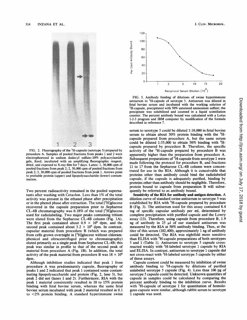
FIG. 3. Antibody binding of dilutions of swine hyperimmuneantiserum to 3H-capsule of serotype 5. Antiserum was diluted infetal bovine serum and incubated with the working solution of3H-capsule, precipitated with 50% saturated ammonium sulfate; theprecipitate was solubilized and counted in a liquid scintillationcounter. The percent antibody bound was calculated with a Lotus1-2-3 program and IBM computer by modification of the formuladescribed in reference 7.
1 2 3FIG. 2. Fluorography of the 3H-capsule (serotype 5) prepared by
procedure A. Samples of pooled fractions from peaks 1 and 2 were
electrophoresed in sodium dodecyl sulfate-10% polyacrylamidegels, fixed, incubated with an amplifying fluorographic reagent,dried, and exposed to X-ray film for 7 days. Lanes: 1, 30,000 cpm ofpooled fractions from peak 2; 2, 50,000 cpm of pooled fractions frompeak 2; 3, 30,000 cpm of pooled fractions from peak 1. Arrows pointto probable protein (upper) and lipopolysaccharide (lower) contam-ination.
Two percent radioactivity remained in the pooled superna-tants after washing with Cetavlon. Less than 1% of the totalactivity was present in the ethanol phase after precipitationor in the phenol phase after extraction. The total [3H]glucoserecovered in the capsule preparation prior to SepharoseCL-4B chromatography was 0.18% of the total [3H]glucoseused for radiolabeling. Two major peaks containing tritiumwere eluted from the Sepharose CL-4B column (Fig. 1A).The first peak contained about 0.9 x 10⁶ dpm, and thesecond peak contained about 3.2 x 10⁶ dpm. In contrast,capsular material from procedure B (which was preparedfrom cells grown overnight in [3H]glucose without chloram-phenicol and ultracentrifuged prior to chromatography)eluted primarily as a single peak from Sepharose CL-4B; thispeak was similar in profile to that of the second peak ofmaterial from procedure A (Fig. 1B). In addition, the totalactivity of the peak material from procedure B was 18 x 106dpm.Although inhibition studies indicated that peak 1 from
procedure A was predominately capsule, fluorography ofpeaks 1 and 2 indicated that peak 1 contained some contam-inating lipopolysaccharide and protein (Fig. 2, lane 3), butpeak 2 did not (lanes 1 and 2). Furthermore, RIA with thepeak 1 material consistently resulted in 10 to 15% proteinbinding with fetal bovine serum, whereas the same fetalbovine serum incubated with peak 2 material resulted in 0.1to <2% protein binding. A standard hyperimmune swine
serum to serotype 5 could be diluted 1:10,000 in fetal bovineserum to obtain about 50% protein binding with the 3H-capsule prepared from procedure A, but the same serum
could be diluted 1:55,000 to obtain 50% binding with 3H-capsule prepared by procedure B. Therefore, the specificactivity of the 3H-capsule prepared by procedure B was
apparently higher than the preparation from procedure A.Subsequent preparations of 3H-capsule from serotype 1 weremade following the protocol for procedure B, and fractions11 to 17 from the Sepharose CL-4B column were concen-trated for use in the RIA. Although it is conceivable thatproteins other than antibody could bind the radiolabeledcapsule, if the capsule is adequately purified, binding byproteins other than antibody should be negligible. Therefore,protein bound to capsule from preparation B will subse-quently be referred to as antibody bound.
Sensitivity of the RIA for antibody and antigen detection. Adilution curve of standard swine antiserum to serotype 5 wasestablished by RIA with 3H-capsule prepared by procedureB (Fig. 3). The antiserum used for this assay contained 8.4mg of specific capsular antibody per ml, determined bycomplete precipitation with purified capsule and the Lowryassay (13). Therefore, using capsule from procedure B, 1.9ng of antibody in 25 ,ul of our standard antiserum was
measured by the RIA at 50% antibody binding. Thus, at thetiter of this serum (102,400), approximately 1 ng of antibodycould be detected. The RIA was eightfold more sensitivethan ELISA with 3H-capsule preparations of both serotypes5 and 1 (Table 1). Antiserum to serotype 5 capsule cross-reacted weakly with 3H-labeled serotype 1 capsule by RIAand ELISA. In contrast, antiserum to serotype 1 capsule didnot cross-react with 3H-labeled serotype 5 capsule by eitherof these assays.
Capsule content could be measured by inhibition of swineantibody binding to 3H-capsule by dilutions of purifiedunlabeled serotype 5 capsule (Fig. 4). Less than 100 pg ofserotype 5 capsule could be detected. Unknown quantities ofcapsule in samples could be calculated by comparing thepercent antibody binding to the inhibition curve. Resultswith 3H-capsule of serotype 1 for quantitation of homolo-gous capsule were similar, although rabbit serum to serotype1 capsule was used.
20
J. CLIN. MICROBIOL.
0
0
0
\.-l.0z.
i..u.,.ï
i.f
..t
t.
on July 17, 2018 by guesthttp://jcm
.asm.org/
Dow
nloaded from
RIA TO DETECT A. PLEUROPNEUMONIAE ANTIBODY OR CAPSULE 315
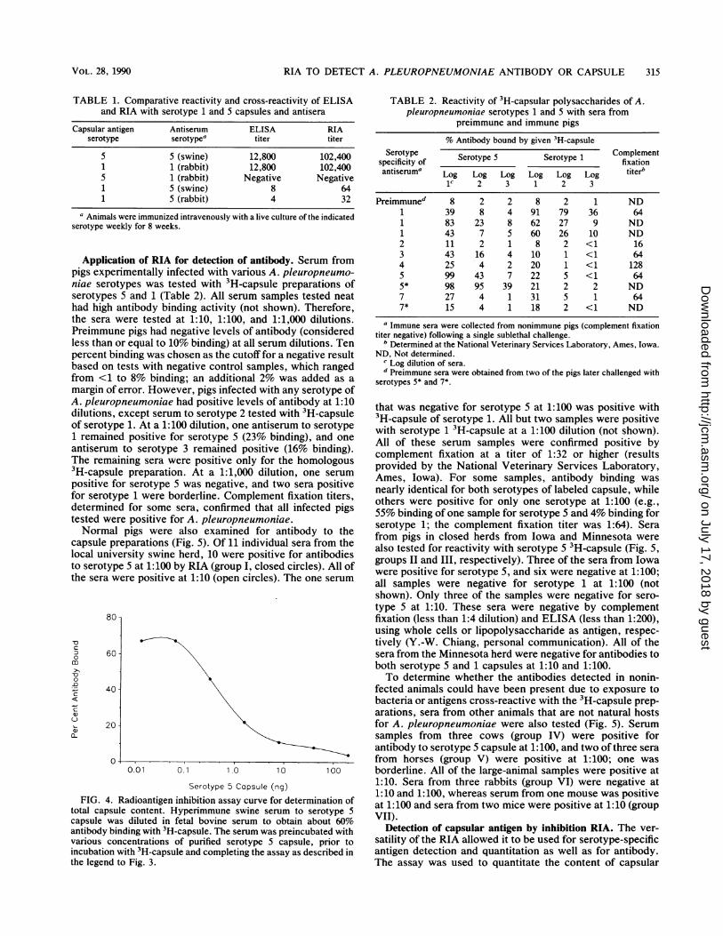
TABLE 1. Comparative reactivity and cross-reactivity of ELISAand RIA with serotype 1 and 5 capsules and antisera
Capsular antigen Antiserum ELISA RIAserotype serotypea titer titer
5 5 (swine) 12,800 102,4001 1 (rabbit) 12,800 102,4005 1 (rabbit) Negative Negative1 5 (swine) 8 641 5 (rabbit) 4 32
a Animals were immunized intravenously with a live culture of the indicatedserotype weekly for 8 weeks.
Application of RIA for detection of antibody. Serum frompigs experimentally infected with various A. pleuropneumo-niae serotypes was tested with 3H-capsule preparations ofserotypes 5 and 1 (Table 2). All serum samples tested neathad high antibody binding activity (not shown). Therefore,the sera were tested at 1:10, 1:100, and 1:1,000 dilutions.Preimmune pigs had negative levels of antibody (consideredless than or equal to 10% binding) at all serum dilutions. Tenpercent binding was chosen as the cutofffor a negative resultbased on tests with negative control samples, which rangedfrom <1 to 8% binding; an additional 2% was added as amargin of error. However, pigs infected with any serotype ofA n11rn»Zm»nha 1aitxAosifathslzetlM. sacLdilutionof serc1 remaantiserThe re3H-cappositivfor serdetertestedNor
capsulelocal uto serothe ser
-o
c:
-D
Co
c:
c:
a,)
v)
FIG.
total cacapsuleantibod
vanousincubatethe lege
TABLE 2. Reactivity of 3H-capsular polysaccharides of A.pleuropneumoniae serotypes 1 and 5 with sera from
preimmune and immune pigs
% Antibody bound by given 3H-capsuleSerotype Serotype 5 Serotype 1 Complement
specificity of fixation
antiseruma Log Log Log Log Log Log titerb1C 2 3 1 2 3
Preimmuned 8 2 2 8 2 1 ND1 39 8 4 91 79 36 641 83 23 8 62 27 9 ND1 43 7 5 60 26 10 ND2 il 2 1 8 2 <1 163 43 16 4 10 1 <1 644 25 4 2 20 1 <1 1285 99 43 7 22 5 <1 645* 98 95 39 21 2 2 ND7 27 4 1 31 5 1 647* 15 4 1 18 2 <1 ND
a Immune sera were collected from nonimmune pigs (complement fixationtiter negative) following a single sublethal challenge.
b Determined at the National Veterinary Services Laboratory, Ames, Iowa.ND, Not determined.
C Log dilution of sera.d Preimmune sera were obtained from two of the pigs later challenged with
serotypes 5* and 7*.
uUyfpn:urmLonrLaue iiau pos~itive ievei oiUntidI.I U l that was negative for serotype 5 at 1:100 was positive withns, except serum to serotype 2 tested with,H-capsule 3H-capsule of serotype 1. All but two samples were positive)type 1. At a 1:100 dilution, one antiserum to serotype with serotype 1 3H-capsule at a 1:100 dilution (not shown).ined positive for serotype 5 (23% binding), and one All of these serum samples were confirmed positive by.um to serotype 3 remained positive (16% binding). complement fixation at a titer of 1:32 or higher (resultsmanning sera were positive only for the homologous provided by the National Veterinary Services Laboratory,)sul prparaion Ata 1: diltioserane sitiv Ames, Iowa). For some samples, antibody binding wase for serotype 5 was negative, and two nearly identical for both serotypes of labeled capsule, whiletype 1 were borderline. Complement fixation tigers, others were positive for only one serotype at 1:100 (e.g.,wined for some sera, confirmed that ail infected pigs 55% binding of one sample for serotype 5 and 4% binding forwere positive for A. pleuropneumoniae. serotype 1; the complement fixation titer was 1:64). Seramal pigs were also examined for antibody to the from pigs in closed herds from Iowa and Minnesota weree preparations (Fig. 5). 0f il individual sera from the also tested for reactivity with serotype 5 3H-capsule (Fig. 5,university swine herd, 10 were positive forantibodiesely). Three of the sera from Iowatype 5 at 1:100 by R1A (group T, closed circles). Ail Of grup -ladII epcey.The ftesr rmIw)type.5at1:.0 by RIA (group 1, closedr . A of were positive for serotype 5, and six were negative at 1:100;-a were positive at 1:10 (open circles). The one serum all samples were negative for serotype 1 at 1:100 (not
shown). Only three of the samples were negative for sero-type 5 at 1:10. These sera were negative by complement
80 fixation (less than 1:4 dilution) and ELISA (less than 1:200),using whole cells or lipopolysaccharide as antigen, respec-tively (Y.-W. Chiang, personal communication). All of the
60 sera from the Minnesota herd were negative for antibodies toboth serotype 5 and 1 capsules at 1:10 and 1:100.To determine whether the antibodies detected in nonin-
40 fected animals could have been present due to exposure tobacteria or antigens cross-reactive with the 3H-capsule prep-arations, sera from other animals that are not natural hosts
20 for A. pleuropneumoniae were also tested (Fig. 5). Serumsamples from three cows (group IV) were positive forantibody to serotype 5 capsule at 1:100, and two ofthree sera
i 0 . from horses (group V) were positive at 1:100; one was0.0l o 1 1.0o 0o 10 borderline. All of the large-animal samples were positive at
Serotype 5 Capsule (ng) 1:10. Sera from three rabbits (group VI) were negative at1:10 and 1:100, whereas serum from one mouse was positive4. Radioantigen inhibition assay curve for determination of 1:10 and sera fromtwo miceewerepositivea 1
ipsleconen. .yeimn .wn eu osrtpat 1:100 and sera from two mice were positive at 1:10 (groupLpsule content. Hypenimmune swine serum to serotype S5 .rT
was diluted in fetal bovine serum to obtain about 60% VII).[y binding with 3H-capsule. The serum was preincubated with Detection of capsular antigen by inhibition RIA. The ver-concentrations of purified serotype 5 capsule, prior to satility of the RIA allowed it to be used for serotype-specific
ion with 3H-capsule and completing the assay as described in antigen detection and quantitation as well as for antibody.nd to Fig. 3. The assay was used to quantitate the content of capsular
VOL. 28, 1990
on July 17, 2018 by guesthttp://jcm
.asm.org/
Dow
nloaded from

316 INZANA ET AL.
100
90
80*
70-
c
om
soe
a-0
60
50 -
40 -
30 -
20 -
10 -
o
o
o
ooo
ooo
o
8oo
oSa
o
o
.
o ooox
Animal
FIG. 5. Binding of antibody from v
3H-capsule of serotype 5 A. pleuropneui1:10 (O) or 1:100 (@) in fetal bovine seîRIA. Animal groups: I, swine maintainedcenter; II, swine maintained at a closedswine maintained at a closed researchnormal cattle; V, normal horses; VI, ncmice.
antigen produced by three strains ofto confirm the lack of capsule produmutant (10) (Table 3). In addition, atml (or 100 pg total) could be detectswine infected with strain J45 A.from lung tissue of mice and swine i
The capsule content ranged from 15lung to >100 nglml in lesions of infe
DISCUSSIONOur 3H-capsule was initially preç
described by Anderson (3). Howevertritium obtained in the capsule by thhigh as that reported by Anderson (3'inherent differences in capsule protween A. pleuropneumoniae and hboth. Therefore, we modified the 1
TABLE 3. Radioantigen inhibition assay for quantitation ofA. pleuropneumoniae serotype 5 capsule
Specimen Capsule contentper ml
A. pleuropneumoniaeJ45" ............................ 130 ± 18 ,ug178a ............................. 19 ± 4 ugK17a ............................. 155 ± 25 ,ugK17_Cb.............................. <4 ng
ExtractsInfected mouse lung (J45)' ............................ 15.3 + 2.5 ngNoninfected mouse lung' ............................ <4 ngInfected swine lung (J45)C ............................ 107 + 19 ngNoninfected swine lung' ............................ <4 ngNasal swabs from infected pig (J45)C ............... 25 ± 2 ngNasal swabs from noninfected pigsC ................ <4 nga Serotype 5 strain.b Noncapsulated mutant of strain K17.
0 c Mean capsule content of at least three different samples ± the standarddeviation for sampled data.
growing the cells overnight in the presence of [3H]glucoseand adding ultracentrifugation to the purification process.3H-capsule purified from A. pleuropneumoniae followingovernight growth in the labeled compound greatly increasedthe specific activity of the capsule preparation. The addition
* of ultracentrifugation and recovery of selected fractionsfrom Sepharose CL-4B chromatography resulted in at leastas pure a preparation as when stationary-phase cells were
* incubated with [3H]glucose in the presence of chloramphen-icol. Ultracentrifugation removed most of the material that
O previously eluted in the void volume, which, in addition to* 0 capsule, contained lipopolysaccharide and protein, and ma-o terial that nonspecifically reacted with fetal calf serum., * The RIA had the flexibility of directly measuring antibody
Iv v vu vil to capsule or, by inhibition, antigen. The sensitivity of theRIA exceeded that of any test for A. pleuropneumoniae yet
Groups described. With our standard antiserum, <100 pg of capsule,variouss animal species to or about 1 ng of antibody to capsule, could be detected andmoniae. Sera were diluted measured. Due to the sensitivity of the RIA, it was notrum prior to testing in the surprising that some weak cross-reactivity of antiserum tolat the Virginia Tech swine serotype 5 capsule occurred with serotype 1 capsule. Ofresearch herd in Iowa; III, interest, however, was that antiserum to serotype 1 capsuleherd in Minnesota; IV, did not cross-react with serotype 5 capsule. Though some-
rmal rabbits; VII, normal what less sensitive, these results were confirmed by ELISA.The serotype 1 capsule of A. pleuropneumoniae has thestructure [(4)-,-D-GlcpNAc-(1->6)-a-D-Galp-(l-PO4)]n (85%
serotype 5 bacteria and acetylated at position 6 of P-D-GlcpNAc), while the struc-ced by a noncapsulated ture of the serotype Sa capsule is [->6)-at-D-GlcpNAc-(1->5)-least 4 ng of capsule per P-D-dOclAp-(2--]n (1, 2); the serotype 5b capsule has theed from nasal swabs of same structure as Sa except that 3-D-Glcp is linked 1->4 topleuropneumoniae and ,-D-dOclAp (E. Altman, J.-R. Brisson, D. W. Griffith, andinfected with strain J45. M. B. Perry, unpublished data). Therefore, serotype 1 and 5nglml in infected mouse capsules share N-acetylglucosamine as a common compo-,cted swine lung. nent. This amino sugar may compose part of a weak, butcommon, antigenic epitope of each capsule, which may only
be detected with hyperimmune antiserum and a sensitiveassay. Furthermore, the A. pleuropneumoniae capsules are
vared by the procedure composed of common sugars that occur in many capsularr, the specific activity of polysaccharides (11). In fact, the structure of the capsule ofis procedure was not as A. pleuropneumoniae serotype 1 is identical to that of H.). This is possibly due to influenza type c, except that the serotype 1 capsule is)duction or release be- acetylated at the 6-position of N-acetylglucosamine, rathert. influenza type b or than the 3-position in the type c capsule (11). It is difficult toprocedure primarily by compare the sensitivity and specificity of the RIA with those
J. CLIN. MICROBIOL.
on July 17, 2018 by guesthttp://jcm
.asm.org/
Dow
nloaded from
RIA TO DETECT A. PLEUROPNEUMONIAE ANTIBODY OR CAPSULE 317
of other serologic tests currently in use, except for theELISA described previously (10), because other assays havenot used purified capsule or monospecific antiserum in theassays. When whole cells are used as antigen in assays,preparation of the cells may result in decapsulation. Thus, anegative result by complement fixation, but positive by RIA,may have been due to the lower sensitivity of the assaycombined with the diminished amount of capsule present.Nonetheless, the sensitivity of the RIA described is so highthat cross-reactions not previously detected by other assayswould not be unexpected unless the sera are properlydiluted.Most normal pigs tested for antibody to A. pleuropneumo-
niae were positive when the sera were tested neat, and manywere still positive at 1:10. At 1:100, however, most seranegative for A. pleuropneumoniae by other assays werenegative by RIA. One antiserum to serotype 1 and oneantiserum to serotype 3 remained weakly positive to sero-type 5 3H-capsule at 1:100. However, since we did not havepreimmune sera from these pigs, it is possible they may havebeen exposed to serotype 5. Mittal et al. (14) have reportedthat pigs may be coinfected with more than one serotype,particularly serotypes 5 and 1, and that such coinfections canbe detected with more sensitive assays. The positive anti-body results at higher concentrations were likely due toantibodies made in response to cross-reacting antigens hav-ing components common to those in A. pleuropneumoniaecapsules (e.g., glucose, galactose, N-acetylglucosamine,etc.). Our evidence that sera positive for antibodies to thecapsule were not necessarily specific for A. pleuropneumo-niae was demonstrated by the positive responses of animalsthat were not natural hosts for A. pleuropneumoniae. Inaddition, large animals that may naturally be exposed tomore bacteria (horses and cattle) were positive at higherdilutions of antiserum than laboratory animals (rabbits andmice). Three of the antisera with known serotype specificitywere negative when tested at 1:1,000 (one to serotype 5 andtwo to serotype 1). Therefore, dilution of sera to 1:1,000resulted in false-negative results. It is likely that the opti-mum dilution of serum for testing by RIA would be 1:200 or1:400.A variety of variables have been identified that affect
antibody binding in the RIA for H. influenza type b capsule(4); these include the concentration and size distribution ofthe radiolabeled antigen, as well as the dilution and affinitybinding of the sera. Differences in specific activity of cap-sules prepared by procedures A and B clearly affected thesensitivity of the RIA. Variations due to differences in sizecould be controlled by pooling the same fractions fromSepharose CL-4B chromatography.
In addition to antibody detection, the amount of antibodyto the capsule could be quantitated by comparing the percentantibody bound in a test serum with that of a standardantiserum, in which the amount of antibody to capsule wasdetermined by precipitation. Quantitative assays for anti-body to the capsule of H. influenza type b are commonlyused to determine the antibody response to capsular vac-cines and whether sera contain protective levels of antibodyto capsule (4).When used in combination with a standard antiserum, the
competitive inhibition RIA can be used to quantitate theamount of capsule present in samples or produced bybacteria. Antigen detection is particularly well suited to theRIA because of its sensitivity. Thus, antigen could bedetected in nasal swabs of pigs or lung lesions of infectedmice and pigs. Detection of capsule in nasal swabs is
particularly useful because the procedure is noninvasive andbecause recovery of A. pleuropneumoniae from nasal swabsmay be compromised due to overgrowth by less fastidiousbacteria.The major disadvantages of the RIA are that it is not
suitable for field testing and requires the use of radioiso-topes, making it predominately a research tool. Further-more, because of the widespread occurrence of A. pleuro-pneumoniae in pigs and the potential for mixed infectionsdue to more than one serotype, we were not able to testenough known positive and negative sera to determine theexact dilution of serum required to minimize false-negativeand false-positive results. However, the RIA should behighly useful as a standard assay by which to compare futurediagnostic tests and for research involving the capsule andcapsular antibody of A. pleuropneumoniae.
ACKNOWLEDGMENTS
We are grateful to Richard F. Ross, Martha Mulks, and CarlosPijoan for providing positive and negative swine serum samples,Porter Anderson for valuable advice, and Stephen Boyle for reviewof the manuscript.
This work was supported, in part, by grant BIO 88-006 from theVirginia Center for Innovative Technology.
LITERATURE CITED
1. Altman, E., J.-R. Brisson, and M. B. Perry. 1986. Structuralstudies of the capsular polysaccharide from Haemophilus pleu-ropneumoniae serotype 1. Biochem. Cell Biol. 64:707-716.
2. Altman, E., J.-R. Brisson, and M. B. Perry. 1987. Structure ofthe capsular polysaccharide of Haemophilus pleuropneumoniaeserotype 5. Eur. J. Biochem. 170:185-192.
3. Anderson, P. 1978. Intrinsic tritium labeling of the capsularpolysaccharide antigen of Haemophilus influenza type b. J.Immunol. 120:866-870.
4. Anderson, P., R. A. Insel, S. Porcelli, and J. I. Ward. 1987.Immunochemical variables affecting radioantigen-binding as-says of antibody to Haemophilus influenza type b capsularpolysaccharide in childrens' sera. J. Infect. Dis. 156:582-590.
5. Brandreth, S. R., and I. M. Smith. 1985. Prevalence of pig herdsaffected by pleuropneumonia associated with Haemophiluspleuropneumoniae in eastern England. Vet. Rec. 117:143-147.
6. Farr, R. S. 1958. A quantitative immunochemical measure ofthe primary interaction between I*BSA and antibody. J. Infect.Dis. 103:239-262.
7. Gotschlich, E. C. 1971. A simplification of the radioactiveantigen binding test by a double label technique. J. Immunol.107:910-911.
8. Heildelberger, M., C. M. MacLeod, S. J. Kaiser, and B. Robin-son. 1946. Antibody formation in volunteers following injectionof pneumococci or their type-specific polysaccharides. J. Exp.Med. 82:303-320.
9. Inzana, T. J. 1987. Purification and partial characterization ofthe capsular polymer of Haemophilus pleuropneumoniae sero-type 5. Infect. Immun. 55:1573-1579.
10. Inzana, T. J., and B. Mathison. 1987. Serotype specificity andimmunogenicity of the capsular polymer of Haemophilus pleu-ropneumoniae serotype 5. Infect. Immun. 55:1580-1587.
11. Kenne, L., and B. Lindberg. 1983. Bacterial polysaccharides, p.287-363. In G. R. Aspinall (ed.), The polysaccharides, vol. 2.Academic Press, Inc., New York.
12. Laemmli, U. K. 1970. Cleavage of structural proteins during theassembly of the head of bacteriophage T4. Nature (London)227:680-685.
13. Lowry, O. H., N. J. Rosebrough, A. L. Farr, and R. J. Randall.1951. Protein measurement with the Folin phenol reagent. J.Biol. Chem. 193:265-275.
14. Mittal, K. R., R. Higgins, and S. Larivière. 1983. Identificationand serotyping of Haemophilus pleuropneumoniae by coagglu-
VOL. 28, 1990
on July 17, 2018 by guesthttp://jcm
.asm.org/
Dow
nloaded from
J. CLIN. MICROBIOL.
tination test. J. Clin. Microbiol. 18:1351-1354.15. Nielsen, R. 1986. Serological characterization of Actinobacillus
pleuropneumoniae strains and proposal of a new serotype:serotype 12. Acta Vet. Scand. 27:452-455.
16. Rapp, V. J., R. F. Ross, and B. Z. Erickson. 1985. Serotyping ofHaemophilus pleuropneumoniae by a rapid slide agglutinationand indirect fluorescent antibody tests in swine. Am. J. Vet.Res. 46:185-192.
17. Rapp, V. J., R. F. Ross, and T. F. Young. 1985. CharacterizationofHaemophilus spp. isolated from healthy swine and evaluationof cross-reactivity of complement-fixing antibodies to Hae-mophilus pleuropneumoniae and Haemophilus taxon "minorgroup." J. Clin. Microbiol. 22:945-950.
18. Rosendal, S., and K. R. Mittal. 1985. Serological cross-reac-tivity between a porcine Actinobacillus strain and Haemophiluspleuropneumoniae. Can. J. Comp. Med. 49:164-170.
19. Schultz, R. A., R. F. Ross, A. Gunnarsson, and R. Nielsen. 1983.Serotyping of 50 different isolates of Haemophilus pleuropneu-moniae from swine pneumonia in Iowa and surrounding states.Vet. Med. Small Anim. Clin. 78:1451-1453.
20. Schultz, R. A., T. F. Young, R. F. Ross, and D. R. Jeske. 1982.Prevalence of antibodies to Haemophilus pleuropneumoniae inIowa swine. Am. J. Vet. Res. 43:1848-1851.
21. Sebunya, T. N. K., and J. R. Saunders. 1982. Haemophiluspleuropneumoniae infections in swine: a review. Am. J. Vet.Res. 182:1331-1337.
318 INZANA ET AL.
on July 17, 2018 by guesthttp://jcm
.asm.org/
Dow
nloaded from