Detection and Automated Scoring of Dicentric Chromosomes ...
10
Biology Contribution Detection and Automated Scoring of Dicentric Chromosomes in Nonstimulated Lymphocyte Prematurely Condensed Chromosomes After Telomere and Centromere Staining Radhia M’kacher, PhD,* Elie El Maalouf, PhD,* ,y Georgia Terzoudi, PhD, z Michelle Ricoul, PhD,* Leonhard Heidingsfelder, MS, x Ionna Karachristou, PhD, z Eric Laplagne, MS, k William M. Hempel, PhD,* Bruno Colicchio, PhD, y Alain Dieterlen, PhD, y Gabriel Pantelias, PhD, z and Laure Sabatier, PhD* *Laboratoire de Radiobiologie et Oncologie, Commissariat a`l’Energie Atomique, Fontenay-aux- Roses, France; y Laboratoire Mode´lisation Intelligence Processus Syste`mes (MIPS)eGroupe TIIM3D, Universite´de Haute-Alsace, Mulhouse, France; z Laboratory of Radiobiology & Biodosimetry, National Center for Scientific Research Demokritos, Athens, Greece; x MetaSystems, Altlussheim, Germany; and k Pole Concept, Paris, France Received Jun 20, 2014, and in revised form Sep 27, 2014. Accepted for publication Oct 24, 2014. Summary Premature chromosome condensation (PCC) enables the direct observation of cytogenetic damage in non- stimulated human interphase peripheral blood lympho- cytes. Conventional uniform staining of PCC fusions does not allow the identification of dicentrics, the most Purpose: To combine telomere and centromere (TC) staining of premature chromo- some condensation (PCC) fusions to identify dicentrics, centric rings, and acentric chromosomes, making possible the realization of a doseeresponse curve and automa- tion of the process. Methods and Materials: Blood samples from healthy donors were exposed to 60 Co irradiation at varying doses up to 8 Gy, followed by a repair period of 8 hours. Prema- ture chromosome condensation fusions were carried out, and TC staining using pep- tide nucleic acid probes was performed. Chromosomal aberration (CA) scoring was carried out manually and automatically using PCC-TCScore software, developed in our laboratory. Results: We successfully optimized the hybridization conditions and image capture parameters, to increase the sensitivity and effectiveness of CA scoring. Dicentrics, Reprint requests to: Laure Sabatier, PhD, Commissariat a ` l’Energie Atomique, Laboratoire de Radiobiologie et Oncologie, Route du Pano- rama, 92265 Fontenay aux Roses, France. Tel: (þ33) 146-548- 755; E-mail: [email protected] This work was supported by grants from the European Community’s Seventh Framework Program (EURATOM) contracts Fission-2011-249689 (DoReMi) and Fission-2011-295513 (RENEB). This work was also supported in part by a Commissariat a ` l’Energie Atomique grant from the Nucle ´aire, Radiologique, Biologique, Chimique (NRBC) -C2. Conflict of interest: none. Supplementary material for this article can be found at www.redjournal.org. AcknowledgmentsdThe authors thank Corina Cuceu and Wala Najar for their valuable technical assistance. Int J Radiation Oncol Biol Phys, Vol. 91, No. 3, pp. 640e649, 2015 0360-3016/$ - see front matter Ó 2015 The Authors. Published by Elsevier Inc. This is an open access article under the CC BY-NC-ND license (http:// creativecommons.org/licenses/by-nc-nd/3.0/). http://dx.doi.org/10.1016/j.ijrobp.2014.10.048 Radiation Oncology International Journal of biology physics www.redjournal.org
Transcript of Detection and Automated Scoring of Dicentric Chromosomes ...

International Journal of
Radiation Oncologybiology physics
www.redjournal.org
Biology Contribution
Detection and Automated Scoring of DicentricChromosomes in Nonstimulated LymphocytePrematurely Condensed Chromosomes AfterTelomere and Centromere StainingRadhia M’kacher, PhD,* Elie El Maalouf, PhD,*,y
Georgia Terzoudi, PhD,z Michelle Ricoul, PhD,*Leonhard Heidingsfelder, MS,x Ionna Karachristou, PhD,z
Eric Laplagne, MS,k William M. Hempel, PhD,* Bruno Colicchio, PhD,y
Alain Dieterlen, PhD,y Gabriel Pantelias, PhD,z andLaure Sabatier, PhD*
*Laboratoire de Radiobiologie et Oncologie, Commissariat a l’Energie Atomique, Fontenay-aux-Roses, France; yLaboratoire Modelisation Intelligence Processus Systemes (MIPS)eGroupe TIIM3D,Universite de Haute-Alsace, Mulhouse, France; zLaboratory of Radiobiology & Biodosimetry, NationalCenter for Scientific Research Demokritos, Athens, Greece; xMetaSystems, Altlussheim, Germany; andkPole Concept, Paris, France
Received Jun 20, 2014, and in revised form Sep 27, 2014. Accepted for publication Oct 24, 2014.
Summary
Premature chromosomecondensation (PCC) enablesthe direct observation ofcytogenetic damage in non-stimulated human interphaseperipheral blood lympho-cytes. Conventional uniformstaining of PCC fusions doesnot allow the identificationof dicentrics, the most
Reprint requests to: Laure Sabatier, PhD,
Atomique, Laboratoire de Radiobiologie et O
rama, 92265 Fontenay aux Roses, France
755; E-mail: [email protected]
This work was supported by grants from th
Seventh Framework Program (EURATOM) cont
(DoReMi) and Fission-2011-295513 (RENEB
Int J Radiation Oncol Biol Phys, Vol. 91, No. 3
0360-3016/$ - see front matter � 2015 The A
creativecommons.org/licenses/by-nc-nd/3.0/).
http://dx.doi.org/10.1016/j.ijrobp.2014.10.048
Purpose: To combine telomere and centromere (TC) staining of premature chromo-some condensation (PCC) fusions to identify dicentrics, centric rings, and acentricchromosomes, making possible the realization of a doseeresponse curve and automa-tion of the process.Methods and Materials: Blood samples from healthy donors were exposed to 60Coirradiation at varying doses up to 8 Gy, followed by a repair period of 8 hours. Prema-ture chromosome condensation fusions were carried out, and TC staining using pep-tide nucleic acid probes was performed. Chromosomal aberration (CA) scoring wascarried out manually and automatically using PCC-TCScore software, developed inour laboratory.Results: We successfully optimized the hybridization conditions and image captureparameters, to increase the sensitivity and effectiveness of CA scoring. Dicentrics,
Commissariat a l’Energie
ncologie, Route du Pano-
. Tel: (þ33) 146-548-
e European Community’s
racts Fission-2011-249689
). This work was also
supported in part by a Commissariat a l’Energie Atomique grant from the
Nucleaire, Radiologique, Biologique, Chimique (NRBC) -C2.
Conflict of interest: none.
Supplementary material for this article can be found at
www.redjournal.org.
AcknowledgmentsdThe authors thank Corina Cuceu and Wala Najar
for their valuable technical assistance.
, pp. 640e649, 2015
uthors. Published by Elsevier Inc. This is an open access article under the CC BY-NC-ND license (http://

Volume 91 � Number 3 � 2015 Dicentric: detection and automation in PCC fusions 641
important biomarkers for
recent exposure to radiation.We have combined telomereand centromere staining ofPCC fusions to identify di-centrics, making it possibleto establish the first di-centrics radiation doseere-sponse curve for aPCC fusion. This improve-ment permits the automationof the process. This newapproach can be used forbiological dosimetry in radi-ation emergency medicineand also for patient follow-upwhere the rapid and accurateestimation of the dose ofgenotoxic agents is consid-ered to be a high priority.centric rings, and acentric chromosomes were rapidly and accurately detected, leadingto a linear-quadratic doseeresponse curve by manual scoring at up to 8 Gy. UsingPCC-TCScore software for automatic scoring, we were able to detect 95% of dicen-trics and centric rings.Conclusion: The introduction of TC staining to the PCC fusion technique has madepossible the rapid scoring of unstable CAs, including dicentrics, with a level ofaccuracy and ease not previously possible. This new approach can be used for bio-logical dosimetry in radiation emergency medicine, where the rapid and accuratedetection of dicentrics is a high priority using automated scoring. Because thereis no culture time, this new approach can also be used for the follow-up of patientstreated by genotoxic therapy, creating the possibility to perform the estimationof induced chromosomal aberrations immediately after the blood draw.� 2015 The Authors. Published by Elsevier Inc. This is an open access article underthe CC BY-NC-ND license (http://creativecommons.org/licenses/by-nc-nd/3.0/)
Introduction
The current gold standard technique for biological dosim-etry for recent exposure to ionizing radiation consists of thescoring of dicentric chromosomes (dicentrics) on meta-phases in peripheral blood lymphocytes because of thespecificity, precision, and sensitivity (0.1 Gy) of thisapproach (1). However, this approach presents severalmajor limitations due to the low mitotic index of irradiatedcells (especially after high doses), the selection of cellsharvested at metaphase, and the long time needed for cellcultures to reach metaphase (2). These limitations hinderthe application of this approach in cases where the timeafter exposure needs to be reduced, such as triage afteraccidental exposure.
Cell fusionemediated premature chromosome conden-sation (PCC), first described by Johnson and Rao in 1970(3), enables the visualization of chromosome aberrations(CAs) directly in interphase cells. This technique wasadapted for the purpose of biological dosimetry with theuse of a polyethylene glycolebased methodology for cellfusion of human lymphocytes with mitotic PCC inducerChinese hamster ovary (CHO) cells (4, 5).
This approach presents a major advantage in that dam-age can be observed within hours after blood sampling (3hours vs 49 hours for the gold standard technique) (6).However, the major drawback of this approach is thecomplexity of the scoring process and the type of damagethat can be scored. After Giemsa staining of PCC fusions, itis possible to score the number of human chromosomalpieces and rings, as well as the number of acentric chro-mosomes in excess of the background frequency. Thescoring of acentric chromosomes is limited by the influenceof reparation, which decreases the frequency of this
aberration. Dicentrics, which are the most specific to irra-diation, cannot be visualized owing to the specificmorphology of interphase condensed chromosomes and theinability to detect centromeric regions after uniform stain-ing. Early studies to detect centromeric regions used theC-banding technique (7), permitting the visualization ofcentromere and heterochromatin regions. This approach,however, gave poor reproducibility and accuracy. Otherstudies have introduced fluorescence in situ hybridization(FISH) to visualize centromeric regions or specific chro-mosomes to score CAs of PCC fusions using centromeresor whole-chromosome DNA probes (6). However, thestandard FISH technique requires 2 (for DNA centromereor simple chromosome painting) to 5 days (M-FISH)to perform, which is the major limitation of this techniquefor biological dosimetry, aside from the high cost of theprobes (8).
Despite the detection of centromere sequences usingDNA probes, PCC fusion has not been widely adopted. Theintroduction of peptide nucleic acid (PNA) probes to thescoring of radiation-induced CAs, with short hybridizationtimes, high specificity, increased signal intensity, and lowercost, could open new horizons for this application (9-11).However, the long, brightly staining, interstitial telomeresequences of CHO cells impede the detection of the muchshorter and more dimly stained telomere sequences inhuman lymphocytes (12).
In the present study we have shown that telomere andcentromere (TC) staining permits the easy detection ofdicentrics, centric rings, and acentric chromosomes oflymphocyte PCCs using optimized hybridization condi-tions and fusion capture of interphase chromosomes. Themanual scoring of these CAs proved to be straightforwardand reliable. This improvement has made it possible to

M’kacher et al. International Journal of Radiation Oncology � Biology � Physics642
establish the first dicentric doseeresponse curve for aPCC fusion. In addition, a software tool was developed(PCC-TCScore) for the automation of dicentric andcentric ring scoring in PCC fusions. Using this approach,it was possible to generate a doseeresponse curve fordicentric and centric rings indistinguishable from thatgenerated by manual scoring.
Methods and Materials
Lymphocyte isolation and irradiation procedure
Peripheral blood lymphocytes were isolated using Ficollmedium (Ficoll; Biochrom AG, Berlin, Germany), placedin Roswell Park Memorial Institute medium (Gibco-BRL,Grand Island, NY) supplemented with 10% fetal bovineserum (Eurobio, Courtaboeuf, France) and antibiotics(penicillin and streptomycin; Gibco-BRL), irradiated using60Co (dose rate 0.5 Gy/min) at 5 doses (0, 2, 4, 6, and 8 Gy)at room temperature, and maintained at 37�C for 8 hoursafter irradiation before their fusion to mitotic CHO cells.The choice of this time point is linked to the repair ofdouble-strand breaks (DSBs) during the first hours afterirradiation and the formation of dicentrics 8 hours afterirradiation.
Premature chromosome condensation
Premature chromosome condensation was performed aspreviously described (4, 13) using CHO cells. Thecondensed human chromosomes were easily distinguishedfrom the CHO chromosomes according to their morpho-logic characteristics. Please see Supplementary Data E1(available online at www.redjournal.com) for details. Thisstep requires 3 hours.
TC staining
Telomeres and centromeres of PCC fusions were stainedusing the Q-FISH technique with a Cy-3-labeled PNAprobe specific for TTAGGG of telomere and a fluoroscienisothiocyanate-labeled PNA probe specific for centromeresequences (both from Panagene, Daejon, South Korea)(14) (described in Supplementary Data E2; available on-line at www.redjournal.com).
Slide scanning, interphase acquisition, and CAdetection
The PCC fusions were automatically detected usingMSearch software (MetaSystems, Altlussheim, Germany).Telomere and centromereestained PCC fusions wereacquired using automated acquisition module Autocaptsoftware (MetaSystems, version 3.9.1) using a Zeiss Plan-
Apochromat 63�/1.40 oil and CoolCube 1 Digital HighResolution CCD Camera (Carl Zeiss, Jena, Germany).Manual scoring of PCC aberrations was performed usingIsis software (version 5.5; MetaSystems).
For automatic scoring of CA after TC staining of PCCfusions, the acquired gallery was exported as a series ofRGB bitmap images exploitable by any image-processingsoftware. The 3-channel image contained the DAPI (40,6-diamidino-2-phenylindole) information in the blue channel,and the telomere and centromere information in the red andgreen channels, respectively. Using the software tooldeveloped in our laboratory (PCC-TCScore), the chromo-somes were automatically segmented on the DAPI channel,where those of CHO were detected and filtered. A spatialposition map of the telomeres and centromeres was createdusing information from the red and green channels. Thechromosomes were then defined by classifying each objectaccording to its corresponding attributes, and finally eachwas assigned to a corresponding aberration class (dicentric,centric ring, and acentric chromosome). The results mustthen be validated by the operator (Supplementary Data E3;available online at www.redjournal.com).
Scoring of CAs and calculated DSBs
Using the same slide, 2 types of manual scoring wereperformed: the first scoring was performed using uniformstaining (in these analyses, reverse DAPI). Only rings andexcess acentric chromosomes were scored. The secondscoring was performed using TC staining to score di-centrics, centric rings, and acentric chromosomes frominterstitial deletions without telomere staining, acentricchromosomes with only 1 telomere, representing terminaldeletions, and acentric chromosomes with 2 telomeresderived from the fusion of 2 acentric chromosomesaccompanying the formation of dicentrics or acentricrings. Resulting DSBs were calculated. Using uniformstaining, rings with the accompanying acentric chromo-some were counted as 2 DSBs. Acentric chromosomeswere counted as one DSB. All of the information providedby TC staining allowed us to precisely calculate thenumber of DSBs that generated the CA: either a dicentricor a centric ring with a fragment containing 4 telomereswas considered as 2 DSBs. Excess acentric chromosomesare considered as resulting from 1 DSB for terminal de-letions with only 2 telomeres and 2 DSB for interstitialdeletion fragments with no telomeres. The formula usedto calculate the DSB is provided in Supplementary DataE4 (available online at www.redjournal.com).
Automatic scoring was performed only on TC-stainedPCC fusions because there is no existing image analysissystem for uniformly stained PCC fusions.
To avoid observer bias, the slides were prepared, coded,hybridized, and analyzed blindly.
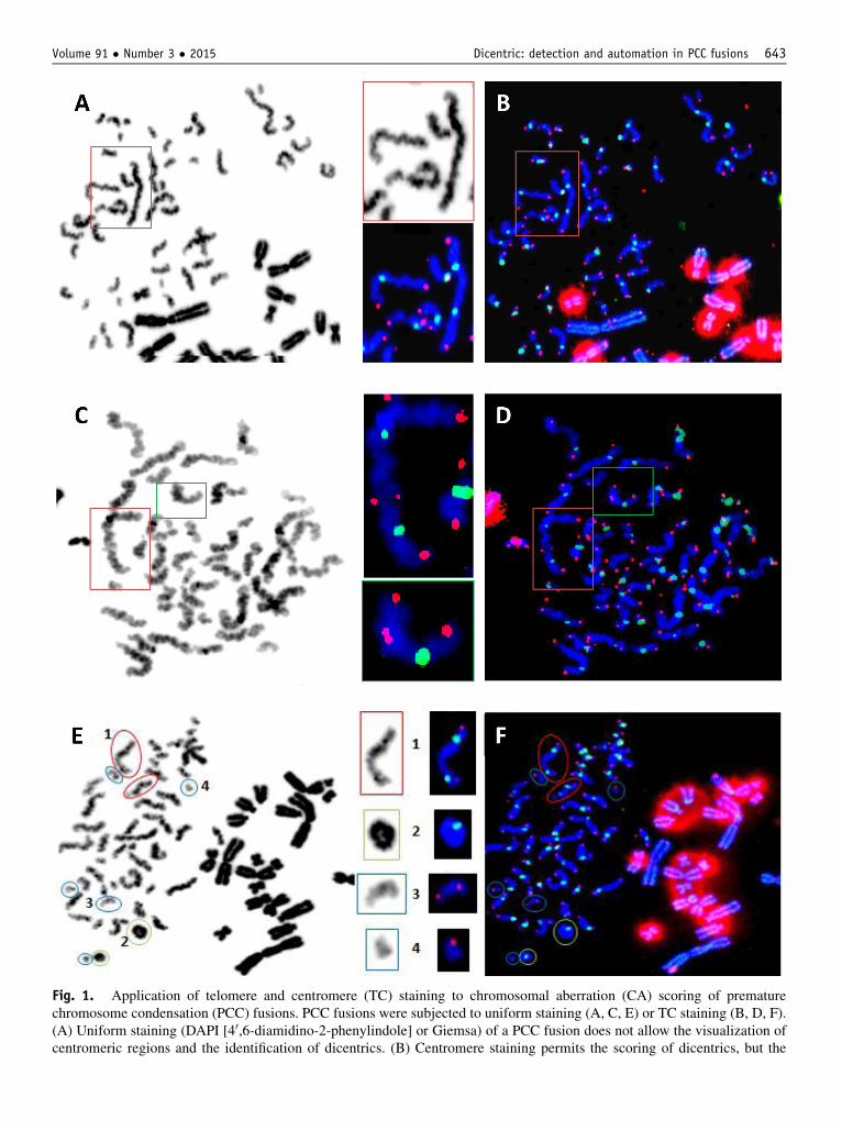
ig. 1. Application of telomere and centromere (TC) staining to chromosomal aberration (CA) scoring of prematurehromosome condensation (PCC) fusions. PCC fusions were subjected to uniform staining (A, C, E) or TC staining (B, D, F).A) Uniform staining (DAPI [40,6-diamidino-2-phenylindole] or Giemsa) of a PCC fusion does not allow the visualization ofentromeric regions and the identification of dicentrics. (B) Centromere staining permits the scoring of dicentrics, but the
Volume 91 � Number 3 � 2015 Dicentric: detection and automation in PCC fusions 643
Fc(c

Table 1 Frequencies and distribution of chromosomal aberrations 8 hours after irradiation, detected after manual scoring usingtelomere and centromere staining
Dose(Gy)
Cellsscored
No.dicentrics
No.rings
No.excessacentrics
Dicentricsþ ring/cell
Dicentrics/cell SE
Dicentrics distribution
s2/y U0 1 2 3 4 5 6 7 8 9
0 101 1 0 1 0.009 0.009 0.009 100 1 0 0 0 0 0 0 0 0 1 02 186 62 16 164 0.419 0.333 0.042 134 43 8 1 0 0 0 0 0 0 1.027 0.2624 164 198 49 266 1.506 1.207 0.085 55 45 43 17 4 0 0 0 0 0 0.99 �0.086 139 362 58 331 3.025 2.604 0.136 11 23 38 26 27 9 3 2 0 0 0.91 �0.698 124 472 83 373 4.475 3.806 0.175 4 8 15 28 31 16 12 7 2 1 0.86 �1.08
Abbreviations: s2/y Z index of dispersion; SE Z standard bar deviation; U Z variance.
M’kacher et al. International Journal of Radiation Oncology � Biology � Physics644
Statistical analysis
A script in R was developed taking into account theInternational Atomic Energy Agency recommendations (1).The curves generated in this study are primarily based ongeneralized linear models using the glm function of the stan-dard stats package. Details are provided in SupplementaryData E4 (available online at www.redjournal.com).
Results
Optimization of capture parameters after TCstaining of PCC fusions
After developing a specific classifier to automaticallysearch for the PCC fusions, the optimization of PCCfusion capture permitted better visualization of thecentromere regions and telomeric sequences, thus helpingto resolve overlapping chromosomes, which is difficult inthe absence of TC staining (Fig. 1A). However, the cap-ture of telomere sequences has to be adapted to take intoaccount the fact that the interstitial telomere sequences ofCHO are much longer than those of human lymphocytes(Fig. 1B). Image capture was carried out using a fixedtime by which the signal for the CHO telomeres wassaturated, whereas human telomeres were easily visual-ized. Detection of centromere and telomere signals leadsto the more precise scoring not only of dicentrics but alsoof CA, with better characterization of acentric chromo-somes (Fig. 1A-F).
demarcation of the chromosome ends requires telomere stainindifficulty in determining the chromosome ends after uniformelimination of false dicentrics and for the robust scoring of ac(circled in red), leaving only the possibility to detect rings (circleTC staining of a PCC fusion. After telomere (red) and centromedicentrics, 2 green centromere signals with 2 red telomere signal(2); and acentric chromosomes, no green signal with either 2 (numbers shown in E and F.
Manual detection of unstable CAs includingdicentrics after TC staining of PCC fusions
After uniform staining, it is possible to score DSBs solelyaccording to the presence of excess acentric chromosomesand rings (Fig. 1D) in PCC fusions. After TC staining itwas possible to detect dicentrics, centric and acentric rings,and different types of acentric chromosomes (Fig. 1F).
Table 1 shows the number of PCC cells scored manu-ally and the frequency of excess fragments and ringchromosomes detected after uniform staining, as well asthe frequency of dicentrics, centric rings, and excessacentric chromosomes after TC staining. In addition, thedistribution of dicentrics per cell and their associated s2/Yand U values obtained from 0 to 8 Gy are shown. APoisson distribution was observed at each dose with nodispersion.
A linear doseeresponse curve was obtained upon thescoring of acentric chromosomes after uniform staining(Fig. 2A), confirming previously published data (Table E1;available online at www.redjournal.com). However, usingTC staining, a linear-quadratic doseeresponse curve wasobtained, and a significant difference was observed betweenthe 2 curves (P<10�4). The number of acentric chromo-somes detected after TC staining was up to 2- to 3-foldgreater at higher exposures than that after uniform stain-ing (Fig. 2A). Figure 2B shows the frequency of differenttypes of acentric chromosomes. We observed a higherfrequency of acentric chromosomes with only 1 telomereinduced by a terminal deletion. This higher frequency canbe related to incomplete DNA repair at 8 hours after higherdoses of exposure.
g as demonstrated in this figure (boxed in red). (C) Thestaining. (D) The importance of telomere staining for theentric chromosomes. (E) Dicentrics are difficult to detectd in green) and acentric chromosomes (circled in blue). (F)re (green) staining, the following criteria were established:s (1); ring chromosomes, 1 green signal without a red signal3) or 1 red signal (4). Numbers in parenthesis refer to the

Uniform staining
0
0
0 0
02
2
2 2
24
4
4 4
46
6
6 6
68
8
8 8
8
Manual scoringAutomatic scoring Manual scoring
Automatic scoring
Uniform staining
5
4
3
2
1
0
Dic+
Ring
s /
Cell
Dose [Gy]
Dose [Gy]0 2 4 6 8
Dose [Gy]
Dose [Gy] Dose [Gy]
Dose [Gy]
Without telomereWith 1 telomereWith 2 telomeres
0
1
2
3
4
5
Acen
tric
chr
omos
omes
/cel
l0
1
2
3
4
5
Abbe
rati
ons
/ Ce
ll
Dic+Rings after TC stainingRings after uniform staining
TC stainingUniform staining
0
5
10
15
DSB
/ Ce
ll
0
5
10
15
DSB
/ Ce
ll
0
2
4
6
8
Acen
tric
chr
omos
omes
/cel
l
TC staining
A B
C D
E F
Fig. 2. Manual and automated chromosomal aberration (CA) scoring in premature chromosome condensation (PCC)fusions after uniform or telomere and centromere (TC) staining. In all cases, error bars represent the 95% confidencelimits. (A) Doseeresponse curves obtained after the scoring of all CAs after uniform or TC staining. (B) Doseeresponsecurves obtained after the scoring of different types of CAs after TC staining. (C) Doseeresponse curves obtained afterthe scoring of dicentrics (Dic) and rings after TC or uniform staining. (D) Doseeresponse curves based on the estimatednumber of double-strand breaks (DSBs) after the scoring of dicentrics (Dic) and rings after TC or uniform staining. (E)Comparison of doseeresponse curves for dicentrics and rings per cell after manual or automated scoring after TCstaining. No significant difference was observed. (F) Comparison of doseeresponse curves, including that for uniformstaining for calculated DSB per cell.
Volume 91 � Number 3 � 2015 Dicentric: detection and automation in PCC fusions 645
After uniform staining, a linear doseeresponse curvewas obtained upon the scoring of rings in the PCC fusion(Fig. 2C) with no saturation. After TC staining, a linear-quadratic doseeresponse curve was obtained with nosaturation at up to 8 Gy (Fig. 2C). A significant differencewas observed between the doseeresponse curve obtainedfor dicentrics plus rings after TC scoring and that ob-tained for rings only, after uniform staining (P<10�6)(Fig. 2C).
Using the calculated DSBs based on all unstableCAs, a linear doseeresponse curve was obtained afteruniform staining and a linear-quadratic curve afterTC staining. The calculated DSBs after TC stainingwere significantly higher than those observed afteruniform staining (PZ.005) (Fig. 2D). The curve fitcoefficients for all doseeresponse curves are shownin Supplementary Data E5 (available online atwww.redjournal.com).

M’kacher et al. International Journal of Radiation Oncology � Biology � Physics646
Automated scoring of unstable CAs after TCstaining of PCC fusions
The reliable and robust detection of CAs in PCC fusionsallows the possibility to automate their scoring, which hasnot been previously possible. Figure 3A shows a simplifiedversion of the workflow used by PCC-TCScore. The3-channel image contains the DAPI information in the bluechannel and the telomere and centromere information inthe red and green channels, respectively. The humanchromosomes are filtered to CHO chromosomes using thesaturated red channel zone and complementary informationabout large objects from the DAPI channel (blue block),and the centromeres and telomeres are detected. Thechromosomes are then defined by the number of telomeresand centromeres and finally classified according to theaberration class (Supplementary Data E3; available onlineat www.redjournal.com). This processing scheme allowsfor batch processing and report generation using a highlyintuitive and interactive user interface designed for rapidreview and correction by the user (Fig. 3B). The re-quirements for the use of this software are minimal,allowing its use on any computer, and images can betreated regardless of the type of image capture platformused. An average of 7 seconds per image was requiredusing the software, compared with 2 to 3 minutes formanual scoring.
Linear-quadratic doseeresponse curves for automatedscoring of dicentrics and centric rings, but not taking intoaccount acentric chromosomes, were obtained (Fig. 2E).No significant difference was observed between manualand automated scoring of PCC fusions using PCC-TCScoreat up to 8 Gy, whereas those for calculated DSBs based onall CAs (Fig. 2F) did show a significant difference in favorof manual scoring because the current version of the soft-ware does not accurately detect acentric chromosomes(Table E2; available online at www.redjournal.com).
Discussion
The dicentric chromosome assay is the “gold standard”biodosimetry method for recent exposure to ionizing ra-diation (1). Nonetheless, the non-compressible timerequired to obtain the metaphases remains the majorobstacle for obtaining a rapid dose estimate, and theapplication of this technique for triage of a populationafter a mass exposure event. The PCC fusion techniqueinduces the premature condensation of chromosomes,allowing rapid scoring of radiation-induced CA, but thecomplexity of the technique and subsequent analysis ofthe data restrict its use. Given that we have recentlydemonstrated the high efficiency of TC staining in thescoring of dicentrics and rings in metaphases (15), wedecided to combine this approach with the PCC fusiontechnique. Thus, in this study we introduce for the firsttime the scoring of dicentrics, centric rings, and acentric
chromosomes in PCC fusions after TC staining using PNAprobes, permitting the reliable detection of all unstableCAs in PCC fusions with a high level of precision andsensitivity. This amelioration allows the automation of thescoring of dicentrics and centric rings. The application ofautomated software to detect aberrations in PCC fusionswould represent a major advance in biological dosimetryafter an accident, as well as in the area of radioprotectionin general, where conventional cytogenetics is not suffi-ciently sensitive to detect the effects of exposure to lowdoses (16).
Many applications of the PCC fusion technique havebeen used in research and the clinic. In particular, drug-induced PCC assays have been extensively used toinvestigate the mechanisms underlying the formation ofradiation-induced aberrations (17, 18) or bystander effects(19). Premature chromosome condensation has also beenused to perform cytogenetic evaluation of quiescent andsenescent cells, as well as to increase the number ofmitotic cells within tumor samples, allowing for rapidkaryotyping after a very short-term culture, permitting theevaluation of karyotype diversity (20). It has also beenused to contribute to clinical prediction (21) and the cy-togenetic investigation of various diseases (22, 23).Another important application of this technique is theassessment of genotoxic risk immediately after exposureto various reagents (24), and it has also proven useful forinvestigating chromosomal damage immediately afterirradiation, as well as the kinetics of CA formation (25).The PCC technique has been shown to be compatible witha number of chromosome staining techniques, includingC-banding, centromere detection by FISH, and chromo-some painting (7, 8, 26-29). It has also been recognized asa powerful biodosimetric tool in an internationally stan-dardized technical report (1). Despite the utility of thistechnique, the scoring of CAs in PCC fusions has,nevertheless, remained technically challenging and timeconsuming, limiting routine use, even in research, despitethe advantages associated with the absence of cell cultureand the ability to analyze interphase chromosomes. Assuch, this technique is used solely by a few specialists. Inthis work we chose to perform PCC fusions using theCHO cell line as opposed to the drug-induced PCC assaybecause it does not allow a significant time reductioncompared with cell culture (44 hours vs 48 hours formetaphase). In contrast, we performed PCC fusion 8hours after irradiation, allowing time for DNA repair andthe occurrence of dicentrics, centric rings, and acentricchromosomes (3 hours vs 49 hours for metaphase).Indeed, in the case of accidental exposure, the time delayafter irradiation, including blood sampling, is variable,and would probably exceed 4 hours without including theadditional time for transport and treatment. Because thetime of DNA repair may vary between individuals, andowing to the conditions of storage/transport, the ability toscore DSBs allows the estimation of the absorbed doseindependently of DNA repair processes. It is for this

Red Channel (SPOR) Blue Chanel
(DAPI)
Local subtractive mean filter
Green Chanel (FITC)
Auto-Thresholding (Otsu)
Multi-levellocal thresholding
Thresholding std/2Filtering small structures filtering nuclei
Telo-FilterDistance MAP filter
Histogram andSTD based thresholding
Apply Chromo Mask
Spatial detection
apply DAPI Mask
spatial detection
CHO Zone map CHO excluding Filter
create Chromosome mask
Chromosomes Modeling
Classification
GUI & Report
LoG Filter(Laplacien of Gaussian)
A
B
ig. 3. Summary of the automation process used to develop the softward PCC-TCScore. (A) Chinese hamster ovary (CHO)hromosome is defined as being larger than the human one with an intense telomere signal. To filter out the CHO chro-osomes and create a human chromosome map, a CHO zone map is constructed using the saturated red channel zone andomplementary information about large objects from the DAPI (40,6-diamidino-2-phenylindole) channel (blue block).
Volume 91 � Number 3 � 2015 Dicentric: detection and automation in PCC fusions 647
Fcmc

M’kacher et al. International Journal of Radiation Oncology � Biology � Physics648
reason that it is indispensable to take into account allCAs, including all the different types of acentric chro-mosomes, made possible by TC staining, for estimatingthe dose.
After uniform staining, we have scored ring chromo-somes and acentric fragments. The frequency of chromo-some rings is in agreement with previously published datafor 8 hours after exposure (30) but higher than that obtainedfor chemically induced PCC rings (31).
The introduction of TC staining with PNA probes,with higher signal intensity and a short hybridizationtime, has permitted the identification of centromeric re-gions and telomere sequences, allowing the precisedetermination of the number of centromeres in each ob-ject and the limits of each chromosome to remove allambiguity in the scoring of CAs. This improvement hasallowed the scoring of all unstable CAs, including di-centrics, and the calculation of the number of resultingDSBs with higher precision, because of the detection ofinterstitial acentric chromosomes due to telomere stain-ing. Telomere and centromere staining represents a sig-nificant improvement compared with other studies thathave used centromere staining alone, which was the firstattempt to detect dicentrics in PCC fusions, but manyuncertainties remained, and there was poor detection ofacentric chromosomes.
The combination of telomere and centromere stainingwith PCC fusion thus represents an important advance inthe detection of unstable CAs in PCC fusions. Importantly,because of the higher signal obtained using PNA probes,the manual scoring of dicentrics, centric rings, and acentricchromosomes can be directly carried out at the microscopein the absence of an image analysis system.
After the successful detection of CAs in PCC fusionsand the establishment of a doseeresponse curve, the secondstep of this work was the semiautomation of the scoring ofthese aberrations using a new software that we havedeveloped, called PCC-TCScore, which is independent ofthe image analysis system used. Using PCC-TCScore wehave successfully detected on average 95% of dicentricsand centric rings compared with manual scoring, with thelevel of automated detection at or near 100% below 4 Gy,
Telomere location is mapped by filtering red channel informatiresult using half of the standard deviation as a threshold. Data ardetect centromeres, a LoG filter is applied to the green channelmation and the data standard deviation value. Results are confichromosome mask, and the telomere, and centromere location arScreen shot from PCC-TCScore showing a premature chromosoat 4 Gy. The numbers of centromeres, dicentrics, centric rings,software detected 3 dicentrics (circled in red), 3 acentric chromoyellow), permitting the end-user to examine these anomalies in mare difficult to segment, the software does not illuminate the Prections. Although the process is fully automated, PCC-TCScorcation using the attractive and user-friendly interface.
falling to 95% at 6 to 8 Gy. This high rate of detection ismade possible owing to the robust recognition of CAs afterTC staining, the high quality of the PCC fusions, and thevalidation step by the observer.
It may be equally important to apply these tools to thefield of low dose exposure, which is currently a growingpublic health concern. The PCC fusion technique associ-ated with whole-chromosome painting FISH on 3 chro-mosomes has been successfully used to measure thebiological effects of CT scans and to estimate the absorbeddose (28). Such information is key to advancing ourknowledge in the field of low-dose exposure to better defineradiation protection standards. Moreover, this assay iscurrently used in only a limited number of laboratories. Itsapplication for biological dosimetry in an emergencyresponse to radiation exposure would need to be precededby assimilation of the assay into qualifying laboratories. Inboth cases the introduction of TC staining will permit morewidespread use of PCC fusion techniques and the exploi-tation of its full potential.
Conclusion
In the present work we demonstrate that the contributionof TC staining to the detection of CAs, including di-centrics, eliminates the time-dependent problem ofunderestimating the frequency of acentric chromosomesowing to their reparation encountered when using uniformstaining. This approach creates new possibilities for thescoring of unstable CAs when conventional techniques areinadequate and when the rapidity of the answer is aprerequisite.
The accurate detection of each type of CA has allowedthe development of PCC-TCScore for the scoring of di-centrics and centric rings from captured images of PCCfusions. It will open up new horizons for biologicaldosimetry, in particular for low, high, and partial-bodyirradiation, as well as for cytogenetic studies concerningchromosomal instability, the heterogeneity of the karyo-type, and when cell culture is not possible. Furthermore,individual responses to genotoxic agents could be
on using a local subtractive mean filter and binarizing thee restricted to the human chromosome mask (red block). To, followed by a threshold calculated using histogram infor-ned to the human chromosome mask. Finally, the humane merged for chromosome modeling and classification. (B)me condensation (PCC) fusion from lymphocytes irradiatedand acentric chromosomes are provided. In this case, thesomes (circled in green), and 2 abnormal clusters (circled inore detail. In the case of overlapping chromosomes, whichCC fusion but allows the operator to perform manual cor-e allows the operator to validate the chromosome classifi-

Volume 91 � Number 3 � 2015 Dicentric: detection and automation in PCC fusions 649
monitored, including the follow-up of patients and theirindividual response to treatment by radiation orchemotherapy.
References
1. International Atomic Energy Agency. Cytogenetic Dosimetry Appli-
cations in Preparedness for and Response to Radiation Emergencies.
EPR-Biodose 2011. Vienna: IAEA; 2011.
2. Dossou J, Lartigau E, M’Kacher R, et al. Biological dosimetry after
total body irradiation (TBI) for hematologic malignancy patients. Int J
Radiat Oncol 2000;46:123-129.
3. Johnson RT, Rao PN. Mammalian cell fusion: Induction of premature
chromosome condensation in interphase nuclei. Nature 1970;226:
717-722.
4. Pantelias GE, Maillie HD. A simple method for premature chromo-
some condensation induction in primary human and rodent cells using
polyethylene glycol. Somat Cell Genet 1983;9:533-547.
5. Pantelias GE, Maillie HD. The use of peripheral blood mononuclear
cell prematurely condensed chromosomes for biological dosimetry.
Radiat Res 1984;99:140-150.
6. Darroudi F, Natarajan AT, Bentvelzen PA, et al. Detection of total- and
partial-body irradiation in a monkey model: A comparative study of
chromosomal aberration, micronucleus and premature chromosome
condensation assays. Int J Radiat Biol 1998;74:207-215.
7. Pantelias GE, Iliakis GE, Sambani CD, Politis G. Biological dosimetry
of absorbed radiation by C-banding of interphase chromosomes in
peripheral blood lymphocytes. Int J Radiat Biol 1993;63:349-354.
8. Darroudi F, Bergs JW, Bezrookove V, et al. PCC and COBRA-FISH a
new tool to characterize primary cervical carcinomas: To assess hall-
marks and stage specificity. Cancer Lett 2010;287:67-74.
9. Boei JJ, Vermeulen S, Natarajan AT. Analysis of radiation-induced
chromosomal aberrations using telomeric and centromeric PNA
probes. Int J Radiat Biol 2000;76:163-167.
10. Muller P, Schmitt E, Jacob A, et al. COMBO-FISH enables high
precision localization microscopy as a prerequisite for nanostructure
analysis of genome loci. Int J Mol Sci 2010;11:4094-4105.
11. Puerto S, Marcos R, Ramirez MJ, et al. Induction, processing and
persistence of radiation-induced chromosomal aberrations involving
hamster euchromatin and heterochromatin.MutatRes2000;469:169-179.
12. Suto Y, Akiyama M, Gotoh T, et al. A modified protocol for accurate
detection of cell fusion-mediated premature chromosome condensation
in human peripheral blood lymphocytes. Cytologia 2013;78:97-103.
13. Terzoudi GI, Singh SK, Pantelias GE, Iliakis G. Premature chromo-
some condensation reveals DNA-PK independent pathways of chro-
mosome break repair. Int J Oncol 2008;33:871-879.
14. Pottier G, Viau M, Ricoul M, et al. Lead exposure induces telomere
instability in human cells. PloS One 2013;8. e67501.
15. M’kacher R, El Maalouf E, Ricoul M, et al. New tool for biological
dosimetry: Reevaluation and automation of the gold standard method
following telomere and centromere staining. Mutat Res-Fund Mol M
2014;770:45-53.
16. Pernot E, Hall J, Baatout S, et al. Ionizing radiation biomarkers for
potential use in epidemiological studies.Mutat Res 2012;751:258-286.
17. Pantelias GE, Terzoudi GI. Functional cell-cycle chromatin confor-
mation changes in the presence of DNA damage result into chromatid
breaks: A new insight in the formation of radiation-induced chromo-
somal aberrations based on the direct observation of interphase
chromatin. Mutat Res 2010;701:27-37.
18. Terzoudi GI, Hatzi VI, Donta-Bakoyianni C, Pantelias GE. Chromatin
dynamics during cell cycle mediate conversion of DNA damage into
chromatid breaks and affect formation of chromosomal aberrations:
Biological and clinical significance. Mutat Res 2011;711:174-186.
19. Terzoudi GI, Donta-Bakoyianni C, Iliakis G, Pantelias GE. Investi-
gation of bystander effects in hybrid cells by means of cell fusion and
premature chromosome condensation induction. Radiat Res 2010;173:
789-801.
20. Heng HH, Bremer SW, Stevens JB, et al. Genetic and epigenetic
heterogeneity in cancer: A genome-centric perspective. J Cell Physiol
2009;220:538-547.
21. Hittelman WN, Broussard LC, McCredie K. Premature chromosome
condensation studies in human leukemia. I. Pretreatment characteris-
tics. Blood 1979;54:1001-1014.
22. Neitzel H, Neumann LM, Schindler D, et al. Premature chromosome
condensation in humans associated with microcephaly and mental
retardation: A novel autosomal recessive condition. Am J Hum Genet
2002;70:1015-1022.
23. Sreekantaiah C, Bhargava MK, Shetty NJ. Premature chromosome
condensation in human cervical carcinoma. Cancer Genet Cytogenet
1987;24:263-269.
24. Bolzan AD, Paez GL, Bianchi MS. FISH analysis of telomeric repeat
sequences and their involvement in chromosomal aberrations induced
by radiomimetic compounds in hamster cells. Mutat Res 2001;479:
187-196.
25. Pantelias GE, Maillie HD. The measurement of immediate and
persistent radiation-induced chromosome damage in rodent primary
cells using premature chromosome condensation. Health Phys 1985;
49:425-433.
26. Durante M, George K, Yang TC. Biodosimetry of ionizing radiation
by selective painting of prematurely condensed chromosomes in
human lymphocytes. Radiat Res 1997;148(5 Suppl):S45-S50.
27. Evans JW, Chang JA, Giaccia AJ, et al. The use of fluorescence in
situ hybridisation combined with premature chromosome conden-
sation for the identification of chromosome damage. Br J Cancer
1991;63:517-521.
28. M’Kacher R, Violot D, Aubert B, et al. Premature chromosome
condensation associated with fluorescence in situ hybridisation detects
cytogenetic abnormalities after a CT scan: Evaluaton of the low-dose
effect. Radiat Prot Dosim 2003;103:35-40.
29. Prasanna PG, Blakely WF. Premature chromosome condensation in
human resting peripheral blood lymphocytes for chromosome aber-
ration analysis using specific whole-chromosome DNA hybridization
probes. Methods Mol Biol 2005;291:49-57.
30. Lamadrid Boada AI, Romero Aguilera I, Terzoudi GI, et al. Rapid
assessment of high-dose radiation exposures through scoring of cell-
fusion-induced premature chromosome condensation and ring chro-
mosomes. Mut Res 2013;757:45-51.
31. Lindholm C, Stricklin D, Jaworska A, et al. Premature chromosome
condensation (PCC) assay for dose assessment in mass casualty
accidents. Radiat Res 2010;173:71-78.