Designing of Cytotoxic and Helper T Cell Epitope Map ...
37
doi.org/10.26434/chemrxiv.12253463.v1 Designing of Cytotoxic and Helper T Cell Epitope Map Provides Insights into the Highly Contagious Nature of the Pandemic Novel Coronavirus SARS-CoV2 Seema Mishra Submitted date: 07/05/2020 • Posted date: 11/05/2020 Licence: CC BY-NC-ND 4.0 Citation information: Mishra, Seema (2020): Designing of Cytotoxic and Helper T Cell Epitope Map Provides Insights into the Highly Contagious Nature of the Pandemic Novel Coronavirus SARS-CoV2. ChemRxiv. Preprint. https://doi.org/10.26434/chemrxiv.12253463.v1 This study provides key insights into the contagious nature of SARS-CoV2 through analyses of T cell epitopes designed from SARS-CoV2 proteome. All top-scoring cytotoxic T cell epitopes from ORF1ab and helper T cell epitopes identified from all proteins were utilized to provide crucial insights into its pathogenesis. These T cell epitopes can be used as prophylactic or therapeutic multi-subunit vaccine or as diagnostic tools. File list (17) download file view on ChemRxiv PaperSeema.pdf (290.84 KiB) download file view on ChemRxiv Supplementary Figure S10.doc (340.00 KiB) download file view on ChemRxiv muscleEnvelope (1.10 KiB) download file view on ChemRxiv muscleMembrane (2.66 KiB) download file view on ChemRxiv muscleNS3 (2.08 KiB) download file view on ChemRxiv muscleNS6 (364.00 B) download file view on ChemRxiv muscleNucleocapsid (5.98 KiB) download file view on ChemRxiv muscleORF1ab (76.25 KiB) download file view on ChemRxiv muscleORF7a (617.00 B) download file view on ChemRxiv muscleORF8 (820.00 B) download file view on ChemRxiv muscleSpike (20.55 KiB) download file view on ChemRxiv S4_Supplementary material_PickPocket.xls (2.97 MiB)
Transcript of Designing of Cytotoxic and Helper T Cell Epitope Map ...
doi.org/10.26434/chemrxiv.12253463.v1
Designing of Cytotoxic and Helper T Cell Epitope Map Provides Insightsinto the Highly Contagious Nature of the Pandemic Novel CoronavirusSARS-CoV2Seema Mishra
Submitted date: 07/05/2020 • Posted date: 11/05/2020Licence: CC BY-NC-ND 4.0Citation information: Mishra, Seema (2020): Designing of Cytotoxic and Helper T Cell Epitope Map ProvidesInsights into the Highly Contagious Nature of the Pandemic Novel Coronavirus SARS-CoV2. ChemRxiv.Preprint. https://doi.org/10.26434/chemrxiv.12253463.v1
This study provides key insights into the contagious nature of SARS-CoV2 through analyses of T cell epitopesdesigned from SARS-CoV2 proteome. All top-scoring cytotoxic T cell epitopes from ORF1ab and helper T cellepitopes identified from all proteins were utilized to provide crucial insights into its pathogenesis. These T cellepitopes can be used as prophylactic or therapeutic multi-subunit vaccine or as diagnostic tools.
File list (17)
download fileview on ChemRxivPaperSeema.pdf (290.84 KiB)
download fileview on ChemRxivSupplementary Figure S10.doc (340.00 KiB)
download fileview on ChemRxivmuscleEnvelope (1.10 KiB)
download fileview on ChemRxivmuscleMembrane (2.66 KiB)
download fileview on ChemRxivmuscleNS3 (2.08 KiB)
download fileview on ChemRxivmuscleNS6 (364.00 B)
download fileview on ChemRxivmuscleNucleocapsid (5.98 KiB)
download fileview on ChemRxivmuscleORF1ab (76.25 KiB)
download fileview on ChemRxivmuscleORF7a (617.00 B)
download fileview on ChemRxivmuscleORF8 (820.00 B)
download fileview on ChemRxivmuscleSpike (20.55 KiB)
download fileview on ChemRxivS4_Supplementary material_PickPocket.xls (2.97 MiB)
download fileview on ChemRxivS5_Supplementary material_NetCTLpan_results.xls (11.73 MiB)
download fileview on ChemRxivS6_Supplementary material_NetMHCIIpan_results.xls (15.68 MiB)
download fileview on ChemRxivSupplementary table S1.xls (10.50 KiB)
download fileview on ChemRxivSupplementary table S2.xls (358.00 KiB)
download fileview on ChemRxivSupplementary table S3.xls (197.00 KiB)
Designing of cytotoxic and helper T cell epitope map provides insights into the
highly contagious nature of the pandemic novel coronavirus SARS-CoV2
Seema Mishra
Department of Biochemistry
School of Life Sciences
University of Hyderabad, India
Email: [email protected]
Abstract:
Novel coronavirus, SARS-CoV2, has emerged one of the deadliest pathogens of this century creating a
pandemic. Belonging to betacoronavirus family, it spreads through human contact and even through
asymptomatic transmission. Till date, there is no known treatment in the form of drugs or vaccines,
despite several attempts since it emerged. Several vaccines are in pre-clinical and two in clinical trials
as of 4th April 2020 as per WHO document. Here, in order to develop subunit vaccine, attempts have
been made to find globally conserved epitopes from all ten SARS-CoV2 proteins as there is no clear
information on the virulence of these proteins. Using computational tools, a ranked list of probable
immunogenic, promiscuous epitopes generated through all three main stages of antigen processing and
presentation pathway has been generated. Moreover, on the way to finding these epitopes, several
useful insights were gleaned. One of the most important insights is that all of the proteins in this
pathogen present unique epitopes, so that if one protein's function is hindered by the immune system,
other proteins can continue to assist in virus survival. Due to presence of these unique epitopes in all
SARS-CoV2 proteins, a stronger immune response generated may lead to immunopathology and
consequently, less chances of human survival. These epitopes, after due validation in vitro, may thus
need to be presented to the human body in that form of subunit vaccine that avoids such
immunopathologies.
Introduction
Novel coronavirus (SARS-CoV2), also known as 2019-nCoV, causes Covid19 disease with significant
mortality rate. As there is currently no known cure, vaccine design and development is urgently
required. Despite 77 drugs against viral spike protein being identified by world's fastest supercomputer,
Summit, (1), Immunoinformatics tools will prove crucial (2, 3). As of 4th April 2020, WHO has put
forward a draft which identifies 2 vaccines in clinical evaluation and 60 candidate vaccines in preclinical
evaluation (4). This study presents several such novel cytotoxic and helper T cell epitopes against
ORF1ab protein and helper T cell epitopes against all other proteins.
SARS-CoV2 genome submitted by CDC, Atlanta (GenBank accession number: MT106054.1
submitted on 24-Feb-2020) is 29882 bp in length. Being 100% identical to reference sequence,
NC_045512.2 from Wuhan, China, it harbors multiple structural, non-structural and accessory proteins
essential or playing a role at various stages of the viral life cycle. This SARS-CoV2 genome is found
82.3% identical to SARS-CoV genome (NC_004718.3), using NCBI BLASTn tool. T cell epitopes
against several proteins in SARS and MERS species have been identified (5, 6). In brief, the sequence
of proteins in its RNA genome as per this GenBank accession information is as follows: 5′-ORF1ab-S
(Spike/Surface)–ORF3a-E (Envelope)-M (Membrane)-ORF6-ORF7a-ORF8-N (Nucleocapsid)-
ORF10-polyA tail-3', which are usually seen in betacoronaviruses (7). ORF1ab, a polyprotein, encodes
several non-structural proteins, 15 in number identified in this genome sequence annotation, including
RNA-dependent RNA polymerase (RdRP). The role of structural proteins is determined from their
homology to SARS-CoV as well as few experiments (8). Expression, localization and function of some
SARS-CoV2 accessory proteins is as yet unclear, although several such proteins have been
characterized in SARS-CoV (9) and the roles may be similar in the two viruses. Sequencing studies
suggest that the most abundant transcript was N RNA followed by S, ORF7a, ORF3a, ORF8, M, E,
ORF6, and ORF7b (10; ORF7b is identified in this paper). In view of scarcity of data on relevance and
roles of these proteins, any one or more of these proteins may act as prime vaccine candidates. Hence,
all of these proteins were used for T cell epitope prediction for the purpose of peptide-based subunit
vaccine design and further analyses. The fact that this approach may be better also arises from previous
studies on related SARS-CoV virus (11), wherein more than 50% of the patients had T cell responses
against at least one of the two proteins tested, and 25% showed responses against both proteins.
The advantages of peptide subunit vaccine as opposed to DNA and live attenuated virus vaccine
is that they do not contain live components and so are considered safe. Moreover, they present an
antigen or a set of antigens to the immune system with lower risk of side effects (12). These are also
applicable to people with weakened immune response, which the old people have and are, therefore,
prime targets in this SARS-CoV2 infection. Henec, this study has been done with the objectives of
finding novel CTL and HTL epitopes and helping glean many important insights along the way.
Results and Discussion:
Cytotoxic T lymphocyte (CTL) epitopes
Two prediction algorithms were used to generate a consensus list of nonameric CTL epitopes. The
consensus list was chosen to increase prediction accuracy from two different algorithms. While
NetCTLpan uses neural network algorithm, PickPocket works on the basis of position-specific weight
matrices. NetCTLpan, in addition to HLA binding, also predicts TAP-transporter binding and C-
terminal proteasome cleavage predictions. Total number of CTL epitopes generated was 9621 across
ten SARS-CoV2 proteins including ORF1ab polyprotein. A total of 122 epitopes were enlisted. These
common, promiscuous CTL epitopes are enlisted in Table 1 as ranked order for ORF1ab. For other
proteins, it may be observed/enlisted from (13). It is seen that out of a few common promiscuous
epitopes for surface protein across prediction algorithms (13), one of these epitopes, FVFLVLLPL,
signal peptide in surface/spike protein, has been found to harbor a mutation, L5F, in many strains of 13
countries in distinct phylogenetic clades and L8V/W mutation is present in Hong Kong (14). These
authors further suggest that L5F mutation might be a sequencing artifact. The highest number of
common top-ranking epitopes is seen in the case of nsp7 of ORF1ab followed by ORF10, ORF8,
ORF6 and ORF3a proteins. Among structural proteins, envelope protein provided the highest number
of such epitopes. Venn diagram analysis showed no common epitopes at all across proteins and alleles.
Even though SARS-CoV2 RBD (331-527) is shown to harbor epitopes for eliciting neutralizing
antibodies (14, 15), this region is not present in this data for CTL epitopes. However, receptor-binding
motif (RBM) region (437-508), the ACE-2 binding motif of this RBD provided immunogenic HTL
epitopes which are detailed below in section on promiscuous HTL epitopes. Immunogenicity prediction
of these proteins (Table 2) showed that 71 of these 122 epitopes had a positive immunogenicity score.
Further, conserved residues between SARS-CoV2 and other HCoV and MERS species were found
from multiple sequence alignments and found in several of these epitopes (Supplementary fig. S1). As
the NCBI RefSeq sequence of SARS-CoV was unclear in proper annotations for respective proteins, it
could not be used in MSA studies. It is observed that most of the epitopes with conserved residues
belonged to ORF1ab region (table 2), and epitopes belonging to this region may act as vaccine
candidates targeting MERS and other HCoV species, in addition to SARS-CoV2.
Table 1: Top ranked sequences of CTL epitopes common across HLA supertypes (HLA-A*01:01,
HLA-A*02:01, HLA-A*03:01, HLA-A*24:02, HLA-A*26:01, HLA-B*07:02, HLA-B*08:01,
HLA-B*27:05, HLA-B*39:01, HLA-B*40:01, HLA-B*58:01, HLA-B*15:01) and across the two
prediction algorithms used for SARS-CoV2 ORF1ab polyprotein.
Leaderprotein(nsp1)
nsp2 nsp3 nsp4 3C-like Proteinase
nsp6 nsp7 nsp8 nsp9 nsp10 RdRP Helicase
3'-5'exonuclease
EndoRNAse
Ribose methyltransferase
HVGEIPVAY
NMMVTNNTF
LVAEWFLAY (not among top scorers in many alleles)
VVAFNTLLF VVAFNTLLFall alleles exceptHLA-A*02:01,
HLA-A*03:01,
HLA-B*07:02,
HLA-B*08:01,
HLA-B*27:05,
HLA-B*39:01,
HLA-B*40:01
QTFSVLACY
MLVYCFLGYall alleles except HLA-A*02:01,
HLA-A*24:02,
HLA-*07:02,
HLA-B*39:01,
HLA-B*58:01,
MVSLLSVLL
SSLPSYAAF
CTDDNALAY
FAVDAAKAY
MVMCGGSLY among top scoring inall allelles, except
HLA-A*02:01,HLA-A*24:02,HLA-B*08:01,
HLA-B*39:01,
YVFCTVNALall except
HLA-A*01:01,HLA-A*03:01,HLA-A*24:02,HLA-B*58:01
HSIGFDYVY
present in HLA-A*01:01,HLA-A*03:01,HLA-A*26:01,HLA-B*07:02,HLA-B*27:05,HLA-B*40:01,HLA-B*58:01,HLA-B*15:01;
TTLPVNVAF-all alleles,
YVMHANYIF-all alleles exceptHLA-A*03:01,HLA-B*27:05
VMVELVAEL
RTILGSALL
LIISVTSNYall alleles except
HLA-A*02:01,
HLA-A*24:02,
HLA-B*08:01,
HLA-B*39:01,
SLLSVLLSM
ISMDNSPNL
KSDGTGTIY
STVLSFCAF
TMADLVYAL
among top scoring inall allelles, except
HLA-A*01:01,HLA-A*03:01,HLA-A*24:02,HLA-B*08:01,HLA-B*27:05,
HLA-B*58:01,
FAIGLALYY (top-scorer in few allelesin HLA-A*01:01, HLA-A*26:01, HLA-B*58:01,HLA-B*15:01)
VSIINNTVY-all except HLA-A*02:01
YSLFDMSKF- all exceptHLA-A*02:01,HLA-B*08:01,HLA-B*39:01
HVQLSLPVL
VSFCYMHHM
FLARGIVFMall alleles except
KMVSLLSVL
AMQTMLFTM
GTGTIYTEL
PANSTVLSF
LMIERFVSLall alleles exceptA*01:01,
LLLDDFVEI-all except HLA-A*03:01
HLA-A*01:01,
HLA-A*03:01,
HLA-A*24:02,
HLA-B*27:05,
HLA-B*40:01,
HLA-B*58:01,
A*03:01,
A*26:01,
HLA-B*58:01,
and HLA-B*27:05
HLKDGTCGL
FLNRFTTTL
CTSVVLLSV
TTFTYASAL
TELEPPCRF
FGGASCCLY
SQLGGLHLL-all exceptHLA-A*01:01,HLA-A*03:01,HLA-A*26:01 ,HLA-B*07:02,
PQLEQPYVF
HSMQNCVLK allalleles exceptHLA-A*01:01,
HLA-A*02:01,
HLA-A*24:02,
HLA-A*26:01,
HLA-B*07:02,
HLA-B*08:01,
HLA-B*27:05,
HLA-B*39:01,
HLA-B*40:01,
HLA-B*58:01,
HLA-B*15:01
TSVVLLSVL
YFIKGLNNL
ITVTPEANM
QLEQPYVFI
KLWAQCVQL
ALAYYNTTK
VLSFCAFAV
RTAPHGHVM
VLLSVLQQL
GMVLGSLAA
YLASGGQPI
EMLDNRATL
FVLALLSDL (in some, after around top 30)
EAFEKMVSL
DVKCTSVVL
LAKDTTEAF
LHNDILLAK
LSMQGAVDI
VQLHNDILL
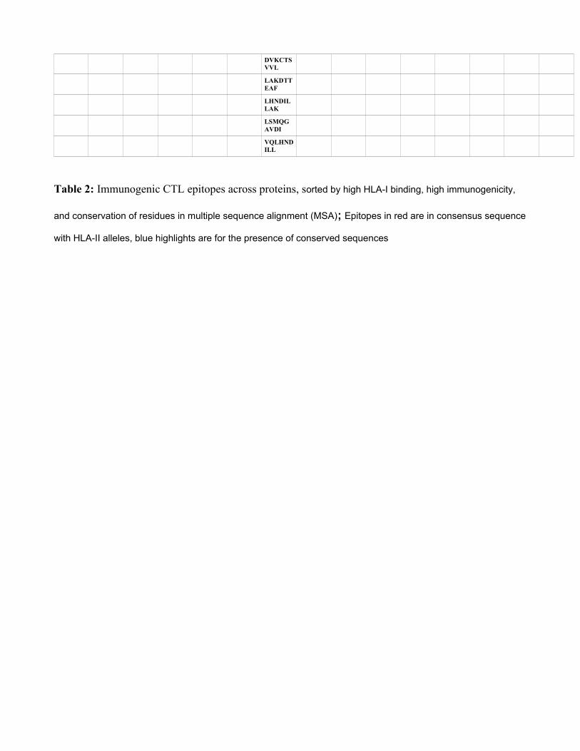
Table 2: Immunogenic CTL epitopes across proteins, sorted by high HLA-I binding, high immunogenicity,
and conservation of residues in multiple sequence alignment (MSA); Epitopes in red are in consensus sequence
with HLA-II alleles, blue highlights are for the presence of conserved sequences
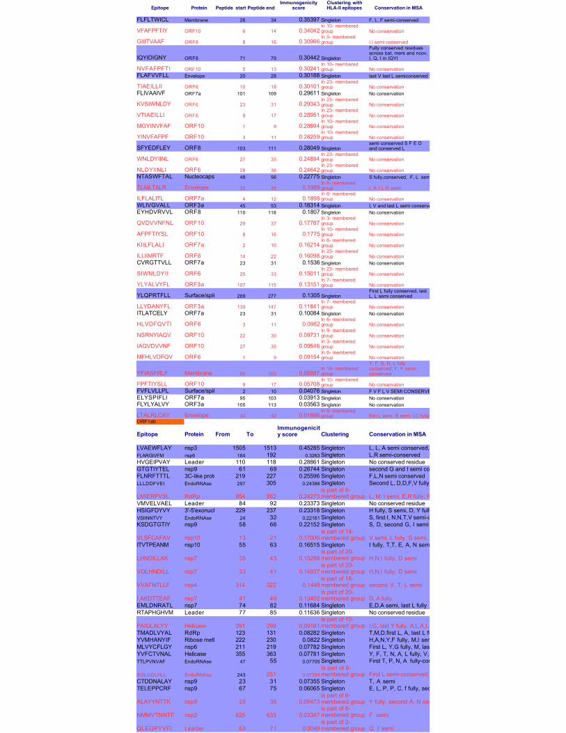
Epitope Protein Peptide start Peptide end Conservation in MSA
FLFLTWICL Membrane 26 34 0.35397 Singleton F, L, F semi-conserved
VFAFPFTIY ORF10 6 14 0.34042 No conservation
GIIITVAAF ORF8 8 16 0.30966 I,I semi conserved
IQYIDIGNY ORF8 71 79 0.30442 Singleton
NVFAFPFTI ORF10 5 13 0.30241 No conservationFLAFVVFLL Envelope 20 28 0.30188 Singleton last V last L semiconserved
TIAEILLII ORF6 10 18 0.30101 No conservationFLIVAAIVF ORF7a 101 109 0.29611 Singleton No conservation
KVSIWNLDY ORF6 23 31 0.29343 No conservation
VTIAEILLI ORF6 9 17 0.28951 No conservation
MGYINVFAF ORF10 1 9 0.28694 No conservation
YINVFAFPF ORF10 3 11 0.28259 No conservation
SFYEDFLEY ORF8 103 111 0.28049 Singleton
WNLDYIINL ORF6 27 35 0.24894 No conservation
NLDYIINLI ORF6 28 36 0.24642 No conservationNTASWFTAL Nucleocapsid 48 56 0.22775 Singleton S fully,conserved, F, L semiconserved
TLAILTALR Envelope 30 38 0.1989 L A I L R semi
ILFLALITL ORF7a 4 12 0.1895 No conservationWLIVGVALL ORF3a 45 53 0.18314 Singleton I, V and last L semi conservedEYHDVRVVL ORF8 110 118 0.1807 Singleton No conservation
QVDVVNFNL ORF10 29 37 0.17787 No conservation
AFPFTIYSL ORF10 8 16 0.1775 No conservation
KIILFLALI ORF7a 2 10 0.16214 No conservation
ILLIIMRTF ORF6 14 22 0.16098 No conservationCVRGTTVLL ORF7a 23 31 0.1536 Singleton No conservation
SIWNLDYII ORF6 25 33 0.15011 No conservation
YLYALVYFL ORF3a 107 115 0.13151 No conservation
YLQPRTFLL Surface/spike 269 277 0.1305 Singleton
LLYDANYFL ORF3a 139 147 0.11841 No conservationITLATCELY ORF7a 23 31 0.10084 Singleton No conservation
HLVDFQVTI ORF6 3 11 0.0982 No conservation
NSRNYIAQV ORF10 22 30 0.09731 No conservation
IAQVDVVNF ORF10 27 35 0.09546 No conservation
MFHLVDFQV ORF6 1 9 0.09154 No conservation
YFIASFRLF Membrane 95 103 0.06887
FPFTIYSLL ORF10 9 17 0.05708 No conservationFVFLVLLPL Surface/spike 2 10 0.04076 Singleton F V F L V SEMI CONSERVEDELYSPIFLI ORF7a 95 103 0.03913 Singleton No conservationFLYLYALVY ORF3a 105 113 0.03563 Singleton No conservation
LTALRLCAY Envelope 34 42 0.01886 first L semi, R semi, LC fullyORF1ab
Epitope Protein From To Clustering Conservation in MSA
LVAEWFLAY nsp3 1505 1513 0.45285 Singleton L, L, A semi conserved, Y fully conservedFLARGIVFM nsp6 184 192 0.3263 Singleton L,R semi-conservedHVGEIPVAY Leader 110 118 0.28861 Singleton No conserved residueGTGTIYTEL nsp9 61 69 0.26744 Singleton second G and I semi conserved, E, L fully conservedFLNRFTTTL 3C-like proteinase 219 227 0.25596 Singleton F,L,N semi conservedLLLDDFVEI EndoRNAse 297 305 0.24386 Singleton Second L, D,D,F,V fully conserved; I semi-conserved
LMIERFVSL RdRp 854 862 0.24273 L, M, I semi, E,R fully, F semi, V, S, L fullyVMVELVAEL Leader 84 92 0.23373 Singleton No conserved residueHSIGFDYVY 3'-5'exonuclease 229 237 0.23318 Singleton H fully, S semi, D, Y fully, V semi, Y fullyVSIINNTVY EndoRNAse 24 32 0.22161 Singleton S, first I, N,N,T,V semi-conservedKSDGTGTIY nsp9 58 66 0.22152 Singleton S, D, second G, I semi
VLSFCAFAV nsp10 13 21 0.17009 V semi, L fully, S semi, second F fullyA semi, V fullyITVTPEANM nsp10 55 63 0.16515 Singleton I fully, T,T, E, A, N semi
LHNDILLAK nsp7 35 43 0.15288 H,N,I fully, D semi
VQLHNDILL nsp7 33 41 0.14937 H,N,I fully, D semi
VVAFNTLLF nsp4 314 322 0.1449 second V, T, L semi
LAKDTTEAF nsp7 41 49 0.13402 D, A fullyEMLDNRATL nsp7 74 82 0.11684 Singleton E,D,A semi, last L fullyRTAPHGHVM Leader 77 85 0.11636 Singleton No conserved residue
FAIGLALYY Helicase 291 299 0.09181 I,G, last Y fully, A,L,A,L,Y semiTMADLVYAL RdRp 123 131 0.08282 Singleton T,M,D,first L, A, last L fully, Y semiYVMHANYIF Ribose methytransferase222 230 0.0822 Singleton H,A,N,Y,F fully, M,I semiMLVYCFLGY nsp6 211 219 0.07782 Singleton First L, Y,G fully, M, last L, last Y semiYVFCTVNAL Helicase 355 363 0.07781 Singleton Y, F, T, N, A, L fully, V, V semiTTLPVNVAF EndoRNAse 47 55 0.07705 Singleton First T, P, N, A fully-conserved, second T, V, V,F semi-conserved
SQLGGLHLL EndoRNAse 243 251 0.07388 First L semi-conserved, G,G,L,H,L,L fully-conservedCTDDNALAY nsp9 23 31 0.07355 Singleton T, A semiTELEPPCRF nsp9 67 75 0.06065 Singleton E, L, P, P, C, f fully, second E and R semi
ALAYYNTTK nsp9 28 36 0.05473 Y fully, second A, N semi
NMMVTNNTF nsp2 625 633 0.03347 F semi
QLEQPYVFI Leader 63 71 0.0049 Q, I semi
Immunogenicityscore
Clustering withHLA-II epitopes
In 10- memberedgroupIn 5- memberedgroup
Fully conserved residuesacross bat, mers and ncov,I, Q, I in IQYI
In 10- memberedgroup
In 23- memberedgroup
In 23- memberedgroupIn 23- memberedgroupIn 10- memberedgroupIn 10- memberedgroup
semi conserved S F E Dand conserved L
In 23- memberedgroupIn 23- memberedgroup
In 8- memberedgroupIn 6- memberedgroup
In 3- memberedgroupIn 10- memberedgroupIn 6- memberedgroupIn 23- memberedgroup
In 23- memberedgroupIn 7- memberedgroup
First L fully conserved, lastL, L semi conserved
In 7- memberedgroup
In 6- memberedgroupIn 9- memberedgroupIn 3- memberedgroupIn 6- memberedgroup
In 14- memberedgroup
Y, F, S, R, L fullyconserved, F, F semi-conserved
In 10- memberedgroup
In 8- memberedgroup
Immunogenicity score
is part of 6-membered group
is part of 14-membered group
is part of 20-membered groupis part of 20-membered groupis part of 16-membered groupis part of 20-membered group
is part of 10-membered group
Is part of 8-membered group
is part of 6-membered groupis part of 6-membered groupis part of 2-membered group
During these CTL epitope identification studies, it was also found that many epitopes same as SARS-
CoV epitopes found previously in spike, membrane, nucleocapsid and ORF3a proteins (16) were in the
lower ranking positions, in the case of different alleles, and many were not common across epitopes, so
confidence could not be gathered in enlisting these. However, in ORF3a case, one epitope harbouring
both CD8+ and CD4+ T cell epitopes, PLQASLPFGWLVIGV, among the 3 most frequently
recognized by T cells (17) was also present among top-ranked ones in our study (Table 1a). Purely for
the sake of information to the readers, these T cell epitope data recognized in humans /transgenic
mouse in case of SARS-CoV that are same/similar to lower ranking T cell epitopes in SARS-CoV2 are
provided as supplementary table S1.
Promiscuous helper T cell (HTL) epitopes:
All of the ten SARS-CoV2 proteins, predicted or otherwise, were also studied for helper T cell epitope
generation using well validated prediction tool, NetMHCIIpan, in addition to an immunogenicity
prediction tool, CD4episcore, which predicts epitopes based on both HLA-binding and
immunogenicity. Prominent HLA-II alleles studied using NetMHCIIpan were HLA DRB1 alleles,
specifically, DRB1*01:01, DRB1*03:01, DRB1*07:01, DRB1*09:01, DRB1*10:01, DRB1*11:01 and
DRB1*15:01, because these alleles are found to be frequent across populations ranging from North
America, India, Japan, China, Africa and Europe (allelefrequencies.net). The alleles in CD4episcore
studied are: HLA-DRB1:03:01, HLA-DRB1:07:01, HLA-DRB1:15:01, HLA-DRB3:01:01, HLA-
DRB3:02:02, HLA-DRB4:01:01 and HLA-DRB5:01:01.
Helper T lymphocyte epitopes are typically 15 amino acids residues long. High throughput data for
these epitopes was analysed manually to identify common epitopes across alleles and 10 coronaviral
proteins.
From NetMHCIIpan studies, a total of 1802 HTL epitopes (same epitope is predicted to be bound to
multiple alleles) selected till rank 2% which are strong binders (or till rank 10%, weak binders in case
strong binders were not found) were generated. Among these epitopes, 649 epitopes (15-mer) were
found to be immunogenic by CD4episcore across all alleles. Another immunogenicity prediction tool,
ITcell, was used to predict immunogenic epitopes across two alleles DRB1*01:01 and DRB1*15:01 as
it uses PDB files for TCR and there was no structure for other alleles in PDB. Also, ITcell predicts 12-
mer HTL epitopes. Taking ITcell results into account, top scoring common immunogenic epitopes to
both these immunogenicity prediction tools were 95 in number and were taken for further analysis.
These also included some of the epitopes for other HLA-DRB1 alleles studied. This can be explained
on the basis of observations that among all HLA-II molecules, there exists a high degree of repertoire
overlap, reflecting multiple binding partners. This is most probably due to backbone interactions rather
than anchor residues playing a major role (18). Many of the top-scoring immunogenic epitopes were
common among the two immunogenicity prediction tools, and top 50 high scoring candidate epitopes
are tabulated in Table 3. A complete list of these and other epitope candidates are provided in
Supplementary Table S2. This list also provides immunogenic HTL epitopes in RBM region (437-508),
the ACE-2 binding motif of RBD of Surface protein, which has been shown to elicit neutralizing
antibodies (14). The whole dataset of HLA-I and HLA-II epitopes across these mentioned as well as
other alleles is available as supplementary information (Supplementary Tables S4, S5 and S6).
Table 3: Top 50 immunogenic sequences from CD4episcore and ITcell tools, Red Colored fonts: common
to IT cell immunogenicity epitopes sorted by DRB1*0101 score, Blue highlights: common to ITcell
immunogenicity epitopes sorted by DRB1*1501 score, Yellow highlights: Immunogenic candidates from
CD4episcore and common to ITcell and different from Grifoni et al Cell Host and Microbe, 2020 paper with
patent; also those in blue highlights that are different from Grifoni etal., 2020 paper have been put into text.
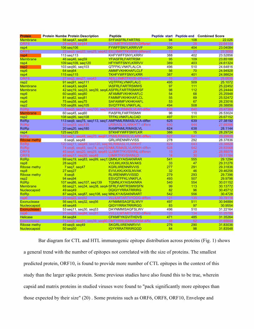
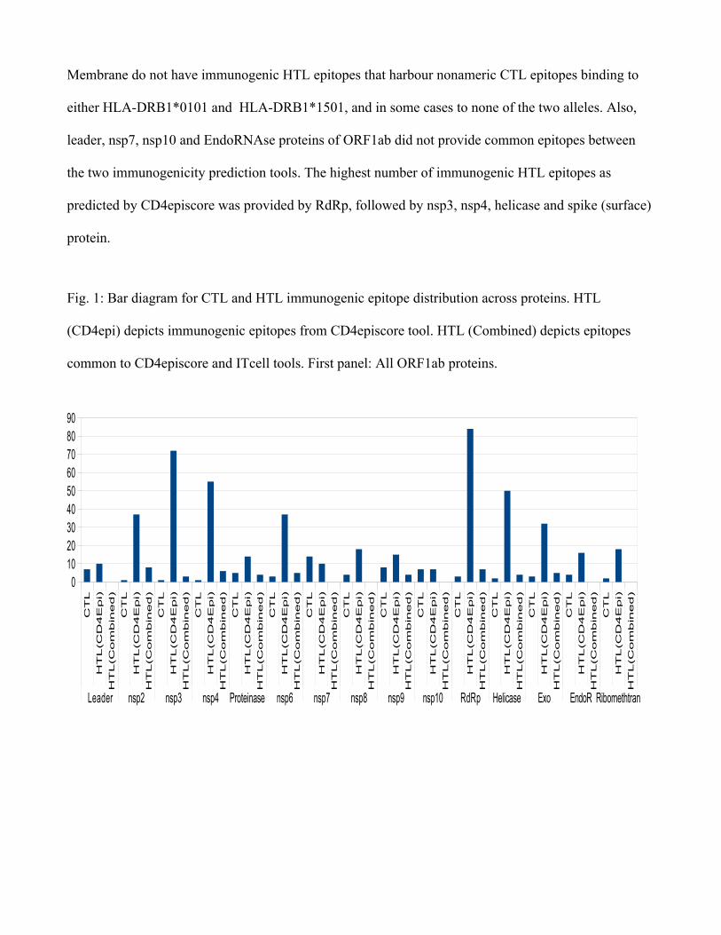
Bar diagram for CTL and HTL immunogenic epitope distribution across proteins (Fig. 1) shows
a general trend with the number of epitopes not correlated with the size of proteins. The smallest
predicted protein, ORF10, is found to provide more number of CTL epitopes in the context of this
study than the larger spike protein. Some previous studies have also found this to be true, wherein
capsid and matrix proteins in studied viruses were found to "pack significantly more epitopes than
those expected by their size" (20) . Some proteins such as ORF6, ORF8, ORF10, Envelope and
Protein Protein Number Protein Description Peptide Peptide start Peptide end Combined ScoreMembrane 58 seq47, seq58 SYFIASFRLFARTRS 94 108 22.026ORF6 28 seq28, seq44 ILLIIMRTFKVSIWN-Different from Grifoni14 28 22.64144nsp4 106 seq106 FYWFFSNYLKRRVVF 390 404 23.04084ORF6 22 seq3, seq22, seq25, seq42 EILLIIMRTFKVSIW-different from Grifoni13 27 23.3002nsp4 113 seq113 KHFYWFFSNYLKRRV 388 402 23.52856Membrane 46 seq46, seq59 YFIASFRLFARTRSM 95 109 23.85188nsp4 109 seq109, seq120 HFYWFFSNYLKRRVV 389 403 24.61324nsp2 103 seq90, seq103 QTFFKLVNKFLALCA 496 510 24.64816nsp6 64 seq64 AMMFVKHKHAFLCLF 56 70 24.94928nsp4 115 seq115 TKHFYWFFSNYLKRR 387 401 24.98624ORF6 27 seq2, seq27, seq43 AEILLIIMRTFKVSI-different from Grifoni15 29 25.0036nsp2 91 seq91, seq111 VQTFFKLVNKFLALC 495 509 25.1072Membrane 43 seq43, seq64 IASFRLFARTRSMWS 97 111 25.22452Membrane 42 seq19, seq33, seq39, seq42, seq65ASFRLFARTRSMWSF 98 112 25.24444nsp6 60 seq60, seq80 AFAMMFVKHKHAFLC 54 68 25.25948nsp6 81 seq62, seq81 FAMMFVKHKHAFLCL 55 69 25.52472nsp6 75 seq58, seq75 SAFAMMFVKHKHAFL 53 67 26.23016nsp2 105 seq89, seq105 SVQTFFKLVNKFLAL 494 508 26.38856RdRp 5 seq5, seq71, seq100, seq165MPNMLRIMASLVLAR-different from Grifoni626 640 26.47384Membrane 60 seq45, seq60 FIASFRLFARTRSMW 96 110 26.53024nsp2 108 seq95, seq108 TFFKLVNKFLALCAD 497 511 26.67152RdRp 113 seq9, seq75, seq113, seq172AMPNMLRIMASLVLA-different from Grifoni625 639 27.38192RdRp 53 seq53, seq135 LRIMASLVLARKHTT-different from Grifoni630 644 27.69396RdRp 20 seq20, seq180 RAMPNMLRIMASLVL 624 638 28.1144nsp4 125 seq125 STKHFYWFFSNYLKR 386 15 28.29724RdRp 152 seq1, seq48, seq70, seq91, seq96, seq152, seq164PNMLRIMASLVLARK-different from Grifoni627 641 28.29936Ribose methyltransferase 6 seq6, seq48 GRLIIRENNRVVISS 278 292 28.37836RdRp 120 seq11, seq49, seq120, seq140, seq174MLRIMASLVLARKHT 629 643 28.38828RdRp 74 seq6, seq45, seq74, seq102, seq142, seq167NMLRIMASLVLARKH-different from Grifoni628 642 28.83568ORF6 45 seq4, seq20, seq29, seq45 LLIIMRTFKVSIWNL-different from Grifoni15 29 28.88076ORF6 48 seq17, seq30, seq48 LIIMRTFKVSIWNLD 16 30 29.11184RdRp 99 seq19, seq83, seq99, seq131QMNLKYAISAKNRAR 541 555 29.1294nsp8 28 seq28 VVLKKLKKSLNVAKS 33 47 29.31276Ribose methyltransferase 7 seq7, seq47 KGRLIIRENNRVVIS 277 291 29.31836nsp8 27 seq27 EVVLKKLKKSLNVAK 32 46 29.46256Ribose methyltransferase 8 seq8 RLIIRENNRVVISSD 279 293 29.7396nsp2 94 seq94 ESVQTFFKLVNKFLA 493 507 29.9796RdRp 107 seq86, seq107, seq139 TQMNLKYAISAKNRA 540 554 30.01152Membrane 68 seq21, seq34, seq38, seq44, seq68SFRLFARTRSMWSFN 99 113 30.13772Nucleocapsid 49 seq49 DQIGYYRRATRRIRG 82 96 30.45712RdRp 87 seq24, seq87, seq108, seq129MNLKYAISAKNRART 542 556 30.4728nsp6 22 seq22, seq92 VLLILMTARTVYDDG-different from Grifoni121 135 30.78116Exonuclease 56 seq15, seq32, seq56 AYNMMISAGFSLWVY 497 511 30.94884Nucleocapsid 48 seq48 QIGYYRRATRRIRGG 83 97 30.9554Exonuclease 53 seq11, seq30, seq53 DAYNMMISAGFSLWV 496 510 31.22164nsp6 91 seq20, seq84, seq91 VVLLILMTARTVYDD-different from Grifoni120 134 31.25796Helicase 84 seq84 CFKMFYKGVITHDVS 471 485 31.35264Exonuclease 71 seq3, seq31, seq39, seq60, seq71DMTYRRLISMMGFKM-different from Grifoni48 62 31.65644Ribose methyltransferase 49 seq9, seq49 SKGRLIIRENNRVVI 276 290 31.83036Nucleocapsid 50 seq50 IGYYRRATRRIRGGD 84 98 31.83548
Membrane do not have immunogenic HTL epitopes that harbour nonameric CTL epitopes binding to
either HLA-DRB1*0101 and HLA-DRB1*1501, and in some cases to none of the two alleles. Also,
leader, nsp7, nsp10 and EndoRNAse proteins of ORF1ab did not provide common epitopes between
the two immunogenicity prediction tools. The highest number of immunogenic HTL epitopes as
predicted by CD4episcore was provided by RdRp, followed by nsp3, nsp4, helicase and spike (surface)
protein.
Fig. 1: Bar diagram for CTL and HTL immunogenic epitope distribution across proteins. HTL
(CD4epi) depicts immunogenic epitopes from CD4episcore tool. HTL (Combined) depicts epitopes
common to CD4episcore and ITcell tools. First panel: All ORF1ab proteins.
CT
L
HT
L(C
D4E
pi)
HT
L(C
om
bin
ed)
CT
L
HT
L(C
D4E
pi)
HT
L(C
om
bin
ed)
CT
L
HT
L(C
D4E
pi)
HT
L(C
om
bin
ed)
CT
L
HT
L(C
D4E
pi)
HT
L(C
om
bin
ed)
CT
L
HT
L(C
D4E
pi)
HT
L(C
om
bin
ed)
CT
L
HT
L(C
D4E
pi)
HT
L(C
om
bin
ed)
CT
L
HT
L(C
D4E
pi)
HT
L(C
om
bin
ed)
CT
L
HT
L(C
D4E
pi)
HT
L(C
om
bin
ed)
CT
L
HT
L(C
D4E
pi)
HT
L(C
om
bin
ed)
CT
L
HT
L(C
D4E
pi)
HT
L(C
om
bin
ed)
CT
L
HT
L(C
D4E
pi)
HT
L(C
om
bin
ed)
CT
L
HT
L(C
D4E
pi)
HT
L(C
om
bin
ed)
CT
L
HT
L(C
D4E
pi)
HT
L(C
om
bin
ed)
CT
L
HT
L(C
D4E
pi)
HT
L(C
om
bin
ed)
CT
L
HT
L(C
D4E
pi)
HT
L(C
om
bin
ed)
Leader nsp2 nsp3 nsp4 Proteinase nsp6 nsp7 nsp8 nsp9 nsp10 RdRp Helicase Exo EndoR Ribomethtran
0102030405060708090
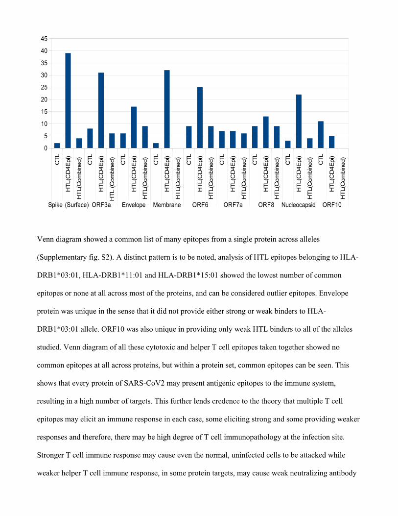
Venn diagram showed a common list of many epitopes from a single protein across alleles
(Supplementary fig. S2). A distinct pattern is to be noted, analysis of HTL epitopes belonging to HLA-
DRB1*03:01, HLA-DRB1*11:01 and HLA-DRB1*15:01 showed the lowest number of common
epitopes or none at all across most of the proteins, and can be considered outlier epitopes. Envelope
protein was unique in the sense that it did not provide either strong or weak binders to HLA-
DRB1*03:01 allele. ORF10 was also unique in providing only weak HTL binders to all of the alleles
studied. Venn diagram of all these cytotoxic and helper T cell epitopes taken together showed no
common epitopes at all across proteins, but within a protein set, common epitopes can be seen. This
shows that every protein of SARS-CoV2 may present antigenic epitopes to the immune system,
resulting in a high number of targets. This further lends credence to the theory that multiple T cell
epitopes may elicit an immune response in each case, some eliciting strong and some providing weaker
responses and therefore, there may be high degree of T cell immunopathology at the infection site.
Stronger T cell immune response may cause even the normal, uninfected cells to be attacked while
weaker helper T cell immune response, in some protein targets, may cause weak neutralizing antibody
CTL
HT
L(C
D4E
pi)
HT
L(C
om
bin
ed)
CTL
HT
L(C
D4E
pi)
HTL
(Com
bin
ed)
CTL
HT
L(C
D4E
pi)
HT
L(C
om
bin
ed)
CTL
HT
L(C
D4E
pi)
HT
L(C
om
bin
ed)
CTL
HT
L(C
D4E
pi)
HT
L(C
om
bin
ed)
CTL
HT
L(C
D4E
pi)
HT
L(C
om
bin
ed)
CTL
HT
L(C
D4E
pi)
HT
L(C
om
bin
ed)
CTL
HT
L(C
D4E
pi)
HT
L(C
om
bin
ed)
CTL
HT
L(C
D4E
pi)
HT
L(C
om
bin
ed)
Spike (Surface) ORF3a Envelope Membrane ORF6 ORF7a ORF8 Nucleocapsid ORF10
0
5
10
15
20
25
30
35
40
45
responses as well as weak CTL response at varying times during infection. Very recently, one study has
pointed to this immune dysregulation (21) in Covid19 patients with IL6-mediated low HLA-DR
expression with sustained cytokine production. Another correspondence paper also pointed to a
cytokine storm in context (22). Antibody-mediated enhancement of immune response is also not ruled
out and can be seen from the fact that all the epitopes present in the list of dominant B cell epitopes
(Table 4 in ref 23) belonging to surface, membrane and nucleocapsid protein, are unique, and there
may be a higher non-neutralizing antibody level in Covid19 patients, like in the case of dengue viruses
(23).
While this study was at writing stage, two studies on T cell epitope generation using all
proteins (24, 25) were published. This present study is different from Grifoni et al. 2020 (24) study in
that two prediction tools with very different algorithms, one using neural network and another using
postion-specific weight matrices were employed to generate a list of common epitopes, thereby
increasing prediction accuracy. Also, Grifoni et al, 2020 focussed mostly on previous SARS
coronavirus epitope similarity for predicting epitopes, while this paper identified several novel epitopes
across all ten proteins using two different prediction algorithms in each case. Further, this epitope list
comprises of common top-scoring epitopes with a higher accuracy and is restricted to highly frequent
HLA alleles across population. Also, in view of the several mutations in SARS-CoV2 genome distinct
from SARS-CoV, these epitopes not found from SARS-CoV similarity may be potentially more
immunogenic. Most of the novel HTL and CTL epitopes were distinct from the epitopes predicted by
Grifoni et al., 2020, and were found among top 100 immunogenic candidates predicted by
CD4episcore as well as those in common to ITcell predictions (Supplementary table S2). There was no
supplementary material on the website or sequence information of the epitopes in the study from
Nguyen et al., 2020 (25). Further, their work did not take into account TAP transporter binding
predictions as well as HLA-II binding studies, while this study used all three stages of MHC processing
and presentation pathway: proteasomal cleavage, TAP transporter binding and MHC class I and II-
binding as well as immunogenicity studies into account for predictions.
Clustering analysis:
All 1924 CTL and HTL top-most epitopes (122 CTL epitopes and 1802 HTL epitopes) across the
proteins studied, of which 1096 were non-redundant, unique epitopes, were then clustered using IEDB
epitope cluster analysis tool (26) to make further biologically meaningful decisions. Results analyzed
suggested that many epitopes were clustered around one consensus sequence (Supplementary table S3).
The total number of clusters (including subclusters) was 244, and 66 epitopes were singletons not
present in a cluster.
The larger clusters harbouring consensus sequences were:
VDFQVTIAEILLIIMRTFKVSIWNLDYIINLIIKN (23 members),
KLWAQCVQLHNDILLAKDTTEAFEKMVSLLSVLLSM and
TQHQPYVVDDPCPIHFYSKWYIRVGARKSAPLIEL (20 members each). These clusters across
proteins and alleles may be considered immunodominant epitopes and tested first among the ranked
list of epitopes.
Among immunogenic 122 CTL epitopes from IEDB and 666 HTL epitopes from CD4episcore,
again HLVDFQVTIAEILLIIMRTFKVSIWNLDYIINLII topped the list. Further, among the same
immunogenic 122 CTL and 95 HTL epitopes common to two prediction algorithms, CD4episcore and
ITcell, VTIAEILLIIMRTFKVSIWNLDYIINL belonging to ORF6 again topped. Moreover,
PIHFYSKWYIRVGARKSAPLIEL belonging to ORF8 and MGYINVFAFPFTIYSLL belonging to
ORF10 were also among the top 3 clustered sequences. It is of interest to note that sequences in the
consensus sequence MGYINVFAFPFTIYSLL belonging to ORF10 are weak binders to all the HLA-
DRB1 alleles studied while the same sequences are strong binders to all HLA-I supertypes studied.
Table S3: Consensus and singleton sequences generated using IEDB Clustering tool
Crossreactivity studies:
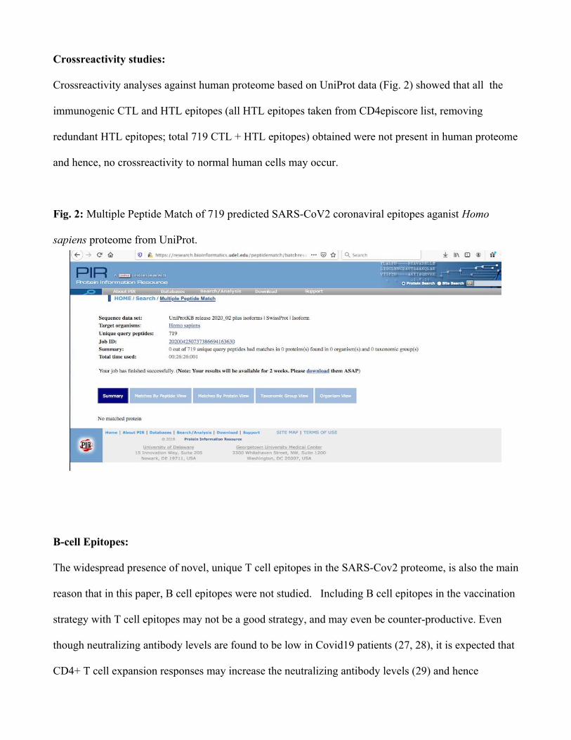
Crossreactivity analyses against human proteome based on UniProt data (Fig. 2) showed that all the
immunogenic CTL and HTL epitopes (all HTL epitopes taken from CD4episcore list, removing
redundant HTL epitopes; total 719 CTL + HTL epitopes) obtained were not present in human proteome
and hence, no crossreactivity to normal human cells may occur.
Fig. 2: Multiple Peptide Match of 719 predicted SARS-CoV2 coronaviral epitopes aganist Homo
sapiens proteome from UniProt.
B-cell Epitopes:
The widespread presence of novel, unique T cell epitopes in the SARS-Cov2 proteome, is also the main
reason that in this paper, B cell epitopes were not studied. Including B cell epitopes in the vaccination
strategy with T cell epitopes may not be a good strategy, and may even be counter-productive. Even
though neutralizing antibody levels are found to be low in Covid19 patients (27, 28), it is expected that
CD4+ T cell expansion responses may increase the neutralizing antibody levels (29) and hence
quantifying CD4+T cell responses using IFN-gamma ELISPOT assays will be useful. This is so done
in order to minimize the possible immune system backfiring (21, 22) due to the presence of too many
overlapping as well as non-overlapping epitopes in multi-subunit vaccines. It is suggested that helper T
cell epitopes be chosen so as to elicit an immune response robust enough to prime and maintain
antibody responses, as well as keep the immunopathology under check. In the proven scenario of
immune system backfiring , it may be one possible mechanism by which SARS-CoV2 may be acting at
its deadliest nature. It is indeed, a dangerous pathogen to control, although for effective
immunotherapy at a global scale, efforts should already be underway using these ranked list of
epitopes. Almost all of its proteins may pose as foreign agents to the human immune system, with each
protein contributing several unique, different immunogenic epitopes. This horde of foreign proteins
brings down an avalanche of immune system molecules to the infection site, in order to fight the virus.
But instead of immune protection, this may lead to immune enhancement or allergic inflammation at
the infection site. These analyses show that coronavirus genome has evolved to be a unique genome.
Even as this study is important in pointing out the possible mechanisms in contagious nature of SARS-
CoV2 , more evidence is required in the form of experiments.
While many of the proteins studied are found to be expressed and also their functions known by
virtue of homology with SARS-CoV, many of the novel ORFs including ORF8 and ORF10 need to be
experimentally tested for their expression and functional validation. Experimental MHC-peptide
binding and T cell assays are now required for in vitro testing for further refinement and development
as potent immunogens to be incorporated as components of subunit vaccines.
Conclusions:
Utilizing all ten SARS-CoV2 proteins, predicted or otherwise, a ranked list of CTL and HTL
epitopes with high HLA binding affinity, high TAP transport efficiency and high C-terminal
proteasomal cleavage ranking has been generated. Utilizing alleles predominant in whole world
population, two different prediction algorithms were implemented in identification of common epitopes
for consensus. Immunogenicity scores for these epitopes have also been predicted in order to further
narrow down the list to key few epitopes that can be experimentally tested. Peptide matching with
human proteome showed no indication of possible crossreactivity. These epitopes are provided to the
scientific community for further in vitro and in vivo assays and saving their time and costs involved in
our urgent bid to tackle SARS-CoV2 infections and ensuing death. This work provides esential
information for developing prophylactic and therapeutic interventions and for understanding human
immune system responses to this virus.
Materials and Methods:
Genome sequence:
The genome sequence of novel coronavirus was retrieved from GenBank accession number
MT106054.1/RefSeq sequence number NC_045512.2 and the corresponding proteins were retrieved.
RefSeq sequences of all of the proteins present in this genomic sequence, ORF10 protein
(YP_009725255.1), nucleocapsid phosphoprotein (YP_009724397.2), ORF8 protein (GenBank:
QID21074.1, no RefSeq sequence identified for ORF8), ORF7a protein (YP_009724395.1), ORF6
protein (YP_009724394.1), membrane glycoprotein (YP_009724393.1), envelope protein
(YP_009724392.1), ORF3a protein (YP_009724391.1), surface glycoprotein (YP_009724390.1),
ORF1ab (YP_009724389.1) were analysed in order to cover the entire genome of SARS-CoV2 in view
of absence of data on its virulent proteins. Within ORF1ab (full protein accession number:
YP_009724389.1), the accession number of the following proteins taken were as follows: leader
protein-YP_009725297.1, nsp2 -YP_009725298.1 , nsp3 -YP_009725299.1, nsp4- YP_009725300.1,
3C-like proteinase -YP_009725301.1, nsp6 -YP_009725302.1, nsp7 -YP_009725303.1, nsp8
-YP_009725304.1, nsp9 -YP_009725305.1, nsp10 -YP_009725306.1, RNA-dependent RNA
polymerase -YP_009725307.1, helicase -YP_009725308.1, 3'-to-5' exonuclease -YP_009725309.1,
endoRNAse -YP_009725310.1 and 2'-O-ribose methyltransferase -YP_009725311.1. Fasta sequences
of all of these proteins were taken as inputs in several T cell epitope prediction and analysis tools.
Cytotoxic T cell epitopes prediction:
NetCTLpan version 1.1 (http://www.cbs.dtu.dk/services/NetCTLpan/, 30 ) and PickPocket version 1.1
( http://www.cbs.dtu.dk/services/PickPocket/, 31) were used. All the parameters used were default
parameters. Nonameric peptide epitopes were selected. Epitopes from NetCTLpan were ranked
according to the combined score using all three different methods, and epitopes from PickPocket
algorithm were sorted by affinity (IC50 values in nM). In order to increase prediction accuracy, high
scoring epitopes common to both these algorithms (among top 10 in PickPocket and same epitopes
among high scoring ones in NetCTLpan) were fished out. 12 HLA supertypes (HLA-A*01:01, HLA-
A*02:01, HLA-A*03:01, HLA-A*24:02, HLA-A*26:01, HLA-B*07:02, HLA-B*08:01, HLA-
B*27:05, HLA-B*39:01, HLA-B*40:01, HLA-B*58:01, HLA-B*15:01) as present in both
algorithms were used (2). For ORF1ab proteins, promiscuous epitopes were selected among top 30
candidates, as not many common epitopes could be found from NetCTLpan and PickPocket.
Helper T cell epitope prediction:
NetMHCIIpan version 3.2 (http://www.cbs.dtu.dk/services/NetMHCIIpan/, 32) was used to predict
helper T cell epitopes across several HLA-DRB1 alleles, specifically, DRB1*01:01, DRB1*03:01,
DRB1*07:01, DRB1*09:01, DRB1*10:01, DRB1*11:01 and DRB1*15:01. It works on the basis of
quantitative MHC-peptide binding affinity data obtained from the Immune Epitope Database. A
consensus list of 15 amino acids long ranked epitopes was generated. For generating top ranked
epitopes, these were sorted using descending order of percent rank. Percent rank is normalized
prediction score, comparing to prediction of a set of random peptides (32). The epitopes with %rank
<2% and <10% were considered strong and weak binders, respectively.
Immunogenicity prediction:
Immunogenicity is a characteristic property of peptide epitopes that can elicit an immune response.
High binding affinity to HLA alleles is not a sufficient criterion for high immunogenicity. Therefore,
all the epitopes that were generated as a consensus were checked for their immunogenicity. Immune
Epitope database (IEDB) immunogenicity tool (http://tools.iedb.org/immunogenicity/, 33) was used to
generate a list of immunogenic CTL eptopes. Immunogenicity of a peptide-MHC complex is
predicted based on the physicochemical properties of amino acids and their positions in the predicted
peptide. Specifically, amino acids with large and aromatic side chains and positions 4-6 are more
important to the immunogenicity of the peptide being presented. Ranking was done after sorting from
higher to lower immunogenicity score (33). For helper T cell epitopes immunogenicity prediction,
CD4episcore (34) and ITcell (35) were used. CD4episcore was developed using neural networks and
combines HLA binding and immunogenicity prediction and outputs a list of immunogenic peptides
using a combined score. The authors combined immunogenicity and HLA binding scores, using the
median percentile rank score (HLA_score) of the 7-allele method (ranging from 0 to 100) and
combined it with their neural network-based immunogenicity score. This combined score is calculated
as follows:
Combined score: (alpha * Imm score) + ((1-alpha) * HLA_score), where alpha is optimized to 0.4.
The 7 alleles used are: "HLA-DRB1:03:01","HLA-DRB1:07:01","HLA-DRB1:15:01","HLA-
DRB3:01:01","HLA-DRB3:02:02","HLA-DRB4:01:01","HLA-DRB5:01:01". The whole HTL epitope
sequence list belonging to each protein was given as an input and IEDB-recommended combined
method was selected for scoring. Lower combined scores imply higher immunogenicity according to
the authors developing this prediction tool. The immunogenic vs non-immunogenic epitopes cutoff was
a combined score of 50 as per CD4episcore paper.
ITcell works on the basis of three stages of MHC-II processing and presentation pathway. These
three stages are, in the authors' (35) own words: "....antigen cleavage, MHCII presentation, and TCR
recognition. First, antigen cleavage sites are predicted based on the cleavage profiles of cathepsins S,
B, and H. Second, for each 12-mer peptide in the antigen sequence we predict whether it will bind to a
given MHCII, based on the scores of modeled peptide-MHCII complexes. Third, we predict whether or
not any of the top scoring peptide-MHCII complexes can bind to a given TCR, based on the scores of
modeled ternary peptide-MHCII-TCR complexes and the distribution of predicted cleavage sites". The
scores are given as normalized Z-scores with negative scores implying higher immunogenicity. The
epitope sequences as well as PDB files for TCR molecules corresponding to their cognate MHC alleles
were given as an input. The PDB ID for files for HLA-DRB1*01:01 and HLA-DRB1*15:01 alleles are
1FYT.pdb and 1YMM.pdb, respectively. PDB files for all other alleles were not available.
Clustering
As globally conserved epitopes are relevant at this time to contain and treat coronavirus infection,
clustering approach was used to find patterns among disparate datasets. In order to group epitopes into
several clusters, IEDB epitope cluster analysis tool (26) was applied. All the topmost CTL and HTL
epitopes across proteins targets were used as inputs with minimum sequence identity threshold as 70%.
Cluster-break algorithm was applied for clear representative sequence.
Cross-reactivity analysis:
All the immunogenic CTL and HTL epitopes obtained were used to search against human proteome
data from UniProt database (2020_02 release, 181,292,975 sequences as of date 06-05-2020) for any
matches to human proteome, thus avoiding cross-reactivity. For this, Multiple Peptide Match tool
(https://research.bioinformatics.udel.edu/peptidematch/batchpeptidematch.jsp) of Protein Information
Resource was used.
Multiple Sequence Alignment:
MUSCLE (https://www.ebi.ac.uk/Tools/msa/muscle/) was used to generate multiple sequence
alignments of all SARS-CoV2 proteins with corresponding proteins in other HCoV and MERS species.
The species chosen and their GenBank accession IDs were: Alpha-CoV: HCoV-NL63 (NC_005831.2),
HCoV-229E (NC_002645.1); Beta-CoV: HCoV-OC43 (NC_006213.1), HCoV-HKU1 (NC_006577.2),
MERS CoV (NC_019843.3) and SARS-CoV2 (SARS-CoV2, accession IDs same as above). Spike
protein sequence for SARS-CoV was taken from UniProt (P59594). In view of different/unclear
annotations, it was difficult to get corresponding protein sequences from SARS-CoV (RefSeq
accession ID NC_004718.3). There are no human CoVs in gamma/delta CoV categories. In addition,
bat coronavirus RaTG13 sequences (MN996532.1) were also used.
Acknowledgments:
This author acknowledges the tireless help of researchers working towards SARS-CoV2 control and
submitting data to GenBank without which these sequence analyses using Immunoinformatics would
not have been possible.
Conflict of interest: This author declares that there is no conflict of interest.
References:
References:
1. Smith, Micholas; Smith, Jeremy C. (2020). Repurposing Therapeutics for COVID-
19:Supercomputer-Based Docking to the SARS-CoV-2 Viral Spike Protein and Viral Spike
P r o t e i n - H u m a n A C E 2 I n t e r f a c e . C h e m R x i v . P r e p r i n t .
https://doi.org/10.26434/chemrxiv.11871402.v4
2. Seema Mishra and Subrata Sinha (2009). ‘Immunoinformatics and modeling perspective of T
cell epitope-based cancer immunotherapy: a holistic picture’ J Biomol Struct Dyn. 27(3),
pp.293-306.
3. Seema Mishra and Subrata Sinha (2006). ‘Prediction and molecular modeling of T cell epitopes
derived from placental alkaline phosphatase for use in cancer immunotherapy’. J Biomol Struct
Dyn. 24(2), pp.109-121.
4. WHO: h t tps : / /www.who. in t /b luepr in t /pr ior i ty-d iseases /key-act ion/Novel -
Coronavirus_Landscape_nCoV-4april2020.pdf?ua=1
5. Wang, Y-D Fion Sin W-Y, Xu, G-B et al., (2004) T-Cell Epitopes in Severe Acute Respiratory
Syndrome (SARS) Coronavirus Spike Protein Elicit a Speci fic T-Cell Immune Response in
Patients Who Recover from SARS. Journal of Virology, 78: 5612–5618
6. Shi J, Zhang J, Li S, Sun J, Teng Y, Wu M, et al. (2015) Epitope-Based Vaccine Target
Screening against Highly Pathogenic MERS-CoV: An In Silico Approach Applied to Emerging
Infectious Diseases. PLoS ONE 10(12): e0144475. doi:10.1371/journal.pone.0144475
7. Anthony R. Fehr and Stanley Perlman (2015). Coronaviruses: An Overview of Their
Replication and Pathogenesis, Methods Mol Biol. 1282: 1–23.
8. Wrapp D, Wang N, Corbett, KS, Goldsmith, JA et al. Cryo-EM structure of the 2019-nCoV
spike in the prefusion conformation Science 367, pp. 1260-1263 DOI:
10.1126/science.abb2507
9. Narayanan, K., Huang, C., & Makino, S. (2008). SARS coronavirus accessory proteins. Virus
Research, 133(1), 113–121. https://doi.org/10.1016/j.virusres.2007.10.009
10. Kim et al., 2020, The Architecture of SARS-CoV-2 Transcriptome Cell 181, 1–8
https://doi.org/10.1016/j.cell.2020.04.011
11. Oh HL, Chia A, Chang CX, Leong HN, Ling KL, Grotenbreg GMet al. Engineering T cells
specific for a dominant severe acute respiratory syndrome coronavirus CD8 T cell epitope. J
Virol 85: 10464–10471. (2011).
12. Vartak, A and Sucheck, S. J. Recent Advances in Subunit Vaccine Carriers. Vaccines 4, 12;
doi:10.3390/vaccines4020012 (2016).
13. Mishra, S. T Cell Epitope-Based Vaccine Design for Pandemic Novel Coronavirus 2019-nCoV.
ChemRxiv. Preprint. https://doi.org/10.26434/chemrxiv.12029523.v2 (2020).
14. Korber B, Fischer WM, Gnanakaran S, et al. (2020) Spike mutation pipeline reveals the
emergence of a more transmissible form of SARS-CoV-2. bioRxiv 2020.04.29.069054; doi:
https://doi.org/10.1101/2020.04.29.069054
15. Quinlan BD, Mou H, Zhang L, Guo Y, He W, Ojha A, et al., The SARS-CoV-2 receptor-binding domain elicits a potent neutralizing response without antibody-dependent enhancementbioRxiv 2020.04.10.036418; doi: https://doi.org/10.1101/2020.04.10.036418.
16. Wang, C., Li, W., Drabek, D. et al. A human monoclonal antibody blocking SARS-CoV-2
infection. Nat Commun 11, 2251 (2020). https://doi.org/10.1038/s41467-020-16256-y
17. Janice Oh H-L, Gan S K-E, Bertoletti A and T Y-J (2012) Understanding the T cell immune
response in SARS coronavirus infection. Emerging Microbes and Infections(2012)1,e23;
doi:10.1038/emi.2012.26.
18. Li CK, Wu H, Yan H, Ma S, Wang L, Zhang M et al. T cell responses to whole SARS
coronavirus in humans. J Immunol 2008; 181: 5490–5500.
19. Greenbaum J, Sidney J, Chung J, Brander C, Peters B, Sette A. Functional classification of
class II human leukocyte antigen (HLA) molecules reveals seven different supertypes and a
surprising degree of repertoire sharing across supertypes. Immunogenetics 2011; 63:325–35.
20. Diez-Rivero CM, Reche PA (2012) CD8 T Cell Epitope Distribution in Viruses Reveals
P a t t e r n s o f P r o t e i n B i o s y n t h e s i s . P L o S O N E 7 ( 8 ) : e 4 3 6 7 4 .
https://doi.org/10.1371/journal.pone.0043674
21. Giamarellos-Bourboulis EJ, Netea MG, Rovina N, et al. Complex Immune Dysregulation in
COVID-19 Patients with Severe Respiratory Failure [published online ahead of print, 2020 Apr
17]. Cell Host Microbe. 2020;S1931-3128(20)30236-5. doi:10.1016/j.chom.2020.04.009
22. Mehta P, McAuley, DF et al., COVID-19: consider cytokine storm syndromes and
immunosuppression. The Lancet 395: 10229, P1033-1034, (2020).
23. Dejnirattisai W, Jumnainsong A, Onsirisakul, N et al., Cross-Reacting Antibodies Enhance
Dengue Virus Infection in Humans Science 328, 745-748 (2010 )
24. Grifoni et al., A Sequence Homology and Bioinformatic Approach Can Predict Candidate
Targets for Immune Responses to SARS-CoV-2, Cell Host & Microbe (2020),
https://doi.org/10.1016/j.chom.2020.03.002
25. Nguyen, A Julianne K. David, J K, Maden SK et al., Human leukocyte antigen susceptibility
map for SARS-CoV-2. J. Virol. doi:10.1128/JVI.00510-20.
26. Sandeep Kumar Dhanda, Kerrie Vaughan, Veronique Schulten, Alba Grifoni, Daniela
Weiskopf, John Sidney, Bjoern Peters, Alessandro Sette: Development of a novel clustering
tool for linear peptide sequences . Immunology (2018) doi:https://doi.org/10.1111/imm.12984
27. Fan Wu et al. Neutralizing antibody responses to SARS-CoV-2 in a COVID-19 recovered 1
patient cohort and their implications, MedRxiv. https://doi.org/10.1101/2020.03.30.20047365
(2020).
28. Haveri, A. et al. Serological and molecular findings during SARS-CoV-2 infection: the first
case study in Finland, January to February 2020 Euro Surveill. 25(11): 2000266 (2020).
29. Nayak JL, Fitzgerald TF, Richards KA, et al. CD4+ T-cell expansion predicts neutralizing
antibody responses to monovalent, inactivated 2009 pandemic influenza A(H1N1) virus
subtype H1N1 vaccine. J Infect Dis. 2013 Jan 15;207(2):297-305. doi: 10.1093/infdis/jis684.
30. Stranzl, T., Larsen, M. V., Lundegaard, C., & Nielsen, M. . NetCTLpan: pan-specific MHC
class I pathway epitope predictions. Immunogenetics, 62(6), 357–368 (2010).
https://doi.org/10.1007/s00251-010-0441-4
31. Zhang, H., Lund, O., & Nielsen, M. The PickPocket method for predicting binding
specificities for receptors based on receptor pocket similarities: application to MHC-peptide
b i n d i n g . B i o i n f o r m a t i c s ( O x f o r d , E n g l a n d ) , 2 5 ( 1 0 ) , 1 2 9 3 – 1 2 9 9 .
https://doi.org/10.1093/bioinformatics/btp137 (2009).
32. Karosiene, E., Rasmussen, M., Blicher, T., Lund, O., Buus, S., & Nielsen, M. NetMHCIIpan-
3.0, a common pan-specific MHC class II prediction method including all three human MHC
class II isotypes, HLA-DR, HLA-DP and HLA-DQ. Immunogenetics, 65(10), 711–724.
https://doi.org/10.1007/s00251-013-0720-y (2013).
33. Calis JJA, Maybeno M, Greenbaum JA, Weiskopf D, De Silva AD, Sette A, Kesmir C, Peters
B. Properties of MHC class I presented peptides that enhance immunogenicity. PloS Comp.
Biol. 8(1):361. (2013)
34. Dhanda SK, Karosiene E, Edwards L, et al., Predicting HLA CD4 immunogenicity in human
populations. Frontiers in Immunology 2018 doi: 10.3389/fimmu.2018.01369
35. Schneidman-Duhovny D, Khuri N, Dong GQ, Winter MB, Shifrut E, Friedman N, et al. (2018)
Predicting CD4 T-cell epitopes based on antigen cleavage, MHCII presentation, and TCR
recognition. PLoS ONE 13(11): e0206654.


download fileview on ChemRxivPaperSeema.pdf (290.84 KiB)
Surface: Venn Diagram of all HLA-II binding epitopes
ORF3a
Envelope: Venn Diagram of all HLA-II binding epitopes
Membrane: Venn Diagram of all HLA-II binding epitopes
ORF6: Venn Diagram of all HLA-II binding epitopes
ORF7a: Venn Diagram of all HLA-II binding epitopes
ORF8: Venn Diagram of all HLA-II binding epitopes
Nucleocapsid: Venn Diagram of all HLA-II binding epitopes
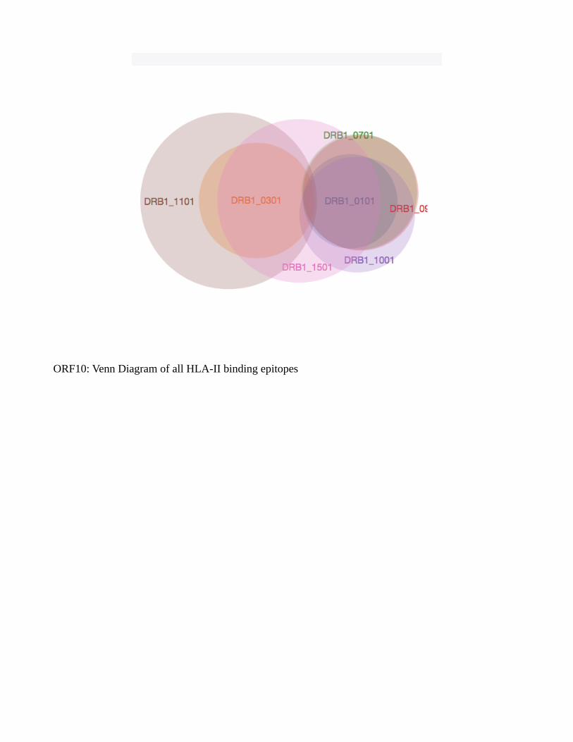
ORF10: Venn Diagram of all HLA-II binding epitopes

download fileview on ChemRxivSupplementary Figure S10.doc (340.00 KiB)
Other files
download fileview on ChemRxivmuscleEnvelope (1.10 KiB)
download fileview on ChemRxivmuscleMembrane (2.66 KiB)
download fileview on ChemRxivmuscleNS3 (2.08 KiB)
download fileview on ChemRxivmuscleNS6 (364.00 B)
download fileview on ChemRxivmuscleNucleocapsid (5.98 KiB)
download fileview on ChemRxivmuscleORF1ab (76.25 KiB)
download fileview on ChemRxivmuscleORF7a (617.00 B)
download fileview on ChemRxivmuscleORF8 (820.00 B)
download fileview on ChemRxivmuscleSpike (20.55 KiB)
download fileview on ChemRxivS4_Supplementary material_PickPocket.xls (2.97 MiB)
download fileview on ChemRxivS5_Supplementary material_NetCTLpan_results.xls (11.73 MiB)
download fileview on ChemRxivS6_Supplementary material_NetMHCIIpan_results.xls (15.68 MiB)
download fileview on ChemRxivSupplementary table S1.xls (10.50 KiB)
download fileview on ChemRxivSupplementary table S2.xls (358.00 KiB)
download fileview on ChemRxivSupplementary table S3.xls (197.00 KiB)


![HCV structure - VHPB...• specific immunity to HCV (antigen presentation on MHC class I and II molecules, HCV-specific cytotoxic and helper [type 1 and 2] T-cells, B-lymphocytes [antibody](https://static.fdocuments.in/doc/165x107/5f219090bc55d9200f3a1c0d/hcv-structure-a-specific-immunity-to-hcv-antigen-presentation-on-mhc-class.jpg)