ميحرلا نمحرلا الله مسب · T helper cells, whose main function is to secrete...
36
ن الرحيم الرحم بسم
Transcript of ميحرلا نمحرلا الله مسب · T helper cells, whose main function is to secrete...

بسم هللا الرحمن الرحيم

OVERVIEW OF THE IMMUNE SYSTEM

Immunology :is a relatively new science which has always been linked to the science of
microbiology. This is because the main function of the immune system is to combat
infections caused by different microbes.
The immune response to infection includes two parts:
I. Innate Immunity
1. This is present in all normal individuals since birth and at all times.
2. It includes many different resistance mechanisms such as chemicals and cells present
in the body.
3. It is very important at the beginning of an infection.
II. Acquired Immunity (Adaptive Immunity)
1. This is a specific immune response directed against a particular pathogen. It occurs
during the lifetime of a person as an adaptive response to infection with that
particular pathogen.
2. It is associated with the development of Immunological memory, so that in many
cases it gives life-long protection against that same pathogen. It is very effective in
combating infection, but there is a delay of a few days until it can start being
effective.
The Components of the Immune System •The granulocytes, monocytes and lymphocytes are the main cells of the immune system.
•Innate immunity depends mainly on the granulocytes (neutrophils, eosinophils and basophils) and monocytes.
• Acquired immunity depends mainly on the lymphocytes.
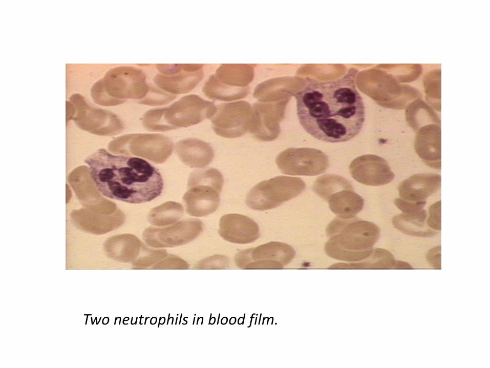
A brief description of the role of each cell in the immune response: A-Neutrophils
1. They are phagocytic cells that are capable of engulfing and digesting foreign agents.
2. They are the most numerous and most important cells of the innate immune system.
3. They are short-lived (few hours), yet they form 60-80% of total blood leucocytes. They are absent
from normal tissue, but when there is an infection at a certain place, certain chemicals
(chemotactic factors) are released to attract neutrophils from the blood to the site of infection.
4. Pus is formed mainly of dead neutrophils.
B-Eosinophils
1. They are mainly of importance in defence against helminthic parasite infections.
2. They may kill such parasites by releasing the toxic contents of their granules onto such extracellular
parasites.
3. They also play an important role in allergic reactions, which are reactions that occur in some people
against harmless antigens present in the environment.
4. Eosinophils also have phagocytic properties.
C-Basophils:
found in the blood in very low concentrations. Their function is probably similar to the of mast cells.

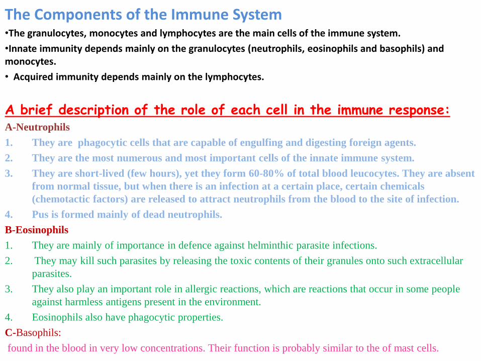
Two neutrophils in blood film.
Blood film showing a monocyte (left) and two neutrophils

Human red blood cells (red), activated platelets (purple)
and white blood cells - monocyte (green) and T
lymphocyte (orange).

D-Mast cells
1. are found in the tissues either around the blood vessels or in the submucosa.
2. They play a very important role in allergic reactions.
3. They possess granules containing a number of important mediators such as histamine.
4. Release of these mediators leads to the manifestations of allergy and inflammation.
E-Monocytes
1. Monocytes are present in the blood and continuously leave it to go to the tissues where they complete their
maturation and become macrophages.
2. Examples are the Kupffer cells of the liver and the alveolar macrophages in the lung.
F- Macrophages
1. Phagocytosis .
2. Antigen presentation: Macrophages help to 'show' or 'present' part of the foreign agents they have eaten to T
cells, so that the T cells can start responding to them. Thus, they are among a group of cells called antigen
presenting cells (APCs).
3. Secretion: They secrete chemical mediators called cytokines, e.g. interleukins.
4. Direct cytotoxicity: They may kill targets without engulfing them. Helminthic parasites which are too large to be
engulfed can be killed by macrophages releasing their toxic contents onto them.
5. Tumour cells can also be killed in a similar way.

G -Lymphocytes |They include T and B lymphocytes as well as natural killer "cells:
G1:T lymphocytes
1. They are produced in the bone marrow, but complete their maturation in the thymus.
2. They comprise around 75% of peripheral blood lymphocytes. 3.
There are two main kinds of T cells:
T lymphocytes :are produced in the bone marrow, but complete their maturation in the thymus. They comprise around 75% of peripheral blood lymphocytes
1. T helper cells, whose main function is to secrete cytokines which help other cells of the immune system.
2. T cytotoxic cells, whose main function is to kill infected cells and other abnormal cells, such as tumor cells.
The ratio of T helper / T cytotoxic cells is around 2:1.
G2:B lymphocytes
1. They are produced in the bone marrow, where they complete their maturation.
2. They comprise around 10% of peripheral blood lymphocytes.
3. When B cells become active, they change into plasma cells which secrete protein molecules called antibodies, or immunoglobulins.
4. These antibodies then circulate in the blood and have a very important role in many immune reactions.
G3:Natural Killer cells (NK cells)
1. They are large granular lymphocytes which can be distinguished from B and T lymphocytes.
2. They constitute 10-15% of peripheral blood lymphocytes.
3. They are capable of killing abnormal or infected cells in a manner similar to cytotoxic T cells, but differ from them in the way they recognize their targets.

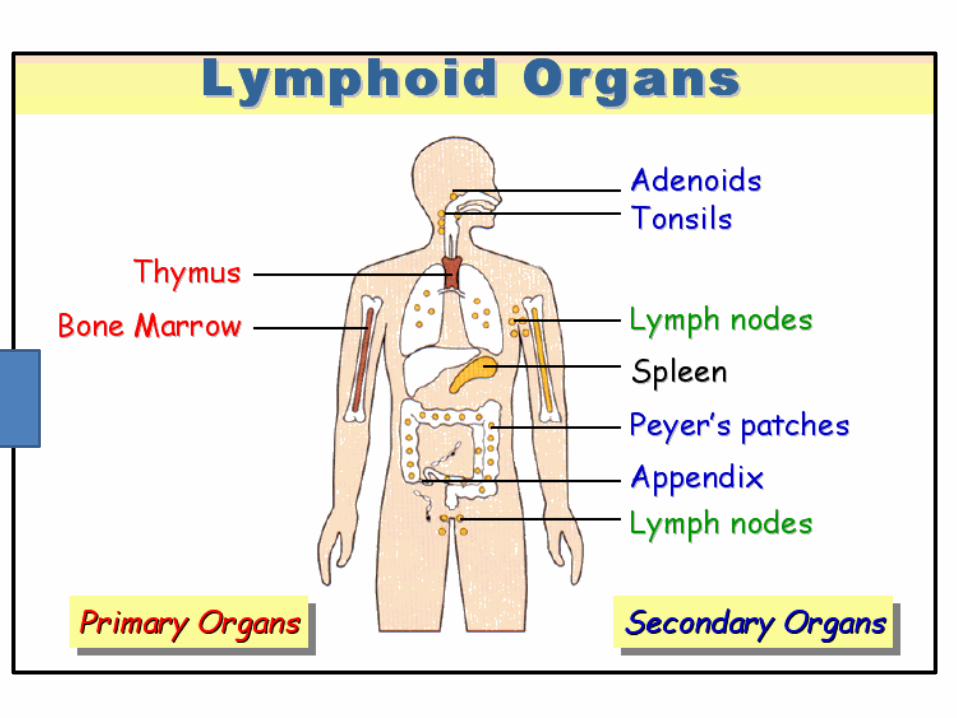
The Lymphoid Organs They are defined as organized tissues where lymphocytes interact with other non-lymphoid cells that are important either in their maturation or in starting an acquired immune response.
They are divided into:
I. Primary (central) lymphoid organs
This is where lymphocytes complete their maturation, becoming mature (adult) lymphocytes. They are:
1. The bone marrow: where the B cells complete their maturation. (Thus, the bone marrow is not only the place of origin of all blood cells, but also acts as a central lymphoid organ).
2. The thymus: where the T cells complete their maturation and differentiation.
II. Secondary (peripheral) lymphoid organs
They are the places where lymphocytes can meet antigens, leading to activation of the lymphocytes.
The secondary lymphoid organs include the spleen, lymph nodes and various mucosal associated lymphoid tissue (MALT):
1. The lymph nodes are highly organized structures. The B lymphocytes are localized in follicles in the cortex of the lymph node, while T lymphocytes are more diffusely distributed in the paracortical area.
2. The spleen is mainly composed of red pulp, which is the site of RBCs disposal. The lymphocytes surround the arterioles entering the organ, forming the white pulp. The inner part of this is called the periarteriolar lymphoid sheath, containing mainly T cells and is surrounded by a B cell crorona.
3. The gut-associated lymphoid tissue (GALT) includes the tonsils, adenoids, appendix and Peyer's patches.
4. The bronchial-associated lymphoid tissue (BAL T) includes similar but more diffusely organized collections of lymphocytes which protect the respiratory epithelium.
5. Other mucosal sites contain similar Collections.

A typical active lymphnode, e.g. in an infection

Circulation of Lymphocytes between Blood and Lymph:
1. Small B and T lymphocytes that have matured, but have not yet met antigen are called naive lymphocytes. They leave the bone marrow and thymus, respectively, and circulate continually from the blood into secondary lymphoid organs, such as the lymph nodes.
2. Microbial antigens are drained from the site of infection through the afferent lymphatic vessels into the lymph nodes.
3. Not any lymphocyte seeing an antigen will recognize it. This is because lymphocytes are very specific for the antigens they recognize. Lymphocytes which recognize a certain antigen undergo a series of changes which make them ready to start working against the antigen to which they are specific.
The changes which occur are:
a) Activation: they become Iymphoblasts.
b) Proliferation: rapid multiplication.
c) Differentiation: they change into effector cells, capable of being effective against the invading agent:
•The B cell changes into a plasma cell, capable of secreting antibody.
•The cytotoxic T cell becomes capable of killing infected cells.
•The helper T cell becomes capable of producing cytokines.

continued
3 . They now leave the lymph nodes through the efferent lymphatic vessels and return to the
blood through the thoracic duct. From the blood, they reach the peripheral tissues where
they start functioning to eliminate the specific infection which started their activation.
• Some of the antigen-specific lymphocytes produced by these events remain after the antigen has
been eliminated.
• These are called memory cells and are the basis 'of Immunological memory which ensures a more
rapid and effective response on a second meeting with the same pathogen and, therefore, gives long
lasting immunity.
• Immunological memory is the most important biological sequence of the development of acquired
immunity.

1-Naive lymphocytes enter lymph nodes from blood
3-Lymphocytes return via thoracic duct to blood and from there To tissues
Infected peripheral tissue
Lymph node
2-Antigens from sites of lnfection reach lymph nodes via lymphatics
Fig
. (1)
: C
ircu
lati
on
of
lym
ph
ocy
tes
be
twe
en
blo
od
an
d
lym
ph
.

The clonal selection theory As mentioned before , T and B lymphocytes are very specific. They recognize
antigen by receptors present on their surface.
1. Every naive Lymphocyte has a single type of receptor of a certain specificity. Only those lymphocytes which meet the antigen which their receptors recognize, will undergo activation, proliferation and differentiation.
2. The result is a clone (family) of identical daughter cells, all having identical receptors, which can bind the same antigen wherever they find it.
3. Thus, antigen specificity is maintained in the daughter cells, all having identical receptors, which can bind the same antigen cells.
4. This is called the clonal selection theory because a certain clone of cells is selected from the pool of naive lymphocytes.
5. lymphocytes having receptors specific for self molecules (a person's own molecules) are normally deleted at an early stage in lymphoid cell development and are, therefore, absent from the pool of mature lymphocytes.
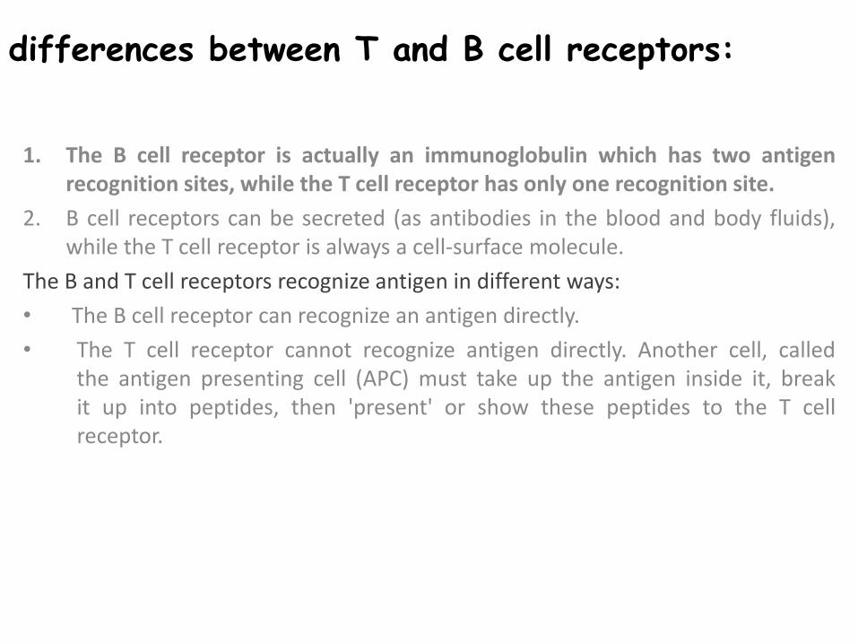
differences between T and B cell receptors:
1. The B cell receptor is actually an immunoglobulin which has two antigen recognition sites, while the T cell receptor has only one recognition site.
2. B cell receptors can be secreted (as antibodies in the blood and body fluids), while the T cell receptor is always a cell-surface molecule.
The B and T cell receptors recognize antigen in different ways:
• The B cell receptor can recognize an antigen directly.
• The T cell receptor cannot recognize antigen directly. Another cell, called the antigen presenting cell (APC) must take up the antigen inside it, break it up into peptides, then 'present' or show these peptides to the T cell receptor.

Recognition & Effector Mechanisms of Acquired Immunity
These are the mechanisms by which pathogens are detected and destroyed in a successful acquired immune response.
It is very important to know that different pathogens have different lifestyles and, therefore, need different mechanisms for detection, recognition and destruction:
1. B cells recognize antigen outside cells, where most bacteria are found .
2. T cells can detect antigens generated inside the cell, as those belonging to viruses or intracellular bacteria.
I. Effector Mechanisms of Antibodies
a) Antibodies work mainly to eliminate extracellular pathogens and their toxins. Antibodies are found in plasma and extracellular fluids.
b) Immunity mediated by antibodies is called humoral immunity. it is so- called because body fluids were once known as humors.
c) In general, when antibodies bind to pathogens, they help in combating infection in many ways.
d) They may block access of the pathogen to body cells. They may also start a process which eventually leads to lysis of the pathogen.
e) All pathogens bound by antibody are eventually delivered to phagocytes for ingestion, degradation and removal from the body.

II. Effector Mechanisms of T Cells
1) Pathogens are accessible to antibodies only in the blood and extracellular spaces.
2) However, all viruses and some bacteria and parasites replicate inside cells, where they cannot be detected by antibodies. The destruction of these invaders is the function of the T lymphocytes.
3) This is called cell-mediated immunity. In addition, T cells offer important help to B cells.

The functions of T cells may be summarized as follows:
Cytotoxic T (Tc) cells These recognize body cells infected with virus. Antigens from replicating viruses are
displayed on the surface of infected cells, where they are recognized by the cytotoxic T cells. These cells directly kill the infected cells before viral replication is complete and new viruses are released to infect other cells.
Helper T (Th) cells The function of these cells is mainly production of cytokines. There are two main kinds of helper T cells, grouped according to the type of cytokines they produce and the main cells they help:
a- T helper 1 (Th1) cells
These cells mainly secrete cytokines which help in activation of macrophages. This means making macrophages more capable of killing any bacteria inside them. This is very important because some bacteria, such as M. tuberculosis, after being ingested by macrophages, resist digestion and can survive for a long time inside.
b-T helper 2 (Th2) cells
These cells playa very important role in destruction of extracellular pathogens. They secrete certain cytokines which help in activation of B cells, so that they can become plasma cells and produce antibodies to deal with those pathogens.

Failure of Host Defence Mechanisms 1. In some cases, a certain aspect or aspects of the immune system
are deficient and the immune system is unable to free our body of infections which known as immunodeficiency.
2. In very severe immunodeficiency diseases, adaptive immunity is completely eliminated and death occurs early in life.
3. In less severe failures, there are specific recurrent infections. AIDS is a disease in which a virus infects T helper cells and destroys them. The person suffers from infections in which T helper cells normally play an important role in defence.
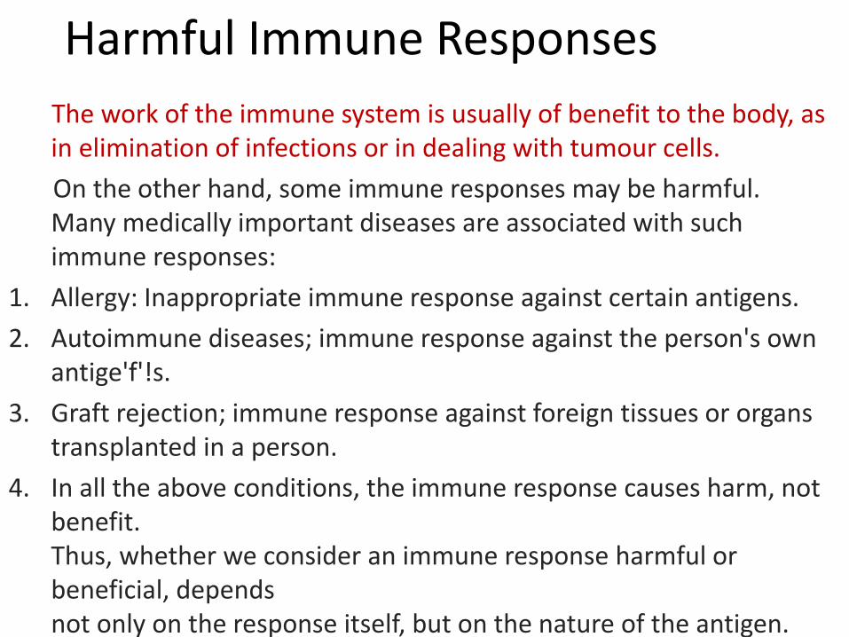
Harmful Immune Responses The work of the immune system is usually of benefit to the body, as
in elimination of infections or in dealing with tumour cells.
On the other hand, some immune responses may be harmful. Many medically important diseases are associated with such immune responses:
1. Allergy: Inappropriate immune response against certain antigens.
2. Autoimmune diseases; immune response against the person's own antige'f'!s.
3. Graft rejection; immune response against foreign tissues or organs transplanted in a person.
4. In all the above conditions, the immune response causes harm, not benefit. Thus, whether we consider an immune response harmful or beneficial, depends not only on the response itself, but on the nature of the antigen.

Immunity
• Body defense against
exogenous(microbes) and
endogenous(tumor cells) agents.


Two neutrophils in blood film.

Blood film showing a monocyte (left) and two neutrophils

Macrophage Attacking E.coli. Macrophage Attacking E.coli. Macrophage Attacking E.coli.

Eosinophil in blood film.

Monocyte with ingested malaria parasite.

Non-specific Immunity Specific Immunity
Response is antigen-independent Response is antigen-dependent
There is immediate maximal response There is a lag time between exposure and
maximal response
Not antigen-specific Antigen-specific
Exposure results in no immunologic memory Exposure results in immunologic memory
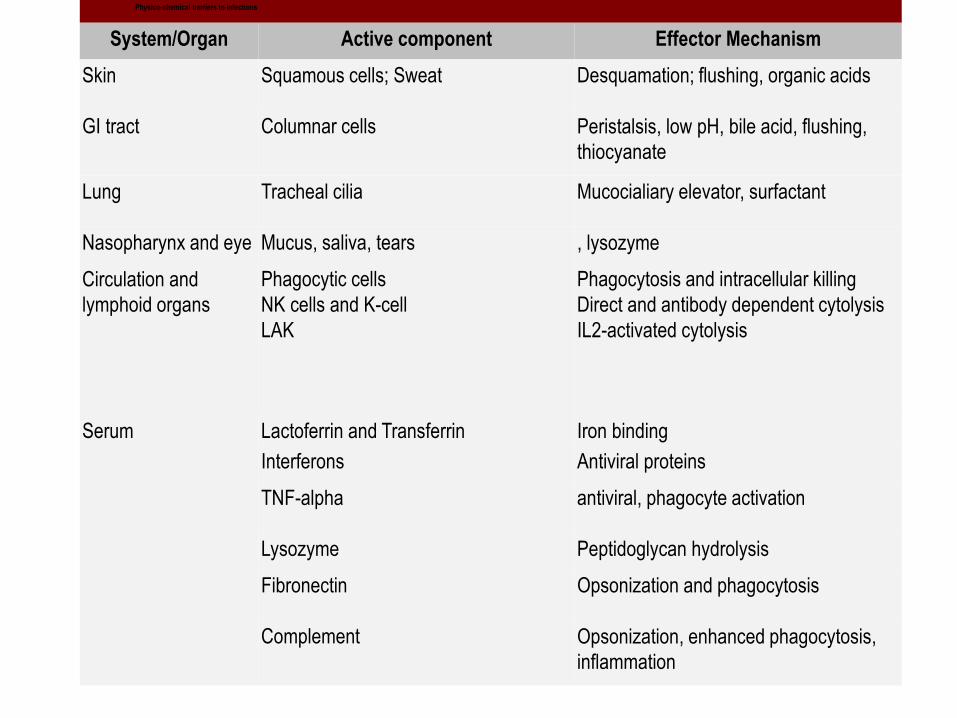
Physico-chemical barriers to infections
System/Organ Active component Effector Mechanism
Skin Squamous cells; Sweat Desquamation; flushing, organic acids
GI tract Columnar cells Peristalsis, low pH, bile acid, flushing,
thiocyanate
Lung Tracheal cilia Mucocialiary elevator, surfactant
Nasopharynx and eye Mucus, saliva, tears , lysozyme
Circulation and
lymphoid organs
Phagocytic cells
NK cells and K-cell
LAK
Phagocytosis and intracellular killing
Direct and antibody dependent cytolysis
IL2-activated cytolysis
Serum Lactoferrin and Transferrin Iron binding
Interferons Antiviral proteins
TNF-alpha antiviral, phagocyte activation
Lysozyme Peptidoglycan hydrolysis
Fibronectin Opsonization and phagocytosis
Complement Opsonization, enhanced phagocytosis,
inflammation

Characteristics of cells involved in non-specific resistance
Effector cell
Identifying marker(s) and/or function
CD3 Ig Fc CD Phagocytosis
Neutrophil
Macrophage
NK cell
K-cells
LAK cell
Eosinophil
-
-
-
-
-
-
-
-
-
-
-
-
IgG
IgG
IgG
IgG
?
IgE
CD67
CD14
CD56 & 16
?
?
CD67
+
+
-
-
?
-