Designing multifunctional quantum dots for bioimaging...
29
This journal is c The Royal Society of Chemistry 2010 Chem. Soc. Rev. Designing multifunctional quantum dots for bioimaging, detection, and drug delivery Pavel Zrazhevskiy,w a Mark Senaw b and Xiaohu Gao* a Received 23rd December 2009 DOI: 10.1039/b915139g The emerging field of bionanotechnology aims at revolutionizing biomedical research and clinical practice via introduction of nanoparticle-based tools, expanding capabilities of existing investigative, diagnostic, and therapeutic techniques as well as creating novel instruments and approaches for addressing challenges faced by medicine. Quantum dots (QDs), semiconductor nanoparticles with unique photo-physical properties, have become one of the dominant classes of imaging probes as well as universal platforms for engineering of multifunctional nanodevices. Possessing versatile surface chemistry and superior optical features, QDs have found initial use in a variety of in vitro and in vivo applications. However, careful engineering of QD probes guided by application-specific design criteria is becoming increasingly important for successful transition of this technology from proof-of-concept studies towards real-life clinical applications. This review outlines the major design principles and criteria, from general ones to application-specific, governing the engineering of novel QD probes satisfying the increasing demands and requirements of nanomedicine and discusses the future directions of QD-focused bionanotechnology research (critical review, 201 references). 1. Introduction The development of materials, structures and systems with physical dimensions of 1 to 100 nanometers (nm) has a tremendous impact on the advancement of a wide range of fields including catalysis, computing, photonics, energy, and medicine. As a result, interest in nanotechnology has increased dramatically during the last decade. The National Nanotechnology Initiative budget, for example, has expanded by approximately 6 times since 2000. 1 In contrast to widely used bulk counterparts, nanomaterials possess novel unusual and useful physicochemical properties that emerge at minute length scales. Metallic nanostructures in the presence of an electromagnetic field, for example, exhibit electron density oscillations which are highly sensitive to environmental perturbations. Iron oxide nanoparticles become super- paramagnetic, exhibiting field-inducible magnetic dipoles. Carbon nanotubes possess remarkable tensile strength and controllable electrical conductivity. Semiconductor nano- particles emit tunable and spectrally narrow fluorescence light upon excitation. These structures have been synthesized in a variety of shapes, sizes and configurations, and the theoretical a Department of Bioengineering, University of Washington, 3720 15th Avenue NE, Seattle, WA 98195, USA. E-mail: [email protected]; Tel: +1 206-543-6562 b Department of Bioengineering, University of California, Berkeley, 306 Stanley Hall #1762, Berkeley, CA 94720, USA Pavel Zrazhevskiy Pavel Zrazhevskiy received his BS degree in Bioengineering from the University of Washington in 2006. In 2007 he became a graduate student in Dr Xiaohu Gao’s laboratory at the Department of Bio- engineering, University of Washington. His research interests span fields of nano- technology, molecular engi- neering, and medicine with focus on design and engineering of quantum dot based tools for biomedical applications. He received the NSF’s Graduate Research Fellowship in 2009. Mark Sena Mark Sena obtained a BS degree in Bioengineering at the University of Washington while working in the labora- tory of Dr Xiaohu Gao with a focus on the biosensing applications of quantum dots for small-scale diagnostic devices. He recently joined the Joint Graduate Group in Bioengineering between the University of California, Berkeley and San Francisco. His current research interests include virus-based bionano- materials for energy and sensing applications and microfluidic protein diagnostics for point-of-care testing. He received the NSF’s Graduate Research Fellowship in 2010. w These authors contributed equally to this work. CRITICAL REVIEW www.rsc.org/csr | Chemical Society Reviews Downloaded by UNIVERSITY OF WASHINGTON on 22 August 2010 Published on 09 August 2010 on http://pubs.rsc.org | doi:10.1039/B915139G View Online
Transcript of Designing multifunctional quantum dots for bioimaging...

This journal is c The Royal Society of Chemistry 2010 Chem. Soc. Rev.
Designing multifunctional quantum dots for bioimaging, detection,
and drug delivery
Pavel Zrazhevskiy,wa Mark Senawb and Xiaohu Gao*a
Received 23rd December 2009
DOI: 10.1039/b915139g
The emerging field of bionanotechnology aims at revolutionizing biomedical research and
clinical practice via introduction of nanoparticle-based tools, expanding capabilities of existing
investigative, diagnostic, and therapeutic techniques as well as creating novel instruments and
approaches for addressing challenges faced by medicine. Quantum dots (QDs), semiconductor
nanoparticles with unique photo-physical properties, have become one of the dominant classes of
imaging probes as well as universal platforms for engineering of multifunctional nanodevices.
Possessing versatile surface chemistry and superior optical features, QDs have found initial use in
a variety of in vitro and in vivo applications. However, careful engineering of QD probes guided
by application-specific design criteria is becoming increasingly important for successful transition
of this technology from proof-of-concept studies towards real-life clinical applications. This
review outlines the major design principles and criteria, from general ones to application-specific,
governing the engineering of novel QD probes satisfying the increasing demands and
requirements of nanomedicine and discusses the future directions of QD-focused
bionanotechnology research (critical review, 201 references).
1. Introduction
The development of materials, structures and systems with
physical dimensions of 1 to 100 nanometers (nm) has a
tremendous impact on the advancement of a wide range of
fields including catalysis, computing, photonics, energy,
and medicine. As a result, interest in nanotechnology has
increased dramatically during the last decade. The National
Nanotechnology Initiative budget, for example, has expanded
by approximately 6 times since 2000.1 In contrast to widely
used bulk counterparts, nanomaterials possess novel unusual
and useful physicochemical properties that emerge at minute
length scales. Metallic nanostructures in the presence of an
electromagnetic field, for example, exhibit electron density
oscillations which are highly sensitive to environmental
perturbations. Iron oxide nanoparticles become super-
paramagnetic, exhibiting field-inducible magnetic dipoles.
Carbon nanotubes possess remarkable tensile strength and
controllable electrical conductivity. Semiconductor nano-
particles emit tunable and spectrally narrow fluorescence light
upon excitation. These structures have been synthesized in a
variety of shapes, sizes and configurations, and the theoretical
aDepartment of Bioengineering, University of Washington,3720 15th Avenue NE, Seattle, WA 98195, USA.E-mail: [email protected]; Tel: +1 206-543-6562
bDepartment of Bioengineering, University of California, Berkeley,306 Stanley Hall #1762, Berkeley, CA 94720, USA
Pavel Zrazhevskiy
Pavel Zrazhevskiy received hisBS degree in Bioengineeringfrom the University ofWashington in 2006. In 2007he became a graduate studentin Dr Xiaohu Gao’s laboratoryat the Department of Bio-engineering, University ofWashington. His researchinterests span fields of nano-technology, molecular engi-neering, and medicine withfocus on design and engineeringof quantum dot based tools forbiomedical applications. Hereceived the NSF’s GraduateResearch Fellowship in 2009.
Mark Sena
Mark Sena obtained a BSdegree in Bioengineering atthe University of Washingtonwhile working in the labora-tory of Dr Xiaohu Gao with afocus on the biosensingapplications of quantum dotsfor small-scale diagnosticdevices. He recently joinedthe Joint Graduate Group inBioengineering between theUniversity of California,Berkeley and San Francisco.His current research interestsinclude virus-based bionano-materials for energy and
sensing applications and microfluidic protein diagnostics forpoint-of-care testing. He received the NSF’s Graduate ResearchFellowship in 2010.
w These authors contributed equally to this work.
CRITICAL REVIEW www.rsc.org/csr | Chemical Society Reviews
Dow
nloa
ded
by U
NIV
ER
SIT
Y O
F W
ASH
ING
TO
N o
n 22
Aug
ust 2
010
Publ
ishe
d on
09
Aug
ust 2
010
on h
ttp://
pubs
.rsc
.org
| do
i:10.
1039
/B91
5139
GView Online

Chem. Soc. Rev. This journal is c The Royal Society of Chemistry 2010
framework explaining the unique optical, chemical and
electronic properties of nanomaterials has been built. Mean-
while, nanomaterials have been incorporated in a variety of
useful products ranging from stain-repellent fabrics and
nanoparticle-containing sunscreens to lipid-encapsulated
anticancer drugs and sensitive bioanalytical tools. With
the number of nanotechnology-based patents growing
exponentially,2 such items are rapidly appearing on the
market. As new applications are developed, especially in such
critical fields as energy generation and medicine, the impact of
nanotechnology on the economy and on society will become
increasingly more profound.
One of the most promising applications of nanotechnology
has been in the area of biomedical research. Nanoscale sensors
find their use in sensitive molecular diagnostics and high
throughput bioanalytics, while nanoparticle-based drug
carriers enable spatial and temporal control of drug delivery
and release. Of great interest are organic and inorganic
nanostructures that incorporate radiolabels and contrast
agents for in vivo imaging techniques, such as Positron
Emission Tomography (PET), Computed Tomography (CT),
Single Photon Emission Computed Tomography (SPECT),
Magnetic Resonance Imaging (MRI), sonography, and optical
imaging. In combination with these macroscale modalities,
nanoscale probes are important tools for molecular imaging—
visualization, characterization, and quantification of bio-
logical processes at the molecular level within living systems.3,4
Fluorescent semiconductor nanoparticles, commonly referred
to as quantum dots (QDs), represent a particularly interesting
class of probes well-suited for advanced fluorescence imaging
applications, such as multiplexed quantitative analysis of
cellular phenotypes, real-time monitoring of intracellular
processes, and in vivo molecular imaging.5–12 Exhibiting many
supreme characteristics compared to conventional fluoro-
phores, including size-tunable and spectrally narrow light
emission along with efficient light absorption throughout
a wide spectrum, improved brightness with outstanding
resistance to photobleaching and degradation, and extremely
large Stokes shift, QDs greatly expand the capabilities of
fluorescence imaging. Furthermore, QDs provide a suitable
platform for engineering of multifunctional nanodevices with
capabilities of exploiting multiple imaging modalities or
merging imaging and therapeutic functionalities within a
single nanoparticle.
Utilization of unique photo-physical and chemical
properties rendered by QDs for addressing challenging issues
raised by biomedical research has promoted development of
novel imaging probes, traceable drug delivery vehicles, and
multifunctional nanocomposites. Active exploration of
QD-based biomedical applications has resulted in more than
300% increase in related peer-reviewed publications since 2002
(based on PubMed and Nature.com searches). This review
provides a synopsis of the key achievements in nanoscience
that have initiated the work on utilizing QDs for biomedical
applications and discusses recent developments that have
converted QDs into clinically relevant tools. The brief over-
view of the photophysical properties and surface engineering
strategies describes design principles guiding development of
QDs into imaging probes and drug delivery vehicles. In-depth
discussion of cell and tissue molecular profiling along with
live-cell and in vivo molecular imaging presents the current
state of the QD-based diagnostic and therapeutic applications
and outlines potential future directions within these areas of
research. Finally, review of the QD-based nanocomposites
provides an introduction to an exciting emerging field of
multimodal imaging and nano-therapeutics.
2. General principles for engineering of QD probes
QDs are semiconductor nanoparticles often made from
hundreds to thousands of atoms of group II and VI elements
(e.g. CdSe and CdTe) or group III and V elements (e.g. InP
and InAs). Bulk semiconductors are materials with a relatively
small band gap (less than 4 eV) between the valence and
conduction bands, thus behaving like insulators at ambient
conditions and exhibiting electrical conductivity only under
external stimulation. Electrons in the ground state that are
typically localized to individual atoms (i.e. comprising valence
band) can be promoted to higher energy levels where electrons
are free to move throughout the material (i.e. populate the
conduction band) by supplying an amount of energy that
exceeds the band gap. In certain cases, relaxation of an
electron results in the release of bandgap energy in the form
of light (fluorescence). QDs are crystalline particles that range
from 2 to 10 nanometers in diameter. Physical size smaller
than the exciton Bohr radius results in a 3-dimensional
quantum confinement of charge carriers within the QD and
limits the number of possible energy states that an electron can
occupy (Fig. 1), thus giving nanoparticles novel properties
not achievable in bulk materials.13–15 Additionally, the
relatively small size comparable to that of large biomolecules
(e.g. antibodies) aids in engineering of biologically functional
materials.
The inorganic nanoparticle core provides a rigid foundation
for the development of QD probes. Manipulation of the
core chemical composition, size, and structure controls
the photo-physical properties of the probe. However, bare
Xiaohu Gao
Prof. Xiaohu Gao received hisBS degree in chemistry fromNankai University, China in1998, his PhD degree inchemistry from IndianaUniversity, Bloomington in2004, and his postdoctoraltraining from the Departmentof Biomedical Engineering atGeorgia Tech and EmoryUniversity. In 2005, hebecame a faculty member inthe Department of Bio-engineering and the Centerfor Nanotechnology at theUniversity of Washington,
Seattle. His research is focused on biomedical nanotechnology,molecular engineering, molecular imaging, and therapeutics. Hehas authored or co-authored over 50 papers and book chapters;and he is also a recipient of the NSF CAREER Award.
Dow
nloa
ded
by U
NIV
ER
SIT
Y O
F W
ASH
ING
TO
N o
n 22
Aug
ust 2
010
Publ
ishe
d on
09
Aug
ust 2
010
on h
ttp://
pubs
.rsc
.org
| do
i:10.
1039
/B91
5139
GView Online
This journal is c The Royal Society of Chemistry 2010 Chem. Soc. Rev.
nanoparticles usually cannot interact with biological systems
and do not possess any biological functionality. Careful design
of coating materials that can encapsulate the QD core and
shield it from the environment yields biocompatible probes
with controllable physicochemical properties. Further
decoration of the QDs with biomolecules imparts the bio-
functionality and enables probe interaction with biological
systems. Therefore, preparation of QD-based probes and
nanodevices represents a multi-step process. Each step is
guided by individual design principles aimed at controlling
optical, physical and chemical properties of the final probe
(Fig. 2).
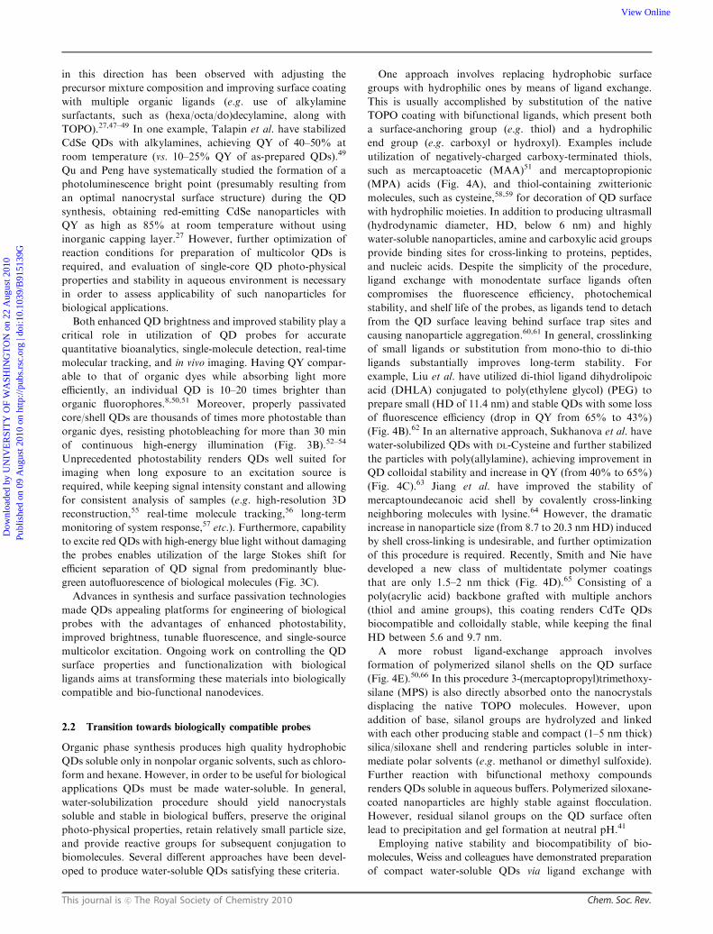
2.1 Design of the quantum dot core
The QD core defines optical properties of the probe and
represents a structural scaffold for engineering of nanodevices.
In general, the QD core should be compact and highly stable
with precisely controlled nanoparticle size distribution,
geometry, chemical composition, and surface chemistry.
Initial reports on preparation of semiconductor nanoparticles
utilized QD synthesis in aqueous solutions and yielded
particles with poor fluorescence efficiencies and large size
variation. Advancements in synthetic procedures and surface
chemistry have enabled production of water-soluble QDs with
higher quantum yield (QY, up to 40–50%) and relatively
narrow size distribution (exhibiting spectral emission width
of B50 nm for CdTe/CdSe particles16 and down to 19 nm for
ZnSe QDs17). However, aqueous synthesis still suffers from
poor control over the QD photo-physical and chemical
properties. A major leap towards synthesis of highly uniform
colloidal CdSe QDs was made in 1993 by Bawendi and
coworkers by developing a high-temperature organometallic
procedure,18 which is now widely used for synthesis of QDs for
a variety of applications. In this procedure pyrolysis of
organometallic precursors at high temperature yields nucleation
and growth of nanocrystals, while coordination of trioctyl
phosphine/trioctyl phosphine oxide (TOP/TOPO) base with
unsaturated metal atoms on the QD surface prevents the
formation of bulk semiconductor. Yet, utilization of a highly
toxic and unstable Cd precursor (dimethyl cadmium) imposes
restrictions on the equipment and reaction conditions and
limits flexibility in the QD core design. A leap towards large-
scale preparation of high-quality QDs has been made by
Peng et al. using alternative cheap precursor materials
(such as CdO).19,20 Relatively mild and simple reaction
conditions along with slower nucleation and growth rates offer
extensive flexibility in engineering of QD chemical composi-
tion, geometry, and photo-physical properties. Precise kinetic
control over a nanoparticle growth achieved with organo-
metallic procedure enables preparation of QD populations
with narrow size distribution. Therefore, as the difference in
energy between the discrete ground and excited states
increases with increasing degree of confinement (i.e. decreasing
particle size), the size of the band gap and, consequently, the
color of emitted light can be fine-tuned by adjusting the QD
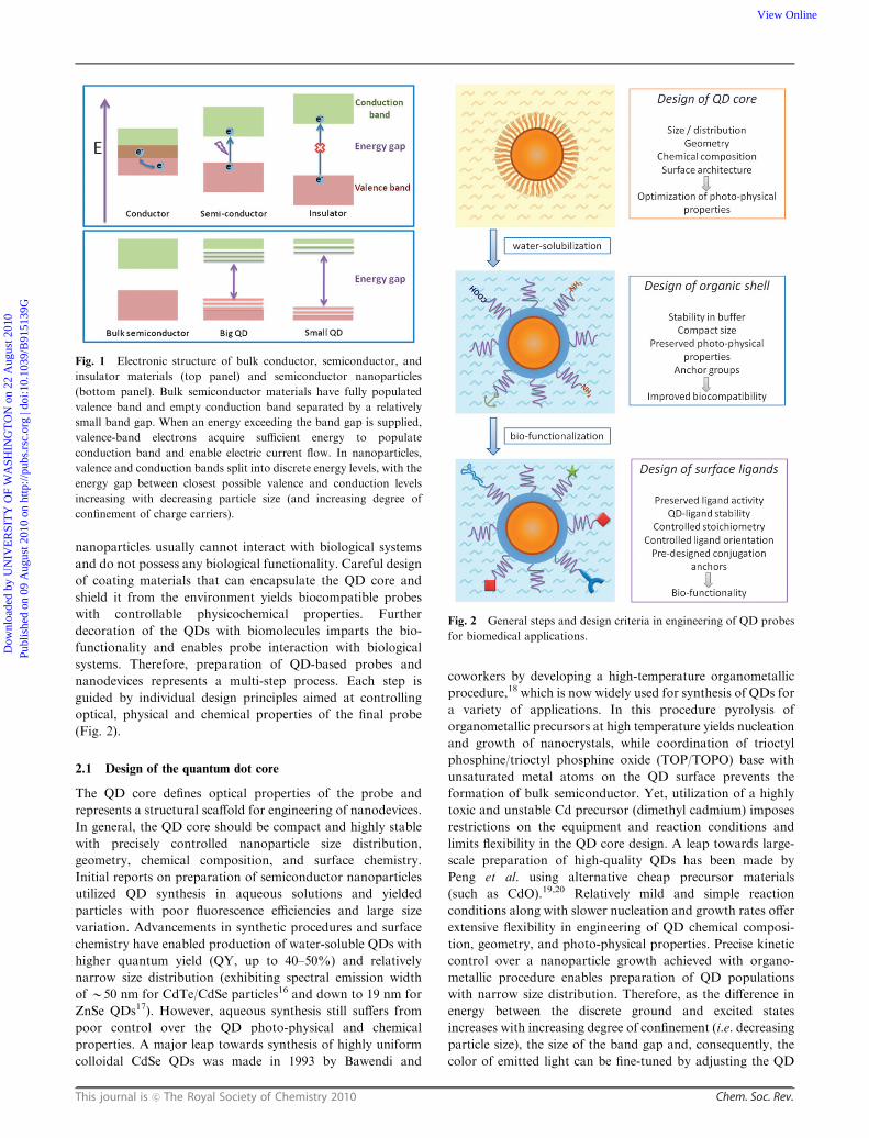
Fig. 1 Electronic structure of bulk conductor, semiconductor, and
insulator materials (top panel) and semiconductor nanoparticles
(bottom panel). Bulk semiconductor materials have fully populated
valence band and empty conduction band separated by a relatively
small band gap. When an energy exceeding the band gap is supplied,
valence-band electrons acquire sufficient energy to populate
conduction band and enable electric current flow. In nanoparticles,
valence and conduction bands split into discrete energy levels, with the
energy gap between closest possible valence and conduction levels
increasing with decreasing particle size (and increasing degree of
confinement of charge carriers).
Fig. 2 General steps and design criteria in engineering of QD probes
for biomedical applications.
Dow
nloa
ded
by U
NIV
ER
SIT
Y O
F W
ASH
ING
TO
N o
n 22
Aug
ust 2
010
Publ
ishe
d on
09
Aug
ust 2
010
on h
ttp://
pubs
.rsc
.org
| do
i:10.
1039
/B91
5139
GView Online

Chem. Soc. Rev. This journal is c The Royal Society of Chemistry 2010
size (Fig. 3A).21 With optimization of reaction conditions and
utilization of size focusing via re-injection of precursors, an
emission spectral width below 20 nm has been achieved.22–24
Further band gap engineering by varying the chemical
composition of nanocrystals has produced QDs emitting light
from the UV, throughout the visible, and into the infrared
spectra (400–4000 nm).21,24–30
Narrow size-tunable light emission has proven to be highly
beneficial for multiplexed molecular labeling (e.g. for pheno-
typing cell populations31 or detection of molecular signatures
of cancer),32 as little or no cross-talk between adjacent colors
enables simultaneous detection and quantification of multiple
fluorescence signals. Furthermore, high electron density of
QDs and direct correlation between the particle size/composition
and emission wavelength facilitate detailed evaluation of low-
resolution fluorescence images with high-resolution imaging
modalities—multiplexed imaging based on particle size can be
achieved with transmission electron microscopy (TEM),33
while that based on particle chemical composition—with
electron spectroscopic imaging (ESI).34 The multiplexing
capability of QDs is complemented by efficient light absorp-
tion over a broad spectral range (hundreds of nanometers),
as essentially any photon in UV-visible range with energy
exceeding the band gap can be absorbed without damaging the
nanoparticle. Unlike organic fluorophores, the molar extinction
coefficient of QDs gradually increases toward shorter wave-
length, allowing multicolor QDs to be simultaneously excited
by a single high-energy light source (e.g. UV lamp), thus
eliminating the need for multiple excitation sources, reducing
the cost of imaging instrumentation, and simplifying data
analysis.
While providing good control over the particle size, the
original organometallic procedure produces QDs with low
QY, compromising the utility of such particles as fluorescent
probes. Moreover, TOPO-coated QDs are unstable with
respect to photooxidation, resulting in effective degradation
of nanocrystals and potential QD toxicity due to release of free
Cd ions.13 Both issues arise from the relatively large number of
atoms exposed on the surface of nanoparticles. In the nano-
scale regime, surface atoms play a major role in determining
the catalytic, electronic, and optical properties. As the radius
of a spherical particle decreases, the ratio of its surface area to
volume rapidly increases placing larger number of atoms on
the surface.35 Surface atoms lack neighbors with which to
form chemical bonds and thus possess unoccupied electron
orbitals. Commonly referred to as dangling bonds or surface
trap sites, these orbitals can trap charge carriers and either
prevent or delay electron-hole recombination and subsequent
photon emission, thus reducing the fluorescence QY.36,37
Furthermore, such sites might exhibit enhanced chemical
reactivity and compromise chemical stability of the nano-
particles. In order to prevent some of these undesirable
characteristics, dangling bonds can be saturated by organic
and inorganic capping layers.
Several groups have developed high-bandgap-energy
inorganic shells (e.g. CdS and ZnS) several atomic layers thick
that effectively passivate the photoactive core of QDs.38–40 The
wider band gap of the shell efficiently confines the exciton to
the core, reducing nonradiative relaxation pathways and
increasing QY.41 Careful choice of core and shell materials
as well as optimization of the shell thickness are necessary to
minimize the lattice strain between the core and shell and
maximize the QD photo-physical properties. Although thin
shells (1–2 monolayers) often produce the highest fluorescence
yields, thicker shells (4–6 monolayers) provide more core
protection from photooxidation and degradation.42 For
example, Peng et al. have observed confinement of the
hole created during excitation within the CdSe core by a
higher-band gap CdS shell.40 As a result of such confinement,
hole-dependent photo-oxidative processes that cause QD
degradation and result in the loss of fluorescence are impeded.
Also, a thicker shell might significantly reduce QD blinking
(intermittence in light emission) associated with charge
trapping and un-trapping at surface defects of a nanomaterial
or due to charge ejection from the QD (Auger ionization)
followed by recombination process.43–46 Since blinking might
cause signal fluctuations in ultrasensitive detection, loss of
distance information when movement of a single molecule is
observed, and spectral jumping (change in the emission peak
position), its elimination is often desirable.
Alternative approaches aim at achieving better fluorescence
efficiency by optimizing the surface structure of nanocrystals
and minimizing the number of surface trap sites. Some success
Fig. 3 Unique photo-physical properties of QD probes. (A) Narrow
size-tunable light emission profile enables precise control over the
probe color via varying the nanoparticle size. (B) Outstanding photo-
stability of QDs enables real-time monitoring of probe dynamics and
accurate quantitative analysis, whereas quick photobleaching of
organic dyes limits such applications. (C) Capability of absorbing
high-energy (UV-blue) light without damaging the probe and emitting
fluorescence with a large Stokes shift enables efficient separation of the
QD signal over the fluorescent background. Reprinted from ref. 54,
Copyright (2005), with permission from Elsevier.
Dow
nloa
ded
by U
NIV
ER
SIT
Y O
F W
ASH
ING
TO
N o
n 22
Aug
ust 2
010
Publ
ishe
d on
09
Aug
ust 2
010
on h
ttp://
pubs
.rsc
.org
| do
i:10.
1039
/B91
5139
GView Online
This journal is c The Royal Society of Chemistry 2010 Chem. Soc. Rev.
in this direction has been observed with adjusting the
precursor mixture composition and improving surface coating
with multiple organic ligands (e.g. use of alkylamine
surfactants, such as (hexa/octa/do)decylamine, along with
TOPO).27,47–49 In one example, Talapin et al. have stabilized
CdSe QDs with alkylamines, achieving QY of 40–50% at
room temperature (vs. 10–25% QY of as-prepared QDs).49
Qu and Peng have systematically studied the formation of a
photoluminescence bright point (presumably resulting from
an optimal nanocrystal surface structure) during the QD
synthesis, obtaining red-emitting CdSe nanoparticles with
QY as high as 85% at room temperature without using
inorganic capping layer.27 However, further optimization of
reaction conditions for preparation of multicolor QDs is
required, and evaluation of single-core QD photo-physical
properties and stability in aqueous environment is necessary
in order to assess applicability of such nanoparticles for
biological applications.
Both enhanced QD brightness and improved stability play a
critical role in utilization of QD probes for accurate
quantitative bioanalytics, single-molecule detection, real-time
molecular tracking, and in vivo imaging. Having QY compar-
able to that of organic dyes while absorbing light more
efficiently, an individual QD is 10–20 times brighter than
organic fluorophores.8,50,51 Moreover, properly passivated
core/shell QDs are thousands of times more photostable than
organic dyes, resisting photobleaching for more than 30 min
of continuous high-energy illumination (Fig. 3B).52–54
Unprecedented photostability renders QDs well suited for
imaging when long exposure to an excitation source is
required, while keeping signal intensity constant and allowing
for consistent analysis of samples (e.g. high-resolution 3D
reconstruction,55 real-time molecule tracking,56 long-term
monitoring of system response,57 etc.). Furthermore, capability
to excite red QDs with high-energy blue light without damaging
the probes enables utilization of the large Stokes shift for
efficient separation of QD signal from predominantly blue-
green autofluorescence of biological molecules (Fig. 3C).
Advances in synthesis and surface passivation technologies
made QDs appealing platforms for engineering of biological
probes with the advantages of enhanced photostability,
improved brightness, tunable fluorescence, and single-source
multicolor excitation. Ongoing work on controlling the QD
surface properties and functionalization with biological
ligands aims at transforming these materials into biologically
compatible and bio-functional nanodevices.
2.2 Transition towards biologically compatible probes
Organic phase synthesis produces high quality hydrophobic
QDs soluble only in nonpolar organic solvents, such as chloro-
form and hexane. However, in order to be useful for biological
applications QDs must be made water-soluble. In general,
water-solubilization procedure should yield nanocrystals
soluble and stable in biological buffers, preserve the original
photo-physical properties, retain relatively small particle size,
and provide reactive groups for subsequent conjugation to
biomolecules. Several different approaches have been devel-
oped to produce water-soluble QDs satisfying these criteria.
One approach involves replacing hydrophobic surface
groups with hydrophilic ones by means of ligand exchange.
This is usually accomplished by substitution of the native
TOPO coating with bifunctional ligands, which present both
a surface-anchoring group (e.g. thiol) and a hydrophilic
end group (e.g. carboxyl or hydroxyl). Examples include
utilization of negatively-charged carboxy-terminated thiols,
such as mercaptoacetic (MAA)51 and mercaptopropionic
(MPA) acids (Fig. 4A), and thiol-containing zwitterionic
molecules, such as cysteine,58,59 for decoration of QD surface
with hydrophilic moieties. In addition to producing ultrasmall
(hydrodynamic diameter, HD, below 6 nm) and highly
water-soluble nanoparticles, amine and carboxylic acid groups
provide binding sites for cross-linking to proteins, peptides,
and nucleic acids. Despite the simplicity of the procedure,
ligand exchange with monodentate surface ligands often
compromises the fluorescence efficiency, photochemical
stability, and shelf life of the probes, as ligands tend to detach
from the QD surface leaving behind surface trap sites and
causing nanoparticle aggregation.60,61 In general, crosslinking
of small ligands or substitution from mono-thio to di-thio
ligands substantially improves long-term stability. For
example, Liu et al. have utilized di-thiol ligand dihydrolipoic
acid (DHLA) conjugated to poly(ethylene glycol) (PEG) to
prepare small (HD of 11.4 nm) and stable QDs with some loss
of fluorescence efficiency (drop in QY from 65% to 43%)
(Fig. 4B).62 In an alternative approach, Sukhanova et al. have
water-solubilized QDs with DL-Cysteine and further stabilized
the particles with poly(allylamine), achieving improvement in
QD colloidal stability and increase in QY (from 40% to 65%)
(Fig. 4C).63 Jiang et al. have improved the stability of
mercaptoundecanoic acid shell by covalently cross-linking
neighboring molecules with lysine.64 However, the dramatic
increase in nanoparticle size (from 8.7 to 20.3 nm HD) induced
by shell cross-linking is undesirable, and further optimization
of this procedure is required. Recently, Smith and Nie have
developed a new class of multidentate polymer coatings
that are only 1.5–2 nm thick (Fig. 4D).65 Consisting of a
poly(acrylic acid) backbone grafted with multiple anchors
(thiol and amine groups), this coating renders CdTe QDs
biocompatible and colloidally stable, while keeping the final
HD between 5.6 and 9.7 nm.
A more robust ligand-exchange approach involves
formation of polymerized silanol shells on the QD surface
(Fig. 4E).50,66 In this procedure 3-(mercaptopropyl)trimethoxy-
silane (MPS) is also directly absorbed onto the nanocrystals
displacing the native TOPO molecules. However, upon
addition of base, silanol groups are hydrolyzed and linked
with each other producing stable and compact (1–5 nm thick)
silica/siloxane shell and rendering particles soluble in inter-
mediate polar solvents (e.g. methanol or dimethyl sulfoxide).
Further reaction with bifunctional methoxy compounds
renders QDs soluble in aqueous buffers. Polymerized siloxane-
coated nanoparticles are highly stable against flocculation.
However, residual silanol groups on the QD surface often
lead to precipitation and gel formation at neutral pH.41
Employing native stability and biocompatibility of bio-
molecules, Weiss and colleagues have demonstrated preparation
of compact water-soluble QDs via ligand exchange with
Dow
nloa
ded
by U
NIV
ER
SIT
Y O
F W
ASH
ING
TO
N o
n 22
Aug
ust 2
010
Publ
ishe
d on
09
Aug
ust 2
010
on h
ttp://
pubs
.rsc
.org
| do
i:10.
1039
/B91
5139
GView Online
Chem. Soc. Rev. This journal is c The Royal Society of Chemistry 2010
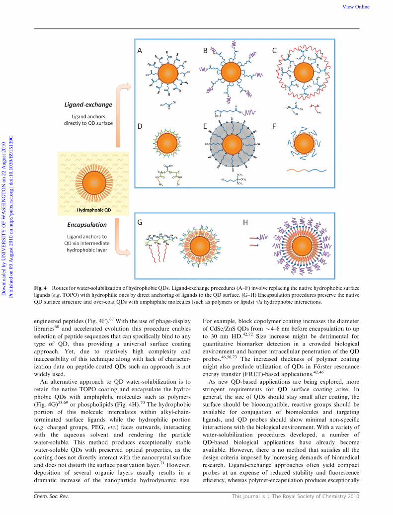
engineered peptides (Fig. 4F).67 With the use of phage-display
libraries68 and accelerated evolution this procedure enables
selection of peptide sequences that can specifically bind to any
type of QD, thus providing a universal surface coating
approach. Yet, due to relatively high complexity and
inaccessibility of this technique along with lack of character-
ization data on peptide-coated QDs such an approach is not
widely used.
An alternative approach to QD water-solubilization is to
retain the native TOPO coating and encapsulate the hydro-
phobic QDs with amphiphilic molecules such as polymers
(Fig. 4G)53,69 or phospholipids (Fig. 4H).70 The hydrophobic
portion of this molecule intercalates within alkyl-chain-
terminated surface ligands while the hydrophilic portion
(e.g. charged groups, PEG, etc.) faces outwards, interacting
with the aqueous solvent and rendering the particle
water-soluble. This method produces exceptionally stable
water-soluble QDs with preserved optical properties, as the
coating does not directly interact with the nanocrystal surface
and does not disturb the surface passivation layer.71 However,
deposition of several organic layers usually results in a
dramatic increase of the nanoparticle hydrodynamic size.
For example, block copolymer coating increases the diameter
of CdSe/ZnS QDs from B4–8 nm before encapsulation to up
to 30 nm HD.42,72 Size increase might be detrimental for
quantitative biomarker detection in a crowded biological
environment and hamper intracellular penetration of the QD
probes.46,56,73 The increased thickness of polymer coating
might also preclude utilization of QDs in Forster resonance
energy transfer (FRET)-based applications.42,46
As new QD-based applications are being explored, more
stringent requirements for QD surface coating arise. In
general, the size of QDs should stay small after coating, the
surface should be biocompatible, reactive groups should be
available for conjugation of biomolecules and targeting
ligands, and QD probes should show minimal non-specific
interactions with the biological environment. With a variety of
water-solubilization procedures developed, a number of
QD-based biological applications have already become
available. However, there is no method that satisfies all the
design criteria imposed by increasing demands of biomedical
research. Ligand-exchange approaches often yield compact
probes at an expense of reduced stability and fluorescence
efficiency, whereas polymer-encapsulation produces exceptionally
Fig. 4 Routes for water-solubilization of hydrophobic QDs. Ligand-exchange procedures (A–F) involve replacing the native hydrophobic surface
ligands (e.g. TOPO) with hydrophilic ones by direct anchoring of ligands to the QD surface. (G–H) Encapsulation procedures preserve the native
QD surface structure and over-coat QDs with amphiphilic molecules (such as polymers or lipids) via hydrophobic interactions.
Dow
nloa
ded
by U
NIV
ER
SIT
Y O
F W
ASH
ING
TO
N o
n 22
Aug
ust 2
010
Publ
ishe
d on
09
Aug
ust 2
010
on h
ttp://
pubs
.rsc
.org
| do
i:10.
1039
/B91
5139
GView Online
This journal is c The Royal Society of Chemistry 2010 Chem. Soc. Rev.
stable and bright particles at an expense of increased size.
Therefore, engineering of novel coatings that combine the
protective features of encapsulation procedures with the compact-
ness of small ligands represents an active area of research.
2.3 Development of bio-functional QD nanodevices
In order to utilize high quality QDs for bioimaging, detection,
and drug delivery applications, bio-functionality has to be
added to otherwise inert nanoparticles. This is usually
achieved by decorating QDs with proteins, peptides, nucleic
acids, or other biomolecules that mediate specific interactions
with living systems. Surface engineering is thus crucial not
only for tuning the fundamental properties of nanomaterials
and rendering them stable and soluble in different environ-
ments, but also for creating nanoparticle–biomolecule hybrids
capable of participating in biological processes. Such hybrids
should combine useful properties of both materials involved,
i.e. optical properties of the nanocrystals and biological
functions of ligands attached.
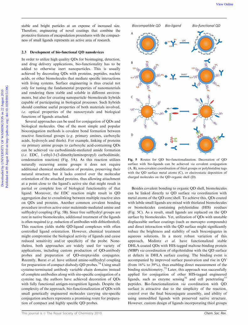
Several approaches can be used for conjugation of QDs and
biological molecules. One of the most simple and popular
bioconjugation methods is covalent bond formation between
reactive functional groups (e.g. primary amines, carboxylic
acids, hydroxyls and thiols). For example, linking of proteins
via primary amine groups to carboxylic acid-containing QDs
can be achieved via carbodiimide-mediated amide formation
(i.e. EDC, 1-ethyl-3-(3-dimethylaminopropyl) carbodiimide,
condensation reaction) (Fig. 5A). As this reaction utilizes
naturally occurring amine groups it does not require
additional chemical modification of proteins, preserving their
natural structure; but it lacks control over the molecular
orientation of the attached proteins, thus allowing attachment
at a point close to the ligand’s active site that might result in
partial or complete loss of biological functionality of that
ligand. Moreover, the EDC reaction might result in QD
aggregation due to crosslinking between multiple reactive sites
on QDs and proteins. Another common covalent bonding
procedure involves active ester maleimide-mediated amine and
sulfhydryl coupling (Fig. 5B). Since free sulfhydryl groups are
rare in native biomolecules, additional treatment of the ligands
is often required (e.g. reduction of antibodies with dithiothreitol).
This reaction yields stable QD-ligand complexes with often
controlled ligand orientation. However, chemical treatment
might compromise the biological activity of ligands and cause
reduced sensitivity and/or specificity of the probe. None-
theless, both approaches are widely used for variety of
applications, including custom production of QD-antibody
probes and preparation of QD-streptavidin conjugates.
Recently, Barat et al. have utilized amine-sulfhydryl coupling
for preparation of compact diabody-QD probes.74 Using small
cysteine-terminated antibody variable chain domains instead
of complete antibodies along with site-specific conjugation of a
cysteine tag, the authors have achieved decoration of QDs
with fully functional antigen-recognition ligands. Despite the
complexity of the approach, bio-functionalization of QDs with
small genetically engineered molecules carrying site-specific
conjugation anchors represents a promising route for prepara-
tion of compact and highly specific QD probes.
Besides covalent bonding to organic QD shell, biomolecules
can be linked directly to QD surface via coordination with
metal atoms of the QD core/shell. To achieve this, QDs coated
with labile small ligands are mixed with thiolated biomolecules
or biomolecules containing polyhistidine (HIS) residues
(Fig. 5C). As a result, small ligands are replaced on the QD
surface by biomolecules. Yet, utilization of QDs with unstable
displaceable surface coatings (such as mercapto compounds)
and direct interaction with the QD surface might significantly
reduce the brightness and stability of such bioconjugates in
aqueous solutions. In a more robust variation of this
approach, Medintz et al. have functionalized stable
DHLA-coated QDs with HIS-tagged maltose-binding protein
(MBP) via coordination of oligohistidine with the QD surface
at defects in DHLA surface coating. The binding event is
accompanied by improved surface passivation and rise in QY
(from 16% to 39%), thus enabling direct measurement of the
binding stoichiometry.75 Later, this approach was successfully
applied for conjugation of other HIS-tagged engineered
ligands, such as enzyme sensing76 and cell penetrating77
peptides. Bio-functionalization via coordination with QD
surface is attractive due to the simplicity of the reaction,
control over the final bioconjugate assembly, and ability of
using unmodified ligands with preserved native structure.
However, custom design of ligands incorporating thiol groups
Fig. 5 Routes for QD bio-functionalization. Decoration of QD
surface with bio-ligands can be achieved via covalent conjugation
(A, B), non-covalent coordination of thiol groups or polyhistidine tags
with the QD surface metal atoms (C), or electrostatic deposition of
charged molecules on the QD organic shell (D).
Dow
nloa
ded
by U
NIV
ER
SIT
Y O
F W
ASH
ING
TO
N o
n 22
Aug
ust 2
010
Publ
ishe
d on
09
Aug
ust 2
010
on h
ttp://
pubs
.rsc
.org
| do
i:10.
1039
/B91
5139
GView Online

Chem. Soc. Rev. This journal is c The Royal Society of Chemistry 2010
or HIS-tags is often complex and suitable only for small
biomolecules with relatively simple structures.
Non-covalent self-assembly of engineered proteins on the
surface of QDs with preserved organic shell prevents direct
access to inorganic QD core and exhibits minimal effect on the
photo-physical properties (Fig. 5D). In one example a fusion
protein has been utilized as an adaptor for immunoglobulin G
(IgG) coupling.78,79 Electrostatic interaction between the
positively charged leucine zipper domain of an adaptor protein
and the negatively charged QD shell stably deposits the
adaptor protein to the QD surface, while the protein G
domain specifically captures the antibody Fc region. The
resulting assembly features precise control over the antibody
orientation and eliminates any chemical modification of
IgG, thus preserving its activity. However, this procedure is
often limited to conjugation of specific classes of ligands
(e.g. antibodies). Moreover, the size of such bioconjugates is
large due to a number of thick biomolecule layers deposited on
the QD surface.
Recent achievements in merging nanoparticle encapsulation
and bioconjugation steps and design of pre-functionalized
surface coatings promise to provide more compact, stable,
and biocompatible nanoparticles with controlled density and
orientation of ligands attached. Amphiphilic polymers with a
maleic anhydride backbone are being actively explored for this
purpose. In organic anhydrous solvents, such polymers
encapsulate TOPO-coated QDs and introduce reactive anhydride
groups on the surface. In basic aqueous buffers anhydride
rings are quickly hydrolyzed, yielding negatively charged
carboxylic acid groups and rendering QDs water soluble.69
More importantly, anhydride groups are highly reactive
towards amine-containing molecules, thus allowing covalent
conjugation of a variety of biomolecules to the polymer chains
without the need for post-encapsulation modification.80,81
Choice of the bio-conjugation approach depends on
availability of ligands with suitable functional groups and on
specific application requirements. However, common design
criteria involve preserved QD photo-physical properties and
ligand bio-functionality, controlled ligand orientation and
binding stoichiometry, compact probe size, and good stability
in physiological environment. As these criteria can be satisfied
in only few specific cases, improvement of existing bio-
conjugation techniques and design of novel application-
specific water-solubilization and bioconjugation approaches
remains an active area of research. With the development of
stable and bio-functional QD probes these materials will
become nanoscience building blocks82 with flexible properties
that could be further optimized for specific applications
including biomedical imaging, detection, and nano-therapeutics.
3. QD probes for in vitro applications
In the last decade, surface engineering and bio-functionalization
techniques have transformed semiconductor nanocrystals into
complex cellular probes capable of interaction with bio-
molecules and direct participation in biological processes. In
1998, two seminal Science papers first demonstrated that
semiconductor nanoparticles could be made water-soluble
and used as biological imaging probes.50,51 One approach
utilized silica shell encapsulation chemistry in order to produce
QDs for a single-excitation dual-color cell staining.50 When
derivatized with trimethoxysilylpropyl urea and acetate
groups, green QDs preferentially labeled the cell nucleus,
and when derivatized with biotin, red QDs labeled F-actin
filaments pre-treated with phalloidin-biotin and streptavidin.
The second paper was the first to demonstrate the ligand-
exchange approach to QD water-solubilization.51 Subsequent
conjugation of transferrin produced QD probes that were
endocytosed by live HeLa cells resulting in punctate cell
staining, while IgG bioconjugates were used in an aggrega-
tion-based immunoassay. Since then, a multitude of surface
engineering techniques for QD solubilization and bio-
functionalization have been developed, enabling application-
specific design of QD probes. Such probes have found their use
in a variety of in vitro applications, such as histological
evaluation of cells and tissue specimens, single molecule
detection and real-time tracking, long-term live-cell imaging,
and study of intracellular processes.
3.1 Molecular pathology
Fluorescence microscopy is a widely used optical imaging
modality for evaluation of phenotypes of healthy cells as
well as for detection of molecular signatures of diseases.
Histological techniques, such as fluorescence in situ hybridization
(FISH) and immunohistochemistry (IHC), enable detection of
nucleic acids and protein biomarkers within cells and tissue
specimens with a high degree of sensitivity and spatial
resolution. Organic fluorophores have been widely used in
these applications, either as stains for highlighting cell
structures or as specific probes for labeling biomarkers.
However, applicability of organic fluorophores in multiplexed
and quantitative analysis for molecular profiling, a powerful
technique for study of complex molecular networks underlying
physiological and pathological processes, is limited by the
quick photobleaching, spectral overlap between probes, and
the need to excite fluorophores at unique wavelengths. QD
probes, on the other hand, exhibit photophysical properties
well-suited for this application.83,84 Despite the relatively
recent introduction into biomedical research, QDs have
already proven to be a powerful tool for sensitive quantitative
molecular profiling of cells and tissues, providing unique
identification of individual cell lineages and uncovering
molecular signatures of pathological processes.84,85 Utilization
of QDs for staining of fixed cells and tissue specimens does not
impose strict requirements on the probe biocompatibility,
toxicity, or stability in biological media. However, careful
design of the probe size, surface properties, and image
processing algorithms is essential for this application.
The hydrodynamic size of the QD-ligand bioconjugate
should be minimized in order to achieve good penetration of
the probes within the cross-linked intracellular compartments
of fixed cells. Membrane-bound compartments, such as
nucleus and mitochondria, represent especially difficult targets
for QD staining. For example, Wu et al. have investigated the
utility of QD-streptavidin and QD-antibody bioconjugates for
simultaneous labeling of membrane-associated Her2 receptor
and of a nuclear antigen in breast cancer cells (Fig. 6).53 While
Dow
nloa
ded
by U
NIV
ER
SIT
Y O
F W
ASH
ING
TO
N o
n 22
Aug
ust 2
010
Publ
ishe
d on
09
Aug
ust 2
010
on h
ttp://
pubs
.rsc
.org
| do
i:10.
1039
/B91
5139
GView Online

This journal is c The Royal Society of Chemistry 2010 Chem. Soc. Rev.
staining of cell surface antigens is reliable and effective,
staining of cytoplasmic and nuclear markers is more variable,
resulting from the relatively large size of the probes. In another
example, Tholouli et al. have employed the biotin-streptavidin
linkage for preparation of QD-oligonucleotide probes for
FISH-based studies of mRNA.86 Biotinylated DNA probes
pre-incubated with QD-Streptavidin conjugates enable
detection of 3 mRNA targets in a 1-step FISH procedure.
Yet, pre-conjugation of multiple oligonucleotides to QDs
significantly increases the overall size of the probe, thus
requiring specimen permeabilization with proteinase K, which
necessarily degrades cell and tissue architecture and destroys
most of the protein-based biomarkers useful for IHC studies.
Chan et al. have resolved this issue by developing a more
controlled procedure for pre-conjugation of exactly one
oligonucleotide probe per QD via biotin-streptavidin linkage.87
Starting with QD-streptavidin conjugates, excess streptavidin
sites are blocked with biocytin (water soluble biotin deriva-
tive), and only a few biotinylated oligonucleotides are allowed
to bind. Further purification of QD-oligo conjugates with
agarose gel electrophoresis yields relatively small mono-
oligonucleotide FISH probes suitable for multiplexed mRNA
detection under mild specimen permeabilization. As a result,
a combined QD-based FISH-IHC procedure has been
developed to compare cellular distribution patterns of
vesicular monoamine transporter (Vmat2) mRNA and
immunoreactivity of tyrosine hydroxylase in dopaminergic
neurons.87 In general, with larger QD probes, stronger
permeabilization of specimens with detergents and/or enzymes
might be required to obtain sufficient intracellular access;
however, chemical treatment might damage the target
molecules, thus reducing staining sensitivity and providing
inaccurate quantitative information about biomarker expression
levels. Furthermore, entrapment of larger QD probes within
cells hampers post-staining washing of unbound probes and
reduces the specificity of staining. Therefore, engineering of
more compact probes is highly beneficial.
QD surface engineering is critical for minimizing the non-
specific binding of QD probes to biomolecules, a common
reason of reduced staining signal-to-noise ratio and decreased
sensitivity and specificity of the target detection. Majority of
the non-specific binding results from electrostatic interactions,
when highly charged QD probes are used, and from hydro-
phobic interactions, when QDs with exposed hydrophobic
regions or partially hydrophobic ligands are used. Decoration
of QDs with uncharged hydrophilic moieties (e.g. PEG) and
zwitterionic molecules produces highly water-soluble and
stable probes while efficiently eliminating non-specific inter-
actions. For example, QD probes used in the majority of
published research have a layer of PEG that shields the QD
core from the environment and provides anchor points for
ligand attachment. Popularity of QD-PEG comes from the
outstanding non-fouling properties of PEG as well as high
stability of probes in a wide range of experimental conditions,
which facilitates engineering of QD probes for virtually any
application. However, addition of a PEG layer often results
in increased particle HD leading to the detrimental size-
dependent consequences described above. Zwitterionic
coatings, on the other hand, become utilized more often as
smaller probes are being developed. Featuring densely packed
alternating positively and negatively charged groups, these
coatings do not favor electrostatic or hydrophobic interactions
while providing an overall neutral well-hydrated surface.
However, zwitterionic coatings tend to show high pH-sensitivity,
thus imposing more stringent requirements on bioconjugation
and staining conditions. Alternatively, the QD surface can be
completely over-coated with large biomolecules (e.g. proteins)
shielding the QD from the environment and mimicking the
native functionality of the ligand; yet, possible dramatic
increase in probe size renders this approach most appropriate
for labeling of extracellular targets.
The high brightness and photostability of QD probes
enables sensitive and robust measurement of the biomarker
expression levels. However, accurate quantitative analysis of
multiple biomarkers and comparison of their relative levels
of expression within a single specimen further demand
standardization of image acquisition and processing algorithms.
Extraction and analysis of individual QD spectra from a
composite image can be achieved with spectral imaging.84,88
Generally, spectral imaging systems incrementally apply
narrow band-pass filters and collect a series of images for each
wavelength band over a specified spectrum, thus providing
spectral information for each pixel of an image. Deconvolution
of known emission profiles from the resulting composite image
separates different probe signals from each other and from the
background fluorescence. However, quantitative comparison
of different biomarkers in multiplexed staining might be
compromised by the strong signal enhancement of larger
Fig. 6 Labeling of surface and intracellular targets with QD probes.
In single-color examples membrane-associated Her2 receptors are
detected with primary antibodies and QD-labeled secondary IgG
(A, green), while intracellular nuclear antigens (B, red) and microtubules
(C, red) are visualized with primary IgG/secondary IgG-biotin/
QD-Streptavidin cascade. Both labeling routes can be applied
simultaneously for a two-color staining (D). The nuclei are counter-
stained with Hoechst 33 342 (blue) in A and C. Reprinted by
permission from Macmillan Publishers Ltd.,53 copyright (2003).
Dow
nloa
ded
by U
NIV
ER
SIT
Y O
F W
ASH
ING
TO
N o
n 22
Aug
ust 2
010
Publ
ishe
d on
09
Aug
ust 2
010
on h
ttp://
pubs
.rsc
.org
| do
i:10.
1039
/B91
5139
GView Online

Chem. Soc. Rev. This journal is c The Royal Society of Chemistry 2010
(red) QD and reduction of smaller (green-blue) QD signals.
For example, Ghazani and coworkers have demonstrated
three-color staining of lung carcinoma xenografts for epidermal
growth factor receptor (EGFR), E-cadherin, and cytokeratin
with 655, 605, and 565 nm QD-based assays and noticed
significant enhancement of 655 nm signal over 565 nm one,
attributing this phenomenon to FRET from smaller to larger
QDs.89 Further, the discordance in fluorescence intensity of
individual probes directly relates to light absorption properties
of QDs, as larger QDs possess larger absorption cross-sections
and thus collect light more efficiently. The effect of FRET
depends on the density and distribution of biomarkers, which
is hard to predict and account for during quantitative analysis.
However, differences in photo-physical properties of
individual probes can be readily characterized in advance
and incorporated into signal analysis algorithms. In a recent
study, Yezhelyev et al. have demonstrated the multiplexed
labeling and quantification of three clinically significant breast
cancer markers—Her2, ER, and PR—on formalin-fixed
paraffin-embedded (FFPE) breast cancer cells.32 In order to
account for signal enhancement of red QDs and compare
expression levels of biomarkers within one sample, acquired
data is adjusted according to the relative QD intensities
(QD655:QD605:QD565 = 8 : 4 : 1 as measured in a separate
experiment for equal QD concentrations), yielding relative
biomarker abundance consistent to that obtained with
conventional techniques (IHC, Western blot, and FISH). This
technology has been further validated by the detection and
quantification of a panel of five biomarkers on FFPE breast
cancer tissue biopsies (Fig. 7).
Future advancements in the area of QD-based molecular
pathology will be centered around highly multiplexed quanti-
tative molecular profiling. Engineering of more compact and
sensitive QD probes with outstanding stability and non-
fouling properties will, therefore, remain the major focus of
research in this area. Modification of the band gap by tuning
the QD chemical composition, for example, might enable
shifting QD emission into deep blue90 or far red30 region,
while keeping the particle size constant within 4–6 nm range.
However, further reduction of the QD inorganic core size
below 3–4 nm might be highly challenging. Meanwhile,
significant probe size reduction can be achieved via engineering
of the compact organic coating layers and ligands that offer
great design flexibility. Substitution of thick shells with thinner
zwitterionic coatings, development of mono-valent probes,
and utilization of smaller targeting ligands (e.g. peptides and
aptamers) will, thus, become essential for engineering of
robust and stoichiometric QD probes and their translation
to clinical diagnostics.
3.2 Real-time monitoring of dynamic molecular processes
Staining of fixed cells and tissue specimens provides information
on biomarker expression and distribution; however, the study
of intracellular molecular pathways underlying the physio-
logical and pathological processes is limited by the static
nature of this technique. Real-time imaging of live cells, on
the other hand, enables the study of highly complex and
dynamic biological processes that occur at molecular level.
While the relatively large size of QD probes often hampers
cellular entry and intracellular targeting, access to the bio-
markers expressed on the cell membrane is usually readily
achievable. Consequently, the majority of applications
reported in the literature describe dynamics of membrane
proteins (e.g. receptor diffusion) and membrane-associated
processes (e.g. endocytosis and intracellular trafficking) rather
than monitoring of intracellular targets. As a general
guideline, QD probes for real-time live cell imaging should
Fig. 7 Multiplexed labeling of breast cancer tissue biopsies. Normal-
ization of the fluorescence according to relative QD intensities is
required for accurate quantitative analysis of biomarker expression.
Reproduced with permission from ref. 32. Copyright 2007 Wiley-VCH
Verlag GmbH & Co. KGaA.
Dow
nloa
ded
by U
NIV
ER
SIT
Y O
F W
ASH
ING
TO
N o
n 22
Aug
ust 2
010
Publ
ishe
d on
09
Aug
ust 2
010
on h
ttp://
pubs
.rsc
.org
| do
i:10.
1039
/B91
5139
GView Online

This journal is c The Royal Society of Chemistry 2010 Chem. Soc. Rev.
have compact size and high stability in biological buffers and
cell culture media, exhibit high brightness and photostability
for single-molecule imaging, show no toxicity or interference
with cell physiology throughout the duration of experiment,
and possess biological functionality for interaction with target
biomolecules.
Majority of the QD probes used for live cell imaging employ
a well-characterized and robust PEG coating as a universal
non-fouling shield against protein adsorption. Resistance to
protein binding conveys high stability in a wide range of
buffers as well as cell culture media, precluding QD aggrega-
tion, non-specific interaction with cells, and off-target effects
(e.g. receptor activation, enhanced endocytosys, etc.). In
addition, such a coating efficiently protects the QD core and
preserves the beneficial photo-physical properties. Being 10–20
times brighter and orders of magnitude more photostable than
organic fluorophores, QDs are well-suited for sensitive single-
probe detection and long-term probe monitoring.50,51 In
combination with advanced imaging techniques (e.g. 3-D
tracking confocal microscopy,91 pseudo total internal
reflection fluorescence microscopy,56 oblique angle fluorescence
microscopy,92 etc.), QD probes enable the study of active and
passive molecular transport mechanisms in high-background
environments. Furthermore, distinguishing single QD probes
from the small QD aggregates by the characteristic
fluorescence intermittency (or blinking) improves the accuracy
of measurement by eliminating the contribution of QD
clusters. Outstanding resistance to photobleaching and
degradation enables probe monitoring for several hours or
days. For example, Jaiswal et al. have utilized this property for
visualization of QD endocytic uptake and specific cell-surface
labeling of P-glycoprotein transporters over the course of 14 h,
acquiring images at a rate of 1 frame per minute.93 Localization
of particles within the endosomes of live HeLa cells and
D. discoideum amoebae could be monitored over the course
of more than a week with minimal loss of QD fluorescence.
Accurate examination of physiological processes in native
environment is often hard to achieve, as any chemical
modification introduced to the system (e.g. labeling with a
fluorophore or expression of a foreign reporter protein) might
potentially change intramolecular interactions and interfere
with normal cell physiology. This issue is especially keen for
QD-based studies, since biomolecules must be tagged
with bulky (sometimes several times larger than the studied
biomolecule) probes. Therefore, design of QD probes that
introduce minimal changes to the cell physiology and
lack short-term cyto-toxicity is essential for the QD-based
investigation of dynamic molecular processes. Much success in
overcoming this challenge has been achieved in the study of
cell receptor diffusion and interaction. In a single-molecule
imaging study, Dahan et al. have used QDs for labeling of
individual glycine receptors on the surface of cultured spinal
neurons and tracking the receptor diffusion in and out of
synaptic cleft (Fig. 8).73 Differential 2-D diffusion coefficients
of receptors have been measured over time spans 240 times
longer than previously achieved using organic dyes as tags,
with 4 to 8-fold better spatial resolution, and with a signal to
noise ratio almost an order of magnitude higher. While the
steric effect of QD probes could not be assessed through this
study, relative characterization of receptor diffusion patterns
within the synaptic, perisynaptic, and extrasynaptic regions
was achieved. In another study, QDs have been used to reveal
a previously unknown receptor diffusion mechanism for
recovery from synaptic depression in neurons.94 Tracking of
the rapid lateral diffusion of QD-labeled AMPA glutamate
receptors have shown diffusion behavior comparable to that of
organic dye-labeled receptors, while providing a robust
fluorescence signal for the duration of experiment. Murcia
et al. have demonstrated that labeling of individual cell
membrane lipids with QDs does not affect lipid diffusion
(as compared to dye-labeled lipids), while enhanced brightness
of the probe enables high-speed single molecule tracking at
1000 frames per second.92 Overall, it has been shown by
several studies that QD probes do not significantly interfere
with the diffusion of labeled biomolecules on the cell
membrane, thus permitting both absolute measurement of
diffusion coefficients and self-consistent relative studies of
biomolecule diffusion under varying conditions.
Besides providing insight on the molecular dynamics of cell
membrane components, QD probes facilitate the detailed
Fig. 8 Labeling of individual glycine receptors in cultured spinal
neurons. QD probes label glycine receptors throughout somato-
dendritic compartment (A) and can be located adjacent to (B, arrowhead)
or in front of (B, arrow) inhibitory synaptic boutons. TEM examination
reveals QD clustering at the extrasynaptic (C), perisynaptic (D), and
synaptic (E) regions. Reprinted from ref. 73 with permission from
AAAS. Copyright (2008).
Dow
nloa
ded
by U
NIV
ER
SIT
Y O
F W
ASH
ING
TO
N o
n 22
Aug
ust 2
010
Publ
ishe
d on
09
Aug
ust 2
010
on h
ttp://
pubs
.rsc
.org
| do
i:10.
1039
/B91
5139
GView Online
Chem. Soc. Rev. This journal is c The Royal Society of Chemistry 2010
study of such important processes as endocytosis and intra-
cellular trafficking. Due to the relatively small size, individual
QDs can be uptaken by the cells via endocytosis, incorporated
within the endosomes, and transported like any other endo-
cytosed cargo without interfering with the mechanism of the
process, thus representing a useful model system for the study
of these phenomena. For example, Cui et al. have studied the
dynamics of axonal internalization and neuronal retrograde
transport of the nerve growth factor (NGF) by tagging native
NGF with QDs.56 While recording an average retrograde
endosome movement speed consistent with previous bulk
measurements of NGF transport, real-time monitoring of
individual QD-NGF-containing endosomes has revealed a
‘‘stop-and-go’’ behavior and occasional anterograde move-
ment, thus providing insight on the diversity in transport
mechanisms. In another study Zhang et al. have utilized the
unique size and pH-dependent fluorescence of QDs for the
study of the dynamics of synaptic vesicles during multiple
rounds of neuronal transmission without perturbing the
vesicle cycling.95 Monitoring of individual QD-loaded
synaptic vesicles has enabled characterization of complete
vesicle fusion (full-collapse fusion) and transient fusion
(so-called kiss-and-run behavior) with respect to time and
frequency of impulse firing, and uncovered new aspects of
neurotransmitter release and replenishment mechanisms.
Efficient specific interaction with cell components requires
otherwise inert QD probes to possess biological functionality,
which is usually conveyed by decoration of QDs with targeting
biomolecules. Often such moieties are represented by the
receptor ligands attached to QD surface either covalently or
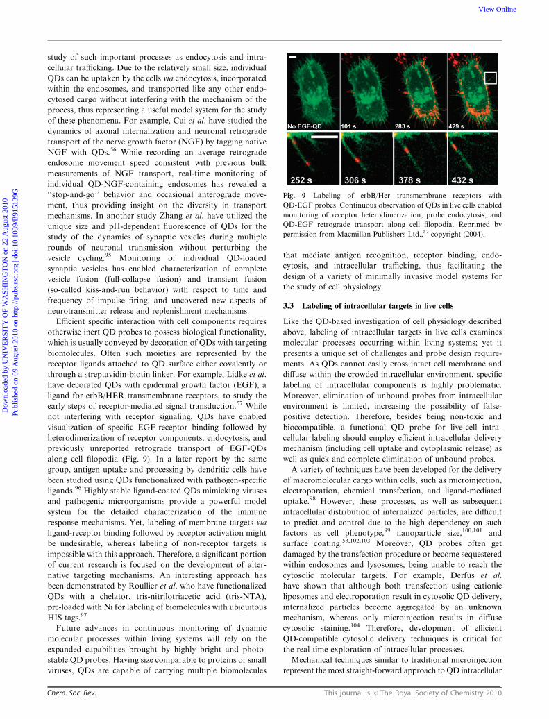
through a streptavidin-biotin linker. For example, Lidke et al.
have decorated QDs with epidermal growth factor (EGF), a
ligand for erbB/HER transmembrane receptors, to study the
early steps of receptor-mediated signal transduction.57 While
not interfering with receptor signaling, QDs have enabled
visualization of specific EGF-receptor binding followed by
heterodimerization of receptor components, endocytosis, and
previously unreported retrograde transport of EGF-QDs
along cell filopodia (Fig. 9). In a later report by the same
group, antigen uptake and processing by dendritic cells have
been studied using QDs functionalized with pathogen-specific
ligands.96 Highly stable ligand-coated QDs mimicking viruses
and pathogenic microorganisms provide a powerful model
system for the detailed characterization of the immune
response mechanisms. Yet, labeling of membrane targets via
ligand-receptor binding followed by receptor activation might
be undesirable, whereas labeling of non-receptor targets is
impossible with this approach. Therefore, a significant portion
of current research is focused on the development of alter-
native targeting mechanisms. An interesting approach has
been demonstrated by Roullier et al. who have functionalized
QDs with a chelator, tris-nitrilotriacetic acid (tris-NTA),
pre-loaded with Ni for labeling of biomolecules with ubiquitous
HIS tags.97
Future advances in continuous monitoring of dynamic
molecular processes within living systems will rely on the
expanded capabilities brought by highly bright and photo-
stable QD probes. Having size comparable to proteins or small
viruses, QDs are capable of carrying multiple biomolecules
that mediate antigen recognition, receptor binding, endo-
cytosis, and intracellular trafficking, thus facilitating the
design of a variety of minimally invasive model systems for
the study of cell physiology.
3.3 Labeling of intracellular targets in live cells
Like the QD-based investigation of cell physiology described
above, labeling of intracellular targets in live cells examines
molecular processes occurring within living systems; yet it
presents a unique set of challenges and probe design require-
ments. As QDs cannot easily cross intact cell membrane and
diffuse within the crowded intracellular environment, specific
labeling of intracellular components is highly problematic.
Moreover, elimination of unbound probes from intracellular
environment is limited, increasing the possibility of false-
positive detection. Therefore, besides being non-toxic and
biocompatible, a functional QD probe for live-cell intra-
cellular labeling should employ efficient intracellular delivery
mechanism (including cell uptake and cytoplasmic release) as
well as quick and complete elimination of unbound probes.
A variety of techniques have been developed for the delivery
of macromolecular cargo within cells, such as microinjection,
electroporation, chemical transfection, and ligand-mediated
uptake.98 However, these processes, as well as subsequent
intracellular distribution of internalized particles, are difficult
to predict and control due to the high dependency on such
factors as cell phenotype,99 nanoparticle size,100,101 and
surface coating.53,102,103 Moreover, QD probes often get
damaged by the transfection procedure or become sequestered
within endosomes and lysosomes, being unable to reach the
cytosolic molecular targets. For example, Derfus et al.
have shown that although both transfection using cationic
liposomes and electroporation result in cytosolic QD delivery,
internalized particles become aggregated by an unknown
mechanism, whereas only microinjection results in diffuse
cytosolic staining.104 Therefore, development of efficient
QD-compatible cytosolic delivery techniques is critical for
the real-time exploration of intracellular processes.
Mechanical techniques similar to traditional microinjection
represent the most straight-forward approach to QD intracellular
Fig. 9 Labeling of erbB/Her transmembrane receptors with
QD-EGF probes. Continuous observation of QDs in live cells enabled
monitoring of receptor heterodimerization, probe endocytosis, and
QD-EGF retrograde transport along cell filopodia. Reprinted by
permission from Macmillan Publishers Ltd.,57 copyright (2004).
Dow
nloa
ded
by U
NIV
ER
SIT
Y O
F W
ASH
ING
TO
N o
n 22
Aug
ust 2
010
Publ
ishe
d on
09
Aug
ust 2
010
on h
ttp://
pubs
.rsc
.org
| do
i:10.
1039
/B91
5139
GView Online

This journal is c The Royal Society of Chemistry 2010 Chem. Soc. Rev.
delivery, as virtually no modification of QD probes already
available for extracellular labeling is required. For example,
peptide-functionalized QD probes delivered to the cytoplasm
via microinjection successfully exploit active peptide-specific
transport mechanisms to reach target compartments, nucleus
and mitochondria, within several hours after delivery
(Fig. 10A).104 In another example, Yum et al. have utilized
gold-coated boron nitride nanotubes (with a diameter of 50 nm)
to deliver QDs within the cytoplasm or nucleus of live HeLa
cells with consequent 30-minute monitoring of QD diffusion
within those compartments (Fig. 10B).105 Linking the
ubiquitous QD-Streptavidin probes onto the nanotubes via
reducible disulfide bonds enables delivery of intact QDs to a
controlled intracellular location without much damage to the
cell. While the QD probes used in this study did not carry
targeting ligands, the technique can be expanded to deliver
functionalized QDs as well. However, being quite labor-
intensive and low-throughput, both techniques might only
find use in limited single-cell studies. Aiming at high-throughput
intracellular delivery, Park et al. have engineered arrays of
vertically aligned carbon nanosyringes that, upon cell growth
on top of them, provide cytosolic access for injection of
unmodified QDs (Fig. 10C).106 Efficient and consistent
delivery of QD probes within large cell populations promises
to enable studies of cell heterogeneity, inter-cellular communi-
cation, and cell population response to changing exogenous
factors; yet complex manufacturing of the arrays as well as the
unpredictable effect of changed surface topology on the cell
physiology might hamper wide use of this technique.
Non-mechanical approaches are gaining increasing
popularity due to the potential for high-throughput robust
QD intracellular delivery with minimal intrusion to cell
physiology. Functionalization of QDs with engineered
peptides, small versatile biomolecules, might provide great
flexibility in tuning the QD interaction with cell components
(Fig. 10D).107 Linking of short peptide sequences to the QD
surface can be achieved by a variety of methods, including
covalent conjugation to existing functional groups,108–110
electrostatic adsorption,79 biotin-streptavidin binding,111,112
and direct coordination to the nanocrystal surface via HIS
sequences.76,77 In general, highly cationic peptides facilitate
enhanced interaction with the cell membrane and QD inter-
nalization, whereas additional targeting moieties govern
intracellular distribution. For example, Delehanty et al. have
modified QDs with HIS-tagged cell penetrating peptide based
on the HIV-1 Tat protein motif, achieving efficient internaliza-
tion of QDs via endocytosis,77 while Rozenzhak et al. have
added the nuclear localization sequence for nuclear targeting
and apoptotic GH3 domain for triggering cell death.112 Other
groups have explored similar cationic peptides, such as
polyarginine111 and polylysine,113 for achieving cell entry.
Despite the versatility of QD-peptide conjugates for labeling
of intracellular targets, this approach still suffers from the
uncontrolled probe aggregation and lysosomal sequestration
inside cells.
Recent work on QD cell uptake and intracellular targeting
has focused on employing endocytosis/pinocytosis as a
universal delivery mechanism and endosome destabilization/
lysis as a cytosolic access route.114 One strategy involves QD
cell-loading using osmotic lysis of pinocytic vesicles (Fig. 10E).
Efficient uptake is first achieved by inducing pinocytosis by
incubation of cells in a hypertonic solution followed by vesicle
osmotic lysis and cytoplasmic release by switching to
hypotonic medium. Utilization of external control over the
osmotic strength of cell medium requires no modification to
QD probes and enables uniform loading of intact single QD
probes to all cells within the population. For example, Courty
et al. have utilized this approach to load QD-tagged kinesin
motors to living HeLa cells and monitor single-motor
movement within the cytoplasm.115 However, drastic change
in extracellular conditions is not compatible with QD loading
of fragile cells, and external triggering of osmotic lysis might
require extensive optimization of procedure due to wide
heterogeneity in cell response to changing culture conditions.
A more robust approach involves engineering of on-demand
endosome-disrupting capacity within the QD probes. To
achieve intracellular delivery of unmodified QD probes, Kim
et al. have utilized 100-nm external biodegradable delivery
vesicles made of poly(D,L-lactide-co-glycolide) (PLGA) polymer.116
Functionalization of PLGA surface with antibodies enables
interaction with cell surface markers, thus inducing efficient
and specific cellular uptake, whereas PLGA charge reversal
within low-pH endosomal environment causes membrane
destabilization and endosomal escape. Finally, degradation
of the polymeric vehicle within the cytoplasm releases the QD
payload for specific labeling of intracellular targets. Aiming at
Fig. 10 Mechanical (A–C) and non-mechanical (D–F) routes for
intracellular delivery of bio-functional QDs within live cells. (A)
Microinjection enables intracellular loading of unmodified QD probes
along with carrier solution on a cell-by-cell basis. (B) Delivery with
nanotubes offers precise control over QD delivery location, but
requires QD anchoring to nanotubes via reducible linkers. (C) High-
throughput microinjection via nanosyringe arrays delivers unmodified
QDs within large cell population, but changes the surface topology for
cell growth. (D) QDs functionalized with cell-penetrating peptides
might employ endosome-mediated and non-endosomal pathways
(depending on the peptide structure), offering flexibility in tuning the
QD-cell interaction. (E) Pinocytosis enables uptake of unmodified QD
probes with consequent cytoplasmic distribution. (F) Utilization of
active receptor-mediated QD uptake via endocytosis followed by
endosomal escape via proton-sponge effect represents a highly efficient
non-invasive delivery method with specific targeting capabilities.
Dow
nloa
ded
by U
NIV
ER
SIT
Y O
F W
ASH
ING
TO
N o
n 22
Aug
ust 2
010
Publ
ishe
d on
09
Aug
ust 2
010
on h
ttp://
pubs
.rsc
.org
| do
i:10.
1039
/B91
5139
GView Online

Chem. Soc. Rev. This journal is c The Royal Society of Chemistry 2010
containing all functionalities within single QD probes, Duan
and Nie have coated QDs with hybrid poly(ethylene glycol)/
polyethylenimine (PEG/PEI) polymers producing nano-
particles with a reasonably small HD (15–22 nm) and
endosome-disrupting capacity and yet good stability and
biocompatibility (Fig. 10F).117 The high amine content of
PEI conveys endosomal lysis through the proton sponge
effect—buffering of the endosome acidification by the amines
of the polymer backbone followed by an increase in counter-
ion (mostly chlorine) concentration, build-up of osmotic
pressure, and eventually endosome rupture118—while highly
hydrophilic PEG layer provides QD protection from the
environment, prevents aggregation, and precludes undesirable
non-specific interactions with biomolecules. As a result, when
incubated with live HeLa cells, such QDs are internalized,
escape from the endosomes, and become distributed through-
out the cytosol. However, ligand exchange and direct inter-
action of PEI with the QD surface necessarily cause an
undesirable drop in fluorescence QY and detection sensitivity.
Retention and modification of a stable coating should preclude
such adverse changes in QD photo-physical properties. In one
example, Yezhelyev et al. have decorated negatively-charged
polymer-coated QDs with tertiary amines, thus producing
proton-absorbing QD probes that efficiently achieve intra-
cellular endosomal release while featuring bright fluorescence
and good colloidal stability.119
Currently, a wide range of potential techniques for QD
intracellular delivery is being developed and perfected. Yet,
another major obstacle—inability to remove unbound probes
and determine whether QD probes have reached their intra-
cellular targets—still remains largely unexplored. As a result,
both the sensitivity and specificity of intracellular labeling
suffer from the dependency on the relative number of probes
that enter a cell. If too few probes are internalized, incomplete
or dim labeling of targets may occur; whereas too many
probes might lead to a high degree of background fluorescence
and false-positive detection. Thus, effective intracellular
labeling requires either active elimination of unbound probes
by the cells or utilization of QD sensors that alter the
wavelength or intensity of fluorescence signal upon target
recognition.
A promising technology for real-time sensing of target
recognition is based on the nonradiative energy transfer
(FRET) from the QD to acceptor/quencher molecules. In this
approach intracellular target binding is accompanied by the
change in QD-acceptor proximity and, therefore, fluorescence
intensity, thus distinguishing bound probes from the back-
ground. In order to achieve efficient FRET sensing, QD
probes must feature compact shell/linker structure (allowing
sufficient proximity between the QD core and acceptor for
nonradiative coupling), offer overlapping emission/absorption
spectra for efficient energy transfer, and exploit suitable routes
of excitation. Since the QD fluorescence wavelength can be
tuned by adjusting the nanocrystal size and/or chemical
composition, QD emission spectrum can be precisely matched
with the absorption peak of an arbitrary acceptor molecule,
ensuring maximum spectral overlap and efficiency of energy
transfer. Broad QD absorption profile and large Stokes shift,
on the other hand, enable probe excitation by wavelengths of
light tens to hundreds of nanometers shorter than the emission
peak, reducing non-FRET excitation of acceptor molecules
and increasing the signal-to-noise ratio. Furthermore, as QDs
are relatively large, multiple acceptor molecules can be
attached to their surface for tuning the degree of energy
transfer. Therefore, satisfying majority of the design require-
ments, QDs have been successfully used as FRET probes in a
wide variety of sensing schemes detecting conformational
changes as well as binding and cleavage events.5 For example,
Fig. 11 Two-photon (top panel) and one-photon (bottom panel)
excitation of QD-Cy3 FRET system. QDs are efficiently excited by
both methods, enabling fluorescence of conjugated Cy3 molecules via
FRET. However, only two-photon excitation precludes non-FRET
excitation of Cy3 dye, whereas conventional one-photon fluorescence
imaging produces significant background via direct Cy3 excitation.
Reproduced with permission from ref. 121. Copyright 2007 Wiley-VCH
Verlag GmbH & Co. KGaA.
Dow
nloa
ded
by U
NIV
ER
SIT
Y O
F W
ASH
ING
TO
N o
n 22
Aug
ust 2
010
Publ
ishe
d on
09
Aug
ust 2
010
on h
ttp://
pubs
.rsc
.org
| do
i:10.
1039
/B91
5139
GView Online

This journal is c The Royal Society of Chemistry 2010 Chem. Soc. Rev.
in order to monitor molecular interactions within live cells,
McGrath et al. have taken advantage of FRET between
QD-transferrin probes and dye-transferrin conjugates.120
During receptor mediated endocytosis, dimerization of
transferrin receptors results in increased proximity between
QDs and acceptor dyes, thus enabling FRET. Yet, accidental
non-FRET excitation of acceptor dye was unavoidable with
standard single-photon imaging modality in this study. To
minimize this artifact, Clapp et al. have utilized a two-photon
QD excitation route that significantly enhances the signal-to-
noise ratio of intracellular FRET (Fig. 11).121 Since QDs have
two-photon absorption cross sections several orders of
magnitude larger than typical organic dyes, undesirable
two-photon excitation (840 nm) of acceptor Cy3 dye is
dramatically reduced in comparison to single-photon
excitation (488 nm). Despite the promising initial labeling of
cell surface markers and study of endocytosis, sensing of
intracellular targeting with QD probes still remains to be
shown. A number of challenges, such as probe stability,
reproducibility of bioconjugation, detection sensitivity, and
reliability of image acquisition and processing, need to be
addressed before FRET-based QD sensors can be widely
adapted for labeling of intracellular targets in living cells.
Employing active exocytosis of functional QD probes, on
the other hand, provides an attractive route for intracellular
imaging without the strict requirements of FRET sensing.
However, this goal is hard to achieve due to the lack of known
efficient exocytosis mechanisms and poor understanding of the
intracellular behavior of QDs. The most common approach
for achieving QD exocytosis is to incubate cells in starvation
medium; yet poor efficiency of QD elimination and significant
changes in normal cell physiology introduced by starvation
might preclude from the real-time study of physiological
processes. Therefore, engineering of not only intra-
cellular delivery and targeting functionalities, but also efficient
exocytosis mechanisms within the QD probes will become
a major focus of future research in the area of live cell
imaging.
4. QD probes for in vivo imaging
Fluorescence in vivo imaging with QD probes promises to
greatly expand the capabilities of existing imaging modalities,
providing access to high-resolution multiplexed vascular
imaging, intraoperative image guidance, real-time cell tracking,
and in vivo molecular targeting.122 MRI, CT, PET, and
SPECT have become widely used imaging techniques for
examination of internal structures, molecular targets, and
metabolic processes in vivo. Nevertheless, PET and SPECT
(which are based on detection of radioactive labels) suffer from
poor spatial resolution, while MRI and CT (which are based
on tissue contrast) primarily provide structural information
and offer poor sensitivity. Fluorescence imaging with QD
probes, on the other hand, can be performed in a multiplexed
format with varying temporal and spatial resolution. For
example, bulk QD measurements (e.g. in a whole-animal
or whole-organ context) can be achieved via fluorescence
reflectance imaging,123 whereas high-resolution examination
of QD staining is available from intravital fluorescence
microscopy124,125 or post-operative histological examination
of excised tissues. The versatility of QDs provides vast
flexibility in engineering of probes for a variety of in vivo
imaging applications—blood circulation time, degradation
and excretion routes, specific interaction with biomolecules
and cells, and biodistribution along with QD photo-physical
properties can be potentially controlled via the probe design
based on the needs of a particular application. Implementation
of such control, however, is not trivial as behavior of QDs in a
highly heterogeneous and aggressive in vivo environment is still
poorly explored. Gaining thorough understanding of the
interaction between QDs and physiological systems and
learning how to manipulate these interactions represent
essential milestones towards benefitting from the novel
in vivo imaging capabilities featured by the QD probes.
4.1 General design considerations for in vivo QD probes
Impressive progress has been achieved in engineering of
bright, stable, and biocompatible probes for live cell imaging.
The absence of adverse effects on cell physiology and lack of
obvious short-term cytotoxicity encourage further exploration
of whether QD probes can be made suitable for in vivo
applications. Unlike in vitro applications, where experimental
conditions can be strictly controlled by the investigator,
the physiological environment presents complex and often
unpredictable responses to foreign materials. Therefore,
in vivo imaging with QD probes imposes another level of
requirements for the probe design, most important of which
are: biocompatible and non-toxic nanoparticle coatings with
integrated non-fouling functionality for reduced nonspecific
interactions in highly heterogeneous biological environments;
reliable control over the QD biodistribution, degradation, and
excretion pathways for reduced toxicity; and applicability of
QD probes in non-invasive or minimally invasive intravital
imaging for long-term observation of QD dynamics in vivo.
Development of novel and optimization of existing intravital
imaging techniques is primarily governed by the specific
application requirements and, thus, will be discussed in detail
later. The more important issue of QD biocompatibility and
biodistribution (and associated potential toxicity), on the
other hand, is relevant to all QD-based in vivo applications.
Great variability in the structure and composition of the
semiconductor core, particle coating, and biomolecular
functionalities impede systematic investigation of QD inter-
action with biological systems and modes of toxicity.126–128
While early studies have shown severe cytotoxicity originating
from photo-oxidation of unprotected CdSe QDs and release of
Cd2+ ions,129,130 later in vitro and in vivo experiments with
protected nanoparticles have not uncovered significant
QD-associated adverse changes. Within different reports,
potential QD toxicity has been attributed to size-based
effects,100,102 Cd2+ release,129,131,132 and ROS (reactive oxygen
species) induced oxidative stress.133–135 None of these mecha-
nisms can be universally applied towards characterization of
all existing or newly developed QD probes. Yet, the potential
accumulation of QDs within the body and release of toxic
Cd2+ ions seems to be the most prominent concern within the
scientific community. Therefore, elucidation of mechanisms
Dow
nloa
ded
by U
NIV
ER
SIT
Y O
F W
ASH
ING
TO
N o
n 22
Aug
ust 2
010
Publ
ishe
d on
09
Aug
ust 2
010
on h
ttp://
pubs
.rsc
.org
| do
i:10.
1039
/B91
5139
GView Online

Chem. Soc. Rev. This journal is c The Royal Society of Chemistry 2010
for preventing in vivo QD accumulation and degradation has
become a priority in bio-nanotechnology research (Fig. 12).
Probably the safest and most desirable approach to
addressing the toxicity issue is engineering of QD probes that
are quickly and completely eliminated from the body via renal
or bile excretion pathways without triggering uptake by the
reticulo-endothelial system (RES) and avoiding degradation
pathways. This approach seems especially favorable in light of
sparse information on in vivo QD degradation mechanisms
and long-term effect of QD accumulation in organs.
Systematic investigation of QD biodistribution performed by
Choi et al. in mice has identified a nanoparticle hydrodynamic
size renal clearance threshold of 5.5 nm.58 When delivered
systemically, small cysteine-coated QDs are readily excreted
into the bladder with minimal accumulation in the liver (4.5%)
and kidneys (2.6%), whereas larger particles exhibit significant
liver uptake (26.5%). Further, the importance of non-fouling
zwitterionic surface coatings in inhibiting the protein
absorption and retaining the original nanoparticle hydro-
dynamic size has been emphasized. In contrast, QDs featuring
charged surface undergo serum protein adsorption and
increase in HD to more than 15 nm. Working towards
preparation of compact QD probes, Law et al. have
synthesized ultrasmall (3–5 nm in diameter) cysteine-coated
CdTe/ZnTe QDs and tested biodistribution of these probes in
mice, finding no QDs in liver and spleen 2 weeks post-
injection,59 whereas Choi et al. have employed cysteine-coated
CdSe(ZnCdS) QDs functionalized with cyclic-RGD peptide
for tumor targeting in mouse model and observed complete
elimination of 65% of injected QDs within first 4 h, while
detecting specific tumor labeling (with tumor-to-background
ratio of 6.9).136 However, the few ultrasmall QDs currently
available suffer from poor photo-physical properties, while
preparation of better protected and bio-functionalized
probes often increases the QD size, thus making renal
clearance difficult. Furthermore, quick renal clearance is often
undesirable, as prolonged QD circulation is required for
specific targeting, high-sensitivity imaging, and therapeutic
potency. Therefore, high molecular weight coatings are
routinely applied to QD probes to increase their circulation
time and improve bioavailability. Ballou et al. have
emphasized the importance of coating with high molecular
weight PEG to reduce rapid clearance of QDs by liver and
bone marrow,137 and Prencipe et al. have achieved remarkably
long blood circulation of nanomaterials encapsulated with
branched PEG.138 Meanwhile, high doses of particles with
positively charged amine or negatively charged carboxyl
groups have been shown to initiate coagulation cascades
resulting in pulmonary thrombosis and death.139 Therefore,
utilization of stimulus-responsive biodegradable ligands for
dynamic tuning of the QD size might become a promising
design route for future in vivo probes. Such ligands would
ensure prolonged QD blood circulation and/or interaction
with target cells, while eventually detaching from the QD
surface and releasing single nanoparticles with original size
below 5.5 nm capable of efficient renal excretion.
In some cases complete elimination of QD probes from
the body via renal excretion or other means might prove
challenging or undesirable. Engineering of ultra-stable QDs
encapsulated with inert biocompatible materials might prove
helpful in this situation. If QD integrity within a human body
can be retained for many years, biological systems might never
be exposed to heavy metal components of the QD core. For
example, Ballou et al. have indicated that intact PEG-coated
QDs remain in bone marrow and lymph nodes of mice for
several months after injection,137 yet Fitzpatrick et al. have
detected signs of QD degradation (spectral blue-shifting) over
a course of two years.140 While organic coatings, such as
polymers and lipids, might still degrade due to exposure to
biological environment, utilization of more stable inorganic
materials should protect the cores of QD probes for extended
periods of time. Alternatively, as cadmium poisoning results
Fig. 12 Potential routes for elimination of Cd-associated QD toxicity.
Dow
nloa
ded
by U
NIV
ER
SIT
Y O
F W
ASH
ING
TO
N o
n 22
Aug
ust 2
010
Publ
ishe
d on
09
Aug
ust 2
010
on h
ttp://
pubs
.rsc
.org
| do
i:10.
1039
/B91
5139
GView Online

This journal is c The Royal Society of Chemistry 2010 Chem. Soc. Rev.
from a quick release of large amounts of this metal into a
bloodstream, its preferential accumulation in kidneys, and
consequent nephrotoxicity, slow degradation of QD probes
within a human body followed by urinary excretion might
offer a way of safe and efficient elimination of QD components
over a long term. Since up to 30 ug/day of dietary Cd
(coming from fish, vegetables, and other sources) can be
consumed by a healthy adult without adverse effects on kidney
function,141 this approach seems to be feasible. Adapting
technology developed for controlled drug release towards
QD encapsulation with biodegradable polymers might provide
one way of achieving control over QD in vivo degradation.
Despite the proven potential for QD excretion or stable
shielding, increasing safety concerns urge for complete
elimination of toxic components from in vivo probes and
design of biocompatible and non-toxic QDs. In one approach,
Yong et al. have prepared Cd-free InP/ZnS QDs and utilized
these probes for targeting of pancreatic cancer cell lines;142
however, low QY (B30%) and large size (B30 nm in
diameter) might limit utility of such probes for in vivo imaging.
Higher-quality probes with QY of up to B60% and HD
of 17 nm have been developed by Li et al. on the basis of
CuInS2/ZnS QDs.143 Further, engineering of low-cost, non-
toxic, and potentially biodegradable in vivo imaging probes
might become available through utilization of recently
developed technology for preparation of water-soluble QDs
made of silicon144,145—an inert, biocompatible, and abundant
material. However, while being an attractive approach,
Cd-free QDs still suffer from poor stability and inferior
photo-physical properties compared to high-quality QDs
made of toxic materials (such as CdSe). Therefore, improving
biocompatibility of potentially toxic QD probes remains a
sound and highly promising alternative, and elimination or
reduction of cadmium interaction with live cells seem to be the
cornerstone of such approaches.
4.2 Vascular imaging
One of the most common in vivo applications of QDs is
fluorescence contrast imaging of the blood vasculature and
lymphatic drainage system.146 Intravenously injected QDs can
highlight morphological abnormalities in vasculature, model
biodistribution of nanoparticle-based drug delivery vehicles,
and monitor the blood circulation dynamics, whereas intra-
dermally delivered QDs can map the lymphatic basins along
with sentinel lymph nodes (SLN) and uncover disease-related
transport mechanisms (e.g. tumor metastasis pathways).
Furthermore, the multicolor nature of QD probes makes it
possible to investigate separate vascular systems in a multi-
plexed manner, providing insight into the intricate blood and
lymph circulation networks within organs and tissues. In
clinical practice, the ability to map tumor vasculature and
lymphatic drainage pathways might not only enhance the
accuracy of diagnostics, but also provide intraoperative image
guidance for more effective and less invasive tumor and lymph
node resection. Therefore, real-time vascular imaging with
QDs has the potential to improve our understanding of
vasculature-related physiological and pathological processes
as well as advance clinical diagnostics and therapy.
With such a great potential, this application requires
virtually no additional surface engineering of QD probes
satisfying general requirements for in vivo use, as no extra-
vasation, organ selectivity, cellular uptake, and specific target
binding are necessary. However, prolonged circulation and
enhanced stability in physiological conditions are often
desirable for reliable data collection and long-term monitoring
of probe biodistribution. Therefore, the major design focus in
engineering of QD contrast agents for vascular imaging
should be placed on synthesis of non-fouling and possibly
biodegradable coatings that will efficiently protect the QD
core, evade RES uptake and renal filtration for the duration
of experiment, and then enable eventual particle degradation
and excretion. Further, engineering of fluorescence imaging
systems suitable for deep-tissue in vivo imaging will be
indispensable for the success of QD-based angiography.
Pioneering studies done by Larson et al. have demonstrated
the value of QD probes for the dynamic imaging of the blood
vasculature of skin and adipose tissue in live mice.147 The
relatively large size and high stability of polymer-encapsulated
QDs have provided bright and persistent fluorescence contrast
after intravenous injection. Performing line scans across
capillaries and monitoring the propagation of QD fluores-
cence, the investigators have been able to measure blood flow
velocities. At the same time, the large two-photon excitation
action cross-section of QD probes has enabled nearly back-
ground-free vasculature imaging at tissue depths of several
hundred microns with two-photon fluorescence intravital
microscopy (Fig. 13). However, the surface coating used in
this study was not specifically designed for prolonged QD
blood circulation, and the fate of QD probes was not
investigated. Ballou et al. have systematically studied the effect
of additional PEG coating on the circulation half-life and
biodistribution of polymer-coated QDs using whole-animal
real-time fluorescence reflectance imaging.137 QDs decorated
with long-chain methoxy-PEG have shown significantly longer
circulation half-life compared to non-modified QDs; however,
the PEG shell has failed to significantly reduce the RES uptake
and sequestration of particles within liver and spleen, thus
limiting the blood circulation to only a few hours. Moreover,
extravasation of QD-PEG probes into surrounding tissues has
been observed even for large particles, which might result from
the non-specific interaction between QDs and endothelial cells
and cause increased fluorescence background detrimental
for dynamic vascular imaging. Yet, even a shorter blood
circulation time is often sufficient for detailed vascular
imaging. In one example Stroh et al. have combined two-
photon intravital microscopy, blue-emitting QDs encapsulated
in PEG-phospholipid micelles, and a transgenic mice
model with GFP-expressing perivascular cells to study the
morphology of the tumor vasculature.148 Following systemic
administration, QDs highlight the vessel boundary providing a
clear picture of tumor vessel morphology while resisting
extravasation for at least 30 min, whereas GFP fluorescence
indicates the distribution of perivascular cells. Poor QD
extravasation has been employed by Kim et al. for studying
the patho-physiology of viral infection of the central nervous
system in mice.149 Using intravital two-photon microscopy,
QD extravasation from brain microvasculature has been
Dow
nloa
ded
by U
NIV
ER
SIT
Y O
F W
ASH
ING
TO
N o
n 22
Aug
ust 2
010
Publ
ishe
d on
09
Aug
ust 2
010
on h
ttp://
pubs
.rsc
.org
| do
i:10.
1039
/B91
5139
GView Online

Chem. Soc. Rev. This journal is c The Royal Society of Chemistry 2010
monitored as a measure of disease-associated vascular injury
and blood-brain barrier breakdown.
Initial studies on QD-based blood vasculature imaging
outline the numerous beneficial features of QD probes for this
application as well as emphasize the urge for novel ‘‘stealth’’
coatings that would efficiently prevent interaction with
biomolecules, recognition by the immune system, and extra-
vasation, thus improving the probe circulation half-life and
imaging accuracy. In addition, future coatings might feature
controlled biodegradation functionality, enabling disintegration
of bulky QD probes into smaller components that could be
safely eliminated from the bloodstream via renal filtration.
Engineering of QD contrast agents for lymphangiography
and lymph node mapping is governed by less strict and some-
what different design principles. Unlike probes for blood
vessel imaging, QDs need to be small enough to get trans-
ported from interstitial space into lymph vessels, and yet large
enough to be trapped in lymph nodes (in general particles with
HD 5–50 nm are retained). However, neither the particle size
(within 20–50 nm range) nor the surface charge has shown
significant effect on the SLN mapping, providing more
flexibility for probe design.150 More importantly, this is
probably the only in vivo application where QD long-term
toxicity and excretion routes do not present a major concern,
as labeled SLNs and tissues are often removed during surgery.
In an early demonstration of the clinical potential for real-
time intraoperative imaging, NIR-QDs (emitting at 840–860 nm)
coated with oligomeric phosphine have been injected intra-
dermally either into the paws of mice or into the thighs of
pigs and monitored with combined IR/visible reflectance
videography.151 Intermediate size (15–20 nm HD) enables
efficient lymphatic transport and accumulation of QDs within
the SLNs, preventing further leakage to lymphatic system
(Fig. 14). Importantly, labeled tissue can be clearly visualized
through the skin before an incision is made, during surgery,
and after node resection. In a follow-up study this technique
has been utilized for mapping SLNs and identifying lymphatic
drainage pathways from the lung tissue in pigs with 100%
accuracy (as assessed by the conventional isosulfan blue and
radioactive isotope labeling).152
Aiming to image the lymphatic system beyond the SLNs,
Zimmer et al. have employed small (HD o 10 nm) QDs with
tunable emission from 694 to 812 nm for sequential mapping
of up to 5 lymph nodes following subcutaneous injection
(Fig. 15).153 To achieve NIR emission from such small
particles InAs/ZnSe core/shell composition (instead of
common Cd-based QDs) and compact DHLA-PEG coating
have been used. While decoration with short 8-unit PEG
molecules increases the QD size in buffer (from 5.3 to 8.7 nm),
it efficiently resists non-specific binding of proteins, thus
retaining compact probe dimensions in biological environ-
ment. Labeling of distant lymph nodes beyond SLNs has also
been observed for much larger polymer-coated QDs following
direct injection into tumors in live mice, yet the mechanism has
been attributed to bypass routes rather than to QD escape
from SLNs.150 Recently, the multiplexing capability of QDs
has been exploited for in vivo imaging of 5 different lymphatic
basins in mice (Fig. 16). Following intracutaneous injection of
5 types of polymer-coated carboxy-QDs ranging in emission
wavelength from 565 to 800 nm (HD 15–19 nm) into the paws,
ears and chin of mice, Kobayashi et al. have monitored the
transport of QDs through lymphatic networks and accumula-
tion in SLNs.154 Further passage of QD probes to secondary
draining lymph nodes was significantly inhibited, possibly due
Fig. 13 In vivo imaging of blood vasculature with QDs. Large QDs
remain within the blood vessels providing good image contrast (A),
whereas FITC-labeled dextran quickly extravasates creating high
background (B). Two-photon microscopy enables deeper-tissue
imaging with QDs, highlighting not only the superficial vasculature
(C), but also capillary network up to 250 mm deep within the tissue
(D). Reprinted from ref. 147 with permission from AAAS. Copyright
(2003).
Fig. 14 Sentinel lymph node mapping with NIR QDs. Intradermally
injected QDs efficiently accumulate in SLN, enabling SLN visualiza-
tion through the skin and image-guided lymph node resection.
Reprinted by permission from Macmillan Publishers Ltd.,151 copyright
(2004).
Dow
nloa
ded
by U
NIV
ER
SIT
Y O
F W
ASH
ING
TO
N o
n 22
Aug
ust 2
010
Publ
ishe
d on
09
Aug
ust 2
010
on h
ttp://
pubs
.rsc
.org
| do
i:10.
1039
/B91
5139
GView Online

This journal is c The Royal Society of Chemistry 2010 Chem. Soc. Rev.
to non-specific binding between negatively charged QD coating
and proteins resulting in an increase in probe size.
Cardiovascular and lymphatic angiography have been two
of the most successful QD-based in vivo imaging applications.
In combination with fluorescence reflectance imaging, QDs
highlight macroscopic structures on a whole-animal or whole-
organ scale and serve as visual tags for image-guided surgery;
two-photon intravital microscopy provides high-resolution
examination of superficial vessels and their surrounding
tissues; and emerging advanced imaging techniques, such as
multiphoton microscopy with a needle-like gradient index lens
for deep-tissue imaging,155 promise to enable detailed studies
of intact vasculature deep within organs. Yet, further transla-
tion of this technology into clinical practice will heavily
depend on engineering of non-toxic, non-fouling, and bio-
degradable QD coatings as well as stable and bright QD cores.
4.3 In vivo cell tracking
Cell behavior heavily depends on the physical and chemical
cues received from the local environment. Simulation of such
cues with in vitro live-cell imaging studies is often limited by
the inability to accurately reconstruct the complex physio-
logical conditions, while observation of cells in their native
niche in vivo is hampered by poor resolution and sensitivity of
conventional imaging techniques. Even though fluorescent
dyes and proteins have enabled visualization of static cancer
and stem cell distributions as well as short-term cell dynamics
studies,156 quick photobleaching, poor image contrast in high
fluorescence background conditions, and often intrusive
genetic modifications of cells greatly diminish the potential
of in vivo studies over extended spatiotemporal intervals. QD
probes, on the other hand, are well-suited for intracellular
tagging and long-term cell tracking required for monitoring of
cell migration during development, stem cell differentiation,
immune system activation, oncogenesis, and tumor metastasis.
For example, Dubertret et al. have shown that QDs could be
microinjected directly into Xenopus embryos in order to
perform continuous cell lineage tracing over the course of
embryonic development up to the tadpole stage, while exhibiting
no evident toxicity or interference with cell division.70
Engineering of QD tags for in vivo cell tracking is based on
two major design principles—efficient non-intrusive cell
loading of QDs (either ex vivo or in vivo) and sensitive
intravital fluorescence imaging—in addition to satisfying the
general requirements for in vivo QD probes. Specific strategies
for achieving QD cell loading ex vivo discussed in section 3.3
have been successfully used in the majority of cell tracking
reports due to relative simplicity of procedure and availability
of established cell transfection techniques. Pre-loaded cells can
be easily introduced in vivo either via systemic administration
or locally depending on the application. Yet, extraction of cells
from the body for ex vivo loading might be labor-intensive and
inefficient, while manipulation with cells under artificial
environment might alter cell physiology. Therefore, in vivo
QD cell loading techniques have recently been developed to
address this issue. For example, Slotkin et al. have described
two novel methods for QD loading of neural stem and
progenitor cells in developing mouse embryos: ultrasound-
guided biomicroscopy injection, in which carboxy-QDs are
directly infused into embryo brains under the control of
ultrasound imaging, and in utero electroporation, in which
QDs are injected intracerebrally, followed by the application
of three 33-volt electrical pulses.157 Embryos appeared to
develop normally post-labeling, while QD-tagged cells were
able to differentiate and migrate. In another example,
Jayagopal et al. have systemically administered antibody-
functionalized QD probes for labeling of neutrophils and
leukocytes in order to continuously visualize cell dynamics
(including rolling, adhesion, and extravasation) within the
retinal vasculature of rats for over an hour.158 However,
extracellular labeling with bulky QD-antibody probes might
be only appropriate for short-term tracking of blood cells due
to hampered extravasation and poor QD anchoring onto the
cell surface. Despite current technical limitations, in vivo QD
cell loading represents a highly promising technology for
Fig. 15 Sequential mapping of several lymph nodes with compact
QDs. Small size enables QD probes to escape SLN and travel along the
lymphatic system to distant lymph nodes. Reprinted with permission
from ref. 153. Copyright 2006 American Chemical Society.
Fig. 16 Multiplexed in vivo and ex vivo imaging of separate lymphatic
networks with QD accumulation in SLNs. Reprinted with permission
from ref. 154 Copyright 2007 American Chemical Society.
Dow
nloa
ded
by U
NIV
ER
SIT
Y O
F W
ASH
ING
TO
N o
n 22
Aug
ust 2
010
Publ
ishe
d on
09
Aug
ust 2
010
on h
ttp://
pubs
.rsc
.org
| do
i:10.
1039
/B91
5139
GView Online

Chem. Soc. Rev. This journal is c The Royal Society of Chemistry 2010
specific multiplexed tagging of cells within the physiological
environment. Future advances in this field will likely employ
compact QD probes with stealth coatings for efficient
extravasation, non-immunogenic targeting ligands for specific
cell recognition, and ligand-mediated active uptake of QDs for
robust cell tagging.
Sensitive and minimally invasive intravital fluorescence
imaging is another important component of QD-based
in vivo cell tracking. Visible light is efficiently absorbed and
scattered by tissues, thus severely limiting the depth of
fluorescence imaging. Yet, similar to vascular imaging
described above, several established and newly developed
techniques can be successfully applied for cell tracking as well.
In an early example, Voura et al. have studied metastatic
tumor cell extravasation into the lung tissue using DHLA-
coated QDs for cell tagging and two-photon emission-
scanning microscopy for post-mortem examination of excised
tissue specimens.159 With the aid of lipofectamine, five groups
of cells have been loaded ex vivo with QDs ranging in emission
from 510 to 610 nm and then intravenously injected into live
mice. High-resolution imaging of whole-mounted mouse lungs
have enabled clear spectral separation of individual QD
signals from each other and from tissue autofluorescence, thus
facilitating study of interaction between different tumor cell
populations within the same animal. However, the ‘‘snap-shot’’
nature of this approach conceals the dynamics of cell
migration. Gao et al., on the other hand, have demonstrated
the noninvasive whole-animal imaging of subcutaneously
injected QD-tagged cancer cells in live mice using fluorescence
reflectance imaging (Fig. 17).71 TAT or polylysine internaliza-
tion peptides conjugated to the particle surface enable millions
of orange QDs to be introduced into cancer cells ex vivo
without affecting their ability to grow into tumors, while the
high QD brightness facilitates clear visualization of implanted
tumors over the background autofluorescence. In another
example, Stroh et al. have utilized two-photon intravital
microscopy for tracking the transport of bone marrow-derived
progenitor cells through tumor vessels in real time (Fig. 18).148
Cells loaded with TAT-functionalized orange-emitting QDs
have been injected into the carotid artery of live mice along
with blue-emitting QDs for vessel contrast. As two spectrally
distinct QD types can both be excited by 800 nm two-photon
light, simultaneous highlighting of tumor vasculature
and continuous cell tracking at B1 frame/s are possible.
A 3-dimensional tracking of individual transplanted haemato-
poietic stem cells has been recently achieved by Lo Celso et al.
(Fig. 19).160 While fluorescent dyes have been used for cell
labeling and QDs have been only utilized for microvasculature
imaging, this technique can be readily expanded to QD tagging
of cells and multiplexed high-resolution 3D cell tracking,
Fig. 17 In vivo imaging of implanted QD-tagged tumor cells.
(A) Bright QD tags (B) enable visualization of tumor cells through
skin with a non-invasive whole-animal fluorescence imaging, whereas
organic dye (C) signal is indistinguishable from autofluorescence.
(D) Imaging of subcutaneously implanted QD-loaded microbeads
shows the potential for multiplexed in vivo cell detection and tracking.
Reprinted by permission from Macmillan Publishers Ltd.,71 copyright
(2004).
Fig. 18 Intravital tracking of a single bone marrow-derived progenitor
cell. Seven images taken with 1 s intervals are superimposed to show
the movement of a QD-labeled cell (red) through a tumor blood vessel.
Vasculature is highlighted with blue QDs. Reprinted by permission
from Macmillan Publishers Ltd.,148 copyright (2005).
Fig. 19 3-Dimensional tracking of dye-labeled haematopoietic cells
(white) within bone marrow. QDs outline the vasculature, while bone
collagen is visualized with second harmonic generation (blue) and
osteoblasts—viaGFP fluorescence (green). 3-D reconstruction enables
detailed study of bone marrow structure and precise localization of
cells within their niches. Reprinted by permission from Macmillan
Publishers Ltd.,160 copyright (2008).
Dow
nloa
ded
by U
NIV
ER
SIT
Y O
F W
ASH
ING
TO
N o
n 22
Aug
ust 2
010
Publ
ishe
d on
09
Aug
ust 2
010
on h
ttp://
pubs
.rsc
.org
| do
i:10.
1039
/B91
5139
GView Online

This journal is c The Royal Society of Chemistry 2010 Chem. Soc. Rev.
thus providing access to real-time dynamic studies of cell
interaction within physiological niches.
Even in light of the promising achievements in QD-based
cell tracking, the shallow depth of fluorescence imaging and
significant tissue autofluorescence either limit cell tracking to
subcutaneous layers or rely on intraoperative imaging if
tracking within deeper tissues is required. To address this issue
and improve the signal-to-background ratio of in vivo imaging
So and coworkers have developed self-illuminating
QD-luciferase probes for non-invasive fluorescence imaging.161
After intravenous injection of QD-loaded glioma cells,
accumulation of cells within lungs is readily observed via
fluorescence of QDs excited by bioluminescence resonance
energy transfer process, while traditional fluorescence spectral
imaging utilizing external excitation source fails to detect QD
emission due to attenuation of short-wavelength excitation
light and strong tissue autofluorescence (Fig. 20). However,
utilization of potentially immunogenic components and
requirement for supplying the substrate coelenterazine put
limitations on utility of this technology for in vivo cell tracking.
Steady advancements in ex vivo QD cell loading, promising
initial results of in vivo QD cell tagging, and development of
sensitive in vivo fluorescence imaging techniques suggest that
3-dimensional multiplexed in vivo cell tracking for the study of
dynamic cell migration phenomena might become available in
the near future. Achieving this objective will require engineering
of novel QD surface ligands for improved cell targeting,
biocompatibility, and uptake in vivo as well as employing
novel intravital imaging modalities for more sensitive deep-
tissue QD detection.
4.4 In vivo targeted molecular imaging
QD-based fluorescence molecular imaging represents an
attractive technique for the detection of specific biomarker-
expressing cells in vivo. While being simple and inexpensive in
comparison to other targeted molecular imaging modalities, it
provides a powerful tool for studying complex physiological
phenomena (e.g. activation of immune response), detecting
diseased cells and tissues (e.g. tumors), and evaluating the
pharmacokinetics and biodistribution of targeted nano-
particle-based drug delivery vehicles in a whole-animal
context. As discussed in previous sections, QDs have been
successfully used as probes for in vitro molecular profiling of
cells and tissues and as cell tags and vasculature contrast
agents for in vivo imaging. However, QD-based targeted
molecular imaging in vivo remains a significant challenge due
to the lack of specificity in heterogeneous physiological
conditions and pervasive foreign-particle clearance mechanisms.
Therefore, engineering of specific targeting functionalities
that efficiently exploit passive and active targeting path-
ways (Fig. 21), while not compromising the biocompatibility,Fig. 20 In vivo imaging of self-illuminating QD probes. QDs excited
via bioluminescence resonance energy transfer from conjugated
luciferase are clearly visible with non-invasive whole-animal imaging (A),
whereas the signal from same probes illuminated by an external short-
wavelength excitation source is indistinguishable from the strong
tissue autofluorescence (B). Reprinted by permission from Macmillan
Publishers Ltd.,161 copyright (2006).
Fig. 21 Routes for in vivo QD targeting. (A) Abnormal highly
permeable tumor blood vasculature and poor tumor lymphatic
drainage enable passive QD targeting and accumulation within the
tumor via enhanced permeability and retention effect. Similar mecha-
nism can be employed for labeling the regions with damaged or
abnormal vasculature. (B) Intact endothelium represents a significant
barrier for QD extravasation. Labeling of biomarkers specifically
expressed on the vasculature of a particular organ or tissue
(e.g. tumor site) provides an efficient way of targeted in vivo imaging
with bulky QD probes. (C) When QD extravasation is possible,
active targeting of biomarkers expressed on the cell surface enables
specific labeling deep within the tissues of interest, while reducing
non-specific QD accumulation in non-targeted areas.
Dow
nloa
ded
by U
NIV
ER
SIT
Y O
F W
ASH
ING
TO
N o
n 22
Aug
ust 2
010
Publ
ishe
d on
09
Aug
ust 2
010
on h
ttp://
pubs
.rsc
.org
| do
i:10.
1039
/B91
5139
GView Online

Chem. Soc. Rev. This journal is c The Royal Society of Chemistry 2010
extravasation, and blood circulation half-life of QD probes,
represents the major focus of ongoing research.
In a pioneering study performed by Akerman et al., green
and red MAA-coated QDs coupled to peptides with affinity
for lung endothelium and tumor vasculature have been
intravenously injected into live mice.162 Post mortem
evaluation of tissue sections revealed remarkably specific QD
targeting. Certainly, utilization of unstable and dim QD probes
might not satisfy current requirements for in vivo QD probes,
but the idea of using targeting peptides for preparation of
compact and biocompatible probes holds a great potential, as
long as enzyme-mediated peptide degradation and possible
immunogenicity are carefully evaluated. For example,
Cai et al. have used polymer-encapsulated NIR CdTe/ZnS
core/shell QDs functionalized with cyclic RGD (arginine–glycine–
aspartic acid) peptides for targeting integrin avb3 (a biomarker
up-regulated in cancerous tissue during proliferation,
metastasis, and angiogenesis) following systemic administration
in tumor-bearing mice.163 Specific in vivo tumor labeling was
clearly detectable with a whole-animal hyper-spectral imaging.
However, significant RES uptake of QD probes even with a
MW 2000 Da PEG spacer calls for thorough investigation of
the underlying cause of interaction with immune system
and demonstrates the need for further improvements in QD
surface functionalization.
Functionalization of QDs with antibodies or antibody
fragments represents an alternative strategy for preparation
of targeted in vivo QD probes. Utilization of antibodies is
probably more straightforward compared to targeting
peptides, as an extensive library of specific antibodies is widely
available. However, significant increase in probe size and the
potential immunogenicity of foreign proteins puts this
approach under scrutiny. For example, Gao et al. have
successfully utilized bulky PEG-coated QD-antibody conju-
gates for detection of human prostate tumors grown in mice
with non-invasive whole-animal fluorescence spectral imaging.71
Employing both passive targeting (i.e. accumulation of QDs
within the tumor via enhanced permeability and retention
effect) and active targeting (i.e. QD binding to tumor cells
with antibodies against prostate-specific membrane antigen,
PSMA), QDs efficiently labeled the tumor tissue. Notably,
particles solubilized by an amphiphilic triblock copolymer and
modified with PEG have demonstrated remarkable stability in
physiological conditions (with circulation half-life of 5–8 h).
However, poor extravasation characteristics limit the use of
large QD probes primarily for imaging of tumors with leaky
vasculature.
Immunogenicity of targeting antibodies has been empha-
sized by Jayagopal et al.158 In order to prevent phagocyte
recognition and uptake of IgG fragment-conjugated QD
probes when administered in immune-competent rats, the
authors have passivated the Fc domains on the QD surface
with anti-Fc F(ab)2 fragments. Recently, single-chain Fv anti-
body fragments with high affinity for EGF receptors have been
conjugated to QDs for specific labeling of pancreatic
tumors.164 These ligands eliminate the problem of Fc binding
and uptake in vivo and can be conjugated to nanoparticles at
higher surface densities than whole antibodies due to their
relatively small size (B26 vs. B160 kDa). While this approach
is not as accessible as targeting with whole antibodies and care
is required to preserve the specificity of antigen recognition
during Fv fragment preparation and conjugation, engineering
of compact non-immunogenic QD probes remains highly
attractive.
One of the major advantages of QDs as in vivo molecular
imaging probes is their compatibility with both whole-animal
imaging as well as high-resolution intravital microscopy
modalities, thus permitting examination of biological
processes at multiple length scales in live animals. In a
whole-animal context QDs can highlight diseased tissue and
organ uptake, while at the cellular level single particles can be
tracked as they are transported through tissue and interact
with molecular targets on the surface of cells. For example,
single-particle tracking in vivo has also been demonstrated by
Tada et al. through the use of high resolution 3-D confocal
real-time intravital microscopy technique.165 QDs conjugated
to trastuzumab (a monoclonal antibody against the HER2/
neu receptor) have been visualized within HER2-overexpressing
human breast cancer tumors grown in mice. Following injec-
tion through the tail vein, vascular transport, extravasation,
specific binding, and cellular internalization of single QD
probes can be followed over the course of 24 h. Specific
interactions between probes and target cells have been
observed in real time at a sampling rate of 30 frames/s with
30 nm spatial resolution, and particle trajectories have been
quantitatively analyzed in order to determine six rate-
associated stages of nanoparticle transport.
QD-based in vivo targeted molecular imaging has been
demonstrated with different targeting ligands and QD
coatings, at different spatial resolutions and time scales, and
for different target localizations. With all the heterogeneity of
used probe designs, it is still not clear which route will yield the
best QD probe for this application. Yet, the likely candidate
probe will feature outstanding biomimetic capabilities,
compact size, improved imaging depth, and small, stable,
and non-immunogenic targeting ligands. Having achieved this,
further probe development will address the long-term fate of
QDs within the body and explore the possible degradation and
excretion mechanisms.
5. Engineering of multifunctional nanodevices
The emerging field of nanomedicine seeks to revolutionize
medical diagnostics and therapy through the development of
multifunctional nanodevices. Recent advancements in the
engineering of QD probes and the promising benefits this
technology can bring have dictated a shift of focus from the
synthesis of single-component probes towards the design of
complex nanostructures composed of multiple targeting,
imaging, and therapeutic modules. For example, QDs inte-
grated with MRI contrast agents or radionuclides can be used
for dual-mode imaging, whereas when combined with drugs or
nucleic acid therapeutics, QDs can serve as traceable delivery
vehicles. Like single-component QD probes, these nano-
composites can be potentially targeted to specific disease
biomarkers using antibodies, affinity peptides, or aptamers.
In general, QDs are used as universal scaffolds for the attach-
ment of extra components and targeting ligands due to their
Dow
nloa
ded
by U
NIV
ER
SIT
Y O
F W
ASH
ING
TO
N o
n 22
Aug
ust 2
010
Publ
ishe
d on
09
Aug
ust 2
010
on h
ttp://
pubs
.rsc
.org
| do
i:10.
1039
/B91
5139
GView Online

This journal is c The Royal Society of Chemistry 2010 Chem. Soc. Rev.
large surface area and modular surface chemistry. At the same
time, nanoparticles are small enough to be incorporated into
larger delivery vehicles, which have higher loading capacity
and can be used for multistage targeting and payload release.
The numerous combinations in possible composition and
structure of QD-based multifunctional nanodevices make
systematic analysis and formulation of generic design
principles challenging. While the general and application-
specific criteria outlined in previous sections remain relevant,
a number of additional nuances related to extra components
and expanded probe functionality must be addressed on a
case-by-case basis.
5.1 QD probes for multimodal imaging
Fluorescence imaging with QDs features high sensitivity and
resolution, multiplexed and real-time detection, and quantita-
tive analysis. With such a degree of utility, QDs greatly expand
the capabilities of MRI and PET probes highly suitable for
non-invasive in vivo imaging. For example, QD-based nano-
composites containing magnetic nanoparticles (MNPs) and
gadolinium (Gd) chelates are useful for combined fluores-
cence/MRI imaging, while probes containing radionuclides
enable fluorescence/PET imaging. With utilization of integrated
imaging techniques, such nanocomposites synergistically
combine the strengths of individual modalities, facilitating
the correlation of images with different resolution and tissue/
molecular specificity. Among these, fluorescence imaging can
be especially useful for post-operative histological evaluation
of excised tissues or for intraoperative image guidance,166
which are not possible using MRI or PET imaging agents
alone. Enhanced functionality of nanocomposite probes for
multimodal imaging promises to help clinicians to better
understand, diagnose and treat diseases, such as cancer167
and cardiovascular disorders,168 and allows for accurate
collection and analysis of structural and metabolic data.169,170
For this application, QDs are unique from other fluorophores
in their high brightness and photostability, which permit
long-term continuous imaging in photon-limited conditions,
along with modular surface chemistry, which enables
conjugation of QDs with additional imaging components.
The design of nanodevice architectures that preserve the
imaging capabilities of individual components (especially the
QD fluorescence), while featuring compact structure with
desirable surface chemistry and targeting functionality
represents the major focus of current research in the area of
multimodal imaging.
QDs and MRI contrast agents have been incorporated in
several types of multimodal imaging probes including hybrid
fluorescent/superparamagnetic nanocrystals,171–173 polymeric
nanobeads,174,175 and lipid-based nanocomposites.176,177 Due
to their high relaxivity, MNP-based imaging probes are well
suited for high-contrast MRI imaging.170 Moreover, MNPs
are efficiently attracted by the magnetic field, thus facilitating
quick separation of probes from complex solutions
(e.g. blood). However, potential quenching of QD fluorescence
by closely located MNPs represents a major concern. For
example, superparamagnetic MNP-QD heterostructures
prepared by either growing a semiconductor shell on top of
the magnetic core (Fig. 22A)171 or by growing a separate QD
attached directly to the magnetic core172 show relatively poor
fluorescence properties (QY 2.3–9.7% for blue-emitting core/
shell particles and 38% for red-emitting dimers). Therefore,
functionalization of QDs with alternative MRI agents or
utilization of rigid linkers physically separating the QD and
MNP cores to reduce quenching might yield brighter probes.
In an alternative approach, QDs and chelated Gd contrast
agents have been incorporated within the polymer-based
nanobeads (Fig. 22B).174 Self-assembly of negatively charged
MPA-coated QDs, Gd chelates and chitosan (a cationic
biopolymer) via electrostatic interactions produce B50 nm
HD nanobeads that not only preserve the QD photo-physical
properties, but also improve the QY, possibly due to better
Fig. 22 Engineering of QD-based nanocomposites for dual-modal imaging. Fluorescence/MRI probes can be prepared by synthesizing QD-MNP
heterostructures (A) or incorporating QDs and paramagnetic compounds (e.g. Gd chelates) either within larger nanostructures (B) or on a single-
QD platform (C). Fluorescence/PET probes can be made by functionalizing the QD surface with radionuclides (e.g. 64Cu) along with other
targeting and therapeutic moieties (D). Plasmonic fluorescent probes incorporate gold nanoparticles and nanospheres either directly attached to
the QD surface or grown around the QD in the form of shell (E).
Dow
nloa
ded
by U
NIV
ER
SIT
Y O
F W
ASH
ING
TO
N o
n 22
Aug
ust 2
010
Publ
ishe
d on
09
Aug
ust 2
010
on h
ttp://
pubs
.rsc
.org
| do
i:10.
1039
/B91
5139
GView Online

Chem. Soc. Rev. This journal is c The Royal Society of Chemistry 2010
passivation of the nanocrystal surface. Moreover, incorporation
of multiple QDs and Gd chelates within the single nanobead
significantly improves the sensitivity of dual fluorescence/MRI
imaging. However, the increased size of the probes often
hampers extravasation and interaction with cells when
administered systemically in vivo. Preparation of a single-QD
dual-imaging probe has been demonstrated by Koole et al.176
QD cores protected by the biocompatible silica shell are
further coated by a lipid bilayer containing PEGylated,
Gd-linked, and RGD peptide-linked lipids (Fig. 22C).
Resulting probes with dual fluorescence/paramagnetic
functionalities feature B60 nm HD, B25–30% QY, and
specific targeting for avb3 integrin. Besides providing a flexibleplatform for attachment of additional imaging and targeting
modules, silica-lipid encapsulation efficiently prevents Cd
leakage and physically separates MRI agents from the QD
core. When incubated with endothelial cells in vitro nano-
particles are readily internalized by the cells; however, the
signal strength of both fluorescence detection and MRI are
limited by the low content of imaging agents within each
particle and significantly reduced QY.
Recently, Bruns et al. have generated B250 nm nanosomes
by separately incorporating hydrophobic QDs and MNPs into
the core of targeted lipoproteins.178 Real-time MRI of live
mice has enabled quantitative analysis of the rapid liver
clearance kinetics with high spatiotemporal resolution,
whereas post-mortem fluorescence microscopy of tissue
sections has revealed lipoprotein biodistribution in the liver
tissue and specific uptake of nanosomes by hepatocytes.
Incorporation of both QD and MNP reporters within single
nanosomes will enable dual-modal imaging with these probes.
Despite the promising results, this technology is not yet
suitable for dual-modal in vivo imaging as liver uptake is
highly undesirable for most of the applications. Further
incorporation of non-fouling surface coatings and targeting
ligands, however, will enable highly sensitive MR/fluorescence
vascular imaging and cell tracking with nanosomes.
PET represents another non-invasive in vivo imaging
modality that might benefit from added QD-based
fluorescence detection. At the same time, QD biodistribution
and deep-tissue imaging can be achieved with a significantly
lower (10–20 fold) QD dose when done in combination with
PET agents.179,180 In one example, Cai et al. have quantified
the ability of intravenously injected QDs to target tumors in
mice by both fluorescence and PET in vivo.179 NIR QDs have
been conjugated to 64Cu radionuclides and to RGD peptides
for integrin avb3 targeting (Fig. 22D). Ex vivo PET and NIR
fluorescence imaging of excised tumors and organs was
strongly correlated (R2 = 0.93), demonstrating efficacy of
combined preoperative/intraoperative PET/fluorescence for
image-guided surgery. However, the relatively large probe size
hampers extravasation and accumulation in tumor, while the
extensive modification with targeting peptides enhances RES
uptake. Nevertheless, this technology promises to enable
sensitive quantitative evaluation of QD biodistribution and
tumor targeting efficacy using non-invasive PET imaging.
Recently, Schipper et al. have further employed the power of
real-time 3D-PET imaging to measure the uptake kinetics of
non-targeted 64Cu-QDs of various sizes (5, 10, and 20 nm) and
surface coatings (polymer, peptide, PEG) injected into live
mice.181 Minimal changes in QD surface chemistry or physical
dimensions introduced by the Cu modification ensure the
accuracy of the model system. Utilization of well-characterized
sensitive PET contrast agents and bright versatile QDs in
compact nanocomposites makes dual PET/fluorescence
imaging a valuable tool for the quantitative analysis of nano-
particle biodistribution at different resolutions and time scales.
Engineering of plasmonic fluorescent probes for opto-
electronics and nanophotonics represents a new and exciting
research direction in the field of multimodal imaging. In a
pioneering study, Mokari et al. have grown gold tips on the
CdSe quantum rods, thus obtaining dual-modal optical probes
with unique capability of site-specific self-assembly and
biomolecule attachment.182 However, direct coupling between
two materials significantly quenched the QD fluorescence.
A systematic investigation of the effect of nanocomposite
architecture on optical properties has been done by Jin and
Gao.183 They were the first to synthesize QD-Au nanocomposites
that retained fluorescence while exhibiting plasmonic features,
thus being potentially useful multimodal imaging probes.
Stable and highly bright (QY 75%) lipid-PEG-encapsulated
QDs have been used as a platform for probe preparation.
Concurrent gold shell growth on the QD surface introduces
the plasmonic properties (Fig. 22E). However, fluorescence
QY drops to only 18% following the Au shell deposition. The
thickness of the shell as well as physical separation between the
QD and Au has been determined to be responsible for
fluorescence quenching. In fact it has been shown that an
increase in the QD-Au gap from 3.1 to 4.8 nm improves the
fluorescence (QY 39%), while an increase in the Au shell
thickness from 2 to 5 nm nearly completely quenches the
QDs. Therefore, due to the high quenching capacity of
plasmonic materials, design of plasmonic fluorescent probes
in general requires physical separation of the materials as well
as careful optimization of the probe architecture.
A wide variety of radioactive, optically, and magnetically-
responsive agents can be incorporated into composite nano-
structures. Engineering of multi-component nanodevices is
opening the door to novel multimodal imaging and sensing
applications. Currently, such probes are being incorporated
into a variety of imaging applications in laboratory settings,
while in the near future QD-based nanocomposites may enable
preoperative diagnostics, intraoperative image guidance,
and high-resolution post-operative histological evaluation of
excised tissues.
5.2 QD-based therapeutic nanocomposites
Multifunctional QD-based nanocomposites for drug delivery
represent an extremely beneficial tool for nanomedicine, as
functionalities for drug loading, targeting, controlled release,
and monitoring of pharmacokinetics and biodistribution can
be incorporated within a single unit.184 Moreover, nanodevices
with integrated sensing modality can be used to monitor the
drug release in real time. However, the extensive capabilities of
therapeutic nanocomposites require strict design criteria
guiding the development of this class of nanoparticles. In
general, nanotherapeutics must be made of biocompatible
Dow
nloa
ded
by U
NIV
ER
SIT
Y O
F W
ASH
ING
TO
N o
n 22
Aug
ust 2
010
Publ
ishe
d on
09
Aug
ust 2
010
on h
ttp://
pubs
.rsc
.org
| do
i:10.
1039
/B91
5139
GView Online
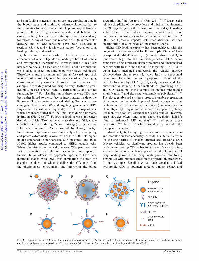
This journal is c The Royal Society of Chemistry 2010 Chem. Soc. Rev.
and non-fouling materials that ensure long circulation time in
the bloodstream and optimized pharmacokinetics, feature
functionalities for overcoming multiple physiological barriers,
possess sufficient drug loading capacity, and balance the
carrier’s affinity for the therapeutic agent with its tendency
for release. Many of the criteria related to the QD intracellular
delivery and in vivo targeting have been discussed in
sections 3.3, 4.3, and 4.4, while this section focuses on drug
loading, release, and sensing.
QDs feature versatile surface chemistry that enables
attachment of various ligands and loading of both hydrophilic
and hydrophobic therapeutics. However, being a relatively
new technology, QD-based drug delivery is not as robust and
well-studied as other liposome or polymer-based techniques.
Therefore, a more common and straightforward approach
involves utilization of QDs as fluorescent markers for tagging
conventional drug carriers. Liposomes and micelles, for
example, are widely used for drug delivery, featuring great
flexibility in size, charge, rigidity, permeability, and surface
functionality.185 For visualization of these vesicles, QDs have
been either linked to the surface or incorporated inside of the
liposomes. To demonstrate external labeling, Weng et al. have
conjugated hydrophilic QDs and targeting ligands (anti-HER2
single-chain Fv antibody fragments) to PEG-phospholipids,
which are incorporated into the lipid layer during liposome
hydration (Fig. 23A).186 Following loading with anticancer
drug doxorubicin (Dox), targeted, traceable, and fairly stable
(15–30% Dox loss during 2-month storage) drug delivery
vehicles are obtained. As determined by flow-cytometry,
functionalized liposomes show remarkably selective targeting
and potent cytotoxicity in vitro, with 900 to 1800-fold higher
uptake compared to non-targeted QD-liposomes, and 16 to
30-fold higher uptake compared to HER2-negative cells.
When administered systemically in vivo, QD-liposomes have
B3 h circulation half-life and accumulate in implanted
tumors. In an alternative approach, liposomes have been
internally loaded with QDs, thus eliminating the need for
chemical conjugation while shielding the QD tags from
the physiological environment and improving the blood
circulation half-life (up to 5 h) (Fig. 23B).187,188 Despite the
relative simplicity of the procedure and minimal requirements
for QD tag design, both external and internal QD loading
suffer from reduced drug loading capacity and poor
fluorescence intensity, as surface attachment of more than 2
QDs per liposome impedes cell internalization, whereas
incorporation of QDs inside of liposomes is sparse.
Higher QD loading capacity has been achieved with the
polymeric drug delivery vehicles. For example, Kim et al. have
incorporated MitoTracker dye (a model drug) and QDs
(fluorescent tag) into 100 nm biodegradable PLGA nano-
composites using a microemulsion procedure and functionalized
particles with trastuzumab for HER2 targeting (Fig. 23C).116
Upon ligand mediated endocytosis the vehicles undergo
pH-dependent charge reversal, which leads to endosomal
membrane destabilization and cytoplasmic release of the
particles followed by PLGA hydrolysis, dye release, and clear
mitochondria staining. Other methods of producing drug-
and QD-loaded polymeric composites include microfluidic
emulsification189 and electrostatic assembly of polyplexes.190,191
Therefore, established synthesis protocols enable preparation
of nanocomposites with improved loading capacity that
facilitate sensitive fluorescence detection (via incorporation
of multiple QD tags) and enhanced therapeutic potency
(via high drug content) essential for in vivo studies. However,
large particles often suffer from short circulation half-life
(due to enhanced RES uptake)192,193 and poor tissue
penetration,148 both of which significantly impede the
therapeutic potential.
Individual QDs, having high surface area to volume ratio
and modular surface chemistry, provide a suitable platform
for the engineering of smaller targeted and traceable drug
delivery vehicles. As significant progress has already been
made in engineering QD probes for targeted in vivo imaging,
a major focus is now being placed on developing novel
drug loading routes and drug loading/release monitoring
capabilities with minimal effect on the overall QD properties.
In one example, Bagalkot et al. have covalently linked
hydrophilic QDs to aptamers targeted against PSMA and
Fig. 23 Engineering of QD-based therapeutic nanocomposites. QDs can be used as tags for labeling of larger drug carriers, such as liposomes
(A, B) and polymeric nanoparticles (C), or as single-QD platforms for traceable drug loading and delivery (D–F).
Dow
nloa
ded
by U
NIV
ER
SIT
Y O
F W
ASH
ING
TO
N o
n 22
Aug
ust 2
010
Publ
ishe
d on
09
Aug
ust 2
010
on h
ttp://
pubs
.rsc
.org
| do
i:10.
1039
/B91
5139
GView Online
Chem. Soc. Rev. This journal is c The Royal Society of Chemistry 2010
loaded Dox via intercalation within the double-stranded CG
portion of the aptamers at B1 : 1 ratio (Fig. 23D).194 Drug
loading/release sensing has been achieved with carefully
designed bi-FRET arrangement. By matching the QD emission
with Dox absorption the drug loading can be monitored via
QD fluorescence quenching. At the same time, as Dox
fluorescence is also efficiently quenched by the aptamer, the
overall Dox-loaded targeted QD probes remain in a
non-emitting state. Upon specific delivery into PSMA-
expressing prostate cancer cells, slow Dox release can be
monitored by recovery of both green QD and red Dox
fluorescence. Yet, the drug loading is strictly limited by the
number of aptamers conjugated to each QD, thus requiring
administration of higher QD dose in order to reach a
therapeutic response.
The low drug loading capacity of single QDs can be
employed for delivery of highly potent therapeutics, such as
siRNA, a 22 bp-long double-stranded RNA that efficiently
suppresses expression of specific genes by initiating cleavage of
corresponding mRNA inside cells.195 Most often siRNA is
loaded on the hydrophilic surface of QDs either via covalent
conjugation or electrostatic interaction. For example, Derfus
et al. have functionalized QDs with tumor-homing peptides
and covalently conjugated anti-GFP siRNA via a cleavable
disulfide linker to suppress GFP expression in HeLa cells
(Fig. 23E).196 Utilization of a cleavable linker ensures high
stability of a QD-siRNA complex, while providing efficient
intracellular siRNA release essential for effective interference.
Walther et al., on the other hand, have absorbed 80–100 bp
cyanine-labeled RNAs via electrostatic interaction with
cationic internalization peptides coated on the QD surface,
achieving intracellular siRNA delivery.197 However, both
delivery approaches suffer from endosomal sequestration of
the particles and require additional treatment of cells with
endosome-disrupting agents (cationic liposomes196 or
chloroquine)197 for efficient cytosolic release.
QD carriers that have intrinsic endosomal escape
functionality along with targeting and siRNA carrying
capacity have recently gained increasing attention due to their
capability of one-step efficient and high-throughput siRNA
delivery. To achieve this goal, fusogenic peptides109,198 that
disrupt membranes via hydrophobic interactions and tertiary
amine-rich polymers117,119 that elicit the proton sponge effect
have been utilized. In one example, Yezhelyev et al. have
modified amphiphilic polymer-coated QDs with tertiary
amines and tuned the surface charge to balance the competing
electrostatic effects of QD-siRNA binding and intracellular
siRNA release (Fig. 23F).119 In a similar report, Qi and Gao
have used a pre-modified amphiphilic polymer for QD
encapsulation and siRNA loading, demonstrating improved
knockdown of the HER2/neu gene in SK-BR-3 cells.199
Importantly, QD carriers protect adsorbed siRNA from
enzymatic degradation and enable real-time monitoring of
FITC-labeled siRNA loading/release via FRET quenching of
FITC signal by QDs and restoration of green fluorescence
upon cytoplasmic siRNA release.
Drug carriers based on single QDs are often small (o20 nm)
relative to conventional delivery vehicles, thus facilitating
improved extravasation from vasculature and accumulation
in target organs.184,200,201 At the same time, compact probes
with zwitterionic surfaces are much more efficient in avoiding
opsonization and RES uptake.58 However, the amount of drug
delivered per QD is limited by the particle’s surface area,
which must be shared with other ligands for molecular
targeting and/or cell internalization. Therefore, the most
promising in vivo application of single-QD nanotherapeutics
will be in monitoring the delivery of highly potent drugs
(such as siRNA) and modeling the biodistribution of other
nanoparticle-based drug carriers with similar surface
properties and physical dimensions, yet higher drug loading
capacities (e.g. polymeric nanoparticles). Future work will
likely focus on incorporation of environmentally responsive
materials for controlled drug release, non-fouling surface
coatings for improved biodistribution, and multistage targeting
functionality for enhanced therapeutic specificity.
6. Conclusions
The application of nanoparticles for the multi-parameter
comprehensive study of physiological and pathological
processes in biomedical research as well as for advanced
diagnostics and therapy in clinical practice presents a surging
trend in nanomedicine. QDs, in particular, have emerged as
one of the most promising classes of nanoparticles for bio-
medical imaging, drug delivery, and sensing due to their
unique photo-physical properties and versatile surface
chemistry. Biofunctionalization of inorganic QD cores
facilitates interaction of nanoparticles with biological systems
and enables direct participation in biological processes. As a
result, QD probes have been utilized in a wide variety of
applications spanning in vitro molecular pathology, live cell
imaging, and in vivo drug delivery and tracing. With each
application QDs have opened new horizons of multiplexed
quantitative detection, sensitive high-resolution fluorescence
imaging, and long-term real-time monitoring of probe
dynamics. Aiming at expanding QD functionality even
further, early steps have been made towards engineering of
QD-based multi-functional nanodevices that promise to
combine the benefits of multiple imaging modalities and
incorporate the imaging, drug loading, and sensing capacities
within a single nanoparticle. Yet, currently available QD
probes are far from being ideal, leaving plenty of room for
improvement of existing and development of novel nano-
particle designs. In this review we have discussed the future
directions of QD-based bio-nanotechnology research and
outlined the major design principles and criteria, from general
ones to application-specific, governing the engineering of
novel QD probes satisfying increasing demands and require-
ments of nanomedicine.
Acknowledgements
This work was supported in part by NIH (R01CA131797,
R01CA140295), NSF, and the Department of Bioengineering
at the University of Washington. X.G. thanks the NSF for
a Faculty Early Career Development award (CAREER).
P.Z. thanks the UW Center for Nanotechnology for a UIF
fellowship and the National Science Foundation for Graduate
Dow
nloa
ded
by U
NIV
ER
SIT
Y O
F W
ASH
ING
TO
N o
n 22
Aug
ust 2
010
Publ
ishe
d on
09
Aug
ust 2
010
on h
ttp://
pubs
.rsc
.org
| do
i:10.
1039
/B91
5139
GView Online

This journal is c The Royal Society of Chemistry 2010 Chem. Soc. Rev.
Research Fellowship. M.S. thanks Mary Gates Endowment
for Students for Research Scholarship, and Amgen Scholars
Program. We are also grateful to Shivang Dave for critical
reading of the manuscript.
Notes and references
1 Nanoscale Science Engineering and Technology Subcommittee(NSET), Committee on Technology (CT) and National Scienceand Technology Council (NSTC), 2009, 44.
2 H. Chen, M. C. Roco, X. Li and Y. Lin, Nat. Nanotechnol., 2008,3, 123–125.
3 T. F. Massoud and S. S. Gambhir, Genes Dev., 2003, 17, 545–580.4 D. A. Mankoff, J. Nucl. Med., 2007, 48, 18N, 21N.5 I. L. Medintz and H. Mattoussi, Phys. Chem. Chem. Phys., 2009,11, 17–45.
6 \I. L. Medintz, H. Mattoussi and A. R. Clapp, Int. J. Nanomedicine,2008, 3, 151–167.
7 R. D. Misra, Nanomedicine, 2008, 3, 271–274.8 U. Resch-Genger, M. Grabolle, S. Cavaliere-Jaricot, R. Nitschkeand T. Nann, Nat. Methods, 2008, 5, 763–775.
9 A. M. Smith, H. Duan, A. M. Mohs and S. Nie, Adv. DrugDelivery Rev., 2008, 60, 1226–1240.
10 E. Tholouli, E. Sweeney, E. Barrow, V. Clay, J. A. Hoyland andR. J. Byers, J. Pathol., 2008, 216, 275–285.
11 Y. Xing and J. Rao, Cancer Biomark, 2008, 4, 307–319.12 P. Zrazhevskiy and X. Gao, Nano Today, 2009, 4, 414–428.13 A. P. Alivisatos, J. Phys. Chem., 1996, 100, 13226–13239.14 L. E. Brus, J. Chem. Phys., 1984, 80, 4403–4409.15 R. Rossetti, S. Nakahara and L. E. Brus, J. Chem. Phys., 1983,
79, 1086–1088.16 R. S. Zeng, T. T. Zhang, J. C. Liu, S. Hu, Q. Wan, X. M. Liu,
Z. W. Peng and B. S. Zou, Nanotechnology, 2009, 20, 095102.17 Y. G. Zheng, Z. C. Yang and J. Y. Ying, Adv. Mater., 2007, 19,
1475–1479.18 C. B. Murray, D. J. Norris and M. G. Bawendi, J. Am. Chem.
Soc., 1993, 115, 8706–8715.19 Z. A. Peng and X. G. Peng, J. Am. Chem. Soc., 2001, 123, 183–184.20 Z. A. Peng and X. Peng, J. Am. Chem. Soc., 2002, 124, 3343–3353.21 A. P. Alivisatos, Science, 1996, 271, 933–937.22 L. S. Li, N. Pradhan, Y. Wang and X. Peng, Nano Lett., 2004, 4,
2261–2264.23 X. Peng, J. Wickham and A. P. Alivisatos, J. Am. Chem. Soc.,
1998, 120, 5343–5344.24 X. H. Zhong, Y. Y. Feng, W. Knoll and M. Y. Han, J. Am.
Chem. Soc., 2003, 125, 13559–13563.25 S. Kim, B. Fisher, H. J. Eisler and M. Bawendi, J. Am. Chem.
Soc., 2003, 125, 11466–11467.26 J. M. Pietryga, R. D. Schaller, D. Werder, M. H. Stewart,
V. I. Klimov and J. A. Hollingsworth, J. Am. Chem. Soc., 2004,126, 11752–11753.
27 L. Qu and X. Peng, J. Am. Chem. Soc., 2002, 124, 2049–2055.28 A. M. Smith, X. Gao and S. Nie, Photochem. Photobiol., 2004, 80,
377–385.29 X. H. Zhong, M. Y. Han, Z. L. Dong, T. J. White and W. Knoll,
J. Am. Chem. Soc., 2003, 125, 8589–8594.30 R. E. Bailey and S.M. Nie, J. Am. Chem. Soc., 2003, 125, 7100–7106.31 P. K. Chattopadhyay, D. A. Price, T. F. Harper, M. R. Betts,
J. Yu, E. Gostick, S. P. Perfetto, P. Goepfert, R. A. Koup,S. C. De Rosa, M. P. Bruchez and M. Roederer, Nat. Med.,2006, 12, 972–977.
32 M. V. Yezhelyev, A. Al-Hajj, C. Morris, A. I. Marcus, T. Liu,M. Lewis, C. Cohen, P. Zrazhevskiy, J. W. Simons, A. Rogatko,S. Nie, X. Gao and R. M. O. Regan, Adv. Mater., 2007, 19,3146–3151.
33 B. N. Giepmans, T. J. Deerinck, B. L. Smarr, Y. Z. Jones andM. H. Ellisman, Nat. Methods, 2005, 2, 743–749.
34 R. Nisman, G. Dellaire, Y. Ren, R. Li and D. P. Bazett-Jones,J Histochem. Cytochem., 2004, 52, 13–18.
35 G. Oberdorster, E. Oberdorster and J. Oberdorster, Environ.Health Perspect., 2005, 113, 823–839.
36 G. W. Bryant and W. Jaskolski, J. Phys. Chem. B, 2005, 109,19650–19656.
37 D. E. Gomez, M. Califano and P. Mulvaney, Phys. Chem. Chem.Phys., 2006, 8, 4989–5011.
38 B. O. Dabbousi, J. Rodriguez-Viejo, F. V. Mikulec, J. R. Heine,H. Mattoussi, R. Ober, K. F. Jensen and M. G. Bawendi, J. Phys.Chem. B, 1997, 101, 9463–9475.
39 M. A. Hines and P. Guyot-Sionnest, J. Phys. Chem., 1996, 100,468–471.
40 X. Peng, M. C. Schlamp, A. V. Kadavanich and A. P. Alivisatos,J. Am. Chem. Soc., 1997, 119, 7019–7029.
41 W. C. Chan, D. J. Maxwell, X. Gao, R. E. Bailey, M. Han andS. Nie, Curr. Opin. Biotechnol., 2002, 13, 40–46.
42 I. L. Medintz, H. T. Uyeda, E. R. Goldman and H. Mattoussi,Nat. Mater., 2005, 4, 435–446.
43 A. L. Efros and M. Rosen, Phys. Rev. Lett., 1997, 78, 1110–1113.44 X. Michalet, F. Pinaud, T. D. Lacoste, M. Dahan, M. P. Bruchez,
A. P. Alivisatos and S. Weiss, Single Mol., 2001, 2, 261–276.45 M. Nirmal, B. O. Dabbousi, M. G. Bawendi, J. J. Macklin,
J. K. Trautman, T. D. Harris and L. E. Brus, Nature, 1996, 383,802–804.
46 F. Pinaud, X. Michalet, L. A. Bentolila, J. M. Tsay, S. Doose,J. J. Li, G. Iyer and S. Weiss, Biomaterials, 2006, 27, 1679–1687.
47 R. Comparelli, F. Zezza, M. Striccoli, M. L. Curri, R. Tommasiand A. Agostiano, Mater.Sci. Eng., C, 2003, 23, 1083–1086.
48 J. J. Li, Y. A. Wang, W. Guo, J. C. Keay, T. D. Mishima,M. B. Johnson and X. Peng, J. Am. Chem. Soc., 2003, 125,12567–12575.
49 D. V. Talapin, A. L. Rogach, A. Kornowski, M. Haase andH. Weller, Nano Lett., 2001, 1, 207–211.
50 M. Bruchez, Jr., M. Moronne, P. Gin, S. Weiss andA. P. Alivisatos, Science, 1998, 281, 2013–2016.
51 W. C. Chan and S. Nie, Science, 1998, 281, 2016–2018.52 X. Gao and S. Nie, Trends Biotechnol., 2003, 21, 371–373.53 X. Wu, H. Liu, J. Liu, K. N. Haley, J. A. Treadway, J. P. Larson,
N. Ge, F. Peale and M. P. Bruchez, Nat. Biotechnol., 2003, 21,41–46.
54 X. Gao, L. Yang, J. A. Petros, F. F. Marshall, J. W. Simons andS. Nie, Curr. Opin. Biotechnol., 2005, 16, 63–72.
55 F. Tokumasu and J. Dvorak, J. Microsc., 2003, 211, 256–261.56 B. Cui, C. Wu, L. Chen, A. Ramirez, E. L. Bearer, W. P. Li,
W. C. Mobley and S. Chu, Proc. Natl. Acad. Sci. U. S. A., 2007,104, 13666–13671.
57 D. S. Lidke, P. Nagy, R. Heintzmann, D. J. Arndt-Jovin,J. N. Post, H. E. Grecco, E. A. Jares-Erijman and T. M. Jovin,Nat. Biotechnol., 2004, 22, 198–203.
58 H. S. Choi, W. Liu, P. Misra, E. Tanaka, J. P. Zimmer, B. IttyIpe, M. G. Bawendi and J. V. Frangioni, Nat. Biotechnol., 2007,25, 1165–1170.
59 W. C. Law, K. T. Yong, I. Roy, H. Ding, R. Hu, W. Zhao andP. N. Prasad, Small, 2009, 5, 1302–1310.
60 J. Aldana, Y. A. Wang and X. Peng, J. Am. Chem. Soc., 2001,123, 8844–8850.
61 W. R. Algar and U. J. Krull, ChemPhysChem, 2007, 8, 561–568.62 W. Liu, M. Howarth, A. B. Greytak, Y. Zheng, D. G. Nocera,
A. Y. Ting and M. G. Bawendi, J. Am. Chem. Soc., 2008, 130,1274–1284.
63 A. Sukhanova, J. Devy, L. Venteo, H. Kaplan, M. Artemyev,V. Oleinikov, D. Klinov, M. Pluot, J. H. Cohen and I. Nabiev,Anal. Biochem., 2004, 324, 60–67.
64 W. Jiang, S. Mardyani, H. Fischer and W. C. W. Chan, Chem.Mater., 2006, 18, 872–878.
65 A. M. Smith and S. Nie, J. Am. Chem. Soc., 2008, 130,11278–11279.
66 D. Gerion, F. Pinaud, S. C. Williams, W. J. Parak, D. Zanchet,S. Weiss and A. P. Alivisatos, J. Phys. Chem. B, 2001, 105,8861–8871.
67 F. Pinaud, D. King, H. P. Moore and S. Weiss, J. Am. Chem.Soc., 2004, 126, 6115–6123.
68 S. R. Whaley, D. S. English, E. L. Hu, P. F. Barbara andA. M. Belcher, Nature, 2000, 405, 665–668.
69 T. Pellegrino, L. Manna, S. Kudera, T. Liedl, D. Koktysh,A. L. Rogach, S. Keller, J. Radler, G. Natile and W. J. Parak,Nano Lett., 2004, 4, 703–707.
70 B. Dubertret, P. Skourides, D. J. Norris, V. Noireaux,A. H. Brivanlou and A. Libchaber, Science (New York, N.Y.),2002, 298, 1759–1762.
Dow
nloa
ded
by U
NIV
ER
SIT
Y O
F W
ASH
ING
TO
N o
n 22
Aug
ust 2
010
Publ
ishe
d on
09
Aug
ust 2
010
on h
ttp://
pubs
.rsc
.org
| do
i:10.
1039
/B91
5139
GView Online

Chem. Soc. Rev. This journal is c The Royal Society of Chemistry 2010
71 X. Gao, Y. Cui, R. M. Levenson, L. W. Chung and S. Nie, Nat.Biotechnol., 2004, 22, 969–976.
72 A. M. Smith, H. Duan, M. N. Rhyner, G. Ruan and S. Nie, Phys.Chem. Chem. Phys., 2006, 8, 3895–3903.
73 M. Dahan, S. Levi, C. Luccardini, P. Rostaing, B. Riveau andA. Triller, Science, 2003, 302, 442–445.
74 B. Barat, S. J. Sirk, K. E. McCabe, J. Li, E. J. Lepin, R. Remenyi,A. L. Koh, T. Olafsen, S. S. Gambhir, S. Weiss and A. M. Wu,Bioconjugate Chem., 2009, 20, 1474–1481.
75 I. L. Medintz, A. R. Clapp, H. Mattoussi, E. R. Goldman,B. Fisher and J. M. Mauro, Nat. Mater., 2003, 2, 630–638.
76 I. L. Medintz, A. R. Clapp, F. M. Brunel, T. Tiefenbrunn,H. T. Uyeda, E. L. Chang, J. R. Deschamps, P. E. Dawson andH. Mattoussi, Nat. Mater., 2006, 5, 581–589.
77 J. B. Delehanty, I. L. Medintz, T. Pons, F. M. Brunel,P. E. Dawson and H. Mattoussi, Bioconjugate Chem., 2006, 17,920–927.
78 E. R. Goldman, G. P. Anderson, P. T. Tran, H. Mattoussi,P. T. Charles and J. M. Mauro, Anal. Chem., 2002, 74, 841–847.
79 H. Mattoussi, J. M. Mauro, E. R. Goldman, G. P. Anderson,V. C. Sundar, F. V. Mikulec and M. G. Bawendi, J. Am. Chem.Soc., 2000, 122, 12142–12150.
80 M. T. Fernandez-Arguelles, A. Yakovlev, R. A. Sperling,C. Luccardini, S. Gaillard, A. S. Medel, J. M. Mallet,J. C. Brochon, A. Feltz, M. Oheim and W. J. Parak, Nano Lett.,2007, 7, 2613–2617.
81 C. A. J. Lin, R. A. Sperling, J. K. Li, T. Y. Yang, P. Y. Li,M. Zanella, W. H. Chang and W. G. J. Parak, Small, 2008, 4,334–341.
82 A. P. Alivisatos, ACS Nano, 2008, 2, 1514–1516.83 R. J. Byers, D. Di Vizio, F. O’Connell, E. Tholouli,
R. M. Levenson, K. Gossage, D. Twomey, Y. Yang,E. Benedettini, J. Rose, K. L. Ligon, S. P. Finn, T. R. Goluband M. Loda, J. Mol. Diagn., 2007, 9, 20–29.
84 L. D. True and X. Gao, J. Mol. Diagn., 2007, 9, 7–11.85 K. K. Jain, Expert Opin. Pharmacother., 2005, 6, 1463–1476.86 E. Tholouli, J. A. Hoyland, D. Di Vizio, F. O’Connell,
S. A. Macdermott, D. Twomey, R. Levenson, J. A. Yin,T. R. Golub, M. Loda and R. Byers, Biochem. Biophys. Res.Commun., 2006, 348, 628–636.
87 P. Chan, T. Yuen, F. Ruf, J. Gonzalez-Maeso and S. C. Sealfon,Nucleic Acids Res., 2005, 33, e161.
88 R. M. Levenson, Lab. Med., 2004, 35, 244–251.89 A. A. Ghazani, J. A. Lee, J. Klostranec, Q. Xiang, R. S. Dacosta,
B. C. Wilson, M. S. Tsao and W. C. Chan, Nano Lett., 2006, 6,2881–2886.
90 X. H. Zhong, Z. H. Zhang, S. H. Liu, M. Y. Han and W. Knoll,J. Phys. Chem. B, 2004, 108, 15552–15559.
91 N. P. Wells, G. A. Lessard and J. H. Werner, Anal. Chem., 2008,80, 9830–9834.
92 M. J. Murcia, D. E. Minner, G. M. Mustata, K. Ritchie andC. A. Naumann, J. Am. Chem. Soc., 2008, 130, 15054–15062.
93 J. K. Jaiswal, H. Mattoussi, J. M. Mauro and S. M. Simon, Nat.Biotechnol., 2003, 21, 47–51.
94 M. Heine, L. Groc, R. Frischknecht, J. C. Beique, B. Lounis,G. Rumbaugh, R. L. Huganir, L. Cognet and D. Choquet,Science, 2008, 320, 201–205.
95 Q. Zhang, Y. Li and R. W. Tsien, Science, 2009, 323, 1448–1453.96 A. Cambi, D. S. Lidke, D. J. Arndt-Jovin, C. G. Figdor and
T. M. Jovin, Nano Lett., 2007, 7, 970–977.97 V. Roullier, S. Clarke, C. You, F. Pinaud, G. G. Gouzer,
D. Schaible, V. Marchi-Artzner, J. Piehler and M. Dahan, NanoLett., 2009, 9, 1228–1234.
98 J. B. Delehanty, H. Mattoussi and I. L. Medintz, Anal. Bioanal.Chem., 2009, 393, 1091–1105.
99 S. Barua and K. Rege, Small, 2009, 5, 370–376.100 W. Jiang, B. Y. Kim, J. T. Rutka and W. C. Chan, Nat.
Nanotechnol., 2008, 3, 145–150.101 I. Nabiev, S. Mitchell, A. Davies, Y. Williams, D. Kelleher,
R. Moore, Y. K. Gun’ko, S. Byrne, Y. P. Rakovich,J. F. Donegan, A. Sukhanova, J. Conroy, D. Cottell,N. Gaponik, A. Rogach and Y. Volkov, Nano Lett., 2007, 7,3452–3461.
102 J. Lovric, H. S. Bazzi, Y. Cuie, G. R. Fortin, F. M. Winnik andD. Maysinger, J. Mol. Med., 2005, 83, 377–385.
103 B. A. Kairdolf, M. C. Mancini, A. M. Smith and S. Nie, Anal.Chem., 2008, 80, 3029–3034.
104 A. M. Derfus, W. C. W. Chan and S. N. Bhatia, Adv. Mater.,2004, 16, 961–966.
105 K. Yum, S. Na, Y. Xiang, N. Wang and M. F. Yu, Nano Lett.,2009, 9, 2193–2198.
106 S. Park, Y. S. Kim, W. B. Kim and S. Jon, Nano Lett., 2009, 9,1325–1329.
107 M. Zhou and I. Ghosh, Biopolymers, 2007, 88, 325–339.108 J. M. de la Fuente, M. Fandel, C. C. Berry, M. Riehle, L. Cronin,
G. Aitchison and A. S. Curtis, ChemBioChem, 2005, 6, 989–991.109 Y. E. Koshman, S. B. Waters, L. A. Walker, T. Los, P. de Tombe,
P. H. Goldspink and B. Russell, J. Mol. Cell. Cardiol., 2008, 45,853–856.
110 S. L. Sewell and T. D. Giorgio, Mater. Sci. Eng., C, 2009, 29,1428–1432.
111 B. C. Lagerholm, M. Wang, L. A. Ernst, D. H. Ly, H. Liu,M. P. Bruchez and A. S. Waggoner,Nano Lett., 2004, 4, 2019–2022.
112 S. M. Rozenzhak, M. P. Kadakia, T. M. Caserta,T. R. Westbrook, M. O. Stone and R. R. Naik, Chem. Commun.,2005, 2217–2219.
113 J. Silver and W. Ou, Nano Lett., 2005, 5, 1445–1449.114 L. Qi and X. Gao, Expert Opin. Drug Delivery, 2008, 5, 263–267.115 S. Courty, C. Luccardini, Y. Bellaiche, G. Cappello and
M. Dahan, Nano Lett., 2006, 6, 1491–1495.116 B. Y. Kim, W. Jiang, J. Oreopoulos, C. M. Yip, J. T. Rutka and
W. C. Chan, Nano Lett., 2008, 8, 3887–3892.117 H. Duan and S. Nie, J. Am. Chem. Soc., 2007, 129, 3333–3338.118 N. D. Sonawane, F. C. Szoka, Jr. and A. S. Verkman, J. Biol.
Chem., 2003, 278, 44826–44831.119 M. V. Yezhelyev, L. Qi, R. M. O’Regan, S. Nie and X. Gao,
J. Am. Chem. Soc., 2008, 130, 9006–9012.120 N. McGrath and M. Barroso, J. Biomed. Opt., 2008, 13, 031210.121 A. R. Clapp, T. Pons, I. L. Medintz, J. B. Delehanty,
J. S. Melinger, T. Tiefenbrunn, P. E. Dawson, B. R. Fisher,B. O. Rourke and H. Mattoussi, Adv. Mater., 2007, 19,1921–1926.
122 L. A. Bentolila, Y. Ebenstein and S. Weiss, J. Nucl. Med., 2009,50, 493–496.
123 E. E. Graves, R. Weissleder and V. Ntziachristos, Curr. Mol.Med., 2004, 4, 419–430.
124 R. K. Jain, L. L. Munn and D. Fukumura, Nat. Rev. Cancer,2002, 2, 266–276.
125 M. Makale, Methods Enzymol., 2007, 426, 375–401.126 R. Hardman, Environ. Health Perspect., 2006, 114, 165–172.127 J. Lee, G. D. Lilly, R. C. Doty, P. Podsiadlo and N. A. Kotov,
Small, 2009, 5, 1213–1221.128 N. A. Monteiro-Riviere, A. O. Inman and L. W. Zhang, Toxicol.
Appl. Pharmacol., 2009, 234, 222–235.129 A. M. Derfus, W. C. W. Chan and S. N. Bhatia,Nano Lett., 2004,
4, 11–18.130 T. Zhang, J. L. Stilwell, D. Gerion, L. Ding, O. Elboudwarej,
P. A. Cooke, J. W. Gray, A. P. Alivisatos and F. F. Chen, NanoLett., 2006, 6, 800–808.
131 A. Hoshino, N. Manabe, K. Fujioka, K. Suzuki, M. Yasuharaand K. Yamamoto, J. Artif. Organs, 2007, 10, 149–157.
132 Y. Su, Y. He, H. Lu, L. Sai, Q. Li, W. Li, L. Wang, P. Shen,Q. Huang and C. Fan, Biomaterials, 2008, 30, 19–25.
133 S. J. Cho, D. Maysinger, M. Jain, B. Roder, S. Hackbarth andF. M. Winnik, Langmuir, 2007, 23, 1974–1980.
134 T. C. King-Heiden, P. N. Wiecinski, A. N. Mangham,K. M. Metz, D. Nesbit, J. A. Pedersen, R. J. Hamers,W. Heideman and R. E. Peterson, Environ. Sci. Technol., 2009,43, 1605–1611.
135 A. Nel, T. Xia, L. Madler and N. Li, Science, 2006, 311, 622–627.136 H. S. Choi, W. Liu, F. Liu, K. Nasr, P. Misra, M. G. Bawendi
and J. V. Frangioni, Nat. Nanotechnol., 2009, 5, 42–47.137 B. Ballou, B. C. Lagerholm, L. A. Ernst, M. P. Bruchez and
A. S. Waggoner, Bioconjugate Chem., 2004, 15, 79–86.138 G. Prencipe, S. M. Tabakman, K. Welsher, Z. Liu,
A. P. Goodwin, L. Zhang, J. Henry and H. Dai, J. Am. Chem.Soc., 2009, 131, 4783–4787.
139 J. Geys, A. Nemmar, E. Verbeken, E. Smolders, M. Ratoi,M. F. Hoylaerts, B. Nemery and P. H. Hoet, Environ. HealthPerspect., 2008, 116, 1607–1613.
Dow
nloa
ded
by U
NIV
ER
SIT
Y O
F W
ASH
ING
TO
N o
n 22
Aug
ust 2
010
Publ
ishe
d on
09
Aug
ust 2
010
on h
ttp://
pubs
.rsc
.org
| do
i:10.
1039
/B91
5139
GView Online

This journal is c The Royal Society of Chemistry 2010 Chem. Soc. Rev.
140 J. A. Fitzpatrick, S. K. Andreko, L. A. Ernst, A. S. Waggoner,B. Ballou and M. P. Bruchez, Nano Lett., 2009, 9, 2736–2741.
141 S. Satarug, M. R. Haswell-Elkins and M. R. Moore, Br. J. Nutr.,2000, 84, 791–802.
142 K. T. Yong, H. Ding, I. Roy, W. C. Law, E. J. Bergey, A. Maitraand P. N. Prasad, ACS Nano, 2009, 3, 502–510.
143 L. Li, T. J. Daou, I. Texier, T. T. Kim Chi, N. Q. Liem andP. Reiss, Chem. Mater., 2009, 21, 2422–2429.
144 Z. H. Kang, Y. Liu, C. H. A. Tsang, D. D. D. Ma, X. Fan,N. B. Wong and S. T. Lee, Adv. Mater., 2009, 21, 661–664.
145 J. H. Park, L. Gu, G. von Maltzahn, E. Ruoslahti, S. N. Bhatiaand M. J. Sailor, Nat. Mater., 2009, 8, 331–336.
146 B. Ballou, L. A. Ernst, S. Andreko, J. A. Fitzpatrick,B. C. Lagerholm, A. S. Waggoner and M. P. Bruchez, MethodsMol. Biol., 2009, 574, 63–74.
147 D. R. Larson, W. R. Zipfel, R. M. Williams, S. W. Clark,M. P. Bruchez, F. W. Wise and W. W. Webb, Science, 2003,300, 1434–1436.
148 M. Stroh, J. P. Zimmer, D. G. Duda, T. S. Levchenko,K. S. Cohen, E. B. Brown, D. T. Scadden, V. P. Torchilin,M. G. Bawendi, D. Fukumura and R. K. Jain, Nat. Med.,2005, 11, 678–682.
149 J. V. Kim, S. S. Kang, M. L. Dustin and D. B. McGavern,Nature, 2009, 457, 191–195.
150 B. Ballou, L. A. Ernst, S. Andreko, T. Harper, J. A. Fitzpatrick,A. S. Waggoner and M. P. Bruchez, Bioconjugate Chem., 2007,18, 389–396.
151 S. Kim, Y. T. Lim, E. G. Soltesz, A. M. De Grand, J. Lee,A. Nakayama, J. A. Parker, T. Mihaljevic, R. G. Laurence,D. M. Dor, L. H. Cohn, M. G. Bawendi and J. V. Frangioni,Nat. Biotechnol., 2004, 22, 93–97.
152 E. G. Soltesz, S. Kim, R. G. Laurence, A. M. DeGrand,C. P. Parungo, D. M. Dor, L. H. Cohn, M. G. Bawendi,J. V. Frangioni and T. Mihaljevic, Ann. Thorac. Surg., 2005, 79,269–277; discussion 269-277.
153 J. P. Zimmer, S. W. Kim, S. Ohnishi, E. Tanaka, J. V. Frangioniand M. G. Bawendi, J. Am. Chem. Soc., 2006, 128, 2526–2527.
154 H. Kobayashi, Y. Hama, Y. Koyama, T. Barrett, C. A. Regino,Y. Urano and P. L. Choyke, Nano Lett., 2007, 7, 1711–1716.
155 M. J. Levene, D. A. Dombeck, K. A. Kasischke, R. P. Molloyand W. W. Webb, J. Neurophysiol., 2004, 91, 1908–1912.
156 E. J. Sutton, T. D. Henning, B. J. Pichler, C. Bremer andH. E. Daldrup-Link, Eur. Radiol., 2008, 18, 2021–2032.
157 J. R. Slotkin, L. Chakrabarti, H. N. Dai, R. S. Carney, T. Hirata,B. S. Bregman, G. I. Gallicano, J. G. Corbin and T. F. Haydar,Dev. Dyn., 2007, 236, 3393–3401.
158 A. Jayagopal, P. K. Russ and F. R. Haselton, BioconjugateChem., 2007, 18, 1424–1433.
159 E. B. Voura, J. K. Jaiswal, H. Mattoussi and S. M. Simon, Nat.Med., 2004, 10, 993–998.
160 C. Lo Celso, H. E. Fleming, J. W. Wu, C. X. Zhao, S. Miake-Lye,J. Fujisaki, D. Cote, D. W. Rowe, C. P. Lin and D. T. Scadden,Nature, 2009, 457, 92–96.
161 M. K. So, C. Xu, A. M. Loening, S. S. Gambhir and J. Rao, Nat.Biotechnol., 2006, 24, 339–343.
162 M. E. Akerman, W. C. Chan, P. Laakkonen, S. N. Bhatia andE. Ruoslahti, Proc. Natl. Acad. Sci. U. S. A., 2002, 99,12617–12621.
163 W. Cai, D. W. Shin, K. Chen, O. Gheysens, Q. Cao, S. X. Wang,S. S. Gambhir and X. Chen, Nano Lett., 2006, 6, 669–676.
164 L. Yang, H. Mao, Y. A. Wang, Z. Cao, X. Peng, X. Wang,H. Duan, C. Ni, Q. Yuan, G. Adams, M. Q. Smith, W. C. Wood,X. Gao and S. Nie, Small, 2009, 5, 235–243.
165 H. Tada, H. Higuchi, T. M. Wanatabe and N. Ohuchi, CancerRes., 2007, 67, 1138–1144.
166 M. F. Kircher, U. Mahmood, R. S. King, R. Weissleder andL. Josephson, Cancer Res., 2003, 63, 8122–8125.
167 W. Cai and X. Chen, J. Nucl. Med., 2008, 49(suppl_2), 113S–128S.168 W. J. Mulder, D. P. Cormode, S. Hak, M. E. Lobatto, S. Silvera
and Z. A. Fayad, Nat. Clin. Pract. Cardiovasc. Med., 2008,5(Suppl 2), S103–111.
169 W. Cai and X. Chen, Small, 2007, 3, 1840–1854.170 J. Cheon and J. H. Lee, Acc. Chem. Res., 2008, 41, 1630–1640.
171 J. Gao, B. Zhang, Y. Gao, Y. Pan, X. Zhang and B. Xu, J. Am.Chem. Soc., 2007, 129, 11928–11935.
172 J. Gao, W. Zhang, P. Huang, B. Zhang, X. Zhang and B. Xu,J. Am. Chem. Soc., 2008, 130, 3710–3711.
173 H. Gu, R. Zheng, X. Zhang and B. Xu, J. Am. Chem. Soc., 2004,126, 5664–5665.
174 W. B. Tan and Y. Zhang, Adv. Mater., 2005, 17, 2375–2380.175 T. R. Sathe, A. Agrawal and S. Nie, Anal. Chem., 2006, 78,
5627–5632.176 R. Koole, M. M. van Schooneveld, J. Hilhorst, K. Castermans,
D. P. Cormode, G. J. Strijkers, C. de Mello Donega,D. Vanmaekelbergh, A. W. Griffioen, K. Nicolay, Z. A. Fayad,A. Meijerink and W. J. Mulder, Bioconjugate Chem., 2008, 19,2471–2479.
177 W. J. Mulder, G. J. Strijkers, G. A. van Tilborg, D. P. Cormode,Z. A. Fayad and K. Nicolay, Acc. Chem. Res., 2009, 42, 904–914.
178 O. T. Bruns, H. Ittrich, K. Peldschus, M. G. Kaul,U. I. Tromsdorf, J. Lauterwasser, M. S. Nikolic, B. Mollwitz,M. Merkel, N. C. Bigall, S. Sapra, R. Reimer, H. Hohenberg,H. Weller, A. Eychmuller, G. Adam, U. Beisiegel and J. Heeren,Nat. Nanotechnol., 2009, 4, 193–201.
179 W. Cai, K. Chen, Z. B. Li, S. S. Gambhir and X. Chen, J. Nucl.Med., 2007, 48, 1862–1870.
180 M. L. Schipper, Z. Cheng, S. W. Lee, L. A. Bentolila, G. Iyer,J. Rao, X. Chen, A. M. Wu, S. Weiss and S. S. Gambhir, J. Nucl.Med., 2007, 48, 1511–1518.
181 M. L. Schipper, G. Iyer, A. L. Koh, Z. Cheng, Y. Ebenstein,A. Aharoni, S. Keren, L. A. Bentolila, J. Li, J. Rao, X. Chen,U. Banin, A. M. Wu, R. Sinclair, S. Weiss and S. S. Gambhir,Small, 2009, 5, 126–134.
182 T. Mokari, E. Rothenberg, I. Popov, R. Costi and U. Banin,Science, 2004, 304, 1787–1790.
183 Y. Jin and X. Gao, Nat. Nanotechnol., 2009, 4, 571–576.184 O. C. Farokhzad and R. Langer, ACS Nano, 2009, 3, 16–20.185 M. L. Immordino, F. Dosio and L. Cattel,Nanomedicine, 2006, 1,
297–315.186 K. C. Weng, C. O. Noble, B. Papahadjopoulos-Sternberg,
F. F. Chen, D. C. Drummond, D. B. Kirpotin, D. Wang,Y. K. Hom, B. Hann and J. W. Park, Nano Lett., 2008, 8,2851–2857.
187 K. T. Al-Jamal, B. Tian, A. Cakebread, J. M. Halket andK. Kostarelos, Mol. Pharmaceutics, 2009, 6, 520–530.
188 W. T. Al-Jamal, K. T. Al-Jamal, P. H. Bomans, P. M. Frederikand K. Kostarelos, Small, 2008, 4, 1406–1415.
189 C. H. Yang, K. S. Huang, Y. S. Lin, K. Lu, C. C. Tzeng,E. C. Wang, C. H. Lin, W. Y. Hsu and J. Y. Chang, Lab Chip,2009, 9, 961–965.
190 H. H. Chen, Y. P. Ho, X. Jiang, H. Q. Mao, T. H. Wang andK. W. Leong, Mol. Ther., 2008, 16, 324–332.
191 W. B. Tan, S. Jiang and Y. Zhang, Biomaterials, 2007, 28,1565–1571.
192 P. C. Rensen, L. A. Sliedregt, M. Ferns, E. Kieviet, S. M. vanRossenberg, S. H. van Leeuwen, T. J. van Berkel andE. A. Biessen, J. Biol. Chem., 2001, 276, 37577–37584.
193 K. Ogawara, M. Yoshida, K. Higaki, T. Kimura, K. Shiraishi,M. Nishikawa, Y. Takakura and M. Hashida, J. ControlledRelease, 1999, 59, 15–22.
194 V. Bagalkot, L. Zhang, E. Levy-Nissenbaum, S. Jon,P. W. Kantoff, R. Langer and O. C. Farokhzad, Nano Lett.,2007, 7, 3065–3070.
195 K. A. Whitehead, R. Langer and D. G. Anderson, Nat. Rev. DrugDiscovery, 2009, 8, 129–138.
196 A. M. Derfus, A. A. Chen, D. H. Min, E. Ruoslahti andS. N. Bhatia, Bioconjugate Chem., 2007, 18, 1391–1396.
197 C. Walther, K. Meyer, R. Rennert and I. Neundorf, BioconjugateChem., 2008, 19, 2346–2356.
198 N. M. Moore, C. L. Sheppard, T. R. Barbour andS. E. Sakiyama-Elbert, J. Gene Med., 2008, 10, 1134–1149.
199 L. Qi and X. Gao, ACS Nano, 2008, 2, 1403–1410.200 S. M. Moghimi, A. C. Hunter and J. C. Murray, Pharmacol. Rev.,
2001, 53, 283–318.201 S. D. Perrault, C. Walkey, T. Jennings, H. C. Fischer and
W. C. Chan, Nano Lett., 2009, 9, 1909–1915.
Dow
nloa
ded
by U
NIV
ER
SIT
Y O
F W
ASH
ING
TO
N o
n 22
Aug
ust 2
010
Publ
ishe
d on
09
Aug
ust 2
010
on h
ttp://
pubs
.rsc
.org
| do
i:10.
1039
/B91
5139
GView Online