Design Awards in 2015 - SHIMADZU CORPORATION2015 Good Design Award. The Good Design Award is a...
26
Shimadzu ISSUE 3 Pharmaceutical Analysis and more... 03 ISSN 2188-0484 SJ15_0033
Transcript of Design Awards in 2015 - SHIMADZU CORPORATION2015 Good Design Award. The Good Design Award is a...

Shimadzu
ISSUE 3
Pharmaceutical Analysis and more...
03
ISSN 2188-0484SJ15_0033

Director’s note

I am Shuzo Maruyama, the new General Manager of the Analytical and Measuring Instruments Division. I replace Teruhisa Ueda, who was promoted to President and Chief Executive Officer this past June.
Prior to this position, I spent three years and a half as president of Shimadzu Scientific Instruments, Inc., the U.S. subsidiary of Shimadzu Corporation. During my presidency there, I experienced many collaborations, the biggest of which was the agreement with the University of Texas at Arlington. A more recent one is with the Milwaukee Institute for Drug Discovery, which we introduce in this issue. Those experiences have convinced me that only collaborative works with customers can advance new technologies that will bring the true solutions to the world.
Recently, we established two innovation centers. One is at our U.S. headquarters in Columbia, Maryland to develop close collaborations with universities, government agencies, and industry centers in order to capitalize on the cutting-edge research being conducted in and around the area. The Baltimore-Washington, DC area is home to many research institutes in the fields of clinical research and health care, such as the National Institutes of Health, its affiliated National Cancer Institute, and Johns Hopkins University. We plan to utilize this geographic location to the fullest in order to develop solutions with customers. The other is the Shimadzu China Mass Spectrometry Center at the Beijing Branch of Shimadzu (China) Co., Ltd., a Shimadzu subsidiary in China. This center’s purpose will be to increase Shimadzu’s share of the mass spectrometry market and to promote cutting-edge joint research and development in China. We will be opening other innovation centers worldwide for the same purpose in the near future.
Shimadzu Journal has featured various collaborative research projects in specific application fields since its launch in October 2013. This issue focuses on pharmaceuticals. One of the featured articles is about the collaboration with Associate Professor Alexander Arnold from the Milwaukee Institute for Drug Discovery at the University of Wisconsin-Milwaukee, mentioned above, while another article discusses a collaboration with Mitsubishi Tanabe Pharma Corporation, Japan. In addition, this issue contains information on other applicable topics, as well as the latest news and applications.
Lastly, the year 2015 marks Shimadzu’s 140th anniversary. Our long history is a testament to the relationships we’ve built with customers throughout the world who utilize our technologies and solutions to meet their needs. Guided by our corporate philosophy: “Contributing to Society through Science and Technologies” and this year’s slogan: “Design the future”, we will strive to fulfill customers’ wishes more than ever. We are motivated by the depth of future opportunities, and it’s our sincere desire to exceed expectations and establish high-quality customer relationships.
We hope that you and Shimadzu build new era together, hand in hand, and that this journal will be of great help to all of you. We always welcome your feedback.
Yours Sincerely,
Shuzo Maruyama
General Manager, Analytical & Measurement Instruments Division
Dear Reader,
General Manager Analytical &

Featuring Pharmaceutical Analysis
CONTENTS
Insight from Customer
Professor Alexander (Leggy) Arnold from Milwaukee Institute for Drug Discovery 61We interviewed Dr. Arnold, aka Leggy Arnold, an Associate Professor in the Department of Chemistry and Biochemistry, Principle Investigator of the Shimadzu Laboratory for Advanced and Applied Analytical Chemistry and Founding Member of the Milwaukee Institute for Drug Discovery (MIDD) at the University of Wisconsin-Milwaukee (UWM).
Drug Discovery
Metabolic Studies of Drug Candidates for Neurological Disorders and Asthma Based on GABAA Receptor Subtype Selective Ligands using Mass Spectrometry
64
Development of pre-clinical experimental models to understand the in vivo metabolic performance of a drug is important in the field of drug discovery. In the present study, an in vitro microsomal assay was designed to evaluate the metabolic stability of GABAA receptor subtype selective ligands using microsomes and S9 fractions of human and mouse liver extracts. A LC-MS/MS method was developed to quantify the amount of drug degrading over a period of time using verapamil as internal standard.
Interview
The Challenge of Discovery and Development of Pharmaceutical Products Using Imaging Mass Spectrometry: iMScope TRIO
69
Imaging mass spectrometry (IMS), which uses MALDI-TOF/MS analysis to identify the presence of target substances in biosamples such as tissue samples, is a cutting-edge measurement technology that is becoming increasingly popular in medical and biological research. We sat down with Hidefumi Kaji, PhD, of Mitsubishi Tanabe Pharma Corporation’s DMPK (Drug Metabolism and Pharmacokinetics) Research Laboratories Research Division, and asked him about his use of this technology.
Topics
Renovated Showroom for Analytical & Measuring Instruments Newly Opened “Science Plaza” 78Shimadzu Corporation has renovated its showroom for analytical & measuring instruments at its headquarters and opened a new facility called “Science Plaza”.
Topics
Design Awards in 2015 79Shimadzu wins two design awards this year.
Topics
SHIMADZU TECH TOUR at WCMic 2015 80Shimadzu held a TECH TOUR at the 10th World Congress for Microcirculation and about 140 attendees from all over the world joined it.
New Products
LCMS-8060, Py-Screener (Py-GC/MS Screening System for Phthalate Esters), AOC-6000 (Autosampler for GCMS) 81
Drug Discovery
Utility of High-resolution MALDI imaging in Drug Discovery: Histological Distribution of Gentamicin in Proximal Renal Tubules of Rats
72
MS/MS imaging of gentamicin was performed from high-selectivity analysis of endogenous metabolites in biological tissue. High-resolution (10 µm) imaging of rat renal cortex showed that gentamicin is specifically distributed in proximal renal tubules. A new matrix application method is useful for sensitive MALDI imaging.
Posters from Recent Conferences 76These articles were selected by Shimadzu. Relating to pharmaceutical analysis, they are from posters presented at recent international conferences in 2015, such as ASMS and HPLC. They feature a variety of instruments we produce and include cutting-edge technologies.

Dr. Arnold, thank you very much for spending some time for this interview. At first, could you outline the research and let us know what discovery and achievements have been made so far?
Why are you interested in this research? What is the goal?
Professor Alexander (Leggy) Arnold from Milwaukee Institute for Drug Discovery
Could you tell us why you chose Shimadzu as your partner when you established this new lab?
We interviewed Dr. Arnold, aka Leggy Arnold, who is an Associate Professor in the Department of Chemistry and Biochemistry, Principle Investigator of the Shimadzu Laboratory for Advanced and Applied Analytical Chemistry and Founding Member of the Milwaukee Institute for Drug Discovery (MIDD) at the University of Wisconsin-Milwaukee (UWM). One area of his research is focused to elucidate the biological and pharmacological role of vitamin D receptor–coregulator interaction by using small molecule probes that in turn are developed into drug candidates for metabolic disease and cancer. His lab supports high throughput screening, medicinal chemistry, biochemistry and molecular biology. The relationship with Shimadzu started in 2014, when the “Shimadzu Laboratory for Advanced and Applied Analytical Chemistry” was established within the MIDD.
Our research is focused on the identification of new synthetic and natural compounds that modulate the function of the vitamin D receptor. The vitamin D receptor is a transcription factor that induces the production of RNA molecules, which in turn are used to generate essential proteins in our body. The vitamin D receptor, as the name implies, binds vitamin D and its metabolites. We have been working together with the NIH Chemical Genomics Center and applied high throughput screening to find new compounds that modulate the function of the vitamin D receptor. We have developed several different compounds that bind the receptor directly or those that bind the receptor surface to modulate the interaction with coregulatory proteins that are essential for gene transcription. We observed that a specific irreversible inhibitor class of compounds exhibited antiproliferative effects. Based on that finding, we developed a compound against leukemia and another compound that reduces the growth of ovarian tumors.
We are all familiar with vitamin D as an essential vitamin that prevents bone diseases, improves kidney function and is an important molecule for healthy skin and hair growth. The molecule that regulates the majority of processes is the vitamin D receptor. I believe that we can target the vitamin D receptor with small molecules to fight diseases that need this receptor in order to thrive. One of the most used chemotherapeutics in the United States for fighting breast cancer is Tamoxifen. Tamoxifen is binding a closely related receptor called the estrogen receptor. Like the vitamin D receptor, the estrogen receptor is essential for many life processes. We are able to target this receptor with small molecules to successfully fight breast cancer. I’m convinced that that the vitamin D receptor has a similar potential and can be targeted by smart compounds to reduce the growth of tumors especially those that express high levels of this receptor like skin cells, white blood cells, and many more. In addition, we continue our work on the development of new treatments for kidney diseases, osteoporosis and immune diseases.
How are our instruments helping you?
Insight from Customer
61
In particular, our research is supported by the MIDD. Our director, Dr. Douglas Stafford, worked in conjunction with Shimadzu to establish an “Analytical Chemistry Center of Excellence” at the UWM. Dr. Stafford was able to secure funding from the University of Wisconsin System for a Center concept to not only support basic research across our campus, but also serve as a regional resource for industrial collaborations and chemistry education. Shimadzu was very generous by providing matching funds in form of instrumentation and long-term support and therefore enabled the establishment of one of the most sophisticated mass spectroscopy facilities in the nation.
Our Shimadzu Laboratory for Advanced Applied and Analytical Chemistry facility is equipped with a range of new instruments and is housed in a brand new 2,000 sq. ft. laboratory in UWM’s new Kenwood Interdisciplinary Research Center. Our instrumentation includes Shimadzu products such as the MALDI-7090 with AccuSpot and CHIP. We and other researchers are using this flagship MALDI TOF-TOF platform for proteomics. In particular, our group is identifying the binding site of our irreversible VDR inhibitors. In addition, we are conducting metabolomic research by identifying new catabolic products of vitamin D to determine their biological activity in respect to the vitamin D receptor. Our group is interested in pinpointing the most metabolically active tissues for the conversion of vitamin D to the most potent ligand of VDR 1,25-dihydroxyvitamin D3.

Finally, could you share any requests that you have with respect to analytical and measuring instrument vendors?
We are very happy with our new instrumentation platform, which is already having tremendous impact on cross-disciplinary research and education at UWM. Yet, the analytical facility is relative young and we are still exploring expanded areas of research as we look towards new collaborations with both academic and industrial partners.
It was significant to know what you think of us and our collaboration. We will strive to meet your request more than ever. Thank you very much.
What are Shimadzu’s strengths compared to other vendors (not limited to the instruments)?
We are especially satisfied with the Shimadzu software. It’s very intuitive and user friendly and has common operating design across the various instruments. It has several safety features that prevents operating errors and encourages preventative maintenance. This is especially important with a large user group, including a large number of students, and a multidisciplinary institute like the MIDD. More importantly, Shimadzu has excellent support with respect to people and knowledge. I would like to highlight one particular technical specialist Dr. Nishi Rochelle. As a talented analytical chemist, she has trained many of your staff and students and helped design and troubleshoot crucial first experiments that lead to preliminary results for our NIH and NSF grants and jumpstarted new collaboration and research. Many other excellent Shimadzu employees have given us special attention, which differentiates Shimadzu from other instrument companies and enabled the rapid start-up of a very sophisticated center.
Insight from Customer
62
Our LCMS-IT-TOF is particularly useful for high resolution mass spectrometry. Several years back only a tri-sector mass spectrometer could achieve such resolution, however we routinely determine the exact mass of new compounds synthesized here at UWM and our neighboring institutions and universities. In addition, we are using the unlimited fragmentation capabilities of this instrument for structural analysis of unknown metabolites of vitamin D. Our vitamin D receptor–coregulator inhibitors have anti-tumor activities in respect of leukemia and ovarian cancer. We are utilizing our LCMS-8040 Triple Quad instrument with high speed positive/negative ionization switching for the quantification of drug candidate characteristics such as microsomal stability, pharmacokinetics, and tissue distribution. In addition to our specific drug discovery and development programs, instruments such as the GCMS QP-2010 Ultra with DI probe, LCMS-2020 single quad, FTIR-IR Tracer NIR/FAR, and a UV-2600 UV-Vis Spectrophotometer support the wide range of basic research within our Department of Chemistry and Biochemistry. The Shimadzu Laboratory is meeting its goal to be a unique platform for both research and chemistry education.

Insight from Customer
63
1. Margaret L Guthrie, Preetpal S. Sidhu, Emily K. Hill, Timothy C. Horan, Premchendar Nandhikonda, Kelly Teske, Feleke, Nina Y. Yuan, Marina Sidorko, Revathi Kodali, James M. Cook, Lanlan Han, Nicholas R. Silvaggi, Daniel D. Bikle, Richard G. Moore, Rakesh K. Singh, Leggy A. Arnold “Anti-tumor activity of 3-indolylmethanamines 31B and PS121912” Anticancer Research 2015 accepted.
2. Preetpal S. Sidhu, Kelly Teske, Belaynesh Feleke, Nina Y. Yuan, Margaret L. Guthrie, Grant B. Fernstrum, Nishita D. Vyas, Lanlan Han, Joshua Preston, Jonathan W. Bogart, Nicholas R. Silvaggi, James M. Cook, Rakesh K. Singh, Daniel D. Bikle, Leggy A. Arnold “Anticancer Activity of VDR-Coregulator Inhibitor PS121912” Cancer Chemotherapy and Pharmacology 2014, 74(4), 787-798.
3. Kelly Teske, Premchendar Nandhikonda, Jonathan W. Bogart, Belaynesh Feleke, Preetpal Sidhu, Nina Yuan, Joshua Preston, Robin Goy, Rakesh K. Singh, Daniel D. Bikle, James M. Cook, Leggy A. Arnold “Identification of VDR antagonists among nuclear receptor ligands using virtual screening” Nuclear Receptor Research 2014, 1, 1-8.
4. Preetpal S. Sidhu, Nicholas Nassif, Megan M. McCallum, Kelly Teske, Belaynesh Feleke, Nina Y. Yuan, Premchendar Nandhikonda, James M. Cook, Rakesh K. Singh, Daniel D. Bikle, Leggy A. Arnold “Vitamin D Receptor-Coactivator Binding Inhibitors” ACS Med Chem Lett 2014, 5(2), 199-204.
5. Kelly Teske, Premchendar Nandhikonda, Jonathan W. Bogart, Belaynesh Feleke, Preetpal Sidhu, Nina Yuan, Joshua Preston, Robin Goy, Leggy A. Arnold “Modulation of Transcription mediated by the Vitamin D Receptor and the Peroxisome Proliferator-Activated Receptor δ in the presence of GW0742 analogs” Journal of Biomolecular Research and Therapeutic 2014, 3, e111.
6. Megan M. McCallum, Alan J. Pawlak, William R. Shadrick, Anton Simeonov, Ajit Jadhav, Adam Yasgar, David J. Maloney, Leggy A. Arnold “A Fluorescence-Based High Throughput Assay for the Determination of Small Molecule-Human Serum Albumin Protein Binding” Analytical and Bioanalytical Chemistry 2014, 406(7), 1867-1875.
7. Katherine M.J. McMurray, Margaret G. Distler, Preetpal Sidhu, James M. Cook, Leggy A. Arnold, Abraham A. Palmer, Leigh D. Plant ”Glo1 inhibitors for neuropsychiatric and anti-epileptic drug development” Biochemical Society Transactions 2014, 42(2), 461-467.
8. Nada Kawar, Shannon Maclaughlan, Timothy C. Horan, Alper Uzun, Thilo S. Lange, Kyu K. Kim, Russell Hopson, Ajay P. Singh, Preetpal S. Sidhu, Kyle A. Glass, Sunil Shaw, James F. Padbury, Nicholi Vorsa, Leggy A. Arnold, Richard G. Moore, Laurent Brard, and Rakesh K. Singh “PT19c, Another Nonhypercalcemic Vitamin D2 Derivative, Demonstrates Antitumor Efficacy in Epithelial Ovarian and Endometrial Cancer Models” Genes and Cancer 2013, 4(11-12), 524-534.
9. Premchendar Nandhikonda, Adam Yasgar, Athena M. Baranowski, Preetpal S. Sidhu, Megan M. McCallum, Alan J. Pawlak, Kelly Teske, Belaynesh Feleke, Nina Y. Yuan, Chinedum Kevin, Daniel D. Bikle, Steven D. Ayers, Paul Webb, Ganesha Rai, Anton Simeonov, Ajit Jadhav, David Maloney, Leggy A. Arnold “PPARδ agonist GW0742 interacts weakly with multiple nuclear receptors including the vitamin D receptor” Biochemistry 2013, 52, 4193-4203.
10. Megan M. McCallum, Premchendar Nandhikonda, Jonathan J. Temmer, Charles Eyermann, Anton Simeonov, Ajit Jadhav, Adam Yasgar, David Maloney, Leggy A. Arnold ”High-Throughput Identification of Promiscuous Inhibitors from Screening Libraries with the Use of a Thiol-Containing Fluorescent Probe” Journal of Biomolecular Screening 2013, 18(6), 705-713.
Here are his recent publications:

Metabolic Studies of Drug Candidates for NeurologicalDisorders and Asthma Based on GABAA Receptor SubtypeSelective Ligands using Mass Spectrometry
Revathi Kodali1, Margaret L. Guthrie1, Michael M. Poe1, Michael R. Stephen1, Rajwana Jahan1, Charles W. Emala2, James M. Cook1, Douglas Stafford1, and Leggy A. Arnold1
1: Department of Chemistry and Biochemistry and Milwaukee Institute for Drug Discovery, University of Wisconsin-Milwaukee, Milwaukee, WI 53211, USA.2: Department of Anesthesiology, College of Physicians and Surgeons of Columbia University, New York, New York 10032, USA.
Development of pre-clinical experimental models to understand the in vivo metabolic performance of a drug is important in the field of drug discovery. GABA-ergic drugs are historically used for the treatment of neurological disorders such as neuropathic pain, schizophrenia and anxiety but recently have shown potential to treat asthma. In the present study, an in vitro microsomal assay was designed to evaluate the metabolic stability of GABAA receptor subtype selective ligands using microsomes and S9 fractions of human and mouse liver extracts. A LC-MS/MS method was developed to quantify the amount of drug degrading over a period of time using verapamil as internal standard. Herein, we will report the development, analysis and standardization of a liver microsome stability assay using the Shimadzu LCMS-8040 triple quadrupole instrument at the MIDD.
Abstract
Drug Discovery
64
IntroductionDrug metabolism is process of converting hydrophobic xenobiotic to highly water soluble species by biochemical modification, facilitating the elimination of drugs from the body. Metabolic stability refers to susceptibility of drugs to bio-transformational enzymes such as cytochrome P450, which are abundant in the liver. Microsomes and S9 fractions are subcellular fractions of liver tissue. Microsomes are vesicles derived from the endoplasmic reticulum containing CYP 450 enzymes responsible for phase I biotransformation reactions. The S9 fraction is a mixture of microsomes and cytosol containing both phase I and Phase II metabolic enzymes.
CYP 450 system
NADPH
FAD
FMN
Preg
O2
H2O
17OHP
NADP+
Heme
P450CytosolCytosol
Endoplasmic Reticulum

Drug Discovery
65
Phases of metabolism
The isoform composition and catalytic activity of cytochrome P450 enzymes varies for different animal species. Therefore, in vitro metabolic studies are carried out with micosomes derived from different animals and humans to identify interspecies variation. The P450 isoform CYP3A4 is known to be mainly involved in metabolism of xenobiotic. Microsomes and S9 are obtained by differential high speed centrifugation from liver homogenate.
Studies in different animal species
Phase I ReactionsOxidationReductionHydrolysis
Phase I ReactionsGlucunorideGlycineGlutathione, Acetylation etc
Drug Active
Metabolite
NADPH NADP+
Conjugated Drug
CYP enzymes Transferases
Glucuronide Sulphate Glutathione Methyl
Phase I Phase II
Cytosol
Drug Discovery
66
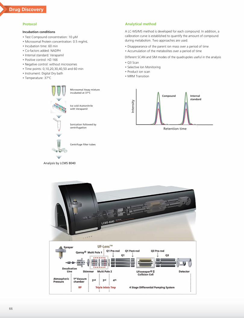
Protocol Analytical method
• Test Compound concentration: 10 µM• Microsomal Protein concentration: 0.5 mg/mL• Incubation time: 60 min• Co-factors added: NADPH• Internal standard: Verapamil• Positive control: HZ-166• Negative control: without microsomes• Time points: 0,10,20,30,40,50 and 60 min• Instrument: Digital Dry bath• Temperature: 37°C
A LC-MS/MS method is developed for each compound. In addition, a calibration curve is established to quantify the amount of compound during metabolism. Two approaches are used.
Different SCAN and SIM modes of the quadrupoles useful in the analysis
• Disappearance of the parent ion mass over a period of time• Accumulation of the metabolites over a period of time
• Q3 Scan• Selective Ion Monitoring• Product ion scan• MRM Transition
Incubation conditions
Microsomal Assay mixtureincubated at 37°C
Ice cold Acetonitrilewith Verapamil
Sonication followed by centrifugation
Analysis by LCMS 8040
Centrifuge filter tubes
Inte
nsi
tyRetention time
Compound Internalstandard

Drug Discovery
67
Metabolic parameters
Data analysis HZ-166
Peak area and % remaining values
Time(min)
0
10
20
30
40
50
60
815599
702717
693332
655521
608029
604208
561988
99.98
87.53
84.60
80.23
76.24
74.73
69.06
Test compoundPeak area
586830
543057
550156
515666
558567
566352
561427
VerapamilPeak area % remaining
0 10 20 30 40 50 60 704.0
4.2
4.4
4.6
4.8
5.0
Time (min)
ln (%
pea
k ar
ea r
atio
)
HZ-166CMD 45
0 10 20 30 40 50 60 704.2
4.4
4.6
4.8
5.0
Time (min)
ln (%
pea
k ar
ea r
atio
)
t1/2= 0.693 slope
V= incubation volume [protein]
Intrinsic clearance = (V*0.693) t1/2
Metabolic rate = slope*[analyte] [protein]
Compounds analyzed
N
N
N
N
O
O
HZ-166Chemical Formula: C21H16N4O2
Molecular Weight: 356.38
CDM-45 Xhe-III-74
Perc
ent
rem
ain
ing
Xhe-III-74 analogs
Perc
ent
rem
ain
ing
HZ-166 analogs

Drug Discovery
68
References
• Donglu Zhang et al, Acta Pharmaceutica Sinica B. 2012, 2, 549• P. Anzenbacher et al, Cell. Mol. Life Sci. 2001 58, 737• Pawe Baranczewski et al, Pharmacological Reports, 2006, 58, 341• Xiaoyan Li et al, J. M. Med. Chem. Res. 2002, 11, 504• George Gallos et al, Am. J. Lung Cell Mol. Physiol. 2011, 302, L248• McGinnity DF et al, Drug Metabolism and Disposition, 2004, 32, 1247• Wang-Hui Jing et al, Journal of Pharmaceutical and Biomedical Analysis, 2013, 77,175
Conclusion and future
During our investigation, we observed that mouse microsomes degrade compounds more quickly than human microsomes. We are currently in process to characterize these metabolites in order to guide the synthesis of new analogs for the research group of Prof. Cook. We will expand this method to pharmacokinetic studies in the near future.
Acknowledgements
Financial support: National Institute of Neurological Disorders and Stroke, National Institute of Mental Health, National Heart, Lung and Blood Institute, University of Wisconsin Milwaukee, UWM Research Foundation, UWM Department of Chemistry and Biochemistry, and UWM Graduate School.

The Challenge of Discovery and Development of Pharmaceutical Products Using Imaging Mass Spectrometry: iMScope TRIO
Imaging mass spectrometry (IMS), which uses MALDI-TOF/MS analysis to identify the presence of target substances in biosamples such as tissue samples, is a cutting-edge measurement technology that is becoming increasingly popular in medical and biological research. The technology has potential not only for drug delivery and pharmacokinetics research, but also as a useful tool in biomarker research, and increasing numbers of measurements are now carried out using the iMScope TRIO. We sat down with Hidefumi Kaji, PhD, of Mitsubishi Tanabe Pharma Corporation’s DMPK (Drug Metabolism and Pharmacokinetics) Research Laboratories, Research Division, and asked him about his use of this technology.
*Affiliates and titles of the interviewee are current as of the time of reporting.
You have been using the iMScope TRIO at the laboratory inShimadzu’s Sanjo Works for around two and a half years. Whatsort of research is it mainly used in?
I work in the DMPK Research Laboratories, and am engaged mainly in the creation of pharmaceutical products, specifically the area of preclinical DMPK. In my work, I aim to use the iMScope TRIO to examine the distribution of pharmaceutical candidate compounds in animal organs and tissue, and evaluate fluctuations in endogenous substances, biomarkers, and so on.
Introduction
Could you please give us an actual example of pharmacokineticanalysis?
Of course. We used the iMScope TRIO to evaluate the distribution of a certain low molecular weight compound in rats. The compound we used is characterized by the fact that it bonds specifically to, and accumulate, the melanin in the eyeball. If a compound under development builds up by bonding to melanin or in some other way, we need to be careful about the photosensitivity of the compound. However, it’s not always the case that if a compound bonds with melanin and is distributed within it, it demonstrates toxicity when it decomposes as a result of photosensitivity. Such understanding of what sort of degradation product occurs as a result of light is important in predicting the risk of toxicity. Furthermore, if we can establish that a compound dissipates relatively quickly, it usually means there will be no problems. From the pharmacokinetic standpoint, it is very important in the creation of safe drugs that we ascertain a compound’s profile at an early stage of development, to see if it bonds to and accumulate melanin and what degradation products might occur.
iMScope TRIO was its spatial resolution.
That’s right. The instrument is characterized by having a microscope at the front. The laser spec has improved significantly in the past few years, meaning that we can now perform high-resolution analysis, but I think that when analyzing a sample slice, the key point is being able to ascertain what point on the sample was being measured when the image in question was obtained. Having a microscope means that we can observe a part in detail and match results with what we are actually measuring. This has allowed us to correctly analyze at super-high resolution for the first time. Anyone who
Did you get more insightful analysis results thanks to using this technology?
Well, we’re still at the basic research level, but we analyze biomarker fluctuation, which tells us what endogenous substances are active in a particular pathology caused by administration of a compound. For example, if gentamycin is administered and causes a disorder, it is assumed that the concentration of a particular endogenous
Pharmacokinetic Analysis of Low Molecular Weight Compounds (1)
Are there any other examples you can give us?
There is a substance called gentamycin, which is known to express toxicity within the kidneys in rats. There have been few reports that have proven where within the kidney it is distributed and expresses toxicity, and so we looked at whether it is possible to use IMS technology to evaluate this. Specifically, we implemented verification using 10-micron super-high-resolution analysis. Results showed that, when we looked at the distribution of the compound at a resolution that allowed the recognition of proximal tubules and glomerulus, gentamycin was distributed in proximal tubules, but had not built up in almost any other areas. In other words, we were able to confirm that the buildup of gentamycin in proximal tubules is the mechanism for toxicity expression.
Pharmacokinetic Analysis of Low Molecular Weight Compounds (2)
Interview
69
Hidefumi Kaji, Ph DGroup Manager
DMPK Research Laboratories, Research DivisionMitsubishi Tanabe Pharma Corporation
actually uses the instrument would be delighted by this. It is extremely important to position accurately and understand what part of the sample slice is being analyzed. The higher the resolution, the more likely it is that, just by moving five or ten microns, we are looking at a completely different place. The use of IMS has made it possible to confirm the specific distribution of the compound under evaluation within retinal melanin at super-high resolution, down to five microns. Many people have been amazed that we are able to examine samples in such detail. When people see the iMScope TRIO’s high-resolution analysis in action for the first time, the impact is enormous.

Interview
70
So you are talking not only about drug discovery that aims for acure, but also going a little further to the diagnostic stage. Earlydiagnosis and early cure are obviously important, but it isextremely interesting to hear how you are seeking biomarkersuseful for determining the effectiveness of prognosis.
What are some of the conventional methods for evaluatingtissue distribution?
In general, for a single project, a labeled compound is not synthesized at the point at which there are still multiple candidate compounds. Unlabeled compounds are administered, and organs subsequently removed, after which quantitative analysis is performed using LCMS on the homogenate of those organs.
And do you use autoradiography (ARG) after that, as part of theresearch process?
Yes. As the research stage progresses and we narrow down the compounds, we use radio-isotope (RI) labeled compounds to evaluate where a compound is distributed within the body after administration, where it builds up, and how long it takes before it is excreted, with the aim of applying for approval as a pharmaceutical product.
The non-targeting approach? That’s a particular feature of thismethod. How about any disadvantages?
There is no guarantee that all compounds being measured can be ionized. That’s the main disadvantage. In contrast to marker analysis, mass spectrometry can’t detect non-ionized compounds, so it’s not possible to use it to evaluate absolutely any and every compound.
That’s true. That’s the reason whole-body ARG probably won’t be replaced completely with IMS, isn’t it? How about sensitivity?
This is a very general comment, but MALDI currently compares poorly with LCMS and LC-MS/MS.
We are pressing ahead with improvements to sensitivity inpretreatment, thanks to Mitsubishi Tanabe Pharma’s cooperation.
Yes, and then we need to think about what IMS is targeting, how much we can expect from it, and what it will be used for. If the target compound is highly ionized, which allows evaluation of effective dosage, then IMS can be used in efficacy mechanism analysis. But if it’s not, then looking at efficacy evaluation levels might be difficult. When evaluating toxicity mechanisms, however, the dose and distribution quantity of the candidate compounds is sufficient, making their levels detectable. If IMS can be used for that purpose, it will offer attractive evaluation methods.
The microscopic level resolution is also part of the comparisonto ARG, isn’t it?
Given the problem of ionization, it is difficult to imagine that IMS completely replaces ARG when considering the current quantitative ARG results. The iMScope TRIO performs best when it is used not to evaluate whole-body distribution, but rather when it is focused on a
What are the advantages and disadvantages of IMS incomparison with ARG?
The advantages are, firstly and most importantly, that we can perform evaluation at an early stage, prior to having to narrow the compounds down to one. The second relates to the greatest attribute of the mass spectrometer. With ARG, it is not possible to distinguish between the unaltered substance and metabolite distributions from marker tissue distribution information. In particular, when there are multiple metabolites present, the distribution and buildup information obtained is for a mixture of behaviors, where we don’t know the ratio of the metabolites. Using a mass spectrometer allows
Furthermore, it may be effective in the evaluation of safety considering the difference between animal species. One example is a case where some data shows a particular toxicity present in rats, which is caused by a metabolite that is only produced in rats, and therefore cannot be created in humans. Not only ARG but also mass spectrometry offers visual information. If it is possible to demonstrate that the toxicity is caused not by unaltered substance but rather by metabolites that are specific to rats, we can consider that the toxicity may not occur in humans.
Comparison with Autoradiography (ARG)
That’s very interesting!
As I just said, the great advantage is that it can be used not only for compounds but also for evaluating biomarkers that are active at the same time.
substance will have risen compared with its state prior to administration. If we administer a drug or a compound that is a candidate for development, and the endogenous substance is reduced, allowing recovery from the disorder, the fluctuation in said endogenous substance indicates pathological improvement indirectly. Additionally, if the phenomenon is caused by the predicted efficacy mechanism of the development candidate compound, analyzing the phenomenon demonstrates that the drug has worked in line with its concept. Furthermore, if comprehensive analysis allows identification of types of endogenous substance, this means it may in the future allow the discovery of biomarkers that are clinically effective. This is an extremely attractive prospect.
us to see the distribution of each of the unaltered substances and metabolites. Members of the safety research section in our company have also said that they would like to use it to investigate toxicity mechanisms. This is because toxicity is sometimes caused by unaltered substances, and sometimes by metabolites.

Interview
71
I see. In other words, it’s not a replacement for ARG, it’s atechnology with the potential to provide supplementary information that cannot be obtained using ARG, and thus further help progress research and development in drug discovery.
Exactly. It’s not possible to do everything using IMS, but we can use it with sample slices, to roughly check whether or not the target substances are present. Once we know where they are, we can perform more detailed analysis using mass spectrometry.
the future?
They are mainly to do with quantitative analysis. Pretreatment is important for this. Particularly, how evenly you can apply the matrix with good reproducibility. An automatic matrix application device is required for this. I think that the vaporization-type iMLayer, actually, is a very good device. When I present my results at seminars and so on, researchers looking at improving sensitivity and quantitative evaluation are always very interested in the automation aspect of the instrument.
We have had high expectations right from the start in regard to the uniqueness of the concept behind imaging mass spectrometry. Recently, not only have significant advances been made in the hardware, but significant progress has also been made in the field of pretreatment technology research aimed at practical utilization. My conversation with Mr. Kaji confirmed that IMS is being applied nowadays not only in basic medical research, but also in a variety of settings at the forefront of drug discovery research.In response to this level of anticipation from our users, I feel strongly encouraged to continue with technological research that aims to “Realize Our Wishes for the Well-Being of Both Mankind and the Earth.”
So you are talking about using IMS not only at an earlier stagein drug discovery than ARG, but also after entering the clinical phase. I remember that in one of your lectures you said that, forbiomarker research, the fluctuation results for biomarkers in theclinical phase would lead to the possibility of finding the nexttarget. I was very impressed when I heard that.Finally, do you have any opinions or hopes regarding theanalytical instruments industry as a whole?
Chasing after the development compounds themselves is my major task using IMS, but I’d like to talk about another area. I am currently responsible for preclinical DMPK, and use laboratory animals for efficacy evaluation. I’d like to use the technology more in these evaluations. If the disease mechanism is a new one, efficacy evaluation becomes difficult. I’d like to discover biomarkers that can be used in that evaluation. If those biomarkers are reflected in human blood parameters when the drug goes into clinical trials, they can be used as indicators, and as markers to monitor treatment effectiveness when the drug is actually administered.
Hopes for the Future
Interviewer’s CommentsBoth methods have advantages don’t they? And these advantagescan be used, rather than merely making up for the disadvantages.
We certainly hope that you will continue to collaborate with uson the development of analysis instruments that can evaluatetreatment as a whole, not only for diagnosis but also to confirmprognosis. Thank you for your insights and your time today.
Within my area of work, which is mainly preclinical DMPK, we handle a lot of samples that originate in clinical research, and there is an increasing tendency towards working with materials actually used for clinical research. Since pharmaceutical companies exist to promote the health of patients, I would like to see Shimadzu working on the development of instruments that do not simply perform analysis for the sake of analysis, but rather take a step further towards the bigger vision of health care, for example by cooperating with us on the development of methods and instruments for use in analyzing markers that predict disease.
iMScope TRIO: Imaging Mass Microscope*Sales area: All areas excluding North America
certain type of tissue. This is because it allows high-resolution analysis. I think it is more appropriate to use it to look in a focused way at kidney tissue, brain tissue or liver tissue, and to get an accurate idea of how compounds, metabolites and biomarkers are distributed and fluctuate within those settings.

LC-MS/MS
Utility of high resolution MALDI imaging in drug discovery: Histological distribution of gentamicin in proximal renal tubules of rats
Hidefumi Kaji1; Hiroyuki Hashimoto1; Masayoshi Saito1; Takushi Yamamoto2; Noriyuki Ojima2
1: Mitsubishi Tanabe Pharma Corporation, Saitama, Japan2: Shimadzu Corporation, Kyoto, Japan
▶ MS/MS imaging of gentamicin was performed from high-selectivity analysis of endogenous metabolites in biological tissue.
▶ High resolution (10 µm) imaging of rat renal cortex showed that gentamicin is specifically distributed in proximal renal tubules.
▶ New matrix application method is useful for the sensitive MALDI imaging.
Overview
In pharmacology and toxicology, localization of a drug molecule in the target tissue of organs provides very important in vivo biological information. Imaging mass spectrometry (IMS) is increasingly used in drug discovery and development during preclinical studies. Gentamicin is an antibiotic to treat infections, but it possesses renal toxicity. As it is excreted in the urine, the kidney tissues of a person being treated with gentamicin are almost constantly bathed in gentamicin. The objective of the study is to define the specific distribution of the parent drug in proximal renal tubules of rats dosed gentamicin using MALDI imaging.
1. Introduction
Drug Discovery
72
At 2 h after single intravenous (I.V.) administration (3, 10, or 30 mg/kg) of gentamicin to male SD rats, 5-µm sections taken from the single kidney of each rat were prepared. The tissue sections were coated with CHCA by sublimation using an automated sample treatment system (iMLayer™, Shimadzu Corporation, Japan), and analyzed using an ion trap-time of flight (IT-TOF) tandem mass spectrometer equipped with MALDI source†. This instrument is a combination of an optical microscope which allows the observation of high-resolution morphological images, with a mass spectrometer which identifies and visualizes the distribution of specific molecules. The other kidney was used for determining the concentrations of gentamicin by LC-MS/MS.
We analyzed gentamicin C1, because it had the highest efficacy.
2. Methods
Structures of Gentamicin
Sample preparation
iMLayer™
(Matrix Vapor Deposition System, Shimadzu)High reproducibility and minute matrix crystals
Imaging Mass Microscope
Laser: Nd:YAG laser (335 nm)Collision energy: 50Laser shot: 200 shot/pixcelLaser diameter: 50 or 10 µmMatrix: CHCA
Observation MALDI imaging
Movement
IMS
I.V. administration Extraction of kidneys
Homoginized kidney
Cryosectioned (10 µm)
Pretreatment
Snap-freeze inliquid nitrogen
Matrix application
Spray: 10 mg/mL CHCA, 0.1% TFA in 60% acetonitrile solutionSublimation: time; 20 min, thickness; ca.2 µmNebulizer: applied methanol mist, 5 min, particle size: <5 µmPre-coated matrix: sublimation before thaw-mounted tissue section
*Part of this work was presented in a poster session of the 63th ASMS Conference, May 13 - June 4, 2015, St. Luis.

Drug Discovery
73
Optimization of matrix application method
3. Results
MS spectrum of standard gentamicin C1 (2 mg/mL, CHCA)
MS/MS spectrum of standard gentamicin C1 (m/z 478.322)
*: S. Shimma et al, J. Mass Spectrom., 48: 1285–1290, 2013.
Peak intensity: 12,731 Peak intensity: 24,291
Peak intensity: 154,514 Peak intensity: 239,536
(m/z: 478.3 → 322.2)The peak intensities of gentamicin C1 by Method 4) rose 20 times from those by Method 1).
Matrix
Analyte
Tissue section
microscope glass plate
425.0 450.0 475.0 500.0m/z
0.0
2.5
(x100,000)
478.322
461.296447.282450.292
433.267
506.137430.266 480.154 500.301
50 100 150 200 250 300 350 400 450 500m/z
0.0
1.0
2.0
(x100,000)
322.196
461.296
Data acquisition was performed in product ion scan (MS/MS) mode and abundance of gentamicin C1 was selectively monitored by focusing on its specific fragment ion, m/z 322.196.
1) Spray 2) Sublimation + Spray
3) Sublimation + Nebulizer*) 4) Pre-coated matrix + Sublimation + Nebulizer
1) 2)
3) 4)
Matrix (sublimation)
Matrix (pre-coated)

Drug Discovery
74
Abundance of gentamicin C1 was higher in the renal cortex than in the renal medulla. The signal intensities of m/z 322 of gentamicin within the kidney in distribution images corresponded well with the gentamicin concentrations.
Gentamicin Distribution in Rat Kidney
Vehicle
Optical image
MALDI image
Strong
Weak
(m/z: 478.3 → 322.2)
Pixel size: 50 µm
Optical image (H&E stained)
Concentration (by LC-MS/MS)
30.3 µg/g 93.6 µg/g 129.5 µg/g
3 mg/kg 10 mg/kg 30 mg/kg
MS/MS spectrum of gentamicin C1 in rat kidney dosed 30 mg/kg of gentamicin

Drug Discovery
75
High resolution (10 µm) imaging of rat renal cortex showed that gentamicin is specifically distributed in proximal renal tubules. Research areas and target compounds involved in pharmaceutical drug discovery is very wide ranging. Imaging mass spectrometry using Mass Microscope is one powerful tool for the drug discovery (pharmacokinetics, pharmacological mechanisms, toxicity), owing to its ability to easily combine morphological observation from the optical microscope and localization of target molecules from the mass spectrometer image.
The image of the specific distribution of gentamicin in proximal renal tubules of rats was detected with a high resolution of 10 µm (pixel size), and found to be similar to the distribution of the immunostaining assay of gentamicin reported previously*.
*: K. Fujiwara, Yakugaku Zasshi, 131(6): 949-960, 2011Pixel size: 10 µm
100 µm
4. ConclusionsThis work was supported by Safety Research Laboratories of Mitsubishi Tanabe Pharma Corporation.
* The MS instrument used in this poster is currently not for sale in the United States.
5. Acknowledgments
Precise Localization of Gentamicin in Renal Cortex
Blue boundary: glomerulus, Yellow boundary: distal renal tubulesThe signal is not detected in these domains.
Strong
Weak
Optical image (H&E stained) Overlap image MALDI image

Selection 1 Pharmaceutical, Life Science
A Fully Automated, Bottom-up Approach for MALDI-TOF MS Based Discovery WorkflowsBottom-up workflows have been a staple of mass spectrometry-based proteomic approaches. Most of these protocols require overnight digestion, sample clean-up and, when involving MALDI, fraction collection, dry down and sample matrix deposition. We present in this work a fully automated solution for MALDI-TOF MS-based peptide mapping experiments. Using an online digestion, desalt, reversed phase separation and fraction collection platform, we were able to decrease the experimental time from over 18 hours to less than 30 min (reversed phase separation included, acquisition time not included) while substantially improving sequence coverage.
Selection 3 Pharmaceutical, Life Science
Characterization of Products Formed by Forced Degradation of Etodolac Using LC/MS/MSForced degradation is a process, whereby the natural degradation rate of a drug product or drug substance is accelerated by the application of an additional stress. The ICH guidelines indicate that stress testing is designed to determine the stability of the molecule by knowing degradation pathways in order to identify the likely degradation products. In this work, acid, base, peroxide, thermal and UV degradation products of Etodolac were studied using the LCMS-8040 Triple Quadrupole Mass Spectrometer.
Selection 4 Pharmaceutical, Life Science
Comprehensive Two-Dimensional HPLC Analysis Coupled with Mass Spectrometric Detection and Informative Data Processing for Lipid AnalysisIn lipidomics, phospholipids are the attractive targets of analysis since lipids are important and essential components of biological membranes. However, a conventional HPLC system with a single separation mode performs poorly on biological lipid sample, because it contains various kinds of lipids with common moieties that govern their behavior on column. In such a case, comprehensive two-dimensional (2D) LC will be a powerful tool.
Selection 5 Pharmaceutical, Life Science
Comprehensive Two-Dimensional HPLC and Informative Data Processing for Pharmaceuticals and Lipids
Kakkonto, a traditional Chinese drug, consists of natural plants and contains many compounds such as ephedrine, glycyrrhizic acid, and cinnamic acid. We separated such a complex matrix using the Shimadzu Nexera-e comprehensive two-dimensional liquid chromatograph, with a photodiode array detector. We also characterized phospholipids using Nexera-e coupled with triple quadrupole and ion-trap-TOF mass spectrometers quantitatively and qualitatively, respectively.
Selection 6 Pharmaceutical, Life Science
Determination of Leachables in Orally Inhaled and Nasal Drug Products (OINDP) by GCMS/MS
Orally Inhaled and Nasal Drug Products (OINDP) are developed for treating asthma or chronic obstructive pulmonary diseases by delivering drug substances directly to the respiratory tract. OINDPs are present in inhaler devices, which may contain polymers, elastomers and other components from which minute quantities of material may migrate/leach into a product and enter the respiratory tract along with the therapeutic agent. In this study, we tried to develop a highly sensitive method for quantitation of 40 leachables in OINDP products by using the Shimadzu GCMS-TQ8040.
Selection 7 Pharmaceutical, Life Science
Development of Simplified Quantitative Method for Tryptic Digested C-reactive Protein by Using Online SPE Coupled to Triple Quadrupole Mass Spectrometer
Method development for accurate quantification of biomarker protein is a very important step for researchers in pharmaceutical and clinical fields. In this study, we tried to develop a simple and high-sensitivity quantification method for C reactive protein (CRP), the level of which rises as a result of in ammation etc., by using online SPE coupled to a triple quadrupole mass spectrometer without any complicated sample enrichment and depletion.
Selection 8 Pharmaceutical, Life Science
Direct Analysis of 10 Antipsychotics in Serum by the Online System Integrating Solid-Phase Extraction with UHPLC-MS/MS
A fully-automatic system integrating solid-phase extraction with ultra-high performance liquid chromatography-tandem mass spectrometry was successfully applied to the direct analysis of ten antipsychotics in serum.
Selection 2 Pharmaceutical, Life Science
Application of a Sensitive Liquid Chromatography-Tandem Mass Spectrometric Method for Determination of Octreotide in Human PlasmaA fast and sensitive method for detecting octreotide in human plasma by ultra high performance liquid chromatography-tandem mass spectrometry was developed. The lower limit of quantitation was 5.0 pg/mL and the results of other parameters for method validation were good.
These articles were selected by Shimadzu. Relating to pharmaceutical analysis, they are from posters presented at recent international conferences in 2015, such as ASMS and HPLC. They feature a variety of instruments we produce and include cutting-edge technologies. Please obtain the articles of your interest through the links on the titles.
Posters from Recent Conferences

Selection 9 Pharmaceutical, Life Science
Evaluation of a Novel Glass Vial Overcoming Adsorption Effect for Pharmaceutical DrugsThe majority of pharmaceutical drugs are still small molecule drugs. Adsorption onto consumables when in sample preparation is regarded as a big issue. To overcome the adsorption effect, we developed and evaluated a novel glass vial “LabTotal vial” which is highly inert to basic compounds and perfect for highly sensitive LC/MS analyses.
Selection 10 Pharmaceutical, Life Science
Flexible Automated Sample Preparation Workflows: Modified Automated Systems for Specific Immuno-MS and MS WorkflowsThe automation of sample preparation has become an increasingly important component for reproducible and operator-independent experiments. The Perfinity Workstation has provided researchers established applications in affinity capture and digestion for targeted proteomic workflows, utilizing specific affinity, IMER and reversed phase components coupled directly to a mass spectrometer. This work outlines novel strategies that are being utilized for automated online and offline sample preparation to achieve these specific goals.
Selection 11 Pharmaceutical, Life Science
Highly Sensitive Quantitative Analysis of Amoxicillin and Clavulanic Acid from Plasma Using LC/MS/MSAmoxicillin and clavulanic acid is a combination drug product in which amoxicillin is a β-lactam antibiotic, and clavulanic acid is a β-lactamase inhibitor. In this combination, clavulanic acid acts by preventing the destruction of antibiotic amoxicillin which is used to treat various bacterial infections. An LC-MS/MS method has been developed for highly sensitive quantitation of these molecules from plasma using the Shimadzu LCMS-8040 triple quadrupole mass spectrometer.
Selection 12 Pharmaceutical, Life Science
Integration of Amino Acid, Acylcarnitine and Steroids Analysis in Single FIA/LC-MS/MS PlatformTraditionally, analysis of a steroid such as 17-hydroxyprogesterone is done by immunoassays but LC-MS/MS is an attractive analytical alternative because it results in a reduction of false positives and a more accurate quantitative performance. The requirements against steroid analysis by LC-MS/MS are getting more stringent. In this study, we present a strategy for performing both AA/AC and steroids analysis within a single LC-MS/MS platform.
Selection 13 Pharmaceutical, Life Science
Non-convex Quasi-norm-based Normalization of MALDI-MS Imaging DataWe explore the use of non-convex quasi-norms as a normalization factor for MALDI-MS Imaging datasets. As a lower norm parameter is used, results show less artefacts but less uniform normalization range. A trade-off value should be chosen. Results are shown for decreasing concentrations of angiotensin peptide by itself and mixed with E.Coli (DH5 ) suspension.
Selection 14 Pharmaceutical, Life Science
N-terminal Charge-driven de novo Sequencing by using ASDF-incorporated Curved Field ReflectronFixing a strong charge at the N-terminus of a peptide has been reported to be an effective derivatization for de novo sequencing. While giving high proton affinity at the N-terminus of a peptide facilitates the generation of a- and b-ions, it is impossible to differentiate isobaric Ile/Leu residues. Side chain fragmentation generated by high-energy CID on MALDI-TOF/TOF has a potential to overcome this issue. We report a discrimination of Ile/Leu and a capability of the assignment of Gln/Lys, applying a newly developed MALDI-TOF/TOF, which has high resolution in MS/MS and a high energy CID at 20keV, for analysis of N-terminal derivatized peptides.
Selection 15 Pharmaceutical, Life Science
On-line Supercritical Fluid Extraction/Supercritical Fluid Chromatography: A Novel Approach to the Cleaning Validation for pharmaceutical ManufacturingCleaning validation is necessary to establish the quality and safety of pharmaceutical drug products. Recently, HPLC has become preferable because of the growing need for the individual analysis of products. Before the HPLC analysis, however, manual processes such as a sample extraction and a sample condensation are required, which may affect the quality of results. Thus, we evaluated the application of a novel on-line supercritical fluid extraction/chromatography system for the cleaning validation.
Selection 16 Pharmaceutical, Life Science
Quantification and Structural Characterization of Glycans and Glycopeptides by TQMS: the Energy-resolved Oxonium Ion Monitoring (Erexim) PlatformWe have previously reported on using a triple quadrupole mass spectrometer to reproducibly monitor glycan-derived low molecular weight ions (oxonium ions) to acquire, within milliseconds, a profile of fragmentation patterns at a range of CID collision energies. This, which we termed the energy-resolved oxonium ion monitoring (Erexim) profile, reflects the glycan structure in such a way that isobaric structures can be clearly distinguished and assigned. Here we have developed new software to fully support Erexim data acquisition and analysis.
Selection 17 Pharmaceutical, Life Science
Structural Analysis of Isomeric Chemicals by Using High Resolution MS/MS on Curved Field Reflectron Incorporated with a Novel Focusing TechnologyWhile resolution of MS has greatly improved, no matter how high it becomes, MS/MS remains essential to conduct analysis of isomeric chemicals, and the analysis is thought to still be a significant task. Recently, we incorporated a new focusing technology into the curved field reflectron, which enabled obtaining 10,000 resolution of MS/MS. We will apply the new curved field reflectron toward ambiguous assignment and differentiation of isomeric chemicals.
Selection 18 Pharmaceutical, Life Science
Transfer of USP Normal Phase Method for the Analysis of Cortisone Acetate to the Nexera UC SFC SystemThe USP monographs are widely referenced to ensure the quality of drug substances. The USP monographs offer reliable and robust methods. However, some of these methods, especially those using normal phase HPLC, require the use of harmful, even toxic solvents. This application note shows an example of the analysis of cortisone acetate tablets according to the USP monograph.
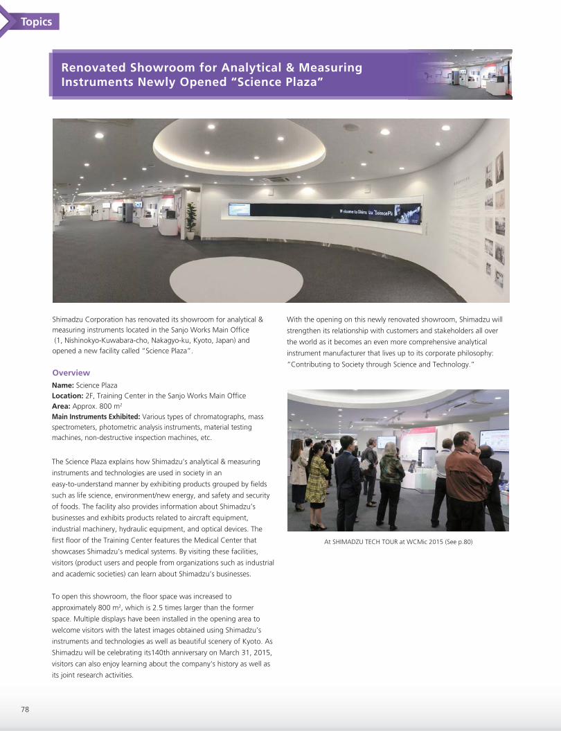
Renovated Showroom for Analytical & Measuring Instruments Newly Opened “Science Plaza”
Shimadzu Corporation has renovated its showroom for analytical & measuring instruments located in the Sanjo Works Main Office (1, Nishinokyo-Kuwabara-cho, Nakagyo-ku, Kyoto, Japan) and opened a new facility called “Science Plaza”.
Name: Science PlazaLocation: 2F, Training Center in the Sanjo Works Main OfficeArea: Approx. 800 m2
Main Instruments Exhibited: Various types of chromatographs, massspectrometers, photometric analysis instruments, material testing machines, non-destructive inspection machines, etc.
The Science Plaza explains how Shimadzu’s analytical & measuring
instruments and technologies are used in society in an
easy-to-understand manner by exhibiting products grouped by fields
such as life science, environment/new energy, and safety and security
of foods. The facility also provides information about Shimadzu’s
businesses and exhibits products related to aircraft equipment,
industrial machinery, hydraulic equipment, and optical devices. The
first floor of the Training Center features the Medical Center that
showcases Shimadzu’s medical systems. By visiting these facilities,
visitors (product users and people from organizations such as industrial
and academic societies) can learn about Shimadzu’s businesses.
To open this showroom, the floor space was increased to
approximately 800 m2, which is 2.5 times larger than the former
space. Multiple displays have been installed in the opening area to
welcome visitors with the latest images obtained using Shimadzu’s
instruments and technologies as well as beautiful scenery of Kyoto. As
Shimadzu will be celebrating its140th anniversary on March 31, 2015,
visitors can also enjoy learning about the company's history as well as
its joint research activities.
Overview
Topics
78
With the opening on this newly renovated showroom, Shimadzu will
strengthen its relationship with customers and stakeholders all over
the world as it becomes an even more comprehensive analytical
instrument manufacturer that lives up to its corporate philosophy:
“Contributing to Society through Science and Technology.”
At SHIMADZU TECH TOUR at WCMic 2015 (See p.80)

Topics
79
Design Awards in 2015
Shimadzu wins two design awards in 2015.
Shimadzu’s i-Series integrated HPLC system was recognized with a
2015 Good Design Award.
The Good Design Award is a comprehensive design-promotion
system, hosted by the Japan Institute for Design Promotion since
1957. The Institute selects winners based on a variety of criteria, with
a focus on how the design will enrich our lives, industries, and society.
The guiding principles of the Good Design Award are as follows:
- HUMANITY Emerging creative power for products and
conceptual embodiments
- HONESTY Insight toward contemporary society
- INNOVATION Conceptual power to pioneer the future
- ESTHETICS Imaginative power for a prosperous lifestyle culture
- ETHICS Thinking power to shape society and the
environment
The i-Series was designed to improve the efficiency of the customer’s
workflow by realizing a PC-free environment that allows operators to
control instruments and to check system status and chromatograms
from anywhere, and offering improved performance, usability, and
automation. The i-Series of integrated HPLCs combines leading-edge
functionality and performance with intuitive design in a compact body
in order to support applications from routine analysis to R&D
Learn more about it at:
http://www.g-mark.org/award/describe/42673?token=qXbaw9KJsa&locale=en
i-Series integrated HPLC system PDA-5000 optical emission spectrometer
Shimadzu’s PDA-5000 Optical Emission Spectrometer(*) has received a
2015 iF DESIGN AWARD, a world-renowned label for excellent
designs for more than 60 years. The evaluation criteria of this award
are listed below:
- AESTHETICS
- PRACTICABILITY
- EXECUTION
- INNOVATION
- USABILITY / ERGONOMICS
- BRAND FIT
- SAFETY
- ENVIRONMENTAL IMPACT
- TARGET GROUP FIT
The PDA-5000 offers ease of use and a highly innovative and reliable
design. The curved design at the front of the instrument removes any
sense of oppressiveness and makes the instrument more accessible. In
addition, a beautiful arched and ergonomically designed handle
improves operability. Taking into account user ergonomics and
workflows, the PDA-5000 offers a new and innovative work
environment.
Learn more about it at:
http://exhibition.ifdesign.de/entrydetails_en.html?mode=aus&offset=49
* This product is only sold in China and South-East Asia.

SHIMADZU TECH TOUR at WCMic 2015
Tour Highlights
Medical Center
Topics
80
The SHIMADZU TECH TOUR was held to learn about innovative technologies for medical diagnostic imaging applications and analytical &
measuring devices at the 10th World Congress for Microcirculation in 2015. It was followed by an official welcome reception. The TECH TOUR
introduced Shimadzu’s state-of-the-art mass spectrometers and angiography systems, and provided an historical overview of the company since
its foundation in Kyoto in 1875.
The Medical Center showroom gathers medical diagnostic imaging
systems(*), including angiography and mammography systems.
* For more details about these products, please visit our medical systems site at http://www.shimadzu.com/med/
Science PlazaA variety of Shimadzu’s latest analytical instruments(*) covering mass
spectrometry and imaging apparatus is displayed in the Science Plaza
showroom.
* For more details about these products, please visit our analytical and measuring instruments site at http://www.shimadzu.com/an/
Old Relics inside the Head OfficeA pillar hole - the basis of an
ancient building - was excavated for
archaeological research prior to the
construction of our new main
building at the Head Office.

81
New Productssststsctsctsctsctuctucuc stsstsctsctsctctucucucuuuduccuuudududuodododrodroProProProPrw Pw Pw w eweweweweNe duduodroPrw Pw duododrodProProPrw Pw weweweweweNeNeNNNNeNNNeeNeNNeeNeNNNNNNNNNNN
LCMS-8060
The LCMS-8060 is a ground breaking innovation in mass spectrometry, helping to transform LC-MS/MS data quality through an unmatched combination of sensitivity and speed. This vision of a triple quadrupole mass spectrometer with the highest sensitivity and ultra-fast technologies results in an innovation that really matters.
Features- Unprecedented Sensitivity- Fusion of Sensitivity and Speed- Outstanding Durability
Click here>>
Designed to push the limits of LC-MS/MS quantitation for applications requiring the highest sensitivity and robustness
Py-Screener (Screening System for Phthalate Esters)
The Py-Screener is a pyrolyzer GC/MS (Py-GC/MS) system with special software and a sampling toolkit to selectively detect and quantify phthalate esters thermally extracted from samples.
Features- Easy to Operate Even for Novices- Easy to Operate Using Special Software- Tabular Display of Concentrations and Criteria Clarifies the Results
Click here>>
Designed to screen for phthalate esters in polymers
AOC-6000 (Multifunctional Autosampler)
The AOC-6000 is capable of exchanging syringe modules automatically.
Features- Automatic Switching between Three GC/MS Sample Injection Modes- Simple to Operate from GCMSsolution- Automated Pretreatment Maximizes Analysis Efficiency
Dramatically improves GC/MS analysis productivity
Click here>>

www.shimadzu.com/an/
For Research Use Only. Not for use in diagnostic procedures.The content of this publication shall not be reproduced, altered or sold for any commercial purpose without the written approval of Shimadzu. The information contained herein is provided to you "as is" without warranty of any kind including without limitation warranties as to its accuracy or completeness. Shimadzu does not assume any responsibility or liability for any damage, whether direct or indirect, relating to the use of this publication. This publication is based upon the information available to Shimadzu on or before the date of publication, and subject to change without notice.
© Shimadzu Corporation, 2015
Shimadzu Corporation Celebrates Its 140th Anniversary This Year
SHIMADZUHISTORY
Shimadzu Corporation was founded in Kyoto in 1875, by Genzo Shimadzu Sr., a Buddhist altar craftsman, manufacturing physics and chemistry instruments.Later, Shimadzu Corporation grew significantly under the leadership of Genzo’s eldest son, Genzo Jr., who showed a rare talent as a remarkable inventor.In this way, Shimadzu progressed hand-in-hand with science, building itself into a leader in technology.This culture of “Contributing to Society through Science and Technology” has been inherited by each new generation of employees at Shimadzu Corporation and now is being expanded globally through the growth of the Shimadzu Group, which continues to be rooted in science and committed to Shimadzu’s management principle “Realizing Our Wishes for the Well-being of both Mankind and the Earth”.
1875 Genzo Shimadzu Sr. established a business in the Kiyamachi-Nijo-Minami district in Kyoto.
1909 Created the first medical X-ray device in Japan.
1919 Established the Sanjo Works.
1923 Established the Berlin office.
1930 Genzo Shimadzu Jr. was recognized by the Emperor as one of the ten greatest inventors of Japan.
1934 Started to manufacture spectroscopy and X-ray analysis systems.
1956 Developed Japan’s first gas chromatograph.
1963 Established the New York office.
1975 Established SSI in the U.S.
1989 Acquired Kratos in the U.K. Kratos, at the time it was acquired
2002 Koichi Tanaka was awarded the Nobel Prize in Chemistry
2015 140th anniversary of the company’s foundation.
2012 Established the Shimadzu Group brand statement “Excellence in Science”.
1917 Reorganized as a joint-stock company with a capital of 2 million yen.
Printed in Japan, December, 2015