Descrição de técnica radiológica contrastada de esôfago ...
6
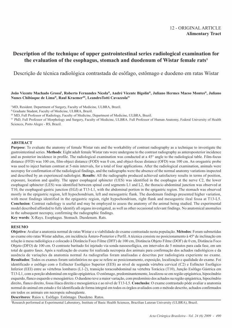
Acta Cirúrgica Brasileira - Vol. 24 (6) 2009 - 490 12 - ORIGINAL ARTICLE Alimentary Tract Description of the technique of upper gastrointestinal series radiological examination for the evaluation of the esophagus, stomach and duodenum of Wistar female rats¹ Descrição de técnica radiológica contrastada de esôfago, estômago e duodeno em ratas Wistar João Vicente Machado Grossi I , Roberto Fernandes Nicola II , André Vicente Bigolin II , Juliano Hermes Maeso Montes II , Juliano Nunes Chibiaque de Lima II , Raul Kraemer III , LeandroTotti Cavazzola IV I MD, Resident. Department of Surgery, Faculty of Medicine, ULBRA, Brazil. II Graduate Student, Faculty of Medicine, ULBRA, Brazil. III MD, Full Professor of Radiology, Faculty of Medicine, Department of Medicine, ULBRA, Brazil. IV PhD, Full Professor of Morphology and Surgery, Faculty of Medicine, ULBRA. Full Professor of Human Anatomy, Federal University of Health Sciences, Porto Alegre - RS, Brazil. ABSTRACT Purpose: To evaluate the anatomy of female Wistar rats and the workability of contrast radiography as a technique to investigate the gastrointestinal series. Methods: Eight adult female Wistar rats were undergone to the contrast radiography as anteroposterior incidence and as posterior incidence in profile. The radiological examination was conducted at a 45° angle to the radiological table. Film-focus distance (FFD) was 100 cm, film-object distance (FOD) was 0 cm, and object-focus distance (OFD) was 100 cm. An orogastric probe was used to inject barium contrast at 5-min intervals, for a total of four applications. After the radiological examination, animals were necropsy for confirmation of the radiological findings, and the radiographs were the absence of the normal anatomy variations inspected and described by an experienced radiologist. Results: All the radiographs produced achieved satisfactory results in terms of position, exposure, location and quality. The upper esophageal sphincter (UES) was identified in the esophagus at the nerve C2, the lower esophageal sphincter (LES) was identified between spinal cord segments L1 and L2, the thoracic-abdominal junction was observed at T10, the esophageal-gastric junction (EGJ) at T13-L1, with the abdominal portion in the epigastric region. The stomach was observed mostly in the epigastric region, left hypochondrium, left and mesogastric flank. The duodenum findings presented higher variation, with most findings identified in the epigastric region, right hypochondrium, right flank and mesogastric ileal fossa at T13-L5. Conclusion: Contrast radiology is useful and may be employed to assess the anatomy of the animal being studied. The experimental model described afforded to fully identify all organs investigated, as well as other occasional relevant findings. No anatomical anomalies in the subsequent necropsy, confirming the radiographic findings. Key words: X-Rays. Esophagus. Stomach. Duodenum. Rats. RESUMO Objetivo: Avaliar a anatomia normal de ratas Wistar e a viabilidade do exame contrastado nesta população. Métodos: Foram submetidas ao exame oito ratas Wistar adultas, em incidência Ântero-Posterior e Perfil. A técnica consiste no posicionamento à 45º de inclinação em relação à mesa radiológica e colocado à Distância Foco Filme (DFF) de 100 cm, Distância Objeto Filme (DOF) de 0 cm, Distância Foco Objeto (DFO) de 100 cm. O contraste baritado foi injetado via sonda nasoesofágica, em intervalos de 5 minutos para cada fase, em um total de quatro fases. Após a realização do exame foi realizada necropsia dos animais para confirmação dos achados radiológicos e da ausência de variações da anatomia normal As radiografias foram analisadas e descritas por radiologista experiente no exame. Resultados: Todos os exames foram satisfatórios no que se refere ao posicionamento, exposição, localização e qualidade do exame. Foi identificado o esôfago com o Esfíncter Esofágico Superior (EES) ao nível de segunda vértebra cervical (C2) e Esfíncter Esofágico Inferior (EEI) entre as vértebras lombares (L1-2), transição toracoabdominal na vértebra Torácica (T10), Junção Esôfago Gástrica em T13-L1, com a porção abdominal em região epigástrica. O estômago, predominantemente, localizou-se em região epigástrica, hipocôndrio esquerdo, flanco esquerdo e mesogástrico. O duodeno teve a maior variação, com predomínio dos achados na região epigástrica, hipocôndrio direito, flanco direito, fossa ilíaca direita e mesogástrica e ao nível de T13-L5. Conclusão: O exame contrastado pôde avaliar a anatomia normal do animal em estudo e foi identificada de forma integral em todos os órgãos avaliados com o método descrito, achados confirmados em todos os animais em necropsia subseqüente. Descritores: Raios x. Esôfago. Estômago. Duodeno. Ratos. 1 Research performed at Experimental Laboratory, Institute of Basic Health Sciences, Brazilian Luteran University (ULBRA), Brazil.
Transcript of Descrição de técnica radiológica contrastada de esôfago ...
Acta Cirúrgica Brasileira - Vol. 24 (6) 2009 - 490
12 - ORIGINAL ARTICLEAlimentary Tract
Description of the technique of upper gastrointestinal series radiological examination forthe evaluation of the esophagus, stomach and duodenum of Wistar female rats¹
Descrição de técnica radiológica contrastada de esôfago, estômago e duodeno em ratas Wistar
João Vicente Machado GrossiI, Roberto Fernandes NicolaII, André Vicente BigolinII, Juliano Hermes Maeso MontesII, JulianoNunes Chibiaque de LimaII, Raul KraemerIII, LeandroTotti CavazzolaIV
I MD, Resident. Department of Surgery, Faculty of Medicine, ULBRA, Brazil.II Graduate Student, Faculty of Medicine, ULBRA, Brazil.III MD, Full Professor of Radiology, Faculty of Medicine, Department of Medicine, ULBRA, Brazil.IV PhD, Full Professor of Morphology and Surgery, Faculty of Medicine, ULBRA. Full Professor of Human Anatomy, Federal University of HealthSciences, Porto Alegre - RS, Brazil.
ABSTRACTPurpose: To evaluate the anatomy of female Wistar rats and the workability of contrast radiography as a technique to investigate thegastrointestinal series. Methods: Eight adult female Wistar rats were undergone to the contrast radiography as anteroposterior incidenceand as posterior incidence in profile. The radiological examination was conducted at a 45° angle to the radiological table. Film-focusdistance (FFD) was 100 cm, film-object distance (FOD) was 0 cm, and object-focus distance (OFD) was 100 cm. An orogastric probewas used to inject barium contrast at 5-min intervals, for a total of four applications. After the radiological examination, animals werenecropsy for confirmation of the radiological findings, and the radiographs were the absence of the normal anatomy variations inspectedand described by an experienced radiologist. Results: All the radiographs produced achieved satisfactory results in terms of position,exposure, location and quality. The upper esophageal sphincter (UES) was identified in the esophagus at the nerve C2, the loweresophageal sphincter (LES) was identified between spinal cord segments L1 and L2, the thoracic-abdominal junction was observed atT10, the esophageal-gastric junction (EGJ) at T13-L1, with the abdominal portion in the epigastric region. The stomach was observedmostly in the epigastric region, left hypochondrium, left and mesogastric flank. The duodenum findings presented higher variation,with most findings identified in the epigastric region, right hypochondrium, right flank and mesogastric ileal fossa at T13-L5.Conclusion: Contrast radiology is useful and may be employed to assess the anatomy of the animal being studied. The experimentalmodel described afforded to fully identify all organs investigated, as well as other occasional relevant findings. No anatomical anomaliesin the subsequent necropsy, confirming the radiographic findings.Key words: X-Rays. Esophagus. Stomach. Duodenum. Rats.
RESUMOObjetivo: Avaliar a anatomia normal de ratas Wistar e a viabilidade do exame contrastado nesta população. Métodos: Foram submetidasao exame oito ratas Wistar adultas, em incidência Ântero-Posterior e Perfil. A técnica consiste no posicionamento à 45º de inclinação emrelação à mesa radiológica e colocado à Distância Foco Filme (DFF) de 100 cm, Distância Objeto Filme (DOF) de 0 cm, Distância FocoObjeto (DFO) de 100 cm. O contraste baritado foi injetado via sonda nasoesofágica, em intervalos de 5 minutos para cada fase, em umtotal de quatro fases. Após a realização do exame foi realizada necropsia dos animais para confirmação dos achados radiológicos e daausência de variações da anatomia normal As radiografias foram analisadas e descritas por radiologista experiente no exame.Resultados: Todos os exames foram satisfatórios no que se refere ao posicionamento, exposição, localização e qualidade do exame. Foiidentificado o esôfago com o Esfíncter Esofágico Superior (EES) ao nível de segunda vértebra cervical (C2) e Esfíncter EsofágicoInferior (EEI) entre as vértebras lombares (L1-2), transição toracoabdominal na vértebra Torácica (T10), Junção Esôfago Gástrica emT13-L1, com a porção abdominal em região epigástrica. O estômago, predominantemente, localizou-se em região epigástrica, hipocôndrioesquerdo, flanco esquerdo e mesogástrico. O duodeno teve a maior variação, com predomínio dos achados na região epigástrica, hipocôndriodireito, flanco direito, fossa ilíaca direita e mesogástrica e ao nível de T13-L5. Conclusão: O exame contrastado pôde avaliar a anatomianormal do animal em estudo e foi identificada de forma integral em todos os órgãos avaliados com o método descrito, achados confirmadosem todos os animais em necropsia subseqüente.Descritores: Raios x. Esôfago. Estômago. Duodeno. Ratos.1Research performed at Experimental Laboratory, Institute of Basic Health Sciences, Brazilian Luteran University (ULBRA), Brazil.

Grossi JVM et al
491 - Acta Cirúrgica Brasileira - Vol. 24 (6) 2009
Introduction
The contrast radiological examination of the esophagus,stomach and duodenum is greatly useful in routine clinical andsurgical evaluations1.
The veterinary radiology has utilized radiologicalexaminations as a diagnosis tool, and the literature shows examplesof investigations conducted with small animals2,3.
The contrast radiology affords to visualize severalesophageal changes. The technique describes lesions like thicknessor filling defects (suggestive of cancer), peptic stenoses, ulcers,as well as hiatal hernias. One of the limitations in study is theidentification of small masses and the initial stages of the disease4,6.
The conduction of contrast radiology in accordance withan appropriate technique is of major importance in the effectivediagnosis of gastrointestinal conditions. The exposure factors foran adequate basic radiological examination include peak kilovoltage(kVp) and milliamperage per second (mAs). The factors that evaluatethe quality of the radiological image include: (I) density, whichdetermines the degree the film is blackened (mAs); (II) contrast,which differentiates the density of the adjacent areas (kVp); (III)detail, which implies the clarity of structures (and that may varydue to movement, whether voluntary or not); and (IV) distortion,which is the wrong representation of size or format of the object asdefined by the film-focus distance (FFD) and beam divergence13,15.
Considering the lack of a description of a radiology techniqueand of anatomic findings, location reference or position of the esophagus,stomach and duodenum in rats, we adopted an anatomical animalmodel based on subdivisions into quadrants and relative heightagainst the vertebral column. This normality model is fundamentalfor the comparison of post-surgical studies of the esophagus,stomach and duodenum of rodents, in future research11,13-15.
To assess the radiological method utilized in terms of thequality and identification of the esophagus, stomach a duodenumof female Wistar rats.
Methods
This paper, is an original, retrospective study of randomizedexperimental design. The study sample consisted of 8 adult femaleWistar rats with 16-week-old and weighing between 170 and 240 g.Randomized performed by arbitrarily assigning the rats a set ofinitials. All animals were maintained in individual cages with twoanimals in each cage, in a controlled temperature environment(22-24°C). Water and feed were ad libitum.
Inclusion criteria were:1) Female Wistar rat, age between 15 and 16 weeks;2) Not siblings;3) No previous treatment;
Exclusion criteria were:1) Visible anatomical distortion, observed before the
radiological examination;2) Anatomical distortion observed as of necropsy.
Images were examined by an experienced radiologist,who was blinded to the identification of the specimen analyzed. Invirtue of the non-availability of male rats it was used only femalerats. The radiological findings thus recorded were latterlycompared to the necropsy findings. The radiological equipmentused had a fixed base, with the X-ray tube being always positionedparallel to the film and at a 45° inclination to the radiologicaltable. The radiological beam was emitted at 36 kW and 32 mA.Impression was conducted in mamographic ECRAM film.Photographic images were captured using a digital camera.
The present study was approved by the Ethics Committeein Research, Lutheran University Luteran of Brazil (ULBRA). Theprocedures and management of animals were supervised by theveterinary in charge of the institutions bioterium.
The procedures were conducted according to the stagesdescribed below:
I. Fasting. A 12-h fasting period was observed, thoughwith water offered continuously.
II. Anesthesia. Intramuscular injections of Atropin SC10 (0.044 mg / kg body weight) were applied. After, the animalsunderwent general anesthesia using a mixture of xylazine 2% andketamine 50 mg as intramuscular injections (0.1 ml / 100 g bodyweight).
III. Positioning and probing. Animals were placed indorsal recumbency at a 45° angle to the horizontal plane of theradiological table. (Figure 1) The film compartment was supportedby the upper limbs at a film-focus distance (FFD) of 100 cm,film-object distance (FOD) of 0 cm, and object-focus distance(OFD) of 100 cm. Orogastric probing was conducted using a nr. 4probe, placed 7 cm away from the oral cavity. The first radiographwas taken at anteroposterior incidence (AP) for confirmation ofthe position at the distal esophagus end.
IV. Injection of the contrast. The contrast (barium sulfate100%) was administered 4 times at 5-min intervals. Contrastvolumes were 0.3, 0.6, 0.3, and 0.3 ml, totaling 1.2 ml. If the organto be visualized was not filled, an extra volume of 0.4 ml wasinjected. After the first injection of contrast (0.3 ml) the secondradiograph was taken, which revealed the esophagus andthe esophagogastric junction, as well as the gastric content.Subsequently, 0.6 ml contrast was administered, reaching thegastroduodenal junction. The duodenum-jejunum junction wasfilled at the third phase, with the injection of the 0.3-ml contrastvolume. If satisfactory, after the third phase the rat was placedon right lateral decubitus for the conduction of the profileX-ray (P).
V. Necropsy. Immediately after the end of the procedure,the animals were administered a 10% potassium chlorideintracardiac injection, while still anesthetized at plane three.
VI. Verification of the technique. The radiologicalexaminations obtained were sent, after revelation, to be evaluatedby a radiologist trained in RESD. The radiologist was instructed toassess the quality of the examination and to identify the anatomicalstructures typical of the species used.

Acta Cirúrgica Brasileira - Vol. 24 (6) 2009 - 492
Description of the technique of upper gastrointestinal series radiological examination for the evaluation of the esophagus, stomach and duodenumof Wistar female rats
Results
The analysis of the images revealed that in all the 8animals studied, the regimens and positions were appropriate toafford the quality required for the assessment of the esophagus,stomach and duodenum (Figure 2).
FIGURE 1 - Position of the specimens on the radiological table
FIGURE 2 - Nine abdominal quadrants. Longitudinal divisions aredefined by the hemiclavicular lines. Transversal divisions aredefined by the lower L1 border, transpyloric plane, and line on theiliac crests on the lower L5 edge
As regards the species studied, the exam is considerednormal when shows the usual structures forming the upperdigestive tract. Apart from this, the assessment of the positions hasto consider the specific details of the rat anatomy, as the presenceof 13 thoracic vertebrae and of 6 lumbar vertebrae, for instance(Figure 3).
The esophagus was visualized showing its initial portionat the second cervical vertebra (C2) the upper esophageal sphincter(UES) and located posterior to the trachea. The esophagus wasidentified in all the animals. The probe position was initially chosento be between the thoracic vertebrae T10-13. Hemidiaphragms werepositioned at T10 in all exams conducted. The esophageal-gastricjunction was observed in the epigastric region and at T13-L1, inanteroposterior and posterior incidences. The lower esophagealsphincter (LES) was also observed at L1-2.
The thoracic-abdominal junction was observed at T11 inthe esophageal hiatus, in all animals studied. The location of thelower esophageal sphincter (LES) and the hemidiaphragms withL1 and T10, respectively (Figure 4).
As regards the stomach, the gastric cardia was locatedin the epigastric region at T13 and L1, angular incisure at L1-2,fundus of stomach in the epigastric region, and left hypochondriumat T13-L1. The gastric body was also observed in the mesogastricregion and left flank at L1-3, the antrum in the epigastric region atT13-L2, the pylorus in the epigastric region at T13-L1.
The duodenum was filled with contrast to the fourthduodenal portion, shown in the epigastric region, righthypochondrium, right flank, right iliac fossa, and mesogastricregion, at T13-L5 (Figure 5).

Grossi JVM et al
493 - Acta Cirúrgica Brasileira - Vol. 24 (6) 2009
FIGURE 3 - Superimposition of the abdominal quadrantsin the anteroposterior incidence
FIGURE 4 - Presence of contrast in the esophagus,stomach and duodenum in the anteroposterior incidence
FIGURE 5 - Visualization of the lateral contrast radiography of the esophagus, stomach and duodenum

Acta Cirúrgica Brasileira - Vol. 24 (6) 2009 - 494
Description of the technique of upper gastrointestinal series radiological examination for the evaluation of the esophagus, stomach and duodenumof Wistar female rats
The necropsy was carried out by median mento-pubicincision (Figure 6) and search for an anatomic changes, and didnot reveal any structure that had not been detected by contrast
FIGURE 6 – a. Necropsy showing the median incision and the location of the organs in the abdominal cavity.b. Necropsy showing the thoracic and abdominal locations of the esophagus, as well as the stomach and the duodenum
During the second and third radiological examination,two cases of barium aspiration were observed. This event wasvisualized in the airways, trachea, main bronchia and bronchialtree.
To visualize the duodenum, an extra injection of contrastwas required for two animals.
Discussion
The aim of contras radiology is the generation ofradiological findings that may demonstrate changes as stenosis,fistulas, pervisouness of anasotmoses, and diverticula14. Someauthors report the deformities of the esophagus after contrastinvestigation in Barret’s esophagus, which may assist in thescreening of patients with gastro-esophageal reflux symptoms5,7,8,13.In the present study, no such changes were observed, whichreveals the normal conditions of the specimens studied.
In the post-operative procedures of stomach andesophagus surgeries, it is possible to search for secondarydeformities that appear after the surgical procedures, exactly whenthe trauma will not be surgical the esophagography may havesensitivity varying of 50- 90% of the times13-16.
In the esophageal perforation, the diagnosis by contrast
examination reveals the occurrence of contrast runoffs in 90% ofthe cases investigated9,10.
The contrast radiology of the upper gastrointestinal seriesdid not show contrast runoff occurrences, in any of the specimensinvestigated, or any type of lesion that was visible even after theconduction of subsequent necropsy14.
It was possible to clearly and safely determine all theanatomical aspects of the organs studied (location, anatomicalrelationships, morphological characteristics), which were confirmedby subsequent laparotomy. This finding revealed that the techniqueas proposed in the present study was an appropriate radiologicalapproach to investigate the normal morphology of the esophagus,stomach and duodenum in Wistar female rats.
In two of the eight exams conducted, the contrast wasvisualized in the airways, due to the speed it was injected viagastrointestinal probe, impaired by the clearance of the esophagus.What the 100% were already foreseen with the barium sulfate use16.
This occurred at the first times the contrast was injected,and was not observed again afterwards.
Among the findings that justified the exclusion of the animalsfrom controlled studies, it is important to consider the possibilityof the occurrence any other abnormality that compromises theconduction of anterior evaluations in these animals.
radiography of the upper gastrointestinal series. In like manner,neither findings nor anatomical changes that led to the exclusionof one specimen were observed.
Grossi JVM et al
495 - Acta Cirúrgica Brasileira - Vol. 24 (6) 2009
Conclusion
The radiological investigation was satisfactory andafforded to identify all the evaluated structures. Contrast radiologyis useful and may be employed to assess the anatomy of the animalbeing studied.The experimental model described afforded to fullyidentify all organs investigated, as well as other occasionalrelevant findings. No anatomical anomalies in the subsequentnecropsy, confirming the radiographic findings.
References
1. Dibble C, Levine MS, Rubesin SE, Laufer I, Katzka DA. Detection ofreflux esophagitis on double-contrast esophagrams and endoscopy usingthe histologic findings as the gold standard. Abdom Imaging. 2004;29:421-5.2. Styles RA, Gibb SP, Tarshis A, Silverman ML, Scholz FJ.Esophagogastric polyps: radiographic and endoscopic findings. Radiology.1985;154:307-11.3. Gupta S, Levine MS, Rubesin SE, Katzka DA, Laufer I. Usefulness ofbarium studies for differentiating benign and malignant strictures of theesophagus. AJR Am J Roentgenol. 2003;180:737-44.4. Levine MS, Goldstein HM. Fixed transverse folds in the esophagus: asign of reflux esophagitis. AJR Am J Roentgenol. 1984;43:275-8.5. Seth NG. Barium studies in patients with barrett’s esophagus:importance of focal areas of esophageal deformity. AJR Am J Roentgenol.1994;163:65-7.6. Ponsaing LG, Kiss K, Loft A, Jensen LI, Hansen MB. Diagnosticprocedures for submucosal tumors in the gastrointestinal tract. World JGastroenterol. 2007;13(24):3301-10.7. Katada N, Hinder RA, Smyrk TC, Hiki Y, Kakita A. Duodenoesophagealreflux induces apoptosis in rat esophageal epithelium. Dig Dis Sci.1999;44(2):301-10.
8. Melo LL, Kruel CD, Kliemann LM, Cavazzola LT, Boeno Rda L, SilberPC, Grossi RS. Influence of surgically induced gastric and gastroduodenalcontent reflux on esophageal carcinogenesis-experimental model in Wistarfemale rats. Dis Esophagus. 1999;12(2):106-15.9. Pera M, Brito MJ, Poulsom R, Riera E, Grande L, Hanby A, Wright NADuodenal-content reflux esophagitis induces the development of glandularmetaplasia and adenosquamous carcinoma in rats. Carcinogenesis.2000;21(8):1587-91.10. Miwa K, Miyashita T, Hattori T. Reflux of duodenal or gastroduodenalcontents induce esophageal carcinoma in rats. Nippon Rinsho.2004;62(8):1433-8.11. Zhang F, Altorki NK, Wu YC, Soslow RA, Subbaramaiah K,Dannenberg AJ Duodenal reflux induces cyclooxygenase-2 in theesophageal mucosa of rats: evidence for involvement of bile acids.Gastroenterology. 2001;121(6):1391-9.12. Clark GW, Smyrk TC, Mirvish SS, Anselmino M, Yamashita Y, HinderRA, DeMeester TR, Birt DF. Effect of gastroduodenal juice and dietaryfat on the development of Barrett’s esophagus and esophageal neoplasia:an experimental rat model. Ann Surg Oncol. 1994;1(3):252-61.13. César JMS, Petroianu A, Gouvêa AP, Alvin DR. Reopening of thegastroduodenal pylorus after its closure in rats. J Surg Res. 2008;144:89–93.14. Weigelt JA, Thal ER, Snyder WH 3rd, Fry RE, Meier DE, Kilman WJ.Diagnosis of penetrating cervical esophageal injuries. Am J Surg.1987;154:619-22.15. Ginai AZ, ten Kate FJ, ten Berg GM, Hoornstra K. Experimentalevaluation of various available contrast agents for use in the uppergastrointestinal tract in case of suspected leakage: effects on mediastinum.Br J Radiol. 1985;58(691):585-92.16. Vaezi MF, Baker ME, Achkar E, Richter JE. Timed bariumoesophagram: better predictor of long term success after pneumaticdilation in achalasia than symptom assessment. Gut. 2002;50(6):765–70.
Conflict of interest: noneFinancial source: none
Correspondence:João Vicente Machado GrossiRua Carlos Trein Filho, 1271/40196450-120 Porto Alegre – RS BrazilPhone: (55 51)[email protected]
Received: April 09, 2009Review: June 13, 2009
Accepted: July 28, 2009
How to cite this articleGrossi JVM, Nicola RF, Bigolin AV, Montes JHM, Lima JNC, Kraemer R, Cavazzola LT. Description of the technique of uppergastrointestinal series radiological examination for the evaluation of the esophagus, stomach and duodenum of Wistar female rats. ActaCir Bras. [serial on the Internet] 2009 Nov-Dec;24(6). Available from URL: http://www.scielo.br/acb