Derivation, propagation and differentiation of human stem ...
94
Derivation, propagation and differentiation of human stem and progenitor cells Mathilda Zetterström Axell Centre for Brain Repair and Rehabilitation, Department of Clinical Neuroscience and Rehabilitation Institute of Neuroscience and Physiology at Sahlgrenska Academy University of Gothenburg 2009
Transcript of Derivation, propagation and differentiation of human stem ...

Derivation, propagation and differentiation
of human stem and progenitor cells
Mathilda Zetterström Axell
Centre for Brain Repair and Rehabilitation,
Department of Clinical Neuroscience and Rehabilitation
Institute of Neuroscience and Physiology
at Sahlgrenska Academy
University of Gothenburg
2009
¨
Mathilda Zetterström Axell
Tryck: Intellecta infolog
Göteborg 2009
ISBN 978-91-628-7841-2
Cover illustration: Devil-shaped human embryonic stem cell colony, Cellartis
AB, Dundee, Scotland

3
Derivation, propagation and differentiation of human
stem and progenitor cells
Mathilda Zetterström Axell
Centre for Brain Repair and Rehabilitation, Institute of Neuroscience and
Physiology at Sahlgrenska Academy, University of Gothenburg, 2009
Abstract
Neuronal loss is a common feature of many neurological disorders, including stroke,
Parkinson’s disease, Alzheimer’s disease and traumatic brain injury. Human
embryonic stem cells (hESCs) and hESC-derived neural progenitors (NPs) may
provide a number of new ways for studying and treating diseases and injuries in the
brain. Studying the proliferation and differentiation characteristics of hESCs and NPs
is important for three main reasons: 1, they represent an almost unlimited source of
cells for neuron replacement therapies after neurodegeneration in the brain; 2, they
are a good source of normal human cells for studying functional genomics,
proteomics or for drug screening; and 3, they allow us to study early human brain
development.
The general aims of this thesis were four-fold: 1, to develop efficient and simple
methods for the large scale propagation of hESCs and hESC-derived NPs; 2, to
optimise NP differentiation into mature neurons and glia; 3, to find suitable materials
to promote migration and differentiation of stem and progenitor cells, and; 4, to
uncover critical differentiation factors expressed in common between neuroblasts in
the rostral migratory stream (RMS; the only long distance cell migration system in
the human brain) and that of hESC-derived NPs.
To address these aims, we used a range of techniques including cell culture,
morphometric analysis, immunocytochemistry, immunohistochemistry and RT-PCR.
Here we report the development of an improved method for the transfer and culture
of undifferentiated hESCs in the absence of a cell feeder-layer, which is more cost
effective and reduces the contact with murine feeder cells that render the hESCs
4
unusable for future transplantation into humans. We have also developed a simple
method for producing NPs from hESCs, suitable for large scale expansion and long
term propagation of NPs. The production of large quantities of NPs allows us to
readily compare the properties of NPs in culture to those in the human brain.
Studying the differentiation of hESCs on permissive substrates has also been a focus
and is of importance because of the relevance to the developing and adult human
brain, where a complex extracellular matrix exists as scaffolding for neuronal
development. We found electrospun fibrous scaffolds suitable for propagation and
differentiation of hESCs, deriving predominantly tyrosine hydroxylase positive
neurons indicating a dopaminergic fate. Finally, we studied the adult human brain for
the presence of progenitor cells with migratory characteristics. We used a
combination of serial sectioning, immunostaining and RT-PCR of human post-
mortem brain material. This was the first study to reveal the presence of a human
RMS by which neuroblasts migrate long distances from the subventricular zone to
the olfactory bulb where they differentiate into mature neurons. Further, we
discovered a number of differentiation factors expressed (Pax6, NCAM, DCX, βIII-
tubulin) in common between the human RMS neuroblasts and hESC-derived NPs.
Taken together, this thesis reveals improved ways to propagate and differentiate
hESCs in culture, and has uncovered common differentiation factors present in both
human neuroblasts and NPs. These studies further our understanding of human brain
development, allow large scale production of NPs for further study, and may one day
be useful for treating central nervous system disorders.
Key Words
Human embryonic stem cells, neural progenitor cells, stem cells, differentiation,
propagation, migration, cell culturing, rostral migratory stream, electrospun scaffolds

5
Populärvetenskaplig sammanfattning på svenska
Förlust av nervceller är en gemensam nämnare för många neurologiska
sjukdomar som stroke, Parkinsons sjukdom, Alzheimers sjukdom och
traumatisk hjärnskada. Den vuxna hjärnans kapacitet att reparera sig själv är
begränsad varför mycket forskning fokuserar på att kunna ersätta och
reparera skadad hjärnvävnad. Humana embryonala stamceller (hESC;
omogna, självreplikerande, kan bilda alla celltyper i den vuxna kroppen) och
neurala progenitorceller (NPC; självreplikerande, förstadium till mogna
hjärnceller) deriverade från hESC kan ge oss nya sätt att studera och
behandla skador på hjärnan efter sjukdom eller trauma. Detta genom att förse
oss med en nästan oändlig källa av celler för att studera geners och proteiners
funktion, för läkemedelutveckling, för att studera tidig utveckling av den
mänskliga hjärnan och för utveckling av transplantationsterapier.
Det är känt sedan tidigare att progenitorceller förflyttar sig en lång sträcka via
en specifik bana/”motorväg” (rostral migratory stream; RMS) i den vuxna
hjärnan hos gnagare. Vi har här för första gången identifierat migrerande
progenitorceller (neuroblaster) i den vuxna mänskliga hjärnan och visar även
för första gången att RMS även finns hos människan. Dessa neuroblaster
visade sig uttrycka flera markörer (Pax6, NCAM, DCX och βIII-tubulin)
gemensamt med NPC deriverade från hESCs, enligt en ny enkel, effektiv och
billig metod som vi utvecklat här. Stora mängder NPC behövs bl.a. för att
kunna jämföra egenskaper hos NPC i odling med de i hjärnan. Traditionellt
sett odlas hESC på att stödlager av så kallade feederceller (bindvävsceller
från mus eller människa). Dessa feederceller utsöndrar näring och bidrar
dessutom med andra idag ej kända faktorer, vilka bidrar till att behålla hESC
i ett stabilt omoget stadium. Vi har här även utvecklat ett förbättrat och stabilt
protokoll för överföring av hESC till ett feederfritt odlingsunderlag och
vidare expandering. Feederfri odling minskar risken för kontamination av
skadliga molekyler från feedercellerna till hESC, vilket måste elimineras för

6
att kunna använda cellerna för transplantationsterapier. För att ta ytterligare
steg mot transplantationsterapier och för att lättare kunna styra mognaden av
hESC till specifika nervcellstyper har vi har tagit fram ett biokompatibelt 3-
dimentionellt material som är lätt att odla hESC på/i vilket främjar bildandet
av dopaminerga nervceller. Det är denna celltyp som dör vid Parkinsons
sjukdom.
Sammanfattningsvis så har våra studier bidragit till att förbättra och utveckla
mer effektiva metoder för att odla hESC och få dem att bilda nervceller. Vi
har även hittat faktorer involverade i mognadsprocessen gemensamma hos
neuroblaster i den vuxna mänskliga hjärnan och hos NPC deriverade från
hESC enligt vår metod. Våra studier främjar förståelsen för hjärnans
utveckling och visar att vi kan producera stora mängder NPC för vidare
studier, något som i framtiden kan vara mycket viktigt för att behandla skador
på centrala nervsystemet (CNS).

7
Papers included in the thesis
I. Eva Sjögren-Jansson, Mathilda Zetterström, Karina Moya, Jenny
Lindqvist, Raimund Strehl, and Peter S. Eriksson. "Large-Scale Propagation
of Four Undifferentiated Human Embryonic Stem Cell Lines in a Feeder-
Free Culture System". Developmental Dynamics, 233:1304–1314, 2005.
II. Mathilda Zetterström Axell, Suzana Zlateva, Maurice A. Curtis. "A
method for rapid derivation and propagation of neural progenitors from
human embryonic stem cells". In manuscript.
III. Björn Carlberg*, Mathilda Zetterström Axell*, Ulf Nannmark, Johan Liu,
H. Georg Kuhn. "Electrospun polyurethane scaffolds for proliferation and
neuronal differentiation of human embryonic stem cells".* equal
contribution. Biomed. Mater. 4 (2009) 045004.
IV. Maurice A. Curtis, Monica Kam, Ulf Nannmark, Michelle F. Anderson,
Mathilda Zetterström Axell, Carsten Wikkelso, Stig Holtås, Willeke M. C.
van Roon-Mom, Thomas Björk-Eriksson, Claes Nordborg, Jonas Frisén,
Michael Dragunow, Richard L. M. Faull, Peter S. Eriksson. "Human
neuroblasts migrate to the olfactory bulb via a lateral ventricular extension".
Science. 2007 Mar 2;315(5816):1243-9. Epub 2007 Feb 15.
Additional papers not included in the thesis;
Camilla Karlsson, Katarina Emanuelsson, Fredrik Wessberg, Kristina Kajic,
Mathilda Zetterström Axell, Peter S. Eriksson, Anders Lindahl, Johan
Hyllner, Raimund Strehl. “Human embryonic stem cell-derived mesenchymal
progenitors-Potential in regenerative medicine”. Stem Cell Res. 2009 May
19.

8
Abbreviations
ALP - alkaline phosphatase
AS - Akademiska Sjukhuset
ASCs - adult stem cells
BMPs - bone morphogenetic proteins
BrdU - bromodeoxyuridine
CN - caudate nucleus
CNS - central nervous system
CSF - cerebrospinal fluid
DAB - 3,3 diaminobenzidine
DAPI - 4´-6´Diamidino-2-
phenylindole
DCX - doublecortin
DG - dentate gyrus
D-MEM - Dulbecco’s modified eagle
medium
DMEM/F12 - DMEM/nutrient
mixture F-12
DMF - n,n-dimethylformamide
DMSO - dimethyl sulfoxide
EBs - embryoid bodies
ECM - extracellular matrix
EGF - epidermal growth factor
ELISA - enzyme-linked
immunosorbent assay
En1 - engrailed 1
ESCs - embryonic stem cells
FBS - fetal bovine serum
FGFs - fibroblast growth factors
FISH - fluorescence in situ
hybridization
GAPDH - glyceraldehyde-3-
phosphate dehydrogenase
Gbx - gastrulation brain homebox
GFAP - glial fibrillary astrocytic
protein
HBSS - Hank’s Balanced Salt
Solution
hEF - human embryonic fibroblasts
hESCs - human embryonic stem cells
ICM - inner cell mass
iPSCs - induced pluripotent stem cells
LV - lateral ventricle
mEF - mouse embryonic fibroblasts
MRI - magnetic resonance imaging
NCAM - neural cell adhesion
molecule
NEAA - non essential amino acids
NPs - neural progenitors
NSCs - neural stem cells
OB - olfactory bulb
Oct-4 - POU Transcription Factor-4
Olig2 - oligodendrocyte lineage
transcription factor 2
OT - olfactory tract
Otx - orthodentical homologue
Pax - paired box

9
PBS - phosphate buffered saline
PCNA - proliferating cell nuclear
antigen
PD - Parkinson’s disease
PEST - penicillin-streptomycin
PFA - paraformaldehyde
PH3 - phosphorylated histone H3
PSA - polysialic acid
RA - retinoic acid
RMS - rostral migratory stream
RT-PCR - reverse transcriptase-
polymerase chain reaction
SA - Sahlgrenska University hospital
SCID - severe combined
immunodeficient
SD - standard deviation
SEM - scanning electron microscopy
SGZ - subgranular zone
Shh - sonic hedgehog
Sox - sex determining region of Y-
chromosome
SR - serum replacement
SSEA - stage specific embryonic
antigens
SVZ - subventricular zone
TEM - transmission electron
microscopy
TFs - transcription factors
TGFβ - transforming growth factor
beta
THF - tetrahydrofuran
TRA - tumour rejection antigen
TUNEL - terminal deoxynucleotidyl
transferase-mediated deoxyuridine
triphosphate nick end labeling
VOE - ventriculo-olfactory extension
VONS - ventriculo-olfactory
neurogenic system
VZ - ventricular zone

10
Table of contents
Abstract________________________________________________3
Populärvetenskaplig sammanfattning på svenska_______________5
Papers included in the thesis _______________________________7
Abbreviations ___________________________________________8
Background ___________________________________________15
Stem cells from concept to thought____________________________ 15
What is a stem cell? _____________________________________________ 15
Stem cells at different levels of maturation ___________________________ 16
Stem cells in the embryo _________________________________________ 17
Stem cells in the developing brain__________________________________ 18
Differentiation and migration of neural progenitor cells_______________ 18
Neural inducing/directing signals ________________________________ 19
Neural inducing molecules _____________________________________ 20
Stem cells in the adult brain_______________________________________ 21
Neurogenesis and gliogenesis ___________________________________ 21
The rostral migratory stream (RMS) ______________________________ 22
Migration and differentiation inducing molecules ___________________ 22
The function of the olfactory system______________________________ 23
Differentiation from embryo to adult brain_____________________ 24
Differentiation _________________________________________________ 24
Neural stem cells _______________________________________________ 24
Neuroectodermal cell type markers _________________________________ 24
Generation of and in vitro culturing methods for human embryonic
stem cells _________________________________________________ 27
Derivation of a human embryonic stem cell line _______________________ 27
Characterization of an hESC line___________________________________ 30

11
Feeder-free culture of hESCs______________________________________ 31
Neural progenitors from hESCs____________________________________ 32
Potential benefits of embryonic stem cell research _______________ 32
Problems to be overcome for the success of cell-based therapies __________ 33
Stem cell therapy in neurological disorders___________________________ 34
Tissue engineering _________________________________________ 35
Scaffolds for hESC propagation and differentiation ____________________ 35
Aim of these studies _____________________________________37
Experimental procedures _________________________________38
Ethical approval ___________________________________________ 38
Human embryonic stem cell (hESC) lines (paper I, II, III) ________ 38
Preparation of conditioned VitroHES-medium (paper I and II) ____ 38
Transfer of hESCs to Matrigel (paper I) _______________________ 39
Viability study on hESC clusters dissociated mechanically vs.
enzymatically (paper I) _____________________________________ 40
The hESC cluster sizes after dissociation (paper I)_______________ 40
Passage of Matrigel propagated hESCs (paper I, II and III) _______ 40
Derivation and propagation of neural progenitor cells (paper II)___ 41
In vitro differentiation of hESC-derived neural progenitor cells (paper
II) _______________________________________________________ 42
Electrospun fiber for co-culture and differentiation of hESCs (paper
III) ______________________________________________________ 42
Human tissue collection (paper IV) ___________________________ 43

12
Characterization of undifferentiated hESCs, NPs, mature derivates,
and RMS neuroblasts (paper I, II, III, IV)______________________ 44
Immunocytochemistry (paper I, II, III) ______________________________ 44
Immunohistochemistry (paper IV)__________________________________ 44
Alkaline phosphatase (ALP) expression (paper I, II) ___________________ 47
Telomerase activity (paper I)______________________________________ 47
Karyotyping and fluorescence in situ hybridization (FISH) (paper I) _______ 48
Teratomas (paper I) _____________________________________________ 48
Reverse transcriptase-polymerase chain reaction (RT-PCR) (paper I, II, IV) _ 49
TUNEL staining (paper IV)_______________________________________ 52
Cryopreservation of Matrigel cultured hESCs and hESC-derived NPs
(paper I and II) ____________________________________________ 52
Statistics (paper II, III) _____________________________________ 53
Electrospun polymer fiber generation (paper III)________________ 53
Surface morphology and structural properties of electrospun polymer
scaffolds (paper III) ________________________________________ 54
Scanning electron microscopy (SEM), (paper III) _______________ 55
Transmission electron microscopy (TEM), (paper IV)____________ 55
Magnetic resonance imaging (MRI) of human brains (paper IV) ___ 56
Results and discussion ___________________________________57
Paper I ___________________________________________________ 57
Mechanical dissociation is more efficient than enzymatic dissociation when
transferring hESC cultures to feeder-free conditions____________________ 58
The cluster size after dissociation is important for transfer and for passage __ 59
The conditioned medium was optimal from mEFs in passage 2, day 1-3____ 60
The percentage of mitotic cells was similar in feeder and feeder-free hESC
cultures ______________________________________________________ 60

13
Successful cryopreservation by slow rate freezing and rapid thawing of feeder-
free hESC cultures ______________________________________________ 60
Our hESCs maintained pluripotency and other hESC characteristics after
transfer to feeder-free conditions___________________________________ 61
Paper II __________________________________________________ 62
Matrigel propagated hESCs for NP generation ________________________ 62
Gelatine and laminin substrates function equally well for cell attachment and
NP derivation__________________________________________________ 63
Rosette formations in passage 1____________________________________ 63
FGF2 is required for the derivation and maintenance of NPs _____________ 64
Cell density affects cell fate_______________________________________ 64
Neuroectodermal markers are expressed by our NP cultures _____________ 65
PAX6 and Sox1 gene expression in the NP cultures ____________________ 66
Sox3 gene expression in the NP cultures and its mature derivates _________ 66
Gradually declining Oct-4 expression required for NP derivation _________ 67
GFAP is expressed by undifferentiated NPs and its derivates_____________ 67
Mature neurons and glial cells are derived from the NP populations _______ 67
Some mesodermal markers are found in the NP cultures ________________ 68
Paper III _________________________________________________ 68
Human ESCs attach and proliferate on electrospun fibrous scaffolds _______ 68
A neuronal cell fate was induced in cells grown on electrospun scaffolds ___ 69
The 3-dimentional scaffolds affect hESC cell fate determination __________ 70
The interaction between hESCs and the scaffolds were shown by SEM
micrographs ___________________________________________________ 71
Paper IV _________________________________________________ 71
The anatomical location of the RMS in the human brain ________________ 71
Cell death is not the fate of most of the RMS progenitor cells ____________ 72
Progenitors in the RMS have migratory proteins and a migratory morphology 72
Ultrastructural studies verify that the human SVZ contains cells with migratory-
like morphology________________________________________________ 73

14
Ultrastructural studies reveal progenitors at all levels of the RMS that have
migratory morphology___________________________________________ 74
Directed migration of progenitors in human VONS ____________________ 74
Progenitor cells become neurons in the OB___________________________ 75
The human RMS is organized around a tubular extension of the lateral ventricle
that reaches the OB _____________________________________________ 75
Pax6, Olig 2, and DCX gene expression is consistent with differentiation along
the VONS ____________________________________________________ 76
Conclusions and Significance _____________________________77
Conclusions from paper I ___________________________________ 77
Conclusion from paper II ___________________________________ 77
Conclusions from Paper III __________________________________ 78
Conclusions from paper IV __________________________________ 78
Responses to given aims__________________________________79
Acknowledgements______________________________________80
References ____________________________________________82

15
Background
Stem cells from concept to thought
What is a stem cell?
All stem cells, regardless of their source, have three important characteristics
that distinguish them from other types of cells in the body; 1, they are capable
of dividing and self-renewal for long periods; 2, they are unspecialized cells;
and 3, and they give rise to all specialized cell types. Under certain
physiologic or experimental conditions, they can be induced to become
tissue- or organ-specific cells with special functions such as the beating cells
of the heart muscle or dopamine producing neurons of the brain [1].
Most work on stem cells is done with either embryonic stem cells (ESCs) or
adult stem cells (ASCs) from rodent or primate species. ESCs are primitive
(undifferentiated) cells derived from a preimplantation embryo with the
capacity to self-replicate indefinitely. They have the potential to become any
cell type of the adult body, thus they are said to be pluripotent. The ASCs (or
somatic) on the other hand are undifferentiated cells found in a differentiated
tissue that typically generate the cell types of the tissue in which they reside.
They can self-renew, with limitations, and they can differentiate to form cell
types of tissues other than the type in which they reside [1]. Only very
recently (in 2006) a new type of stem cell was derived. Researchers made a
breakthrough by identifying conditions that would allow some specialized
adult cells to be "reprogrammed" genetically to assume a stem cell-like state.
These new stem cells are called induced pluripotent stem cells (iPSC; [2]).

16
Stem cells at different levels of maturation
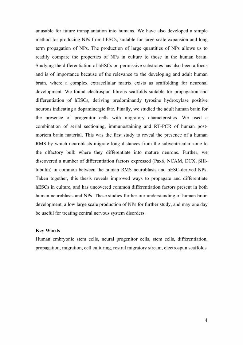
The ultimate stem cell is the fertilized egg, the zygote, which has the ultimate
potential, since it can generate a fetus, and is thus said to be totipotent (figure
1). As the totipotent cells divide it generates pluripotent stem cells that have
the unique ability to self-renew indefinitely and can generate all cell types of
the adult body [3]. Although, they do not have the capacity to generate a
fetus, since the pluripotent cells lack the ability to generate the placenta and
other tissue necessary for development in the uterus. As the pluripotent cells
differentiate, their capacity for self-renewal becomes limited and they gain
the potential to differentiate. As time goes on they become multipotent like
neural stem cells (NSCs) that can only generate the cell types of the tissue in
which they reside, that is neurons and glial cells. Finally, a unipotent stem
cell is a specialised cell in adult organisms capable of differentiating along
only one lineage. The adult stem cells in many differentiated, undamaged
tissues are typically unipotent and give rise to just one cell type under normal
conditions (figure 1), [1].
17
Figure 1; Stem cells from conception to thought. A schematic figure of stem cells with neural
capacity at different levels of maturation.
Stem cells in the embryo
The fertilised egg undergoes multiple divisions to generate a blastocyst. The
blastocyst is primarily composed of three structures; 1, the outer trophoblast,
which is the layer of cells that surrounds the blastocoel; 2, a hollow cavity
inside the blastocyst; 3, the inner cell mass (ICM), which is a group of cells at
one end of the blastocoel that develop into the embryo proper [1]. From the
ICM regionalised cellular differentiation takes place to form three major
embryonic germ layers; endoderm, mesoderm, and ectoderm. These cells go

18
on to form all the tissue types of the body in a strictly temporal and spatial
order [4]. The ectoderm (external layer) gives rise to neural cells and skin;
the mesoderm (middle layer) gives rise to muscle and blood cells; the
endoderm (internal layer) gives rise to the internal organs [1, 3], (figure 2).
Figure 2; A schematic
figure of the stem cell
development in a
blastocyst, from the
fertilized egg (zygote) to
the gastrulation stage. The
embryonic germ layers
(mesoderm, endoderm and
ectoderm) are the source of
all cell types in the adult
body.
Stem cells in the developing brain
Differentiation and migration of neural progenitor cells
In the embryo, precursor cells are located in the ventricular zone (VZ), in the
inner most cell layer surrounding the lumen of the neural tube. These
precursors undergo a number of cell divisions and the postmitotic
differentiated cells then migrate away from the ependymal zone towards the
marginal zone. Extracellular factors direct the cells either to become neurons
or glial cells and the migration of neurons in several regions occurs by
neurons migrating along specialized type of glial cells called radial glia [5].
The generation of new functional neurons is a complex process that is tightly
controlled by extrinsic signals and that is characterized by stage-specific gene
expression programs and cell biological processes. Although, the
19
transcription factors (TFs) regulating such stage-specific developmental steps
in adult neurogenesis are largely unknown [6].
Neural inducing/directing signals
Neural induction represents the earliest step in the determination of
ectodermal cell fates [7]. The neuroectoderm is first seen as a sheet of cells,
the neural plate, which differentiates to finally generate the three major cell
types in the nervous system; neurons, astrocytes and oligodendrocytes.
Neuralization of the pluripotent embryonic stem cells is coupled to inductive
signals, highly conserved among different species, driving this pathway [7].
These signals include bone morphogenetic proteins (BMPs; downregulation),
fibroblast growth factors (FGFs), Wnt-proteins, Sonic hedgehog (Shh),
retinoic acid (RA) and other TFs like paired box (Pax), engrailed (En1),
gastrulation brain homebox (Gbx) and orthodentical homologue (Otx). The
inductive signals are secreted either by the cells themselves or by neighboring
tissues in unique spatial and temporal order during the embryonic
development [5, 8]. These signals (Wnts, FGFs and RA) direct the ectoderm
germ layer to transform into the neural plate, then further into the neural tube,
but also to maintain the neural fate and subdivision of the neural tube into
spinal cord, fore- mid- and hindbrain [5, 8]. The “organizer” region/node
both induce and organize these neuralizing signals by secreting factors like
noggin, chordin and follistatin (inducing neural signals or inhibiting
mesodermal signals), activin, members of the BMPs (BMP4 and BMP7) and
FGF3 [7]. The FGF3 signal has an active role in the neural induction through
inactivation of the BMPs. BMPs are multi-functional growth factors that
belong to the transforming growth factor beta (TGFβ) superfamily [9]. BMP
signaling plays critical roles in heart, neural and cartilage development. The
activity of BMPs is regulated extracellularly by several families of secreted,
negatively-acting factors like noggin, chordin and follistatin. These BMP
20
antagonists participate in the control of a diverse range of embryonic
processes, such as establishment of the dorsal-ventral axis, neural induction,
and formation of joints in the developing skeletal system. The ongoing
process of neurogenesis in the adult brain also requires inhibition of BMP
ligand activity [10]. BMP inhibition is a conserved feature across all species
and stand as as the hallmark of neural induction. This inhibition may be
achieved through distinct mechanisms in different species, at the level of
transcriptional regulation of BMP messages, by the clearance of secreted
BMP proteins by multiple inhibitors and, possibly, by other mechanisms such
as translational control that are necessary to ensure a complete elimination of
BMP signals [7].
Neural inducing molecules
The combination of FGF, TGFβ, activin, Wnts, antagonist of BMP signalling
and other growth factors have been reported to sustain human embryonic
stem cells (hESCs) in an undifferentiated state [11-18]. The morphogen
FGF2 has the ability to effect in both neuralizing embryos and in keeping the
hESCs in an undifferentiated state [19-21]. The TFs FGF8, Wnts and Shh are
required for generation of midbrain dopamine neurons from hESCs [22],
while a combination of the factors RA and Shh have a central role in
directing ESCs into more mature neurons [23]. Many Sox (sex determining
region of Y-chromosome) transcription factors play important roles in
regulating cell differentiation. The numerous members of this family are
organized in several subgroups according to structural identities found within
the proteins [24]. Further, neural cell adhesion molecule (NCAM) is a
member of the Ig superfamily of adhesion molecules [25] and its expression
of non-polysialylated NCAM enables interactions that induce differentiation
and neurite outgrowth while reducing proliferation and motility. The
underlying mechanism leading to differentiation consists of enabling

21
heterophilic NCAM signals at homotypic cell-cell contacts that otherwise are
prevented by polysialyation [26, 27].
Stem cells in the adult brain
Neurogenesis and gliogenesis
During adult neurogenesis, NSCs generate functional neurons through a
coordinated series of steps, including cell fate specification, migration,
axonal and dendritic growth, and synaptic integration into the brain [28]. In
the adult brain neurogenesis occurs in the subventricular zone (SVZ) of the
lateral ventricle and in the subgranular zone (SGZ) of the hippocampal
dentate gyrus (DG), while in newborns the neurogenesis is mainly focused in
the VZ. In the adult SVZ, the glial fibrillary astrocytic protein (GFAP)+, S-
100 calcium-binding protein beta subunit (S100B)-, Nestin+, Sox2+
quiescent population of radial cells called the B cells, give rise to Mash1+
transient amplifying progentiors (the C cells; [29, 30], which in turn give rise
to polysialic acid (PSA)-NCAM+ neuroblasts (A-cells) that migrate towards
the olfactory bulb (OB) through what is known as the rostral migratory
stream (RMS), in all mammals including humans [30, 31]. Within the OB
these new neurons differentiate into two types of interneurons; granular
neurons and periglomerular neurons [28]. On the other hand, in the SGZ,
GFAP+, Sox2+, Nestin+ radial glia-like cells, believed to be quiescent NSCs
[32], give rise to transient amplifying progenitors which in turn give rise to
doublecortin (DCX)+ neuroblasts that give rise to local granule cells
presumably glutamatergic excitatory neurons [28]. In contrast, the
gliogenesis occurs throughout the whole central nervous system (CNS) [5].

22
The rostral migratory stream (RMS)
The RMS (figure 3) is the main pathway by which newly born SVZ cells
reach the OB in rodents, rabbits and primates. However, the RMS in the adult
human brain has been elusive. In the rodent brain the RMS contains
progenitor cells that migrate from the SVZ, adjacent to the lateral ventricle,
out to the OB. The RMS takes a course rostral to the striatum and then the
cells migrate forward in the olfactory tract (OT) to the OB. The human
forebrain follows the basic structural organization of the mammalian brain,
but is extensively developed compared to the rodent. The human OB, and
hence the olfactory interneuron replacement system, is comparatively smaller
than in rodents and is anatomically organized differently and therefore the
RMS has remained elusive in the human brain [31, 33].
Migration and differentiation inducing molecules
Many factors affect cell migration and differentiation in the adult human
brain. Four important factors that regulate differentiation and migration of
neural progenitors along the human ventriculo-olfactory neurogenic system
(VONS; figure 3) are Pax6, oligodendrocyte lineage transcription factor 2
(Olig2), DCX, and PSA-NCAM. The human VONS include the SVZ, the
RMS, the OT, and the OB [31]. Pax6 induces differentiation, important for
the fate specification of progenitor cells into periglomerular neurons in the
OB [34]; Olig2 inhibits olfactory neuron differentiation; DCX promotes cell
migration; and PSA-NCAM is expressed by migrating cells and promotes
migration (see Rutishauser 2007/8 for review) [35, 36]. Both proteins PSA-
NCAM and DCX are important for cell migration in rodent RMS [37-39].
23
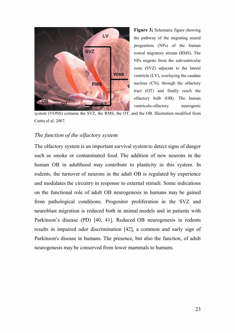
Figure 3; Schematic figure showing
the pathway of the migrating neural
progenitors (NPs) of the human
rostral migratory stream (RMS). The
NPs migrate from the subventricular
zone (SVZ) adjacent to the lateral
ventricle (LV), overlaying the caudate
nucleus (CN), through the olfactory
tract (OT) and finally reach the
olfactory bulb (OB). The human
ventriculo-olfactory neurogenic
system (VONS) contains the SVZ, the RMS, the OT, and the OB. Illustration modified from
Curtis el al. 2007.
The function of the olfactory system
The olfactory system is an important survival system to detect signs of danger
such as smoke or contaminated food. The addition of new neurons in the
human OB in adulthood may contribute to plasticity in this system. In
rodents, the turnover of neurons in the adult OB is regulated by experience
and modulates the circuitry in response to external stimuli. Some indications
on the functional role of adult OB neurogenesis in humans may be gained
from pathological conditions. Progenitor proliferation in the SVZ and
neuroblast migration is reduced both in animal models and in patients with
Parkinson’s disease (PD) [40, 41]. Reduced OB neurogenesis in rodents
results in impaired odor discrimination [42], a common and early sign of
Parkinson's disease in humans. The presence, but also the function, of adult
neurogenesis may be conserved from lower mammals to humans.

24
Differentiation from embryo to adult brain
Differentiation
Differentiation is the process by which unspecialized stem cells give rise to
specialized cells (figure 2). During this process the cell passes through
several stages, and becomes more specialized at each step. The internal
signals for differentiation are controlled by a cell’s genes, carrying coded
instructions for all cellular structures and functions. The external signals on
the other hand, come from chemicals secreted by other cells, physical contact
with neighboring cells, and certain molecules in the microenvironment. Many
of the triggers for these inside and outside signals for differentiation process
is not yet fully understood and many questions about stem cell differentiation
remains. To address these questions may give us new ways to control stem
cell differentiation in vitro, thereby growing cells or tissues that can be used
for specific purposes such as cell-based therapies or drug screening [1].
Neural stem cells
NSCs are primary progenitors that give rise to neurons and glia in the
embryonic, neonatal and adult brain. NSCs divide asymmetrically and often
amplify the number of progeny they generate via symmetrically dividing to
form intermediate progenitors [30]. NSCs in the brain are considered to be
restricted in terms of cell fate and will only give rise to three major cell types
of the CNS: neurons and two categories of non-neuronal cells, astrocytes and
oligodendrocytes. The NSCs are thus said to be multipotent (figure 1), [1].
Neuroectodermal cell type markers
Most markers used to identify neural cells are not limited to neural cells but
may also be expressed by other non-neural cell types. To define a specific
cell type therefore requires a large battery of markers (show presence or

25
absence of expression) combined with morphological and functional
indicators. Through the differentiation process hESCs alter their gene
expression profile and hence their protein production. The TF Oct4 (Octamer
Transcription Factor-3) is expressed by pluripotent cells of the ICM (the
ESCs) and by the primitive ectoderm [43-45]. Oct4 gradually decreases with
the onset of neural progenitor (NP) markers like Sox2, Pax6, Nestin,
musashi1, and NCAM. A rapid downregulation of the POU Transcription
Factor-4 (Oct-4) in the differentiating hESCs results in a extraembryonic cell
fate rather than a neural differentiation [11, 46], suggesting that transiently
sustained levels of Oct4 expression may be required for in vitro
differentiation of hESCs into neural lineages. Rapid down regulation of Oct4
expression in hESCs might promote the formation of primitive endoderm [12,
46-49]. Neuroectodermal cells within the neural tube are characterized by
expression of several markers including Sox1, Sox2, nestin [50-52], musashi-
1 [53] and NCAM [54, 55]. Sox1, Sox2 and Sox3 have equivalent functions.
The TF Sox2 belongs to the highly conserved Sox gene family [56]. The
Sox2 protein is essential for neural induction of the ectoderm, expressed in
neuroepithelial stem cells during embryonic development [57], and retains
neural progenitor identity thus counteracts neuronal differentiation [58]. Sox2
has also been detected in some differentiated neurons and is expressed in the
adult brain, by subtypes of postmitotic neurons. [50-55]. Nestin, a neural
intermediate filament protein, is expressed during early developmental stages
and during regenerative processes in muscle and neuronal cells. It is a
primary marker for the identification of neuroepithelial/neural progenitor
cells [59, 60], but is also widely expressed in the developing embryo e.g. in
endocrine progenitor cells, vascular endothelial cells [61], testis [62] and
skeletal muscle [63]. Musashi-1 is an RNA-binding protein that is highly
expressed in neural progenitor cells, including neural stem cells. Musashi-1 is
gradually down-regulated during the course of neural differentiation and

26
localized in the cytoplasm in embryonic neural progenitor cells [53, 64, 65].
Pax6, a member of the paired box gene family, is expressed in developing
and adult brain [66], and is one of the key factors for CNS patterning [67]. It
has a dual role in controlling both the degree of adult neurogenesis and
periglomerular neuron fate, and was shown to be crutial for generation of
neuronal progenitors as well as for direction of neurons towards the
glomerular layer and acquisition of a dopaminergic phenotype [34]. Olig2
promotes adult oligodendrogliogenesis, opposes the neurogenic role of Pax6,
and specifies the transient amplifying precursor state of neural progenitors in
the adult brain [34]. DCX, a microtubule-associated protein expressed in
migrating neuroblasts, promotes cell migration [68]. DCX is a widely used
marker for newly generated neurons in mammals, and has a critical function
in the movement of newly generated neurons in the adult brain. DCX is
required for nuclear translocation and maintenance of bipolar morphology
during migration in the adult forebrain [38]. DCX is necessary for embryonic
radial migration and migration of adult SVZ cells [39]. Another important
marker for neuroblasts is PSA and it is attached exclusively to NCAM. PSA
on NCAM is developmentally regulated thus playing a prominent role in
different forms of neural plasticity spanning from embryonic to adult nervous
system including axonal growth, cell migration, synaptic plasticity, neuronal-
glial plasticity, embryonic and adult neurogenesis [36]. PSA-NCAM is a
molecule abundant in the developing nervous system, although absent during
the early phases of neurogenesis [69]. During CNS development PSA-
NCAM is considered a marker of immature NPs [70]. PSA-NCAM is
associated with most parts of the olfactory system, including the RMS [71].
βIII-tubulin (TuJ1) is an early neuronal marker [72].

27
Generation of and in vitro culturing methods for human
embryonic stem cells
Human ESCs are derived from embryos generated through in vitro
fertilization procedures and donated for research after informed consent by
the donor pair. They are not derived from eggs fertilized in a woman's body.
The hESCs in vitro cannot give rise to a complete organism, because they do
not have the three dimensional environment that is essential for embryonic
development in vivo, and they lack the trophectoderm and other tissue that
support fetal development [1].
Derivation of a human embryonic stem cell line
To generate a hESC line in vitro several steps are taken;
Figure 4; Schematic figure describing the process of deriving a human embryonic stem cell
line (hESC) from a preimplantaion embryo at the blastocyst stage, by isolating the inner cell
mass (ICM). The ICM is transferred to a culture dish coated with mouse embryonic feeder
(mEFs) cells. Outgrowths of the ICM is dissociated into small pieces and transferred to a new
culture dish coated with new mEFs. The small cell pieces will attach and divide to form hESC
colonies, thus a hESC line is formed.
1. Culturing of the preimplantation embryo to one of the earliest stages
of embryonic development called the blastocyst (figure 4).

28
2. The hESC line is established by isolation of the ICM from the 4-5
day old blastocyst (figure 4). Isolation of the ICM can be done by
spontaneous hatching or by isolation of the ICM by enzymatic
treatment to remove the zona pellucida [73].
3. The ICM is then plated on to a tissue culture dish precoated with
mEF or human embryonic fibroblasts (hEF) in defined hESC
medium (nutrition mix including serum; [20, 74]), (figure 4). The
feeder cells in the bottom of the culture dish provide the ICM cells a
sticky surface to which they can attach. Also, the feeder cells release
nutrients into the culture medium. The ICM cells divide and spread
over the surface of the dish. The feeder layer cells are treated (by
irradiation or enzymatic mitomycine C treatment) so that they can
not divide.
4. After 9-15 days of culturing the ICM-derived outgrowths are
dissociated into small pieces by enzymatic (dispase or collagenase
IV) or mechanical treatment (figure 5A) and replated on fresh mEF
or hEF layers in new hESC medium.
5. Individual colonies with undifferentiated morphology are then
selected and mechanically dissociated into small pieces (figure 5A)
and replated under the same conditions, thus generating a hESC line
(figure 4).
6. Through the process of re-plating or subculturing, the cells can be
repeated many times over and for many months/years. Each cycle of
subculturing the cells is referred to as a passage that results in the
expansion of the cell cultures. When growing hESCs on feeder cell

29
layers, the colonies are mechanically cut every five days and the
pieces are then transferred to new feeder layers in fresh hESC
culturing media. When growing without feeder layers (figure 5B) the
hESCs are treated with an enzyme every 5-8 days to detach the
colonies from the culture surface. To grow the hESCs without mEF
cells is a significant scientific advantage since the risk of viruses or
other macromolecules being transmitted to the human cells is
eliminated [1].
7. Once the cell line is established, the original cells yield millions of
ESCs. Human ESCs that have proliferated in cell culture for six
months or more without differentiating, are pluripotent, and appear
genetically normal are referred to as an hESC line. At any stage in
the process, batches of cells can be frozen and shipped to other
laboratories for further culture and experimentation [1].
Figure 5; Undifferentiated human embryonic stem cell (hESC) colonies. A. Manual passage
by cutting the hESC colony propagated on a layer of mitomycine C treated mouse embryonic
feeder (mEFs) cells. B. Feeder-free culture of hESCs on Matrigel without the use of an mEF
layer.
A B

30
Characterization of an hESC line
To date there is not an agreed upon standard battery of tests that measure the
cells' fundamental properties, however, several different kinds of tests can be
used to say that you have a true ESC line [1, 3, 75] and these are;
1. Identification of specific cell surface markers associated with
undifferentiated hESC, cell surface antigens; SSEA-3 and SSEA-4
(stage specific embryonic antigens), the lack of SSEA-1 which is up
regulated in differentiated cells, TRA-1-60 and TRA-1-81 (tumour
rejection antigen 1).
2. The expression of alkaline phosphatase (ALP; enzyme).
3. Morphological appearance; growing in tight monolayer colonies with
spherical cells devoid of processes.
4. Proof of pluripotency, generation of progeny from all three
embryonic germ layers (mesoderm, endoderm and ectoderm). This
can be done in vitro by spontaneous differentiation (embryoid bodies;
EBs) or by manipulating the cells to differentiate into specific cell
types of all three germ layers, and in vivo by generation of teratomas
(injecting hESCs into severe combined immunodeficent beige mice;
SCID mice).
5. Growing and subculturing the hESC line for many months to ensure
that the cells are capable of long-term growth and self-renewal [1].
6. Retain pluripotency for at least twelve months, while retaining a
normal karyotype. The chromosomes are examinined under a
microscope to assess whether the chromosomes are damaged or if the
number of chromosomes has changed.
7. Active telomerase (high levels) and long telomeres. High levels of
telomerase activity [3, 20, 74], hTERT (the catalytic component of

31
telomerase) is expressed at high levels in undifferentiated hESCs and
downregulated upon differentiation [76].
8. Determine the presence of TFs that are typically produced by
undifferentiated cells. TF help turn genes on and off at the right time,
which is an important part of the processes of cell differentiation and
embryonic development. Expression of Oct-4 and Nanog, two of the
most important TFs, that function to keep the ESCs in a proliferating
and non-differentiating state [1].
9. Determining whether the cells can be re-grown, or subcultured, after
a cycle of freeze, thawing, and re-plating [1].
Feeder-free culture of hESCs
Human and mouse ESCs are traditionally derived and cultured on mEF layers
[3] but can be propagated on hEFs [77-79] or in a feeder-free environment
[15, 74, 80-83]. Future replacement therapies using hESC-derived cells or
tissues will require that the cells and tissues are produced without contact
with any animal source (xeno-free). Contamination of hESCs grown on
animal feeder layers has been shown [84]. The standard methods for
derivation of hESC lines requires the use of either hEFs or mEFs for co-
culturing [3, 77, 85-87]. Even though today there are feeder-free [14] and
xeno-free [88] derived hESC lines, most hESC lines used today where
initially derived on mEFs and hence are not allowed in human therapies.
Furthermore, the use of hESCs for replacement therapies, functional
genomics, and drug screening also relies on the availability of routine large-
scale culturing protocols for undifferentiated hESCs [83, 89]. The use of
enzymes for dissociation when expanding the cultures makes passaging much
less laborious, thus enabling large scale production of these cells.

32
Neural progenitors from hESCs
The most commonly used protocols for the generation of NPs from hESC
involve multicellular aggregates called EBs, long term culturing, co-cultures
and/or genetic manipulations [59, 60, 90-98]. These methods are often
practically inconvenient and also involve poorly defined medium conditions
that can lead to varying culture conditions. More recent publications describe
the derivation of monolayer cultures of NPs from hESC [46, 99-102]. Even
though the latter protocols are simpler than previously published methods
they still contain multiple steps, the use of conditioned medium, extended
derivation times or the addition of many growth factors. Moreover, little is
reported on long term maintenance, large scale production, and/or cost
efficient generation of stable hESC-derived NP populations in adherent
monolayer cultures. Large cell quantities of NPs will be required for future
replacement therapies, toxicology testing and drug screening in the
pharmaceutical industry.
Potential benefits of embryonic stem cell research
The potential applications of hESCs to human disease are many, as shown by
real advances made recently with human diseases in animal models. Studies
transplanting cells derived from human and monkey ESCs into animal
models have shown correction or partial correction of PD [103, 104] and
hESC- derived oligodendrocytes have improved spinal cord injuries in rats
[105, 106]. Furthermore, in late January 2009, the California-based company
Geron received FDA clearance to begin the first human clinical trial of cells
derived from hESCs in patients with acute spinal cord injury [107]. Perhaps
the most important potential application of hESCs is the generation of cells
and tissues that could be used for cell-based therapies. Today, donated organs
and tissues are often used to replace ailing or destroyed tissue, but the need
for transplantable tissues and organs far outweighs the available supply. Stem

33
cells, directed to differentiate into specific cell types, offer the possibility of a
renewable source of replacement cells and tissues to treat diseases [1] and
disorders throughout the body. Potential cell therapies with differentiated
hESCs include retinal neurons for retinitis pigmentosa, dopaminergic neurons
for PD, and motoneurons and oligodendrocytes for spinal cord injury [108].
For nervous system disorders, a possible restoration of cellular and functional
loss is the goal. Many other diseases could benefit from hESC research, for
instance autoimmune diseases including diabetes, rheumatism, multiple
sclerosis, and lupus; also chronic heart failure (after stroke); end-state kidney
disease; cancer; muscular dystrophy; fibrosis and hepatitis; and burns. Other
benefits from hESC research will be to better understand the complex events
occurring during human development, like finding out which genes regulate
cell differentiation. Furthermore, to understand the kind of errors that cause
abnormal cell differentiation and division causing cancer and birth defects,
chromosomal defects and determination of the proteins stem cells express
during differentiation. In addition, the methods for developing new drugs and
security tests, like screening for toxins, in the pharmaceutical industry could
undergo dramatic changes thanks to the development of hESC based methods
[1].
Problems to be overcome for the success of cell-based therapies
Regenerative medicine in human subjects using transplanted stem cells, or
their progeny, faces serious technical hurdles like; 1, transplantation rejection
and the monitoring of this, 2, efficient guidance of hESCs down the correct
pathway of differentiation using growth factors, and 3, and ensuring that cells
of such great proliferative potential do not develop into cancers [109].
34
Stem cell therapy in neurological disorders
Neuronal loss is a common feature of many neurological disorders, including
stroke, PD, Alzheimer’s disease as well as traumatic brain injury. These
neurological disorders are all highly debilitating diseases that usually require
long-term hospitalization and/or rehabilitation at an enormous cost to the
patient as well as to society. Therefore, there is an urgent need to develop
effective treatments for these patients.
Recent studies have presented results that support the idea that hESCs or
hESC-derivates used as donor cells transplantation may provide a future
method for repairing damaged brain structures as well as enhancing
functional recovery of the brain after stroke [110-113] or PD [95, 103, 114-
116]. The potential of hESCs to differentiate into neural lineages have been
demonstrated both in vivo and in vitro [85, 92, 95, 99, 117-121]. Large
amounts of purified hESC-derived NPs are needed for the creation of an
unlimited source of donor cells for neural transplantation and gene therapy,
for creating standardised transplantation experiments (undifferentiated hESCs
and precursors of other lineages may form tumours and foreign tissues upon
transplantation). Transplantation therapies using stem cells may provide
potential treatment for the restoration of cellular and functional loss in the
nervous system after traumatic brain injury or other neurological disorders.
Stem cells as donor elements will require the availability of a renewable
source of transplantable cells. For this purpose hESCs are the ultimate source
since they can be propagated indefinitely in culture, remaining
undifferentiated and maintaining potential to differentiate into all the cell
types of the adult body. Ideally, they will have optimal survival capacity and
can be differentiated into appropriate region-specific cell types according to
the requirements of the injured host tissue. Although, appropriate functional
integration of grafted cells into the host nervous system and prevention of

35
tumor formation are concerns associated with transplantation studies that
needs to be addressed before commencing with therapies in humans.
Tissue engineering
Tissue engineering is one of the major components of regenerative medicine
and follows the principles of cell transplantation, materials science and
engineering towards the development of biological substitutes that can restore
and maintain normal function in diseased and injured tissues [122]. The most
urgent problem in transplantation medicine is the shortage or lack of suitable
donor organs and tissue. Human ESCs could be utilized as a cellular source
to replace damaged tissue by cell transplantation or implantation of cellular
scaffolds [123].
Scaffolds for hESC propagation and differentiation
Polymer fibers produced via electrospinning, widely studied both
theoretically and experimentally, have been proposed for a number of
applications due to their unique properties, like high surface-to-volume ratio,
related to their small physical dimensions [124-127]. Generally for
biomedical applications electrospun biocompatible or biodegradable polymer
fibers are intended to act as three-dimensional scaffolds mimicking natural in
vivo extracellular matrices and supporting cell proliferation [128-130]. This
physical support provided by the three-dimensional porous polymer networks
make them exceptionally promising within tissue engineering. Numerous
studies have been performed with different applications in mind, for instance
cardiovascular, cartilage, osseous and nervous tissue implants [20, 131-139].
The adult CNS has a limited capacity to recover from damage after trauma or
disease, and has therefore been subjected to intense research efforts
concerning tissue engineering and implants [20, 131-133, 136, 137, 140-145].
Electrospun porous scaffolds, mimicking the natural three-dimensional

36
environment of the in vivo extracellular matrix (ECM) and providing physical
support, have been identified as promising candidates for CNS tissue
engineering. Studies have been published where electrospun fibers are used
as a scaffold to support undifferentiated cells with the intent of differentiating
them into nervous tissue [136, 137]. The nanofibrous scaffold can support
differentiation and promote cell adhesion, indicating that nanofibrous
scaffolds may play an important role in nervous tissue engineering [131,
133].
37
Aim of these studies
The overall aim of this thesis was to improve and derive methods for the
propagation of hESCs and hESC-derived NPs and to study the derivation,
propagation and differentiation of human stem and progenitor cells.
Specific aims
I) To develop an improved and more robust protocol for the
transfer of hESCs to feeder-free conditions (paper I).
II) To develop a culture method that facilitates long-term
propagation and large-scale production of undifferentiated hES
cells in a feeder-free environment (paper I).
III) To develop an efficient protocol for the rapid generation of an
expandable population of fast growing hESC-derived NP cells
with the capacity to generate mature neurons and glial cells in
vitro (paper II).
IV) To find suitable materials to promote migration and
differentiation of stem and progenitor cells (paper III).
V) To find critical differentiation factors and markers expressed in
common between human RMS neuroblasts and hESC-derived
NPs (paper IV).

38
Experimental procedures
Ethical approval
Ethical approval was given for studies of stem cell function and survival in
the adult human brain by the regional etikprövningsnämnden in Gothenburg
(Dnr 448-06). Ethical approval was also given by the research committee at
Uppsala University for the research project concerning the culture of hESCs
(Dnr 00-536).
Human embryonic stem cell (hESC) lines (paper I, II, III)
Initially, the hESC lines were established and maintained on a monolayer of
Mitomycin C (Sigma Aldrich, Sweden) treated mEF cells (Thomson et al.,
1998), and cultured in standard hESC medium (Xu et al., 2001; Amit et al.,
2000), currently manufactured as VitroHES™ medium by Vitrolife AB
(Kungsbacka, Sweden), and characterized according to standard criteria [3,
146]. The hESC lines SA002 [146], SA121 [146], SA167 [83], AS034 [146],
and AS038 [146] were established from blastocysts collected from
Sahlgrenska University hospital (SA) and Akademiska Sjukhuset (AS),
respectively.
Preparation of conditioned VitroHES-medium (paper I and II)
To prepare mEF cells for conditioning of VitroHES-medium or hESC
medium, mEF cells (Mitomycin C treated) where seeded in a culture flask
and cultured to a confluent monolayer for 24 hours in Dulbecco’s Modified
Eagle Medium (D-MEM) supplemented with 1% penicillin-streptomycin
(PEST), 10% fetal bovine serum (FBS) and 2 mM GLUTAMAX-I
supplement (200 mM). The mEFs where then wash with phosphate buffered
saline (PBS) and the culture medium replaced with VitroHES or hESC
medium (0.28 ml/cm2) for a 24 hour conditioning period. Conditioned

39
medium (k-VitroHES or k-hESC medium) was collected every day for up to
three times from the same mEF culture, sterile filtered, and used either fresh
or after freezing (-20ºC) and supplemented with 4 ng/ml of FGF2 prior to
use.
Transfer of hESCs to Matrigel (paper I)
Two different techniques (mechanical or enzymatic dissociation) were
evaluated for the transfer of the hESCs onto rehydrated Matrigel plates. First
the hESC colonies were mechanically cut into small square pieces carefully
detached and transferred to Hanks' Balanced Salt Solution (HBSS) solution.
For the enzymatic dissociation collagenase IV was used for a 30 min
incubation during which repeated mechanical dissociations was preformed
and the process monitored in a microscope. For the mechanical dissociation,
the clusters where only mechanically dissociated with a pipette, and the
process completed at an aggregate cluster size of 400-600 cells/cluster. The
cell suspension generated was then pelleted, washed, resuspended in k-
VitroHES medium, and transferred to rehydrated Matrigel plates at a cluster
density of 10 to 15 clusters/well (6-well plate). Each experiment was repeated
four times in 4 wells/dissociation technique, with the same amount of cells
seeded each time. The number of cells initially used for the two different
dissociation protocols were identical. After two and six days the colony size
and number was calculated. The colony area was calculated by measuring the
X and Y diameter of all hESC colonies with undifferentiated morphology,
allowing for an approximation of cell growth. These colonies consist of a
monolayer of homogenously sized cells making this approximation possible.
Colony area to cell number correlation was calculated and an approximately
linear correlation between colony size and cell number was found in
undifferentiated Matrigel cultures. The area/cell relationship averaged at 82
µm2 with individual cell areas ranging from 30 to 134 µm2.

40
Viability study on hESC clusters dissociated mechanically vs.
enzymatically (paper I)
The viability test was performed by using calcein/ethidium homodimer
(calcein/EthD) kit and a comparison was preformed of the hESCs dissociated
mechanically vs. enzymatically in the transfer step. The dissociated cells
were resuspended in 100 µL calcein/EthD solution respectively and
incubated for 10 minutes in room temperature. Each cell suspension was
placed on a glass slide and a cover glass placed on top. The dead and live
cells were counted manually in a microscope (Nikon Eclipse TE2000-U).
The hESC cluster sizes after dissociation (paper I)
Colonies were dissociated mechanically and enzymatically from equally
sized undifferentiated hESC colonies. Suspensions of cell clusters were then
incubated with Nile-red staining solution (1µM in PBS) for 10 minutes and
photo documented using a fluorescence microscope (Nikon Eclipse TE2000-
U). All clusters were counted and measured using ImageJ image analysis
software.
Passage of Matrigel propagated hESCs (paper I, II and III)
The cell cultures were observed visually by using an inverted microscope.
When ready for passage, the medium was aspirated and a collagenase IV
solution was added to each well and incubated for 15 to 20 minutes. To
facilitate cell detachment from the surface careful mechanical dissociation
was performed followed by another 15 minutes of incubation. The cells were
then washed, resuspended in k-VitroHES medium (paper I) or mEF
conditioned hESC medium (paper II and III) and seeded at a split ratio of 1:3
to 1:6 onto Matrigel. The hESC cultures were passaged every 5 to 6 days and
the medium was changed three times a week.

41
Derivation and propagation of neural progenitor cells (paper II)
At 0 days undifferentiated hESC colonies propagated on Matrigel were
enzymatically dissociated (collagenase IV; 200 U/mL, 30 minutes, 37 oC)
generating a cell suspension containing small cell aggregates and single cells.
The cells were washed in warm basal media, pelleted and resuspended in
warm culturing media. The different culturing media evaluated for NP
derivation and propagation included standard hESC medium, mEF
conditioned hESC medium [83], Dulbecco's modified eagle medium: nutrient
mixture F-12 (DMEM/F12) or neurobasal A medium with various
supplements in different combinations (table 1; paper II). We observed that
the standard hESC medium supplemented with FGF2 was the best media for
the induction and propagation of the NPs from hESCs, hence this is the
media used in the following experiments. The cell suspension was then plated
on to different adhesive substrates (laminin, gelatine, polyornithine/laminin,
polyornithine-coated Primaria treated plastic plates, Primaria plastic plates,
Nunclon treated culture plates or Matrigel plates). Laminin and gelatine were
the substrates used for NP derivation and propagation in the following
experiments. The seeding cell density was 150-200x103cells/cm2 for the NP
generation. Half of the culturing medium was changed three times a week.
Cells were cultured under these conditions for 8 days during which time
neural rosettes [147, 148] were formed. On day 8 the cultures where treated
briefly with collagenase IV to detach the rosettes (flattened cells at the edge
of rosette colonies did not detach), and then mechanically dissociated to a
single cell suspension by gently triturating the cell solution with a Pasteur
pipette. The cells were washed and reseeded under the same conditions as
before. In passage 2 adherent monolayer NP populations were generated and
these cultures where expanded 1:2 or 1:3, every 4 to 7 days or when ~80%
confluent. Passage for expansion of the NP cultures hereafter was preformed
using collagenase IV to generate a single cell suspension that was then re-

42
seeded as high density cultures (50-100x103 cells/cm2; lower cell densities
generated differentiated cultures) to maintain a proliferating NP population.
In vitro differentiation of hESC-derived neural progenitor cells
(paper II)
The hESC derived NPs where differentiated into mature neurons and glial
cells as adherent monolayer cultures on laminin in hESC medium
supplemented with TGF-β1 (10 ng/mL) for 7 days, or supplemented with Shh
(500 ng/mL) and FGF8 (100 ng/mL) for 9 to 16 days, or supplemented with
Shh 500 ng/mL, 40 ng/mL FGF2, and 1% N2, for 14 days. Alternatively, the
NPs where differentiated on laminin in DMEM/F12 medium (supplemented
with 1% PEST, 1% L-glutamine and 1% N2 supplement, 1% non essential
amino acids (NEAA), 0.2% β-mercaptoethanol, and 20 ng/mL of FGF2) for
16 days. The NPs where also differentiated as free floating neurospheres, by
seeding the cell suspension onto a non-adherent substrate, in hESC medium
supplemented with Shh (500 ng/mL) and FGF8 (100 ng/mL), generating
spheres, that where then plated onto an adherent (laminin) surface for 6 days.
Electrospun fiber for co-culture and differentiation of hESCs
(paper III)
Undifferentiated feeder-free Matrigel hESC cultures where dissociated
enzymatically by collagenase IV treatment, washed, and seeded onto
Nunclon treated cell culture plates, together with 1 cm2 sized pieces of
electrospun fiber scaffolds. The culturing medium consisted of standard
hESC medium supplemented with 1 % N2 and 10 ng/mL of FGF2. After 5 to
7 days of co-culture with the fibers the medium was changed to a neural
differentiation medium consisting of neurobasal A basal medium
supplemented with 1 % B27 and 1 % N2, omitting the growth factor FGF2.
Additionally, for long term differentiation culture (18-47 days) 20 ng/mL of

43
epidermal growth factor (EGF) and FGF2 was added to the differentiation
medium. The differentiation medium was changed three times a week and the
co-cultures were allowed to propagate and then differentiate for up to 47
days.
Human tissue collection (paper IV)
The human brain tissue was obtained from the Neurological Foundation of
New Zealand Human Brain Bank at the University of Auckland or from
Sahlgrenska University Hospital, Sweden. These latter patients had been
diagnosed with squamous cell carcinomas at the base of the tongue, in the
larynx or in the pharynx and had received 5-bromo-2´-deoxyuridine (BrdU;
250 mg) dissolved in saline and given as an intravenous infusion (2.5 mg/ml,
100 ml). The BrdU was given to patients to assess the proliferative activity of
the tumor cells [149, 150]. The full consent of all families was obtained prior
to autopsy and the respective University Human Subjects Ethics committees
approved these studies. In all cases pathological examination excluded any
neuropathology. The brains were removed at autopsy and in some cases the
hemispheres were separated and one hemisphere was dissected and frozen
fresh and the other half was fixed or the whole brain was fixed. The brains
were fixed by perfusion through the cerebral arteries, first with PBS with 1%
sodium nitrite and followed by 15% formalin in 0.1 M phosphate buffer, pH
7.4. After perfusion, approximately 5 cm x 5 cm blocks of the brain
containing the midline cortical surface medially, middle frontal gyrus
laterally, thalamus caudally, corpus callosum rostrally, cingulate gyrus
dorsally and the OT and trigone ventrally was dissected and postfixed in the
same fixative for 24 h. The blocks were cryoprotected in 20% sucrose and
then 30% sucrose until equilibrated. The blocks were serially sectioned in a
sagittal or coronal plane on a freezing microtome (50-µm sections) and stored

44
serially in PBS and 0.1% azide. Hemispheres that were unfixed were
dissected, frozen and stored at -80°C until required for further processing.
Characterization of undifferentiated hESCs, NPs, mature
derivates, and RMS neuroblasts (paper I, II, III, IV)
Immunocytochemistry (paper I, II, III)
Immunocytochemistry is a technique used to assess the presence of a specific
protein or antigen in cells by use of a specific antibody that binds the antigen
and thus allow the visualization and examination under a microscope. In
paper I, II, III, and IV this technique was utilized to determine the cell types
present using cell type specific markers. Cell cultures and fiber co-cultures
were washed in PBS, fixed with 4% paraformaldehyde (PFA) for 15 minutes
at room temperature and then washed again three times in PBS. The primary
antibodies and cells were incubated over night at 4°C before being visualized
using appropriate secondary antibodies. Cultures were also incubated with a
cell nuclei stain 4´-6´Diamidino-2-phenylindole (DAPI), at a final
concentration of 0.5 ug/mL for 5 minutes at room temperature, to visualize
all the cell nuclei. The stained cultures were rinsed and mounted using
DAKO fluorescent mounting medium or ProLong Gold and visualized with
an inverted fluorescent microscope. See table 1 for primary and secondary
antibodies and for dilutions. Control staining included omission of either
primary or secondary antibodies and revealed neither non-specific staining
nor antibody cross-reactivity.
Immunohistochemistry (paper IV)
Immunohistochemistry is a technique for localization of proteins of interest
using antibodies raised against specific proteins of interest. In paper IV
sagittal and coronal sections for staining with proliferating cell nuclear
45
antigen (PCNA) were first incubated overnight in a citric acid solution
containing citric acid (Na3 salt) and Na2HPO4 (pH 4.5) before undergoing a
standard antigen retrieval protocol [151]. All sections were incubated in a
50% methanol solution with 1% H2O2 to block endogenous peroxidases in
the tissue. To detect proliferating cells, three PCNA antibodies were used
(table 1), each giving identical results. In three cases detailed quantitative
studies were undertaken on the number of PCNA-positive cells in the RMS.
Images from serial sagittal sections (every 3rd section) were photographed at
x20 magnification. Images were analyzed for PCNA-positive cells using the
FindSpots algorithm (which detects objects based upon their intensity and
size) in the Metamorph (v.6.2.6, Molecular Devices) image analysis program.
To detect neuronally committed neuroblasts we used PSA-NCAM antibody
and DCX (table 1). For fluorescent triple-labeling for BrdU and other
antigens tissue underwent a standard hydrochloric acid pretreatment to enable
BrdU presentation, prior to incubation in BrdU antibodies, Neuronal nuclei
antibodies and GFAP (table 1). For visualization with the light microscope,
species specific biotinylated secondary antibodies (dilution 1:500) were
added serially, followed by Extravidin (dilution 1:1,000; Sigma) and finally
3, 3 diaminobenzidine was used to visualize the staining. The sections were
mounted onto glass slides, dehydrated in graded alcohol, cleared in xylene
and coverslipped. Primary antibodies where detected by addition of species
specific secondary antibodies (table 1). Between each of the steps above, the
sections were washed for 3 x 10 minutes in PBS and 0.1% triton X-100 for
3,3 diaminobenzidine (DAB) labeled sections and tris-buffered saline for
BrdU labeled sections. The fluorescent-labeled sections were mounted and
coverslipped with Citifluor and imaged by using a confocal laser scanning
microscope. Each fluorescent label was imaged serially to eliminate detection
of bleed-through and other artificial fluorescence. The confocal images were
captured in a Z-series with an interslice gap of 1-µm. Bright-field images

46
were taken with a digital camera on a light microscope and the images were
captured in Photoshop. Macro-photographs were captured on a free standing
digital camera. Illustrations were compiled in Illustrator.
Table 1: Primary and secondary antibodies used in this study.
Antibody Source Immunogen Dilution Company
Primary abs for immunocytochemistry (paper I, II, III):
Oct-3/4 mouse Human Oct-3/4 1:100-1:200
Santa Cruz Biotech
SSEA-1 mouse stage specific embryonic antigen-1 1:200 DSHB SSEA-3 Rat stage specific embryonic antigen-1 1:200 DSHB SSEA-4 mouse stage specific embryonic antigen-4 1:200 DSHB Tra-1-60 mouse High molecular weight glycoprotein 1:200 Santa Cruz/SDS Tra-1-81 mouse High molecular weight glycoprotein 1:200 Santa Cruz/SDS
Nestin mouse Intermediate filament protein 1:200-1:500
BD Pharmingen
Sox2 goat the SRY-related HMG-box 1:200 Santa Cruz Biotech
Sox2 mouse the SRY-related HMG-box 1:1000 Chemicon NCAM rabbit Neural cell adhesion molecule 1:500 Chemicon Pax6 mouse Human recombinant Pax6 1:200 Chemicon
Musashi-1 rabbit Neural RNA binding protein. 1:200-1:1000
Chemicon
Internexin rabbit Alpha-internexin 1:750 Chemicon A2B5 mouose Neuron cell surface antigen 1:250 Chemicon βIII-tubulin mouse Human III β-tubulin isotype III 1:200 Sigma-Aldrich
βIII-tubulin rabbit Neuronal class III β-tubulin 1:1000 Biosite
MAP2ab mouse Bovine microtubule associated protein 1:100-1:200
Sigma-Aldrich
NF200 mouse Neurofilamant 200 1:200 Sigma TH mouse Tyrosine hydroxylase 1:2000 Sigma-Aldrich TH rabbit Tyrosine hydroxylase 1:250 Chemicon GFAP rabbit Cow glial fibrillary acidic protein 1:250-500 DAKO GFAP 1:250 Chemicon
APC mouse A recombinant ammino terminal fragment of APC
1:20-100 Calbiochem
GalC rabbit Galactoserebroside 1:75-1:180 Sigma-Adrich GalC mouse Galactoserebroside 1:200 Chemicon Ct-1 rabbit Human cardiotrophin-1 1:200 Chemicon AFP mouse Alfafetoprotein 1:500 Sigma
PH3 rabbit Phospho-Histone H3 1:100-1:150
KeLab
Primary abs for immunohistochemistry (paper IV);
PCNA rabbit Proliferating cell nuclear antigen FL261
1:750 Santa Cruz
PCNA mouse Proliferating cell nuclear antigen PC10 1:500 Santa Cruz PCNA mouse Proliferating cell nuclear antigen 1:500 Chemicon
PSA-NCAM mouse Polysialic acid neural cell adhesion molecule
1:1,000 Gift G Rougon
DCX 1:500 BrdU rat Accurate 1:500 Neuronal nuclei
mouse 1:20

47
GFAP guinea pig
1:250
Secondary abs for immunocytochemistry (paper I, II, III):
Alexa 488 goat Mouse IgG 1:2000 Molecular Probes
Alexa 488 donkey Rabbit IgG 1:2000 Jackson Laboratories
Alexa 594 goat Rabbit IgG 1:2000 Molecular Probes Alexa 555 donkey Mouse IgG 1:2000 Molecular Probes FITC sheep Rabbit IgG 1:800 Chemicon FITC donkey Mouse IgG 1:800 Termo
FITC donkey Rat IgG 1:800 Jackson Laboratories
FITC goat Mouse IgM 1:300 Jackson Laboratories
Texas Red donkey Mouse IgG 1:800 Jackson Laboratories
Cy3 goat Rat IgM 1:300 Jackson Laboratories
Secondary abs for immunohistochemistry (paper IV);
Texas Red Mouse IgM FITC donkey Rat 1:250 Cy3 donkey Mouse 1:250 Cy5 donkey guinea pig 1:250 Alexa 594 donkey Mouse 1:200 Alexa 647 donkey Rabbit 1:200
Alkaline phosphatase (ALP) expression (paper I, II)
Expression of ALP in undifferentiated hESCs and NPs was analyzed
following fixation of cells with citrate-acetone-formaldehyde fixative
solution using a Sigma diagnostics kit.
Telomerase activity (paper I)
Enzyme-linked immunosorbent assay (ELISA), an immunoassay utilizing an
antibody labeled with an enzyme marker such as horseradish peroxidase.
While either the enzyme or the antibody is bound to an immunosorbent
substrate, they both retain their biologic activity; the change in enzyme
activity as a result of the enzyme-antibody-antigen reaction is proportional to
the concentration of the antigen and can be measured spectrophotometrically
or with the naked eye. Many variations of the method have been developed.
In paper I we used a PCR-based ELISA, to analyse the telomerase activity in
Matrigel and mEF cultured hESCs that were first harvested, lysed.
48
Karyotyping and fluorescence in situ hybridization (FISH) (paper I)
Karyotyping is the mapping of the full chromosome set of the nucleus of a
cell. The chromosome characteristics of an individual or a cell line are
usually presented as a systematized array of metaphase chromosomes from a
photomicrograph of a single cell nucleus arranged in pairs in descending
order of size and according to the position of the centromere. In paper I the
hESCs designated for karyotyping were incubated for 1 to 3 hours in
colcemid, dissociated, fixated, mounted on glass slides and the chromosomes
visualized by using a modified Wright’s staining.
In situ hybridization is a technique that localizes specific nucleic acid
sequences within intact chromosomes, eukaryotic cells, or bacterial cells
through the use of specific nucleic acid-labeled probes. Fluorescence in situ
hybridization (FISH) is a type of in situ hybridization in which target
sequences are stained with fluorescent dye so their location and size can be
determined using fluorescence microscopy. This staining is sufficiently
distinct that the hybridization signal can be seen both in metaphase spreads
and in interphase nuclei. For the FISH analysis in paper I, a commercially
available kit (MultiVysion™ PB Multicolour Probe Panel) containing probes
for chromosome 12, 13, 17q, 18, 21 and the sex chromosomes (X and Y) was
used. Slides were analyzed using an inverted microscope equipped with
appropriate filters and software (CytoVision, Applied Imaging).
Teratomas (paper I)
For the teratoma formation experiment, immunodeficient SCID mice (C.B-
17/lcrCrl-scidBR) were used. Matrigel propagated hESC colonies were
enzymatically detached from the surface by using collagenase IV,
mechanically dissociated into small cell aggregates and approximately 50 000
to 100 000 cells/organ were injected under the kidney capsule. Control
animals were treated with Cryo-PBS injections or with primary brain cells
49
from a littermate. The animals were sacrificed eight weeks after injection and
the tumors were immediately fixed in 4% PFA and paraffin embedded. For
histological analysis the teratoma were sectioned to 8 µm and stained with
Alcian Blue/Van Giesson.
Reverse transcriptase-polymerase chain reaction (RT-PCR) (paper I,
II, IV)
PCR is an in vitro method for producing large amounts of specific DNA or
RNA fragments of defined length and sequence from small amounts of short
oligonucleotide flanking sequences (primers). The essential steps include
thermal denaturation of the double-stranded target molecules, annealing of
the primers to their complementary sequences, and extension of the annealed
primers by enzymatic synthesis with DNA polymerase. The reaction is
efficient, specific, and very sensitive. Reverse transcriptase-polymerase chain
reaction (RT-PCR) is a variation of the PCR technique in which cDNA is
made from RNA via reverse transcription. The resultant cDNA is then
amplified using standard PCR protocols. In paper I, II and IV RT-PCR was
used to detect the gene expression of cell type specific genes to determine the
cell types present. Total RNA was extracted from the cell cultures (paper II)
using RNeasy Plus Mini kit. In paper IV total RNA was extracted from the
AOC, OT, OB and from putamen, caudate nucleus and hippocampus (as
controls; paper IV). Reverse transcription of 1 µg of total RNA to cDNA was
performed by using AMV First Strand cDNA Synthesis Kit (paper I) or M-
MLV reverse transcriptase (paper II, IV). The PCR was carried out using 5
uL of cDNA with Platinum Taq DNA Polymerase (paper I) or TaqBead Hot
Start Polymerase (paper II, IV) in a total volume of 50 uL in the presence of
50 pmol of each primer, 1.5-2 mM MgCl2. The primer pair sequences,
number of cycles, annealing temperatures, and product size (bp) are
summarized in table 2 for each gene analyzed. The PCR products were size

50
fractioned by gel electrophoresis using a 1-2% agarose gel electrophoresis
and visualized after ethidium bromide staining or SYBR Safe™ DNA gel
stain using a Fuji LAS-3000 imaging system. The housekeeping gene
glyceraldehyde-3-phosphate dehydrogenase (GAPDH) was used as an
internal positive control (paper II and IV), and β-actin in paper I. For all
mRNA samples a minus RT-PCR step was performed using the mRNA as
template in the PCR reaction and the GAPDH primers to detect any
contaminating DNA in the RNA samples. Human genomic DNA was used as
a positive control for PCR reactions (paper II). Human liver was used as a
positive control and water as negative control for the PCR reaction (paper I).
Comments: In paper IV the Olig2 PCR assay failed in the AOC due to
unknown technical reasons. However, we were able to detect the Olig2
protein within the AOC using immunohistochemistry.
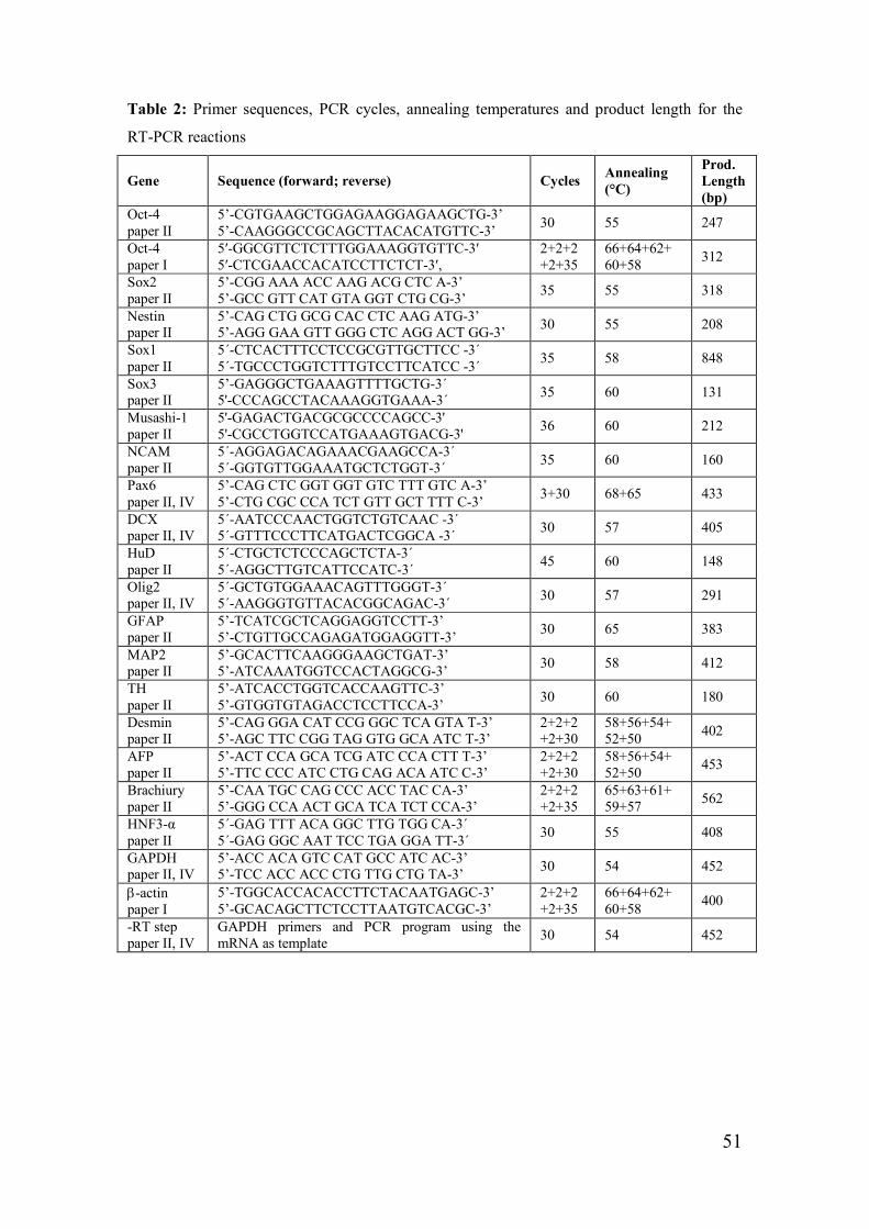
51
Table 2: Primer sequences, PCR cycles, annealing temperatures and product length for the
RT-PCR reactions
Gene Sequence (forward; reverse) Cycles Annealing
(°C)
Prod. Length
(bp)
Oct-4 paper II
5’-CGTGAAGCTGGAGAAGGAGAAGCTG-3’ 5’-CAAGGGCCGCAGCTTACACATGTTC-3’
30 55 247
Oct-4 paper I
5′-GGCGTTCTCTTTGGAAAGGTGTTC-3′ 5′-CTCGAACCACATCCTTCTCT-3′,
2+2+2+2+35
66+64+62+ 60+58
312
Sox2 paper II
5’-CGG AAA ACC AAG ACG CTC A-3’ 5’-GCC GTT CAT GTA GGT CTG CG-3’
35 55 318
Nestin paper II
5’-CAG CTG GCG CAC CTC AAG ATG-3’ 5’-AGG GAA GTT GGG CTC AGG ACT GG-3’
30 55 208
Sox1 paper II
5´-CTCACTTTCCTCCGCGTTGCTTCC -3´ 5´-TGCCCTGGTCTTTGTCCTTCATCC -3´
35 58 848
Sox3 paper II
5’-GAGGGCTGAAAGTTTTGCTG-3´ 5'-CCCAGCCTACAAAGGTGAAA-3´
35 60 131
Musashi-1 paper II
5'-GAGACTGACGCGCCCCAGCC-3' 5'-CGCCTGGTCCATGAAAGTGACG-3'
36 60 212
NCAM paper II
5´-AGGAGACAGAAACGAAGCCA-3´ 5´-GGTGTTGGAAATGCTCTGGT-3´
35 60 160
Pax6 paper II, IV
5’-CAG CTC GGT GGT GTC TTT GTC A-3’ 5’-CTG CGC CCA TCT GTT GCT TTT C-3’
3+30 68+65 433
DCX paper II, IV
5´-AATCCCAACTGGTCTGTCAAC -3´ 5´-GTTTCCCTTCATGACTCGGCA -3´
30 57 405
HuD paper II
5´-CTGCTCTCCCAGCTCTA-3´ 5´-AGGCTTGTCATTCCATC-3´
45 60 148
Olig2 paper II, IV
5´-GCTGTGGAAACAGTTTGGGT-3´ 5´-AAGGGTGTTACACGGCAGAC-3´
30 57 291
GFAP paper II
5’-TCATCGCTCAGGAGGTCCTT-3’ 5’-CTGTTGCCAGAGATGGAGGTT-3’
30 65 383
MAP2 paper II
5’-GCACTTCAAGGGAAGCTGAT-3’ 5’-ATCAAATGGTCCACTAGGCG-3’
30 58 412
TH paper II
5’-ATCACCTGGTCACCAAGTTC-3’ 5’-GTGGTGTAGACCTCCTTCCA-3’
30 60 180
Desmin paper II
5’-CAG GGA CAT CCG GGC TCA GTA T-3’ 5’-AGC TTC CGG TAG GTG GCA ATC T-3’
2+2+2+2+30
58+56+54+ 52+50
402
AFP paper II
5’-ACT CCA GCA TCG ATC CCA CTT T-3’ 5’-TTC CCC ATC CTG CAG ACA ATC C-3’
2+2+2+2+30
58+56+54+ 52+50
453
Brachiury paper II
5’-CAA TGC CAG CCC ACC TAC CA-3’ 5’-GGG CCA ACT GCA TCA TCT CCA-3’
2+2+2+2+35
65+63+61+ 59+57
562
HNF3-α paper II
5´-GAG TTT ACA GGC TTG TGG CA-3´ 5´-GAG GGC AAT TCC TGA GGA TT-3´
30 55 408
GAPDH paper II, IV
5’-ACC ACA GTC CAT GCC ATC AC-3’ 5’-TCC ACC ACC CTG TTG CTG TA-3’
30 54 452
β-actin paper I
5’-TGGCACCACACCTTCTACAATGAGC-3’ 5’-GCACAGCTTCTCCTTAATGTCACGC-3’
2+2+2+2+35
66+64+62+ 60+58
400
-RT step paper II, IV
GAPDH primers and PCR program using the mRNA as template
30 54 452

52
TUNEL staining (paper IV)
Terminal deoxynucleotidyl transferase-mediated deoxyuridine triphosphate
nick end labeling (TUNEL) is an in situ method for detecting areas of DNA
which are nicked during apoptosis. Terminal deoxynucleotidyl transferase is
used to add labeled dUTP, in a template-independent manner, to the 3 prime
OH ends of either single- or double-stranded DNA. The TUNEL assay labels
apoptosis on a single-cell level, making it more sensitive than agarose gel
electrophoresis for analysis of DNA fragmentation. In this study TUNEL
staining was preformed to detect DNA fragmentation to examine if PCNA
expressed in the RMS cells was due to repair or apoptosis instead of
proliferation.
Cryopreservation of Matrigel cultured hESCs and hESC-derived
NPs (paper I and II)
The hESCs colonies or NP cultures were dissociated with collagenase IV
before freezing. After washing and centrifugation, the cells were transferred
to a freezing medium, consisting of k-VitroHES medium or hESC medium
supplemented with 10% dimethyl sulfoxide (DMSO), 4 ng/ml FGF2, and
serum replacement (SR) to a final concentration of 30%, with a cell density 1
million cells per ml freeze medium. The final cell suspension was added to
Nunc CryoTubes and rapidly transferred to a Nalgene freezing container for
storages in -80°C over night or at least for 4 hours before moved to long-term
storage in a Liquid Nitrogen tank. To thaw the cells, k-VitroHES medium or
hESC medium was prepared and preheated to 37°C before thawing the cells
by rapidly transferring the cryotubes to a 37°C water bath until all of the cell
suspension was thawed. The cell suspension was then transferred to the
preheated k-VitroHES medium or hESC medium, for 5 minutes equilibration,
before centrifugation. The cell pellet was washed once, resuspended in the

53
culturing medium and seeded into culturing plates at the same cell density as
before freezing.
Statistics (paper II, III)
Cell counting data was generated from three repeated experiments, counting
at least 10 randomly selected visual fields (40x magnification) per antibody
and experiment. The number of positively stained cells were counted and
expressed as the percentage of positive cells out of the total number of cells.
The total number of cells was quantified by counting the total number of
DAPI stained cell nuclei in all fields of view. The results are presented as
mean ± standard deviation (SD), (paper II and III). In paper III data were
analyzed using Student’s t-test. * p < 0.05 was considered statistically
significant.
Electrospun polymer fiber generation (paper III)
Biocompatible aromatic polyether based polyurethane resin (Desmopan
9370A, Bayer MaterialScience AG) was selected due to its flexible
elastomeric properties, making it a suitable candidate material for electrospun
cell carrier or tissue engineering applications. Polyurethane resin was
dissolved in a solvent composed of 60 wt % tetrahydrofuran (THF, Scharlau)
and 40 wt % n,n-dimethylformamide (DMF, Scharlau), obtaining a solution
with 11 wt % polymer concentration. The homogenized polyurethane
solution was transferred to a 20 ml plastic syringe (Omnifix, Braun). Fibrous
scaffolds were manufactured with a vertical electrospinning setup. The
syringe, loaded with polymer solution, was fitted with a 0.85 mm inner
diameter cannula (1.2 mm outer diameter) (Sterican, Braun) connected to the
positive potential of a high voltage power supply (Gamma High Voltage
Research). A grounded collector was placed at a distance of 22 cm from the
cannula tip. With the described setup (figure 6), scaffolds were manufactured

54
on aluminum foil substrates. Experiments were carried out with the cannula
tip at a potential of 18 kV.
Figure 6; A schematic illustration of the electrospinning process, developed at SMIT Center &
BioNano Systems Laboratory, Department of Microtechnology and Nanoscience at Chalmers
University of Technology. Illustration by Björn Carlberg.
Surface morphology and structural properties of electrospun
polymer scaffolds (paper III)
The surface morphology and structural properties of the electrospun fiber
scaffolds were inspected using scanning electron microscopy (SEM) (JSM-
6301F, JEOL). Prior to electron microscopy samples were sputter coated with
20 nm gold to avoid charging phenomena. The fiber diameter distribution
was determined by image analysis of SEM-micrographs (8 random locations
with 20 samples at each location under 4,000x magnification). Pore diameter
distribution, porosity and total pore volume of the electrospun polyurethane
scaffolds were determined utilizing mercury intrusion porosimetry (AutoPore
III 9410, Micromeritics).
High voltage
aggregate collector
Capillary
containing
polymer solution Beam of polymer
solution
Not to scale

55
Scanning electron microscopy (SEM), (paper III)
SEM is a microscopy technique in which the object is examined directly by
an electron beam scanning the specimen point-by-point. The image is
constructed by detecting the products of specimen interactions that are
projected above the plane of the sample, such as backscattered electrons. To
prepare the samples for SEM analysis, the fibers released from the aluminum
substrate were fixed at different time points of hESC co-culture and
differentiation, using EM-fixative (2 % PF, 2.5 % GA 0.05M Na-kak, pH 7.2,
Na-acid) and treated for SEM analysis.
Transmission electron microscopy (TEM), (paper IV)
Transmission electron microscopy (TEM) is an electron microscope
technique in which the electrons or their reaction products that pass down
through the specimen are imaged below the plane of the specimen while
other electrons are absorbed in the specimen. To prepare the specimen for
TEM a post-fixation procedure was performed after initial post-mortem
fixation of the specimens. Specimens were cut into smaller blocks and
immersion fixed in a modified Karnowsky solution (2% paraformaldehyde,
2.5% glutaraldyde and 0.05M Na-azide in 0.05M Na-cacodylate) overnight
and post fixed in 1% OsO4 and 1% potassium ferrocyanide in 0.1 M Na-
cacodylate for 2 hrs. Specimens were treated with 0.5% uranylacetate en
bloc, dehydrated and embedded in Agar 100 resin. A first series of 0.5µm
sections were cut, stained with a Richardson’s stain and evaluated in a light
microscope. This evaluation made it possible to trim the blocks in order to
make ultrathin (50-60-nm) sections of selected regions for TEM. Electron
microscope sections were examined after contrasting by lead citrate and
uranylacetate in a LEO912AB TEM equipped with a CCD camera.
56
Magnetic resonance imaging (MRI) of human brains (paper IV)
Magnetic resonance imaging (MRI) is a non-invasive method of
demonstrating internal anatomy based on the principle that atomic nuclei in a
strong magnetic field align and radiofrequency energy can be used to force
them out of alignment. As the protons return to alignment radio waves are
generated at different frequencies for different tissue types that can be
reconstructed into computerized images. In order to further visualise the
ventricular extension, we performed MRI using high resolution heavily T2
weighted spin-echo sequences developed to accentuate fluid for the purpose
of tracking cerebrospinal fluid (CSF) leakage in patients with hydrocephalus.
A previous study in humans suggested a nasal route of CSF drainage in man
[152]. It is also a well established clinical phenomenon that patients with
acute hydrocephalus often develop leakage of CSF from the nose (liquorhea).
Therefore we wanted to investigate if the ventriculo-olfactory extension
could be visualized better with MRI in hydrocephalic patients.
Hydrocephalus did not dilate the ventriculo-olfactory extension; we saw
similar results in normotensive patients. The evaluation of the morphology of
the OB was a retrospective analysis and was not part of, or influenced by, the
clinical evaluation. None of the patients had findings on MRI indicating
ongoing CSF leakage. The patients were examined in a 1.5 Tesla MR scanner
with a special T2 weighted sequence designed for high resolution and
maximum contrast between soft tissue and fluid with the following
parameters: 3D TSE (TSE factor 53, DRIVE), TR/TE 1500/130, FOV 130
mm, Reconstructed resolution 0.25 x 0.25 (in plane) x 0.5 (slice thickness),
acquisition time 10 min 9 sec. Images were obtained in the coronal plan with
axial and sagittal reconstructions. All images were examined and interpreted
by a neuroradiologist (S.H.).
57
Results and discussion
Paper I
Future replacement therapies using hESC derived cells or tissues will require
xeno-free culture conditions. The use of hESCs will require the availability of
routine large-scale culturing protocols for undifferentiated hESCs. In this
study, we have developed an efficient technique for the transfer of hESCs
from feeder to feeder-free culture based on mechanical dissociation. On the
other hand, enzymatic dissociation was used for passage of the cultures. The
transfer of cells using the mechanical dissociation technique is more efficient
in terms of generating larger amounts of cells. The current method is a more-
efficient method that leads to higher expansion efficiency combined with
markedly improved purity of undifferentiated cells by virtually eliminating
the presence of differentiated hESCs from the cultures compared with
previously published methods [15, 74]. The pure undifferentiated cultures
generated using the present method are more useful for further applications
such as genetic analysis (DNA array), and differentiation experiments, where
uncontrolled spontaneous differentiation could interfere.
Four different hESC lines SA 002, AS 038, SA 121 [146], and SA 167 were
used in all experiments. The cell lines were propagated on Matrigel for up to
35 passages, and the morphological appearance and other hESC
characteristics remained unaltered even after a cycle of freeze/thawing. All
cultures consisted of well-defined colonies of hESCs without morphological
signs of differentiation. According to the current protocol, the hESCs can be
successfully frozen and thawed using conventional cryopreservation
techniques. Furthermore, this method provides a culturing system that more
closely resembles conditions used for routine propagation of various cell
lines.
58
Mechanical dissociation is more efficient than enzymatic dissociation
when transferring hESC cultures to feeder-free conditions
Enzyme treatment in the transfer step may negatively affect the cell survival,
propagation, and/or differentiation. Therefore, we compared enzymatic
treatment with mechanical dissociation when transferring the hESC cultures
to Matrigel. In the present study, mechanical dissociation was found to be
superior to the enzyme treatment regarding success rate, initial adhesion,
colony size, and propagation of undifferentiated hESC cultures. The colony
sizes (total colony area), after using the mechanical dissociation for transfer,
was significantly larger on day 2 (p < 0.001) and 6 (p < 0.036) after plating,
compared with when using the enzymatic dissociation protocol. The colonies
were dense with sharp edges and a homogenous, compact morphological
appearance, characteristic for undifferentiated hESCs. These features were
maintained throughout the entire propagation period on Matrigel. When
transferring the hESCs to Matrigel, the colonies were mechanically cut from
the feeder, using only the centre part of each colony eliminating the risk of
contaminating the cultures with feeder cells, a possible problem in protocols
where enzymatic dissociation is utilised for the transfer [74]. Furthermore,
the use of enzymes, at the very delicate step of transferring the feeder
cultured hESCs to a feeder-free surface, may cause inactivation of important
surface molecules involved in cell adhesion and growth. The major
components in Matrigel are extracellular matrix proteins, such as collagen
type IV and laminin. Activation of the cell surface integrins upon binding to
extracellular matrix proteins is a crucial step for the regulation of cell
adhesion, survival and proliferation. For example, integrin alpha 1 has a
unique role among the collagen receptors in regulating both in vivo and in
vitro cell proliferation in collagenous matrices [153]. Laminin-specific
receptors, possibly formed by integrin α6 and β1, which are highly expressed
by hESCs [74], may also play a major role in the adhesion of hESC to the
59
matrix surface. Thus, one possibility is that some of the important surface
receptors for attachment or survival might be negatively affected by the
rough initial collagenase IV treatment before the cells have adapted to the
new surface.
The cluster size after dissociation is important for transfer and for
passage
In the present study, the cell cluster size also proved to be of importance for
adhesion, survival, propagation, and maintenance of undifferentiated hESC
cultures on Matrigel. Large aggregates in the cell suspension tended to form
EB-like structures containing differentiated cells, and too small aggregates or
single cell suspensions did not support adhesion and survival of the cells. For
serial passages on Matrigel, the cluster size was also important but not as
crucial as it was for the transfer step. The average cluster size after enzymatic
treatment (used for passage/expansion) was generally smaller than after
mechanical dissociation, and more single cells were found in the enzyme-
treated cell suspensions. The enzymatic treatment produced an average of
1,850 counts with a median size of 34µm2 (ranging from 8 to 535 µm2),
whereas the mechanical treatment resulted in an average of 160 counts with a
size median of 243 µm2 (ranging from 10 to 11,780 µm2). The larger cluster
size generated by mechanical treatment proved to be favourable at the
delicate step of transferring the hESCs to the feeder-free conditions.
However, for passage/expansion on Matrigel, the enzyme dissociation
technique was superior. All our attempts to use mechanical dissociation for
passaging have failed. Mechanical passaging from Matrigel required the use
of considerable mechanical force and resulted in individual floating cells and
differentiated EB-like structures but yielded no analyzable undifferentiated
colonies. Our conclusion, therefore, is that mechanical dissociation for
passaging is inapplicable.
60
The conditioned medium was optimal from mEFs in passage 2,
day 1-3
The quality of the conditioned medium was an important factor for hESC
maintenance on Matrigel. In earlier, unpublished data, it was noted that mEF
cells in passage 2 and no older than 3 days gave the optimal conditions for
coculturing with hESCs. An explanation for this finding may be that, after
day 3, the ability of the feeder cells to produce or release the undefined
factors needed for hESC survival, proliferation, and maintenance gradually
declined. Based on these observations, it was decided to only use the mEF
cells in passage 2 for a maximum of 3 days, for conditioning the VitroHESTM
medium.
The percentage of mitotic cells was similar in feeder and feeder-free
hESC cultures
We calculated the mitotic index to compare the growth rate between our
feeder-free cultures and conventional mEF cultures by quantifying the
number of cells in mitosis as defined by immunoreactivity for phosphorylated
histone H3 (PH3). The mitotic index (percentage of cells in mitosis) was
similar in cultures grown under feeder-free (3.50% ± 0.655) conditions and to
feeder (4.19% ± 0.939) conditions. Furthermore, the doubling time for our
feeder-free cultures was roughly the same (approximately 35 hr) as
previously reported for feeder-free [74, 154] and mEF [20] propagated
hESCs.
Successful cryopreservation by slow rate freezing and rapid thawing
of feeder-free hESC cultures
Slow-rate freezing and rapid thawing methods are most commonly used for
cryopreservation of cell lines [155] and are effective for freezing of mESCs
[156]. However, this method has not been reported as successful for freezing

61
hESC lines. We, however, used this standardized cryopreservation technique
for our feeder-free hESCs and proved it to be efficient; the survival rate was
high and no morphological or cell specific marker differences could be seen
after a cycle of freezing and thawing. This technique has advantages over the
more common but complicated vitrification methods used for freezing of
hESCs [85, 87, 157, 158] in that the risk of contamination is lowered, and it
is less laborious. In vitrification techniques, colonies are cut and frozen in
large pieces compared with the present technique in which a mixture of
single cells and small aggregates were frozen. When freezing large
aggregates using the present technique, the cells did not survive after thawing
(unpublished data). These observations suggested that the size of the
aggregates was very important for survival after thawing, depending on the
freezing technique used.
Our hESCs maintained pluripotency and other hESC characteristics
after transfer to feeder-free conditions
Pluripotency and maintenance of the hESC lines under feeder-free conditions
was demonstrated and compared with previous results for mEF cultures of
the respective cell lines ([146]; table 3, paper I). The characterization was
performed by examining the morphology, expression of undifferentiated
markers, telomerase activity, karyotype, and differentiation in vivo. All the
cell lines used in the present study, cultured under feeder-free conditions,
expressed the ESC TF Oct-4, high levels of ALP reactivity, high levels of
telomerase activity, normal stable karyotype and the expression of ECS
specific cell surface markers SSEA-3, SSEA-4, Tra 1-60, Tra 1-81. Teratoma
formation proved the capacity of the feeder-free propagated hESCs to
differentiate in to all three embryonic germ layers (proof of pluripoteency).
All hESC lines showed similar results in characterization before [146] and
after the transfer to feeder-free cultures (figure 4, 5, 6, 7, 8; paper I), thus,

62
proving the maintenance of pluripotency and other hESC features even when
propagated in a feeder-free environment (on Matrigel). Although, it has been
shown that three independent hESC lines gained chromosomes 12 and 17q
after propagation in feeder-free conditions [102, 159], and this chromosomal
gain was suggested to provide a selective advantage for the propagation of
undifferentiated hESC. We, therefore, performed FISH analyses on all our
cell lines cultured on Matrigel for chromosomes 12 and 17q without detecting
any abnormalities (table 3, paper I).
Paper II
In this study we found gelatine and laminin substrates, together with standard
hESC medium supplemented with FGF2, to efficiently generate proliferating
NPs in only 8 days (figure 1, paper II). These NPs, derived from
undifferentiated feeder-free hESCs cultures on Matrigel, have the potential to
generate mature neurons and glia. The advantage of this simple and novel
method is that it makes the NP generation less laborious and more cost
efficient than previously published protocols [46, 59, 60, 90-102].
Matrigel propagated hESCs for NP generation
In our initial experiments, we found that mEF cultured hESCs did not survive
well on other culture substrates. Using Matrigel propagated hESCs though;
we could derive proliferating cultures on other substrates. We believe that
this was because Matrigel propagated hESCs were better equipped to handle
the harsh conditions used for NP derivation, without feeder cells, resulting in
a much higher cell survival of proliferating cells.

63
Gelatine and laminin substrates function equally well for cell
attachment and NP derivation
It has been shown that Matrigel propagated, undifferentiated hESCs do not
adhere well to gelatine substrates [46], whereas in our study gelatine was
used to efficiently generate NPs. We therefore wanted to know if this
difference in attachment properties could be due to differences in cell lines or
in handling technique. To this aim, we used two well characterized hESC
lines, SA002 and AS034 [146], for this evaluation and each cell line had the
ability to generate NP populations both on gelatine and laminin substrates in
standard hESC medium [20, 74]. Laminin as a substrate promotes neural
differentiation in hESCs [98], but not in combination with standard hESC
medium. Here we demonstrate that gelatine as a substrate functions equally
well as laminin to generate NP cultures in combination with hESC medium
and FGF2; this has not previously been shown. This combination with short
term, adherent cultures and gelatine substrate using only FGF2 as growth
supplement has never before been reported for the generation of NPs from
hESCs.
Rosette formations in passage 1
On gelatine or laminin substrates rosette formations were generated in
passage 1 (figure 1C, paper II). After the first passage on these substrates, the
NPs grew in a homogenous adherent monolayer with elongated cells
displaying a bi- or tri-polar morphology (figure 1D and E, paper II) and
maintained the same morphology for the sequential passage. ALP, expressed
by undifferentiated hESCs [3] and human mesenchymal stem cells (hMSCs)
[160], was not found in our NP cultures (figure 1F and G, paper II),
indicating that the undifferentiated hESCs had undergone a complete shift
from pluripotent hESCs to NPs.

64
FGF2 is required for the derivation and maintenance of NPs
In this study, we found that two hESC lines on two different substrates gave
rise to the same type of NPs with virtually the same gene expression profile,
as shown by RT-PCR (figure 2; paper II) and immunocytochemistry (figure
3; paper II). The growth factor FGF2 seems to play a role in the derivation,
proliferation and maintenance of the progenitor state, as previously reported
[102]. Without the FGF2 addition we observed increased differentiation into
various cell types, hence no generation of a homogenous NP culture.
Cell density affects cell fate
Furthermore, cell density affected the cell fate in our cultures, as previously
reported [161]. In this respect, we observed that a high seeding cell density
was required for rosette formation when deriving the NPs. Low seeding cell
density (<150x103 cells/cm2) resulted in fibroblast-like cell types instead.
This observation was further supported by a study where the authors used a
similar method for the derivation of skeletal myoblasts and found high cell
densities to yield neural cell fates, whereas low cell densities gave rise to
mesenchymal precursors [162]. Furthermore, cell density in the local
environment affects cell-fate determination [161]. Furthermore, we observed
that the derived NP cultures maintained a progenitor state when using a
continuously high cell density (50-100 x103 cells/cm2) when for
passaging/expanding the cultures. We observed that the NPs more readily
differentiated into mature non proliferating cell types when seeding at a low
density (<50 x103 cells/cm2). Also, seeding at a high cell density (>200x103
cells/cm2) when expanding, caused the cultures to form 3-dimensional
colonies instead of monolayer NP cultures.

65
Neuroectodermal markers are expressed by our NP cultures
The undifferentiated neuroectoderm, or NPs, within the neural tube are
characterized by expression of several markers including Sox1, Sox2, nestin
[50-52], musashi-1 [53] and NCAM [54, 55]; consistent with the marker
expression of our NP cultures (figure 2 and 3; paper II). We used a
combination of RT-PCR and immunocytochemical analysis to ensure that not
only the RNA was translated but also the proteins where being expressed.
The immunocytochemical results reviled that our NPs expressed the
progenitor marker nestin (97.8% ± 0.5 of total cells), the neuroectodermal
markers Sox2 (17.2% ± 11.7 of total cells), Pax6 (6.3% ± 1.0 of total cells),
NCAM (96% ± 2.9 of total cells), internexin and A2B5 (glial progenitors), as
well as occasional cells positive for markers of more mature cell types like
the astrocyte marker GFAP, GalC (oligodendrocytes) and βIII -tubulin
(neurons). Furthermore, the progenitor populations displayed no
immunoreactivity for markers of pluripotent hESCs (SSEA-1, 3, and -4, Tra-
1-81), nor markers of other germ layers; mesoderm (Ct-1) or endoderm
(AFP). To confirm the immunocytochemical results RT-PCR was performed
reveling that on both gelatine and laminin substrates, NPs derived from the
hESCs displayed a gene expression for the progenitor marker nestin, the
neuroectodermal markers Sox1, Sox2, Sox3, NCAM, Musashi-1, the dorsal
specification/neuroectodermal marker Pax6, and occasional DCX expression
(figure 2A and B, paper II). Thus, these NPs express many markers (Pax6,
NCAM, DCX, Sox2 and βIII-tubulin) in common with that of the migrating
neuroblasts of the adult human brain [31]. These gene and protein expression
results confirm the derivation of a homogenous population of expandable
NPs using our rapid and simple protocol.
66
PAX6 and Sox1 gene expression in the NP cultures
Simple monolayer protocols for the generation of NPs from mESCs have
previously been described [44], although there are differences between
mESCs and hESCs that prevent the direct transfer of protocols [163].
Furthermore, and in contrast to our novel method, no evidence of large scale
production or long-term culture was given in that study [44]. Human ESCs
express Pax6 before Sox1 in neural differentiation [23], which is the opposite
of previous observations in mESCs. In mESCs Sox1 is instead the earliest
neuroectodermal marker during neural plate and tube formation [164].
During neuroectodermal differentiation of hESCs, early neuroectodermal
cells express Pax6 but not Sox1 (rosettes) and late neuroectodermal cells
express both Pax6 and Sox1 (neural tube-like structures) [23]. The Pax6/Sox1
expressing cells are more mature (corresponding to neuroectodermal cells in
the neural plate/tube that are regionally specified) than the Pax6 expressing
cells (early neuroectodermal cells) [23]. These examples highlight the
importance of studying hESCs and NP cells derived from human material
rather than assuming the mESC data translate to hESC lines. Our NP cultures
expressed both Sox1 and Pax6 indicating that we have mature NPs that are
regionally specified.
Sox3 gene expression in the NP cultures and its mature derivates
Sox3 has previously been described in neural stem cell maintenance where
Sox3 immunoreactivity in hESCs appears upon differentiation to NPs and
then decrease as cells differentiate further into neurons [165]. Although, Sox3
can also persist in specific post-mitotic neuronal populations [165]. We found
that our method also yielded Sox3 expressing NPs, lending further support
for the identity of the NP populations derived using our protocol.
Furthermore, Sox3 gene expression was also found in our differentiated
cultures.

67
Gradually declining Oct-4 expression required for NP derivation
Pluripotent cells of the ICM and primitive ectoderm express the pluripotent
marker Oct-4 [43, 45]. The NPs derived according to our simple and rapid
protocol express the pluripotent marker Oct-4 from passage 1 to at least
passage 11 (later passages were not analyzed for Oct-4 expression), with a
declining expression level as passage number increased. The maintenance of
Oct-4 expression in our NP populations is supported by results from other
studies [46, 166] where the loss of pluripotency and formation of definitive
ectoderm (the progenitor of both surface ectoderm and neuroectoderm) is
marked by down regulation, but not a complete loss, of Oct-4 expression
[166]. Also, Oct-4 is temporally retained before down regulation when
hESCs are induced to become NP-type cells and the cells that lost Oct-4
expression rapidly did not turn into neural cells but rather to flattened
extrembryonic cells [46].
GFAP is expressed by undifferentiated NPs and its derivates
In the present study the proliferating NP populations expressed the astrocytic
marker GFAP, although GFAP is expressed by NSCs of the adult brain [29,
30, 32]. Further, post-mortem human cortical neural progenitor cells express
GFAP among other markers (DCX, EGF-R, nestin, nucleostemin and Sox2)
under proliferating conditions [167]. In addition, our NP populations also
differentiated into mature GFAP expressing astrocytes and approximately
15% of the differentiated NPs were immunopositive for GFAP.
Mature neurons and glial cells are derived from the NP populations
The NPs derived on both laminin and gelatine substrates in hESC medium
generated both mature neurons and glial cells upon differentiation in vitro
resulting in 82.2% ± 8.6 βIII-tubulin (neurons) positive cells, 15.5% ± 2,6
GFAP (astrocytes) positive cells, and 10.4% ± 4.6 GalC (oligodendrocytes)

68
positive cells (figure 4A-C, paper II). Further, MAP2ab (figure 4D, paper II)
and TH positive cell types (figure 4E, paper II) could also be seen. These
immunocytochemical results where also confirmed by RT-PCR analysis of
differentiated NPs, reveling positive expression for GFAP, MAP2, TH and a
negative gene expression for mesodermal and endodermal markers (figure 2A
and 4H, paper II). Furthermore, we found that the NPs maintained their
progenitor characteristics and were able to differentiate into mature
neurons/glia even after freezing and thawing.
Some mesodermal markers are found in the NP cultures
The NP cultures were negative for AFP (endoderm), HNF3-α (endoderm),
Brachiury (mesoderm) and MyoD (mesoderm) gene expression; although,
they expressed Sma and desmin, genes typically associated with the
mesodermal lineage. However, coexpression of neural and mesodemal
markers occurs in mesenchymal stem cells by the differentiation of these
cells to neural cell types [168-170], and also NSC can differentiate into
endothelial lineages (from mesoderm), [171]. Furthermore, Sma and GFAP
positive cells can be derived from the same progenitors and GFAP positive
cortical stem cells turned into Sma positive smooth muscle cells when plated
at a low density [161]. This could potentially explain the Sma expression that
occurred in our cultures, even if only weak gene expression was detected.
Paper III
Human ESCs attach and proliferate on electrospun fibrous scaffolds
The hESCs where able to attach to and proliferate on the electrospun fibrous
scaffolds (figure 4a; paper III) as shown by the data derived from counting of
DAPI stained cell nuclei. A statistically significant increase in cell number
from an average of 9.7 ± 8.1 cells per visual field counted on day 5 to almost

69
10 times the number of cells (91.2 ± 64.1; p < 0.001) on day 18 (figure 4b
and figure 5; paper III) was seen. The cell number remained more constant
after day 18 (figure 4c-d; paper III) and even decreased slightly (figure 5;
paper III) at day 32 (77 ± 51.7; p < 0.001) and 47 (72.9 ± 45; p < 0.001),
indicating that the cells are differentiating (terminally differentiated/mature
cells do not divide) rather than propagating at later time points in co-culture
as a result of the differentiating conditions.
A neuronal cell fate was induced in cells grown on electrospun
scaffolds
The immunocytochemistry results indicate that the hESCs cultured on/in the
fiber structure could be induced to differentiate towards a neuronal cell fate
(figure 4e-l; paper III). After 18 to 47 days of co-culture and differentiation
positive stainings (positive labeling to indicate differentiation had taken
place) were found for the mature neuronal markers MAP2ab (figure 4g-I;
paper III), βIII-tubulin (80-100%; figure 4e, f; paper III) and for the
dopaminergic marker tyrosine hydroxylase (80-100%; TH; figure 4k; paper
III). However, little or no positive staining was observed for the astrocyte
marker GFAP (figure 4e, f, l; paper III), indicating that mostly neuronal cells
were derived in these culture conditions. On the other hand, reference
cultures under the same differentiation conditions, but omitting the fibers,
showed a large proportion of GFAP (95-100%; figure 4m-p; paper III)
positive cell types and only occasional MAP2ab, TH, and GalC positive cells.
These control cultures displayed a negative immunoreactivity for the hESC
marker Oct-4 and the progenitor marker Sox2. Similarly, the differentiated
fiber/hESC co-cultures displayed negative immunoreactivity for Oct-4, Sox2
and nestin, indicating the absence of undifferentiated hESCs and proliferating
progenitors in the cultures. Additionally, only a few cells were positive for
the oligodendrocyte marker GalC, further confirming the conclusion that

70
mature neurons being the primary cell type derived in the co-cultures (data
not shown). Although we can not totally exclude the generation of mature
cell types of other germ layers since we did not include and markers for
mesoderm and endoderm in the immunocytologial analyses made. However,
we could clearly see that virtually all cells where reactive to MAP2ab, TH,
βIII-tubulin, GalC or GFAP.
The 3-dimentional scaffolds affect hESC cell fate determination
Evidently, the results indicate that the three dimensional culture structure
provided by the electrospun scaffolds directs hESC differentiation towards a
neuronal cell fate. Cell fate is determined by intrinsic programs and external
physical cues, for instance cell-cell contact [161]. Indeed, the importance of
external physical cues on cell differentiation and proliferation induced by the
geometrical and topological structures provided by three dimensional
scaffolds mimicking the natural extracellular environment has received
attention and been confirmed in the literature [138, 144, 145, 172].
Proliferation and differentiation of mESCs (E14.5) on electrospun polyamide
surfaces has been studied, demonstrating that physical cues originating from
the topology and structure of these surfaces affect cell signaling pathways
promoting cell proliferation [138]. Similarly, proliferation and differentiation
of hESCs on highly porous PLLA/PLGA three dimensional scaffolds
promotes cell differentiation, connecting these results to the physical and
structural properties of the scaffolds via reference cultures [144, 145].
Furthermore, the structural morphology of the fibrous scaffolds physically
influence stem cell differentiation, in this case towards a neuronal fate [145],
consistent with reports in literature.
71
The interaction between hESCs and the scaffolds were shown by
SEM micrographs
SEM micrographs were acquired to analyze the interaction between cells and
scaffolds. The hESC-derived neuronal cells displayed neurite outgrowths thet
interacted with the scaffold (figure 6a; paper III), elongated neural cell bodies
with neurite outgrowths connecting to and spreading over the nanofibrous
network (figure 6b; paper III), elongated neural cell bodies established
connection through outgrowths (figure 6c; paper III), and the cells exhibit
excellent mechanical attachment to individual fibers of the scaffold (figure
6d; paper III). These results reinforce the potential of utilizing electrospun
polyurethane scaffolds for neural tissue engineering in adult human CNS
repair and rehabilitation and also as cell carriers for enhanced in vitro
culturing of stem cells.
Paper IV
In this study, we provide a characterization of the human VONS containing
the SVZ, the RMS, the OT, and the OB. We demonstrate that the human
RMS is organized around a lateral ventricular extension reaching the OB.
The anatomical location of the RMS in the human brain
Analysis of PCNA-stained sagittal human forebrain sections (counterstained
with a Nissl stain) showed the presence of an RMS-like pathway. The human
RMS takes a caudal path en route from the SVZ to the olfactory cortex
(figure 1; paper IV). The dorsal RMS is a broad band of proliferating cells,
but ventrally there are fewer cells in the RMS (figure 2; paper IV). This work
demonstrates a remarkable similarity between human and rodent olfactory
systems. However, because of the pronounced enlargement of the frontal
cortex in the human forebrain, most of the rostral caudate nucleus (CN), SVZ,
and frontal cortex are located at levels rostral to the olfactory tubercle. The

72
RMS in the human brain must, therefore, take a caudal path before entering
the olfactory tract. In fact, forward rotation of the rodent forebrain by about
75° shows the RMS to be situated in an orientation comparable to that of the
human; the larger frontal cortex and relatively smaller OB in the human
accounts for this geometric difference between the human and the rodent
RMS. Because the human RMS is relatively small and takes a caudal path
before entering the OT (figure 1C; paper IV), rigorous serial sagittal
sectioning of whole forebrains was required to see the long axis of it (figure
1A vs figure 2C; paper IV). Previous attempts to find a human RMS [173]
were probably unsuccessful because of the use of coronal sections in these
studies. Our group has the access to large numbers of normal, well-preserved,
perfused, whole human brains that was necessary for this extensive analysis
of the VONS.
Cell death is not the fate of most of the RMS progenitor cells
To examine whether PCNA was being expressed in RMS cells because of
DNA repair or apoptosis and not proliferation [151], we performed TUNEL
to detect DNA fragmentation. No TUNEL was present in the descending limb
of the RMS despite the presence of many TUNEL-positive cells in the
adjacent CN.
Progenitors in the RMS have migratory proteins and a migratory
morphology
Immunostaining for PSA-NCAM, which is expressed by migrating cells in
the rodent RMS [35, 36], was investigated in the SVZ near the cleft, the
descending and rostral limbs of the RMS, the OT core and periphery, and the
OB. In each region examined, PSA-NCAM–positive cells were present.
Neuronal precursors in the human RMS express the migratory protein PSA-
NCAM. PSA-NCAM is expressed by precursors in the SVZ, suggesting the

73
presence of migratory cells at the start of the RMS (figure 3a; paper IV). ßIII-
tubulin, an immature neuronal marker, was examined in the SVZ, the
descending limb, and the OT. Immature neurons that express both ßIII-
tubulin and PSA-NCAM are present in the RMS, and some mature in the
olfactory bulb and express NeuN. Fiber staining was observed in the SVZ
beneath the gap region and close to the SVZ, as well as in the descending
limb of the RMS (figure 4a-c; paper IV). We performed double labeling and
laser scanning confocal microscopy for PSA-NCAM and ßIII-tubulin, which
demonstrated that in the SVZ PSA-NCAM and ßIII-tubulin are colocalized
on the same bipolar cells (figure 4d-f; paper IV). Double-labeled PSA-
NCAM– and ßIII-tubulin–positive cells were also seen in the OT (figure 4g-j;
paper IV), as well as in the OB.
Ultrastructural studies verify that the human SVZ contains cells with
migratory-like morphology
In the adult mouse brain, where neuronal migration from the SVZ is common,
ultrastructural and immunolabeling studies demonstrated that migrating
neuronal precursors in the SVZ express PSA-NCAM (Doetsch classification,
type A cells) [29, 174]. We sought to determine whether the human SVZ had
similar migratory, type A cells. Electron microscopy (EM) revealed different
types of cells that could be identified on the basis of their nuclear morphology
and overall shape (figure 4K; paper IV). Type A cells had an elongated cell
body and a smooth contour. Their nuclei contained lax chromatin with one or
two small nucleoli. They were located between the myelin layer and the
ependymal layer. Positioned at the interface of the SVZ and myelin layer
were the type B1 cells. These cells contained irregular nuclei with frequent
invaginations and clumped chromatin.

74
Ultrastructural studies reveal progenitors at all levels of the RMS
that have migratory morphology
Next, we wanted to determine the cell types present in the human VONS
compared with the rodent brain, focusing on the OB, the olfactory tract, and
the AOC with light microscopy (LM) and TEM. Collectively, progenitor cells
in the RMS share some ultrastructural features with cells undergoing chain
migration in the rodent RMS [174]. Furthermore, immuno-TEM revealed that
the cells with a migratory-like morphology were also positive for PSA-
NCAM.
Directed migration of progenitors in human VONS
Progenitor cells in the human VONS express PSA-NCAM and DCX, two
proteins important for cell migration in the rodent RMS [37-39]. These cells
have many ultrastructural characteristics that have previously been used as
reliable indicators of neuroblast migration in the rodent RMS [175, 176]. The
close association of the progenitor cells to one another suggests that the
progenitors may migrate in chains, as in rodents; alternatively, the progenitors
may use the extracellular matrix surrounding the ventriculo-olfactory
extension (VOE) [176]. On the basis of the morphological characteristics of
cells within the human VONS, the expression of migratory proteins, the
orientation of the leading processes in the direction of migration, and the
emergence of new neurons in the OB, we conclude that the cells within the
human VONS probably undergo directed migration toward the OB. Studies in
higher mammals have established that insults to the brain can induce
neurogenesis via the recruitment of progenitor cells from the RMS [177]. Our
study provides a foundation for this possibility in the adult human brain.

75
Progenitor cells become neurons in the OB
We used human OB from patients that had been administered BrdU for the
purpose of tracing the development of laryngeal and pharyngeal carcinomas.
BrdU is a thymidine analog and labels cells in the S-phase of the cell cycle
[178, 179]. We double-labeled the OB with neuronal nuclei (NeuN), a mature
neuronal marker, and an antibody against BrdU. Laser-scanning confocal Z-
series analysis revealed the colocalization of BrdU and NeuN in the same
cells in the periglomerular layer (figure 4L; paper IV), indicating that adult-
born neurons exist in the OB.
The human RMS is organized around a tubular extension of the
lateral ventricle that reaches the OB
The VONS contains an extension of the lateral ventricle, the VOE, which
appears to be fluid-filled and is connected via a patent duct to the OB in the
ventral forebrain. Thus, it appears that the human brain contains a continuous
SVZ organized around the VOE that connects the lateral and the olfactory
ventricles. The organization of the RMS around the VOE could support the
notion that the migration of neuroblasts may be influenced by CSF
circulation, as recently suggested [175]. In conclusion, in the adult human
brain proliferating cells migrate from the SVZ adjecent to the lateral ventricle
(LV) overlaying the CN to the OT in the base of the forebrain to the OB. We
performed gross dissection of the OB, this revealed that the rostral OB region
contains a hollow ventricle (figure 5a, b; paper IV). Dissections of the human
OB reveal a hollow OB. The double arrow indicates the width of the
olfactory ventricle. By using MRI scans, we observed a central high signal
consistent with fluid in the center of the OB. Surrounding the central core
there was low-signal (black) OB tissue that was in the shape of the OB
overlying the cribriform plate (figure 5f, h; paper IV). The visualized (with

76
MRI) olfactory ventricle measured about 7 mm in length (figure 5f, g; paper
IV), with a transverse diameter of about 1.5 mm (figure 5h; paper IV).
Pax6, Olig 2, and DCX gene expression is consistent with
differentiation along the VONS
Next, we investigated the differential gene expression pattern in different
parts of the olfactory system. We performed RT-PCR on fresh human brain
homogenates from the AOC, the OT, and the OB. We chose to examine
mRNA for three factors that regulate differentiation of olfactory neurons:
Pax6, which induces differentiation; Olig2, which inhibits differentiation; and
DCX, which promotes cell migration (figure 6M and N; paper IV). GAPDH
was used as an internal control [34, 180, 181]. As expected, Pax6 was highly
expressed in the OB and weakly expressed in the AOC and the OT (figure
6M; paper IV). This is consistent with previous studies showing the
importance of Pax6 for the fate specification of progenitor cells into
periglomerular neurons in the OB [34]. In contrast, expression of Olig2, a TF
thought to inhibit olfactory neuron differentiation, was much higher in the OT
compared with in the OB (figure 6M; paper IV). RT-PCR for DCX revealed
extensive expression in each region of the VONS examined, although the
expression was higher in the OB than in the OT and the AOC (figure 6M;
paper IV). In the rodent brain, DCX is expressed throughout the olfactory
system by migrating progenitors from SVZ to the OB [182]. Therefore, it is
not surprising that we see DCX mRNA evenly abundant in all regions of the
VONS in the human brain. In conclusion, Pax6 expression is increased during
progenitor differentiation in the adult human brain and Olig2 expression
decreases as the neural progenitor cells mature. DCX is evenly abundant in
all regions of the VONS in the human brain (figure 6N; paper IV). To
confirm PCR results immunostaining of various parts of the VONS with
antibodies against Pax6, Olig2, and DCX was performed.
77
Conclusions and Significance
Conclusions from paper I
We have derived a robust and versatile method for the transfer of hESCs to
feeder-free cultures, where propagation of the hESCs in an undifferentiated
state can be carried out without laborious manual cutting and transfer of
colonies. This method is fully comparable to feeder culturing with regard to
differentiation and growth rates as well as for maintaining all the normal
hESC features. This improved method facilitates propagation without
laborious manual cutting and transfer of colonies, fully comparable to feeder
culturing (differentiation, growth rates, maintaining all the normal hESC
features). This feeder-free protocol facilitates large-scale production of
hESCs and makes hESC culturing less dependant on extensive prior
experiences. This culturing system can be effectively used for optimization
experiments of feeder-free hESC cultures in the future, regarding for
example, medium development, comparative studies of the effect of different
substrates, and also facilitate a standardized production of hESCs for various
experiments such as animal studies, where large amounts of cells are
required.
Conclusion from paper II
We have derived a simple and effective method for the generation of
proliferating NP populations. The method is a rapid protocol generating
proliferating adherent monolayer NP cultures on gelatine or laminin substrate
in standard hESC medium supplemented only with FGF2 in 8 days. The NPs
derived according to this novel method express many markers of early
neuroectoderm and progenitors, and can easily be differentiated into mature
neurons and glial cells. The NPs can be frozen slowly and rapidly thawed for
prolonged time storage, and still retain their essential characteristics after

78
thawing. These cultures could serve as a renewable source of hESC-derived
NPs, suitable for large-scale expansion. They may be a valuable source of
cells for various studies of the human neural development, for experimental
work like toxicology testing and drug screening, and as a potential source of
cells for the restoration of cellular and functional loss in the nervous system.
Conclusions from Paper III
In this study electrospun fibrous polyurethane scaffolds have been proven
feasible as a substrate for hESC cultivation and neural differentiation.
Furthermore, cells displayed favorable interaction with the substrate,
spreading outgrowths, establishing connections to adjacent cells and
attaching to individual fibers. Immunocytochemistry results show that fibers
can support neuronal differentiation in hESC cultures, predominantly
dopaminergic, TH positive neurons. Results indicate that physical cues
induced by the fibrous scaffolds direct stem cells towards a neuronal fate.
Hence, these scaffolds exhibit potential as cell carriers in neural tissue
engineering repair and rehabilitation of the adult human CNS.
Conclusions from paper IV
In this paper we demonstrate the presence of a human RMS and illustrate the
neuroblasts in it. The RMS is unexpectedly organized around a lateral
ventricular extension reaching the OB. Further, the RMS ensheathing the
lateral olfactory ventricular extension, as seen by MRI, cell-specific markers,
and electron microscopy, contains progenitor cells with migratory
characteristics and cells that incorporate BrdU and become mature neurons in
the OB.

79
Responses to given aims
I) We have derived a efficient method for the transfer of
undifferentiated hESCs to a feeder-free surface, Matrigel, based
on mechanical dissociation (paper I).
II) Furthermore, this feeder-free method faciclitats non laborious
production of large amounts of undifferentiated hESCs and also
maintain the hESCs in a stable undifferentiated state for long
term cultures (paper I).
III) We have developed a rapid, simple and cost efficient method for
the derivation and propagation of proliferating hESC-derived NP
cultures, with the capacity to generate mature neurons and glial
cells in vitro upon differentiation (paper II).
IV) We have developed a biocompatible electrospun fiber that
promotes propagation and neuronal differentiation of feeder-free
hESC cultures (paper III).
V) We found that many of the markers expressed in the RMS by
migrating neuroblasts also were expressed by the NPs derived
from feeder-free hESC cultures according to our new method
described in paper II, including Pax6, NCAM, DCX, Sox2 and
βIII -tubulin (paper IV).

80
Acknowledgements
Many thanks to;
My first supervisor, the late prof. Peter S. Eriksson, for giving me this
opportunity, for believing in me and for inspiration.
My new supervisor Maurice Curtis, for being an excellent supervisor, for
letting me take part in his fantastic RMS project, for all his help and support.
Eva Kilmare, former Sjögren-Jansson, who took care of me in the lab and
thought me everything I know about cell culturing.
Johan Hyllner, who recruited me for my first position after university studies
and who introduced me to Peter. I’m very grateful!
Michelle Anderson who took really good care of me when I started my PhD
studies and who also has been my co-supervisor almost all the way. I’ve
really missed you at work!
Friends and colleagues at CBR; Helene Andersson, Ina Nodin, Jonas
Faijerson, Ann-Marie Alborn, Jenny Nyberg, Åsa Persson, Charlotta
Lindwall, Linda Paulson, Karin Gustavsson, Cecilia Bull, Birgit Linder, Nina
Hellström, Jenny Zhang, Maire Kalm, Michael Nilsson, Niklas Karlsson,
Georg Kuhn, Christi Kuhn, Axel Jansson, Suzana Zlateva, Karin Hultman,
Sheila Engdahl, Klas Blomgren, Åsa Widestrand, Lizhen Li, Rita Grandér,
Henrik Landgren, Olle Lindberg, Ulrika Wilhelmsson, Milos Pekny and
Marcela Pekna.

81
Everyone at Cellartis AB; Angelica, Raimund, Jenny L, Karina M, Katarina
Em, Katarina A, Jenny G, Marie, Gunilla, Karin, Ulrika F, Gabriella,
Karolina, Josefina, Catharina El, Boo, Petter, Peter, Mikael, Fredrik W,
Ulrika K, Nico, Mats.
Everyone at “Brosklab”; Narimin Bigdeli, Camilla B, Kristina, Tommi T,
Camilla K, Katrin, Anders Lindahl.
Collaborators at Chalmers University of Technology, Department of
Microtechnology and Nanoscience; Rebuan Lim Yee Bun, Björn Carlberg
and Johan Liu.
Gunnel Nordström, Anki Nyberg, Ulla Ohlsson, Carina Mallard, Patrik and
Oskar for help with practical things, administration and IT.
Everyone at the 4th floor for nice lunch conversations.
Ulf Nannmark for collaborations and SEM analysis.
Tack till Familj och Vänner! Det finns inget viktigare!
Tack Inger, Lasse och Maria; min härliga svärfamilj!
Tack Mamma, Pappa, Rasmus, och Ellen med Familjer; den bästa och
tokigaste familj jag kan önska mig!
Tack min underbara Calle och våra fina ungar Wille och Maja; utan er vore
allt värdelöst! Älskar er!

82
References
1. NIH. Stem Cell Basics. [In Stem Cell Information [World Wide Web site]. ] 2009 Tuesday, April 28, 2009 [cited 2009 2009-05-26]; Available at <http://stemcells.nih.gov/info/basics/defaultpage> ].
2. Woltjen, K., et al., piggyBac transposition reprograms fibroblasts to induced pluripotent stem cells. Nature, 2009. 458(7239): p. 766-70.
3. Thomson, J.A., et al., Embryonic stem cell lines derived from human blastocysts. Science, 1998. 282(5391): p. 1145-7.
4. Zhang, S.C., et al., Human embryonic stem cells for brain repair? Philos Trans R Soc Lond B Biol Sci, 2008. 363(1489): p. 87-99.
5. Gilbert, S., Developmental biology 8th edition. 8 ed. 2006: Sinauer Associates, Inc.
6. Haslinger, A., et al., Expression of Sox11 in adult neurogenic niches suggests a stage-specific role in adult neurogenesis. Eur J Neurosci, 2009.
7. Munoz-Sanjuan, I. and A.H. Brivanlou, Neural induction, the default model and embryonic stem cells. Nat Rev Neurosci, 2002. 3(4): p. 271-80.
8. Moore, K.L., Persand, T.V.N., The developing human clinically oriented embryology, 8th edition. 8 ed. 2008: WB Saunders Company.
9. Chen, D., M. Zhao, and G.R. Mundy, Bone morphogenetic proteins. Growth Factors, 2004. 22(4): p. 233-41.
10. Groppe, J., et al., Structural basis of BMP signaling inhibition by Noggin, a novel twelve-membered cystine knot protein. J Bone Joint Surg Am, 2003. 85-A Suppl 3: p. 52-8.
11. Xu, R.H., et al., Basic FGF and suppression of BMP signaling
sustain undifferentiated proliferation of human ES cells. Nat Methods, 2005. 2(3): p. 185-90.
12. Wang, L., et al., Human embryonic stem cells maintained in the absence of mouse embryonic fibroblasts or conditioned media are
capable of hematopoietic development. Blood, 2005. 105(12): p. 4598-603.
13. Xu, C., et al., Basic fibroblast growth factor supports undifferentiated human embryonic stem cell growth without
conditioned medium. Stem Cells, 2005. 23(3): p. 315-23. 14. Klimanskaya, I., et al., Human embryonic stem cells derived without
feeder cells. Lancet, 2005. 365(9471): p. 1636-41. 15. Amit, M., et al., Feeder layer- and serum-free culture of human
embryonic stem cells. Biol Reprod, 2004. 70(3): p. 837-45. 16. Sato, N., et al., Maintenance of pluripotency in human and mouse
embryonic stem cells through activation of Wnt signaling by a

83
pharmacological GSK-3-specific inhibitor. Nat Med, 2004. 10(1): p. 55-63.
17. Kim, S.J., et al., Contribution of the PI3K/Akt/PKB signal pathway to maintenance of self-renewal in human embryonic stem cells. FEBS Lett, 2005. 579(2): p. 534-40.
18. Beattie, G.M., et al., Activin A maintains pluripotency of human embryonic stem cells in the absence of feeder layers. Stem Cells, 2005. 23(4): p. 489-95.
19. Storey, K.G., et al., Early posterior neural tissue is induced by FGF in the chick embryo. Development, 1998. 125(3): p. 473-84.
20. Amit, M., et al., Clonally derived human embryonic stem cell lines maintain pluripotency and proliferative potential for prolonged
periods of culture. Dev Biol, 2000. 227(2): p. 271-8. 21. Lamb, T.M. and R.M. Harland, Fibroblast growth factor is a direct
neural inducer, which combined with noggin generates anterior-
posterior neural pattern. Development, 1995. 121(11): p. 3627-36. 22. Barberi, T., et al., Neural subtype specification of fertilization and
nuclear transfer embryonic stem cells and application in
parkinsonian mice. Nat Biotechnol, 2003. 21(10): p. 1200-7. 23. Li, X.J., et al., Specification of motoneurons from human embryonic
stem cells. Nat Biotechnol, 2005. 23(2): p. 215-21. 24. NCBI. SOX Transcription Factors. [web page] 2009 [cited 2009-
06-12]; http://www.ncbi.nlm.nih.gov/sites/entrez?Db=mesh&Cmd=ShowDetailView&TermToSearch=68055747&ordinalpos=1&itool=EntrezSystem2.PEntrez.Mesh.Mesh_ResultsPanel.Mesh_RVDocSum].
25. Durbec, P. and H. Cremer, Revisiting the function of PSA-NCAM in
the nervous system. Mol Neurobiol, 2001. 24(1-3): p. 53-64. 26. Seidenfaden, R., et al., Polysialic acid directs tumor cell growth by
controlling heterophilic neural cell adhesion molecule interactions. Mol Cell Biol, 2003. 23(16): p. 5908-18.
27. Seidenfaden, R., A. Krauter, and H. Hildebrandt, The neural cell adhesion molecule NCAM regulates neuritogenesis by multiple
mechanisms of interaction. Neurochem Int, 2006. 49(1): p. 1-11. 28. Gage, F.H., Kempermann, G., Song, H., Adult neurogenesis. 2008,
Cold spring Harbor, New York: Cold spring harbor laboratory press. 29. Doetsch, F., J.M. Garcia-Verdugo, and A. Alvarez-Buylla, Cellular
composition and three-dimensional organization of the
subventricular germinal zone in the adult mammalian brain. J Neurosci, 1997. 17(13): p. 5046-61.
30. Merkle, F.T. and A. Alvarez-Buylla, Neural stem cells in mammalian development. Curr Opin Cell Biol, 2006. 18(6): p. 704-9.

84
31. Curtis, M.A., et al., Human neuroblasts migrate to the olfactory bulb via a lateral ventricular extension. Science, 2007. 315(5816): p. 1243-9.
32. Seri, B., et al., Cell types, lineage, and architecture of the germinal zone in the adult dentate gyrus. J Comp Neurol, 2004. 478(4): p. 359-78.
33. Kam, M., et al., The cellular composition and morphological organization of the rostral migratory stream in the adult human
brain. J Chem Neuroanat, 2009. 37(3): p. 196-205. 34. Hack, M.A., et al., Neuronal fate determinants of adult olfactory bulb
neurogenesis. Nat Neurosci, 2005. 8(7): p. 865-72. 35. Bedard, A., et al., The rostral migratory stream in adult squirrel
monkeys: contribution of new neurons to the olfactory tubercle and
involvement of the antiapoptotic protein Bcl-2. Eur J Neurosci, 2002. 16(10): p. 1917-24.
36. Bonfanti, L., PSA-NCAM in mammalian structural plasticity and
neurogenesis. Prog Neurobiol, 2006. 80(3): p. 129-64. 37. Chazal, G., et al., Consequences of neural cell adhesion molecule
deficiency on cell migration in the rostral migratory stream of the
mouse. J Neurosci, 2000. 20(4): p. 1446-57. 38. Koizumi, H., et al., Doublecortin maintains bipolar shape and
nuclear translocation during migration in the adult forebrain. Nat Neurosci, 2006. 9(6): p. 779-86.
39. Ocbina, P.J., et al., Doublecortin is necessary for the migration of adult subventricular zone cells from neurospheres. Mol Cell Neurosci, 2006. 33(2): p. 126-35.
40. Hoglinger, G.U., et al., Dopamine depletion impairs precursor cell proliferation in Parkinson disease. Nat Neurosci, 2004. 7(7): p. 726-35.
41. Freundlieb, N., et al., Dopaminergic substantia nigra neurons project topographically organized to the subventricular zone and stimulate
precursor cell proliferation in aged primates. J Neurosci, 2006. 26(8): p. 2321-5.
42. Lledo, P.M., M. Alonso, and M.S. Grubb, Adult neurogenesis and functional plasticity in neuronal circuits. Nat Rev Neurosci, 2006. 7(3): p. 179-93.
43. Scholer, H.R., et al., New type of POU domain in germ line-specific protein Oct-4. Nature, 1990. 344(6265): p. 435-9.
44. Ying, Q.L., et al., Conversion of embryonic stem cells into
neuroectodermal precursors in adherent monoculture. Nat Biotechnol, 2003. 21(2): p. 183-6.

85
45. Rosner, M.H., et al., A POU-domain transcription factor in early stem cells and germ cells of the mammalian embryo. Nature, 1990. 345(6277): p. 686-92.
46. Gerrard, L., L. Rodgers, and W. Cui, Differentiation of human embryonic stem cells to neural lineages in adherent culture by
blocking bone morphogenetic protein signaling. Stem Cells, 2005. 23(9): p. 1234-41.
47. Niwa, H., J. Miyazaki, and A.G. Smith, Quantitative expression of Oct-3/4 defines differentiation, dedifferentiation or self-renewal of ES cells. Nat Genet, 2000. 24(4): p. 372-6.
48. Hay, D.C., et al., Oct-4 knockdown induces similar patterns of endoderm and trophoblast differentiation markers in human and
mouse embryonic stem cells. Stem Cells, 2004. 22(2): p. 225-35. 49. Wang, G., et al., Noggin and bFGF cooperate to maintain the
pluripotency of human embryonic stem cells in the absence of feeder
layers. Biochem Biophys Res Commun, 2005. 330(3): p. 934-42. 50. Lendahl, U., L.B. Zimmerman, and R.D. McKay, CNS stem cells
express a new class of intermediate filament protein. Cell, 1990. 60(4): p. 585-95.
51. Pevny, L.H., et al., A role for SOX1 in neural determination. Development, 1998. 125(10): p. 1967-78.
52. Wood, H.B. and V. Episkopou, Comparative expression of the mouse Sox1, Sox2 and Sox3 genes from pre-gastrulation to early somite
stages. Mech Dev, 1999. 86(1-2): p. 197-201. 53. Kaneko, Y., et al., Musashi1: an evolutionally conserved marker for
CNS progenitor cells including neural stem cells. Dev Neurosci, 2000. 22(1-2): p. 139-53.
54. Rutishauser, U., NCAM and its polysialic acid moiety: a mechanism for pull/push regulation of cell interactions during development? Dev Suppl, 1992: p. 99-104.
55. Ronn, L.C., B.P. Hartz, and E. Bock, The neural cell adhesion molecule (NCAM) in development and plasticity of the nervous
system. Exp Gerontol, 1998. 33(7-8): p. 853-64. 56. Wegner, M. and C.C. Stolt, From stem cells to neurons and glia: a
Soxist's view of neural development. Trends Neurosci, 2005. 28(11): p. 583-8.
57. Zappone, M.V., et al., Sox2 regulatory sequences direct expression of a (beta)-geo transgene to telencephalic neural stem cells and
precursors of the mouse embryo, revealing regionalization of gene
expression in CNS stem cells. Development, 2000. 127(11): p. 2367-82.
58. Bylund, M., et al., Vertebrate neurogenesis is counteracted by Sox1-3 activity. Nat Neurosci, 2003. 6(11): p. 1162-8.
86
59. Zhang, S.C., et al., In vitro differentiation of transplantable neural precursors from human embryonic stem cells. Nat Biotechnol, 2001. 19(12): p. 1129-33.
60. Reubinoff, B.E., et al., Neural progenitors from human embryonic stem cells. Nat Biotechnol, 2001. 19(12): p. 1134-40.
61. Lardon, J., I. Rooman, and L. Bouwens, Nestin expression in pancreatic stellate cells and angiogenic endothelial cells. Histochem Cell Biol, 2002. 117(6): p. 535-40.
62. Frojdman, K., et al., The intermediate filament protein nestin occurs transiently in differentiating testis of rat and mouse. Differentiation, 1997. 61(4): p. 243-9.
63. Sejersen, T. and U. Lendahl, Transient expression of the intermediate filament nestin during skeletal muscle development. J Cell Sci, 1993. 106 ( Pt 4): p. 1291-300.
64. Sakakibara, S., et al., Mouse-Musashi-1, a neural RNA-binding
protein highly enriched in the mammalian CNS stem cell. Dev Biol, 1996. 176(2): p. 230-42.
65. Imai, T., et al., The neural RNA-binding protein Musashi1
translationally regulates mammalian numb gene expression by
interacting with its mRNA. Mol Cell Biol, 2001. 21(12): p. 3888-900. 66. Stoykova, A. and P. Gruss, Roles of Pax-genes in developing and
adult brain as suggested by expression patterns. J Neurosci, 1994. 14(3 Pt 2): p. 1395-412.
67. Goetz, M., How are neurons specified: master or positional control? Trends Neurosci, 1998. 21(4): p. 135-6.
68. Liu, Y.W., et al., Doublecortin expression in the normal and epileptic adult human brain. Eur J Neurosci, 2008. 28(11): p. 2254-65.
69. Seki, T. and Y. Arai, Expression of highly polysialylated NCAM in
the neocortex and piriform cortex of the developing and the adult rat. Anat Embryol (Berl), 1991. 184(4): p. 395-401.
70. Nguyen, L., et al., Untangling the functional potential of PSA-NCAM-expressing cells in CNS development and brain repair
strategies. Curr Med Chem, 2003. 10(20): p. 2185-96. 71. Bonfanti, L. and D.T. Theodosis, Expression of polysialylated neural
cell adhesion molecule by proliferating cells in the subependymal layer of the adult rat, in its rostral extension and in the olfactory
bulb. Neuroscience, 1994. 62(1): p. 291-305. 72. Doetsch, F. and A. Alvarez-Buylla, Network of tangential pathways
for neuronal migration in adult mammalian brain. Proc Natl Acad Sci U S A, 1996. 93(25): p. 14895-900.
73. Solter, D. and B.B. Knowles, Immunosurgery of mouse blastocyst. Proc Natl Acad Sci U S A, 1975. 72(12): p. 5099-102.
87
74. Xu, C., et al., Feeder-free growth of undifferentiated human
embryonic stem cells. Nat Biotechnol, 2001. 19(10): p. 971-4. 75. Carpenter, M.K., E. Rosler, and M.S. Rao, Characterization and
differentiation of human embryonic stem cells. Cloning Stem Cells, 2003. 5(1): p. 79-88.
76. Lebkowski, J.S., et al., Human embryonic stem cells: culture, differentiation, and genetic modification for regenerative medicine
applications. Cancer J, 2001. 7 Suppl 2: p. S83-93. 77. Amit, M., et al., Human feeder layers for human embryonic stem
cells. Biol Reprod, 2003. 68(6): p. 2150-6. 78. Choo, A.B., et al., Expansion of pluripotent human embryonic stem
cells on human feeders. Biotechnol Bioeng, 2004. 88(3): p. 321-31. 79. Lee, J.B., et al., Available human feeder cells for the maintenance of
human embryonic stem cells. Reproduction, 2004. 128(6): p. 727-35. 80. Amit, M. and J. Itskovitz-Eldor, Feeder-free culture of human
embryonic stem cells. Methods Enzymol, 2006. 420: p. 37-49. 81. Amit, M. and J. Itskovitz-Eldor, Maintenance of human embryonic
stem cells in animal serum- and feeder layer-free culture conditions. Methods Mol Biol, 2006. 331: p. 105-13.
82. Cheon, S.H., et al., Defined feeder-free culture system of human embryonic stem cells. Biol Reprod, 2006. 74(3): p. 611.
83. Sjogren-Jansson, E., et al., Large-scale propagation of four
undifferentiated human embryonic stem cell lines in a feeder-free
culture system. Dev Dyn, 2005. 233(4): p. 1304-14. 84. Martin, M.J., et al., Human embryonic stem cells express an
immunogenic nonhuman sialic acid. Nat Med, 2005. 11(2): p. 228-32.
85. Reubinoff, B.E., et al., Embryonic stem cell lines from human blastocysts: somatic differentiation in vitro. Nat Biotechnol, 2000. 18(4): p. 399-404.
86. Richards, M., et al., Comparative evaluation of various human feeders for prolonged undifferentiated growth of human embryonic
stem cells. Stem Cells, 2003. 21(5): p. 546-56. 87. Hovatta, O., et al., A culture system using human foreskin fibroblasts
as feeder cells allows production of human embryonic stem cells. Hum Reprod, 2003. 18(7): p. 1404-9.
88. Ellerstrom, C., et al., Derivation of a xeno-free human embryonic stem cell line. Stem Cells, 2006. 24(10): p. 2170-6.
89. Kim, M.S., et al., Microfabricated embryonic stem cell divider for
large-scale propagation of human embryonic stem cells. Lab Chip, 2007. 7(4): p. 513-5.

88
90. Peh, G.S., et al., CD133 expression by neural progenitors derived from human embryonic stem cells and its use for their prospective
isolation. Stem Cells Dev, 2009. 18(2): p. 269-82. 91. Carpenter, M.K., et al., Enrichment of neurons and neural precursors
from human embryonic stem cells. Exp Neurol, 2001. 172(2): p. 383-97.
92. Schuldiner, M., et al., Induced neuronal differentiation of human embryonic stem cells. Brain Res, 2001. 913(2): p. 201-5.
93. Schulz, T.C., et al., Directed neuronal differentiation of human embryonic stem cells. BMC Neurosci, 2003. 4: p. 27.
94. Davidson, K.C., et al., Wnt3a regulates survival, expansion, and
maintenance of neural progenitors derived from human embryonic
stem cells. Mol Cell Neurosci, 2007. 36(3): p. 408-15. 95. Hong, S., et al., Neural precursors derived from human embryonic
stem cells maintain long-term proliferation without losing the
potential to differentiate into all three neural lineages, including
dopaminergic neurons. J Neurochem, 2008. 104(2): p. 316-24. 96. Jiang, X., et al., Isolation and characterization of neural crest stem
cells derived from in vitro differentiated human embryonic stem cells. Stem Cells Dev, 2008.
97. Koch, P., et al., A rosette-type, self-renewing human ES cell-derived neural stem cell with potential for in vitro instruction and synaptic
integration. Proc Natl Acad Sci U S A, 2009. 98. Ma, W., et al., Cell-extracellular matrix interactions regulate neural
differentiation of human embryonic stem cells. BMC Dev Biol, 2008. 8: p. 90.
99. Erceg, S., et al., Differentiation of human embryonic stem cells to regional specific neural precursors in chemically defined medium conditions. PLoS ONE, 2008. 3(5): p. e2122.
100. Shin, S., S. Dalton, and S.L. Stice, Human motor neuron
differentiation from human embryonic stem cells. Stem Cells Dev, 2005. 14(3): p. 266-9.
101. Yao, S., et al., Long-term self-renewal and directed differentiation of human embryonic stem cells in chemically defined conditions. Proc Natl Acad Sci U S A, 2006. 103(18): p. 6907-12.
102. Benzing, C., et al., Neural conversion of human embryonic stem cell colonies in the presence of fibroblast growth factor-2. Neuroreport, 2006. 17(16): p. 1675-81.
103. Ben-Hur, T., et al., Transplantation of human embryonic stem cell-derived neural progenitors improves behavioral deficit in
Parkinsonian rats. Stem Cells, 2004. 22(7): p. 1246-55.

89
104. Takagi, Y., et al., Dopaminergic neurons generated from monkey embryonic stem cells function in a Parkinson primate model. J Clin Invest, 2005. 115(1): p. 102-9.
105. Keirstead, H.S., et al., Human embryonic stem cell-derived
oligodendrocyte progenitor cell transplants remyelinate and restore
locomotion after spinal cord injury. J Neurosci, 2005. 25(19): p. 4694-705.
106. Faulkner, J. and H.S. Keirstead, Human embryonic stem cell-derived oligodendrocyte progenitors for the treatment of spinal cord injury. Transpl Immunol, 2005. 15(2): p. 131-42.
107. GeronCorporation. Geron Receives FDA Clearance to Begin World's
First Human Clinical Trial of Embryonic Stem Cell-Based Therapy. [News Release] 2009 January 23, 2009 [cited 2009 2009-05-26]; press release; http://www.geron.com/media/pressview.aspx?id=1148].
108. Erceg, S., M. Ronaghi, and M. Stojkovic, Human embryonic stem cell differentiation toward regional specific neural precursors. Stem Cells, 2008.
109. Penington, D.G. and G.F. Mitchell, Human embryonic stem cells leap the barrier. Med J Aust, 2007. 187(3): p. 139-40.
110. Wei, L., et al., Transplantation of embryonic stem cells
overexpressing Bcl-2 promotes functional recovery after transient
cerebral ischemia. Neurobiol Dis, 2005. 19(1-2): p. 183-93. 111. Tabar, V., et al., Migration and differentiation of neural precursors
derived from human embryonic stem cells in the rat brain. Nat Biotechnol, 2005. 23(5): p. 601-6.
112. Hicks, A.U., et al., Transplantation of human embryonic stem cell-derived neural precursor cells and enriched environment after cortical stroke in rats: cell survival and functional recovery. Eur J Neurosci, 2009.
113. Zhang, P., et al., Transplanted human embryonic neural stem cells survive, migrate, differentiate and increase endogenous nestin
expression in adult rat cortical peri-infarction zone. Neuropathology, 2009.
114. Arenas, E., Stem cells in the treatment of Parkinson's disease. Brain Res Bull, 2002. 57(6): p. 795-808.
115. Brederlau, A., et al., Transplantation of human embryonic stem cell-derived cells to a rat model of Parkinson's disease: effect of in vitro
differentiation on graft survival and teratoma formation. Stem Cells, 2006. 24(6): p. 1433-40.
116. Cho, M.S., D.Y. Hwang, and D.W. Kim, Efficient derivation of functional dopaminergic neurons from human embryonic stem cells
on a large scale. Nat Protoc, 2008. 3(12): p. 1888-94.

90
117. Carpenter, M., et al., Derivation and characterization of neuronal precursors and dopaminergic neurons from human embryonic stem
cells in vitro. Methods Mol Biol, 2006. 331: p. 153-67. 118. Dottori, M. and M.F. Pera, Neural differentiation of human
embryonic stem cells. Methods Mol Biol, 2008. 438: p. 19-30. 119. Itsykson, P., et al., Derivation of neural precursors from human
embryonic stem cells in the presence of noggin. Mol Cell Neurosci, 2005. 30(1): p. 24-36.
120. Itskovitz-Eldor, J., et al., Differentiation of human embryonic stem cells into embryoid bodies compromising the three embryonic germ
layers. Mol Med, 2000. 6(2): p. 88-95. 121. Schuldiner, M., et al., Effects of eight growth factors on the
differentiation of cells derived from human embryonic stem cells. Proc Natl Acad Sci U S A, 2000. 97(21): p. 11307-12.
122. Atala, A., Recent developments in tissue engineering and
regenerative medicine. Curr Opin Pediatr, 2006. 18(2): p. 167-71. 123. Ameen, C., et al., Human embryonic stem cells: current technologies
and emerging industrial applications. Crit Rev Oncol Hematol, 2008. 65(1): p. 54-80.
124. Hohman M M, S.M., Rutledge G and Brenner M P Electrospinning and electrically forced jets. I. Stability theory Physics of Fluids 2001. 13: p. 2201-20.
125. Hohman M M, S.M., Rutledge G and Brenner M P Electrospinning and electrically forced jets. II. Applications Physics of Fluids 2001. 13: p. 2221-36.
126. Theron S A, Z.E.a.Y.A.L., Experimental investigation of the
governing parameters in the electrospinning of polymer solutions
Polymer 2004. 45: p. 2017-30. 127. Thompson C J, C.G.G., Yarin A L and Reneker D H Effects of
parameters on nanofiber diameter determined from electrospinning
model Polymer 2007. 48: p. 6913-22. 128. Lutolf, M.P. and J.A. Hubbell, Synthetic biomaterials as instructive
extracellular microenvironments for morphogenesis in tissue
engineering. Nat Biotechnol, 2005. 23(1): p. 47-55. 129. Venugopal, J., et al., Interaction of cells and nanofiber scaffolds in
tissue engineering. J Biomed Mater Res B Appl Biomater, 2008. 84(1): p. 34-48.
130. Sell, S.A., et al., Cross-linking methods of electrospun fibrinogen scaffolds for tissue engineering applications. Biomed Mater, 2008. 3(4): p. 45001.
131. Bini, T.B., et al., Peripheral nerve regeneration by microbraided poly(L-lactide-co-glycolide) biodegradable polymer fibers. J Biomed Mater Res A, 2004. 68(2): p. 286-95.

91
132. Ahmed, I., et al., Three-dimensional nanofibrillar surfaces covalently modified with tenascin-C-derived peptides enhance neuronal growth
in vitro. J Biomed Mater Res A, 2006. 76(4): p. 851-60. 133. Bini, T.B., et al., Development of fibrous biodegradable polymer
conduits for guided nerve regeneration. J Mater Sci Mater Med, 2005. 16(4): p. 367-75.
134. Sell, S.A., et al., Electrospun polydioxanone-elastin blends: potential for bioresorbable vascular grafts. Biomed Mater, 2006. 1(2): p. 72-80.
135. Sui, G., et al., Poly-L-lactic acid/hydroxyapatite hybrid membrane for bone tissue regeneration. J Biomed Mater Res A, 2007. 82(2): p. 445-54.
136. Yang, F., et al., Electrospinning of nano/micro scale poly(L-lactic acid) aligned fibers and their potential in neural tissue engineering. Biomaterials, 2005. 26(15): p. 2603-10.
137. Yang, F., et al., Characterization of neural stem cells on electrospun poly(L-lactic acid) nanofibrous scaffold. J Biomater Sci Polym Ed, 2004. 15(12): p. 1483-97.
138. Nur, E.K.A., et al., Three-dimensional nanofibrillar surfaces
promote self-renewal in mouse embryonic stem cells. Stem Cells, 2006. 24(2): p. 426-33.
139. Sun, T., et al., Development of a 3D cell culture system for investigating cell interactions with electrospun fibers. Biotechnol Bioeng, 2007. 97(5): p. 1318-28.
140. Nisbet, D.R., et al., Interaction of embryonic cortical neurons on nanofibrous scaffolds for neural tissue engineering. J Neural Eng, 2007. 4(2): p. 35-41.
141. Zietlow, R., et al., Human stem cells for CNS repair. Cell Tissue Res, 2008. 331(1): p. 301-22.
142. Carone, T.W. and J.M. Hasenwinkel, Mechanical and morphological
characterization of homogeneous and bilayered poly(2-hydroxyethyl
methacrylate) scaffolds for use in CNS nerve regeneration. J Biomed Mater Res B Appl Biomater, 2006. 78(2): p. 274-82.
143. Hou, S., et al., The repair of brain lesion by implantation of hyaluronic acid hydrogels modified with laminin. J Neurosci Methods, 2005. 148(1): p. 60-70.
144. Levenberg, S., et al., Neurotrophin-induced differentiation of human embryonic stem cells on three-dimensional polymeric scaffolds. Tissue Eng, 2005. 11(3-4): p. 506-12.
145. Levenberg, S., et al., Differentiation of human embryonic stem cells on three-dimensional polymer scaffolds. Proc Natl Acad Sci U S A, 2003. 100(22): p. 12741-6.

92
146. Heins, N., et al., Derivation, characterization, and differentiation of human embryonic stem cells. Stem Cells, 2004. 22(3): p. 367-76.
147. Zhou, J.M., J.X. Chu, and X.J. Chen, An improved protocol that induces human embryonic stem cells to differentiate into neural cells
in vitro. Cell Biol Int, 2008. 32(1): p. 80-5. 148. Wilson, P.G. and S.S. Stice, Development and differentiation of
neural rosettes derived from human embryonic stem cells. Stem Cell Rev, 2006. 2(1): p. 67-77.
149. Bjork-Eriksson, T., et al., The immunohistochemical expression of DNA-PKCS and Ku (p70/p80) in head and neck cancers:
relationships with radiosensitivity. Int J Radiat Oncol Biol Phys, 1999. 45(4): p. 1005-10.
150. Bjork-Eriksson, T., et al., The lack of correlation between
proliferation (Ki-67, PCNA, LI, Tpot), p53 expression and
radiosensitivity for head and neck cancers. Br J Cancer, 1999. 80(9): p. 1400-4.
151. Tomasevic, G., F. Kamme, and T. Wieloch, Changes in proliferating cell nuclear antigen, a protein involved in DNA repair, in vulnerable
hippocampal neurons following global cerebral ischemia. Brain Res Mol Brain Res, 1998. 60(2): p. 168-76.
152. Lowhagen, P., B.B. Johansson, and C. Nordborg, The nasal route of cerebrospinal fluid drainage in man. A light-microscope study. Neuropathol Appl Neurobiol, 1994. 20(6): p. 543-50.
153. Pozzi, A., et al., Integrin alpha1beta1 mediates a unique collagen-dependent proliferation pathway in vivo. J Cell Biol, 1998. 142(2): p. 587-94.
154. Rosler, E.S., et al., Long-term culture of human embryonic stem cells in feeder-free conditions. Dev Dyn, 2004. 229(2): p. 259-74.
155. Freshney, R.I., A manual of basic technique Culture of animal cells, ed. F. RI. 1994, New York: Wiley-Liss Inc. . p 255-
265. 156. Robertson, E.J., Embryo-derived stem cell lines. Teratocarcinomas
and embryonic stem cells: a practical approach, ed. R. EJ. 1987, Oxford: UK: IRL Press. p 71-112.
157. Reubinoff, B.E., et al., Effective cryopreservation of human
embryonic stem cells by the open pulled straw vitrification method. Hum Reprod, 2001. 16(10): p. 2187-94.
158. Richards, M., et al., Human feeders support prolonged
undifferentiated growth of human inner cell masses and embryonic
stem cells. Nat Biotechnol, 2002. 20(9): p. 933-6. 159. Draper, J.S., et al., Recurrent gain of chromosomes 17q and 12 in
cultured human embryonic stem cells. Nat Biotechnol, 2004. 22(1): p. 53-4.

93
160. Pittenger, M.F., et al., Multilineage potential of adult human
mesenchymal stem cells. Science, 1999. 284(5411): p. 143-7. 161. Tsai, R.Y. and R.D. McKay, Cell contact regulates fate choice by
cortical stem cells. J Neurosci, 2000. 20(10): p. 3725-35. 162. Barberi, T., et al., Derivation of engraftable skeletal myoblasts from
human embryonic stem cells. Nat Med, 2007. 13(5): p. 642-8. 163. Li, Y., et al., Expansion of human embryonic stem cells in defined
serum-free medium devoid of animal-derived products. Biotechnol Bioeng, 2005. 91(6): p. 688-98.
164. Wichterle, H., et al., Directed differentiation of embryonic stem cells into motor neurons. Cell, 2002. 110(3): p. 385-97.
165. Wang, T.W., et al., Sox3 expression identifies neural progenitors in persistent neonatal and adult mouse forebrain germinative zones. J Comp Neurol, 2006. 497(1): p. 88-100.
166. Rathjen, J., et al., Directed differentiation of pluripotent cells to neural lineages: homogeneous formation and differentiation of a
neurectoderm population. Development, 2002. 129(11): p. 2649-61. 167. Schwartz, P.H., et al., Isolation and characterization of neural
progenitor cells from post-mortem human cortex. J Neurosci Res, 2003. 74(6): p. 838-51.
168. Alessandri, G., et al., Isolation and culture of human muscle-derived stem cells able to differentiate into myogenic and neurogenic cell
lineages. Lancet, 2004. 364(9448): p. 1872-83. 169. Kania, G., et al., Somatic stem cell marker prominin-1/CD133 is
expressed in embryonic stem cell-derived progenitors. Stem Cells, 2005. 23(6): p. 791-804.
170. Minguell, J.J., et al., Nonstimulated human uncommitted
mesenchymal stem cells express cell markers of mesenchymal and neural lineages. Stem Cells Dev, 2005. 14(4): p. 408-14.
171. Wurmser, A.E., et al., Cell fusion-independent differentiation of neural stem cells to the endothelial lineage. Nature, 2004. 430(6997): p. 350-6.
172. Berthiaume, F., et al., Effect of extracellular matrix topology on cell structure, function, and physiological responsiveness: hepatocytes
cultured in a sandwich configuration. Faseb J, 1996. 10(13): p. 1471-84.
173. Sanai, N., et al., Unique astrocyte ribbon in adult human brain contains neural stem cells but lacks chain migration. Nature, 2004. 427(6976): p. 740-4.
174. Lois, C., J.M. Garcia-Verdugo, and A. Alvarez-Buylla, Chain
migration of neuronal precursors. Science, 1996. 271(5251): p. 978-81.
94
175. Sawamoto, K., et al., New neurons follow the flow of cerebrospinal fluid in the adult brain. Science, 2006. 311(5761): p. 629-32.
176. Schaar, B.T. and S.K. McConnell, Cytoskeletal coordination during neuronal migration. Proc Natl Acad Sci U S A, 2005. 102(38): p. 13652-7.
177. Arvidsson, A., et al., Neuronal replacement from endogenous
precursors in the adult brain after stroke. Nat Med, 2002. 8(9): p. 963-70.
178. Dolbeare, F., Bromodeoxyuridine: a diagnostic tool in biology and medicine, Part III. Proliferation in normal, injured and diseased
tissue, growth factors, differentiation, DNA replication sites and in
situ hybridization. Histochem J, 1996. 28(8): p. 531-75. 179. Dolbeare, F., Bromodeoxyuridine: a diagnostic tool in biology and
medicine, Part I: Historical perspectives, histochemical methods and
cell kinetics. Histochem J, 1995. 27(5): p. 339-69. 180. Kohwi, M., et al., Pax6 is required for making specific
subpopulations of granule and periglomerular neurons in the olfactory bulb. J Neurosci, 2005. 25(30): p. 6997-7003.
181. Lee, A., et al., Markers of cellular proliferation are expressed in
cortical tubers. Ann Neurol, 2003. 53(5): p. 668-73. 182. Brown, J.P., et al., Transient expression of doublecortin during adult
neurogenesis. J Comp Neurol, 2003. 467(1): p. 1-10.