DEPARTAMENTO DE CIÊNCIAS DA VIDA Luísa Almeida.pdf · 2.3.4.Protein Kinase C Pathway 11 3....
94
Maria Luísa Morais de Almeida 2013 DEPARTAMENTO DE CIÊNCIAS DA VIDA FACULDADE DE CIÊNCIAS E TECNOLOGIA UNIVERSIDADE DE COIMBRA Evaluation of a Dipeptidyl Peptidase IV Inhibitor as a Microvascular Protector in Diabetes
Transcript of DEPARTAMENTO DE CIÊNCIAS DA VIDA Luísa Almeida.pdf · 2.3.4.Protein Kinase C Pathway 11 3....
Maria Luísa Morais de Almeida
2013
DEPARTAMENTO DE CIÊNCIAS DA VIDA
FACULDADE DE CIÊNCIAS E TECNOLOGIA UNIVERSIDADE DE COIMBRA
Evaluation of a Dipeptidyl Peptidase IV Inhibitor as a Microvascular Protector in Diabetes

DEPARTAMENTO DE CIÊNCIAS DA VIDA
FACULDADE DE CIÊNCIAS E TECNOLOGIA UNIVERSIDADE DE COIMBRA
Evaluation of a Dipeptidyl Peptidase IV Inhibitor as a Microvascular Protector in Diabetes
Dissertação apresentada à Universidade de Coimbra para cumprimento dos requisitos necessários à obtenção do grau de Mestre em Bioquímica, realizada sob a orientação científica da Professora Doutora Rosa Fernandes (investigador auxiliar da Universidade de Coimbra) e do Professor Doutor Armando Cristóvão (Universidade de Coimbra)
Maria Luísa Morais de Almeida
2013


ii
TTRRAABBAALLHHOO RREEAALLIIZZAADDOO NNOO::
SSeerrvviiççoo ddee FFaarrmmaaccoollooggiiaa ee TTeerraappêêuuttiiccaa EExxppeerriimmeennttaall ddoo IInnssttiittuuttoo


iv
The important thing in science is not so much to obtain new facts as
to discover new ways of thinking about them.
Sir William Lawrence Bragg
GGoossttaarriiaa ddee ddeeddiiccaarr eessttaa tteessee aa mmaaiiss ddoo qquuee uummaa ppeessssooaa,, mmaaiiss pprreecciissaammeennttee aa
cciinnccoo.. SSããoo sseemm ddúúvviiddaa ooss mmeeuuss hheerróóiiss:: aa mmiinnhhaa FFaammíílliiaa..

vi
AAGGRRAADDEECCIIMMEENNTTOOSS
A execução prática e teórica desta Tese de Mestrado apenas foi possivel com a
contribuição de inúmeras pessoas. No entanto gostaria de tecer especiais agradecimentos,
notadamente:
À minha orientadora, Doutora Rosa Fernandes, toda a confiança depositada em mim, a
partilha de conquistas e desânimos, a atenção e preocupação demonstradas sempre que alguma
dúvida surgia, a disponibilidade absoluta, o acompanhamento e orientação zelosas, os
ensinamentos contínuos bem como as críticas construtivas e a determinação inabalável na busca
por resultados que dava alento sempre que uma experiência era menos bem-sucedida.
Ao meu co-orientador, Doutor Armando Cristóvão, a disponibilidade e amabilidade com
as quais me acolheu no seu laboratório, as conversas e conselhos sempre oportunos com os
quais desafiava o meu conhecimento científico e que em muito contribuíram para a realização e
compreensão deste trabalho.
À instituição onde este trabalho se realizou, o IBILI, com especial enfoque ao Serviço de
Farmacologia e Terapêutica Experimental.
Por fim que não por último, o meu especial agradecimento também:
À Andreia Gonçalves, por me ter guiado por todos os meandros de um laboratório, por
me ter ensinado e ajudado em muitas experiências bem como a ganhar confiança e destreza na
sua realização, pelas amenas cavaqueiras, pelas risadas, pelo apoio e por todos os “vamos fazer
contas” que animavam sempre um pouco mais o nosso dia.
À Catarina Marques por me ter ensinado alguns dos procedimentos que ela empregou,
pela disponibilidade demonstradas e também pelas conversas e trocas de conhecimento.
A todos os restantes colegas do Serviço de Farmacologia e Terapêutica Experimental por
numa ou noutra circunstância me terem ajudado e pela prestabilidade com que o fizeram.
Aos meus Amigos que sempre me apoiaram e pela estima e carinho mútuos sobre os
quais reside esta Amizade.
À minha Nitocas que tanto amo e ao André por fazerem parte da minha vida, por me
terem aturado e apoiado durante todos estes anos.

vii
Ao meu sobrinhito ou sobrinhita, por ir fazer parte integrante de uma nova etapa da
minha vida e por ser um(a) pequeno(a) lutador(a).
À minha Mãe e Pai pelo apoio, amor e confiança incondicional que sempre me
demonstraram. E por serem um exemplo, dando-me por isso o grande orgulho e privilégio de ser
vossa filha.
Finalmente ao meu Becas por me completar, moldar, ajudar a crescer e por querer
sempre um pouco mais de mim, por todos os esmigalhanços de bochecha, mimos e carinhos,
por me apoiar e ajudar a acreditar bem como a confiar mais nas minhas capacidades.

viii
CCOONNTTEENNTTSS
Abbreviations and Acronyms x
Resumo xii
Abstract xiv
CCHHAAPPTTEERR 11:: Introduction
1. Diabetes Mellitus 3 1.1. Brief Story on Diabetes History 3 1.2. Definition and Characterization 4 1.3. Prevalence 4 1.4. Most Common Types 5
1.4.1. Type 1 Diabetes Mellitus 6 1.4.2. Type 2 Diabetes Mellitus 6
2. Diabetic Retinopathy 7 2.1. Definition and Characterization 7 2.2. DR Progression 8 2.3. Major Pathogenic Pathways in Diabetic Retinopathy 9
2.3.1. Polyol Pathway 9 2.3.2. AGEs Pathway 10 2.3.3. Oxidative Stress 10 2.3.4. Protein Kinase C Pathway 11
3. Inflammation 11 3.1. Inflammatory Biomarkers in Diabetic Retinopathy 12 3.2. Role of Inflammation in Blood Retinal Barrier Alteration 14
4. Anti-Diabetic Therapy 16 4.1. Existing Non-Insulin Anti-DiabeticTherapy 16
4.1.1. Incretins 16 4.1.1.1. Incretin Mimetics 17
4.1.1.1.1. Exenatide 17 4.1.2. DPP-IV Inhibitors 18
5. Demand For New Drugs 19 6. Objectives of this Thesis 21
CCHHAAPPTTEERR 22: Methods and Materials
1. BREC Isolation 25 1.1. Subculturing 26 1.2. Freezing Cells 26
ix
1.3. Defrosting Cells 27 1.4. Trypan Blue Assay 27
2. Immunocytochemistry and Confocal Microscopy 28 3. BREC Treatment 29 4. DPP-IV Enzimatic Assay 29 5. Angiogenesis Assay 30 6. Wound-Healing Assay 30 7. BrdU Proliferation Assay 31 8. MTT Metabolic Activity Assay 31 9. Propidium Iodide (PI) Viability Assay 32 10. Sotware 32 11. Statistic Analysis 33
CCHHAAPPTTEERR 33:: Results
1. Characterization of Primary Cultures of BREC 37 2. Sitagliptin Decreases the Activity of DPP-IV in BREC Following Exposure to TNF 38 3. Effect of Sitagliptin in Endothelial Function Following Inflammation 39
CCHHAAPPTTEERR 44: Discussion 47
CCHHAAPPTTEERR 55: Conclusion and Future Perspectives 55
References 59

x
AABBBBRREEVVIIAATTIIOONNSS && AACCRROONNYYMMSS
AAGGEEss AAddvvaanncceedd GGllyyccaattiioonn EEnndd--PPrroodduuccttss
AARR AAddeennoossiinnee RReecceeppttoorr
BBRRBB BBlloooodd RReettiinnaall BBaarrrriieerr
BBSSAA BBoovviinnee SSeerruumm AAllbbuummiinn
DDAAGG DDiiaaccyyllggllyycceerrooll
DDAAPPII 44'',,66--DDiiaammiiddiinnoo--22--PPhheennyylliinnddoollee
DDMM DDiiaabbeetteess MMeelllliittuuss
DDRR DDiiaabbeettiicc RReettiinnooppaatthhyy
RROOSS RReeaaccttiivvee OOxxyyggeenn SSppeecciieess
RRNNSS RReeaaccttiivvee NNiittrrooggeenn SSppeecciieess
RRTT RRoooomm TTeemmppeerraattuurree
DDMMSSOO DDiimmeetthhyyllssuullffooxxiiddee
MMTTTT 33--((44,,55--ddiimmeetthhyylltthhiiaazzooll--22--yyll))--22,,55--ddiipphheennyylltteettrraazzoolliiuumm bbrroommiiddee
DDPPPP--IIVV DDiippeeppttiiddyyll PPeeppttiiddaassee IIVV
TTNNFF TTuummoorr NNeeccrroossiiss FFaaccttoorr
BBRREECC BBoovviinnee RReettiinnaall
TT11DDMM TTyyppee 11 DDiiaabbeetteess MMeelllliiuuss
TT22DDMM TTyyppee 22 DDiiaabbeetteess MMeelllliittuuss
PPKKCC PPrrootteeiinn KKiinnaassee CC
EECC EEnnddootthheelliiaall CCeellllss
EEDDTTAA EEtthhyylleenneeddiiaammiinnee tteettrraaaacceettiicc aacciidd
IILL--11ββ IInntteerrlleeuukkiinn--11 bbeettaa
CCCCLL22 CChheemmookkiinnee ((CC--CC mmoottiiff)) lliiggaanndd 22
CCCCLL55 CChheemmookkiinnee ((CC--CC mmoottiiff)) lliiggaanndd 55
CCXXCCLL88 CChheemmookkiinnee ((CC--XX--CC mmoottiiff)) lliiggaanndd 88 oorr IInntteerrlleeuukkiinn--88 ((IILL--88))
xi
CCXXCCLL1100 cchheemmookkiinnee ((CC--XX--CC mmoottiiff)) lliiggaanndd 1100
CCXXCCLL1122 cchheemmookkiinnee ((CC--XX--CC mmoottiiff)) lliiggaanndd 1122
IICCAAMM IInntteerrcceelllluullaarr AAddhheessiioonn MMoolleeccuullee
VVCCAAMM VVaassccuullaarr CCeellll AAddhheessiioonn MMoolleeccuullee
BBAAEECC BBoovviinnaa AAoorrttiicc EEnnddootthheelliiaall CCeellll
CCDD1188 IInntteeggrriinn bbeettaa--22
TT22DD TTyyppee 22 DDiiaabbeetteess
BBRRBB BBlloooodd RReettiinnaall BBaarrrriieerr
TTZZDD TThhiiaazzoolliiddiinneeddiioonneess
SSTTZZ SSttrreeppttoozzoottoocciinn
DDMMEEMM DDuullbbeeccccoo''ss MMooddiiffiieedd EEaaggllee MMeeddiiuumm
FFBBSS FFeettaall BBoovviinnee SSeerruumm
EECCGGFF EEnnddootthheelliiaall CCeellll GGrroowwtthh FFaaccttoorr
PPFFAA PPaarraaffoorrmmaallddeehhyyddee
PPBBSS PPhhoosspphhaattee BBuuffffeerr SSaalliinnee
VVEEGGFF VVaassccuullaarr EEnnddootthheelliiaall GGrroowwtthh FFaaccttoorr
DDNNAA DDeeooxxyyrriibboonnuucclleeiicc aacciidd
GGFFAAPP GGlliiaall ffiibbrriillllaarryy AAcciiddiicc PPrrootteeiinn
BBrrddUU 55--bbrroommoo--22''--ddeeooxxyyuurriiddiinnee
DDMMSSOO DDiimmeetthhyyllssuullffooxxiiddee
vvWWff vvoonn WWiilllleebbrraanndd ffaaccttoorr
AAMMCC AAmmiinnoommeetthhyyllccoouummaarriinn
MMTTTT 33--((44,,55--DDiimmeetthhyylltthhiiaazzooll--22--yyll))--22,,55--ddiipphheennyylltteettrraazzoolliiuumm bbrroommiiddee
PPII PPrrooppiiddiiuumm IIooddiiddee
RRAAooSSMMCCss RRaatt AAoorrttiicc SSmmooootthh MMuussccllee CCeellllss
xii
RREESSUUMMOO
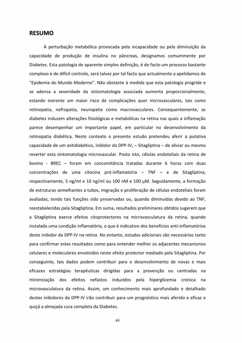
À perturbação metabólica provocada pela incapacidade ou pela diminuição da
capacidade de produção de insulina no pâncreas, designamos comummente por
Diabetes. Esta patologia de aparente simples definição, é de facto um processo bastante
complexo e de difícil controlo, será talvez por tal facto que actualmente a apelidamos de
“Epidemia do Mundo Moderno”. Não obstante à medida que esta patologia progride e
se adensa a severidade da sintomatologia associada aumenta proporcionalmente,
estando inerente um maior risco de complicações quer microvasculares, tais como
retinopatia, nefropatia, neuropatia como macrovasculares. Consequentemente, as
diabetes induzem alterações fisiológicas e metabólicas na retina nas quais a inflamação
parece desempenhar um importante papel, em particular no desenvolvimento da
retinopatia diabética. Neste contexto o presente estudo pretendeu aferir a putativa
capacidade de um antidiabético, inibidor da DPP-IV, – Sitagliptina – de aliviar ou mesmo
reverter esta sintomatologia microvascular. Posto isto, células endoteliais da retina de
bovino - BREC – foram em concomitância tratadas durante 6 horas com duas
concentrações de uma citocina pró-inflamatória – TNF – e de Sitagliptina,
respectivamente, 5 ng/ml e 10 ng/ml ou 100 nM e 100 µM. Seguidamente, a formação
de estruturas semelhantes a tubos, migração e proliferação de células endoteliais foram
avaliadas, tendo tais funções sido preservadas ou, quando diminuídas devido ao TNF,
reestabelecidas pela Sitagliptina. Em suma, resultados preliminares obtidos sugerem que
a Sitagliptina exerce efeitos citoprotectores na microvasculatura da retina, quando
instalada uma condição inflamatória, o que é indicativo dos benefícios anti-inflamatórios
deste inibidor da DPP-IV na retina. No entanto, estudos adicionais são necessários tanto
para confirmar estes resultados como para entender melhor os adjacentes mecanismos
celulares e moleculares envolvidos neste efeito protector mediado pela Sitagliptina. Por
conseguinte, tais dados podem contribuir para o desenvolvimento de novas e mais
eficazes estratégias terapêuticas dirigidas para a prevenção ou centradas na
minimização dos efeitos nefastos induzidos pela hiperglicemia crónica na
microvasculatura da retina. Assim, um conhecimento mais aprofundado e detalhado
destes inibidores da DPP-IV irão contribuir para um prognóstico mais aferido e eficaz e
quiçá a almejada cura completa da Diabetes.

xiii
Palavras-Chave: Retinopatia Diabética, Dipeptidil-peptidase IV, Sitagliptina,
Inflamação, Células Endoteliais da Retina de Bovino (BREC)
xiv
AABBSSTTRRAACCTT
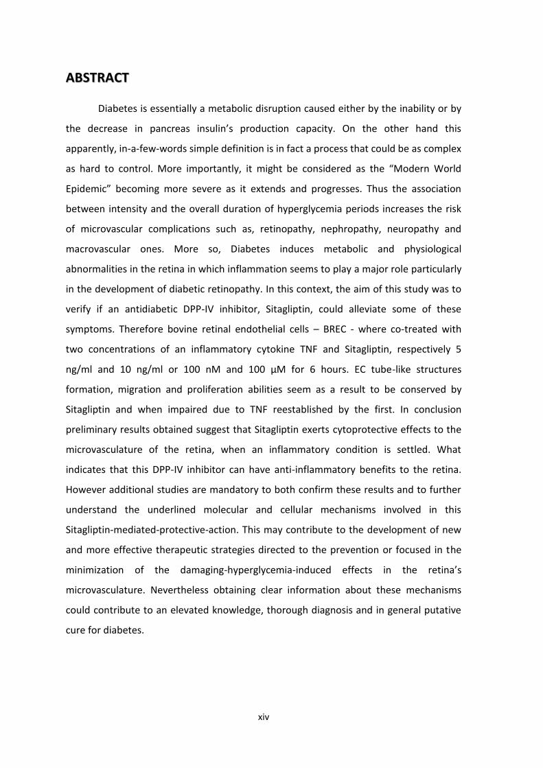
Diabetes is essentially a metabolic disruption caused either by the inability or by
the decrease in pancreas insulin’s production capacity. On the other hand this
apparently, in-a-few-words simple definition is in fact a process that could be as complex
as hard to control. More importantly, it might be considered as the “Modern World
Epidemic” becoming more severe as it extends and progresses. Thus the association
between intensity and the overall duration of hyperglycemia periods increases the risk
of microvascular complications such as, retinopathy, nephropathy, neuropathy and
macrovascular ones. More so, Diabetes induces metabolic and physiological
abnormalities in the retina in which inflammation seems to play a major role particularly
in the development of diabetic retinopathy. In this context, the aim of this study was to
verify if an antidiabetic DPP-IV inhibitor, Sitagliptin, could alleviate some of these
symptoms. Therefore bovine retinal endothelial cells – BREC - where co-treated with
two concentrations of an inflammatory cytokine TNF and Sitagliptin, respectively 5
ng/ml and 10 ng/ml or 100 nM and 100 µM for 6 hours. EC tube-like structures
formation, migration and proliferation abilities seem as a result to be conserved by
Sitagliptin and when impaired due to TNF reestablished by the first. In conclusion
preliminary results obtained suggest that Sitagliptin exerts cytoprotective effects to the
microvasculature of the retina, when an inflammatory condition is settled. What
indicates that this DPP-IV inhibitor can have anti-inflammatory benefits to the retina.
However additional studies are mandatory to both confirm these results and to further
understand the underlined molecular and cellular mechanisms involved in this
Sitagliptin-mediated-protective-action. This may contribute to the development of new
and more effective therapeutic strategies directed to the prevention or focused in the
minimization of the damaging-hyperglycemia-induced effects in the retina’s
microvasculature. Nevertheless obtaining clear information about these mechanisms
could contribute to an elevated knowledge, thorough diagnosis and in general putative
cure for diabetes.

xv
Key Words: Diabetic Retinopathy, Dipeptidyl peptidase IV Sitagliptin,
Inflammation, Bovine Retinal Endothelial Cells (BREC)

CCHHAAPPTTEERR 11 IINNTTRROODDUUCCTTIIOONN


CCHHAAPPTTEERR 11
IInnttrroodduuccttiioonn
3
11.. DDIIAABBEETTEESS MMEELLLLIITTUUSS
11..11.. BBRRIIEEFF SSTTOORRYY AABBOOUUTT DDIIAABBEETTEESS HHIISSTTOORRYY
An Egyptian manuscript from c. 1500 BCE mentioning "passing of too much
urine" is thought to be one of the first diseases description. In addition the first cases
reported are believed to be of type 1 diabetes. Around the same time, Indian physicians
noting the urine would attract ants, also identified the disease and classified it as
madhumeha or "honey urine".
The term "diabetes" or "to pass through" was only used in 230 BCE by the
Greek Appollonius of Memphis. During the Roman Empire the disease was considered
rare with Galen commenting he had only seen two cases during his entire career.
(DiabeWorld, 2012) This is possibly due to ancient people dieting and life-style, or even
because clinical symptoms were only observed during a more advanced stage of the
disease, Galen named the disease "diarrhea of the urine" (diarrhea urinosa)
(MiYDiabetes, 2012).
The earliest surviving work with a detailed reference to diabetes is that of
Aretaeus of Cappadocia, reflecting the beliefs of the "Pneumatic School" he described
the symptoms and the disease course, which he attributed to the moisture and
coldness. He hypothesized a correlation of diabetes with other diseases, discussing
differential diagnosis from the snakebite which also provokes excessive thirst. His work
remained unknown until middle of the 16th century when, in 1552, the first Latin edition
was published in Venice (Diabetes India, 2012).
Moreover type 1 and type 2 diabetes where identified as separate conditions
for the first time by the Indian physicians Sushruta and Charaka in 400-500 CE with type
1 associated with youth and type 2 with being overweight.
The term "mellitus" or "from honey" however was added by the Briton John
Rolle in the late 1700s, when he noticed the urine of a diabetic had a sweet taste
(glycosuria) and to separate the condition from diabetes insipidus, which is also
associated with frequent urination. When Canadians Frederick Banting and Charles
EEvvaalluuaattiioonn ooff aa DDiippeeppttiiddyyll PPeeppttiiddaassee IIVV IInnhhiibbiittoorr aass aa
MMiiccrroovvaassccuullaattuurree PPrrootteeccttoorr iinn DDiiaabbeetteess
4
Herbert Best isolated and purified insulin, in 1921 and 1922, the effective treatment was
developed. This important finding was followed by the development of the long-acting
insulin NPH in the 1940s. (Ahmed A.M. et al., 2002)
11..22.. DDEEFFIINNIITTIIOONN AANNDD CCHHAARRAACCTTEERRIIZZAATTIIOONN
Diabetes Mellitus was considered by the World Health Organization (WHO) as a
complex and heterogeneous metabolic disorder, which is characterized through a
chronic hyperglycemia with atypical carbohydrates, lipids and proteins metabolism,
leading to insulin secretion and/or insulin action deficiency (WHO, 2012).This pathology
may present characteristic symptoms such as thirst, polyuria, blurring of vision, and
weight loss. In its most severe forms, ketoacidosis or a non-ketotic hyperosmolar state
may develop and lead to stupor, coma and, in absence of effective treatment, death.
(WHO, 2012, IDF, 2012))
Moreover the pathogenetic processes involved in the development of diabetes
include pancreatic beta cells destruction with consequent insulin deficiency, and others
that result in resistance to insulin action (Eizirik et al., 2008). This leads to the
progressive development of specific complications, namely retinopathy with potential
blindness, nephropathy that may lead to renal failure, and/or neuropathy with risk of
foot ulcers, amputation and Charcot joints. People with diabetes are also at increased
risk of cardiovascular, peripheral vascular and cerebrovascular disease (IDF, 2012,
Medical News Today, 2012)
11..33.. PPRREEVVAALLEENNCCEE
Worldwide, we have been assisting to an increase in Diabetes prevalence which
is inevitably overwhelming to our already overburdened health system. Hence type 2
Diabetes is the one to be blamed when it comes to this escalating aggravation, not only
due to ever growing world population but also the continuously aging demography.
Nevertheless a massive increase in overweight and obese people due to sedentary and
overall lack of a healthy life style, might as well contribute to a bigger risk of suffering
from this disease (Zimmet et al., 2001)
CCHHAAPPTTEERR 11
IInnttrroodduuccttiioonn
5
Diabetes Mellitus prevalence rates have critically risen in recent years and in
accordance to the International Diabetes Federation (IDF), Diabetes Mellitus is
frequently a non-communicable disease, with high morbidity and mortality rates, just in
2012 alone were registered about 4.8 million casualties. (IDF, 2012)
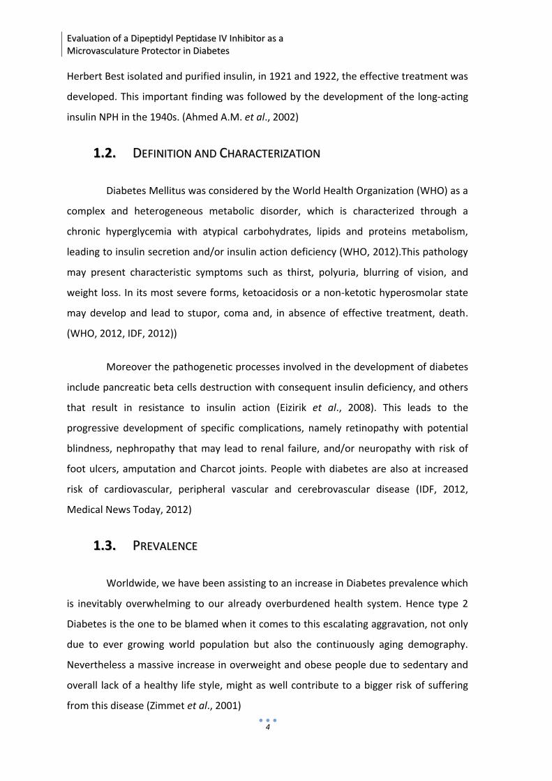
In the present, Diabetes affects about 371 million people (Figure 1.1) and with
an estimated increase to 552 million by 2030, thus representing 8.3% of adult
population. However, these numbers are underestimated due to the undiagnosed
patients, who represent almost half the people with Diabetes (IDF, 2012). Regarding
Portugal, the Sociedade Portuguesa de Diabetologia (Portuguese Diabetes Society)
estimates that 12.7% of the Portuguese adult population will be diabetic, from which
5.4% might still remain undiagnosed (SPD, 2012).
11..44.. MMOOSSTT CCOOMMMMOONN TTYYPPEESS
In accordance to WHO and IDF there are three main types of diabetes, namely:
type 1 diabetes mellitus (T1DM) also called insulin-dependent, type 2 diabetes mellitus
(T2DM) known as insulin-resistance and gestational diabetes
Figure 1.1 - Global prevalence (%) of diabetes mellitus in adult population (20-79 years) in 2012, shown by geographic region (Image taken from IDF Diabetes Atlas, 5th Edition (IDF, 2012)

EEvvaalluuaattiioonn ooff aa DDiippeeppttiiddyyll PPeeppttiiddaassee IIVV IInnhhiibbiittoorr aass aa
MMiiccrroovvaassccuullaattuurree PPrrootteeccttoorr iinn DDiiaabbeetteess
6
11..44..11.. TTYYPPEE 11 DDIIAABBEETTEESS MMEELLLLIITTUUSS
Type 1 diabetes also called as insulin-dependent diabetes, or juvenile-onset
diabetes, is characterized by the presence of anti-GAD, islet cell or insulin antibodies
which identify the autoimmune processes that mediate the destruction of pancreatic
beta cells. Albeit these patients not being necessarily obese they may instead have other
autoimmune disorders such as Graves' disease, Hashimoto's thyroiditis, and Addison's
disease (Betterle et al., 1983).
The first manifestation in some
patients, particularly in children might be
ketoacidosis or modest fasting
hyperglycaemia. A few adults however may
retain residual beta-cell function, sufficient to
prevent ketoacidosis, for many years (Japan
and Pittsburgh Childhood Diabetes Research
Groups, 1985). Eventually, individuals with this
form of Type 1 diabetes often become
dependent on insulin for survival in order to
prevent the development of ketoacidosis
(Zimmet et al.,1995, Willis et al., 1996). At
this stage of the disease, there is little or no insulin secretion (Figure 1.2) as manifested
by low or undetectable levels of plasma C-peptide (Hother et al., 1988) There is however
both a genetic predisposition to autoimmune destruction of beta cells as a relation
between environmental factors, which remains yet known.
11..44..22.. TTYYPPEE 22 DDIIAABBEETTEESS MMEELLLLIITTUUSS
Type 2 diabetes previously encompassed non-insulin dependent diabetes or
adult-onset diabetes, and accounts for at least 90% of all cases of diabetes. It is
characterised by insulin resistance and relative insulin deficiency (Figure 1.3), either or
both of which may be present at the time diabetes is diagnosed.
Figure 1.2 - Carrier proteins involved in type 1 Diabetes (Diabetes Education Online, 2012)

CCHHAAPPTTEERR 11
IInnttrroodduuccttiioonn
7
Type 2 diabetes risk increases with
age, hypertension, dyslipidaemia and overall
lack of physical activity (Zimmet et al., 1992,
Harris et al., 1995). It might be probably
associated with strong familial, likely genetic,
predisposition (Valle et al., 1997, Knowler et
al., 1993). Despite the genetics of this
diabetic form it is still rather complex and not
clearly defined.
It is often, but not always,
associated with overweight, obesity or even
predominantly body fat in the abdominal areas, which itself can cause insulin resistance
and lead to high blood glucose levels. People with type 2 diabetes can often initially
manage their condition through exercise and diet. However, over time most people will
require oral drugs and or insulin (Kissebah et al., 1982).
22.. DDIIAABBEETTIICC RREETTIINNOOPPAATTHHYY
22..11.. DDEEFFIINNIITTIIOONN AANNDD CCHHAARRAACCTTEERRIIZZAATTIIOONN
Diabetic Retinopathy (DR) is a
microvascular complication of diabetes
(Aielo et al., 1998, Davidson et al., 2007), in
early stages of the disease are characterized
by vascular alterations in blood flow, death
of retinal pericytes (perivascular contractile
cells), basement membrane thickening and
subtle increases in vascular
permeability.(Aveleira et al., 2010, Zhang et
al., 2011, Sheetz et al., 2002) As the disease progresses, clear alterations in the vascular
structure, for instance non-perfused vessels, microaneurysms, dot/blot hemorrhages,
Figure 1.3 – Carrier proteins involved in type 12Diabetes (Diabetes Education Online, 2012)
Figure 1.4 - Normal eye anatomy (National Eye Institute, 2012)

EEvvaalluuaattiioonn ooff aa DDiippeeppttiiddyyll PPeeppttiiddaassee IIVV IInnhhiibbiittoorr aass aa
MMiiccrroovvaassccuullaattuurree PPrrootteeccttoorr iinn DDiiaabbeetteess
8
cotton-wool spots, venous beading, vascular loops and significant vascular leakage can
be seen under ophthalmologic examination (Cockburn et al., 1999).
22..22.. DDRR PPRROOGGRREESSSSIIOONN
The vascular changes stated above occur in an early phase – the non-
proliferative stage – of DR, where, as a consequence of increased vascular permeability,
the macular edema formed can eventually lead to vision loss. Later on, in the
proliferative stage of DR, neovascularization takes place on the retinal surface, after that
vascular function becomes impaired by capillary occlusion, non-perfusion and
degeneration. In this stage, severe vision loss or even blindness may as well be caused
by bleeding, hemorrhage and subsequent retinal detachment due to the newly formed
but rather fragile vessels (Davidson et al., 2007).
Diabetic retinopathy progression can be identified into four stages, which were
conveniently enumerated down below:
I. Mild Nonproliferative Retinopathy: At this earliest stage, microaneurysms
occur. They are small areas of balloon-like swelling in the retina's tiny blood vessels.
II. Moderate Nonproliferative Retinopathy: As the disease progresses, some
blood vessels that nourish the retina are blocked.
III. Severe Nonproliferative Retinopathy: Many more blood vessels are
blocked, depriving several areas of the retina with their blood supply. These areas of the
retina send signals to the body to grow new blood vessels for nourishment
IV. Proliferative Retinopathy: At this advanced stage, the signals sent by the
retina for nourishment trigger the growth of new blood vessels. This condition is called
proliferative retinopathy. These new blood vessels are abnormal and fragile. They grow
along the retina and along the surface of the clear, vitreous gel that fills the inside of the
eye.
The mechanisms by which diabetes causes microvascular complications and
disease progression in the retina are not yet fully understood. However, further studies

CCHHAAPPTTEERR 11
IInnttrroodduuccttiioonn
9
in patient samples and animal models have shown that DR could be definitely
characterized as a chronic, hence subclinical inflammation.
22..33.. MMAAJJOORR PPAATTHHOOGGEENNIICC PPAATTHHWWAAYYSS IINN DDIIAABBEETTIICC RREETTIINNOOPPAATTHHYY
The increase in polyol pathway, non-enzymatic glycosylation of proteins,
oxidative stress and protein-c kinase activation by generation of diacylglycerol, are
retinal biochemical and cellular abnormalities induced by chronic hyperglycemia that
lead to vascular impairments. Albeit this, the true mechanism behind hyperglycemia-
induced-DR remains yet known.
22..33..11.. PPOOLLYYOOLL PPAATTHHWWAAYY
Glucose uptake by retinal tissue is insulin-independent existing therefore an
equilibrium between retinal and plasmatic glucose levels. When hyperglycemia is
present it activates the polyol pathway, converting, through aldose reductase, glucose in
sorbitol and then in fructose (by sorbitol dehydrogenase) (Miwa et al., 2003)
Subsequently, in the retina, the intracellular increase in sorbitol concentration
causes osmotic breakdown, damages retinal pericytes, via apoptosis, and thickens
retinal vascular endothelial cell basement membranes leading to closure of retinal
capillaries. These changes in membrane permeability and integrity have been identified
as the key to early cellular pathology (Lorenzi et al., 2007).
Figure 1.5 - Normal Vision and the same scene viewed by a person with Diabetic Retinopathy (National Eye Institute, 2012)

EEvvaalluuaattiioonn ooff aa DDiippeeppttiiddyyll PPeeppttiiddaassee IIVV IInnhhiibbiittoorr aass aa
MMiiccrroovvaassccuullaattuurree PPrrootteeccttoorr iinn DDiiaabbeetteess
10
22..33..22.. AAGGEESS PPAATTHHWWAAYY
The hyperglycemia also leads to lipid and protein glycosylation, whose
oxidation produces glycotoxins and Advanced Glicosilated End-products (AGEs)
(Schleicher et al., 1997). These AGEs can be correlated with Diabetes duration and
severity, being found in the plasma, tissue and vessels walls, exerting their action
through endothelium-expressed receptors, promoting an increase in vascular
permeability and thrombogenicity (Kowluru and Odenbach et al., 2004, Libby et al.,
2007). Nevertheless numerous are the mechanism that control vascular tonicity, that
when Diabetes-activated can lead to vascular hemodynamic regulation loss.
22..33..33.. OOXXIIDDAATTIIVVEE SSTTRREESSSS
There is evidence that oxidative stress, defined as a persistent imbalance
between the production of highly reactive molecular species (chiefly oxygen and
nitrogen) and antioxidant defenses, can lead to tissue damage (Rosen et al., 2001).
Oxidative stress results from increased content of reactive oxygen species (ROS) and/or
reactive nitrogen species (RNS). Examples of ROS include charged species such as
superoxide(O2- •) and the hydroxyl radical (OH•), and uncharged species such as
hydrogen peroxide (Rosen et al., 2001, Irani et al, 2000). There are also data indicating
that ROS formation is a direct consequence of hyperglycemia (Brownlee et al., 2001);
more recent studies have suggested that increased free fatty acids levels may also result
in ROS formation.
Because of their ability to directly oxidize and damage DNA, protein, and lipid,
ROS are believed to play a key direct role in the pathogenesis of late diabetic
complications (Rosen et al., 2001, Nishikawa et al., 2000). In addition, ROS can function
as signaling molecules to activate a number of cellular stress-sensitive pathways that
cause cellular damage, and are ultimately responsible for the late complications of
diabetes. Furthermore, these same pathways are linked to insulin resistance and
decreased insulin secretion (Wolff et al., 1991).

CCHHAAPPTTEERR 11
IInnttrroodduuccttiioonn
11
22..33..44.. PPRROOTTEEIINN KKIINNAASSEE CC PPAATTHHWWAAYY
Studies indicate that enhanced generation of diacylglycerol – a physiologic
activator of PKC – , hence a PKC pathway activation might play a major role in
hyperglycaemia-induced microvascular dysfunction in diabetes by promoting increased
flux through the polyol pathway (Keogh et al., 1997) and the generation of AGEs (Portilla
et al., 2000) and oxidative species result in.
PKC which functions as signaling components for a variety of growth factors,
hormones, neurotransmitters and cytokines, when activated results in numerous
cellular changes, leading to basement membrane thickening and increased production
of vasodilatory prostaglandins as well as vascular endothelial growth factor (VEGF),
which in turn affect vessel permeability and/or blood flow, leading to vascular
impairment (Koya and King et al., 1998, Das Evcimen and King et al.,2007).
33.. IINNFFLLAAMMMMAATTIIOONN
The role of inflammation in the development and progression of diabetic
retinopathy has been studied for a long time but it was in the last years that it started to
get some major attention. Powell and Field in 1960 observed that diabetics treated with
anti-inflammatory agents like “salicylate” had a lower incidence rate of diabetic
retinopathy (Powell et al., 1964). Earlier studies by Lutty and his group identified the
important role of leukocytes in the development of diabetic retinopathy (Lutty et al.,
1997, McLeod et al., 1995) and a subsequent study has established diabetic retinopathy
as an “inflammatory disease” (Adamis et al., 2002).
This inflammatory state starts very early within one week of experimental
diabetes, due to leukocytes accumulation in the vasculature of the retina (Adamis et al.,
2002). Widely observed in diabetic retinopathy the major components of the
inflammatory phenotype progress towards the increase in diabetic macular edema
(retinal vascular permeability) and neovascularization (proliferative diabetic retinopathy)
(Table 1.6).

EEvvaalluuaattiioonn ooff aa DDiippeeppttiiddyyll PPeeppttiiddaassee IIVV IInnhhiibbiittoorr aass aa
MMiiccrroovvaassccuullaattuurree PPrrootteeccttoorr iinn DDiiaabbeetteess
12
IInnccrreeaasseedd EExxpprreessssiioonn ooff IInnffllaammmmaattoorryy AAddhheessiioonn MMoolleeccuulleess IICCAAMM--11
aanndd VVCCAAMM--11 iinn tthhee EEnnddootthheelliiuumm
AAddhheessiioonn LLeeuukkooccyytteess ttoo RReettiinnaall vveesssseellss
IInnccrreeaasseedd EExxpprreessssiioonn ooff CCyyttookkiinneess aanndd GGrroowwtthh ffaaccttoorrss
MMiiccrroogglliiaall CCeellll AAccttiivvaattiioonn
IInnffiillttrraattiioonn ooff MMoonnooccyytteess aanndd NNeeuuttrroopphhiillss
IInnccrreeaasseedd VVaassccuullaarr ppeerrmmeeaabbiilliittyy ((MMaaccuullaarr EEddeemmaa)) aanndd
NNeeoovvaassccuullaarriizzaattiioonn ((PPrroolliiffeerraattiivvee RReettiinnooppaatthhyy))
RReettiinnaall CCeellll DDeeaatthh
Table 1.6 - Components of Diabetic Retinal Inflammation (Adapted from Antonetti et al., 2006 and Adamis et al.,2002)
33..11.. IINNFFLLAAMMMMAATTOORRYY BBIIOOMMAARRKKEERRSS IINN DDIIAABBEETTIICC RREETTIINNOOPPAATTHHYY
The main risk factors for diabetic retinopathy include diabetes duration,
hyperglycemia, hypertension, and dyslipidemia, which only can explain some degree of
variance in the risk of diabetic retinopathy (Nguyen et al., 2009). Studies have therefore
shown the association of multiple systemic inflammatory factors in the progression of
diabetic retinopathy, and a further analysis of diabetic vitreous samples has also
provided insights into novel proinflammatory markers in this process (Schmidt et al.,
1995, Schram et al., 2003).
Furthermore inflammation is a complex biological response of vascular tissues
against harmful stimuli, including damaged cells, irritants, or pathogens being also a
critical step in wound healing (Wagener et al., 2013). This process involves multiple
mediators such as pro-inflammatory cytokines, chemokines and adhesion molecules that
initiate the interaction between leukocytes and the endothelium, guiding directional
leukocyte migration toward infected or injured tissue (Zhang et al., 2011).
First, the pro-inflammatory cytokines (such as tumor necrosis factor (TNF) and
interleukins) and the chemokines (such as CCL2 and CCL5) released from
infected/injured tissue, activate the endothelium to increase expression of adhesion
molecules (such as E-selectin, intercellular adhesion molecule (ICAM)-1, vascular cell

CCHHAAPPTTEERR 11
IInnttrroodduuccttiioonn
13
adhesion molecule (VCAM)-1) and chemokines (Zhang et al., 2011). Then leukocytes,
mediated by chemokines and adhesion molecules, attach to the vessel wall,
transmigrate through the endothelium and linger in the infected or injured tissue until
complete healing (Barreiro et al., 2010). While normal inflammation is beneficial,
excessive or uncontrolled one, like the one seen in DR, can cause tissue injury and
ultimately result in diseases (Barreiro et al., 2010).
Although there are no known pathogens in DR, analysis of inflammatory
molecules in vitreous, serum and retina form diabetic patients or experimental animals
indicate that DR is associated with significant increases in pro-inflammatory cytokines,
chemokines and adhesion molecules (Zhang et al., 2011). High levels of interleukin-1β
(IL-1β) and caspase 1 – its downstream signaling molecule – as well as TNF are
significantly increased in vitreous, retinas and serum from diabetic patients and rats
(Demircan et al., 2006, Vincent et al., 2007, Kowluru et al., 2004). Nevertheless TNF also
appears to be augmented in ocular fibrovascular membranes from patients with DR and
in retinas from rodent model of diabetes mellitus (Joussen et al., 2006, Limb et al.,
1996).
On the other hand, vitreous samples from DR patients also show that certain
chemokines such as CCL2,
CCL5, CXCL8, CXCL10 and
CXCL12 are also upregulated
(Murugeswari et al., 2008,
Meleth et al., 2005).
Furthermore, Increases in IL-6,
ICAM-1 and VCAM-1 have
been shown to be related to
the progression of DR (Meleth
et al., 2005, Adamiec-
Mroczek, 2008) (Figure 1.7).
Figure 1.7 - Inflammatory pathway in Diabetic Retinopathy (Medscape 2011)

EEvvaalluuaattiioonn ooff aa DDiippeeppttiiddyyll PPeeppttiiddaassee IIVV IInnhhiibbiittoorr aass aa
MMiiccrroovvaassccuullaattuurree PPrrootteeccttoorr iinn DDiiaabbeetteess
14
This correlation between increases in inflammatory molecules can also be
associated with recruitment of leukocytes, for example the quantity of neutrophils is
significantly elevated in both retinal and choroidal vessels from diabetic patients and
rhesus monkeys (Kim et al., 2005). Whereas its accumulation can be either correlated
with upregulation of ICAM-1 immunoreactivity in the vessels or associated with capillary
closure (McLeod et al., 1995).
In diabetic rodent models, along with the progression of DR, there is a
cumulative and sustained increase in leukocyte adherence to the retinal vasculature –
leukostasis (Miyamoto et al., 1999). This augment however may be linked to diabetes-
induced increases in ICAM-1 and integrins in endothelial cells and leukocytes,
respectively, since ICAM-1 block or deletion of CD18 (ICAM-1 receptor subunit on
leukocytes) seem to act as a preventive in diabetes-induced leukostasis (Miyamoto et
al., 1999, Joussen et al., 2000, Barouch et al., 2004).
In addition to inflammation-triggered circulatory leukocytes, retinal microglial
cells – resident ocular macrophages – (Fischer et al., 2001) are also most likely to be
involved in DR (Chen et al., 2002), being rapidly activated to release inflammatory
cytokines such as TNF (Yang et al., 2009). On the other hand, studies in wild-type mice
revealed that the treatment with the A2A adenosine receptor agonist resulted in
marked decreases either in tumor necrosis factor (TNF) release or in hyperglycemia-
induced retinal apoptosis (Ibrahim et al., 2011).
33..22.. RROOLLEE OOFF IINNFFLLAAMMMMAATTIIOONN IINN BBLLOOOODD RREETTIINNAALL BBAARRRRIIEERR
AALLTTEERRAATTIIOONN
Briefly, the blood-ocular barrier system is formed by two main barriers: the
blood-aqueous barrier and the blood-retinal barrier (BRB). This barrier, particularly tight
and restrictive, is a physiologic-type, because it regulates ion, protein, and water flux
into and out of the retina. The BRB consists of inner, being formed of tight junctions
between retinal capillary endothelial cells and outer components, and outer BRB which
has tight junctions between retinal pigment epithelial cells. Furthermore, BRB is as
CCHHAAPPTTEERR 11
IInnttrroodduuccttiioonn
15
essential to maintaining the eye as a privileged site as it is indispensable for normal
visual function; in fact it is exactly why alterations of the BRB play a crucial role in the
development of retinal diseases. The 2 most frequent and relevant retinal diseases,
diabetic retinopathy and age-related macular degeneration (AMD), are directly
associated with alterations of the BRB. The first is initiated by an alteration of the inner
BRB, whereas neovascular AMD is a result of an alteration of the outer BRB, being
therefore the macular edema a direct result of alterations in the BRB (Cunha-Vaz et al.,
2010).
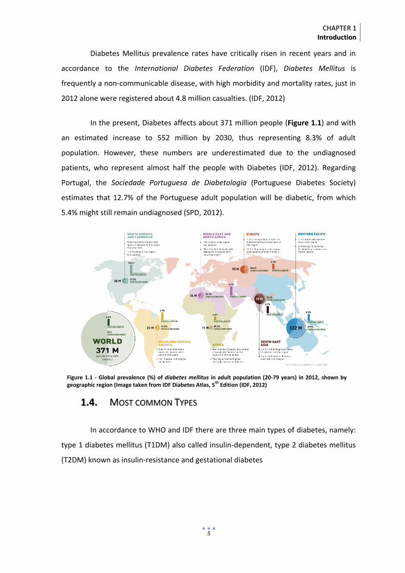
Alteration of the inner blood retinal
barrier (BRB) is the hallmark of diabetic
retinopathy (Aveleira et al., 2010) (Figure 1.8).
And, inflammation is one of the earlier events
in diabetic retinopathy, which there is
increased adhesion of leukocytes to the
microvasculature, resulting in the loss of
endothelial cells and breakdown of the blood-
retinal barrier (Yuuki et al., 2001, Joussen et
al., 2001). Hence the major stages for BRB
alteration are enumerated next: increased
expression of adhesion molecules, such as
ICAM1, VCAM1, P-Selectin, followed by
leukocyte adhesion in the diabetic retina
endothelium (McLeod et al., 1995, Adamis et
al., 2008); next, release of inflammatory
cytokines, vascular permeability factors, and
growth cytokines, what culminates into
modifications in adherens and tight junctional
proteins (for example, VE-Cadherin, Occludin, ZO-1 and Claudin) and also infiltration by
diapedesis of the leukocytes into the retina. Nonetheless all of these steps contribute to
the Blood-Retinal Barrier breakdown.
Figure 1.8 - Disruption of the Neurovascular Unit of the Retina by Diabetes (Antonetti et al., 2012).
EEvvaalluuaattiioonn ooff aa DDiippeeppttiiddyyll PPeeppttiiddaassee IIVV IInnhhiibbiittoorr aass aa
MMiiccrroovvaassccuullaattuurree PPrrootteeccttoorr iinn DDiiaabbeetteess
16
Moreover, Yuuki et al., in animal models of diabetic retinopathy showed that
the inhibition of leukocyte adhesion prevents the loss of pericytes and the formation of
acellular capillaries, leading therefore to the suppression of the blood-retinal barrier
breakdown. Another study also suggested a possible mechanism involving proteolytic
degradation of VE-cadherin which could be possibly linked to diabetes contribution to
BRB (Navaratma et al., 2001).
These recently identified inflammatory risk factors not only serve as potential
biomarkers, but also give insights into the development of potential molecular targets
for treating diabetic retinopathy.
44.. AANNTTII--DDIIAABBEETTIICC TTHHEERRAAPPYY
44..11.. EEXXIISSTTIINNGG NNOONN--IINNSSUULLIINN AANNTTII--DDIIAABBEETTIICC TTHHEERRAAPPYY
At the present time, to treat/control diabetes medicine relies on either the
classic insulin therapy or on the non-insulin antidiabetic one, this is, metformin,
sulphonylureas, meglitinides, thialidonediones, alpha-glucosidase inhibitors, amilin
analogs, GLP-1 receptor agonists and DPP-IV inhibitors. These agents act in different
sites of the organism to amelliorate insulin secretion and/or even its action. (Weiner, L.
et al., 2010).
44..11..11.. IINNCCRREETTIINNSS
In the last decade incretins have become object of attention due to its potential
as a new type 2 Diabetes therapy. Nonetheless this concept was already known almost
half a century ago, once it was observed that orally administered glucose stimulated a
bigger insulin release then the same amount of the injected subtance (Eirick et al.,
1964). From this investigation two hormones, namely glucagon-like-peptide-1 (GLP-1)
and glucose-dependent insulinotropic peptide (GIP), this is, incretin hormones were
found responsible for the signal which led to gastrointestinal tract release of insulin
whenever food was being consumed.

CCHHAAPPTTEERR 11
IInnttrroodduuccttiioonn
17
However it is now known that after being secreted by the gastrointestinal tract
while food is eaten, incretin hormones bind to beta cells receptors in the pancreas,
stimulating insulin secretion as a response to glucose absorption. (Ahrén, 2003). In
healthy individuals it is also thought that incretin effect is responsible for approximately
50 – 70% of insulinic-response to oral glucose, but a smaller secretion in type 2 Diabetes
patients, might suggest that this could be linked to the disease pathogenesis (Nauck et
al., 1986; VilsbØl et al., 2001). Drucker et al., later corroborated with this by showing
that an increase in GLP-1 reduces hyperglycemia (Drucker, 2003).
Consequently, and since GIP is inactive in type 2 Diabetes, a new incretin-like
therapy based on GLP-1 endogenous levels was developed, in which two different
approaches were pursued. The incretin mimetics, for example exenatide, and one
Sitagliptin belongs to, hence the incretin secretors or DPP-IV inhibitors.
44..11..11..11.. IINNCCRREETTIINN MMIIMMEETTIICCSS
Deacon et al, stated that these endogenous peptides (GLP-1 and GIP) were
rapidly removed from circulation by DPP-IV enzyme and renal clearance. So it was
necessary for the production of incretin-based therapies that these molecules had
expand half-lifes and therefore exenatide and liraglutide were produced.
44..11..11..11..11.. EEXXEENNAATTIIDDEE
Exenatide was the first agent to be commercialized in 2005 and in the USA. It is
a synthetic form of exenatide 4, a molecule originally isolated from the venon of the
Heloderma suspectum. The exenatide however is not that analogous to human GLP-1,
only sharing 53% of its molecular sequence. Despite the remaining structural similarity is
enough for it to bind to GLP-1 receptor in order to mimic a significant number of
glucoregulatory actions, not being able however to proceed as a DPP-IV susbstract.
Exenatide decreases fasting and postprandial glucose concentrations through
several mechanisms, also shared with GLP-1. In the pancreas, exenatide all together
stimulates beta cells insulin secretion and suppresses alpha cells glucagon secretion
EEvvaalluuaattiioonn ooff aa DDiippeeppttiiddyyll PPeeppttiiddaassee IIVV IInnhhiibbiittoorr aass aa
MMiiccrroovvaassccuullaattuurree PPrrootteeccttoorr iinn DDiiaabbeetteess
18
reducing for that reason, in a post-prandial state, hepatic glucose production. It also
intervenes in gastric emptiness by slowing it down, promoting a satiety sensation, what
eventually might lead to weight loss in overweight individuals (Linnebjerg et al., 2008).
Nonetheless these events only need to occur in the presence of high circulatory glucose
concentrations, in order to minimize hypoglycemic risk.
In animal studies, exenatide can apparently promote pancreatic islets cells
differentiation and inhibit beta cells apoptosis, altering the balance by increasing islets
mass. Beta cells mass cannot be measured in humans through non-invasive procedures
and in addition their function can only be determined in an indirect manner, for
example, through pro-insulin:insulin ratio and beta cell function homeostasis evaluation
(HOMA-b). These results seem to suggest that GLP-1 receptor agonist as well as DPP-IV
inhibitors enhance beta cell function (Drucker, 2006).
Finally, in contrast with exenatide and due to a higher homology between
liraglutide and endogenous GLP-1, there is a decreased risk of antibody formation
(Marre et al., 2009; Russell-Jones et al., 2009). Nonetheless a clinical relevance for the
antibodies issue remains yet known for both agents.
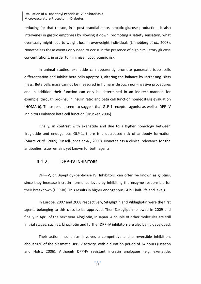
44..11..22.. DDPPPP--IIVV IINNHHIIBBIITTOORRSS
DPP-IV, or Dipeptidyl-peptidase IV, Inhibitors, can often be known as gliptins,
since they increase incretin hormones levels by inhibiting the enzyme responsible for
their breakdown (DPP-IV). This results in higher endogenous GLP-1 half-life and levels.
In Europe, 2007 and 2008 respectively, Sitagliptin and Vildagliptin were the first
agents belonging to this class to be approved. Then Saxagliptin followed in 2009 and
finally in April of the next year Alogliptin, in Japan. A couple of other molecules are still
in trial stages, such as, Linagliptin and further DPP-IV inhibitors are also being developed.
Their action mechanism involves a competitive and a reversible inhibition,
about 90% of the plasmatic DPP-IV activity, with a duration period of 24 hours (Deacon
and Holst, 2006). Although DPP-IV resistant incretin analogues (e.g. exenatide,

CCHHAAPPTTEERR 11
IInnttrroodduuccttiioonn
19
liraglutide) have the same biochemical target than the DPP-IV inhibitors, these last are
only able to increase GLP-1 levels up to physiological values while the others can
increase it by 6-10-fold that of the physiological ones found in the postprandial state
(ElbrØnd et al., 2002, Drucker et al., 2008, Calara et al., 2005).
In addition, among DPP-IV inhibitors glucoregulatory actions are included
insulin secretion stimulation, glucagon secretion inhibition, on the whole improvement
in beta cells function. In spite of this DPP-IV inhibitors seem to not mediate all
endogenous GLP-1 glucoregulatory actions, having therefore little or no effect in gastric
emptiness or satiety for that matter, what might be implied in their neutrality regarding
the weight issue. DPP-IV inhibitors on other hand can be divided into two classes which
are either related to their structural form or their excretory pathway (Nauck et al.,
2009).
First, they are divided due to the presence or absence of the cyano-pirrolidine
ring, since the ones who do possess it, namely Vildagliptin and Saxgliptine are less
selective (not clinically relevant though) than the others who don’t, hence Sitagliptin,
Alogliptin and Linagliptin.
Second, Sitagliptin and Alogliptin remain almost inaltered when kidney-
excreted whereas Saxagliptin and Vildagliptin are metabolized respectively, as an active
metabolite (in the kidney and liver) and an inactive one.
As a final point, Linagliptin is the only DPP-IV inhibitor that could be mainly
suggested to patients with renal insufficiency since it doesn’t have a known renal-
excreted pathway.
55.. DDEEMMAANNDD FFOORR NNEEWW DDRRUUGGSS
Diabetes is a chronic disease that affects a still-rising number of people.
Although the drugs available are initially efficient when trying to achieve the
recommended glycaemic control, in a long term and without constant adjustments or a
combination therapy it becomes difficult to accomplish this kind of management.

EEvvaalluuaattiioonn ooff aa DDiippeeppttiiddyyll PPeeppttiiddaassee IIVV IInnhhiibbiittoorr aass aa
MMiiccrroovvaassccuullaattuurree PPrrootteeccttoorr iinn DDiiaabbeetteess
20
Above and beyond with the exception of TZD’s the available drugs have a
reduced effect on Diabetes progression due to the continuous degradation in pancreatic
beta cells function. Furthermore a higher risk of hypoglycemia and overweight
associated to several therapies also represent huge difficulties for the optimal glycaemic
control.
More importantly it must be taken into consideration both the drug´s glycaemic
reduction rate and the underlined mechanisms that led to this decrease. Subsequently
new drugs beyond aiming for glycaemic control must be projected to control diabetes
through yet unsolvable questions: better tolerance, lingering .efficiency and overall
capacity to act on the actual cause of the disease.
In a previous work from our laboratory, Gonçalves et al, showed that sitagliptin,
prevents oxidative stress, inflammation and even apoptosis in retinal cells and exerts
beneficial effects on the integrity of the blood-retinal barrier in an animal model of type
2 diabetes (Gonçalves et al., 2012). In addition, it also demonstrated, in a model of type
1 diabetes, that sitagliptin can inhibit blood-retinal barrier breakdown, leading to
restored tight junctions organization, induced neuronal cells protection and enhanced
retina inflammatory condition Nevertheless these studies seem as well to indicate that
sitagliptin directly protects the retinal endothelial cells, but, until now, is not clear
whether this drug could have a role in angiogenesis or vascular repair.
CCHHAAPPTTEERR 11
IInnttrroodduuccttiioonn
21
66.. OOBBJJEECCTTIIVVEESS OOFF TTHHIISS TTHHEESSIISS
Diabetes is a metabolic disease increasingly common, with an increasing
prevalence worldwide. It is expected that 220 million people have diabetes and it is
estimated that this number will doubled in the next 30 years. If not controlled, diabetes
can lead to an increased risk of chronic complications including diabetic retinopathy.
Chronic hyperglycemia leads to endothelial cell dysfunction resulting in loss of retinal
pericytes, formation of acellular capillaries, increased vascular permeability and
leukocyte adhesion
The diabetes-induced vascular permeability appears to correlate with the
disruption of the integrity of the tight junctions, which form a complex structure
between endothelial cells, constituting the inner blood-retinal barrier. The chronic
hyperglycaemia induces changes in the levels and distribution of proteins of tight
junctions in the retinal vascular endothelium, which appears to directly contribute to
increased vascular permeability. Diabetes induces metabolic and physiological
abnormalities in the retina and it appears that inflammation plays a major role in the
development of diabetic retinopathy.
Since the microvascular complications are correlated with the severity and
duration of hyperglycemia, it is highly desirable to find drugs that would permit an
improvement in glycemic control and may also have an important role in delaying or
preventing microvascular complications.
Previous work from our laboratory showed that an inhibitor of dipeptidyl
peptidase IV, sitagliptin, prevents oxidative stress, inflammation and apoptosis in retinal
cells and exerts beneficial effects on the integrity of the blood-retinal barrier in an
animal model of type 2 diabetes. In addition, it also showed that sitagliptin inhibits the
breakdown of the blood-retinal barrier, so that tight junctions organization could be
restored, inducing neuronal cells protection and enhancing the inflammatory condition
of the retina in a model of type 1 diabetes. Nevertheless these studies seem to indicate
that sitagliptin directly protects the endothelial cells of the retina. However, until now, is
not clear whether this drug may have a role in angiogenesis or vascular repair.

EEvvaalluuaattiioonn ooff aa DDiippeeppttiiddyyll PPeeppttiiddaassee IIVV IInnhhiibbiittoorr aass aa
MMiiccrroovvaassccuullaattuurree PPrrootteeccttoorr iinn DDiiaabbeetteess
22
For this work, we intend to use the primary cell cultures of bovine retinal
endothelial cells (BREC) to evaluate the potential protective effect of sitagliptin in
angiogenesis induced by a pro-inflammatory cytokine TNF (Wound-Healing, Proliferation
and MTT assay and also PI staining). Finally, another objective is to assess the effect of
sitagliptin on the ability of BREC to form tubular structures like vessels (Angiogenesis
kit).

CCHHAAPPTTEERR 22 MMEETTHHOODDSS AANNDD MMAATTEERRIIAALLSS

CCHHAAPPTTEERR 22
MMeetthhooddss aanndd MMaatteerriiaallss
25
Quoting Erwin Chargaff – Austrian biochemist responsible for unraveling two
rules that lead to the double-helix-DNA structure discovery – “Science is wonderfully
equipped to answer the question 'How?' but it gets terribly confused when you ask the
question 'Why?' !”
Hence, throughout this section it is going to be briefly explained the “how” part
by enumerating methods as well as materials and reagents used during this thesis.
BBIIOOLLOOGGIICCAALL AASSSSAAYYSS
A bioassay (i.e., biological assay) is a procedure for the determination of the
concentration of a particular constituent of a mixture or a measurement of the effects of
a substance on living organisms. Hence they can measure under controlled conditions
the effects of a biologically active substance using an intermediate in vivo or in vitro
tissue or cell model.
11.. BBRREECC IISSOOLLAATTIIOONN
BREC were isolated as previously described (Fernandes et al., 2004). Briefly,
BREC were isolated from retinal capillaries of fresh bovine eyes. Under sterile conditions,
the retinas were separated from other ocular tissues, washed in Dulbecco’s Modified
Eagle Medium (DMEM) (Introgen, Carlbad, CA) and fragments of the contaminating
retinal pigment epithelium were then removed.
Next the retinas were transferred to an enzyme solution containing 100 µg/ml
Pronase (Roche, Mannheim, Germany), 500 µg/ml Collagenase type I (Introven,
Carlsbad, CA, USA), 70 µg/ml DNase (Sigma-Aldrich, St. Louis, MO, USA) and incubated
with agitation for about 20 minutes, at 37°C. After incubation, the retinal digest was
passed through two nylon meshes, of 210 µm and 50 µm. The microvessels retained on
top of the 50 µm nylon mesh were collected in DMEM by centrifugation. The fragments
were resuspended in DMEM containing low glucose concentrations (5.5 mM),
supplemented with 15% (v/v) Fetal Bovine Serum (FBS) (Introgen, Carlsbad, CA, USA), 20
µg/ml Endothelial Growth Factors (ECGF) (Roche, Mannheim, Germany), 100 µg/ml

EEvvaalluuaattiioonn ooff aa DDiippeeppttiiddyyll PPeeppttiiddaassee IIVV IInnhhiibbiittoorr aass aa
MMiiccrroovvaassccuullaattuurree PPrrootteeccttoorr iinn DDiiaabbeetteess
26
heparin and antibiotic/antimycotic solution (100 U/ml Penicillin, 100 µg/ml
streptomycin, 0.25 µg/ml amphotericine B, Sigma-Aldrich, St. Louis, MO, USA).
The cells were plated and grown on 50 µg/ml fibronectin-coated flasks, (Sigma-
Aldrich, St. Louis, MO, USA) in a humidified incubator, at 37°C, in an atmosphere with
95% of air and 5% of CO2. BREC were divided between 7 and 10 days after isolation.
BREC used in all experiences were from passages 3 to 6.
11..11.. SSUUBBCCUULLTTUURRIINNGG
After reaching confluency BRECs were subcultured. The DMEM medium was
removed from culture flasks by aspiration and discarded. BREC were rinsed with 5 ml of
warm sterile Phosphate Buffer Saline (PBS: Na2HPO4 10 mM, KH2PO4 1.8mM, NaCl 137
mM, KCl 2.7 mM, pH 7.4), in order to remove traces of serum which would inhibit the
action of the trypsin. BREC cells were treated with 3 ml of trypsin 0.25% (w/v) (Life
Technologies Corporation, Carlsbad, CA, USA) and incubated at 37° C for approximately
5 minutes. The tryspsinization process was monitored at an inverted microscope. DMEM
medium was added to inhibit further tryptic activity and the cells were dispersed by
repeated pipetting over the surface bearing the monolayer. The cell suspensions were
then centrifuged at 1,000x g for 5 min and cell pellet was resuspended in DMEM
medium and appropriate aliquots of cells were added to new 75 cm2 culture flasks. The
confluency and morphology of BREC were examined every day under an inverted
microscope. BREC were subcultured at a ratio of 1 to 3 in the conditions described
above.
11..22.. FFRREEEEZZIINNGG CCEELLLLSS
Some BREC were stored frozen as stocks in liquid nitrogen using DMEM
medium containing 10% (v/v) DMSO. Some stocks were also frozen at -80 °C for short
periods of time. Cells were harvested following the same protocol used for routine
subculture.The cell pellet was resuspended in DMEM medium and 900µl were aliquot
into each sterile vial containing 100µl of sterile DMSO. The cells were frozen at -80 °C
prior to transfer to liquid nitrogen.

CCHHAAPPTTEERR 22
MMeetthhooddss aanndd MMaatteerriiaallss
27
11..33.. DDEEFFRROOSSTTIINNGG CCEELLLLSS
The BREC were frozen in 1 ml vials that were stored at ‐80° C or in liquid
nitrogen. The vial content was thawed, as fast as possible, in 37°C water bath and
transferred to 5 ml of prewarmed regular BREC culture medium. After centrifugation at
1,000 x g for 5 minutes, the supernatant was removed and the cell pellet was
resuspended in regular BREC culture medium and the cells were allowed to grow in 75
cm2 flasksat 37°C in a humidified incubator gassed with 5% carbon dioxide (CO2) and
95% air.
11..44.. TTRRYYPPAANN BBLLUUEE AASSSSAAYY
The trypan blue assay was used for viability cell counting before plating for
subculturing or for treatments. Trypan blue is an organic dye that is excluded by living
cells with intact plasma membranes, whereas dead or dying cells with the plasma
membrane compromised take up that dye. The viable cells appear brilliant under the
microscope whereas the dying cells appear with a blue color, since the compromised
membrane allow dye uptake. To check cell viability before plating, cells were trypsinized
and resuspended in regular BREC medium. Then 20 µl of cell suspension and 20µl of
trypan blue stain were mixed, and the cells counted using a haemocytometer. The
percentage of viable cells and the cell density (number of cells per unit of volume) were
calculated.
Calculations
The concentration of viable cells/ml was calculated considering the average of
viable cells for each counted square (VCs), and their volume, plus the volume used in
ressuspension:
CCoonncceennttrraattiioonn ooff CCeellll//mmll = VC

EEvvaalluuaattiioonn ooff aa DDiippeeppttiiddyyll PPeeppttiiddaassee IIVV IInnhhiibbiittoorr aass aa
MMiiccrroovvaassccuullaattuurree PPrrootteeccttoorr iinn DDiiaabbeetteess
28
22.. IIMMMMUUNNOOCCYYTTOOCCHHEEMMIISSTTRRYY AANNDD CCOONNFFOOCCAALL MMIICCRROOSSCCOOPPYY
The evaluation of cellular distribution of the proteins in BREC was performed by
immunocytochemistry. BREC culture were plated on fibronectin-coated cover slips.
Twenty-four hours after plating, the medium was removed and the cells were washed
with PBS and fixed in 4% (m/v) paraformaldehyde (PFA) for 10 minutes. Cells were then
permeabilized for 10 minutes in 1% (v/v) Triton X-100 in PBS and blocked with 10 %
normal goat serum for 20 min Primary antibodies (Table 2.1) were diluted in PBS
containing 0.02% (m/v) of BSA and (PBS/BSA). The primary antibodies were then added,
and the cells were incubated for 1 hour in a humid chamber at room temperature.
After incubation, the cells were extensively washed with PBS/BSA (3 washes x 5
min). Specimens were subsequently incubated with secondary antibodies produced in
goat against mouse and rabbit immunoglobulins (IgG), conjugated with Alexa Fluor 568
(1:400)or Alexa Fluor 488 (1:200) fluorochromes (Molecular Probes Inc. OR, USA) and
stained with 4',6-diamidino-2-phenylindole (DAPI) for 1 hour at room temperature. The
coverslips were washed before mounting with Glycergel Dako mounting medium (Dako,
Carpinteria, CA, USA). Negative control conditions were processed and prepared in the
same manner described above with the exception of the primary antibody incubation
that were omitted. Cells were visualized and images acquired on a confocal microscope
(LSM 710, Carl Zeiss, Gottingen, Germany).
MMAARRKKEERRSS DDIILLUUTTIIOONN SSUUPPPPLLIIEERR
MMoonnoocclloonnaall AAnnttii--VViimmeennttiinn aannttiibbooddyy pprroodduucceedd iinn
mmoouussee 11::440000
Sigma-Aldrich, St. Louis, MO,
USA
PPoollyycclloonnaall RRaabbbbiitt AAnnttii--HHuummaann
VVoonn WWiilllleebbrraanndd FFaaccttoorr 11::440000 Dako, Carpinteria, CA, USA
MMoonnoocclloonnaall AAnnttii--CCDD1111bb AAnnttiibbooddyy pprroodduucceedd iinn
mmoouussee 11::110000
Serotec (Bio-Rad
Laboratories, Inc, )
MMoonnoocclloonnaall AAnnttii--GGlliiaall FFiibbrriillllaarryy AAcciiddiicc PPrrootteeiinn
((GGFFAAPP)) aannttiibbooddyy pprroodduucceedd iinn mmoouussee 11::550000
Sigma-Aldrich, St. Louis, MO,
USA
DDAAPPII 11::550000 Life Technologies
Corporation Carlsbad, CA, USA
Table 2.1 – Primary antibodies used for the immunocytochemistry

CCHHAAPPTTEERR 22
MMeetthhooddss aanndd MMaatteerriiaallss
29
33.. BBRREECC TTRREEAATTMMEENNTT
On the day of the treatment, regular BREC medium was replaced by DMEM
medium (without ECGF and heparin) containing 5% FBS for 3 h. Then, BREC were non
treated (only incubated in the medium of the treatments) or treated for 6 hwith: (a) 5
ng/ml or 10 ng/ml TNF (R&D Systems, Minneapolis, MN, USA); (b) 100 nM or 100 µM
DPP-4 inhibitorSitagliptin (Sigma-Aldrich, St. Louis, MO, USA) (c) TNF plus sitagliptin in
the concentrations above mentioned.
44.. DDPPPP--IIVV EENNZZYYMMAATTIICC AASSSSAAYY
To measure the activity of DPP-IV, a fluorometric assay was employed, using H-
Gly-Pro-AMC.HBr (BACHEM, Bubendorf, Switzerland). Gly-Pro-AMC is cleaved by DPP-IV
to release the fluorescent aminomethylcoumarin (AMC).
The reaction was initiated by the addition of the fluorogenic substrate to a final
concentration of 50 µmol/l. The final reaction volume for each well was 150 µl.
Liberation of AMC was monitored, using an excitation wavelength of 360 nm and an
emission wavelength of 460 nm (microplate reader Synergy HT, BioTek, Winooski, VT,
USA), every 5 min for a total of 60 min.
Figure 2.2 - Timeline containing sequential events to be performed in this work
EEvvaalluuaattiioonn ooff aa DDiippeeppttiiddyyll PPeeppttiiddaassee IIVV IInnhhiibbiittoorr aass aa
MMiiccrroovvaassccuullaattuurree PPrrootteeccttoorr iinn DDiiaabbeetteess
30
For comparison of DPP-IV activity between samples, data was plotted as
Relative Fluorescence Units versus time for each sample. The time range over which the
reaction was linear was determined. A trend line for these data points was obtained and
the slopes determined.
55.. AANNGGIIOOGGEENNEESSIISS AASSSSAAYY
In order to assess the capability of BREC to form tube-like structures the
Angiogenesis Assay Kit (EMD Millipore Corporation, Billerica, MA, USA) was used
according to the manufacturer’s protocol. Briefly, ECMatrix gel solution was thawed at
4°C overnight, mixed with ECMatrix diluent buffer, and placed in a 96-well microplate at
37°C for 1 hour to allow the matrix solution to solidify. BRECs were trypsinized and then
seeded onto the surface of the polymerized matrigel (3×10⁴ cells/well). After that, BREC
were treated with TNF (5 and 10ng/ml), sitagliptin (100nM and 100µM) or both. After 6
h, BREC were visualized and photographed under a phase-contrast microscope (Leica
Microsystems (Model DFC350)).
66.. WWOOUUNNDD--HHEEAALLIINNGG AASSSSAAYY
Nearly confluent cell monolayers were scraped using a sterile 200 µL
micropipette tip. The medium and dislodged cells were aspirated, and plates were
replenished with DMEM medium with 5% FBS for 3 hours. Treatment of BREC was
performed as before described.
Images on light microscope (100x magnification; Leica Microsystems, Model
DFC350) were acquired before treatment (t0) and after treatment (t6)Cells present in
the denuded area represent the migration ability of the cells. Results were reported as
the percentage of wound healing using the equation:
% Wound Healing = [1 − (wound length at t6 / mean wound length at t0)] ×
100, where t0 is the time immediately following wounding.

CCHHAAPPTTEERR 22
MMeetthhooddss aanndd MMaatteerriiaallss
31
77.. BBrrddUU PPRROOLLIIFFEERRAATTIIOONN AASSSSAAYY
BREC proliferation was measured using Roche Cell Proliferation ELISA Kit
(Roche Diagnostics Corp., Indianapolis, IN, USA), which measures the BrdU incorporation
during DNA synthesis as the cells replicate. Replacement of the traditionally used [3H]-
thymidine with 5-bromo-2’-deoxyuridine (BrdU) as the pyrimidine analog for DNA
incorporation in dividing cells allows the incorporated BrdU to be detected by
colorimetric immunoassay.
Briefly for this colorimetric assay BREC cells were plated at a density of 2x10⁴
cells per well (150 µl per well), in 96-wells culture plates 24h before treatment. Next,
after replacing the cells in DMEM medium with 5% FBS for 3 hours, theywere treated
with TNF (5 and 10ng/ml), Sitagliptin (100nM and 100µM) or both. Cells were incubated
with BrdU labeling solution during the last 4 hours of treatment. The supernatants were
discarded and the cells were then fixed in 200 μL/well of FixDenat solution for 30 min.
The supernatant was again thoroughly removed and anti-BrdU-POD solution (100
μL/well of 1:100 dilution) was added and incubated for 90 min at room temperature.
The antibody was removed and the cells were washed in PBS and the substrate solution
(TMB) was then added and incubated for 15 minutes. The absorbance of the developed
color was measured at 370 nm (with reference measured at 492 nm) (microplate reader
Synergy HT, BioTek, Winooski, VT, USA). The data of BrdU incorporation are presented
as % of control.
88.. MMTTTT MMEETTAABBOOLLIICC AACCTTIIVVIITTYY AASSSSAAYY
MTT acronym for 3-[4,5-dimethylthiazol-2-yl]-2,5-diphenyl-tetrazolium bromide
is a yellow-colored substance added directly to the medium in the wells, which permits
the evaluation of cell viability. Thus, in living cells MTT becomes reduced into an
insoluble formazan – giving rise to a bluish purple color. BREC cells were plated at a
density of 3x10⁴ cells per well (150 µl per well), in 96-wells culture plates 24h before
treatment. Next, after replacing the cells in DMEM medium with 5% FBS for 3 hours,
they were treated with TNF (5 and 10ng/ml), sitagliptin (100nM and 100µM) or both.

EEvvaalluuaattiioonn ooff aa DDiippeeppttiiddyyll PPeeppttiiddaassee IIVV IInnhhiibbiittoorr aass aa
MMiiccrroovvaassccuullaattuurree PPrrootteeccttoorr iinn DDiiaabbeetteess
32
After treatment, the medium was discarded and the cells were washed with KREBS
solution. Then KREBS (50µl) with MTT at the final concentration of 0.5 mg.ml-1 was
added.
Then the plates were incubated for 4 hours at 37°C in the dark. The resulting
purplish color crystals were later dissolved by adding 100 µl of acidic isopropanol (0.04
M HCl in absolute isopropanol). To solubilize completely this heterogeneous solution,
repetitive pipetting was required as well as room temperature (RT) plate stirring on an
automatic shaker in the dark. The positive control condition was performed by
incubating the cellswith 1mM H2O2 for 1 hour.
The absorbance was measured at 570 nm (using 620 nm as the background
wavelength), using a plate reader spectrophotometer. The percentage of absorbance for
each treated sample was normalized to that of the control.
99.. PPRROOPPIIDDIIUUMM IIOODDIIDDEE ((PPII)) VVIIAABBIILLIITTYY AASSSSAAYY
For this colorimetric assay,BREC were plated on fibronectin-coated glass
coverslips 24h before treatment. Subsequently cells were placed in DMEM medium with
5% FBS for 3 hours and then treated as previously described. After treatment, cells were
stained with 5µM of PI (Sigma-Aldrich Co ) for 10 minutes. After several washes in PBS,
cells were fixed in 4% PFA and the nuclei staining was performed by incubation with
DAPI (1µg.ml-1) for 10 min. The coverslips were washed before mounting with Glycergel
Dako mounting medium (Dako, Carpinteria, CA, USA)The positive control condition was
performed by incubating the cellswith 5mM H2O2 for 10 minutes. Images of the PI and
DAPI staining were captured using the fluorescence microscope (Leica Microsystems,
Model DFC350).
1100.. SSOOFFTTWWAARREE
Image J 1.42n (Wayne Rasband, National Institute of Health, USA) was used for
the quantification of the migration percentage from the control in the Angiogenesis
assay.

CCHHAAPPTTEERR 22
MMeetthhooddss aanndd MMaatteerriiaallss
33
1111.. SSTTAATTIISSTTIICC AANNAALLYYSSIISS
BREC were isolated as previously described (Fernandes et al., 2004). Briefly,
BREC were isolated from retinal capillaries of fresh bovine eyes. Under sterile conditions,
the retinas were separated from other ocular tissues, washed in Dulbecco’s Modified
Eagle Medium (DMEM) (Introgen, Carlbad, CA) and fragments of the contaminating
retinal pigment epithelium were then removed.


CCHHAAPPTTEERR 33 RREESSUULLTTSS


CCHHAAPPTTEERR 33
RReessuullttss
37
11.. CCHHAARRAACCTTEERRIIZZAATTIIOONN OOFF PPRRIIMMAARRYY CCUULLTTUURREESS OOFF BBRREECC
Retinal endothelial cells obtained from bovines were characterized. These cells
have a spindle-fiber shape morphology characteristic of an endothelial cell origin and
the typical phase microscopic image of BREC is shown in Figure 3.1A. In order to
determine the purity of endothelial cell culture, the expression of vWf, GFAP, CD11b,
vimentin proteins in BREC were analysed by immunocytochemistry. Strong staining was
observerd following incubation with anti-vimentin and anti-vWf antibodies, indicating
vWf (endothelial marker) and vimentin (cytoskeletal marker) expression in BREC (Figure
3.1 B and C) while is not the case for GFAP (astrocyte marker) and CD11b (microglia
marker) (Figure 3.1 D and E). Using DAPI (nuclear dye) staining, we identified more than
95% of cells expressing vWf. This finding shows that primary cultures used in this study
are almost of endothelial origin.
FFiigguurree 33..11 - Characterization of bovine retinal endothelial cells (BREC). (A) Phase microscopic image of BREC; (B, C, D and E) Cells were fixed and stained with antibodies directed against respectively vimentin (red), vWf (green), GFAP (red) and CD11b (red), nuclei was counterstained with DAPI (blue) and imaged by confocal microscopy Magnification 400x.
EEvvaalluuaattiioonn ooff aa DDiippeeppttiiddyyll PPeeppttiiddaassee IIVV IInnhhiibbiittoorr aass aa
MMiiccrroovvaassccuullaattuurree PPrrootteeccttoorr iinn DDiiaabbeetteess
38
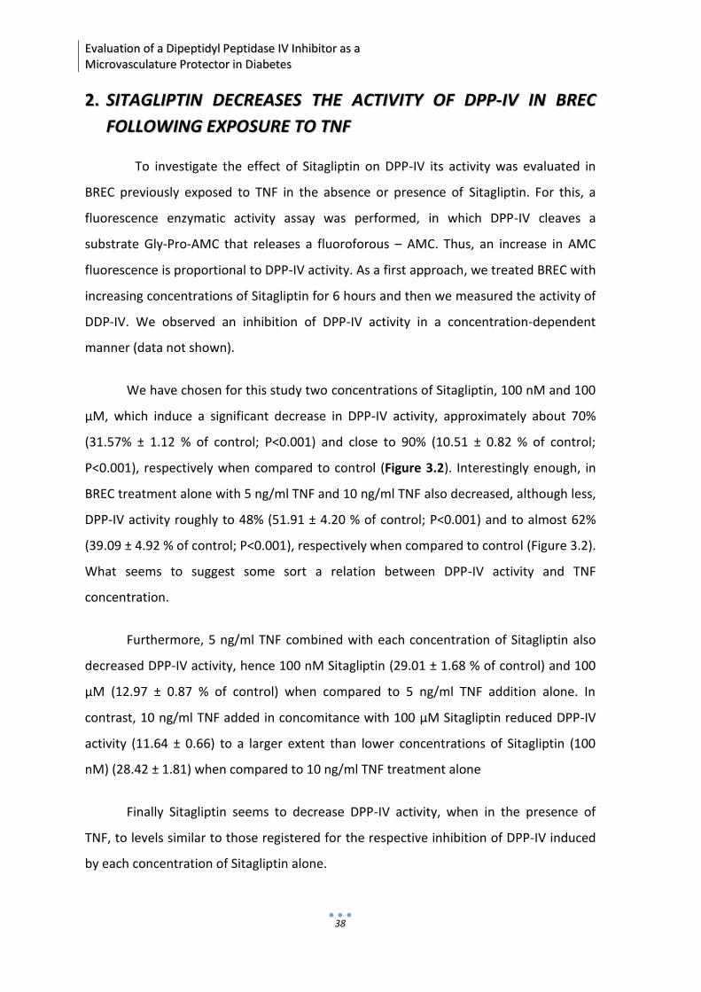
22.. SSIITTAAGGLLIIPPTTIINN DDEECCRREEAASSEESS TTHHEE AACCTTIIVVIITTYY OOFF DDPPPP--IIVV IINN BBRREECC
FFOOLLLLOOWWIINNGG EEXXPPOOSSUURREE TTOO TTNNFF
To investigate the effect of Sitagliptin on DPP-IV its activity was evaluated in
BREC previously exposed to TNF in the absence or presence of Sitagliptin. For this, a
fluorescence enzymatic activity assay was performed, in which DPP-IV cleaves a
substrate Gly-Pro-AMC that releases a fluoroforous – AMC. Thus, an increase in AMC
fluorescence is proportional to DPP-IV activity. As a first approach, we treated BREC with
increasing concentrations of Sitagliptin for 6 hours and then we measured the activity of
DDP-IV. We observed an inhibition of DPP-IV activity in a concentration-dependent
manner (data not shown).
We have chosen for this study two concentrations of Sitagliptin, 100 nM and 100
µM, which induce a significant decrease in DPP-IV activity, approximately about 70%
(31.57% ± 1.12 % of control; P<0.001) and close to 90% (10.51 ± 0.82 % of control;
P<0.001), respectively when compared to control (Figure 3.2). Interestingly enough, in
BREC treatment alone with 5 ng/ml TNF and 10 ng/ml TNF also decreased, although less,
DPP-IV activity roughly to 48% (51.91 ± 4.20 % of control; P<0.001) and to almost 62%
(39.09 ± 4.92 % of control; P<0.001), respectively when compared to control (Figure 3.2).
What seems to suggest some sort a relation between DPP-IV activity and TNF
concentration.
Furthermore, 5 ng/ml TNF combined with each concentration of Sitagliptin also
decreased DPP-IV activity, hence 100 nM Sitagliptin (29.01 ± 1.68 % of control) and 100
µM (12.97 ± 0.87 % of control) when compared to 5 ng/ml TNF addition alone. In
contrast, 10 ng/ml TNF added in concomitance with 100 µM Sitagliptin reduced DPP-IV
activity (11.64 ± 0.66) to a larger extent than lower concentrations of Sitagliptin (100
nM) (28.42 ± 1.81) when compared to 10 ng/ml TNF treatment alone
Finally Sitagliptin seems to decrease DPP-IV activity, when in the presence of
TNF, to levels similar to those registered for the respective inhibition of DPP-IV induced
by each concentration of Sitagliptin alone.
CCHHAAPPTTEERR 33
RReessuullttss
39
33.. EEFFFFEECCTT OOFF SSIITTAAGGLLIIPPTTIINN IINN EENNDDOOTTHHEELLIIAALL FFUUNNCCTTIIOONN
FFOOLLLLOOWWIINNGG IINNFFLLAAMMMMAATTIIOONN
Recent data from our laboratory has shown that sitagliptin prevented the
increase in blood-retinal barrier permeability induced by diabetes and exerted
protective effects against inflammation and pro-apoptotic state in the retina of diabetic
rats, by a mechanism independent of glycemia normalization (Gonçalves et al., 2012). In
this work, to evaluate the potential role of DPP-IV activity in endothelial cells response
to inflammation, neovasculatization, endothelial cell proliferation, migration and
survival, in inflammation conditions were evaluated by using the DPP-IV inhibitor,
Sitagliptin.
To investigate whether inhibition of DPP-IV protects endothelial function
following an inflammatory stimulus, endothelial cells ability to form tube-like structures
was evaluated using an angiogenesis assay.
FFiigguurree 33..22 - Sitagliptin decreases the activity in BREC treated with TNF. BREC were incubated with TNF (5 or 10 ng/ml) in the absence or presence of sitagliptin (100 nM or 100 μM) for 6h. DPP-IV activity was determined using the fluorogenic substrate Gly-Pro-AMC. Data are presented as percentage of control and represent the mean ± SEM of triplicates from four independent experiments. ***P<0.001 vs. control; #P<0.05, ###P<0.001 vs. TNF 5 ng/ml; δδδP<0.001 vs. TNF 10 ng/ml; ANOVA followed by Bonferroni’s post hoc test.

EEvvaalluuaattiioonn ooff aa DDiippeeppttiiddyyll PPeeppttiiddaassee IIVV IInnhhiibbiittoorr aass aa
MMiiccrroovvaassccuullaattuurree PPrrootteeccttoorr iinn DDiiaabbeetteess
40
The results analyzed suggest that in control condition the formed tube-like
structures seem quite organized. After 6 hours of treatment with 5 or 10 ng/ml TNF, a
loss of endothelial cells organization in tubular structures was observed (Figure 3.3).
Treatment with 100 nM sitagliptin in combination with 5 or 10 ng/ml TNF was able to
prevent this effect (Figure 3.3). Similarly, treatment with 100 µM sitagliptin, was also
able to prevent the tube-like structures disorganization, but on the other hand
treatment with Sitagliptin 100 µM and TNF 10 ng/ml seems not to exert the same
effects under inflammatory conditions, since the structures are more brittle and less
arranged (Figure 3.3).
Since our data above suggest that inhibition of DPP-IV may positively regulate
endothelial cell function under inflammation, we next evaluated migration of
endothelial cells, which is an important component of the cascade of events of the
angiogenesis process. To evaluate endothelial cell migration, we performed a scratch-
wound assay, by evaluating migration of endothelial cells into the denuded area.
FFiigguurree 33..33 – Effect of DDP-4 inhibition on angiogenesis upon TNF treatment. BREC were incubated with TNF (5 or 10 ng/ml) in the absence or presence of sitagliptin (100 nM or 100 μM) for 6h. Representative images, from each condition, showing endothelial cells grown on matrigel matrix forming capillary network structures. Duplicates from one independent experiment. Magnification 100x.

CCHHAAPPTTEERR 33
RReessuullttss
41
By analyzing both images (Figure 3.4 A and B) we can observe that 5 ng/ml TNF
can decrease BREC migration when compared to control from (32.74 ± 1.42 % from t0) to
(23.31 ± 1.54 % from t0; P<0.05). More so the highest concentration used of TNF (10
ng/ml) seems to impair to a larger extent and more significantly BREC migration from
(32.74 ± 1.42 % from t0) to (19.10 ± 1.28 % from t0; P<0.001) when compared to control.
Furthermore this impairment induced by both concentrations of TNF seems to be
alleviated in the presence of 100 nM Sitagliptin, from (23.31 ± 1.54 % from t0; P<0.05) to
(40.51 ± 1.62 % from t0; P<0.001) when compared to 5 ng/ml TNF alone and from (19.10
± 1.28 % from t0; P<0.001) to (37.45 ± 1.45 % from t0; P<0.001) when compared to 10
ng/ml TNF alone (Figure 3.4 A and B).
In contrast the highest concentration of Sitagliptin used (100 µM) does not seem
to protect BREC from 10 ng/ml TNF-induced impairment, but in co-treatment with 5
ng/ml TNF a increase is registered from (23.31 ± 1.54 % from t0; P<0.05) to (32.88 ± 1.62
% from t0; P<0.05) when compared to 5 ng/ml TNF alone (Figure 3.4 A and B).
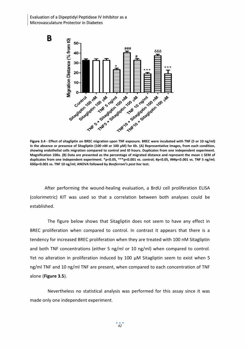
EEvvaalluuaattiioonn ooff aa DDiippeeppttiiddyyll PPeeppttiiddaassee IIVV IInnhhiibbiittoorr aass aa
MMiiccrroovvaassccuullaattuurree PPrrootteeccttoorr iinn DDiiaabbeetteess
42
After performing the wound-healing evaluation, a BrdU cell proliferation ELISA
(colorimetric) KIT was used so that a correlation between both analyses could be
established.
The figure below shows that Sitagliptin does not seem to have any effect in
BREC proliferation when compared to control. In contrast it appears that there is a
tendency for increased BREC proliferation when they are treated with 100 nM Sitagliptin
and both TNF concentrations (either 5 ng/ml or 10 ng/ml) when compared to control.
Yet no alteration in proliferation induced by 100 µM Sitagliptin seem to exist when 5
ng/ml TNF and 10 ng/ml TNF are present, when compared to each concentration of TNF
alone (Figure 3.5).
Nevertheless no statistical analysis was performed for this assay since it was
made only one independent experiment.
FFiigguurree 33..44 - Effect of sitagliptin on BREC migration upon TNF exposure. BREC were incubated with TNF (5 or 10 ng/ml) in the absence or presence of Sitagliptin (100 nM or 100 μM) for 6h. (A) Representative images, from each condition, showing endothelial cells migration compared to control and t0 hours. Duplicates from one independent experiment. Magnification 100x. (B) Data are presented as the percentage of migrated distance and represent the mean ± SEM of duplicates from one independent experiment. *p<0.05, ***p<0.001 vs. control; #p<0.05, ###p<0.001 vs. TNF 5 ng/ml; δδδp<0.001 vs. TNF 10 ng/ml; ANOVA followed by Bonferroni’s post hoc test.

CCHHAAPPTTEERR 33
RReessuullttss
43
After confirming that Sitagliptin exerts protective effects in BREC migration,
proliferation and formation in tube-like structures, a cell viability assay was performed
to support these results.
Primary cultures of BREC were treated during a 6 hour period with two different
concentrations of Sitagliptin (100nM or 100µM) and TNF (5 or 10 ng/ml). After this
period the cell viability was evaluated by MTT colorimetric assay, in which this the
yellow-colored MTT is reduced, by living cells mitochondrial dehydrogenases, to a sort of
purplish-colored formazan precipitate. The absorption of dissolved formazan can be
then correlated with living cells number, hence, being expressed by the percentage of
cell viability in BREC cells. No significant cytotoxicity was registered when BREC were
treated with TNF in the absence or presence of Sitagliptin (Figure 3.6).However, in the
positive control condition, where the cells were exposed to hydrogen peroxide (H2O2)
for 1 hour, an increased cytotoxicity was observed (45.43 ± 6.54 % of control; P <0.001)
when compared to control (Figure 3.6).
FFiigguurree 33..55 - Effect of sitagliptin on BREC proliferation upon exposure to TNF. BREC were incubated with TNF (5 or 10 ng/ml) in the absence or presence of sitagliptin (100 nM or 100 μM) for 6h. Cell proliferation was assessed by BrdU incorporation. Data are presented as percentage of control and represent the mean ± SEM of triplicates from one independent experiment.

EEvvaalluuaattiioonn ooff aa DDiippeeppttiiddyyll PPeeppttiiddaassee IIVV IInnhhiibbiittoorr aass aa
MMiiccrroovvaassccuullaattuurree PPrrootteeccttoorr iinn DDiiaabbeetteess
44
Contr
ol
Sitaglip
tin 1
00 n
M M
Sitaglip
tin 1
00
TNF 5
ng/m
l
TNF 5
ng/m
l + S
itaglip
tin n
M M
TNF 5
ng/m
l + S
itaglip
tin
TNF 1
0 ng/m
l
TNF 1
0 ng/m
l + S
itaglip
tin n
M M
TNF 1
0 ng/m
l + S
itaglip
tin
2O2H
0
50
100
150
***C
ell
via
bil
ity
(% o
f co
ntr
ol)
Theoretically, in the MTT assay, formazan dye color intensity is correlated with
the number of viable cells. However, this assay is dependent of cellular metabolism to
reduce formazan, so in order to confirm the obtained results, additional studies using
the propidium iodide (PI) were performed. Since PI is excluded from live cells intact
membrane and it is retained in dead cells, it can serve as a complementary assay to
MTT.
Furthermore, PI was added at the end of the co-treatments with two different
concentrations of Sitagliptin (100nM e 100µM) and TNF (5 and 10 ng/ml). Qualitative
evaluation of the images was performed by Fluorescence Microscopy.
The representative images below show no cytotoxicity induced either by
Sitagliptin and/or TNF. In addition, a positive control was performed, where the cells
were exposed to 5mM hydrogen peroxide (H2O2) for ten minutes and increased
cytotoxicity was observed due to total positive staining for PI. (Figure 3.7).
FFiigguurree 33..66 – Effecct of sitagliptin on BREC viability after incubation with TNF. BREC were incubated with TNF (5 and 10 ng/ml) and sitagliptin 100 nM or 100 μM for 6h. Cell viability was assessed by MTT assay. Data are presented as percentage of control and represent the mean ± SEM of triplicates from two independent experiments. ***p<0.001

CCHHAAPPTTEERR 33
RReessuullttss
45
FFiigguurree 33..77 - BREC were incubated with TNF (5 or 10 ng/ml) and/or sitagliptin (100 nM or 100 μM) for 6h. Representative images, from each condition, showing cell stained with PI. Duplicates from one independent experiment. Magnification 100x


CCHHAAPPTTEERR 44 DDIISSCCUUSSSSIIOONN

CCHHAAPPTTEERR 44
DDiissccuussssiioonn
49
Diabetic Retinopathy (DR) is one of the most common microvascular
complications in diabetic patients and it is the major cause of blindness in the working-
age population of most developed countries. It is now widely accepted that the
complications of diabetes, including DR, are related to elevation of the plasma glucose
concentration (American Diabetes Association, Implications of the diabetes control and
complications trial 2003; 23: S26-S27).It is also known that lowering blood glucose
concentration significantly decreases the risk of DR development (Kowluru & Chan
2007). Therefore, there is an urgent need to implement novel and more effective
therapeutic strategies.
Several reports have shown that a low-grade inflammation is implicated in the
pathogenesis of DR (Kowluru et al., 2004). Elevated levels of proinflammatory
cytokines have also been detected both in the vitreous of diabetic patients with DR
(Kowluru et al., 2004) and in diabetic retinas (Lutty et al., 1997; Joussen et al., 2001). In
fact, DR shares numerous characteristics of chronic inflammatory disease, such as,
increased vascular permeability, edema, infiltration of inflammatory cells, presence of
cytokines, tissue damage and neovascularization (Antonetti et al., 2006; Kowluru et al.,
2004).
Recently, a new class of antihyperglycemic agents, in which is included
Sitagliptin, have been used for the treatment of Type 2 Diabetes (T2D). Sitagliptin has
been shown to improve glycemic control by enhancing the incretin levels of active
incretin hormones, like GLP-1, and consequently increasing the insulin secretion levels
in humans with T2D (Drucker et al., 2006).
Moreover, it has also been shown that this DPP-IV inhibitor increases
circulating progenitor endothelial cells in diabetic patients with T2D, which may have
an important role in the repair of the damaged vessels (Fadini et al., 2010). Recently,
our group has shown that administration of Sitagliptin to diabetic animals improved
microvascular permeability and ameliorated the detrimental changes associated with
retinal dysfunction. In fact, sitaglitin administration to T1DM and T2DM animal models
reduced tissue inflammation, improving the functionality of the inner blood-retinal
barrier. Nevertheless, the putative role of Sitagliptin in protecting retinal endothelial
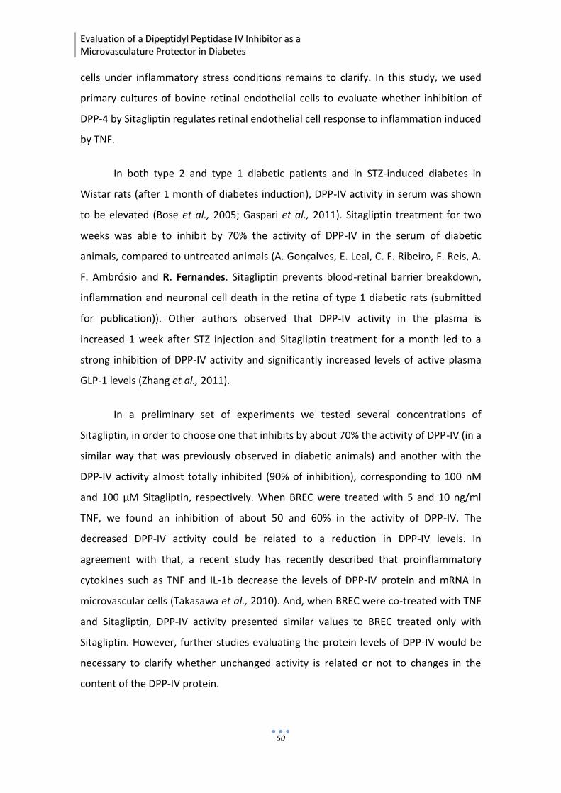
EEvvaalluuaattiioonn ooff aa DDiippeeppttiiddyyll PPeeppttiiddaassee IIVV IInnhhiibbiittoorr aass aa
MMiiccrroovvaassccuullaattuurree PPrrootteeccttoorr iinn DDiiaabbeetteess
50
cells under inflammatory stress conditions remains to clarify. In this study, we used
primary cultures of bovine retinal endothelial cells to evaluate whether inhibition of
DPP-4 by Sitagliptin regulates retinal endothelial cell response to inflammation induced
by TNF.
In both type 2 and type 1 diabetic patients and in STZ-induced diabetes in
Wistar rats (after 1 month of diabetes induction), DPP-IV activity in serum was shown
to be elevated (Bose et al., 2005; Gaspari et al., 2011). Sitagliptin treatment for two
weeks was able to inhibit by 70% the activity of DPP-IV in the serum of diabetic
animals, compared to untreated animals (A. Gonçalves, E. Leal, C. F. Ribeiro, F. Reis, A.
F. Ambrósio and R. Fernandes. Sitagliptin prevents blood-retinal barrier breakdown,
inflammation and neuronal cell death in the retina of type 1 diabetic rats (submitted
for publication)). Other authors observed that DPP-IV activity in the plasma is
increased 1 week after STZ injection and Sitagliptin treatment for a month led to a
strong inhibition of DPP-IV activity and significantly increased levels of active plasma
GLP-1 levels (Zhang et al., 2011).
In a preliminary set of experiments we tested several concentrations of
Sitagliptin, in order to choose one that inhibits by about 70% the activity of DPP-IV (in a
similar way that was previously observed in diabetic animals) and another with the
DPP-IV activity almost totally inhibited (90% of inhibition), corresponding to 100 nM
and 100 µM Sitagliptin, respectively. When BREC were treated with 5 and 10 ng/ml
TNF, we found an inhibition of about 50 and 60% in the activity of DPP-IV. The
decreased DPP-IV activity could be related to a reduction in DPP-IV levels. In
agreement with that, a recent study has recently described that proinflammatory
cytokines such as TNF and IL-1b decrease the levels of DPP-IV protein and mRNA in
microvascular cells (Takasawa et al., 2010). And, when BREC were co-treated with TNF
and Sitagliptin, DPP-IV activity presented similar values to BREC treated only with
Sitagliptin. However, further studies evaluating the protein levels of DPP-IV would be
necessary to clarify whether unchanged activity is related or not to changes in the
content of the DPP-IV protein.
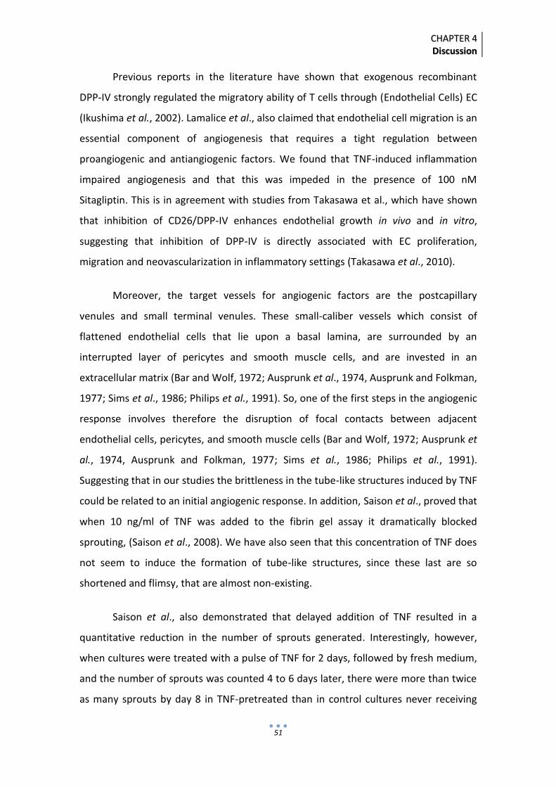
CCHHAAPPTTEERR 44
DDiissccuussssiioonn
51
Previous reports in the literature have shown that exogenous recombinant
DPP-IV strongly regulated the migratory ability of T cells through (Endothelial Cells) EC
(Ikushima et al., 2002). Lamalice et al., also claimed that endothelial cell migration is an
essential component of angiogenesis that requires a tight regulation between
proangiogenic and antiangiogenic factors. We found that TNF-induced inflammation
impaired angiogenesis and that this was impeded in the presence of 100 nM
Sitagliptin. This is in agreement with studies from Takasawa et al., which have shown
that inhibition of CD26/DPP-IV enhances endothelial growth in vivo and in vitro,
suggesting that inhibition of DPP-IV is directly associated with EC proliferation,
migration and neovascularization in inflammatory settings (Takasawa et al., 2010).
Moreover, the target vessels for angiogenic factors are the postcapillary
venules and small terminal venules. These small-caliber vessels which consist of
flattened endothelial cells that lie upon a basal lamina, are surrounded by an
interrupted layer of pericytes and smooth muscle cells, and are invested in an
extracellular matrix (Bar and Wolf, 1972; Ausprunk et al., 1974, Ausprunk and Folkman,
1977; Sims et al., 1986; Philips et al., 1991). So, one of the first steps in the angiogenic
response involves therefore the disruption of focal contacts between adjacent
endothelial cells, pericytes, and smooth muscle cells (Bar and Wolf, 1972; Ausprunk et
al., 1974, Ausprunk and Folkman, 1977; Sims et al., 1986; Philips et al., 1991).
Suggesting that in our studies the brittleness in the tube-like structures induced by TNF
could be related to an initial angiogenic response. In addition, Saison et al., proved that
when 10 ng/ml of TNF was added to the fibrin gel assay it dramatically blocked
sprouting, (Saison et al., 2008). We have also seen that this concentration of TNF does
not seem to induce the formation of tube-like structures, since these last are so
shortened and flimsy, that are almost non-existing.
Saison et al., also demonstrated that delayed addition of TNF resulted in a
quantitative reduction in the number of sprouts generated. Interestingly, however,
when cultures were treated with a pulse of TNF for 2 days, followed by fresh medium,
and the number of sprouts was counted 4 to 6 days later, there were more than twice
as many sprouts by day 8 in TNF-pretreated than in control cultures never receiving
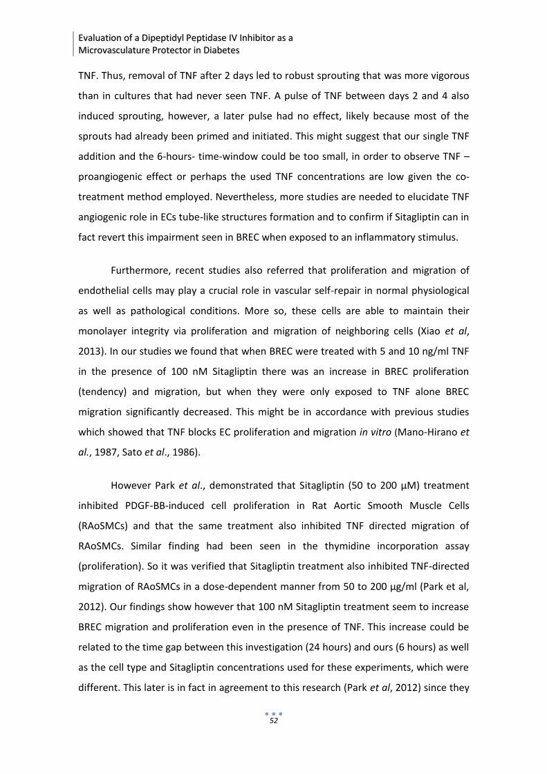
EEvvaalluuaattiioonn ooff aa DDiippeeppttiiddyyll PPeeppttiiddaassee IIVV IInnhhiibbiittoorr aass aa
MMiiccrroovvaassccuullaattuurree PPrrootteeccttoorr iinn DDiiaabbeetteess
52
TNF. Thus, removal of TNF after 2 days led to robust sprouting that was more vigorous
than in cultures that had never seen TNF. A pulse of TNF between days 2 and 4 also
induced sprouting, however, a later pulse had no effect, likely because most of the
sprouts had already been primed and initiated. This might suggest that our single TNF
addition and the 6-hours- time-window could be too small, in order to observe TNF –
proangiogenic effect or perhaps the used TNF concentrations are low given the co-
treatment method employed. Nevertheless, more studies are needed to elucidate TNF
angiogenic role in ECs tube-like structures formation and to confirm if Sitagliptin can in
fact revert this impairment seen in BREC when exposed to an inflammatory stimulus.
Furthermore, recent studies also referred that proliferation and migration of
endothelial cells may play a crucial role in vascular self-repair in normal physiological
as well as pathological conditions. More so, these cells are able to maintain their
monolayer integrity via proliferation and migration of neighboring cells (Xiao et al,
2013). In our studies we found that when BREC were treated with 5 and 10 ng/ml TNF
in the presence of 100 nM Sitagliptin there was an increase in BREC proliferation
(tendency) and migration, but when they were only exposed to TNF alone BREC
migration significantly decreased. This might be in accordance with previous studies
which showed that TNF blocks EC proliferation and migration in vitro (Mano-Hirano et
al., 1987, Sato et al., 1986).
However Park et al., demonstrated that Sitagliptin (50 to 200 µM) treatment
inhibited PDGF-BB-induced cell proliferation in Rat Aortic Smooth Muscle Cells
(RAoSMCs) and that the same treatment also inhibited TNF directed migration of
RAoSMCs. Similar finding had been seen in the thymidine incorporation assay
(proliferation). So it was verified that Sitagliptin treatment also inhibited TNF-directed
migration of RAoSMCs in a dose-dependent manner from 50 to 200 µg/ml (Park et al,
2012). Our findings show however that 100 nM Sitagliptin treatment seem to increase
BREC migration and proliferation even in the presence of TNF. This increase could be
related to the time gap between this investigation (24 hours) and ours (6 hours) as well
as the cell type and Sitagliptin concentrations used for these experiments, which were
different. This later is in fact in agreement to this research (Park et al, 2012) since they
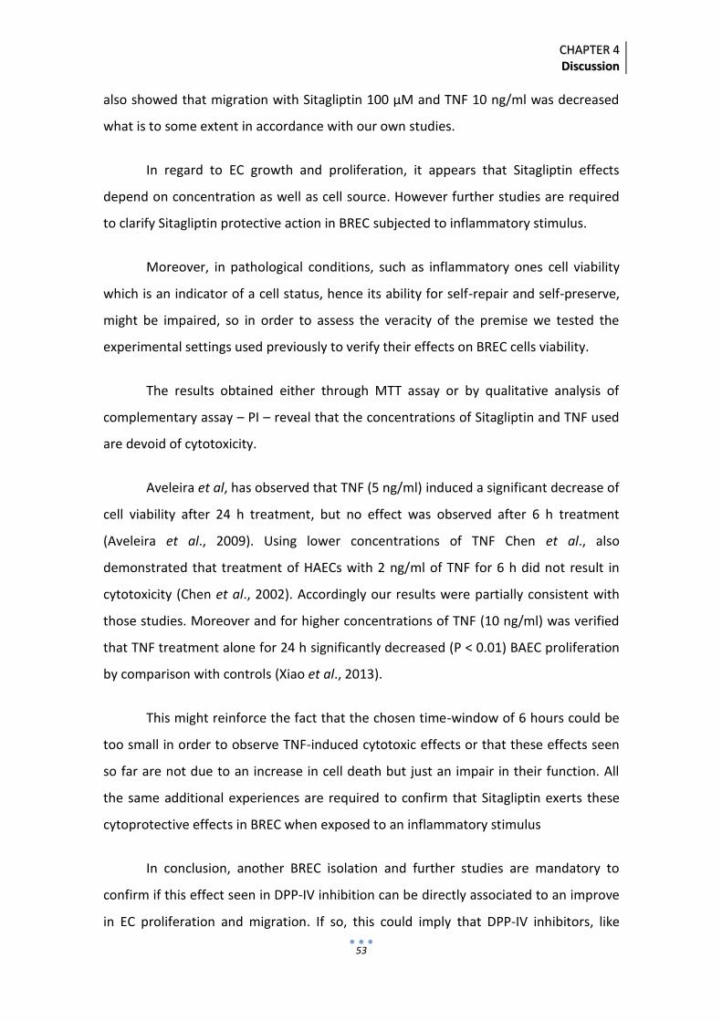
CCHHAAPPTTEERR 44
DDiissccuussssiioonn
53
also showed that migration with Sitagliptin 100 µM and TNF 10 ng/ml was decreased
what is to some extent in accordance with our own studies.
In regard to EC growth and proliferation, it appears that Sitagliptin effects
depend on concentration as well as cell source. However further studies are required
to clarify Sitagliptin protective action in BREC subjected to inflammatory stimulus.
Moreover, in pathological conditions, such as inflammatory ones cell viability
which is an indicator of a cell status, hence its ability for self-repair and self-preserve,
might be impaired, so in order to assess the veracity of the premise we tested the
experimental settings used previously to verify their effects on BREC cells viability.
The results obtained either through MTT assay or by qualitative analysis of
complementary assay – PI – reveal that the concentrations of Sitagliptin and TNF used
are devoid of cytotoxicity.
Aveleira et al, has observed that TNF (5 ng/ml) induced a significant decrease of
cell viability after 24 h treatment, but no effect was observed after 6 h treatment
(Aveleira et al., 2009). Using lower concentrations of TNF Chen et al., also
demonstrated that treatment of HAECs with 2 ng/ml of TNF for 6 h did not result in
cytotoxicity (Chen et al., 2002). Accordingly our results were partially consistent with
those studies. Moreover and for higher concentrations of TNF (10 ng/ml) was verified
that TNF treatment alone for 24 h significantly decreased (P < 0.01) BAEC proliferation
by comparison with controls (Xiao et al., 2013).
This might reinforce the fact that the chosen time-window of 6 hours could be
too small in order to observe TNF-induced cytotoxic effects or that these effects seen
so far are not due to an increase in cell death but just an impair in their function. All
the same additional experiences are required to confirm that Sitagliptin exerts these
cytoprotective effects in BREC when exposed to an inflammatory stimulus
In conclusion, another BREC isolation and further studies are mandatory to
confirm if this effect seen in DPP-IV inhibition can be directly associated to an improve
in EC proliferation and migration. If so, this could imply that DPP-IV inhibitors, like

EEvvaalluuaattiioonn ooff aa DDiippeeppttiiddyyll PPeeppttiiddaassee IIVV IInnhhiibbiittoorr aass aa
MMiiccrroovvaassccuullaattuurree PPrrootteeccttoorr iinn DDiiaabbeetteess
54
Sitagliptin, which are widely used in the treatment of diabetes, may be of potential use
in treating diabetic vascular complications.

CCHHAAPPTTEERR 55 CCOONNCCLLUUSSIIOONN AANNDD
FFUUTTUURREE PPEERRSSPPEECCTTIIVVEESS


CCHHAAPPTTEERR 55
CCoonncclluussiioonn aanndd FFuuttuurree PPeerrssppeeccttiivveess
57
In this thesis we intended to evaluate the potential role of Sitagliptin in
protecting the endothelial cells of the retina under inflammatory stress conditions. So
in order to study the effect of Sitagliptin in the microvasculature of the retina, when an
inflammatory state-TNF-induced is present, were used primary culture of Bovine
Retinal Endothelial Cells (BREC). The fact that much is already known about the
development and pathophysiology of the retinal microvasculature makes this capillary
bed particularly well suited for in vitro studies (Bowman et al, 1982)
Therefore a series of assays were performed on these cells together with 6
hours in co-treatment with TNF and Sitagliptin. Hence endothelial cells ability to form
tube-like structures, migrate and proliferate were analyzed and the results obtained
suggest that Sitagliptin has protective and regenerative effects in these cells when
exposed to an inflammatory-TNF-induced state. More so therapeutic benefits of
Sitagliptin are consequently reinforced by these studies but TNF’s endothelial cells
impairment ability was not yet fully understood.
Nonetheless it is still important to refer that TNF role in angiogenesis continues
to be highly controversial with numerous studies showing that it is either
proangiogenic or antiangiogenic, with the preponderance of published studies
suggesting that TNF blocks EC activation in vitro, but enhances angiogenesis in vivo
(Patterson C et al., 1996, Frater-Schroder M et al., 1987, Mano-Hirano Y et al., 1987,
Sato N et al, 1986, Leibovich SJ et al., 1987).
Therefore to confirm the scientific veracity of our findings it is imperative to do
another BREC cell culture isolation in which all the assays performed throughout this
thesis will be redone and revised, comparing afterwards these future results with the
former ones.

58

59
RREEFFFFEERREENNCCEESS
Abbot, S. E., Kaul, A., Stevens, C. R. and Blake, D. R. (1992). Isolation and culture of synovial
microvascular endothelial cells. Arthritis Rheum. 35, 401-406
Adamiec-Mroczek J, Oficjalska-Mlynczak J.(2008) Assessment of selected adhesion
molecule and proinflammatory cytokine levels in the vitreous body of patients with type 2
diabetes--role of the inflammatory-immune process in the pathogenesis of proliferative
diabetic retinopathy. Graefes Arch Clin Exp Ophthalmol. 2008;246:1665–70.
Adamis AP, Berman AJ. (2008) Immunological mechanisms in the pathogenesis of diabetic
retinopathy. Semin Immunopathol 2008;30:65-84.
Adamis AP. Is diabetic retinopathy an inflammatory disease? (2002) Br J Ophthalmol
2002;86:363-5
Ahmed AM, (2002). History of diabetes mellitus, 2002 Apr, Saudi Med J., 2002
Apr;23(4):373-8.
Aiello LP, Gardner TW, King GL, Blankenship G, Cavallerano JD, Ferris FL, 3rd, et al. (1998)
Diabetic retinopathy. Diabetes Care. 1998;21:143–56
Antonetti DA, Barber AJ, Bronson SK, Freeman WM, Gardner TW, Jefferson LS, et al. (2006)
Diabetic retinopathy: seeing beyond glucose-induced microvascular disease. Diabetes
2006;55:2401-11
Antonetti David A., Ph.D., Klein Ronald, M.D., M.P.H., and GardneThomas W. r, M.D, (2012)
Diabetic Retinopathy Mechanisms of Disease, N Engl J Med, 366:1227-1239, DOI:
10.1056/NEJMra1005073
Ausprunk DH, Falterman K, Folkman J (1978). The sequence of events in the regression of
corneal capillaries. Lab Invest 38:284-296
Ausprunk DH, Falterman K, Folkman J (1978). The sequence of events in the regression of
corneal capillaries. Lab Invest 38:284-296

60
Ausprunk DH, Folkman J (1977). Migration and proliferation of endothelial cells in
preformed and newly formed blood vessels during tumor angiogenesis. Microvasc R^s
14:53-65.
Ausprunk DH, Knighton DR, Folkman I (1974). Differentiation of vascular endothelium in
the chick chorioallantois: a structural and autoradiographic study. Dev Biol 38:237-248.
Aveleira CA, Lin CM, Abcouwer SF, Ambrosio AF, Antonetti DA. (2010) TNF-alpha signals
through PKCzeta/NF-kappaB to alter the tight junction complex and increase retinal
endothelial cell permeability. Diabetes. 2010;59:2872–82.
Bar TH, Wolf JR (1972). The formation of capillary basement membrane during internal
vascularization of the rat's cerebral cortex. Z Zellforsch 133:231-248
Barber AJ, Nakamura M, Wolpert EB, Reiter CE, Seigel GM, Antonetti DA, et al.(2001)
Insulin rescues retinal neurons from apoptosis by a phosphatidylinositol 3-kinase/Akt-
mediated mechanism that reduces the activation of caspase-3. J Biol Chem.
2001;276(35):32814-21
Barouch FC, Miyamoto K, Allport JR, Fujita K, Bursell SE, Aiello LP, et al.(2004) Integrin-
mediated neutrophil adhesion and retinal leukostasis in diabetes. Invest Ophthalmol Vis
Sci. 2000;41:1153–8.
Barreiro O, Martin P, Gonzalez-Amaro R, Sanchez-Madrid F. (2010) Molecular cues guiding
inflammatory responses. Cardiovasc Res. 2010;86:174–82.
Betterle C, Zanette F, Pedini B, Presotto F, Rapp LB, Monsciotti CM et al.(1983) Clinical and
subclinical organ-specific autoimmune manifestations in type 1 (insulin-dependent)
diabetic patients and their first-degree relatives. Diabetologia 1983; 26: 431-36.
Bose AK, Mocanu MM, Carr RD, Brand CL, Yellon DM (2005) Glucagon-like peptide 1 can
directly protect the heart against ischemia/reperfusion injury. Diabetes 54:146-151, 2005
Bowman Phillip D., A. Lorris Betz and Gary W. Goldstein, (1982) In Vitro Primary Culture of
Microvascular Endothelial Cells from Bovine Retina: Selective Growth Using Fibronectin
Coated Substrate and Plasma Derived Serum Society for In Vitro Biology, Vol. 18, No. 7, pp.
626-632
61
Bowman Phillip D., A. Lorris Betz and Gary W. Goldstein, (1982) In Vitro primary Culture of
Microvascular Endothelial Cells from Bovine Retina: Selective Growth Using Fibronectin
Coated Substrate and Plasma Derived Serum Society for In Vitro Biology, Vol. 18, No. 7 ,
pp. 626-632
Brownlee M (1995): Advanced protein glycosylation in diabetes and aging. Annu Rev Med
46 : 223 –234,1995
Brownlee M (2001): Biochemistry and molecular cell biology of diabetic complications.
Nature 414 : 813 –820,2001
Brownlee M, Cerami A (1981) The biochemistry of the complications of diabetes mellitus.
Annu Rev Biochem 50 : 385 –432,1981
Caicedo A, Espinosa-Heidmann DG, Piña Y, Hernandez EP, Cousins SW.(2005) Blood-
derived macrophages infiltrate the retina and activate Muller glial cells under experimental
choroidal neovascularization. Exp Eye Res. 2005; 81(1):38-47
Calara F, Taylor K, Han J, Zabala E, Carr EM, Wintle M, Fineman M.(2005) A randomized,
open-label, crossover study examining the effect of injection site on bioavailability of
exenatide (synthetic exendin-4). Clin Ther 2005; 27: 210– 215
Carmeliet P.(2005) Angiogenesis in life, disease and medicine. Nature 2005;438:932-936.
Chai J, Jones MK, Tarnawski AS. (2004) Serum response factor is a critical requirement for
VEGF signaling in endothelial cel ls and VEGF-induced angiogenesis. Faseb J. 2004;18:1264-
1266.
Chen L, Yang P, Kijlstra A. (2002) Distribution, markers, and functions of retinal microglia.
Ocul Immunol Inflamm. 2002;10:27–39.
Chen Y.H., Lin S.J., Chen J.W., Ku H.H., Chen Y.L. (2002) Magnolol attenuates VCAM-1
expression in vitro in TNF-α-treated human aortic endothelial cells and in vivo in the aorta
of cholesterol-fed rabbits, British Journal of Pharmacology 135,37 -47
Cockburn M., (1999) Diabetic Retinopathy:classification, description and optometric
management, Clinical and Experimental Optometry
62
Cordero O.J., (2009) On the origin of serum CD26 and its altered concentration in cancer
patients:; Cancer Immunol. Immunother. 58, 1723
Crutchfield A, Diller K, Brand J (1999). "Cryopreservation of Chlamydomonas reinhardtii
(Chlorophyta)". European Journal of Phycology 34 (1): 43–52.
doi:10.1080/09670269910001736072
Cunha-Vaz J (2007) Characterization and relevance of different diabetic retinopathy
phenotypes. Dev Ophthalmol 39: 13–30.
Cunha-Vaz J., Bernardes R., Lobo C. (2011) Blood-retinal barrier Eur J Ophthalmol, 2011, 21
(Suppl. 6): S3-S9.
Das Evcimen, N., & King , G. L. (2007) The role of protein kinase C activation and the
vascular complications of Diabetes. Pharmacol Res 55: 498-510
Davidson JA, Ciulla TA, McGill JB, Kles KA, Anderson PW. (2007) How the diabetic eye loses
vision. Endocrine. 2007;32:107–16.
Davison, P. M., Bensch, K. and Karasek, M. A. (1980). Isolation and growth of endothelial
cells from the microvessels of the newborn human foreskin in cell culture. J. Invest. Derm.
75, 316-321.
Davison, P. M., Bensch, K. and Karasek, M. A. (1983). Isolation and long-term serial
cultivation of endothelial cells from the microvessels of the adult human dermis. In Vitro
19, 937-945.
de Courten M, Bennett PH, Tuomilehto J, Zimmet P.(1997) Epidemiology of NIDDM in Non-
Europids. In: Alberti KGMM, Zimmet P, DeFronzo RA, eds. International Textbook of
Diabetes Mellitus. 2nd edn. Chichester: John Wiley, 1997: pp 143-70.
Décio L. Eizirik, Maikel L. Colli, (2009), The role of inflammation in insulitis and β-cell loss in
type 1 diabetes, Nature Reviews Endocrinology 5, 219-226
Demircan N, Safran BG, Soylu M, Ozcan AA, Sizmaz S. (2006) Determination of vitreous
interleukin-1 (IL-1) and tumour necrosis factor (TNF) levels in proliferative diabetic
retinopathy. Eye (Lond) 2006;20:1366–9.

63
Detmar, M., Tenorio, S., Hettmannsperger, U., Ruszczak, Z. and Orfanos, C. E. (1992).
Cytokine regulation of proliferation and ICAM-1 expression of human dermal
microvascular endothelium in vitro. J. Invest. Derm. 98, 147-153.
Diglio, C. A., Grammas, P., Giacomelli, F. and Wiener, J. (1982). Primary culture of rat
cerebral microvascualr endothelial cells. Lab. Invest. 46, 554-563.
Drucker DJ, Buse JB, Taylor K, Kendall DM, Trautmann M, Zhuang D, Porter L.: (2008)
Exenatide once weekly versus twice daily for the treatment of type 2 diabetes: a
randomised, open-label, non-inferiority study. Lancet 2008; 372: 1240– 1250.
Eizirik, D. L., Cardozo, A. K. and Cnop, M.(2008). The role for endoplasmic reticulum stress
in diabetes mellitus. Endocr. Rev. 29 , 42-61
Elbrønd B, Jakobsen G, Larsen S, Agersø H, Jensen LB, Rolan P, Sturis J, Hatorp V,
Zdravkovic M. (2002) Pharmacokinetics, pharmacodynamics, safety, and tolerability of a
single-dose of NN2211, a long-acting glucagon-like peptide 1 derivative, in healthy male
subjects. Diabetes Care 2002; 25: 1398– 1404.
Elrick, H., Stimmler S., JR, C. J. Hlad, Arai, Y, (1964), Plasma Insulin Response to Oral and
Intravenous Glucose Administration, JCEM
Fadini GP, Boscaro E, Albiero M et al. (2010) The oral dipeptidyl peptidase-4 inhibitor
sitagliptin increases circulating endothelial progenitor cells in patients with type 2
diabetes: possible role of stromal-derived factor- 1alpha. Diabetes Care 2010; 33: 1607–
1609.
Fernandes R., Carvalho A. L., Kumagai A., Seica R., Hosoya K.,Terasaki T., Murta J., Pereira
P, Faro C, (2004) Downregulation of retinal GLUT-1 in diabetes by ubiquitinylation. Mol Vis
10:618-628
Fischer F, Martin G, Agostini HT. (2011) Activation of retinal microglia rather than
microglial cell density correlates with retinal neovascularization in the mouse model of
oxygen-induced retinopathy. J Neuroinflammation. 2011;8:120
Folkman, J., Haudenschild, C. C. and Zetter, B. R. (1979). Long-term culture of capillary
endothelial cells. Proc. Nat. Acad. Sci. USA 76, 5217-5221.

64
Follkman, J. (1992). Capillary endothelial cells: The use of microcarrier beads for isolation
and long-term culture. J. Cell Biol. 95, 458a
Frater-Schroder M, Risau W, Hallmann R, Gautschi P, Bohlen P, (1987) Tumor necrosis
factor type alpha, a potent inhibitor of endothelial cell growth in vitro, is angiogenic in
vivo. Proc Natl Acad Sci U S A 1987;84:5277-5281.
Funatsu H, Yamashita H, Ikeda T, Mimura T, Eguchi S, Hori S. (2003) Vitreous levels of
interleukin-6 and vascular endothelial growth factor are related to diabetic macular
edema. Ophthalmology. 2003;110:1690–6.
Gariano RF, Gardner TW. (2005) Retinal angiogenesis in development and disease. Nature.
2005;438(7070):960-6
Gaspari T, Liu H, Welungoda I, Hu Y, Widdop RE, Knudsen LB, Simpson RW, Dear AE (2011)
A GLP-1 receptor agonist liraglutide inhibits endothelial cell dysfunction and vascular
adhesion molecule expression in an ApoE-/- mouse model. Diab Vasc Dis Res 8:117-124,
2011
Ghersi G., Chen W., Lee E.W., Zukowska Z., (2001) Critical role of dipeptidyl peptidase IV in
neuropeptide Y-mediated endothelial cell migration in response to wounding, Peptides 22
453–458.
Goncalves A, Leal E, Paiva A, Teixeira Lemos E, Teixeira F, Ribeiro CF, Reis F, Ambrosio AF,
Fernandes R: Protective effects of the dipeptidyl peptidase IV inhibitor sitagliptin in the
blood-retinal barrier in a type 2 diabetes animal model. Diabetes Obes Metab 14:454-463,
2012
Gutierrez A, Sanda T, Grebliunaite R, Carracedo A, Salmena L, Ahn Y, Dahlberg S, Neuberg
D, Moreau LA, Winter SS, Larson R, Zhang J, Protopopov A, Chin L, Pandolfi PP, Silverman
LB, Hunger SP, Sallan SE, Look AT. (2009) High frequency of PTEN, PI3K, and AKT
abnormalities in T-cell acute lymphoblastic leukemia. Blood. 2009;114:647–650
H. Ikushima, Y. Munakata, S. Iwata, K. Ohnuma, S. Kobayashi, N.H. Dang, C. Morimoto,
(2002) Soluble CD26/dipeptidyl peptidase IV enhances transendothelial migration via its
interaction with mannose 6-phosphate/insulin-like growth factor II receptor, Cell Immunol.
215 ,2002, 106–110.

65
Hama T. (1981) Changes in form of dipeptidyl-aminopeptidase IV in urine from patients
with renal disease:; Clin. Chim. Acta 113, 217
Harhaj NS, Felinski EA, Wolpert EB, Sundstrom JM, Gardner TW, Antonetti DA. (2006) VEGF
activation of protein kinase C stimulates occludin phosphorylation and contributes to
endothelial permeability. Invest Ophthalmol Vis Sci 2006;47:5106-15.
Harris MI, Cowie CC, Stern MP, Boyko ES, Reiber GE, Bennett PH, (1995) eds. Diabetes in
America. 2nd edn. Washington DC: US Government Printing Office, 1995 (NIH publ. No. 95-
1468).
Hawker, J.R.Jr. (2003) J.Immunol.Methods 274, 77-82
Hother-Nielsen O, Faber O, Sørensen NS, Beck-Nielsen H. (1998) Classification of newly
diagnosed diabetic patients as insulin-requiring or non-insulin-requiring based on clinical
and biochemical variables. Diabetes Care 1988; 11: 531-37.
http://diabetes.niddk.nih.gov/dm/pubs/control/
http://diabeworld.com/history.html#&panel1-1
http://miydiabetes.com/index.php/cmspage/6/a-little-history
http://probes.invitrogen.com/media/pis/mp01304.pdf
http://www.diabetes.org/
http://www.diabetesindia.com/diabetes/history_diabetes3.htm
http://www.dtu.ox.ac.uk/generic/publications.php?3#1995
http://www.genwaybio.c
om/elisas/dpp4-assay-kit
http://www.idf.org/
http://www.ihcworld.com/_protocols/general_ICC/introduction.htm

66
http://www.medicalnewstoday.com/info/diabetes/
http://www.nei.nih.gov/
http://www.piercenet.com/browse.cfm?fldID=01041204
http://www.spd.pt/
http://www.who.int/topics/diabetes_mellitus/en/
Ibrahim AS, El-Shishtawy MM, Zhang W, Caldwell RB, Liou GI (2011) A(2A) adenosine
receptor (A(2A)AR) as a therapeutic target in diabetic retinopathy. Am J Pathol 178: 2136–
2145.
Imcke, E., Ruszczak, Z., Mayer-da-Silva, A., Detmar, M. and Orfanos, C. E. (1991).
Cultivation of human dermal microvascular endothelial cells in vitro: immunocytochemical
and ultrastructural characterization and effect of treatment with three synthetic retinoids.
Archs Derm. Res. 283, 149-157.
Japan and Pittsburgh Childhood Diabetes Research Groups. (1985) Coma at onset of young
insulin-dependent diabetes in Japan: the result of a nationwide survey. Diabetes 1985; 34:
1241-46.
Joussen AM, Murata T, Tsujikawa A, Kirchhof B, Bursell SE, Adamis AP (2001) Leukocyte-
mediated endothelial cell injury and death in the diabetic retina. Am J Pathol
2001;158:147-52.
Joussen AM, Poulaki V, Le ML, Koizumi K, Esser C, Janicki H, et al. (2004) A central role for
inflammation in the pathogenesis of diabetic retinopathy. Faseb J. 2004;18:1450–2.
Joussen AM, Poulaki V, Mitsiades N, Kirchhof B, Koizumi K, Dohmen S, et al.(2002)
Nonsteroidal anti-inflammatory drugs prevent early diabetic retinopathy via TNF-alpha
suppression. Faseb J. 2002;16:438–40.
Karasek, M. A. (1989). Microvascular endothelial cell culture. J. Invest. Derm. 3, 33S-38S

67
Kim SY, Johnson MA, McLeod DS, Alexander T, Hansen BC, Lutty GA. (2005) Neutrophils are
associated with capillary closure in spontaneously diabetic monkey retinas. Diabetes.
2005;54:1534–42.
Kissebah AH, Vydelingum N, Murray R, Evans DF, Hartz AJ, Kalkhoff RK et al. (1982)
Relationship of body fat distribution to metabolic complications of obesity. J Clin
Endocrinol Metab 1982; 54: 254-60.
Knowler WC, Nelson RG, Saad M, Bennett PH, Pettitt DJ. (1993) Determinants of diabetes
mellitus in the Pima Indians. Diabetes Care 1993; 16: 216-27.
Kowluru RA, Chan PS.(2007) Oxidative stress and diabetic retinopathy. Exp Diabetes Res
2007; 2007: 43603.
Kowluru RA, Odenbach S. (2004) Role of interleukin-1beta in the development of
retinopathy in rats: Effect of antioxidants. Invest Ophthalmol Vis Sci. 2004;45:4161–6.
Koya D, King GL: (1998) Protein kinase C activation and the development of diabetic
complications. Diabetes 47 : 859 –866,1998
Kuzu, I., Bicknell, R., Harris, A. L., Jones, M., Gatter, K. C. and Mason, D. Y. (1992).
Heterogeneity of vascular endothelial cells with relevance to diagnosis of vascular
tumours. J. Clin. Pathol. 45, 143-148.
Lamalice L. , Le Boeuf F., Huot J., (2007), Endothelial Cell Migration During Angiogenesis,
Circulation Research, 100: 782-794 doi: 10.1161/01.RES.0000259593.07661.1e
Lambeir A.-M.; DPP4 inhibitors for diabetes—what next?, (2008)Biochem. Pharmacol. 76,
1637 2.
Lehmann, J., Retz, M., Sidhu, S.S., Suttmann, H., Sell, M., Paulsen, F., Harder, J.,
Unteregger, G., and Stöckle, M. (2006) Eur. Urology 50 , 141 - 147.
Leibovich SJ, Polverini PJ, Shepard HM, Wiseman DM, Shively V, Nuseir N.(1987)
Macrophage-induced angiogenesis is mediated by tumour necrosis factor-alpha. Nature
1987;329:630-632.
68
Leiting B. (2003) Catalytic properties and inhibition of proline-specific dipeptidyl
peptidases II, IV, and VII; Biochem. J. 371, 525
Leonid Poretsky, (2009). Principles of diabetes mellitus (2nd ed.). New York: Springer. p. 3.
ISBN 978-0-387-09840-1.
Leonid Poretsky, (2009). Principles of diabetes mellitus (2nd ed.). New York: Springer. p. 3.
ISBN 978-0-387-09840-1.
Li J, Wang JJ, Chen D, Mott R, Yu Q, Ma JX, et al. (2009) Systemic administration of HMG-
CoA reductase inhibitor protects the blood-retinal barrier and ameliorates retinal
inflammation in type 2 diabetes. Exp Eye Res 2009;89:71-8.
Limb GA, Chignell AH, Green W, LeRoy F, Dumonde DC.(1996) Distribution of TNF alpha
and its reactive vascular adhesion molecules in fibrovascular membranes of proliferative
diabetic retinopathy. Br J Ophthalmol. 1996;80:168–73.
Lindner B, Seydel U (1983). "Mass spectrometric analysis of drug-induced changes in Na+
and K+ contents of single bacterial cells". Journal of General Microbiology 129 (1): 51–55.
PMID 633967
Linnebjerg H, Park S, et al. (2008). Effect of exenatide on gastric emptying and relationship
to postprandial glycemia in type 2 diabetes. Regul. Pept. 151 (1-3): 123-9.
Lorenzi Mara, (2007) The Polyol Pathway as a Mechanism for Diabetic Retinopathy:
Attractive, Elusive,and Resilient, Diabetic Retinopathy from pathogenesis to treatment,
Volume 2007, Article ID 61038, 10 pages
Lutty GA, Cao J, McLeod DS. (1997) Relationship of polymorphonuclear leukocytes to
capillary dropout in the human diabetic choroid. Am J Pathol 1997;151:707-14.
Maes M.-B. (2007) Dipeptidyl peptidase II (DPPII), a review:; Clin. Chim. Acta 380, 31
Maier R, Weger M, Haller-Schober EM, El-Shabrawi Y, Wedrich A, Theisl A, et al.(2008)
Multiplex bead analysis of vitreous and serum concentrations of inflammatory and
proangiogenic factors in diabetic patients. Mol Vis. 2008;14:637–43.

69
Mano-Hirano Y, Sato N, Sawasaki Y, et al .(1987) Inhibition of tumor-induced migration of
bovine capillary endothelial cells by mouse and rabbit tumor necrosis factor. J Natl Cancer
Inst 1987;78:115-120.
Marguet D. (2000) Enhanced insulin secretion and improved glucose tolerance in mice
lacking CD26:; Proc. Natl. Acad. Sci. USA 97 6874
Marks, R. M. and Penny, R. (1986). Use of diathermy for weeding heterogeneous tissue
cultures. In Vitro 22, 311-316.
Marks, R. M., Czerniecki, M. and Penny, P. (1985). Human dermal microvascular
endothelial cells: an improved method for tissue culture and a description of some singular
properties in culture. In Vitro 21, 627- 635.
Marre M, Shaw J, Brändle M, Bebakar WM, Kamaruddin NA, Strand J, Zdravkovic M, Le Thi
TD, Colagiuri S; (2009) Liraglutide, a once-daily human GLP-1 analogue, added to a
sulphonylurea over 26 weeks produces greater improvements in glycaemic and weight
control compared with adding rosiglitazone or placebo in subjects with Type 2 diabetes
(LEAD-1 SU). Diabet Med. 2009 Mar;26(3):268-78. doi: 10.1111/j.1464-5491.2009.02666.x.
McCarthy, S. A., Kuzu, I., Gatter, K. C. and Bicknell, R. (1991). Heterogeneity of the
endothelial cell and its role in organ preference of tumour metastasis. Trends Pharm.
Sci.12, 462-467.
McLeod DS, Lefer DJ, Merges C, Lutty GA.(1995) Enhanced expression of intracellular
adhesion molecule-1 and P-selectin in the diabetic human retina and choroid. Am J Pathol.
1995;147:642–53.
McLeod DS, Lefer DJ, Merges C, Lutty GA.(1995) Enhanced expression of intracellular
adhesion molecule-1 and P-selectin in the diabetic human retina and choroid. Am J Pathol
1995;147:642-53
Meleth AD, Agron E, Chan CC, Reed GF, Arora K, Byrnes G, et al.(2005) Serum inflammatory
markers in diabetic retinopathy. Invest Ophthalmol Vis Sci. 2005;46:4295–301.
70
Mendez M. G., Kojima S.I., Goldman R. D. (2010) Vimentin induces changes in cell shape,
motility, and adhesion during the epithelial to mesenchymal transition FASEB J June 2010
24:1838-1851;doi:10.1096/fj.09-151639
Miwa, K., Nakamura, J., Hamada, Y., Naruse, K., Nakashima, E., Kato, K., Kasuya, Y., Yasuda,
Y., Kamiya, H., Hotta, N. (2003). The role of polyol pathway in glucose-induced apoptosis of
cultured retinal pericytes. Diabetes Res Clin Pract 60: 1-9.
Miyamoto K, Khosrof S, Bursell SE, Rohan R, Murata T, Clermont AC, et al. (1999)
Prevention of leukostasis and vascular leakage in streptozotocin-induced diabetic
retinopathy via intercellular adhesion molecule-1 inhibition. Proc Natl Acad Sci U S A
1999;96:10836-41.
Mocan MC, Kadayifcilar S, Eldem B. (2006) Elevated intravitreal interleukin-6 levels in
patients with proliferative diabetic retinopathy. Can J Ophthalmol. 2006;41:747–52
Mølbak AG, Christau B, Marner B, Borch-Johnsen K, Nerup J. (1994) Incidence of insulin-
dependent diabetes mellitus in age groups over 30 years in Denmark. Diabet Med 1994;
11: 650-55.
Muir, D., Varon, S ., and Manthorpe, M. (1990) Analytical. Biochem . 185, 377 - 382.
Muller WA.(2003) Leukocyte-endothelial-cell interactions in leukocyte transmigration and
the inflammatory response. Trends Immunol 2003;24:327-34.
Murakami T, Felinski EA, Antonetti DA. (2009) Occludin phosphorylation and ubiquitination
regulate tight junction trafficking and vascular endothelial growth factor-induced
permeability. J Biol Chem 2009;284:21036-46.
Murugeswari P, Shukla D, Rajendran A, Kim R, Namperumalsamy P, Muthukkaruppan V.
(2008) Proinflammatory cytokines and angiogenic and anti-angiogenic factors in vitreous of
patients with proliferative diabetic retinopathy and eales’ disease. Retina. 2008;28:817–24
Nauck M, Stockmann F, Ebert R, et al., (1986) Reduced incretin effect in type 2 (non-
insulin-dependent) diabetes, Diabetologia (1986);29(1): pp. 46–52.
Navaratna D, McGuire PG, Menicucci G, Das A.(2007) Proteolytic degradation of VE-
cadherin alters the blood-retinal barrier in diabetes. Diabetes 2007;56:2380-7.
71
Nguyen TT, Alibrahim E, Islam FM, Klein R, Klein BE, Cotch MF, et al. (2009) Inflammatory,
hemostatic, and other novel biomarkers for diabetic retinopathy: the multi-ethnic study of
atherosclerosis. Diabetes Care 2009;32:1704-9
Nishikawa T, Edelstein D, Brownlee M (2000) The missing link: a single unifying mechanism
for diabetic complications. Kidney Int 58 : 26 –30,2000
Park KS, Lim S, Choi SH, Shin H, Cho BJ, et al. (2012) Effect of a Dipeptidyl Peptidase-IV
Inhibitor, Des-Fluoro-Sitagliptin, on Neointimal Formation after Balloon Injury in Rats. PLoS
ONE 7(4): e35007. doi:10.1371/journal.pone.0035007
Parvez Hossain, M.D., Bisher Kawar, M.D., and Meguid El Nahas, M.D., Ph.D.,
(2007);Obesity and Diabetes in the Developing World — A Growing Challenge, N Engl J
Med, 356:213-215, DOI: 10.1056/NEJMp068177
Patterson C, Perrella MA, Endege WO, Yoshizumi M, Lee ME, Haber E. (1996)
Downregulation of vascular endothelial growth factor receptors by tumor necrosis factor-
alpha in cultured human vascular endothelial cells. J Clin Invest 1996;98:490-496.
Philips GD, Whitehead RA, Knighton DR (1991). Initiation and pattern of angiogenesis in
wound healing in the rat. Am) Anat 192:257-262.
Pichugin Y, Fahy GM, Morin R (2006). "Cryopreservation of rat hippocampal slices by
vitrification". Cryobiology 52 (2):228–240. doi:10.1016/j.cryobiol.2005.11.006. PMID
16403489
Portilla, D., Dai, G., Peters, J. M., Gonzalez, F. J., Crew, M. D., Proia, A. D. (2000). Etomoxir-
induced PPARalpha-modulated enzynes protect during acute renal failure. Am J Physiol
278: 741-750.
Powell ED, Field RA. (1964) Diabetic retinopathy and rheumatoid arthritis. Lancet
1964;2:17-8
Prudence A. E. Scott and Roy Bicknell, (1993), The isolation and culture of microvascular
endothelium, Journal of Cell Science 105, 269-273
Rákoczi G. (1995) Increased urinary dipeptidyl peptidase IV activity in extrahepatic biliary
atresia:; Lancet 345, 864

72
Raza, A., Spiridonidis, C., Ucar, K., Mayers, G., Bankert, R., and Preisler, H.D. (1985) Cancer
Res . 45, 228 3 - 2287.
Ripoll, Brian C. Leutholtz, Ignacio (2011). Exercise and disease management (2nd edition.).
Boca Raton: CRC Press. p. 25. ISBN 978-1-4398-2759-8.
Rosen P, Nawroth PP, King G, Moller W, Tritschler HJ, Packer L: (2001) The role of oxidative
stress in the onset and progression of diabetes and its complications: a summary of a
Congress Series sponsored by UNESCO-MCBN, the American Diabetes Association, and the
German Diabetes Society. Diabetes Metab Res Rev 17 : 189 –212,2001
Russell-Jones D., Vaag A., Schmitz O. et al.(2009) On behalf of the LEAD-5 (Liraglutide
Effect and Action in Diabetes 5) met+SU Study Group. Liraglutide vs insulin glargine and
placebo in combination with metformin and sulphonylurea therapy in type 2 diabetes
mellitus: a randomised controlled trial (LEAD-5 met+SU) . Diabetologia 2009;52:2046-55
Sainson Richard C. A., Johnston Douglas A, Chu Henry C., Holderfield Matthew T, Nakatsu
Martin N, Crampton Steven P, Davis Jaeger, Conn Erin, Hughes Christopher C. W., (2008),
TNF primes endothelial cells for angiogenic sprouting by inducing a tip cell phenotype
Blood May 15, 2008 vol. 111 no. 10 4997-5007
Sato N, Goto T, Haranaka K, et al. (1986) Actions of tumor necrosis factor on cultured
vascular endothelial cells: morphologic modulation, growth inhibition, and cytotoxicity. J
Natl Cancer Inst 1986;76:1113-1121.
Schleicher, E. D., Wagner, E., Nerlich, A. G. (1997). Increased accumulation of the
glycoxidation product N (epsilon)-(carboxymethy) lysine in human tissues in diabetes and
aging. J Clin Invest 99: 457-468.
Schmidt AM, Hori O, Chen JX, Li JF, Crandall J, Zhang J, et al. (1995) Advanced glycation
endproducts interacting with their endothelial receptor induce expression of vascular cell
adhesion molecule-1 (VCAM-1) in cultured human endothelial cells and in mice: A
potential mechanism for the accelerated vasculopathy of diabetes. J Clin Invest
1995;96:1395-403

73
Schor, A. M. and Schor, S. L. (1986). The isolation and culture of endothelial cells and
pericytes from bovine retinal vasculature: a comparative study with large vessel vascular
cells. Microvasc. Res. 32, 21-38.
Schram MT, Chaturvedi N, Schalkwijk C, Giorgino F, Ebeling P, Fuller JH, et al.,(2003)
EURODIAB Prospective Complications Study. Vascular risk factors and markers of
endothelial function as determinants of inflammatory markers in type 1 diabetes: the
EURODIAB Prospective Complications Study. Diabetes Care 2003;26:2165-73
Schroder S, Palinski W, Schmid-Schonbein GW. (1991) Activated monocytes and
granulocytes, capillary nonperfusion, and neovascularization in diabetic retinopathy. Am J
Pathol. 1991;139(1):81-100
Seigel GM, Chiu L, Paxhia A. (2000) Inhibition of neuroretinal cell death by insulin-like
growth factor-1 and its analogs. Mol Vis. 2000;6:157-63.
Sheetz MJ, King GL: (2002) Molecular understanding of hyperglycemia's adverse effects for
diabetic complications. JAMA 288(20), 2579–2588.
Sheetz MJ, King GL: (2002) Molecular understanding of hyperglycemia's adverse effects for
diabetic complications. JAMA 288(20), 2579–2588.
Sims DE (1986). The pericyte. A review. Tissue Cell 18:153- 174.
Sylvester PW. (2011) Optimization of the tetrazolium dye (MTT) colorimetric assay for
cellular growth and viability, Methods Mol Biol.; 716:157-68. doi: 10.1007/978-1-61779-
012-6_9.
Takasawa W., Ohnuma K., Hatano R., Endo Y., Dang N. H., Morimoto C.,( 2010), Inhibition
of dipeptidyl peptidase 4 regulates microvascular endothelial growth induced by
inflammatory cytokines, Biochemical and Biophysical Research Communications 2010; 401
7–12, Elsevier
Takasawa Wataru, Ohnuma Kei, Hatano Ryo, Endo Yuko, Dang Nam H., Morimoto Chikao,
2010, Inhibition of dipeptidyl peptidase 4 regulates microvascular endothelial growth
induced by inflammatory cytokines, Biochemical and Biophysical Research
Communications 401 (2010) 7–12, Elsevier

74
Takeda Y, Kazarov AR, Butterfield CE, Hopkins BD, Benjamin LE, Kaipainen A, Hemler ME.
(2006) Deletion of tetraspanin CD 151 results in decreased pathological angiogenesis in
vivo and in vitro. Blood. Oct 5 2006.[
Thoma R., Structure (2003) Structural basis of proline-specific exopeptidase activity as
observed in human dipeptidyl peptidase-IV: 11 , 9473
Tong Q, Zheng L, Li B, Wang D, Huang C, Matuschak GM, Li D. (2006) Hypoxia-induced
mitogenic factor enhances angiogenesis by promoting proliferation and migration of
endothelial cells.Exp Cell Res.2006; 312:3559-3569
Tuomi T, Type 1 and type 2 diabetes: what do they have in common?, (2005), Diabetes,
Dec 2005, Suppl 2:S40-5.
UKPDS,16, (1995), Overview of six years therapy of type 2 diabetes - a progressive disease
UKPDS Group Diabetes 1995;44:1249-1258
Valle T, Tuomilehto J, Eriksson J. (1997) Epidemiology of NIDDM in Europids. In: Alberti
KGMM, Zimmet P, DeFronzo RA, eds. International Textbook of Diabetes Mellitus. 2nd
edn. Chichester: John Wiley, 1997: pp 125-42.
Verge CF, Gianani R, Kawasaki E, Yu L, Pietropaolo M, Jackson RA et al.(1996) Predicting
type I diabetes in first-degree relatives using a combination of insulin, GAD, and
ICA512bdc/IA-2 autoantibodies. Diabetes 1996; 45: 926-33.
Vilsboll Tina, et al, (2001) Reduced Postprandial Concentrations of Intact Biologically Active
Glucagon-Like Peptide 1 in Type 2 Diabetic Patients, Diabetes, vol. 50, Mar. 2001.
Vincent JA, Mohr S. (2007) Inhibition of caspase-1/interleukin-1beta signaling prevents
degeneration of retinal capillaries in diabetes and galactosemia. Diabetes. 2007;56:224–
30.
Vora K.A., (2009), Genetic ablation or pharmacological blockade of dipeptidyl peptidase IV
does not impact T cell-dependent immune responses:; BMC Immunol. 10, 19
Wagener, F.A.D.T.G.; Carels, C.E.; Lundvig, D.M.S. (2013) Targeting the Redox Balance in
Inflammatory Skin Conditions. Int. J. Mol. Sci. 2013, 14, 9126-9167
75
Wang J, Xu X, Elliott MH, Zhu M, Le YZ., (2010), Müller cell-derived VEGF is essential for
diabetes-induced retinal inflammation and vascular leakage. Diabetes 2010;59:2297-305.
Weiner, Letícia Schwerz; Camargo, Eduardo Guimarães; Silveiro, Sandra Pinho, (2010),
Pharmacotherapy of hyperglycemia in type 2 diabetes mellitus, Review Article
Wellen KE, Hotamisligil GS. (2005) Inflammation, stress, and diabetes., J Clin Invest
2005.;115(5):1111-9.
Willis JA, Scott RS, Brown LJ, Forbes LV, Schmidli RS, Zimmet PZ et al.(1996) Islet cell
antibodies and antibodies against glutamic acid decarboxylase in newly diagnosed adult-
onset diabetes mellitus. Diabetes Res Clin Pract 1996; 33: 89-97.
Wolff SP, Dean RT (1987) Glucose autoxidation and protein modification: the potential role
of ‘autoxidative glycosylation’ in diabetes. Biochem J 245 : 243 –250,1987
Wusteman MC, Pegg DE, Robinson MP, Wang LH, Fitch P (2002). "Vitrification media:
toxicity, permeability, and dielectric properties". CRYOBIOLOGY 44 (1): 24–37.
doi:10.1016/S0011-2240(02)00002-0. PMID 12061845
Xiao J., Song J., Ford A., Wang X. L., Shi Q., Chen L., Vandeberg J., L., (2013), Protective
Effects of Resveratrol on TNF-α-Induced Endothelial Cytotoxicity in Baboon Femoral
Arterial Endothelial Cells, Journal of Diabetes Research, Volume 2013, Article ID 185172, 9
pages, http://dx.doi.org/10.1155/2013/185172
Yang LP, Sun HL, Wu LM, Guo XJ, Dou HL, Tso MO, et al. (2009) Baicalein reduces
inflammatory process in a rodent model of diabetic retinopathy. Invest Ophthalmol Vis Sci.
2009;50:2319–27
Yu D.M.T, (2010) The dipeptidyl peptidase IV family in cancer and cell biology; FEBS J. 277,
1126
Yu D.M.T. (2009) The in vivo expression of dipeptidyl peptidases 8 and 9; J. Histochem.
Cytochem. 57, 1025
Yuuki T, Kanda T, Kimura Y, Kotajima N, Tamura J, Kobayashi I, et al. (2001) Inflammatory
cytokines in vitreous fluid and serum of patients with diabetic vitreoretinopathy. J Diabetes
Complications 2001;15:257-9.

76
Zaruba M.M., Theiss H.D., Vallaster M., (2009), Synergy between CD26/DPP-IVinhibition
and G-CSF improves cardiac function after acute myocardialinfarction, Cell Stem Cell 4313–
323.
Zhang W, Liu H, Rojas M, Caldwell RW, Caldwell RB (2011) Anti-inflammatory therapy for
diabetic retinopathy. Immunotherapy 3: 609-628
Zhang Y, Zhang J, Wang Q, Lei X, Chu Q, Xu GT, Ye W (2011) Intravitreal injection of
exendin-4 analogue protects retinal cells in early diabetic rats. Invest Ophthalmol Vis Sci
52:278-285, 2011
Zimmet PZ. (1995) The pathogenesis and prevention of diabetes in adults. Diabetes Care
1995; 18: 1050-64.
Zimmet PZ. West K. (1991) Lecture 1991: challenges in diabetes epidemiology: from West
to the rest. Diabetes Care 1992; 15: 232-52.
Zimmet, P., Alberti, K.G., Shaw, J. (2001). Global and societal implications of the diabetes
epidemic. Nature 414: 782-787.