Dentistry @ Birmingham Professor Damien Walmsley (Dentistry Admissions Team)
Upload
doktergigiCategory
view
5download
0description

Masticatory Anatomy

The Muscles of Mastication

A. Temporalis Muscle
Origin: temporal fossa & temporal fascia
Insertion: coronoid process & anterior of ramus

A. Temporalis Muscle
Function: Elevation and positioning of the mandible
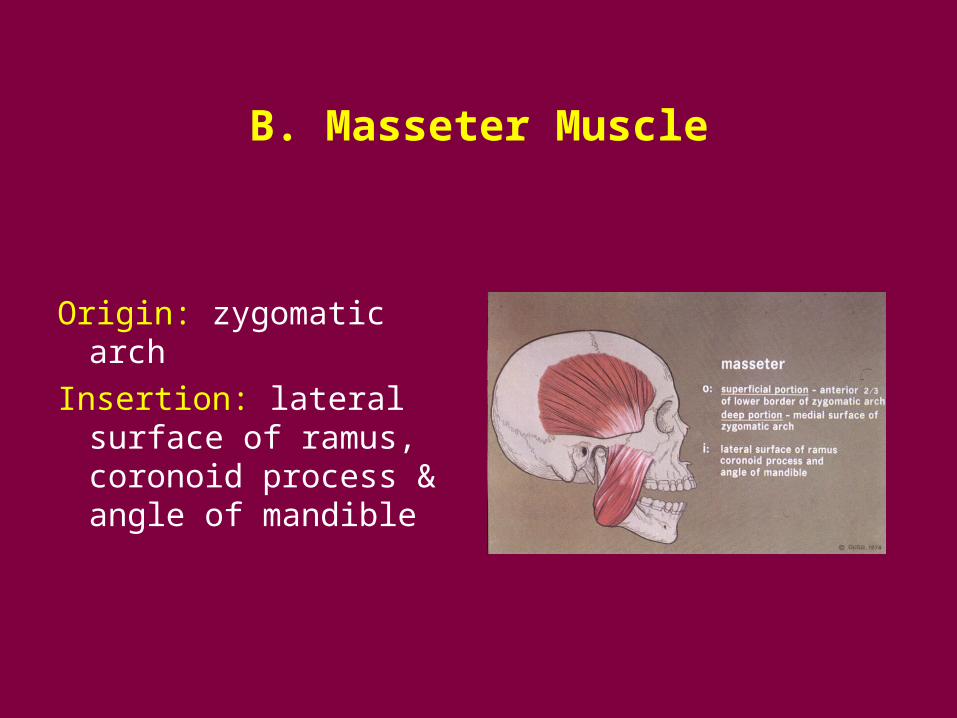
B. Masseter Muscle
Origin: zygomatic arch
Insertion: lateral surface of ramus, coronoid process & angle of mandible
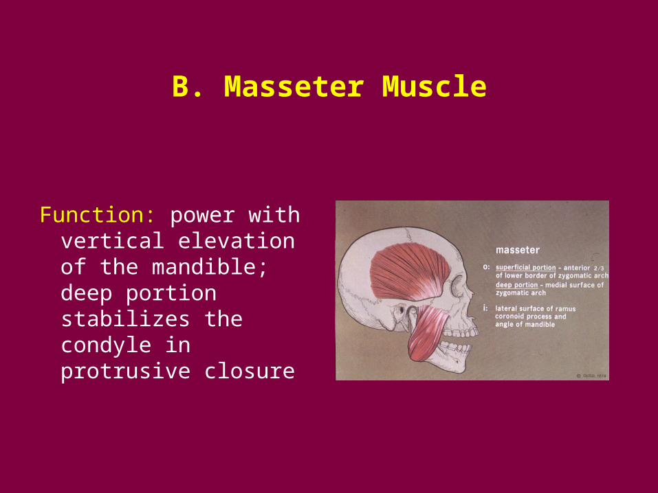
B. Masseter Muscle
Function: power with vertical elevation of the mandible; deep portion stabilizes the condyle in protrusive closure

C. Medial Pterygoid Muscle
Origin: medial surface of lateral pterygoid plate, pyramidal process of palatine bone & Mx tuberosity
Insertion: medial surface of ramus & angle of mandible

C. Medial Pterygoid Muscle
Function: elevation of the mandible, protrusion of the mandible and lateral movement of the mandible with unilateral activation

Pterygomasseteric Sling

D. Inferior Lateral Pterygoid Muscle
Origin: lateral surface of lateral pterygoid plate
Insertion: pterygoid fovea of condyle

D. Inferior Lateral Pterygoid Muscle
Function: protrusion, lateral movement and contributes to opening

E. Superior Lateral Pterygoid Muscle
Origin: infratemporal surface greater wing of the sphenoid bone
Insertion: pterygoid fovea of the condyle and variable to the disc

E. Superior Lateral Pterygoid Muscle
Function: active with the muscles of closure, especially aiding stabilization of the condyle during the power stroke

Temporomandibular Joint Anatomy

“Craniomandibular Joint”: a diarthrodial or synovial lined joint

A compound joint: > 2 components
• Temporal Bone
• Mandibular Condyle
• Articular Disc

A. Temporal Bone
• Cranial component • Mandibular fossa • Articular eminence • Articular surface from
superior fossa to the anterior aspect of the eminence, thickest bone.

B. Mandibular Component: Condyle
• Condylar dimensions: A-P 8-10 mm M-L 15-20 mm
• Articular surface: anterior superior aspect

B. Mandibular Condyle
• Variation side to side in size and shape is common. Response to loading
• Lateral pole anterior to medial pole.

C. Articular Tissue
• Origin: modified periosteum of intramembranous bone, NOT endochondral origin. A consequence of 2 embryonic tissue masses growing towards each other, NOT a single tissue mass cleft to form a joint articulation.

C. Articular Tissue • Function: 1)
load distribution 2) synovial lubrication
• Character: NOT hyaline cartilage, but fibrous in nature 1) avascular 2) NOT innervated 3) resistant to shear, tension forces 4) increased remodeling potential

D. Articular Disc
• Composition: avascular connective tissue, collagenous
• Shape: biconcave in cross section with posterior band, intermediate zone and anterior band
• Position: posterior band edge at “12:00”

D. Articular Disc

E. TM Joint Capsule
• Function: 1) limit extreme ROM 2) synovial lining 3) confines synovial
fluid 4) joint proprioception

F. Mandibular Ligaments
Restrict and limit
joint range of motion
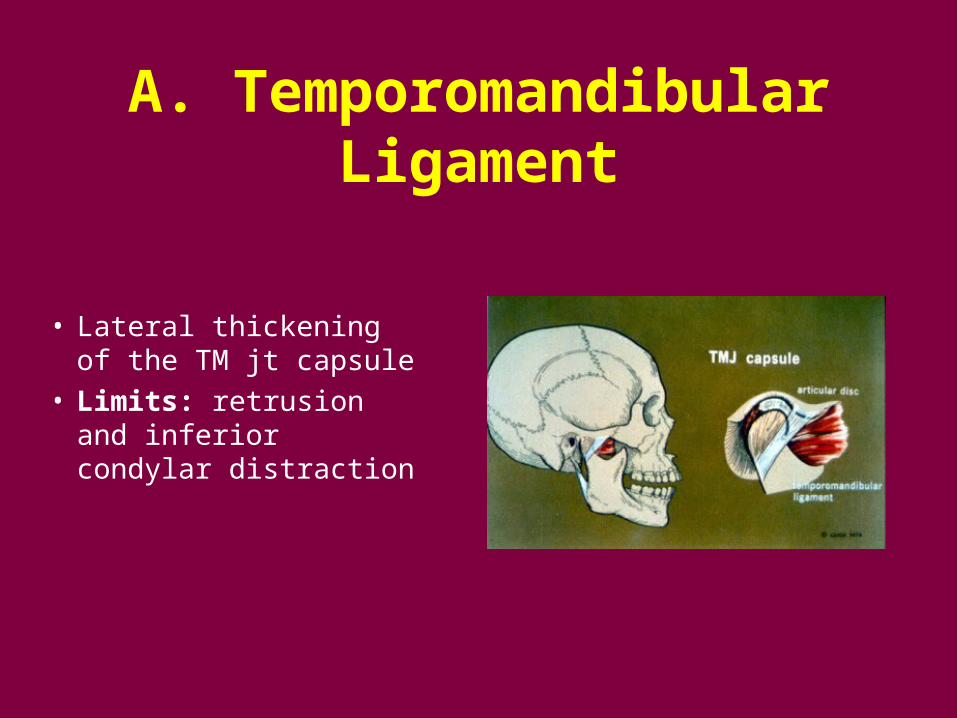
A. Temporomandibular Ligament
• Lateral thickening of the TM jt capsule
• Limits: retrusion and inferior condylar distraction

B. Collateral Ligaments
• Medial and lateral • Limit: medial and
lateral movement of the disc relative to the condyle

C. Accessory Ligaments
• Sphenomandibular Ligament: nonfunctional vestige or remnant of Meckel’s cartilage
• Stylomandibular Ligament: limits extreme jaw protrusion
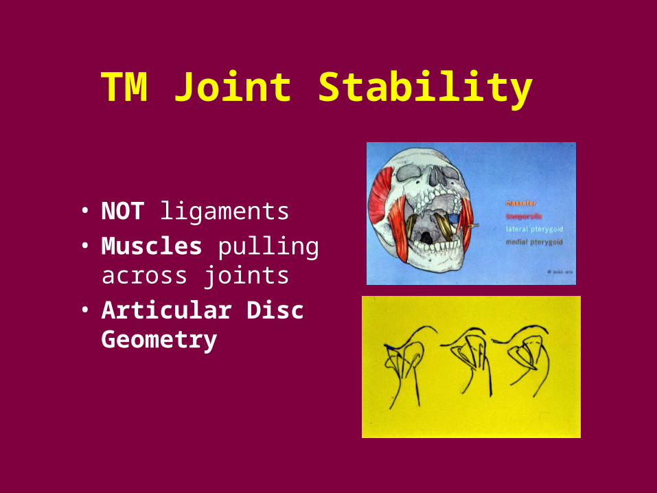
TM Joint Stability
• NOT ligaments • Muscles pulling
across joints • Articular Disc
Geometry

TM Joint Anatomy: sagittal
• Lateral Pterygoid M Superior head insertion- condyle & sometimes disc Inferior head insertion- condyle
• Retrodiscal tissues 1) Superior- elastic 2) Inferior- “check ligament” 3) Loose C.T.- venous compartment and innervated
• Synovial tissues

TM Joint Retrodiscal Tissue

TM Joint Innervation: V3

TM Joint Functional Anatomy
Read pages in Okeson, pp. 22-26!


Centric Relation (Okeson, 2003)
Jaw position with the condyles in their most superoanterior postions in the articular fossae, resting against the posterior slopes of the articular eminences, with the articular discs properly interposed.

Biology of Centric Relation
• Muscloskeletal Stability: a stable position in which to load the joint, mm pull across jts seating condyle
• Clinical Evidence: patients do well with IP coincidental with jaw position in CR (clinical experience)

Biology of Centric Relation
• Clinically repeatable: a reference position for restorative dental procedures
… but NOT immutable! (Celenza FV, JPD 1973)
… (~1 yr?)