DENTAL TRAUMA GUIDELINES - Dalhousie Universityremovpros.dentistry.dal.ca/ewExternalFiles/1-9 IADT...
27
International Association of Dental Traumatology DENTAL TRAUMA GUIDELINES Revised 2012 CONTENT: Section 1. Fractures and luxations of permanent teeth Section 2. Avulsion of permanent teeth Section 3. Traumatic injuries to primary teeth Disclaimer: These guidelines are intended to provide information for health care providers caring for patients with dental injuries. They represent the current best evidence based on literature research and professional opinion. As is true for all guidelines, the health care provider must apply clinical judgment dictated by the conditions present in the given traumatic situation. The IADT does not guarantee favorable outcomes from following the Guidelines, but using the recommended procedures can maximize the chances of success. THESE GUIDELINES ARE ENDORSED BY THE AMERICAN ASSOCIATION OF ENDODONTISTS
Transcript of DENTAL TRAUMA GUIDELINES - Dalhousie Universityremovpros.dentistry.dal.ca/ewExternalFiles/1-9 IADT...

International Association of Dental Traumatology
DENTAL TRAUMA GUIDELINES
Revised 2012
CONTENT: Section 1. Fractures and luxations of permanent teeth Section 2. Avulsion of permanent teeth Section 3. Traumatic injuries to primary teeth Disclaimer: These guidelines are intended to provide information for health care providers caring for patients with dental injuries. They represent the current best evidence based on literature research and professional opinion. As is true for all guidelines, the health care provider must apply clinical judgment dictated by the conditions present in the given traumatic situation. The IADT does not guarantee favorable outcomes from following the Guidelines, but using the recommended procedures can maximize the chances of success.
THESE GUIDELINES ARE ENDORSED BY THE AMERICAN ASSOCIATION OF ENDODONTISTS

INTRODUCTION Traumatic dental injuries (TDIs) occur frequently in children and young adults, comprising 5% of all injuries. Twenty‐five percent of all school children experience dental trauma and 33% of adults have experienced trauma to the permanent dentition, with the majority of the injuries occurring before age 19. Luxation injuries are the most common TDIs in the primary dentition, whereas crown fractures are more commonly reported for the permanent teeth. Proper diagnosis, treatment planning and follow‐up are important to assure a favorable outcome. This update includes a review of the current dental literature using EMBASE, MEDLINE, PUBMED and Scopes searches from 1996‐2011, as well as a search of Dental Traumatology from 2000‐2011. The goal of these guidelines is to provide information for the immediate and urgent care of TDIs. It is understood that some of the subsequent treatment may require secondary and tertiary interventions involving specialists with experience in dental trauma. The IADT published its first set of guidelines in 2001, and updated them in 2007. As with previous guidelines, the working group included experienced investigators and clinicians from various dental specialties and general practice. The current revision represents the best evidence based on the available literature and expert professional judgment. In cases where the data did not appear conclusive, recommendations are based on the consensus opinion of the working group, followed by review by the members of the IADT Board of Directors.
It is understood that guidelines are to be applied with evaluation of the specific clinical circumstances, clinicians’ judgment and patients’ characteristics, including but not limited to compliance, finances and understanding of the immediate and long‐term outcomes of treatment alternatives versus non‐treatment. The IADT cannot and does not guarantee favorable outcomes from adherence to the Guidelines, but believe that their application can maximize the chances of a favorable outcome. These Guidelines offer recommendations for diagnosis and treatment of specific TDIs; however, they provide neither the comprehensive nor the detailed information found in textbooks, in the scientific literature, and most recently in the Dental Trauma Guide (DTG), which can be accessed @ http://www.dentaltraumaguide.org and the IADT website, http://www.iadt‐dentaltrauma.org provides connection to the journal Dental Traumatology and other dental trauma information.
GENERAL RECOMMENDATIONS Special considerations for trauma to primary teeth A young child is often difficult to examine and treat due to lack of cooperation and because of fear. The situation is distressing for both the child and the parents. It is important to keep in mind that there is a close relationship between the apex of the root of the injured primary tooth, and the underlying permanent tooth germ. Tooth malformation, impacted teeth, and eruption disturbances in the developing permanent dentition are some of the consequences that can occur following severe injuries to primary teeth and/or alveolar bone. A child’s maturity and ability to cope with the emergency situation, the time for shedding of the injured tooth and the occlusion, are all important factors that influence treatment. Repeated trauma episodes are frequent in children. Immature versus Mature Permanent Teeth

Every effort should be made to preserve pulpal vitality in the immature permanent tooth to ensure continuous root development. The vast majority of TDIs occur in children and teenagers where loss of a tooth has lifetime consequences. The immature permanent tooth has considerable capacity for healing after traumatic pulp exposure, luxation injury and root fractures. Avulsion of Permanent Teeth The prognosis for avulsed permanent teeth is very much dependent on the actions taken at the place of accident. Promotion of public awareness of first‐aid treatment for the avulsed tooth is strongly encouraged. Treatment choices and prognosis for the avulsed tooth are largely dependent on the vitality of the periodontal ligament (PDL), and the maturity of the root. Patient/Parent Instructions
Patient compliance with follow‐up visits and home care contributes to better healing following a TDI. Both the patients and the parents of young patients should be advised regarding care of the injured tooth/teeth for optimal healing, prevention of further injury by avoidance of participation in contact sports, meticulous oral hygiene, and rinsing with an antibacterial such as Chlorhexidine Gluconate 0.1% alcohol free for 1‐2 weeks. Alternatively, with a young child, it is desirable to apply Chlorhexidine Gluconate to the affected area with a cotton swab. The use of pacifiers should be restricted.

FRACTURES AND LUXATIONS OF PERMANENT TEETH 1. Treatment guidelines for fractures of teeth and alveolar bone Followup Procedures
for fractures of teeth and alveolar bone+
Favorable and Unfavorable outcomes include some, but not necessarily all, of the
following: INFRACTION
Clinical findings Radiographic findings
Treatment
Follow-Up
Favorable Outcome
Unfavorable Outcome
● An incomplete fracture (crack) of the enamel without loss of tooth structure. ● Not tender. If tenderness is observed evaluate the tooth for a possible luxation injury or a root fracture.
● No radiographic abnormalities. ● Radiographs recom- mended: a periapical view. Additional radiographs are indicated if other signs or symptoms are present.
● In case of marked infractions, etching and sealing with resin to prevent discoloration of the infraction lines. Otherwise, no treatment is necessary.
● No follow-up is generally needed for infraction injuries unless they are associated with a luxation injury or other fracture types.
● Asymptomatic ● Positive response to pulp testing. ● Continuing root development in immature teeth.
● Symptomatic ● Negative response to pulp testing. ● Signs of apical periodontitis. ● No continuing root development in immature teeth. ● Endodontic therapy appropriate for stage of root development is indicated.
ENAMEL FRACTURE Clinical findings
Radiographic findings
Treatment
Followup
Favorable Outcome
Unfavorable Outcome
● A complete fracture of the enamel. ● Loss of enamel. No visible sign of exposed dentin. ● Not tender. If tenderness is observed evaluate the tooth for a possible luxation or root fracture injury. ● Normal mobility. ● Sensibility pulp test usually positive.
● Enamel loss is visible. ● Radiographs recom-mended: periapical, occlusal and eccentric exposures. They are recommended in order to rule out the possible presence of a root fracture or a luxation injury. ● Radiograph of lip or cheek to search for tooth fragments or foreign materials.
● If the tooth fragment is available, it can be bonded to the tooth. ● Contouring or restoration with composite resin depending on the extent and location of the fracture.
6-8 weeks C++
1 year C
++
● Asymptomatic ● Positive response to pulp testing. ● Continuing root development in immature teeth. ● Continue to next evaluation.
● Symptomatic ● Negative response to pulp testing. ● Signs of apical periodontitis ● No continuing root development in immature teeth. ● Endodontic therapy appropriate for stage of root development is indicated.
+
= for crown fractured teeth with concomitant luxation injury, use the luxation followup schedule. C
++ = clinical and radiographic examination.

Follow-Up Procedures for fractures of teeth and alveolar bone+
Favorable and Unfavorable outcomes include some, but not necessarily all, of the
following: ENAMEL-DENTIN-
FRACTURE Clinical findings
Radiographic findings
Treatment
Follow-Up
Favorable Outcome
Unfavorable Outcome
● A fracture confined to enamel and dentin with loss of tooth structure, but not exposing the pulp. ● Percussion test: not tender. If tenderness is observed, evaluate the tooth for possible luxation or root fracture injury. ● Normal mobility. ● Sensibility pulp test usually positive.
● Enamel-dentin loss is visible. ● Radiographs recom-mended: periapical, occlusal and eccentric exposure to rule out tooth displacement or possible presence of root fracture. ● Radiograph of lip or cheek lacerations to search for tooth fragments or foreign materials.
● If a tooth fragment is available, it can be bonded to the tooth. Otherwise perform a provisional treatment by covering the exposed dentin with glass- Ionomer or a more permanent restoration using a bonding agent and composite resin, or other accepted dental restorative materials ● If the exposed dentin is within 0.5mm of the pulp (pink, no bleeding) place calcium hydroxide base and cover with a material such as a glass ionomer.
6-8 weeks C++
1 year C
++
● Asymptomatic ● Positive response to pulp testing. ● Continuing root development in immature teeth ● Continue to next evaluation
● Symptomatic ● Negative response to pulp testing. ● Signs of apical periodontitis. ● No continuing root development in immature teeth. ● Endodontic therapy appropriate for stage of root development is indicated.
ENAMEL-DENTIN-PULP FRACTURE
Clinical findings
Radiographic findings
Treatment
Favorable Outcome
Unfavorable Outcome
● A fracture involving enamel and dentin with loss of tooth structure and exposure of the pulp. ● Normal mobility ● Percussion test: not tender. If tenderness is observed, evaluate for possible luxation or root fracture injury. ● Exposed pulp sensitive to stimuli.
● Enamel – dentin loss visible. ● Radiographs recommended: periapical, occlusal and eccentric exposures, to rule out tooth displacement or possible presence of root fracture. ● Radiograph of lip or cheek lacerations to search for tooth fragments or foreign materials.
● In young patients with immature, still developing teeth, it is advantageous to preserve pulp vitality by pulp capping or partial pulpotomy. Also, this treatment is the choice in young patients with completely formed teeth. ● Calcium hydroxide is a suitable material to be placed on the pulp wound in such procedures.
● In patients with mature apical development, root canal treatment is usually the treatment of choice, although pulp capping or partial pulpotomy also may be selected. ● If tooth fragment is available, it can be bonded to the tooth. ● Future treatment for the fractured crown may be restoration with other accepted dental restorative materials.
6-8 weeks C++
1 year C
++
● Asymptomatic. ● Positive response to pulp testing. ● Continuing root development in immature teeth. ● Continue to next evaluation.
● Symptomatic. ● Negative response to pulp testing. ● Signs of apical periodontitis. ● No continuing root development in immature teeth. ● Endodontic therapy appropriate for stage of root development is indicated.
+ = for crown fractured teeth with concomitant luxation injury, use the luxation followup schedule
C++
= clinical and radiographic examination.

Follow-Up Procedures for fractures of teeth and alveolar bone +
Favorable and Unfavorable outcomes include some, but not necessarily all, of the
following: CROWN-ROOT
FRACTURE WITHOUT PULP EXPOSURE
Clinical findings
Radiographic findings
Treatment
Follow-Up
Favorable Outcome
Unfavorable Outcome
● A fracture involving enamel, dentin and cementum with loss of tooth structure, but not exposing the pulp. ● Crown fracture extending below gingival margin. ● Percussion test: Tender. ● Coronal fragment mobile. ● Sensibility pulp test usually positive for apical fragment.
● Apical extension of fracture usually not visible. ● Radiographs recommended: periapical, occlusal and eccentric exposures. They are recommended in order to detect fracture lines in the root.
Emergency treatment ● As an emergency treatment a temporary stabilization of the loose segment to adjacent teeth can be performed until a definitive treatment plan is made. Non-Emergency Treatment Alternatives Fragment removal only ● Removal of the coronal crown-root fragment and subsequent restoration of the apical fragment exposed above the gingival level. Fragment removal and gingivectomy (sometimes ostectomy) ● Removal of the coronal crown-root segment with subsequent endodontic treatment and restoration with a post-retained crown. This procedure should be preceded by a gingivectomy, and sometimes ostectomy with osteoplasty. Orthodontic extrusion of apical fragment ● Removal of the coronal segment with subsequent endodontic treatment and orthodontic extrusion of the remaining root with sufficient length after extrusion to support a post-retained crown. Surgical extrusion ● Removal of the mobile fractured fragment with subsequent surgical repositioning of the root in a more coronal position. Root submergence ● Implant solution is planned. Extraction ● Extraction with immediate or delayed implant-retained crown restoration or a conventional bridge. Extraction is inevitable in crown-root fractures with a severe apical extension, the extreme being a vertical fracture.
6-8 weeks C++
1 year C++
● Asymptomatic ● Positive response to pulp testing. ● Continuing root development in immature teeth ● Continue to next evaluation
● Symptomatic ● Negative response to pulp testing. ● Signs of apical periodontitis. ● No continuing root development in immature teeth. ● Endodontic therapy appropriate for stage of root development is indicated.
+=for crown fractured teeth with concomitant luxation injury, use the luxation followup schedule. C++ =clinical and radiographic examination
Follow-Up Procedures for fractures of teeth and alveolar bone +
Favorable and Unfavorable outcomes include some, but not necessarily all, of the
following: CROWN-ROOT
FRACTURE WITH PULP EXPOSURE
Clinical findings
Radiographic findings
Treatment
Follow-Up
Favorable Outcome
Unfavorable Outcome
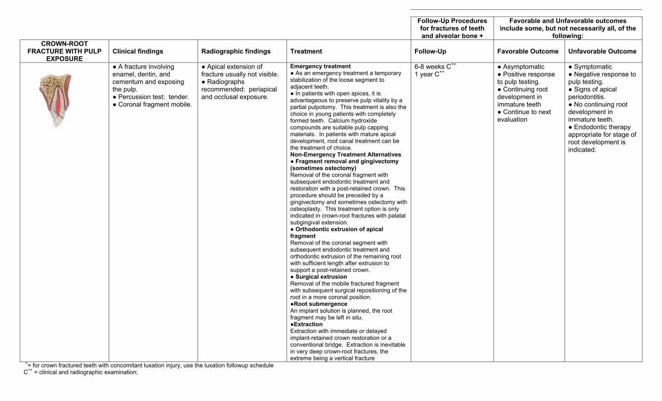
● A fracture involving enamel, dentin, and cementum and exposing the pulp. ● Percussion test: tender. ● Coronal fragment mobile.
● Apical extension of fracture usually not visible. ● Radiographs recommended: periapical and occlusal exposure.
Emergency treatment ● As an emergency treatment a temporary stabilization of the loose segment to adjacent teeth. ● In patients with open apices, it is advantageous to preserve pulp vitality by a partial pulpotomy. This treatment is also the choice in young patients with completely formed teeth. Calcium hydroxide compounds are suitable pulp capping materials. In patients with mature apical development, root canal treatment can be the treatment of choice. Non-Emergency Treatment Alternatives ● Fragment removal and gingivectomy (sometimes ostectomy) Removal of the coronal fragment with subsequent endodontic treatment and restoration with a post-retained crown. This procedure should be preceded by a gingivectomy and sometimes ostectomy with osteoplasty. This treatment option is only indicated in crown-root fractures with palatal subgingival extension. ● Orthodontic extrusion of apical fragment Removal of the coronal segment with subsequent endodontic treatment and orthodontic extrusion of the remaining root with sufficient length after extrusion to support a post-retained crown. ● Surgical extrusion Removal of the mobile fractured fragment with subsequent surgical repositioning of the root in a more coronal position. ●Root submergence An implant solution is planned, the root fragment may be left in situ. ●Extraction Extraction with immediate or delayed implant-retained crown restoration or a conventional bridge. Extraction is inevitable in very deep crown-root fractures, the extreme being a vertical fracture
6-8 weeks C++
1 year C++
● Asymptomatic ● Positive response to pulp testing. ● Continuing root development in immature teeth ● Continue to next evaluation
● Symptomatic ● Negative response to pulp testing. ● Signs of apical periodontitis. ● No continuing root development in immature teeth. ● Endodontic therapy appropriate for stage of root development is indicated.
+= for crown fractured teeth with concomitant luxation injury, use the luxation followup schedule
C++
= clinical and radiographic examination;

Follow-Up Procedures
for fractures of teeth and alveolar bone
Favorable and Unfavorable outcomes include some, but not necessarily all, of the
following:++ ROOT FRACTURE Clinical findings Radiographic findings Treatment Favorable Outcome Unfavorable Outcome ●The coronal segment may be
mobile and may be displaced. ● The tooth may be tender to percussion. ● Bleeding from the gingival sulcus may be noted. ● Sensibility testing may give negative results initially, indicating transient or permanent neural damage. ● Monitoring the status of the pulp is recommended. ● Transient crown discoloration (red or grey) may occur.
● The fracture involves the root of the tooth and is in a horizontal or oblique plane. ● Fractures that are in the horizontal plane can usually be detected in the regular periapical 90
o angle film with
the central beam through the tooth. This is usually the case with fractures in the cervical third of the root. ● If the plane of fracture is more oblique which is common with apical third fractures, an occlusal view or radiographs with varying horizontal angles are more likely to demonstrate the fracture including those located in the middle third.
● Reposition, if displaced, the coronal segment of the tooth as soon as possible. ● Check position radiographically. ● Stabilize the tooth with a flexible splint for 4 weeks. If the root fracture is near the cervical area of the tooth, stabilization is beneficial for a longer period of time (up to 4 months). ● It is advisable to monitor healing for at least one year to determine pulpal status. ● If pulp necrosis develops, root canal treatment of the coronal tooth segment to the fracture line is indicated to preserve the tooth.
4 Weeks S+, C
++
6-8 Weeks C++
4 Months S++
*, C++
6 Months C
++
1 Year C++
5 Years C
++
● Positive response to pulp testing (false negative possible up to 3 months). ● Signs of repair between fractured segments. ● Continue to next evaluation.
● Symptomatic ● Negative response to pulp testing (false negative possible up to 3 months). ● Extrusion of the coronal segment. ● Radiolucency at the fracture line. ● Clinical signs of periodontitis or abscess associated with the fracture line. ● Endodontic therapy appropriate for stage of root development is indicated.
ALVEOLAR FRACTURE Clinical findings Radiographic findings Treatment Follow-Up Favorable Outcome Unfavorable Outcome ● The fracture involves the
alveolar bone and may extend to adjacent bone. ● Segment mobility and dislocation with several teeth moving together are common findings. ● An occlusal change due to misalignment of the fractured alveolar segment is often noted. ● Sensibility testing may or may not be positive.
● Fracture lines may be located at any level, from the marginal bone to the root apex. ● In addition to the 3 angulations and occlusal film, additional views such as a panoramic radiograph can be helpful in determining the course and position of the fracture lines.
● Reposition any displaced segment and then splint. ● Suture gingival laceration if present. ● Stabilize the segment for 4 weeks.
4 Weeks S+, C
++
6-8 Weeks C++
4 Months C
++
6 Months C++
1 Year C++
5 Years C++
● Positive response to pulp testing (false negative possible up to 3 months). ● No signs of apical periodontitis. ●Continue to next evaluation.
● Symptomatic ● Negative response to pulp testing (false negative possible up to 3 months). ● Signs of apical periodontitis or external inflammatory root resorption. ● Endodontic therapy appropriate for stage of root development is indicated.
S
+=splint removal; S
++=splint removal in cervical third fractures.
C++
= clinical and radiographic examination. ++=Whenever there is evidence of external inflammatory root resorption, root canal therapy should be initiated immediately, with the use of calcium hydroxide as an intra-canal medication.

2. Treatment Guidelines for Luxation Injuries
Follow-Up Procedures for luxated permanent teeth
Favorable and Unfavorable outcomes include some, but not necessarily all, of the following:++
CONCUSSION Clinical findings Radiographic findings Treatment Favorable Outcome Unfavorable Outcome ● The tooth is tender to touch
or tapping; it has not been displaced and does not have increased mobility. ● Sensibility tests are likely to give positive results.
●No radiographic abnormalities
● No treatment is needed. ● Monitor pulpal condition for at least one year.
4 Weeks C++
6-8 Weeks C++
1 Year C
++
● Asymptomatic ● Positive response to pulp testing ● False negative possible up to 3 months. ● Continuing root development in immature teeth ● Intact lamina dura
● Symptomatic ● Negative response to pulp testing ● False negative possible up to 3 months ●No continuing root development in immature teeth, signs of apical periodontitis.
● Endodontic therapy appropriate for stage of root development is indicated.
SUBLUXATION Clinical findings Radiographic findings Treatment Follow-Up Favorable Outcome Unfavorable Outcome ● The tooth is tender to touch
or tapping and has increased mobility; it has not been displaced. ● Bleeding from gingival crevice may be noted. ● Sensibility testing may be negative initially indicating transient pulpal damage. ● Monitor pulpal response until a definitive pulpal diagnosis can be made.
●Radiographic abnormalities are usually not found.
● Normally no treatment is needed, however a flexible splint to stabilize the tooth for patient comfort can be used for up to 2 weeks.
2 Weeks S+, C
++
4 Weeks C++
6-8 Weeks C++
6 Months C++
1 Year C++
● Asymptomatic ● Positive response to pulp testing ● False negative possible up to 3 months. ● Continuing root development in immature teeth ● Intact lamina dura
● Symptomatic ● Negative response to pulp testing ● False negative possible up to 3 months ● External inflammatory resorption. ● No continuing root development in immature teeth, signs of apical periodontitis.
● Endodontic therapy appropriate for stage of root development is indicated.
EXTRUSIVE LUXATION Clinical Findings Radiographic findings Treatment Follow-Up Favorable Outcome Unfavorable Outcome ● The tooth appears elongated
and is excessively mobile. ● Sensibility tests will likely give negative results.
●Increased periodontal ligament space apically.
● Reposition the tooth by gently re-inserting It into the tooth socket. ● Stabilize the tooth for 2 weeks using a flexible splint. ● In mature teeth where pulp necrosis is anticipated or if several signs and symptoms indicate that the pulp of mature or immature teeth became necrotic, root canal treatment is indicated.
2 Weeks S+,
C++
4 Weeks C
++
6-8 Weeks C++
6 Months C
++
1 Year C++
Yearly 5 years C++
● Asymptomatic ● Clinical and radiographic signs of normal or healed periodontium. ● Positive response to pulp testing (false negative possible up to 3 months). ● Marginal bone height corresponds to that seen radiographically after repositioning. ● Continuing root development in immature teeth.
● Symptoms and radiographic sign consistent with apical periodontitis. ● Negative response to pulp testing (false negative possible up to 3 months). ● If breakdown of marginal bone, splint for an additional 3-4 weeks. ● External inflammatory root resorption.
● Endodontic therapy appropriate for stage of root development is indicated.
S+=splint removal;
C++
= clinical and radiographic examination. ++=Whenever there is evidence of external inflammatory root resorption, root canal therapy should be initiated immediately, with the use of calcium hydroxide as an intra-canal medication.

Follow-Up Procedures for luxated permanent
teeth
Favorable and Unfavorable outcomes include some, but not necessarily all, of the following:++
LATERAL LUXATION Clinical findings Radiographic findings Treatment Favorable Outcome Unfavorable Outcome ● The tooth is displaced,
usually in a palatal/lingual or labial direction. ● It will be immobile and percussion usually gives a high, metallic (ankylotic) sound. ● Fracture of the alveolar process present. ● Sensibility tests will likely give negative results
● The widened periodontal ligament space is best seen on eccentric or occlusal exposures.
● Reposition the tooth digitally or with forceps to disengage it from its bony lock and gently reposition it into its original location. ● Stabilize the tooth for 4 weeks using a flexible splint. ● Monitor the pulpal condition. ● If the pulp becomes necrotic, root canal treatment is indicated to prevent root resorption.
2 Weeks, C++
4 Weeks S+, C
++
6-8 Weeks C++
6 Months C++
1 Year C
++
Yearly for 5 years C++
● Asymptomatic ● Clinical and radiographic signs of normal or healed periodontium. ● Positive response to pulp testing (false negative possible up to 3 months). ● Marginal bone height corresponds to that seen radiographically after repositioning. ●Continuing root development in immature teeth
● Symptoms and radiographic signs consistent with apical periodontitis. ● Negative response to pulp testing (false negative possible up to 3 months). ● If breakdown of marginal bone, splint for an additional 3-4 weeks. ● External inflammatory root resorption or replacement resorption
● Endodontic therapy appropriate for stage of root development is indicated.
INTRUSIVE LUXATION Clinical findings Radiographic findings Treatment Follow-Up Favorable Outcome Unfavorable ● The tooth is displaced axially
into the alveolar bone. ● It is immobile and percussion may give a high, metallic (ankylotic) sound. ● Sensibility tests will likely give negative results.
●The periodontal ligament space may be absent from all or part of the root. ● The cemento-enamel junction is located more apically in the intruded tooth than in adjacent non-injured teeth, at times even apical to the marginal bone level.
Teeth with incomplete root formation ● Allow eruption without intervention ● If no movement within few weeks, initiate orthodontic repositioning. ● If tooth is intruded more than 7mm, reposition surgically or orthodontically. Teeth with complete root formation: ● Allow eruption without intervention if tooth intruded less than 3mm. If no movement after 2-4 weeks, reposition surgically or orthodontically before ankylosis can develop. ● If tooth is intruded 3-7 mm, reposition
surgically or orthodontically. ● If tooth is intruded beyond 7mm, reposition surgically. ● The pulp will likely become necrotic in teeth with complete root formation. Root canal therapy using a temporary filling with calcium hydroxide is recommended and treatment should begin 2-3 weeks after repositioning. ● Once an intruded tooth has been repositioned surgically or orthodontically, stabilize with a flexible splint for 4 weeks.
2 Weeks, C++
4 Weeks S+, C
++
6-8 Weeks C++
6 Months C++
1 Year C++
Yearly for 5 years C++
● Tooth in place or erupting. ● Intact lamina dura ● No signs of resorption. ● Continuing root development in immature teeth.
● Tooth locked in place/ankylotic tone to percussion. ● Radiographic signs of apical periodontitis ● External inflammatory root resorption or replacement resorption.
● Endodontic therapy appropriate for stage of root development is indicated.
S+=splint removal;
C++
= clinical and radiographic examination. ++= whenever there is evidence of external inflammatory root resorption, root canal therapy should be initiated immediately, with the use of calcium hydroxide as an intra-canal medication.

Page 1 of 11
Avulsion of permanent teeth
Avulsion of permanent teeth is seen in 0.5‐3% of all dental injuries (1,2). Numerous studies show that this injury is one of the most serious dental injuries and the prognosis is very much dependent on the actions taken at the place of accident and promptly after the avulsion (2‐27). Replantation is in most situations the treatment of choice, but cannot always be carried out immediately. An appropriate emergency management and treatment plan are important for a good prognosis. There are also individual situations when replantation is not indicated (e.g. severe caries or periodontal disease, non‐cooperating patient, severe medical conditions (e.g. immunosuppression and severe cardiac conditions) which must be dealt with individually. Replantation may successfully save the tooth, but it is important to realize that some of the replanted teeth have lower chances of long term survival and may even be lost or extracted at a later stage.
Guidelines for the emergency management are useful for delivering the best care possible in an efficient manner. The International Association of Dental Traumatology (IADT) has developed a consensus statement after an update of the dental literature and discussions in expert groups. Experienced international researchers and clinicians from various specialties and general dentistry were included in the groups. In cases in which the data did not appear conclusive, recommendations were based on the consensus opinion and in some situations on majority decision among the IADT board members. All recommendations are not evidence based on a high level. The guidelines should therefore be seen as the current best evidence and practice based on literature research and professionals’ opinion.
Guidelines should assist dentists, other health care professionals and patients in decision making. Also, they should be credible, readily understandable and practical with the aim of delivering appropriate care as effectively and efficiently as possible.It is understood that guidelines are to be applied with judgment of the specific clinical circumstances, clinicians’ judgments and patients’ characteristics, including but not limited to compliance, finances and understanding of the immediate and long‐term outcomes of treatment alternatives versus non‐treatment. The IADT cannot and does not guarantee favorable outcomes from strict adherence to the Guidelines, but believe that their application can maximize the chances of a favorable outcome. Guidelines undergo periodic updates. The following guidelines by the International Association of Dental Traumatology (IADT) represent an updated set of guidelines based on the original guidelines published in 2007 (28‐30).

Page 2 of 11
In these IADT Guidelines for management of avulsed permanent teeth the literature has been searched using Medline and Scopus databases using the search words: avulsion, exarticulation and replantation. The task group has then discussed the emergency treatment in detail and reached consensus of what to recommend today as best practice for the emergency management. This text is aiming at giving the concise, short necessary advice for treatment in the emergency situation. More detailed description of protocols, methods and documentation for clinical assessment and diagnosis of different dental injuries can be found in articles, textbooks and manuals (2,24) and in the interactive web site Dental Trauma Guide http://dentaltraumaguide.org
The final decision regarding patient care remains primarily in the hand of the treating dentist. For ethical reasons it is important that the dentist provides the patient and guardian with pertinent information relating to treatment so also the patient and guardian has as much influence in the decision making process as possible.
First aid for avulsed teeth at the place of accident (2,10,24,25,31‐55)
Dentists should always be prepared to give appropriate advice to the public about first aid for avulsed teeth. An avulsed permanent tooth is one of the few real emergency situations in dentistry. In addition to increasing the public awareness by, e.g. mass media campaigns, healthcare professionals. Guardians and teachers should receive information on how to proceed following these severe unexpected injuries. Also, instructions may be given by telephone to people at the emergency site. Immediate replantation is the best treatment at the place of accident. If for some reasons this cannot be carried out, there are alternatives such as using various storage media
If a tooth is avulsed, make sure it is a permanent tooth (primary teeth should not be replanted)
• Keep the patient calm. • Find the tooth and pick it up by the crown (the white part). Avoid touching the root. • If the tooth is dirty, wash it briefly (max 10 seconds) under cold running water and
reposition it. Try to encourage the patient/guardian to replant the tooth. Once the tooth is back in place, bite on a handkerchief to hold it in position.
• If this is not possible, or for other reasons when replantation of the avulsed tooth is not possible (e.g. an unconscious patient), place the tooth in a glass of milk or another suitable storage medium and bring with the patient to the emergency clinic. The tooth can also be transported in the mouth, keeping it inside the lip or cheek if the patient is conscious. If the patient is very young, he/she could swallow the tooth – therefor it is advisable to get the patient to spit in a container and place the tooth in it. Avoid storage in water!
• If there is access at the place of accident to special storage or transport media (e.g. tissue culture/transport medium, Hanks balanced storage medium (HBSS or saline) such media can preferably be used.
• Seek emergency dental treatment immediately.

Page 3 of 11
The poster ‘Save a Tooth’ is written for the public and is available in several languages: English, Spanish, Portuguese, French, Icelandic, Italian, Arabic and Turkish and can be obtained at the IADT website: http://www.iadt‐dentaltrauma.org.
Treatment guidelines for avulsed permanent teeth (56‐96)
Choice of treatment is related to the maturity of the root (open or closed apex) and the condition of the periodontal ligament cells. The condition of the cells is depending on the storage medium and the time out of the mouth, especially the dry time is critical for survival of the cells. After a dry time of 60 minutes or more all PDL cells are non‐viable. For this reason, the dry time of the tooth, before it was placed replanted or placed in a storage medium, is very important to assess from the patient’s history.
From a clinical point of view it is important for the clinician to roughly assess the condition of the cells by classifying the avulsed tooth into one of the following three groups before starting treatment:
o The PDL cells are most likely viable (i.e. the tooth has been replanted immediately or after a very short time at the place of accident)
o The PDL cells may be viable but compromised. The tooth has been kept in storage medium (e.g. tissue culture medium, HBSS, saline, milk or saliva and the total dry time has been less than 60 min).
o The PDL cells are non viable. Examples of this is when the trauma history tells us that the total extra‐oral dry time has been more than 60 min regardless of if the tooth was stored in an additional medium or not, or if the storage medium was non‐physiologic.
1. Treatment guidelines for avulsed permanent teeth with closed apex
1a. The tooth has been replanted before the patient’s arrival at the clinic
• Leave the tooth in place. • Clean the area with water spray, saline or chlorhexidine. • Suture gingival lacerations, if present. • Verify normal position of the replanted tooth both clinically and radiographically. • Apply a flexible splint for up to 2 weeks (see Splinting). • Administer systemic antibiotics. (see Antibiotics).

Page 4 of 11
• Check tetanus protection (see Tetanus). • Give patient instructions (See Patient instructions). • Initiate root canal treatment 7–10 days after replantation and before splint removal. ( See
Endodontic considerations). Follow up See: Follow‐up procedures
1b. The tooth has been kept in a physiologic storage medium or osmolality balanced medium and/or stored dry, the extraoral dry time has been less than 60 minutes
Physiologic storage media include e.g. tissue culture medium and cell transport media. Examples of osmolality balanced media are HBSS, saline and milk. Saliva can also be used.
• Clean the root surface and apical foramen with a stream of saline and soak the tooth in saline thereby removing contamination and dead cells from the root surface.
• Administer local anesthesia. • Irrigate the socket with saline. • Examine the alveolar socket. If there is a fracture of the socket wall, reposition it with a suitable
instrument. • Replant the tooth slowly with slight digital pressure. Do not use force. • Suture gingival lacerations, if present. • Verify normal position of the replanted tooth both clinically and radiographically. • Apply a flexible splint for up to 2 weeks, keep away from the gingiva. • Administer systemic antibiotics (see: Antibiotics). • Check tetanus protection (see Tetanus). • Give patient instructions (See Patient instructions). • Initiate root canal treatment 7–10 days after replantation and before splint removal (See
Endodontic considerations). Follow up See: Follow‐up procedures
1c. Dry time longer than 60 minutes or other reasons suggesting non‐viable cells
Delayed replantation has a poor long‐term prognosis. The periodontal ligament will be necrotic and not expected to heal. The goal in delayed replantation is, in addition to restoring the tooth for aesthetic, functional and psychological reasons, to maintain alveolar bone contour However, the expected eventual outcome is ankylosis and resorption of the root and the tooth will be lost eventually.
The technique for delayed replantation is:

Page 5 of 11
• Remove attached non‐viable soft tissue carefully e.g. with gauze. The best way to this has not yet been decided (See Future areas of research).
• Root canal treatment to the tooth can be carried out prior to replantation or later (See Endodontic considerations).
• In cases of delayed replantation, root canal treatment should be done either on the tooth prior to replantation, or it can be done 7–10 days later like in other replantation situations (See Endodontic considerations).
• Administer local anesthesia. • Irrigate the socket with saline. • Examine the alveolar socket. If there is a fracture of the socket wall, reposition it with a suitable
instrument. • Replant the tooth. • Suture gingival lacerations, if present. • Verify normal position of the replanted tooth clinically and radiographically. • Stabilize the tooth for 4 weeks using a flexible splint (See Splinting). • Administration of systemic antibiotics (See Antibiotics). • Check tetanus protection (See Tetanus). • Give patient instructions (See Patient instructions).
In order to slow down osseous replacement of the tooth, treatment of the root surface with fluoride
prior to replantation has been suggested (2% sodium fluoride solution for 20 min) but it should not be seen as an absolute recommendation Follow up See: Follow‐up procedures
In children and adolescents ankylosis is frequently associated with infraposition. Careful follow up is required and good communication is necessary to ensure the patient and guardian of this likely outcome. Decoronation may be necessary later when infraposition (>1mm) is seen. For more detailed information of this procedure the reader if referred to textbooks.
2. Treatment guidelines for avulsed permanent teeth with an open apex
Page 6 of 11
2a. The tooth has been replanted before the patient’s arrival at the clinic
• Leave the tooth in place. • Clean the area with water spray, saline or chlorhexidine. • Suture gingival lacerations, if present. • Verify normal position of the replanted tooth both clinically and radiographically. • Apply a flexible splint for up to 2 weeks (see Splinting). • Administer systemic antibiotics (see Antibiotics). • Check tetanus protection (see Tetanus). • Give patient instructions (see Patient instructions). • The goal for replanting still‐developing (immature) teeth in children is to allow for possible
revascularization of the pulp space. If that does not occur, root canal treatment may be recommended (see Endodontic considerations).
Follow up See: Follow‐up procedures
2b. The tooth has been kept in a physiologic storage medium or osmolality balanced medium and/or stored dry, the extraoral dry time has been less than 60 minutes
Physiologic storage media include e.g. tissue culture medium and cell transport media. Examples of osmolality balanced media are HBSS, saline and milk. Saliva can also be used.
• If contaminated, clean the root surface and apical foramen with a stream of saline. • Topical application of antibiotics has been shown to enhance chances for revascularization of
the pulp and can be considered if available (See Antibiotics). • Administer local anesthesia. • Examine the alveolar socket. • If there is a fracture of the socket wall, reposition it with a suitable instrument. • Remove the coagulum in the socket and replant the tooth slowly with slight digital pressure. • Suture gingival lacerations, especially in the cervical area. • Verify normal position of the replanted tooth clinically and radiographically. Apply a flexible
splint for up to 2 weeks (see splinting). • Administer systemic antibiotics (see Antibiotics). • Check tetanus protection (see Tetanus). • Give patient instructions (see Patient instructions). • The goal for replanting still‐developing (immature) teeth in children is to allow for possible
revascularization of the pulp space. The risk of infection related root resorption should be weighed up against the chances of revascularization. Such resorption is very rapid in teeth of children. If revascularization does not occur, root canal treatment may be recommended (see Endodontic considerations).

Page 7 of 11
Follow up See Follow‐up procedures.
2c. Dry time longer than 60 minutes or other reasons suggesting non‐viable cells
Delayed replantation has a poor long‐term prognosis. The periodontal ligament will be necrotic and not expected to heal. The goal in delayed replantation is to restore the tooth to the dentition for aesthetic, functional and psychological reasons and to maintain alveolar contour. The eventual outcome will be ankylosis and resorption of the root. The technique for delayed replantation is:
• Remove attached non‐viable soft tissue carefully e.g. with gauze. The best way to this has not yet been decided (See Future areas of research).
• Root canal treatment to the tooth can be carried out prior to replantation or later (See Endodontic considerations).
• Administer local anesthesia. • Remove the coagulum from the socket with a stream of saline. Examine the alveolar socket. If
there is a fracture of the socket wall, reposition it with a suitable instrument. • Replant the tooth slowly with slight digital pressure. Suture gingival laceration. Verify normal
position of the replanted tooth clinically and radiographically. • Stabilize the tooth for 4 weeks using a flexible splint (See Splinting). • Administer systemic antibiotics (See Antibiotics). • Check tetanus protection (See Tetanus). • Give patient instructions (See Patient instructions).
In order to slow down osseous replacement of the tooth, treatment of the root surface with fluoride prior to replantation (2% sodium fluoride solution for 20 min) has been suggested but it should not be seen as an absolute recommendation
Follow up See Follow‐up procedures.
Ankylosis is unavoidable after delayed replantation and must be taken into consideration. In children and adolescents ankylosis is frequently associated with infraposition. Careful follow up is required and good communication is necessary to ensure the patient and guardian of this likely outcome. Decoronation may be necessary when infraposition (>1mm) is seen. For more detailed information of this procedure the reader if referred to textbooks.
Anesthetics (64‐66)
Patients and guardians are recommended by us to do replantation at the place of accident without anesthesia. In the clinic however, where local anesthetics is available, there is no need not to

Page 8 of 11
omit local anesthesia, especially since there are often concomitant injuries. Concern is sometimes raised whether there are risks of compromising healing by using vasoconstrictor in the anesthesia. Evidence is weak for omitting vasoconstrictor in the oral & maxillofacial region and must be further documented before any recommendations against the use of it can be given (see suggested future areas of research at the end of this article). Block anesthesia (e.g. infraorbital nerve block) may be considered as an alternative to infiltration anesthesia in more severely injured areas and must be related to the clinicians’ experience of such blocking techniques.
Antibiotics (67‐76)
The value of systemic administration of antibiotics in human after replantation is still questionable as clinical studies have not demonstrated its value. Experimental studies have however, usually shown positive effects upon both periodontal and pulpal healing especially when administered topically. For this reason antibiotics are in most situations recommended after replantation of teeth. In addition, the patient’s medical status or concomitant injuries may warrant antibiotic coverage.
For systemic administration tetracycline is the first choice in appropriate dose for patient age and weight the first week after replantation. The risk of discoloration of permanent teeth must be considered before systemic administration of tetracycline in young patients. In many countries tetracycline is not recommended for patients under 12 years of age. A penicillin phenoxymethylpenicillin (Pen V,) or amoxycillin, in an appropriate dose for age and weight the first week, can be given as alternative to tetracycline.
Topical antibiotics (minocycline or doxycycline,1mg per 20ml of saline for 5 minutes soak) appear experimentally to have a beneficial effect in increasing the chance of pulpal space revascularization and periodontal healing and may be considered in immature teeth (2b).
Tetanus ( 2,24,25 ) Refer the patient to a physician for evaluation of need for a tetanus booster if the avulsed tooth
has contacted soil or tetanus coverage is uncertain.
Splinting of replanted teeth (77‐83)
It is considered best practice to maintain the repositioned tooth in correct position, provide patient comfort and improve function Current evidence supports short‐term, flexible splints for splinting of replanted teeth. Studies have shown that periodontal and pulpal healing is promoted if the replanted tooth is given a chance for slight motion and the splinting time is not too long. Given this there is so far no specific type of splint related to healing outcomes. The splint should be placed on the buccal surfaces of the maxillary teeth to enable lingual access for endodontic procedures and to avoid occlusal interference.

Page 9 of 11
Replanted permanent teeth should be splinted up to 2 weeks. Various types of acid etch bonded splints have been widely used to stabilize avulsed teeth because they allow good oral hygiene and are well tolerated by the patients. For a detailed description of how to make a splint the reader is referred to articles, textbooks and manuals and the web site Dental Trauma Guide www.dentaltraumaguide.org
Patient instructions (2,24,25)
Patient compliance with follow‐up visits and home care contributes to satisfactory healing following an injury. Both patients and guardians of young patients should be advised regarding care of the replanted tooth for optimal healing and prevention of further injury.
• Avoid participation in contact sports. • Soft diet for up to 2 weeks. Thereafter normal function as soon as possible • Brush teeth with a soft toothbrush after each meal • Use a chlorhexidine (0.1%) mouth rinse twice a day for 1 week.
Endodontic considerations (84‐94)
If root canal treatment is indicated (teeth with closed apex), the ideal time to begin treatment is 7–10 days post replantation. Calcium hydroxide is recommended as an intra‐canal medication for up to 1 month followed by root canal filling with an acceptable material. Alternatively if an antibiotic‐corticosteroid paste is chosen to be used as an anti‐inflammatory, anti‐clastic intra‐canal medicament, it may be placed immediately or shortly following replantation and left for at least 2 weeks. If the antibiotic in the paste is dechlortetracycline, there is a risk of tooth discoloration and care should be taken to confine the paste to the root canal and avoid contact of the paste with the pulp chamber walls
If the tooth has been dry for more than 60 min before replantation. The root canal treatment may be done extra‐orally prior to replantation
In teeth with open apexes, which have been replanted immediately or kept in appropriate storage media prior to replantation, pulp revascularization is possible. The risk of infection related root resorption should be weighed up against the chances of obtaining pulp space revascularization. Such resorption is very rapid in teeth of children. For very immature teeth root canal treatment should be avoided unless there is clinical or radiographic evidence of pulp necrosis.
Follow up procedures (2, 6‐9,24,25)
Clinical control

Page 10 of 11
Replanted teeth should be monitored by clinical and radiographic control after 4 weeks, 3 months, 6 months, one year and yearly thereafter. Clinical and radiographic examination will provide information to determine outcome. Evaluation may include the findings described as follows.
Favorable outcome
Closed apex. Asymptomatic, normal mobility, normal percussion sound. No radiographic evidence of resorption or periradicular osteitis: the lamina dura should appear normal.
Open apex. Asymptomatic, normal mobility, normal percussion sound. Radiographic evidence of arrested or continued root formation and eruption. Pulp canal obliteration is to be expected.
Unfavorable outcome
Closed apex. Symptomatic, excessive mobility or no mobility (ankylosis) with high‐pitched percussion sound. Radiographic evidence of resorption (inflammatory, infection‐related resorption, or ankylosis‐related replacement resorption). When ankylosis occurs in a growing patient, infraposition of the tooth is highly likely leading to disturbance in alveolar and facial growth over the short, medium and long term.
Open apex. Symptomatic, excessive mobility or no mobility (ankylosis) with high‐pitched percussion sound. In the case of ankylosis, the crown of the tooth will appear to be in an infraposition. Radiographic evidence of resorption (inflammatory, infection‐related resorption, or ankylosis‐related replacement resorption) or absence of continued root formation. When ankylosis occurs in a growing patient, infraposition of the tooth is highly likely to occur leading to disturbance of alveolar and facial growth over the short, medium and long term.
Loss of tooth In cases where teeth are lost in the emergency phase or will be lost later after trauma, discussions with colleagues, where available, who have expertise with managing such cases is prudent especially in growing patients. Ideally these discussions should take place before the tooth shows signs of infraposition. Appropriate treatment options may include decoronation, autotransplantation, resin retained bridge, denture, orthodontic space closure with composite modification and sectional osteotomy. Such treatment decisions are based on a full discussion with the child and parents, clinician’s expertise and aim to keep all options open until maturity is reached. After growth is completed implant treatment can also be considered. The clinician is referred to textbooks and articles for further readings regarding these procedures.
Future areas of research methods discussed but not included as recommendations in the guidelines this time

Page 11 of 11
A number of promising treatment procedures for avulsed teeth have been discussed in the consensus group. Some of these treatment suggestions do have certain experimental evidence and some of them are even used today in clinical practice: According to the group members, there is currently insufficient weight or quality of clinical and/or experimental evidence for some of these methods to be recognized as recommendations in the guidelines this time. These and some other important fields are examples where the group advocates further research and documentation:
⋅ Methods for removal of non‐viable PDL ⋅ Conditioning the PDL with extra oral storage in tissue culture media prior to replantation ⋅ Conditioning the PDL with enamel matrix protein prior to replantation for teeth with short
extra oral periods ⋅ Topical treatment of root surface with fluoride for teeth with long extra oral period ⋅ Revascularization of pulp space and methods promoting this ⋅ Optimal splint types with regards to periodontal and pulpal healing ⋅ Effect on adrenaline content of local anesthesia on healing ⋅ Reducing the inflammation with corticosteroids ⋅ Extra oral root filling of teeth with less than a 60 min drying period ⋅ Use of titanium posts for root elongation and as alternatives to conventional root canal
treatment ⋅ Long term development of alveolar crest following replantation and decoronation
Acknowledgment: The authors wish to express their gratefulness to the Dental Trauma Guide team in Copenhagen, Denmark www.dentaltraumaguide.org for permission to use their illustrations for these Guidelines.

Primary dentition Guidelines
INJURIES IN THE PRIMARY DENTITION 1. Treatment guidelines for fractures of teeth and alveolar bone Follow-up Procedures
for fractures of teeth and alveolar bone
Favorable and Unfavorable outcomes include some, but not necessarily all, of the
following: ENAMEL FRACTURE
Clinical findings Radiographic findings
Treatment
Favorable Outcome Unfavorable Outcome
● Fracture involves enamel. ● No radiographic abnormalities
● Smooth sharp edges.
ENAMEL DENTIN FRACTURE
Clinical findings
Radiographic findings
Treatment
Favorable Outcome Unfavorable Outcome
● Fracture involves enamel and dentin; the pulp is not exposed.
● No radiographic abnormalities The relation between the fracture and the pulp chamber will be disclosed
If possible, seal completely the involved dentin with glass ionomer to prevent microleakage. In case of large lost tooth structure, the tooth can be restored with composite.
3-4 weeks C
CROWN FRACTURE
WITH EXPOSED PULP Clinical findings
Radiographic findings
Treatment
Followup
Favorable Outcome
Unfavorable Outcome
● Fracture involves enamel and dentin and the pulp is exposed.
● The stage of root development can be determined from one exposure.
● If possible preserve pulp vitality by partial pulpotomy. Calcium hydroxide is a suitable material for such procedures. A well-condensed layer of pure calcium hydroxide paste can be applied over the pulp, covered with a lining such as reinforced glass ionomer. Restore the tooth with composite.
● The treatment is depending on the child´s maturity and ability to cope. Extraction is usually the alternative option.
1 week C 6-8 weeks C+R 1 year C+R
● Continuing root development in immature teeth and a hard tissue barrier.
● Signs of apical periodontitis; no continuing root development in immature teeth. Extraction or root canal treatment
C=Clinical examination; R=Radiographic examination

Primary dentition Guidelines
Follow-Up Procedures for fractures of teeth
and alveolar bone
Favorable and Unfavorable outcomes include some, but not necessarily all, of the
following: CROWN-ROOT FRACTURE Clinical findings Radiographic findings Treatment Favorable Outcome Unfavorable Outcome
● Fracture involves enamel, dentin and root structure; the pulp may or may not be exposed. ● Additional findings may include loose, but still attached, fragments of the tooth. ● There is minimal to moderate tooth displacement
● In laterally positioned fractures, the extent in relation to the gingival margin can be seen. One exposure is necessary to disclose multiple fragments
Depending on the clinical findings, two treatment scenarios may be considered:
● Fragment removal only. If the fracture involves only a small part of the root and the stable fragment is large enough to allow coronal restoration.
● Extraction in all other instances
In cases of fragment removal only: 1 week C 6-8 weeks C+R 1 year C(*)
● Asymptomatic; continuing root development in immature teeth
● Symptomatic; signs of apical periodontitis; no continuing root development in immature teeth. .
ROOT FRACTURE Clinical findings Radiographic findings Treatment Follow-Up Favorable Outcome Unfavorable Outcome
●The coronal fragment may be mobile and may be displaced.
● The fracture is usually located mid-root or in the apical third.
• If the coronal fragment is not displaced no treatment is required.
• If the coronal fragment is displaced, extract only that fragment. The apical fragment should be left to be resorbed
• No displacement: 1 week C, • 6-8 weeks C, • 1 year C+R and C(*) each subsequent year until exfoliation. • Extraction 1 year C+R and C(*)each subsequent year until exfoliation.
● Signs of repair between fractured segments. ● Continuous resorption of the left apical fragment
.
None
ALVEOLAR FRACTURE Clinical findings Radiographic findings Treatment Follow-Up Favorable Outcome Unfavorable Outcome
● The fracture involves the alveolar bone and may extend to adjacent bone. ● Segment mobility and dislocation are common findings. ● Occlusal interference is often noted. .
● The horizontal fracture line to the apices of the primary teeth and their permanent successors will be disclosed. ● A lateral radiograph may also give information about the relation between the two dentitions and if the segment is displaced in labial direction
● Reposition any displaced segment and then splint. ● General anesthesia is often indicated. ● Stabilize the segment for 4 weeks. ● Monitor teeth in fracture line
1 week C 3-4 weeks S+C+R 6-8 weeks C+R 1 year C+R and C(*) each subsequent year until exfoliation.
● Normal occlusion ● No signs of apical periodontitis. • No signs of disturbances in the permanent successors
● Signs of apical periodontitis or external inflammatory root resorption of primary teeth. ● Signs of disturbances in the permanent successors require follow-up until full eruption.
S=Splint removal C=Clinical examination; R=Radiographic examination; (C*)=Clinical and radiographic monitoring until eruption of the permanent successor

Primary dentition Guidelines
2. Treatment guidelines for luxation injuries
Follow-Up Favorable and Unfavorable outcomes include some, but not necessarily all, of the
following: CONCUSSION Clinical findings Radiographic findings Treatment Favorable Outcome Unfavorable Outcome
● The tooth is tender to touch. It has normal mobility and no sulcular bleeding. .
No radiographic abnormalities. Normal periodontal space.
● No treatment is needed. Observation. .
1 week C 6-8 weeks C
● Continuing root development in immature teeth
● No continuing root development in immature teeth, periradicular radiolucencies. ● Crown dark discoloration. No treatment is needed unless a fistula develops.
SUBLUXATION Clinical findings Radiographic findings Treatment Follow-Up Favorable Outcome Unfavorable Outcome
● The tooth has increased mobility but has not been displaced. ● Bleeding from gingival crevice may be noted.
Radiographic abnormalities are usually not found. Normal periodontal space. An occlusal exposure is recommended in order to screen for possible signs of displacement or the presence of a root fracture. The radiograph can furthermore be used as a reference point in case of future complications.
● No treatment is needed. Observation. Brushing with a soft brush and use of chlorhexidine 0.12% alcohol-free topically to the affected area with cotton swabs twice a day for one week.
1 week C 6-8 weeks C Crown discoloration might occur. No treatment is needed unless a fistula develops. Dark discolored teeth should be followed carefully to detect sign of infection as soon as possible
● Continuing root development in immature teeth ● Transient red/gray discoloration. A yellow discoloration indicates pulp obliteration and has a good prognosis
● No continuing root development in immature teeth, periradicular radiolucencies. ● A dark persisting discoloration indicating pulp necrosis.
EXTRUSIVE LUXATION Clinical Findings Radiographic findings Treatment Follow-Up Favorable Outcome Unfavorable Outcome
● Partial displacement of the tooth out of its socket. ● The tooth appears elongated and can be excessively mobile. .
Increased periodontal ligament space apically.
• Treatment decisions are based on the degree of displacement, mobility, root formation and the ability of the child to cope with the emergency situation. ● For minor extrusion (< 3mm) in an immature developing tooth, careful repositioning or leaving the tooth for spontaneous alignment can be treatment options.
• Extraction is the treatment of choice for severe extrusion in a fully formed primary tooth.
1 week C 6-8 weeks C+R 6 months C+R 1 year C+R Discoloration might occur. Dark discolored teeth should be followed carefully to detect sign of infection as soon as possible.
● Continuing root development in immature teeth. ● Transient red/gray discoloration. A yellow discoloration indicates pulp obliteration and has a good prognosis.
● No continuing root development in immature teeth, periradicular radiolucencies. ● A dark persisting discoloration indicating pulp necrosis.
C=Clinical examination; R=Radiographic examination

Primary dentition Guidelines
Follow-Up Favorable and Unfavorable outcomes include some, but not necessarily all, of the
following:
LATERAL LUXATION Clinical findings Radiographic findings Treatment Favorable Outcome Unfavorable Outcome ● The tooth is displaced,
usually in a palatal/lingual or labial direction. ● It will be immobile.
Increased periodontal ligament space apically is best seen on the occlusal exposure. And an occlusal exposure can sometimes also show the position of the displaced tooth and its relation to the permanent successor
● If there is no occlusal interference, as is often the case in anterior open bite, the tooth is allowed to reposition spontaneously.
● If minor occlusal interference, slight grinding is indicated.
● When there is more severe occlusal interference, the tooth can be gently repositioned by combined labial and palatal pressure after the use of local anesthesia.
● In severe displacement, when the crown is dislocated in a labial direction, extraction is the treatment of choice.
.
1 week C 2-3 weeks C 6-8 weeks C+R 1 year C+R
● Asymptomatic ● Clinical and radiographic signs of normal or healed periodontium. ● Transient discoloration might occur
● Symptoms and radiographic sign consistent with periodontitis. ● Grey persistent discoloration
INTRUSIVE LUXATION Clinical findings Radiographic findings Treatment Follow-Up Favorable Outcome Unfavorable Outcome ● The tooth is usually
displaced through the labial bone plate, or can be impinging upon the succedaneous tooth bud
When the apex is displaced toward or through the labial bone plate, the apical tip can be visualized and appears shorter than its contra lateral.
When the apex is displaced towards the permanent tooth germ, the apical tip cannot be visualized and the tooth appears elongated
If the apex is displaced toward or through the labial bone plate, the tooth is left for spontaneous repositioning
If the apex is displaced into the developing tooth germ, extract
1 week C 3-4 weeks C + R 6-8 weeks C 6 months C+R ● 1 year C+R and (C*)
● Tooth in place or erupting. ● No or transient discoloration.
● Tooth locked in place ● Radiographic signs of apical periodontitis ● Persistent discoloration ● Damage to the permanent successor.
C=Clinical examination; R=Radiographic examination; (C*)=Clinical and radiographic monitoring until eruption of the permanent successor

Primary dentition Guidelines
AVULSION Clinical findings Radiographic findings Treatment Follow-Up Favorable Outcome Unfavorable Outcome
The tooth is completely out of the socket
A radiographic examination is essential to ensure that the missing tooth is not intruded.
It is not recommended to replant avulsed primary teeth.
1 week C 6 months C + R • 1 year C + R and (C*)
Damage to the permanent successor.
C=Clinical examination; R=Radiographic examination; (C*)=Clinical and radiographic monitoring until eruption of the permanent successor

2012 GUIDELINES COMMITTEES
FRACTURES AND LUXATIONS OF PERMANENT TEETH: Dr. Jens Andreasen, Denmark Dr. Anthony DiAngelis, USA Dr. Kurt Ebeleseder, Austria Dr. David Kenny, Canada Dr. Asgeir Sigurdsson, Iceland Dr. Martin Trope, USA AVULSION OF PERMANENT TEETH: Dr. Lars Andersson, Kuwait Dr. Jens O Andreasen, Denmark Dr. Peter Day, United Kingdom Dr. Geoffrey Heithersay, Australia Dr. Yango Pohl, Germany Dr. Martin Trope, USA TRAUMATIC INJURIES TO PRIMARY TEETH: Dr. Jens Andreasen, Denmark Dr. Marie Therese Flores, Chile Dr. Barbro Malmgren, Sweden Dr. Agneta Robertson, Sweden IADT, BOARD OF DIRECTORS, 2012 Dr. Lars Andersson, President, Kuwait Dr. Anthony J. DiAngelis, President Elect, USA Dr. Lamar Hicks, Secretary/Treasurer, USA Dr. Mitsuhiro Tsukiboshi, Immediate‐Past President, Japan Dr. Giacomo Cavalleri, Italy Dr. Nestor Cohenca, USA Dr. Peter Day, UK Dr. Olle Malmgren, Sweden Dr. Alex J. Moule, Australia Dr. Juan E. Onetto, Chile Dr. Yango Pohl, Germany
References for all Guidelines can be viewed on the originally published works: Link to > > Section 1. Fractures and luxations of permanent teeth
Link to > > Section 2. Avulsion of permanent teeth
Link to > > Section 3. Traumatic injuries to primary teeth