DENTAL IMPLANT TREATMENT - Southern Cross Dental - …€¦ · · 2015-05-15DENTAL IMPLANT...
4
Implant Dentistry Today 24 T he practice of implant dentistry has greatly expanded so that it can enhance the clinician’s ability to restore dentitions and improve the patient’s quality of life. In order to fulfil the high expectations associated with the provision and success of implants, disciplined meticulous observation and adherence to certain details are critical. Successful osseointegration must be accomplished with a placement of the implant to ideally support the proposed restoration. Implants should be considered as a prosthetic procedure with a surgical component. Dental implants are an imperative tool for conservative management of single missing teeth in an otherwise unrestored dentition and have wider applications with both bounded and free-end saddle areas as well as the restoration of largely or full edentulous mouths. The posterior areas of the mouth, especially the first molars often require the replacement of a single tooth (1). EVALUATION Successful patient selection depends on the systemic health of the patient and various local factors around the implant site. A detailed and thorough systemic health review with special emphasis on factors which will influence osseointegration is crucial when assessing implant patients. Several key systemic factors will affect the success of implant therapy (see Chart 1): ALCOHOL: Regular alcohol consumption can lead to changes in bony healing which affects osseointegration and results in implant failure (2). TOBACCO: n increases the chance of implant failure by more than twice than in non-tobacco users (3). n affects both the initial placement of implants and the long-term health and stability of the implant as it has a negative effect on several types of bone grafting which restricts the appropriateness of implant sites (4). DIABETES: Affects healing by affecting microvascular DENTAL IMPLANT TREATMENT By Dr Brenda Baker BDS Hons. (Syd) MSc. Conservative Dentistry (London) Senior Clinical & Technical Support Consultant Southern Cross Dental health and regeneration and the cellular reactions to trauma (5) and has been shown to be associated with more early failures than in non-diabetic patients (6). OSTEOPOROSIS: The agents used to treat osteoporosis are bisphosphonates. Patients receiving i.v bisphosphonates are at greater risk of bisphosphonate-induced osteonecrosis but there have been reports of developing osteonecrosis with oral bisphosphonates. Vascular and heart conditions: Heart medications for either hypertension or anticoagulant therapy can complicate initial implant surgery. SYSTEMIC DISEASE/RADIO/ CHEMOTHERAPY: (SEE CHART 1) Several local factors can affect implant success (Table 1) and the one of most notable being periodontitis. Several reports have shown that periodontal disease increases the chances of periimplantitis (7). DATA COLLECTION Both the complexity of implant placement and subsequent restoration can be managed by thorough and methodical treatment planning procedures (Chart 1). Accompanying perioral and intraoral photographs provide valuable information of local conditions to augment treatment planning and hence provision of successful implant prostheses. All implant treatment cases should start with accurate study models taken with a facebow and mounted on a semi-/fully- adjustable articulator. Mounted models allow: n accurate and easy measurement of residual alveolar ridges and interproximal spaces n assessment of available interocclusal space n assessment of space in the implant receptor site n aesthetic analysis n occlusal analysis. Some single tooth cases can be mounted in the habitual intercuspidation position and other larger rehabilitation cases particularly when there are changes in vertical dimension need to be mounted in centric relation. Implants tolerate axial load better than lateral load (8). In order to avoid excessive lateral loads on a posterior implant, desirable occlusal schemes would include well-designed immediate anterior guidance (9).Implant-supported restorations are more vulnerable to occlusal overloading than natural teeth (10) and any parafunction should be identified as this will affect the long-term predictability of the implant (11). The osseous condition of the intended implant site is an essential part of the diagnostic data. The osseous architecture and quality in relation to the contours of the proposed dental prosthesis have to be evaluated. Radiographic reviews should include: n volumetric assessment of available bone which is best achieved by use of CBCT (12). A CBCT image offers careful measurement of mesiodistal, buccolingual and occlusogingival osseous dimensions and the topography of the edentulous space. n periapical radiographs done with a long- cone paralleling technique (13) are still important to help evaluate adjacent tooth connective tissue attachment and bone levels in the anterior maxillary aesthetic zone. Radiopaque millimetre grids can be superimposed over the film before it is exposed (14). ACHIEVING SUCCESSFUL OUTCOMES: WHAT TO CONSIDER? A. AESTHETICS Aesthetic anterior implant-retained restorations should involve a smile analysis to achieve the most optimal outcome. This is most easily attained by the use of diagnostic intraoral photographs of the anterior tooth display. A smile analysis is done by careful evaluation of the pink, white and black components of the smile. Pink aesthetics relates to the examination of the shape of the gingival tissues around the gingival margins and edentulous spaces. White aesthetics relates to the study of the tooth morphology, position and axial inclination of the clinical crowns within the arch of the aesthetic zone.
Transcript of DENTAL IMPLANT TREATMENT - Southern Cross Dental - …€¦ · · 2015-05-15DENTAL IMPLANT...
Implant Dentistry Today24
The practice of implant dentistry has greatly expanded so that it can enhance the clinician’s ability to restore dentitions and improve
the patient’s quality of life. In order to fulfil the high expectations associated with the provision and success of implants, disciplined meticulous observation and adherence to certain details are critical. Successful osseointegration must be accomplished with a placement of the implant to ideally support the proposed restoration. Implants should be considered as a prosthetic procedure with a surgical component.
Dental implants are an imperative tool for conservative management of single missing teeth in an otherwise unrestored dentition and have wider applications with both bounded and free-end saddle areas as well as the restoration of largely or full edentulous mouths. The posterior areas of the mouth, especially the first molars often require the replacement of a single tooth (1).
EVALUATIONSuccessful patient selection depends on the systemic health of the patient and various local factors around the implant site. A detailed and thorough systemic health review with special emphasis on factors which will influence osseointegration is crucial when assessing implant patients.
Several key systemic factors will affect the success of implant therapy (see Chart 1):
ALCOHOL: Regular alcohol consumption can lead to changes in bony healing which affects osseointegration and results in implant failure (2).
TOBACCO:n increases the chance of implant failure
by more than twice than in non-tobacco users (3).
n affects both the initial placement of implants and the long-term health and stability of the implant as it has a negative effect on several types of bone grafting which restricts the appropriateness of implant sites (4).
DIABETES:Affects healing by affecting microvascular
DENTAL IMPLANT TREATMENTBy Dr Brenda Baker BDS Hons. (Syd) MSc. Conservative Dentistry (London) Senior Clinical & Technical Support Consultant Southern Cross Dental
health and regeneration and the cellular reactions to trauma (5) and has been shown to be associated with more early failures than in non-diabetic patients (6).
OSTEOPOROSIS: The agents used to treat osteoporosis are bisphosphonates. Patients receiving i.v bisphosphonates are at greater risk of bisphosphonate-induced osteonecrosis but there have been reports of developing osteonecrosis with oral bisphosphonates. Vascular and heart conditions: Heart medications for either hypertension or anticoagulant therapy can complicate initial implant surgery.
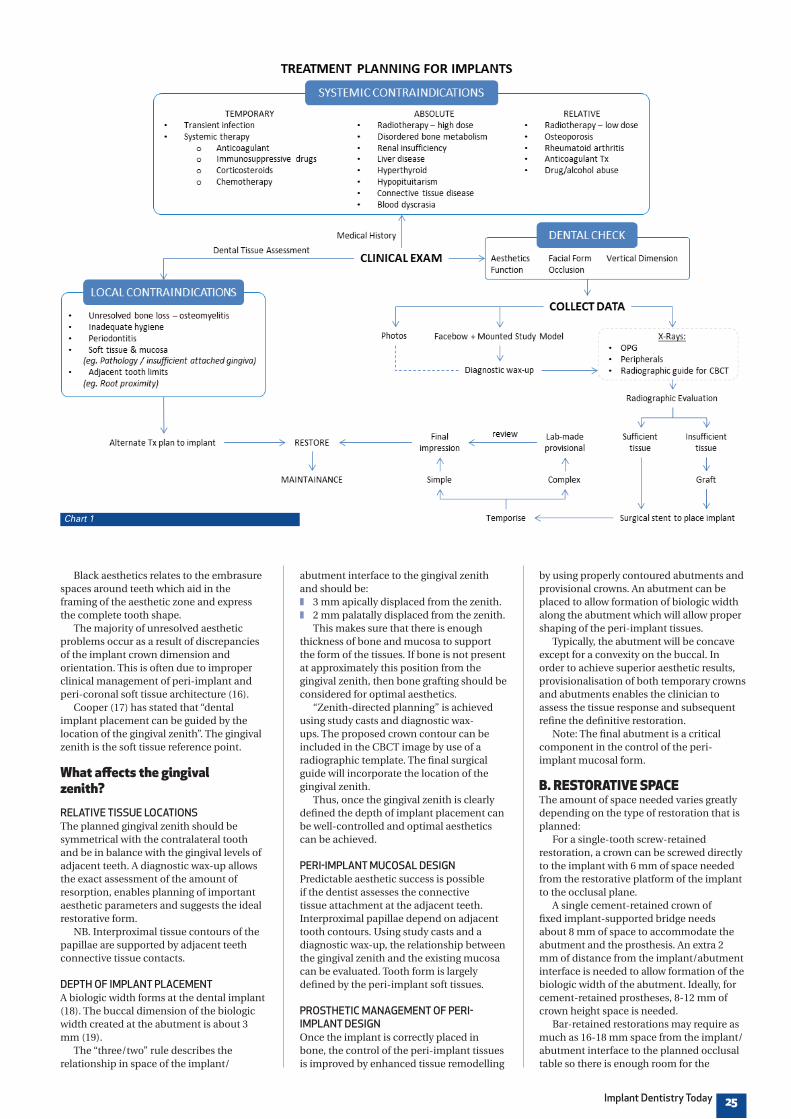
SYSTEMIC DISEASE/RADIO/CHEMOTHERAPY: (SEE CHART 1)Several local factors can affect implant success (Table 1) and the one of most notable being periodontitis. Several reports have shown that periodontal disease increases the chances of periimplantitis (7).
DATA COLLECTIONBoth the complexity of implant placement and subsequent restoration can be managed by thorough and methodical treatment planning procedures (Chart 1).
Accompanying perioral and intraoral photographs provide valuable information of local conditions to augment treatment planning and hence provision of successful implant prostheses.
All implant treatment cases should start with accurate study models taken with a facebow and mounted on a semi-/fully-adjustable articulator.
Mounted models allow:n accurate and easy measurement of
residual alveolar ridges and interproximal spaces
n assessment of available interocclusal space
n assessment of space in the implant receptor site
n aesthetic analysisn occlusal analysis.
Some single tooth cases can be mounted in the habitual intercuspidation position and other larger rehabilitation cases particularly when there are changes in vertical dimension need to be mounted in centric relation.
Implants tolerate axial load better than lateral load (8). In order to avoid excessive lateral loads on a posterior implant, desirable occlusal schemes would include well-designed immediate anterior guidance (9).Implant-supported restorations are more vulnerable to occlusal overloading than natural teeth (10) and any parafunction should be identified as this will affect the long-term predictability of the implant (11).
The osseous condition of the intended implant site is an essential part of the diagnostic data. The osseous architecture and quality in relation to the contours of the proposed dental prosthesis have to be evaluated.
Radiographic reviews should include:n volumetric assessment of available
bone which is best achieved by use of CBCT (12). A CBCT image offers careful measurement of mesiodistal, buccolingual and occlusogingival osseous dimensions and the topography of the edentulous space.
n periapical radiographs done with a long-cone paralleling technique (13) are still important to help evaluate adjacent tooth connective tissue attachment and bone levels in the anterior maxillary aesthetic zone. Radiopaque millimetre grids can be superimposed over the film before it is exposed (14).
ACHIEVING SUCCESSFUL OUTCOMES: WHAT TO CONSIDER?A. AESTHETICSAesthetic anterior implant-retained restorations should involve a smile analysis to achieve the most optimal outcome. This is most easily attained by the use of diagnostic intraoral photographs of the anterior tooth display.
A smile analysis is done by careful evaluation of the pink, white and black components of the smile.
Pink aesthetics relates to the examination of the shape of the gingival tissues around the gingival margins and edentulous spaces.
White aesthetics relates to the study of the tooth morphology, position and axial inclination of the clinical crowns within the arch of the aesthetic zone.
Implant Dentistry Today 25
Black aesthetics relates to the embrasure spaces around teeth which aid in the framing of the aesthetic zone and express the complete tooth shape.
The majority of unresolved aesthetic problems occur as a result of discrepancies of the implant crown dimension and orientation. This is often due to improper clinical management of peri-implant and peri-coronal soft tissue architecture (16).
Cooper (17) has stated that “dental implant placement can be guided by the location of the gingival zenith”. The gingival zenith is the soft tissue reference point.
What affects the gingival zenith?
RELATIVE TISSUE LOCATIONSThe planned gingival zenith should be symmetrical with the contralateral tooth and be in balance with the gingival levels of adjacent teeth. A diagnostic wax-up allows the exact assessment of the amount of resorption, enables planning of important aesthetic parameters and suggests the ideal restorative form.
NB. Interproximal tissue contours of the papillae are supported by adjacent teeth connective tissue contacts.
DEPTH OF IMPLANT PLACEMENTA biologic width forms at the dental implant (18). The buccal dimension of the biologic width created at the abutment is about 3 mm (19).
The “three/two” rule describes the relationship in space of the implant/
abutment interface to the gingival zenith and should be:z 3 mm apically displaced from the zenith.z 2 mm palatally displaced from the zenith.
This makes sure that there is enough thickness of bone and mucosa to support the form of the tissues. If bone is not present at approximately this position from the gingival zenith, then bone grafting should be considered for optimal aesthetics.
“Zenith-directed planning” is achieved using study casts and diagnostic wax-ups. The proposed crown contour can be included in the CBCT image by use of a radiographic template. The final surgical guide will incorporate the location of the gingival zenith.
Thus, once the gingival zenith is clearly defined the depth of implant placement can be well-controlled and optimal aesthetics can be achieved.
PERI-IMPLANT MUCOSAL DESIGNPredictable aesthetic success is possible if the dentist assesses the connective tissue attachment at the adjacent teeth. Interproximal papillae depend on adjacent tooth contours. Using study casts and a diagnostic wax-up, the relationship between the gingival zenith and the existing mucosa can be evaluated. Tooth form is largely defined by the peri-implant soft tissues.
PROSTHETIC MANAGEMENT OF PERI-IMPLANT DESIGNOnce the implant is correctly placed in bone, the control of the peri-implant tissues is improved by enhanced tissue remodelling
by using properly contoured abutments and provisional crowns. An abutment can be placed to allow formation of biologic width along the abutment which will allow proper shaping of the peri-implant tissues.
Typically, the abutment will be concave except for a convexity on the buccal. In order to achieve superior aesthetic results, provisionalisation of both temporary crowns and abutments enables the clinician to assess the tissue response and subsequent refine the definitive restoration.
Note: The final abutment is a critical component in the control of the peri-implant mucosal form.
B. RESTORATIVE SPACEThe amount of space needed varies greatly depending on the type of restoration that is planned:
For a single-tooth screw-retained restoration, a crown can be screwed directly to the implant with 6 mm of space needed from the restorative platform of the implant to the occlusal plane.
A single cement-retained crown of fixed implant-supported bridge needs about 8 mm of space to accommodate the abutment and the prosthesis. An extra 2 mm of distance from the implant/abutment interface is needed to allow formation of the biologic width of the abutment. Ideally, for cement-retained prostheses, 8-12 mm of crown height space is needed.
Bar-retained restorations may require as much as 16-18 mm space from the implant/abutment interface to the planned occlusal table so there is enough room for the
Chart 1
Implant Dentistry Today26
abutment, bar, acrylic and teeth.For simple single-tooth implants, the
“Rules of Six” (20) determine if there is adequate space for successful implant restorations:n 6 mm of inter-radicular spacen 6 mm of buccolingual osseous dimensionn 6 mm of minimum implant lengthn 6 mm of interocclusal distance for
prosthetic and component requirementsn Less than 6 mm distance from the bone
crest to the interproximal contact point for papillary formation
n “Three/two” rule – zenith planning (see above)There are three “rules” for treatment
planning for any implant-retained overdentures or implant-supported fixed prosthesis called the “Rules of 10” :n The inferior/superior dimension of the
mandible must be 10 mm or more.n The interocclusal (restorative) dimension
measured from the ridge crest to the occlusal plane must be 10 mm or more. When planning for implant placement, know the planned position of the prosthetic teeth. Plan DOWN from the occlusal plane and not UP from the osseous crest.
n The restorative dimension for any implant prosthesis has 4 key components each with its own dimensions:z Transmucosal dimension (biologic
width) of about 2 mmz Supramucosal abutment height (0-2
mm ) to allow hygienez Framework height 3-5 mmz Acrylic veneer thickness of more than
2 mm Given that there needs to be space
for location of teeth, frameworks, attachments, retaining abutments (balls, bars, etc) and the biologic width at least 15 mm is advised.
n The anterior/posterior distribution of implants must be at least 10 mm. The aim is to have the distal implant in the distal-most location not impose on the inferior dental nerve.The all-on-four concept requires that
posterior teeth beyond the first premolar are supported by a cantilever. The aim is to reduce or eliminate the cantilever by distal placement of terminal implants.
CONCLUSIONVarious considerations apply with patient selection and implant treatment planning. The systemic and oral health of the patient, careful intraoral assessment of soft tissue and residual supporting tissues and the planned restoration have to be evaluated. As much diagnostic information as possible needs to be collected with photographs, various radiographic images and study models with diagnostic wax-ups of the
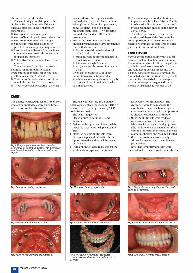
Fig. 9 The final restorations were placed.
Fig. 6 reveals buccal view of abutments in situ.
Fig. 2a – upper healing caps in situ.
Fig.1 This preoperative view illustrated the destructive periodontitis evident with significant attachment loss and associated loss of posterior support.
Fig. 8 The completed implant-supported prostheses were shown to the patient prior to insertion.
Fig. 5 shows occlusal view of abutments.
Fig. 2b – lower healing caps in situ.
Fig. 7 shows occlusal view of abutments.
Fig. 4 reveals the abutments in situ.
Fig. 3 The position and angulation of impression copings is indicated.
The dentist requested upper and lower fixed implant-supported telescopic prostheses with custom-milled abutments.
The aim was to restore 16-26 on the maxilla and 36-46 on the mandible. If there was too much overhang, then only 35-45 would be restored.
The dentist requested:1. Please mount upper model using
facebow.2. Articulate the upper and lower models
using the clear denture duplicates as a bite.
3. Make the custom abutments with a 12 degree taper and milled finish. The option existed to either mill the wax-up or the metal.
4. Duralay bonnets were requested for the abutments for a pick-up impression
for accuracy for the final PFM. The abutments were to be placed in the mouth, then the acrylic bonnets placed over them and then a pick-up impression to check the accuracy of the model.
5. Once the abutments were made, an acrylic temporary restoration was to be fabricated including artificial gingiva to fully replicate the final restoration. These were to be inserted in the mouth and the aesthetics checked and the bite adjusted.
6. Once the provisionals were finally adjusted, the plan was to complete one jaw at a time.Note: The temporary dentures were
included in the case as a guide for aesthetics.
CASE 1:
Implant Dentistry Today 27
proposed prostheses. Adherence to the Rules of 6 and Rules of 10 will allow the treating clinician to plan for sufficient space for restorations which will be both aesthetic and achieve longevity.
Southern Cross Dental would like to thank Dr Lincoln Harris, Queensland who graciously permitted publication of his cases. n
BIBLIOGRAPGHY1. Palmqvist S, Swartz B. Artificial crowns and fixed
partial dentures 18 to 23 years after placement. Int J Prosthodont 1993;6:279-85
2. Heitz-Mayfield LJ. Peri-implant diseases: diagnosis and risk indicators. J Clin Periodontol 2008;35 (Suppl 8):292-304.
3. Baig MG, Rajan M. Effects of smoking on the outcome of implant treatment: a literature review. Indian J Dent Res 2007;18(4): 190-5.
4. Strietzel FP, Reichart PA, Kale A, et al. Smoking interferes with the prognosis of dental implant treatment: a systematic review and meta-analysis. J Clin Periodontol 2007;34(6):523-44.
5. Fiorellini JP, Nevins ML. Dental implant considerations in the diabetic patient. Periodontol 2000 2000;23:73-7.
6. Bornstein MM, Cionca N, Mombelli A. Systemic conditions and treatments as risks for implant therapy. Int J Oral Maxillofac Implants 2009;24(Suppl):12-27.
7. Van Der Weijden GA, van Bemmel KM, Renvert S. Implant therapy in partially edentulous, periodontally compromised patients: a review. J Clin Periodontol 2005;32(5):506-11.
8. Isidor F. Influence of forces on peri-implant bone. Clin Oral Implants Res 2006;17:8-18.
9. Gross MD. Occlusion in implant dentistry. A review of the literature of prosthetic determinants and current concepts. Aust Dent J 2007;53:60-8.
10. Merikske-Stern R, Geering AH, Burgin WB, graf H. Three dimensional force measurements on mandibular implants supporting overdentures. Int J Oral Maxillofac Implants 1992;7:185-94.
11. Handelman M. Surgical guidelines for dental implant placement. Br Dent J 2006;201:139-52.
12. Harris D, Horner K, Grondahl K, et al. E.A.O. guidelines for the use of diagnostic imaging in implant dentistry 2011. A consensus workshop organized by the European Association for Osseointegration at the Medical University of Warsaw. Clin Oral Implants Res 2012;23(11);1243-53.
13. Floyd P, Lamer P, Palmer R. Radiographic treatment planning. R I Dent J 1990;23:5-11.
14. Misch CE. Contemprorary Implant Dentistry. 3rd ed. St Louis: Mosby;2-1-/ [/40.130-45,245-50,280.
15. Bryington M, Ingeborg J. De Kik, Thalji G, Cooper LF. Patient Selection and Treatment Planning for Implant Restorations Dent Clin N Am 58 (2014) 193-206.
16. Jeijer HJ, Stellingsma K, Meijndert L, Raghoebar GM. A new index for rating aesthetics of implant-supported single crowns and adjacent soft tissues – the Implant Crown Aesthetic Index. Clin Oral Implants Res 1999;10:185-94.
17. Cooper LE. Objective CriteriaL Guiding and Evaluating Dental Implant Esthetics. Journal Compilation 2008 Vol 20:3 195-205.
18. Hermann JS, Buser D, Schenk RK, et al. Biologic with around titanium implants. A physiologically formed and stable dimension over time. Clin Oral Implants Res 2000;11:1-11.
19. Kan JY, Rungcharassaeng K, Umezu K, Kois JC. Dimensions of peri-implant mucosa: an evaluation of maxillary anterior single implants in humans. J Periodontol 2003;74:557-62.
20. Cooper LF, Pin-Harry OC. “Rules of Six” – diagnostic and therapeutic guidelines for single-tooth implant success. Compend Contin Educ Dent 2013;34(2):94-8, 100-1;quiz 102,117.
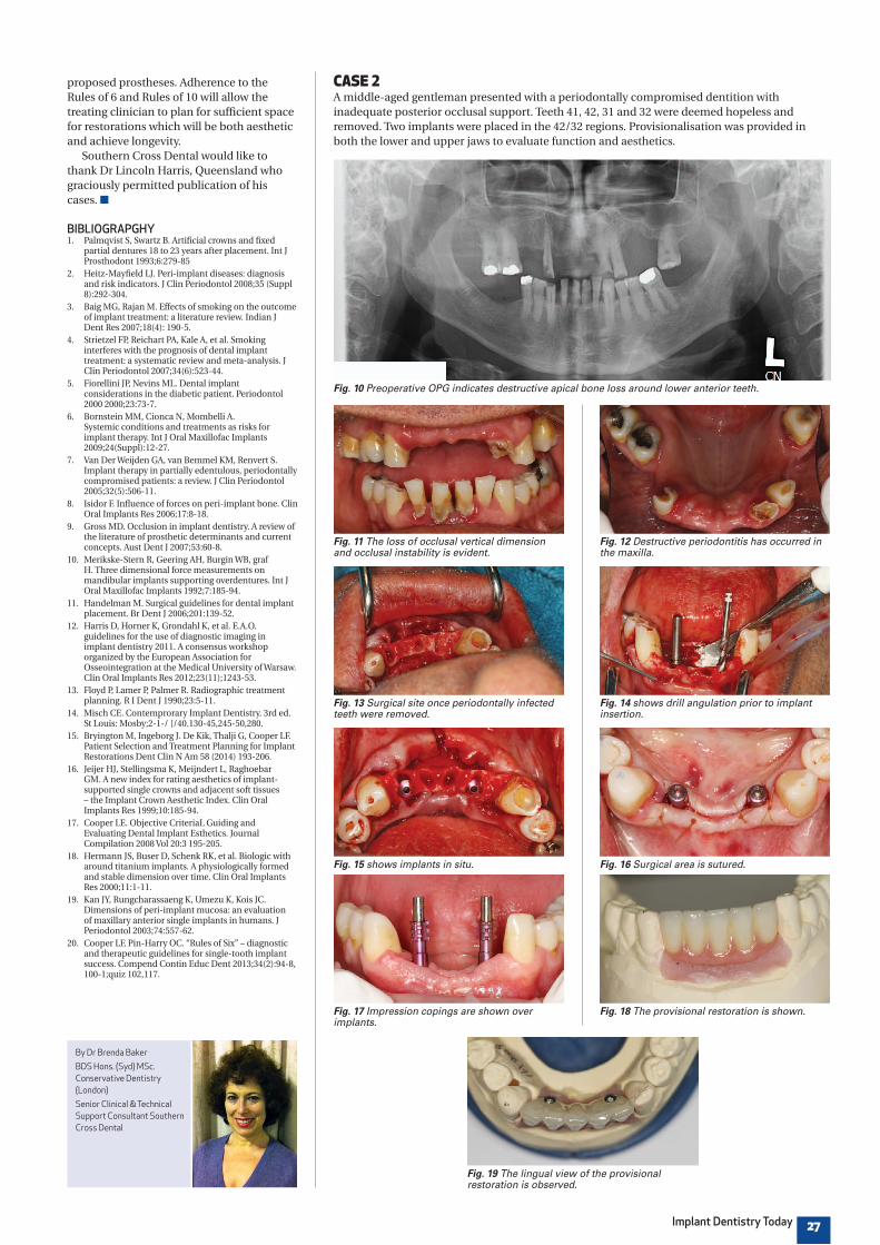
Fig. 19 The lingual view of the provisional restoration is observed.
Fig. 17 Impression copings are shown over implants.
Fig. 15 shows implants in situ.
Fig. 13 Surgical site once periodontally infected teeth were removed.
Fig. 11 The loss of occlusal vertical dimension and occlusal instability is evident.
Fig. 18 The provisional restoration is shown.
Fig. 16 Surgical area is sutured.
Fig. 14 shows drill angulation prior to implant insertion.
Fig. 12 Destructive periodontitis has occurred in the maxilla.
Fig. 10 Preoperative OPG indicates destructive apical bone loss around lower anterior teeth.
CASE 2A middle-aged gentleman presented with a periodontally compromised dentition with inadequate posterior occlusal support. Teeth 41, 42, 31 and 32 were deemed hopeless and removed. Two implants were placed in the 42/32 regions. Provisionalisation was provided in both the lower and upper jaws to evaluate function and aesthetics.
By Dr Brenda Baker BDS Hons. (Syd) MSc. Conservative Dentistry (London) Senior Clinical & Technical Support Consultant Southern Cross Dental