Defective Bud Formation Human Cells Chronically Infected...
15
JOURNAL OF VIROLOGY, Aug. 1976, p. 579-593 Copyright ©D 1976 American Society for Microbiology Vol. 19, No. 2 Printed in U.S.A. Defective Bud Formation in Human Cells Chronically Infected with Subacute Sclerosing Panencephalitis Virus M. DUBOIS-DALCQ,* T. S. REESE, M. MURPHY, AND D. FUCCILLO Infectious Diseases Branch and Laboratory of Neuropathology and Neuroanatomical Sciences, National Institute of Neurological and Communicative Disorders and Stroke, Bethesda, Maryland 20014 Received for publication 20 January 1976 Human prostate cells chronically infected with the Mantooth strain of sub- acute sclerosing panencephalitis (SSPE) virus multiply normally, fuse only occasionally to form giant cells, and yet have twisted intracytoplasmic nucleo- capsids. These cells are able to support replication of vesicular stomatitis virus, although they release only small amounts of SSPE virus. To determine why carrier cells do not produce virus, they were examined with techniques for surface replication, freeze-fracturing, and immunoperoxidase labeling with SSPE antibody. The surface of carrier cells, like that of productive cells, is char- acterized by ridges crowned with viral antigens and devoid of the intramem- brane particles revealed by freeze-fracture techniques. Since surface ridges form where nucleocapsids attach to the membrane, the shape and length of ridges are indicative of the shape and length of the underlying nucleocapsid. Whereas ridges on productive cells are serpentine in shape, those on carrier cells are typically straight or hairpin shaped, and the hairpin ridges are twice as long as serpentine ridges on productive cells. Furthermore, the spacing between ridges on carrier cells is never as small as that in productive infections, so that con- tinuous sheets of viral membrane are never formed. The majority of carrier cells lack the round viral buds observed in productive cells but have, instead, many elongated processes attached to the cell surface. Each of these processes contains one or two hairpin ridges overlying hairpin-shaped nucleocapsids. These "hair- pin buds" are restricted to a single region of the carrier cell surface, whereas viral buds are distributed over the entire surface of productive cells. Thus, there are several structural defects in carrier cells that depend on the specific inter- action of a certain viral strain with a certain cell type. These defects prevent the deployment of viral antigen in some regions of the cell surface, the formation of nucleocapsids of normal length, the coiling of attached nucleocapsids, and the consolidation of sheets of viral membrane into spherical buds with the nucleo- capsids coiled inside. These defects may account for the failure of carrier cells to shed infectious virus. Cultured cells can be chronically infected with measles virus and therefore constitute a tool to study the mechanism of chronic measles infection (11, 12, 22, 25-27, 30, 35, 36). By selec- tion of a less destructive virus, or of cells resist- ant to lysis by the virus, chronically infected measles carrier cells have been obtained with- out the use of antibody (27). These carrier cells show almost no cytopathic effects and produce a small amount, if any, of infectious virus. While they contain intracytoplasmic viral anti- gen as well as nucleocapsids embedded in a granular coat (21, 25), the amount and distribu- tion of membrane antigens vary in different cell lines. In some, no viral antigens are found on the plasma membrane (25), whereas in others a polar distribution has been observed (11). However, no alignment of nucleocapsids or budding of virus has been detected (21, 25). Similar carrier cell lines chronically infected with subacute sclerosing panencephalitis (SSPE) strains of measles virus were described recently (3, 5). These cell lines were derived by co-cultivating indicator cells with cells from brains of SSPE patients. One of these carrier cell lines is characterized by numerous smooth nucleocapsids and the absence of nucleocapsid alignment and viral bud formation on giant cells (7). However, another type of carrier state, characterized by a paucity in giant cells, can be induced by direct infection of cultured cells with an SSPE strain (37). This carrier cell line was chosen for our study because a high per- centage of the cells carried viral antigens (37). 579 on July 7, 2018 by guest http://jvi.asm.org/ Downloaded from
Transcript of Defective Bud Formation Human Cells Chronically Infected...
JOURNAL OF VIROLOGY, Aug. 1976, p. 579-593Copyright ©D 1976 American Society for Microbiology
Vol. 19, No. 2Printed in U.S.A.
Defective Bud Formation in Human Cells ChronicallyInfected with Subacute Sclerosing Panencephalitis Virus
M. DUBOIS-DALCQ,* T. S. REESE, M. MURPHY, AND D. FUCCILLO
Infectious Diseases Branch and Laboratory ofNeuropathology and Neuroanatomical Sciences, NationalInstitute of Neurological and Communicative Disorders and Stroke, Bethesda, Maryland 20014
Received for publication 20 January 1976
Human prostate cells chronically infected with the Mantooth strain of sub-acute sclerosing panencephalitis (SSPE) virus multiply normally, fuse onlyoccasionally to form giant cells, and yet have twisted intracytoplasmic nucleo-capsids. These cells are able to support replication of vesicular stomatitis virus,although they release only small amounts of SSPE virus. To determine whycarrier cells do not produce virus, they were examined with techniques forsurface replication, freeze-fracturing, and immunoperoxidase labeling withSSPE antibody. The surface of carrier cells, like that of productive cells, is char-acterized by ridges crowned with viral antigens and devoid of the intramem-brane particles revealed by freeze-fracture techniques. Since surface ridges formwhere nucleocapsids attach to the membrane, the shape and length of ridges areindicative of the shape and length of the underlying nucleocapsid. Whereasridges on productive cells are serpentine in shape, those on carrier cells aretypically straight or hairpin shaped, and the hairpin ridges are twice as long asserpentine ridges on productive cells. Furthermore, the spacing between ridgeson carrier cells is never as small as that in productive infections, so that con-
tinuous sheets of viral membrane are never formed. The majority of carrier cellslack the round viral buds observed in productive cells but have, instead, manyelongated processes attached to the cell surface. Each ofthese processes containsone or two hairpin ridges overlying hairpin-shaped nucleocapsids. These "hair-pin buds" are restricted to a single region of the carrier cell surface, whereasviral buds are distributed over the entire surface of productive cells. Thus, thereare several structural defects in carrier cells that depend on the specific inter-action of a certain viral strain with a certain cell type. These defects prevent thedeployment of viral antigen in some regions of the cell surface, the formation ofnucleocapsids of normal length, the coiling of attached nucleocapsids, and theconsolidation of sheets of viral membrane into spherical buds with the nucleo-capsids coiled inside. These defects may account for the failure of carrier cells toshed infectious virus.
Cultured cells can be chronically infectedwith measles virus and therefore constitute atool to study the mechanism of chronic measlesinfection (11, 12, 22, 25-27, 30, 35, 36). By selec-tion of a less destructive virus, or of cells resist-ant to lysis by the virus, chronically infectedmeasles carrier cells have been obtained with-out the use of antibody (27). These carrier cellsshow almost no cytopathic effects and producea small amount, if any, of infectious virus.While they contain intracytoplasmic viral anti-gen as well as nucleocapsids embedded in agranular coat (21, 25), the amount and distribu-tion of membrane antigens vary in differentcell lines. In some, no viral antigens are foundon the plasma membrane (25), whereas inothers a polar distribution has been observed
(11). However, no alignment of nucleocapsidsor budding of virus has been detected (21, 25).
Similar carrier cell lines chronically infectedwith subacute sclerosing panencephalitis(SSPE) strains of measles virus were describedrecently (3, 5). These cell lines were derived byco-cultivating indicator cells with cells frombrains of SSPE patients. One of these carriercell lines is characterized by numerous smoothnucleocapsids and the absence of nucleocapsidalignment and viral bud formation on giantcells (7). However, another type of carrier state,characterized by a paucity in giant cells, can beinduced by direct infection of cultured cellswith an SSPE strain (37). This carrier cell linewas chosen for our study because a high per-centage of the cells carried viral antigens (37).
579
on July 7, 2018 by guesthttp://jvi.asm
.org/D
ownloaded from
580 DUBOIS-DALCQ ET AL.
The application of freeze-fracturing and sur-face replication techniques combined with im-munolabeling methods has demonstrated ex-tensive changes in membrane structure duringviral development in productive infection withSSPE virus (9). In the present study, we com-pare the biological properties and structure ofone SSPE carrier cell line with those of cellsproductively infected with measles and SSPEviruses to identify the defects that prevent theproduction of infectious virus in carrier cells.
MATERIALS AND METHODS
Virus and cells. Cells from a continuous cell linederived from a human prostate adenoma (MA 160cells) were infected with the Mantooth strain ofSSPE virus to produce a carrier cell line. The Man-tooth virus was isolated by co-cultivating brain cellsin primary cultures with HeLa cells (17). Free virusisolated from these co-cultures was used after onemore passage in HeLa cells. A carrier cell line desig-nated MA 72046 was established with these virusesin MA 160 cells by M. M. Vincent (MicrobiologicalAssociates, Inc., Bethesda, Md.) by the method ofRustigian (35). For the ultrastructural study, cellsfrom passages 25 to 30 of the carrier cell line (MA72046) were usually studied, but later passages, aswell as uninfected cells (MA 160), were sampledalso. For studies with thin sectioning or freeze-frac-turing, cells were treated with trypsin and seededinto large Falcon plastic flasks. Monolayers of cellswere also grown in 35-mm plastic petri dishes con-taining 12-mm glass cover slips in preparation forsurface replication and immunolabeling experi-ments. In some instances, 1 volume of trypsin-treated carrier cells was co-cultivated with 1 or 2volumes of Vero cells (Flow Laboratories, Inc.,Rockville, Md.) and observed for the occurrence ofcytopathic effect (17). The growth medium for allexperiments consisted of Eagle minimum essentialmedium supplemented with 10% heat-inactivatedfetal calf serum and 100 U of penicillin G and 100 ,ugof streptomycin sulfate per ml. Cultures were incu-bated in a 37°C incubator with 5% CO2.
For comparative studies, productive infectionwith four strains of virus was produced in Vero cells.Viruses were gathered from the fifth passage ofmeasles virus Edmonston B strain, the third pas-sage of SSPE virus Mantooth strain (17), the fourthpassage of SSPE virus Halle strain (9, 15), and thethird passage of SSPE virus McClellan strain (6), allin Vero cells. The SSPE Halle virus was plaquepurified and also studied in MA 160 cells. In allexperiments with measles or SSPE viruses, multi-plicity of infection varied between 0.01 and 1. TheIndiana strain of vesicular stomatitis virus (VSV)was obtained from Robert Lazzarini and inoculatedinto carrier cells at a multiplicity of infection of 10.Titrations ofVSV were done in baby hamster kidney(BHK-21) cells, and plaques were counted at 18 h(4).
Test for virus production and measles antigenic-ity. Free virus and cell-associated virus were as-sayed by the PFU test on Vero cells grown in a
medium containing methylcellulose (4). Hemad-sorption was performed with rhesus monkey eryth-rocytes (24). Indirect fluorescent-antibody stainingwas performed on cells fixed in methanol for thedetection of intracellular antigen (15) and on livingcells for the detection of membrane antigen (20).Indirect immunoperoxidase (IP) labeling was per-formed after glutaraldehyde fixation for the detec-tion of surface antigen as described previously (6, 7).Briefly, various dilutions of SSPE serum with highmeasles antibody titers were first applied to the cellsfor 1 h. Monolayers were then rinsed, and goat anti-human immunoglobulin G coupled to peroxidase(Miles Laboratories, Rehovoth, Israel) was added ata globulin concentration of 0.05 mg/ml. After 1 h,cells were washed overnight, postfixed in glutaral-dehyde, and incubated in a peroxide-diaminobenzi-dine substrate (6). As a control for specificity, in-fected cells were treated similarly with human se-rum with no measles antibodies detectable by thehemagglutination inhibition and fluorescent-anti-body tests.
Electron microscopy. Viral particles and intra-cellular nucleocapsids were examined after negativestaining. For free virus, supernatant fluids werecollected from 3- to 7-day-old cultures, whereas forcytoplasmic nucleocapsids the cells were disruptedby osmotic shock. After clarification, both free virusand nucleocapsids were concentrated at 37,000 x gfor 30 min at 4°C (18). Sediments were suspended ina small amount of culture medium, stained with 2%potassium phosphotungstate (pH 6.8), and layeredon Formvar-coated grids.
For surface replication (2, 9), cells grown on glasscover slips were fixed in 1% glutaraldehyde in 0.1 Mcacodylate buffer for 30 min, rinsed, and postfixed in1% osmium tetroxide in the same buffer for 1 h.Osmium fixation was omitted in immunolabelingexperiments. After fixation, cells were dehydratedquickly and air dried, or, in a few instances, thecritical point drying method was used instead (1).Cover slips with dried cells were placed on top of thespecimen stage of a Balzer 360 M freeze-etch appara-tus, and a replica was made at 105°C by using anelectron beam gun for platinum shadowing.
For freeze-fracturing, fixation was performedwith the aldehyde fixative described previously (9).Cells were then scraped out of the flasks, centri-fuged at 3,000 rpm, rinsed with buffer, progressivelyequilibrated with 25% glycerol in water, frozen inFreon 22, and stored in liquid nitrogen prior to beingfractured in the freeze-etch apparatus at - 118C.All replicas were cleaned in methanol and Chloroxbefore being mounted on Formvar-coated grids.
For thin sectioning, pellets as well as monolayersof immunolabeled cells were postfixed in osmiumtetroxide, stained with uranyl acetate at pH 5, dehy-drated in graded alcohols, and embedded in Epon.Electron micrographs were taken on a Philips 201electron microscope. Some carrier cells were seriallysectioned and mounted on Formvar-coated slotgrids.
Structures seen in surface replicas and negativelystained preparations were measured with a Hew-lett-Packard Digitizer. Student's t test was used forthe statistical analysis.
J. VIROL.
on July 7, 2018 by guesthttp://jvi.asm
.org/D
ownloaded from

BUD FORMATION IN SSPE VIRUS-INFECTED CELLS 581
RESULTS
Biological properties. Table 1 compares theproperties of the SSPE Mantooth virus-infectedcarrier cells with those of Vero cells produc-tively infected with the Halle strain of SSPEvirus. Productive infections of cells with otherSSPE and measles strains were also studied,but their properties were quite similar to theHalle infection. Therefore, the properties ofHalle virus infection given in Table 1 are repre-
sentative of productive infection with otherstrains of measles virus. The carrier cellsshowed few cytopathic changes even 7 daysafter transfer, whereas 3 days after inoculationwith the Halle strain most of the Vero cellswere lysed and detached from the substrate. Asmall proportion of carrier cells had 5 to 10nuclei, whereas giant cells in the productiveinfection had hundreds of nuclei and coveredtwo-thirds of the monolayer even after 2 days.Fusion and lysis were also characteristic ofMA160 cells infected with SSPE Halle strain and ofVero cells infected with SSPE Mantooth andMcClellan strains as well as the Edmonston Bstrain of measles virus. However, in these in-stances, the infection took 2 or 3 days longer tospread over the entire monolayer. The amountof hemadsorption was identical in both carrierand productive cells. In productive infections,the number of Vero cells with intracytoplasmicviral antigen detected by fluorescent antibodyincreased from 50% on day 2 to 100% on day 3after viral inoculation, whereas in the chronicinfection 80 to 90% of the cells contained viralantigen 2 days after a passage, when the cellswere still in the growth phase. Cytoplasmicinclusions of viral antigen were often found incarrier cells undergoing mitosis. Membrane an-
tigen, as detected by fluorescent antibody or IPstaining, was present in both chronic and pro-ductive infection but was evenly distributed incells productively infected with the SSPE Hallestrain and present only at the poles of most ofthe carrier cells (11).Plaque assays of carrier cultures revealed
less than 50 infectious particles per ml of thesupernatant fluid. Freezing and thawing car-
rier cells from the same passage liberated as
many as 105 PFU/ml into the medium. In con-
trast, the supernatant fluids of cultured Verocell monolayers infected with SSPE Mantooth,Halle, and McClellan viruses and measles Ed-monston B viruses had titers of 6 x 10⁵, 1 X 107,1.4 x 105, and 5 x 10⁶ PFU/ml, respectively.The titers of cell-associated virus were 3 to 4logarithmic units lower than those of the cell-free virus. Infectious particles obtained fromthe carrier cultures produced smaller plaquesthat formed after longer incubation periodsthan with control viruses.
In other experiments, monolayers of carriercells were superinfected with measles Edmon-ston B, SSPE Halle, or SSPE Mantooth virus.Cytopathic changes in the carrier cells were
evident in all instances. Infectious particles attiters as high as 6 x 10⁴ PFU/ml were detectedin the supernatant fluid at 72 h after superin-fection with all viruses except SSPE Mantooth.However, our titrations did not allow distinc-tion between the newly assembled superinfect-ing virus and infectious particles from the Man-tooth virus already in the carrier cells. VSValso reproduced well in carrier cell monolayers,resulting in 1.6 x 10⁹ PFU/ml of supernatant,but the normal 12- to 14-h lytic cycle was ex-
tended to more than 30 h.Subcellular structure. The sequence of
membrane changes in carrier cells differedgreatly from those in productive SSPE virusinfection (6, 7, 9, 15, 33, 34). In productive infec-tions in MA 160 or Vero cells infected withSSPE Halle strain, as well as in Vero cellsinfected with SSPE Mantooth strain or themeasles Edmonston B strain, the surface of thegiant cells was covered with twisted ridges seen
in surface replicas (Fig. 1). These were orientedin various directions and assumed sinuousshapes, like an S, or a U shape (Fig. 1). Ridgeswere seen in areas in which the virus-modifiedmembrane protruded over a nucleocapsid lyingunder the plasmalemma (Fig. 2, arrows) (9).
TABLE 1. Properties ofproductive and carrier cell lines
Infective particlesFre- Cells Distribution (PFU/ml)
Cells Cytopathic quency Hemad- with of viral anti- Appearance ofeffect of cell fu- sorption viral an- Cell plaques
sion tigen (%) gen Cell freea asso-ciatedb
Productive" + + + + + + + + 100 Diffuse 107 10: Early and largeCarrier' - + + + 80-90 Polar 0-100 105 Late and various
sizes
Supernatant.b Freeze-thawed.Halle strain in Vero.
" Mantooth strain in human prostate cells.
VOL. 19, 1976
on July 7, 2018 by guesthttp://jvi.asm
.org/D
ownloaded from
~~~~~~ - . t_4
4%4
,~~~~~~~~~~~~~~~~~~~I
8;r_r t f
tAL~ ~ ~ ~ A
.rC ~ -. -s4-
.. .. ~~~~~~~~~~~~~~lr2~~~~~~~~~~
FIG. 1. Surface replica of a human prostate cell (MA 160) acutely infected with the Halle strain ofSSPEvirus. Much of the surface of this giant cell is covered with twisted ridges and viral buds (asterisks). Theridges have a random orientation and assume various shapes such as an S or 0, or a U (short arrows). Most ofthe viral buds are spherical and covered with ridges, which are more closely apposed to each other than on theadjacent membrane. A few buds are club shaped (long arrows). x18,000.
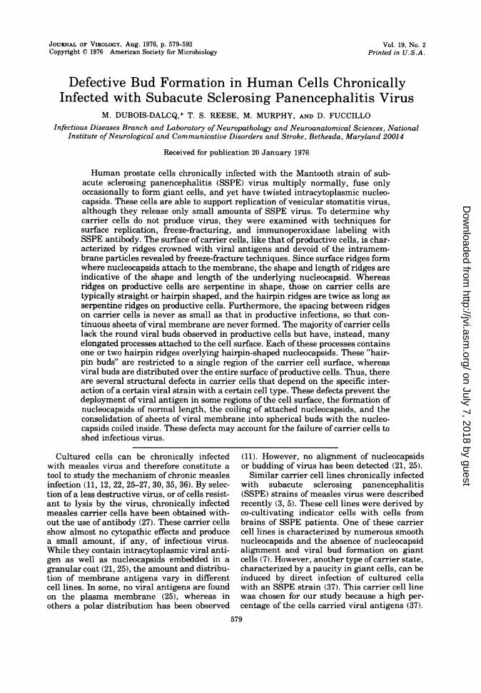
FIG. 2. Thin section of a Vero cell acutely infected with the Halle strain of SSPE virus. Groups of viralbuds and viruses cluster near the surface ofa cell containing nucleocapsids. Ridges are scattered over the cellsurface (arrows). The limiting membrane of the round buds has an increased density due to the presence of acontinuous coat ofsurface projections and ofan internal fuzz around the nucleocapsids. Nucleocapsids are notseen clearly at this magnification. x25,000.
582
on July 7, 2018 by guesthttp://jvi.asm
.org/D
ownloaded from
BUD FORMATION IN SSPE VIRUS-INFECTED CELLS 583
Twisted ridges were also present on the viralbuds, and the spacing between them was lessthan in the surrounding cell membrane (Fig. 1)(9). Most of the viral buds were spherical al-though some were club shaped (Fig. 1, longarrows). That these structures were indeedviral buds is illustrated in Fig. 2, which showsviral buds and particles containing nucleocap-sids in a thin section through a cell at a stage ofviral production identical to that shown in Fig.1. The membrane surrounding viral buds orcovering nucleocapsids at ridges was character-ized by projections on its outer surface and afuzzy material on the inner surface of the mem-brane. Groups of nucleocapsids were also seenin the cytoplasma (Fig. 2).Ninety percent of the carrier cells, when ex-
amined in thin sections (Fig. 3), typically con-tained cytoplasmic viral inclusions similar tothose found in productive cells. However, nonuclear inclusions of the type seen in latestages of productive infection (15, 33, 34) werefound. The cytoplasmic inclusions consisted oftwisted tubules surrounded by a fuzzy coat.Since cytoplasmic nucleocapsids isolated fromthese cells displayed the typical herringbonestructure of pseudomyxovirus nucleocapsids(Fig. 3, left inset) (31), these tubules were as-sumed to be nucleocapsids. The isolated nucleo-capsids were measured and compared withthose obtained from productive cells; no signifi-cant differences in length were found. Sincenucleocapsids are frequently fragmented dur-ing concentration (Fig. 3) (13, 39), comparativemeasurements of their lengths are open toquestion.
Carrier cells usually differ from productivecells by the presence of elongated processesforming groups at one pole of the cell (Fig. 3).Examination of these processes in serial sec-tions showed that they are usually branchedand are extensions of the carrier cell plasmamembrane. Unlike the normal microvilli, theplasma membrane limiting those processes hadan increased density (Fig. 3) similar to thatobserved on round viral buds on productivecells. With higher magnification, it was appar-ent that the characteristic membrane densitywas mainly due to surface projections (Fig. 3,right inset; see Fig. 14). Details of the internalstructure of these processes are given below.
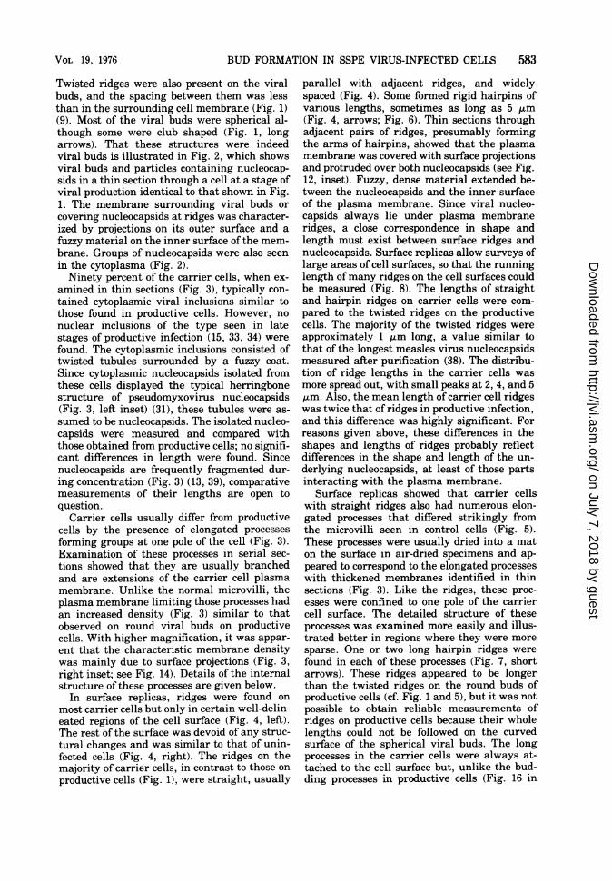
In surface replicas, ridges were found onmost carrier cells but only in certain well-delin-eated regions of the cell surface (Fig. 4, left).The rest of the surface was devoid of any struc-tural changes and was similar to that of unin-fected cells (Fig. 4, right). The ridges on themajority of carrier cells, in contrast to those onproductive cells (Fig. 1), were straight, usually
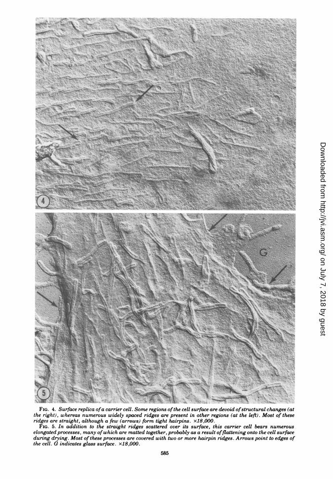
parallel with adjacent ridges, and widelyspaced (Fig. 4). Some formed rigid hairpins ofvarious lengths, sometimes as long as 5 ,um(Fig. 4, arrows; Fig. 6). Thin sections throughadjacent pairs of ridges, presumably formingthe arms of hairpins, showed that the plasmamembrane was covered with surface projectionsand protruded over both nucleocapsids (see Fig.12, inset). Fuzzy, dense material extended be-tween the nucleocapsids and the inner surfaceof the plasma membrane. Since viral nucleo-capsids always lie under plasma membraneridges, a close correspondence in shape andlength must exist between surface ridges andnucleocapsids. Surface replicas allow surveys oflarge areas of cell surfaces, so that the runninglength of many ridges on the cell surfaces couldbe measured (Fig. 8). The lengths of straightand hairpin ridges on carrier cells were com-pared to the twisted ridges on the productivecells. The majority of the twisted ridges wereapproximately 1 ,um long, a value similar tothat of the longest measles virus nucleocapsidsmeasured after purification (38). The distribu-tion of ridge lengths in the carrier cells wasmore spread out, with small peaks at 2, 4, and 5,tm. Also, the mean length of carrier cell ridgeswas twice that of ridges in productive infection,and this difference was highly significant. Forreasons given above, these differences in theshapes and lengths of ridges probably reflectdifferences in the shape and length of the un-derlying nucleocapsids, at least of those partsinteracting with the plasma membrane.
Surface replicas showed that carrier cellswith straight ridges also had numerous elon-gated processes that differed strikingly fromthe microvilli seen in control cells (Fig. 5).These processes were usually dried into a maton the surface in air-dried specimens and ap-peared to correspond to the elongated processeswith thickened membranes identified in thinsections (Fig. 3). Like the ridges, these proc-esses were confined to one pole of the carriercell surface. The detailed structure of theseprocesses was examined more easily and illus-trated better in regions where they were moresparse. One or two long hairpin ridges werefound in each of these processes (Fig. 7, shortarrows). These ridges appeared to be longerthan the twisted ridges on the round buds ofproductive cells (cf. Fig. 1 and 5), but it was notpossible to obtain reliable measurements ofridges on productive cells because their wholelengths could not be followed on the curvedsurface of the spherical viral buds. The longprocesses in the carrier cells were always at-tached to the cell surface but, unlike the bud-ding processes in productive cells (Fig. 16 in
VOL. 19, 1976
on July 7, 2018 by guesthttp://jvi.asm
.org/D
ownloaded from

..~~ ~ ~ ~ ~ ~ ~ ~
FIG. 3. Thin sections through a carrier cell (MA 72046). A cytoplasmic inclusion of twisted viral tubulesoccupies the upper left corner. These nucleocapsids, when concentrated and negatively stained, show theherringbone structure typical ofpseudomyxovirus nucleocapsids, but they are frequently broken (left inset). Afew microvilli (short arrow) are seen in the upper part of the picture, whereas numerous elongated processesoriginate from other regions of the same cell. These processes are sometimes branched or transversally cut(large arrow), and their membranes look thicker than the normal cell membrane. A portion of one of theseprocesses, framed in the center of the picture, is enlarged in the right inset. This process is covered with acontinuous coat of surface projections, and a portion of one internal tubule can be detected (arrows; see Fig.14). x30,000; left inset, x217,O00; right inset, x140,000.
584
on July 7, 2018 by guesthttp://jvi.asm
.org/D
ownloaded from
N<N-N'I
~~~~~~~~~~~~~~~~~~~~~~~~~~~~~~~~~~~~~~~~~~~A- - \6;i
, -A'*-v;*'N to.A2 \.sj ' 'v-i. l. _ 8V--o \t,> V , AY v ' . '
,- t&t,4~4.. ;: a-
St ,-%
s*;'~~~~~~~~~~~' tb,,aL's.|A
(41s*_ w_+ $-'V-"u'\q %( ;.$'s *ir';--2'a.'-
N~~~~~~~~~~~~~~
-A~~~~~~~~~~~~~~~~~~~~~~~-702. *Al ,,* a,-'99t '~ -. i
Agt
FIG. 4. Surface replica ofa carrier cell. Some regions ofthe cell surface are devoid ofstructural changes (atthe right), whereas numerous widely spaced ridges are present in other regions (at the left). Most of theseridges are straight, although a few (arrows) form tight hairpins. x18,000.
FIG. 5. In addition to the straight ridges scattered over its surface, this carrier cell bears numerouselongated processes, many ofwhich are matted together, probably as a result offlattening onto the cell surfaceduring drying. Most of these processes are covered with two or more hairpin ridges. Arrows point to edges ofthe cell. G indicates glass surface. x18,000.
585
on July 7, 2018 by guesthttp://jvi.asm
.org/D
ownloaded from

It.
4,- -
; - w w E--
"S4,~~~~~~~~~~~~~~~~~~~~~
AVO
a-'.
*1 R s 4
t' ''I
,!oS/ r Ct,
§y'w'j'J,,1 It iiD
).. I 'R g ;a~~~.1
Sit,;§#,,
G
v
,.
.'
.;
f: sI
i
!:
A,
FIG. 6. Several of the hairpin type of ridges are scattered over a portion of the surface of a carrier cellsurface. The longest hairpins are approximately four times as long as the shortest ones. x32,000.
FIG. 7. Inside each elongated process in this carrier cell are two intertwined hairpin ridges. The shortarrows indicate the tips ofelongated processes where the separate hairpin ridges are seen clearly. Unlike viralbuds in productively infected cells, the point ofattachment ofthese processes to the cell surface is not narrower
than the rest of the process (long arrows). G indicates glass surface. x31,000.586
.:-.fl
.9A " ,
-:I tf .I '..1 0
.. 4,;*"
'...1; 4
on July 7, 2018 by guesthttp://jvi.asm
.org/D
ownloaded from
BUD FORMATION IN SSPE VIRUS-INFECTED CELLS 587
50
40c
g30
20
* Productive
D2 Chronic
10 L
Length of ridge pm
FIG. 8. Frequency of the lengths of ridges mea-
sured on pictures of surface replicas of productive(black blocks) and carrier cells (clear blocks). Com-parative measurements were made of twisted ridges(productive infection) adjacent to viral buds andrigid hairpin ridges (chronic infection). These meas-
urements of ridges reflect the distribution of lengthsof the nucleocapsids in contact with the plasma-lemma since these nucleocapsids are always coveredby ridges. In productive cells, a sharp peak of ridgelength is found at 1 ,m, whereas in carrier cells thelength distribution is more spread out, with a majorpeak at 2 ,um and smaller ones at multiples of 1 ,um.
reference 9), the necks of these processes were
not narrower than the process itself (Fig. 7 and9, long arrows).
In surface replicas of carrier cells dried by thecritical point method, a fine particulate mate-rial, which was not detectable after air drying,appeared on the crests of ridges (Fig. 9). Someridges on the cell membrane were in continuitywith hairpin ridges on elongated processes(Fig. 9, arrows). The processes were usuallycovered with packed granular subunits, whichdistinguished them from microvilli attached touninfected cells since villi had few granularparticles on their surface (Fig. 10). Indirect IPlabeling with SSPE serum, followed by airdrying and surface replication, was used to de-termine whether the packed granular subunitson ridges were associated with the viral anti-gens on the surface of carrier cells. Hairpinridges were indeed covered with granules oflabel (Fig. 12), and patches of label were alsopresent outside of the ridges, although mostregions of the carrier cell surfaces were devoidof label. The labeling was thought to be specificfor viral antigens since carrier cells treatedwith negative serum did not have granules oflabel on the ridges or the adjacent membraneand their surfaces looked like unlabeled cells(Fig. 11).Freeze-fractured ridges in carrier cells were
characterized by their paucity of intramem-brane particles (Fig. 13), and the finely granu-
lar appearance of the fractured surfaces wassimilar to that observed on the twisted ridges ofproductive cells (9). The number, shape, anddistribution of ridges inside the membrane offreeze-fractured carrier cells were similar tothose of the ridges seen in surface replicas.
Longitudinal sections cut through elongatedprocesses, occasionally passed in the plane of ahairpin-shaped nucleocapsid lying under themembrane. These had the 5-nm transverse pe-riodicity typical of pseudomyxovirus nucleocap-sids (Fig. 14). The more frequent cross sectionsthrough elongated processes revealed the pres-ence of two or four nucleocapsids under themodified membrane, probably corresponding toone or two hairpin nucleocapsids (Fig. 14, rightinset). Three or five viral tubules were occa-sionally found in cross sections of the elongatedprocesses. This suggested that, in addition toone or two hairpin nucleocapsids, one straightviral tubule was also present or that the armsof some hairpins were of unequal lengths. Thespacing between nucleocapsids inside of theelongated processes was approximtely 70 nmand did not differ from the smallest spacingdetected between nucleocapsids under the adja-cent membrane (insets, Fig. 12 and 14). Thislack of difference in spacing contrasts with thelarge difference in spacing characteristic of pro-ductive cells, where membrane-associated nu-cleocapsids were separated by 67 nm and thosein buds by 46 nm (9).
In surface replicas, elongated processes wereheavily labeled by the indirect IP method, andthis labeling was also apparent in thin sections(Fig. 14). On the same cell surface, microvilliadjacent to these labeled elongated processeswere devoid of specific label (Fig. 14, left inset),although both types of surface projections wereequally accessible to the antibody.Groups of elongated processes could be recog-
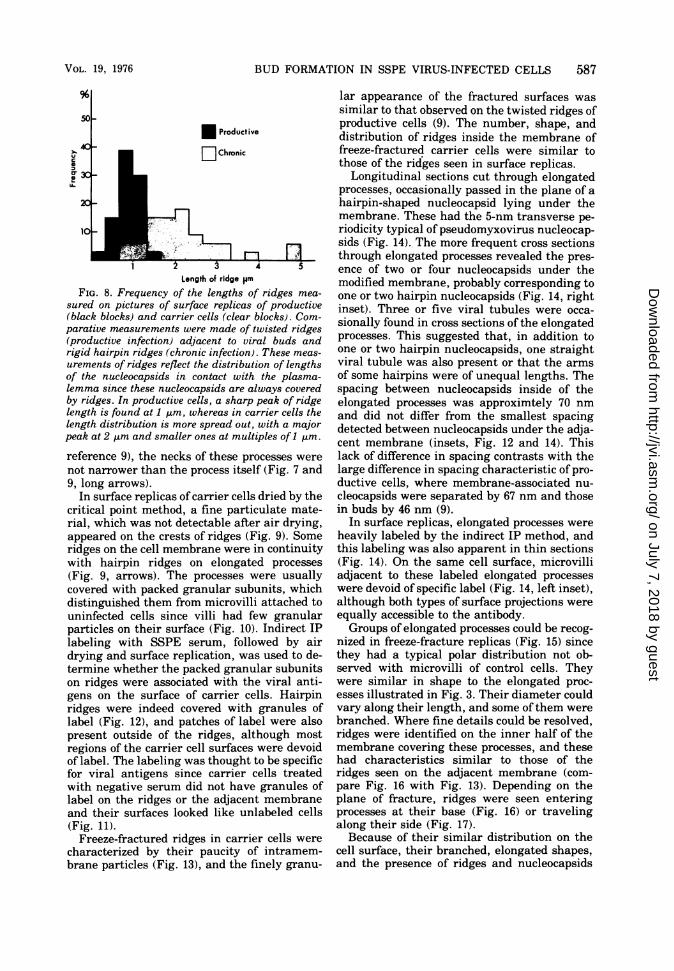
nized in freeze-fracture replicas (Fig. 15) sincethey had a typical polar distribution not ob-served with microvilli of control cells. Theywere similar in shape to the elongated proc-esses illustrated in Fig. 3. Their diameter couldvary along their length, and some of them werebranched. Where fine details could be resolved,ridges were identified on the inner half of themembrane covering these processes, and thesehad characteristics similar to those of theridges seen on the adjacent membrane (com-pare Fig. 16 with Fig. 13). Depending on theplane of fracture, ridges were seen enteringprocesses at their base (Fig. 16) or travelingalong their side (Fig. 17).Because of their similar distribution on the
cell surface, their branched, elongated shapes,and the presence of ridges and nucleocapsids
VOL. 19, 1976
on July 7, 2018 by guesthttp://jvi.asm
.org/D
ownloaded from

'lit , .. , ,
iS -."..
1.
,>-% \t
* V; .
'A. _-
.6..
XE
it I.tSrwr -S *t > o .i tSZ ., ... o9 , t .S...1¢
I,e;.:.
(,0 .i^7'
I
N
K
/
; }s9t?
8 e w _F-j **BX sb. _i4* w*X.8' z -- Sb . .
.. v,' .'. ; vs , . s,' 61 v s,
x _ s b-*s ss b X XP'Ni- > ;^s 2, it t
Po.,- ,.4....?r
Af , 2
FIG. 9. In carrier cells prepared by the critical point drying method, granular subunits are packed at thecrests ofridges and on adjacent elongated processes. A few granules are scattered on the rest ofthe cell surface.Points where ridges on the surrounding cell surface are reflected onto the surface of the elongated processes attheir attachment with the plasmalemma are indicated by arrows. x68,000.
FIG. 10. Microvillus from an uninfected cell that was critical point dried. Normal microvilli are character-ized by scattered granules similar to those on the rest ofthe surface ofuninfected cells. There is no aggregationofgranular subunits like those on elongated processes of carrier cells (Fig. 9). x68,000.
588
.,
.0
./
N
A._s '
1,i.s
'4
4,
..I
.l
,;IAl4
.:
e,S1F
:
S r
,^ '<b...'
f1 r
4
r
.
W.',.s,.
..
4
,..S?'
A
4.
'(12) *zA4 I * ,x,: ' w ' vtw_Wg 'sSoo~~~~~~~~~~~~~~~~~~~~~~~~~~~~~~~~~~~
.1
k
I4
Z.
I,-
k
31
Ak .
I ", I
I.
t6- AtI L
V. -
."w . "." 0 e ;'4
_.- 'v. - tio *
, O A!;. ie ow
.3-V '; 4
on July 7, 2018 by guesthttp://jvi.asm
.org/D
ownloaded from

BUD FORMATION IN SSPE VIRUS-INFECTED CELLS 589
along their length, we concluded that the elon-gated processes seen in surface replicas, in thinsections, and with the freeze-fracture techniquerepresent different aspects of a single popula-tion of viral-induced processes. Small discrep-ancies in their apparent shapes and sizes withdifferent techniques are presumed to be due toseveral factors. First, the dehydration andembedding for thin sectioning or the dehydra-tion for surface replication would produce con-siderable shrinkage, which should not occurduring preparation for freeze-fracturing. Thiscould account for the elongated processes look-ing larger and more rounded in the freeze-frac-ture replica than with the other techniques.Conversely, processes passing out of the planeof freeze-fracture have rounded tips, whichmight be mistaken for true ends of these proc-esses; this effect should make these processeslook shorter after freeze-fracturing than in sur-face replicas.
In passages 25 to 29 of the carrier cells, alarge majority of the cells, approximately 80%,had rigid hairpin ridges and elongated proc-esses, whereas two other cell types, represent-ing 10 to 20% of the cell population, showed adifferent deployment of ridges. One type con-sisted of cells with no structural changes orviral antigen on their surface. The other type ofcell had the twisted ridges and round budscharacteristic of productive infections. Thenumbers of these two types of cell fluctuatedfrom one passage to another. Indeed, after pas-sage 30, cells with twisted ridges and roundbuds increased in number and were mixed withthe cells bearing hairpin ridges. Some cells hadboth round buds and elongated processes withridges. These changes might reflect a morpho-logical transition from the carrier to the pro-ductive type of infection, but this was not sub-stantiated by a significant increase in the titerof free virus. When Vero cells were added tocarrier cells at these passages, fusion to formgiant cells occurred within 2 days (16) althoughVero cells had no effect on earlier passages.
DISCUSSION
The biological properties of our carrier cellline are very similar to those of regulated orcarrier infections established with other mea-sles virus strains (11, 22, 26, 27, 30, 35) as wellas to the properties of the IP-3-Ca subline ofSSPE virus (3). However, none of these previ-ously described carrier cells contains as muchcell-associated virus as do the carrier cells se-lected for the present study. Furthermore, su-perinfection with other SSPE or measlesstrains, as well as with VSV, results in therelease of a substantial amount of virus. WhenVSV is inoculated into carrier cells, typical bul-let-shaped buds are recognized on carrier cells,indicating that VSV can replicate in these cells(unpublished data). Similar experiments withstrains of measles virus showed that the resist-ance of carrier cells to superinfection is limitedto the SSPE strain originally used to create thechronic infection. Therefore, the arrest ofmatu-ration appears to be specific for the MantoothSSPE strain.By comparing surface replicas with thin sec-
tions and combining these approaches with im-munolabeling methods, it has been possible toidentify structural changes in the plasma mem-brane of carrier cells, which differ strikinglyfrom those in productively infected cells (9).Carrier cells typically have straight and hair-pin ridges on their surfaces instead of thetwisted ones that characterize productive cells.Furthermore, hairpin ridges are twice as longas the twisted ridges on productive cells. How-ever, these membrane ridges have several fea-tures in common with those on productive cells.The membrane surface in both is crowned bygranular subunits, which are the sites of viralantigens, and both are formed where a nucleo-capsid contacts a region of the plasma mem-brane lacking intramembrane particles. Hair-pin ridges are also characteristic of the elon-gated processes clustered at one pole of carriercells. The ridges, as well as the size, shape, and
FIG. 11 and 12. Details of hairpin ridges in carrier cells air dried without (Fig. 11) or with (Fig. 12)immunoperoxidase labeling with SSPE serum. In Fig. 12, granules of label 15 to 25 nm in diamneter aretightly packed along the hairpin ridges, whereas in Fig. 11 the hairpin ridge is smooth and bears no granules.The membrane adjacent to the labeled hairpin has only small patches of diffuse reaction product (asterisk).Inset shows a cross section through an unlabeled ridge. Two viral tubules are present under the membrane,each crowned by surface projections (arrows). x45,000; inset, x140,000.
FIG. 13. Freeze-fractured membrane from carrier cell, showing a straight ridge. Along the ridge, themembrane is devoid of the particles typical of the rest of the membrane. x60,OOO.
FIG. 14. Thin section splitting a long process from an immunolabeled carrier cell. The surface of theprocess is strongly labeled, and long viral tubules (arrows), possibly arms ofa single hairpin, lie just underthe membrane. This heavy label is specific on certain regions of the membrane since a microvillus from thesame immunolabeled cell has almost no label (left inset). The right inset shows a cross section through anunlabeled elongated process containing two nucleocapsids that are probably different arms of a hairpinnucleocapsid. Fuzzy projections from the membrane surface are gathered on the membrane over the underly-ing nucleocapsids. x 72,000; left inset, x45,000; right inset, x180,000.
VOL. 19, 1976
on July 7, 2018 by guesthttp://jvi.asm
.org/D
ownloaded from
-44
40-.k t / I 3
s' t',',r',
46
..-i.4,
*,9 f*'q .~
IO~~~~~~~~,q _l;e
I' 6
(i&) > /'''
A;.'bs; #
;'-' ' ) -2\s" ~~~~~~~~4
*o :/5.'t
IS0FIG. 15. Freeze-fracture replica of carrier cells bearing elongated processes similar to the ones shown in
thin sections (Fig. 3). As seen in thin sections, some are branched and vary in diameter along their length.Characteristic structural details of elongated processes are shown below. x53,000.
590
';'^,kq
4'i;
.i k
I., I I
, 1
I...
I.
-- -(, - "
**AkN.. 4i
.~
t-.. .....
.. `,
qw ,1.011,
I
.6 ".,V. 1.N.
l.;I
f.1
';
-14,
N1J.,"I
-LA
(.
. :. '7 .1.
t.l7
I ." 1-
on July 7, 2018 by guesthttp://jvi.asm
.org/D
ownloaded from
BUD FORMATION IN SSPE VIRUS-INFECTED CELLS
distribution of these processes, differentiatethem from normal microvilli. Since each ofthese processes contains one or two hairpin-shaped nucleocapsids contacting the plasma-lemma at the ridges, they are considered to be aform of viral bud, hereafter referred to as "hair-pin buds." Hairpin buds differ from the viralbuds on productive cells by their elongatedshape (32) as well as by the fact that the ridgeswith the underlying nucleocapsids remainseparated by narrow strips of membrane re-sembling that on the rest of the cell surface.How hairpin buds form is not clear. The at-
tached nucleocapsids might bend the mem-brane around them to form a hairpin bud or thehairpin nucleocapsids might migrate into andattach to preexisting processes. Since no de-tached hairpin buds are found, even in serialthin sections, and no other morphological evi-dence has been found that hairpin buds pinchoff from the carrier cell, the hairpin buds ap-pear to be incapable of detaching from the cell.This could be a sufficient explanation for thesmall amount of infectious virus found in thesupernatant of carrier cells.
Straight ridges and hairpin buds are notfound in Vero cells acutely infected with theSSPE Mantooth strain or in human prostatecells acutely infected with the SSPE Hallestrain, so that these structural alterations donot seem to be characteristic of the virus strainor of the cell type used but, rather, of the per-sistent state of infection. During the establish-ment of the chronic infection, carrier cellsmight have been selected on the basis of theirviability. For instance, some host cell factorsmight induce a change in the flexibility of thenucleocapsids. Indeed, a rigid appearance ofthepurified nucleocapsid from four different pseu-domyxoviruses, including measles, has been de-scribed after trypsin treatment of productivelyinfected cells (28, 29, 38). It was proposed thatthe trypsin triggered an enzyme inside the cellsthat might cleave certain nucleocapsid pro-teins, resulting in rigid nucleocapsids. The car-rier cells in the present study may be examplesof the spontaneous occurrence of a rigid nucleo-capsid in situ. However, only the nucleocapsidsinteracting with the membrane display thesecharacteristics, possibly because the enzyme re-sponsible for protein cleavage is present or ac-tive only at the cell membrane.
Since the shape of the nucleocapsids can beinferred from the shape of surface ridges, apossible interpretation of the striking straightcourse of hairpin nucleocapsids, except wherethey are bent on themselves at the apices of thehairpin, is that aligned nucleocapsids in carriercells are more rigid than those in productivecells. However, it must be considered that otherchanges in the viral components or in the hostcell could be responsible for the straight ap-pearance of the nucleocapsid contacting themembrane and the formation of hairpin buds inthe carrier cells. The organization and distribu-tion of the attachment sites may be modified,the nucleocapsid coiling preparatory to viralbud formation might be inhibited, or an unu-sual fluidity of the cell membrane might pre-vent the pulling together of the nucleocapsidsand ridges to exclude intervening host mem-brane from the viral bud (9). For any of thesereasons, normal viral buds might not completetheir maturation in carrier cells.The defective buds, after they have been arti-
ficially detached from the cell, appear to con-tain the complete genomic information neces-sary for replication since they are able to pro-duce numerous plaques. In this respect, theydiffer from defective interfering particles ofmeasles (14). However, some of the plaquesmay also have been produced by normal roundbuds, which are seen occasionally on the sur-face of carrier cells, or by other structures notdetected by our techniques. The longer lengthof the carrier cell nucleocapsids, which usuallyis a multiple of the length of nucleocapsids inproductive cells, suggests that repetition ofgenomic fragments might occur in the hairpinstructures.A typical feature of carrier cells is the polar
distribution of ridges and buds, regardless oftheir structure. It is possible that viral antigensare incorporated into restricted or selected re-gions of the infected-cell membrane since a dif-fuse distribution of antigen is never observed inthe carrier cells. This postulated distribution ofviral antigens differs from that after capping inmeasles-infected cells which can be induced byantibody and implies movement of viral anti-gen within the plane ofthe membrane (23). Thepeculiar polar distribution of surface antigensmight be related to the decreased capacity forcell fusion in carrier cells.
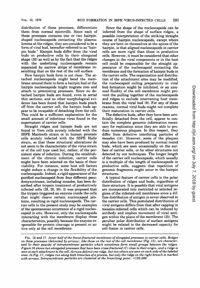
FIG. 16 and 17. Inner halfof the freeze-fractured membrane ofelongated processes in carrier cells. Ridgeson these processes (delimited by arrows), like those on the rest of the cell membrane (Fig. 13), are character-ized by their paucity of intramembrane particles which sometimes form small groups between the ridges.Figure 16 shows two elongated processes that have been cross-fractured (C) close to their origin, and a ridge isseen at each attachment to the cell. Arrows delimit one ridge, but two others are seen on each side ofthe labeledones. In Fig. 17, ridges run along both branches ofa process, but only the ridge on the right branch is markedwith arrows. Intramembrane particles are clustered at the branching point. x120,000.
VOL. 19, 1976 591
on July 7, 2018 by guesthttp://jvi.asm
.org/D
ownloaded from

592 DUBOIS-DALCQ ET AL.
Thus, in carrier cells a defect in incorporationof viral proteins into the cell surface mightcoexist with a defect in nucleocapsid structureor membrane mobility, and either defect mighthave a variable expression from cell to cell andfrom one passage to another. Molecular analy-sis of nucleocapsid RNA and protein from car-rier cells should help clarify the causes of thesedefects. As yet, there is no evidence that thekind of defective bud formation described hereoccurs in any disease, including SSPE. Furtherstudies, using similar techniques, on SSPE pri-mary brain cultures from humans (8) or ani-mals (10, 19) should be done to explore whethersimilar defects in viral maturation can occurspontaneously.
ACKNOWLEDGMENTSWe would like to thank John L. Sever for helpful advice.
The excellent technical help of Kathy Worthington andOtto Gutenson is greatly appreciated.
LITERATURE CITED
1. Anderson, T. F. 1951. Techniques for the preservationof three dimensional structure in preparing speci-mens for the electron microscope. Trans. N.Y. Acad.Sci. 13:130-134.
2. Birdwell, C. R., E. G. Strauss, and J. H. Strauss. 1973.Replication of Sindbis virus. III. An electron micro-scopic study of virus maturation using the surfacereplica technique. Virology 56:429-438.
3. Burnstein, T., L. B. Jacobsen, W. Zeman, and T. T.Chen. 1974. Persistent infection of BSC-1 cells bydefective measles virus derived from subacute scle-rosing panencephalitis. Infect. Immun. 10:1378-1382.
4. Cooper, P. D. 1967. The plaque assay of animal viruses,p. 243-311. In K. Maramorosch and H. Koprowski(ed.), Methods in virology, vol. 3. Academic PressInc., New York.
5. Doi, Y., T. Sanpe, M. Nakajima, S. Okawa, T. Katoh,H. Itoh, T. Sato, K. Oguchi, T. Kumanishi, and T.Tsubaki. 1972. Properties of a cytopathic agent iso-lated from a patient with subacute sclerosing panen-cephalitis in Japan. Jpn. J. Med. Sci. Biol. 25:321-333.
6. Dubois-Dalcq, M., and L. H. Barbosa. 1973. Immuno-peroxidase stain of measles antigen in tissue culture.J. Virol. 12:909-918.
7. Dubois-Dalcq, M., L. H. Barbosa, R. Hamilton, and J.L. Sever. 1974. Comparison between productive andlatent subacute sclerosing panencephalitis viral in-fection in vitro. An electron microscopic and immuno-peroxidase study. Lab. Invest. 30:241-250.
8. Dubois-Dalcq, M., J. M. Coblentz, and A. B. Pleet.1974. Subacute sclerosing panencephalitis. Arch.Neurol. 31:355-363.
9. Dubois-Dalcq, M., and T. S. Reese. 1975. Structuralchanges in the membrane of vero cells infected with aparamyxovirus. J. Cell Biol. 67:551-565.
10. Dubois-Dalcq, M., K. Worthington, 0. Gutenson, andL. H. Barbosa. 1975. Immunoperoxidase labeling ofsubacute sclerosing panencephalitis virus in hamsteracute encephalitis. Lab. Invest. 32:518-526.
11. Ehrnst, A., L. Weiner, and E. Norrby. 1974. Fluctua-tions and distribution of measles virus antigens inchronically infected cells. Nature (London) 248:691-693.
12. Gibson, P. E., and T. M. Bell. 1972. Persistent infection
of measles virus in mouse brain cell cultures infectedin vivo. Arch. Gesamte Virusforsch. 37:45-53.
13. Hall, W. W., and S. J. Martin. 1973. Purification andcharacterization of measles virus. J. Gen. Virol.19:175-188.
14. Hall, W. W., and S. J. Martin. 1974. Defective interfer-ing particles produced during the replication of mea-sles virus. Med. Microbiol. Immunol. 160:155-164.
15. Hamilton, R., L. Barbosa, and M. Dubois-Dalcq. 1973.Subacute sclerosing panencephalitis measles virus:study of biological markers. J. Virol. 12:632-642.
16. Haspel, M. V., P. R. Knight, R. G. Duff, and F. Rapp.1973. Activation of latent measles virus infection inhamster cells. J. Virol. 12:690-695.
17. Horta-Barbosa, L., D. A. Fuccillo, J. L. Sever, and W.Zeman. 1969. Subacute sclerosing panencephalitisisolation of measles virus from a brain biopsy. Nature(London) 221:974.
18. Iwasaki, Y., H. Koprowski, D. Muller, V. ter Meulen,and Y. M. Kackell. 1973. Morphogenesis and struc-ture of a virus in cells cultured from brain tissue fromtwo cases of multiple sclerosis. Lab. Invest. 28:494-500.
19. Johnson, K. P., and E. Norrby. 1974. Subacute scleros-ing panencephalitis (SSPE) agent in hamsters. III.Induction of defective measles infection in hamsterbrain. Exp. Mol. Pathol. 21:166-178.
20. Klein, G., P. Clifford, E. Klein, R. T. Smith, J. Mino-wada, F. M. Kouriosky, and J. H. Burchenal. 1967.Membrane immunofluorescence reactions of Burkittlymphoma cells from biopsy specimens and tissuecultures. J. Natl. Cancer Inst. 39:1027-1044.
21. Knight, P., R. Duff, R. Glaser, and F. Rapp. 1973.Characteristics of the release of measles virus fromlatently infected cells after co-cultivation with BSC-1cells. Intervirology 2:287-298.
22. Knight, P., R. Duff, and F. Rapp. 1972. Latency ofhuman measles virus in hamster cells. J. Virol.10:995-1001.
23. Lampert, P. W., B. S. Joseph, and M. Oldstone. 1975.Antibody-induced capping of measles virus antigenson plasma membrane studied by electron microscopy.J. Virol. 15:1248-1255.
24. Lennette, E. H., and N. J. Schmidt. (ed.). 1969. Diag-nostic procedure for viral and rickettsial infections,4th ed. American Public Health Association, Inc.,New York.
25. Menna, J. H., A. R. Collins, and T. D. Flanagan. 1975.Characterization of an in vitro persistent-state mea-sles virus infection: establishment and virologicalcharacterization of the BGM/MV cell line. Infect.Immun. 11:152-158.
26. Minagawa, T. 1971. Studies on the persistent infectionwith measles virus in HeLa cells. I. Clonal analysis ofcells of carrier cultures. Jpn. J. Microbiol. 15:325-331.
27. Minagawa, T. 1971. Studies on the persistent infectionwith measles virus in HeLa cells. II. The properties ofcarried virus. Jpn. J. Microbiol. 15:333-340.
28. Mountcastle, W. E., R. W. Compans, L. A. Caliguiri,and P. W. Choppin. 1970. Nucleocapsid protein sub-units of simian virus 5, Newcastle disease virus, andSendai virus. J. Virol. 6:677-684.
29. Mountcastle, W. E., R. W. Compans, H. Lackland, andP. W. Choppin. 1974. Proteolytic cleavage of subunitsof the nucleocapsids of the paramyxovirus simianvirus 5. J. Virol. 14:1253-1261.
30. Norrby, E. 1967. A carrier cell line of measles virus inLu 106 cells. Arch. Gesamte Virusforsch. 20:215-224.
31. Norrby, E. C. J., and P. Magnusson. 1965. Some mor-phological characteristics of the internal componentof measles virus. Arch. Gesamte Virusforsch. 17:443-447.
32. Orenstein, J. M., and I. B. Weinstein. 1973. Filamen-
J. VIROL.
on July 7, 2018 by guesthttp://jvi.asm
.org/D
ownloaded from

BUD FORMATION IN SSPE VIRUS-INFECTED CELLS 593
tous forms of enveloped A particles in cell culturesfrom chemically induced rat hepatomas. Cancer Res.33:1998-2004.
33. Raine, C. S., L. A. Feldman, R. D. Sheppard, L. H.Barbosa, and M. B. Bornstein. 1974. Subacute scle-rosing panencephalitis virus. Observations on a neu-
roadapted strain and non neuroadapted strain in or-ganotypic central nervous system cultures. Lab. In-vest. 31:42-53.
34. Raine, C. S., L. A. Feldman, R. D. Sheppard, and M. B.Bornstein. 1973. Subacute sclerosing panencephalitisvirus in cultures of organized central nervous tissue.Lab. Invest. 28:627-640.
35. Rustigian, R. 1966. Persistent infection of cells in cul-ture by measles virus. I. Development and character-istics of HeLa sublines persistently infected withcomplete virus. J. Bacteriol. 92:1792-1804.
36. Rustigian, R. 1966. Persistent infection of cells in cul-ture by measles virus. II. Effect of measles antibodyon persistently infected HeLa sublines and recovery
of a HeLa clonal line persistently infected with in-complete virus. J. Bacteriol. 92:1805-1811.
37. Steele, R. W., D. A. Fuccillo, S. A. Hensen, M. M.Vincent, and J. A. Bellanti. 1976. Specific inhibitoryfactors of-cellular immunity in children with Sub-acute Sclerosing panencephalitis. J. Pediatr. 88:56-62.
38. Waters, D. J., and R. H. Bussell. 1974. Isolation andcomparative study of the nucleocapsids of measlesand canine distemper viruses from infected cells. Vi-rology 61:64-79.
39. Waters, D. J., R. T. Hersh, and R. H. Bussell. 1972.Isolation and characterization of measles nucleocap-sid from infected cells. Virology 48:278-281.
VOL. 19, 1976
on July 7, 2018 by guesthttp://jvi.asm
.org/D
ownloaded from