Decoding Breast Cancer with Quantitative Radiomics ... · Quantitative Radiomics & Radiogenomics:...
53
Decoding Breast Cancer with Quantitative Radiomics & Radiogenomics: Imaging Phenotypes in Breast Cancer Risk Assessment, Diagnosis, Prognosis, and Response to Therapy Maryellen Giger & Yuan Ji The University of Chicago, NorthShore University [email protected] for the TCGA Breast Phenotype Research Group • Analysis funded by The University of Chicago Dean Bridge Fund • Images hosted by NCI TCIA COI: M L Giger is a stockholder in R2/Hologic, a co-founder and equity holder in Quantitative Insights, and receives royalties from Hologic, GE Medical Systems, MEDIAN Technologies, Riverain Medical, Mitsubishi, and Toshiba Giger TCGA 2015
-
Upload
vuongnguyet -
Category
Documents
-
view
217 -
download
0
Transcript of Decoding Breast Cancer with Quantitative Radiomics ... · Quantitative Radiomics & Radiogenomics:...

Decoding Breast Cancer with Quantitative Radiomics & Radiogenomics:
Imaging Phenotypes in Breast Cancer Risk Assessment, Diagnosis, Prognosis, and Response to Therapy
Maryellen Giger & Yuan Ji The University of Chicago, NorthShore University
for the TCGA Breast Phenotype Research Group
• Analysis funded by The University of Chicago Dean Bridge Fund • Images hosted by NCI TCIA COI: M L Giger is a stockholder in R2/Hologic, a co-founder and equity holder in Quantitative Insights, and receives royalties from Hologic, GE Medical Systems, MEDIAN Technologies, Riverain Medical, Mitsubishi, and Toshiba
Giger TCGA 2015

NCI TCGA/TCIA Breast Phenotype Research Group
Radiologists: •Elizabeth Morris – MSKCC •Ermelinda Bonaccio – Roswell •Kathleen Brandt – Mayo •Elizabeth Burnside – U Wisconsin Madison •Basak Dogan – MD Anderson •Marie Ganott – Magee •Jose Net – U Miami •Elizabeth Sutton – MSKCC •Gary Whitman – MD Anderson •Margarita Zuley – U Pittsburgh •H. Carisa Le-Petross – MD Anderson
Human-Extracted Phenotypes Analysis -- MD Anderson
• Arvind Rao
Computer-Extracted Phenotypes & Data analysis/associations University of Chicago • Maryellen Giger • Hui Li • Karen Drukker • Li Lan
NorthShore University • Yuan Ji • Yitan Zhu • Wentian Guo
NCI: • Carl Jaffe • John Freymann • Erich Huang • Justin Kirby • Brenda Fevrier-Sullivan
Mapping of Breast MRI Phenotypes to Histopathology and Genomics
Giger TCGA 2015

Decoding Breast Cancer with Quantitative Radiomics & Radiogenomics:
Imaging Phenotypes in Breast Cancer Risk Assessment, Diagnosis, Prognosis, and Response to Therapy
Purpose: To demonstrate, using the TCGA TCIA breast cancer dataset of MRI images, the role of quantitative radiomics in characterizing the molecular subtypes of breast cancer and associating the magenetic resonance imaging (MRI) computer-extracted image phenotypes with genomic data.
Giger TCGA 2015

Decoding Breast Cancer with Imaging Involves interdisciplinary research:
– Development and/or customization of mathematical image analysis methods for extracting information from biomedical image data (computer vision) - developed from CAD research
– Investigations in the applications of these techniques to gain knowledge in (a) the management of the cancer patient and in (b) the understanding of cancer
Quantitatively Extract Lesion Characteristics
(Computer Vision)
Patient-Specific Image-based Tumor Signature
for Precision Medicine
Data-mining of Computer-Extracted Features on Large
Datasets for Population-based Cancer Discovery Giger TCGA 2015
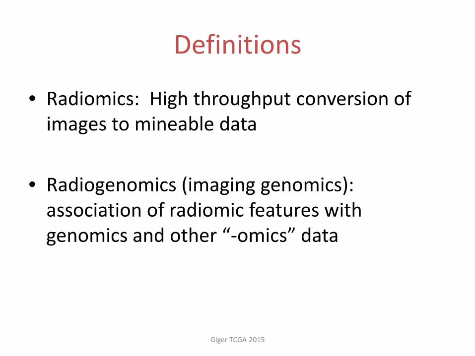
Definitions
• Radiomics: High throughput conversion of images to mineable data
• Radiogenomics (imaging genomics): association of radiomic features with genomics and other “-omics” data
Giger TCGA 2015

Imaging Genomics
Medical Images
Computer Segmentation of
Lesions
Computer-extracted Lesion Features
(size, morphology, texture, kinetics)
Associations and/or Classification Relevant to Clinical or Biological Questions – Develop Predictive Models
Radiologist Descriptors
Histopathology, Molecular
Classification
Genomics Data
Sources
Asks questions about the relationships between features “seen” in medical images and the biology of cancer
Giger TCGA 2015

Imaging Genomics
Medical Images
Computer Segmentation of
Lesions
Computer-extracted Lesion Features
(size, morphology, texture, kinetics)
Associations and/or Classification Relevant to Clinical or Biological Questions – Develop Predictive Models
Radiologist Descriptors
Genomics Data
Sources
Which correlate and which are
complementary???
Asks questions about the relationships between features “seen” in medical images and the biology of cancer
Histopathology, Molecular
Classification
Giger TCGA 2015

Imaging Genomics
Medical Images
Computer Segmentation of
Lesions
Computer-extracted Lesion Features
(size, morphology, texture, kinetics)
Associations and/or Classification Relevant to Clinical or Biological Questions – Develop Predictive Models
Radiologist Descriptors
Genomics Data
Sources
Which correlate and which are
complementary???
Asks questions about the relationships between features “seen” in medical images and the biology of cancer
Lead to Personalized Screening and
Personalized Treatment
Histopathology, Molecular
Classification
Giger TCGA 2015
Radi
omic
s

Analysis & Output of Tumor Signature
Automated Lesion Segmentation, Feature Extraction [volumetrics, morphological, texture, kinetics] and Estimation of the Probability of Malignancy
University of Chicago High-Throughput MRI Phenotyping System (Quantitative Image Analysis Workstation)
Giger et al., RSNA 2010 Giger TCGA 2015

Dataset
Giger TCGA 2015
cancerimagingarchive.net Breast Cancer cases
Clinical /Histopathology /Genomic data
downloaded by TCGA Assembler & Molecular
subtyping / risk of recurrence values by
Perou Lab
Tumor location on MRI determined by
consensus of three of the TCIA radiologists
MRIs of 91 cases (GE 1.5T) collected by TCIA
MRIs of 91 cases downloaded to UChicago for computational
MRI tumor phenotyping (radiomics)
cancergenome.nih.gov

Distribution of the 91 MRI cases
14 19
72
11
77 72
19
80
ER PR HER2 TN
Negative Positive
Giger TCGA 2015

Distribution of the 91 MRI cases
4
55
10 5
10
0
10
20
30
40
50
60
Giger TCGA 2015

Dataset
Giger TCGA 2015
cancerimagingarchive.net Breast Cancer cases
Clinical /Histopathology /Genomic data
downloaded by TCGA Assembler & Molecular
subtyping / risk of recurrence values by
Perou Lab
Tumor location on MRI determined by
consensus of three of the TCIA radiologists
MRIs of 91 cases (GE 1.5T) collected by TCIA
MRIs of 91 cases downloaded to UChicago for computational
MRI tumor phenotyping (radiomics)
cancergenome.nih.gov

Contrast-enhanced MR images of breast
• Tumors have increased blood vessels and differ in microvascular density and vessel permeability
• Gd-DTPA shortens T1 relaxation time which leads to increase of
signal in T1-weighted images
Precontrast Postcontrast Subtraction
Giger TCGA 2015

Dynamic Contrast-Enhanced MRI & Tumor Segmentation
Giger TCGA 2015
Incr
easin
g tim
e
Across slices
4D image analysis

4D DCE MRI images
……
Computerized Tumor Segmentation
Radiologist-indicated Tumor Center
University of Chicago High-Throughput MRI Phenotyping System (Segmentation of the Tumor within the Breast MR image)
ER-negative ER-positive Giger TCGA 2015

3D Breast MRI image
Giger TCGA 2015
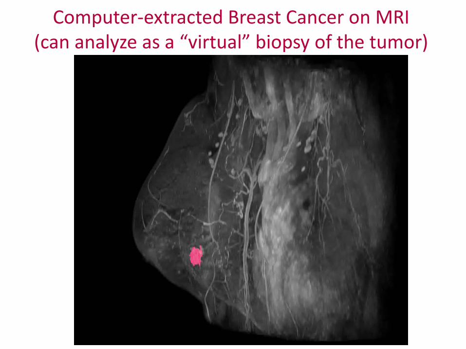
Computer-extracted Breast Cancer on MRI (can analyze as a “virtual” biopsy of the tumor)
Giger TCGA 2015

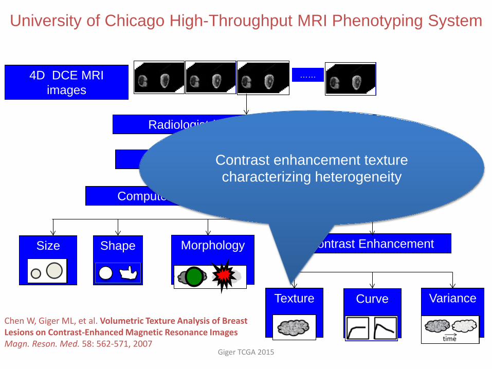
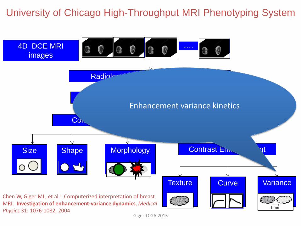
4D DCE MRI images
Computer-Extracted Image Phenotypes (CEIP)
Size
Shape
Morphology
Contrast Enhancement
Texture
Curve
Variance
……
Computerized Tumor Segmentation
Radiologist-indicated Tumor Center
CAD pipeline = radiomics pipeline
University of Chicago High-Throughput MRI Phenotyping System
Giger TCGA 2015

4D DCE MRI images
Computer-Extracted Image Phenotypes (CEIP)
Size
Shape
Morphology
Contrast Enhancement
Texture
Curve
Variance
……
Computerized Tumor Segmentation
Radiologist-indicated Tumor Center
CAD pipeline = radiomics pipeline
University of Chicago High-Throughput MRI Phenotyping System
• Volume • Effective diameter • Maximum linear size • Surface Area
Giger TCGA 2015

4D DCE MRI images
Computer-Extracted Image Phenotypes (CEIP)
Size
Shape
Morphology
Contrast Enhancement
Texture
Curve
Variance
……
Computerized Tumor Segmentation
Radiologist-indicated Tumor Center
CAD pipeline = radiomics pipeline
University of Chicago High-Throughput MRI Phenotyping System
• Sphericity • Irregularity • Surface area/volume
Giger TCGA 2015

4D DCE MRI images
Computer-Extracted Image Phenotypes (CEIP)
Size
Shape
Morphology
Contrast Enhancement
Texture
Curve
Variance
……
Computerized Tumor Segmentation
Radiologist-indicated Tumor Center
CAD pipeline = radiomics pipeline
University of Chicago High-Throughput MRI Phenotyping System
• Margin sharpness • Variance of margin sharpness • Variance of radial gradient
histogram
Giger TCGA 2015

4D DCE MRI images
Computer-Extracted Image Phenotypes (CEIP)
Size
Shape
Morphology
Contrast Enhancement
Texture
Curve
Variance
……
Computerized Tumor Segmentation
Radiologist-indicated Tumor Center
CAD pipeline = radiomics pipeline
University of Chicago High-Throughput MRI Phenotyping System
Enhancement heterogeneity & kinetics of the uptake and washout of the contrast agent during the imaging time
Giger TCGA 2015

Tumors are Heterogeneous: Contrast Enhancement Heterogeneity & Kinetics
Heterogeneity of Tumors:
Giger TCGA 2015
4D DCE MRI images
Computer-Extracted Image Phenotypes (CEIP)
Size
Shape
Morphology
Contrast Enhancement
Texture
Curve
Variance
……
Computerized Tumor Segmentation
Radiologist-indicated Tumor Center
University of Chicago High-Throughput MRI Phenotyping System
Contrast enhancement texture characterizing heterogeneity
Chen W, Giger ML, et al. Volumetric Texture Analysis of Breast Lesions on Contrast-Enhanced Magnetic Resonance Images Magn. Reson. Med. 58: 562-571, 2007
Giger TCGA 2015

4D DCE MRI images
Computer-Extracted Image Phenotypes (CEIP)
Size
Shape
Morphology
Contrast Enhancement
Texture
Curve
Variance
……
Computerized Tumor Segmentation
Radiologist-indicated Tumor Center
University of Chicago High-Throughput MRI Phenotyping System
Chen W, Giger ML, et al.: Automatic identification and classification of characteristic kinetic curves of breast lesions on DCE-MRI. Medical Physics, 33: 2878-2887,2006
Kinetic curve assessment based on most-enhancing voxels within tumor: Uptake, washout, curve shape
Giger TCGA 2015
4D DCE MRI images
Computer-Extracted Image Phenotypes (CEIP)
Size
Shape
Morphology
Contrast Enhancement
Texture
Curve
Variance
……
Computerized Tumor Segmentation
Radiologist-indicated Tumor Center
University of Chicago High-Throughput MRI Phenotyping System
Chen W, Giger ML, et al.: Computerized interpretation of breast MRI: Investigation of enhancement-variance dynamics, Medical Physics 31: 1076-1082, 2004
Enhancement variance kinetics
Giger TCGA 2015

4D DCE MRI images
Computer-Extracted Image Phenotypes (CEIP)
Size
Shape
Morphology
Contrast Enhancement
Texture
Curve
Variance
……
Computerized Tumor Segmentation
Radiologist-indicated Tumor Center
Can be thought of as a non-invasive “virtual biopsy”
University of Chicago High-Throughput MRI Phenotyping System For Breast Tumors
Giger TCGA 2015

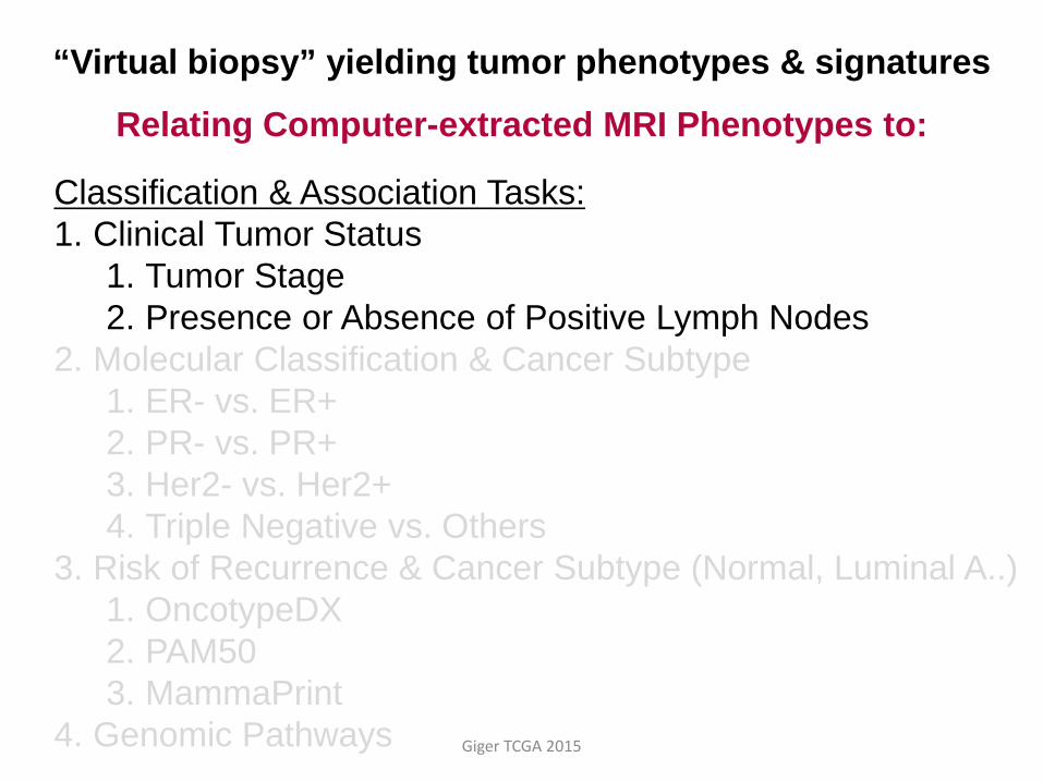
“Virtual biopsy” yielding tumor phenotypes & signatures
Relating Computer-extracted MRI Phenotypes to:
Classification & Association Tasks: 1. Clinical Tumor Status
1. Tumor Stage 2. Presence or Absence of Positive Lymph Nodes
2. Molecular Classification & Cancer Subtype 1. ER- vs. ER+ 2. PR- vs. PR+ 3. Her2- vs. Her2+ 4. Triple Negative vs. Others
3. Risk of Recurrence 1. OncotypeDX 2. PAM50 3. MammaPrint
4. Genomic Pathways Giger TCGA 2015
Classification & Association Tasks: 1. Clinical Tumor Status
1. Tumor Stage 2. Presence or Absence of Positive Lymph Nodes
2. Molecular Classification & Cancer Subtype 1. ER- vs. ER+ 2. PR- vs. PR+ 3. Her2- vs. Her2+ 4. Triple Negative vs. Others
3. Risk of Recurrence & Cancer Subtype (Normal, Luminal A..) 1. OncotypeDX 2. PAM50 3. MammaPrint
4. Genomic Pathways Giger TCGA 2015
“Virtual biopsy” yielding tumor phenotypes & signatures
Relating Computer-extracted MRI Phenotypes to:

MRI-based Phenotypes of Size – predictive of breast cancer tumor stage
TCGA/TCIA Breast Cancer Group cases; University of Chicago Giger Lab computer-extracted image phenotypes Giger TCGA 2015

Classification & Association Tasks: 1. Clinical Tumor Status
1. Tumor Stage 2. Presence or Absence of Positive Lymph Nodes
2. Molecular Classification & Cancer Subtype 1. ER- vs. ER+ 2. PR- vs. PR+ 3. HER2- vs. HER2+ 4. Triple Negative vs. Others
3. Risk of Recurrence 1. OncotypeDX 2. PAM50 3. MammaPrint
4. Genomic Pathways Giger TCGA 2015
“Virtual biopsy” yielding tumor phenotypes & signatures
Relating Computer-extracted MRI Phenotypes to:

From TCIA MRI Radiomics -- ER Negative Breast Cancers tended to have larger size, a more irregular shape, and more heterogeneous in
terms of contrast enhancement
Giger TCGA 2015

From TCIA Radiomics- Triple Negative Breast Cancers tended to have a more irregular shape, and more heterogeneous in terms of contrast
enhancement
Giger TCGA 2015

From the TCIA Radiomics -- Enhancement Texture of Tumor Heterogeneity appears Predictive of Molecular Subtype
4 55
10 5 10
Molecular Subtyping from C. Perou Giger TCGA 2015
Kendall test results for trends; p-value=0.0055

size < 2 cm tumors Kendall test for trends; p-value=0.0435
2 cm < size < 5 cm Kendall test for trends; p-value=0.016
From the TCIA Radiomics -- Enhancement Texture of Tumor Heterogeneity appears Predictive of Molecular Subtype
Molecular Subtyping from C. Perou
Giger TCGA 2015

Classification & Association Tasks: 1. Clinical Tumor Status
1. Tumor Stage 2. Presence or Absence of Positive Lymph Nodes
2. Molecular Classification & Cancer Subtype 1. ER- vs. ER+ 2. PR- vs. PR+ 3. Her2- vs. Her2+ 4. Triple Negative vs. Others
3. Risk of Recurrence from multi-gene assays 1. OncotypeDX 2. PAM50 3. MammaPrint
4. Genomic Pathways Giger TCGA 2015
“Virtual biopsy” yielding tumor phenotypes & signatures
Relating Computer-extracted MRI Phenotypes to:

Giger TCGA 2015
Multi-gene assays of
risk of recurrence
Radiomics for “virtual” biopsy
Computer analysis of Breast MRIs of tumors

Radiomics “Virtual Biopsy” & Risk of Recurrence

Giger TCGA 2015
ROC curves for leave-one-out LDA classifier using computer-extracted MRI phenotypes as decision variable in the tasks of distinguishing between [low+medium] and high risk levels of recurrence for MammaPrint, PAM50 ROR-S (Subtype), and PAM50 ROR-P (Subtype+Proliferation) from Perou
Performance of the MRI Tumor Signatures in the task of predicting Risk of Recurrence
(ROC analysis )

Classification & Association Tasks: 1. Clinical Tumor Status
1. Tumor Stage 2. Presence or Absence of Positive Lymph Nodes
2. Molecular Classification & Cancer Subtype 1. ER- vs. ER+ 2. PR- vs. PR+ 3. Her2- vs. Her2+ 4. Triple Negative vs. Others
3. Risk of Recurrence 1. OncotypeDX 2. PAM50 3. MammaPrint
4. Genomic Pathways Giger TCGA 2015
“Virtual biopsy” yielding tumor phenotypes & signatures
Relating Computer-extracted MRI Phenotypes to:
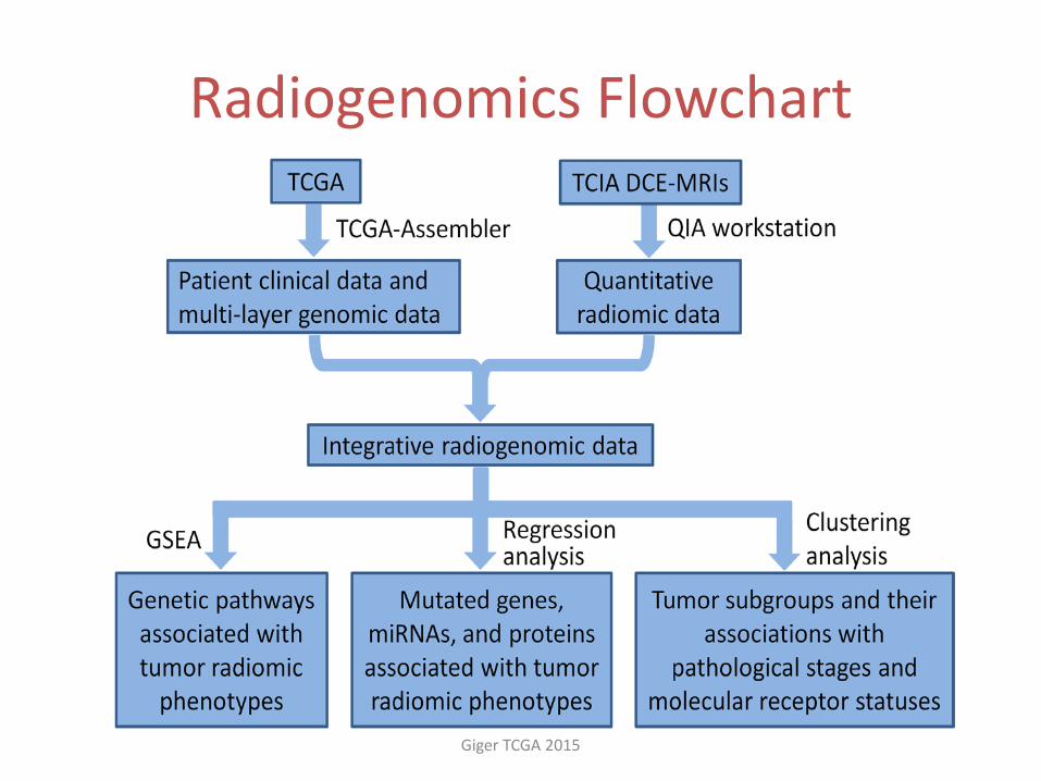
Radiogenomics Flowchart
Giger TCGA 2015

Significant associations between radiomic features and clinical outcomes evaluated by t-tests.
Giger TCGA 2015
Exploratory Cluster Analysis of the MRI Tumor Phenotypes

Radiomics from the MRI tumor “Virtual Biopsy” shows association with Pathway Transcriptional Activities
Zhu et al. submitted
Gige
r la
b Ji
lab
Giger TCGA 2015

Identified significant associations
Giger TCGA 2015

Identified significant associations
Giger TCGA 2015
Size Phenotypes
Gene expressions of
pathways

Identified significant associations
Giger TCGA 2015
Enhancement Texture
Heterogeneity Phenotypes
miRNA expressions
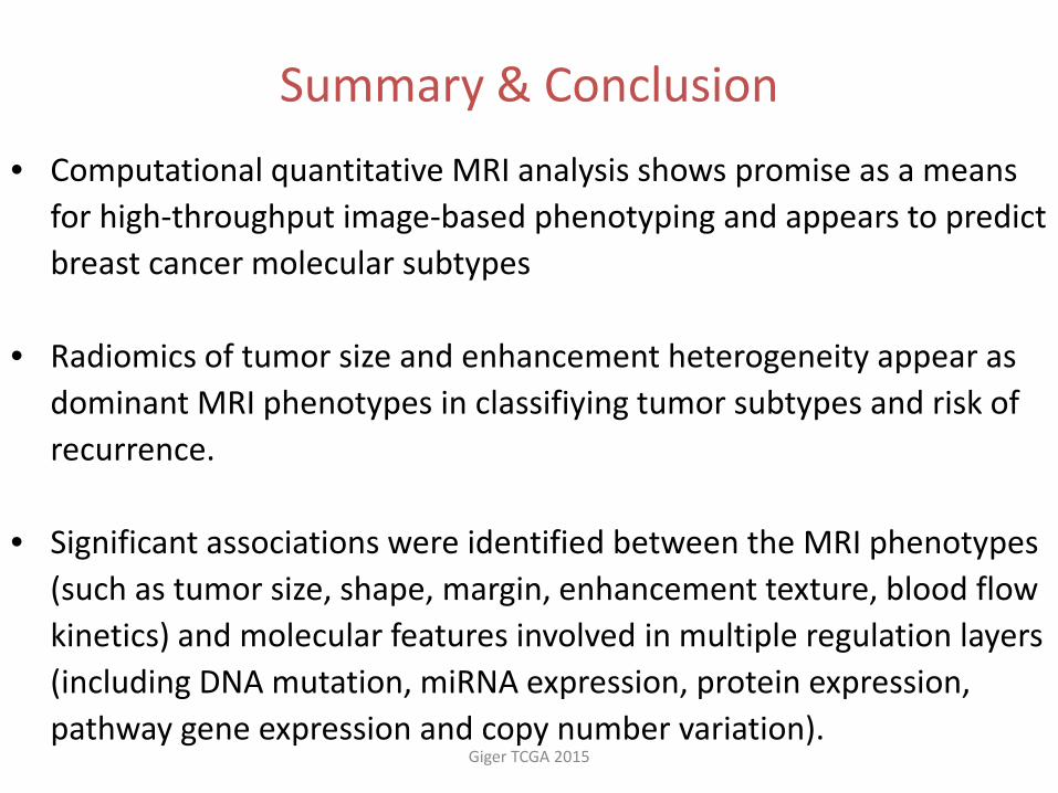
Summary & Conclusion • Computational quantitative MRI analysis shows promise as a means
for high-throughput image-based phenotyping and appears to predict breast cancer molecular subtypes
• Radiomics of tumor size and enhancement heterogeneity appear as
dominant MRI phenotypes in classifiying tumor subtypes and risk of recurrence.
• Significant associations were identified between the MRI phenotypes (such as tumor size, shape, margin, enhancement texture, blood flow kinetics) and molecular features involved in multiple regulation layers (including DNA mutation, miRNA expression, protein expression, pathway gene expression and copy number variation).
Giger TCGA 2015

Summary & Conclusion
• Limitations included a small dataset of only 91 cancers • TCIA is collecting additional images • Investigators are organizing a multi-institutional radiomics network to
collect beyond the TCGA/TCIA
• Identification of radiomics of molecular subtypes of breast tumors is expected to allow for virtual biopsies
• Ongoing research involves relating and merging MRI phenotypes with genomic data to develop improved predictive models
Giger TCGA 2015

Questions
Giger TCGA 2015
• Is it possible to decide targeted therapy based on imaging-genomics association results?
• Can imaging features inform important genomics features?
• Can integration of imaging and genomics features lead to higher power in prediction?
• Can imaging serve as a virtual biopsy? –non-invasive, covers complete tumor, & repeatable

Thank you & please attend our related Workshop & Posters
Giger TCGA 2015
• Workshop: Imaging Resources for the TCGA: Radiology and Pathology Tools for Enabling Science; May 11; 4-5pm and repeated 5-6pm
• Poster 91 • Poster 79 • Poster 105