Cytoskeleton
26
The cytoskeleton 03/20/2022
-
Upload
aftab-badshah -
Category
Science
-
view
81 -
download
0
Transcript of Cytoskeleton
The cytoskeleton
04/15/2023

04/15/2023
Cytoskeleton
The cytoskeleton is the structure consisting of fibrous proteins that occur in the cytoplasm and maintain the shape of the cell.

04/15/2023
Cytoplasm
Microtubules – function in cell division and serve as a "temporary support" for other organelles.
Actin microfilaments are thin threads that function in cell division and cell motility.
Intermediate filaments are between the size of the microtubules and the actin filaments.
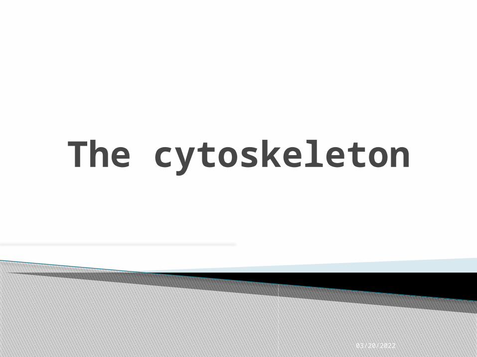
The CytosceletonThe cytoskeleton:
• gives the cell shape,
• anchors some organelles and directs the movement of others,
• may enable the entire cell to change shape or move.
• may play a regulatory role, by mechanically transmitting signals from the cell's surface to its interior.

04/15/2023

04/15/2023
Role of microtubulesHollow tubes with wall that consists of 13 columns of tubulin molecules (25 nm in diameter)
Involved in:
• cell shape maintenance (compression resistance)• cell motility (as in cilia or flagella)• chromosome movement in cell division• Organelle movements

04/15/2023
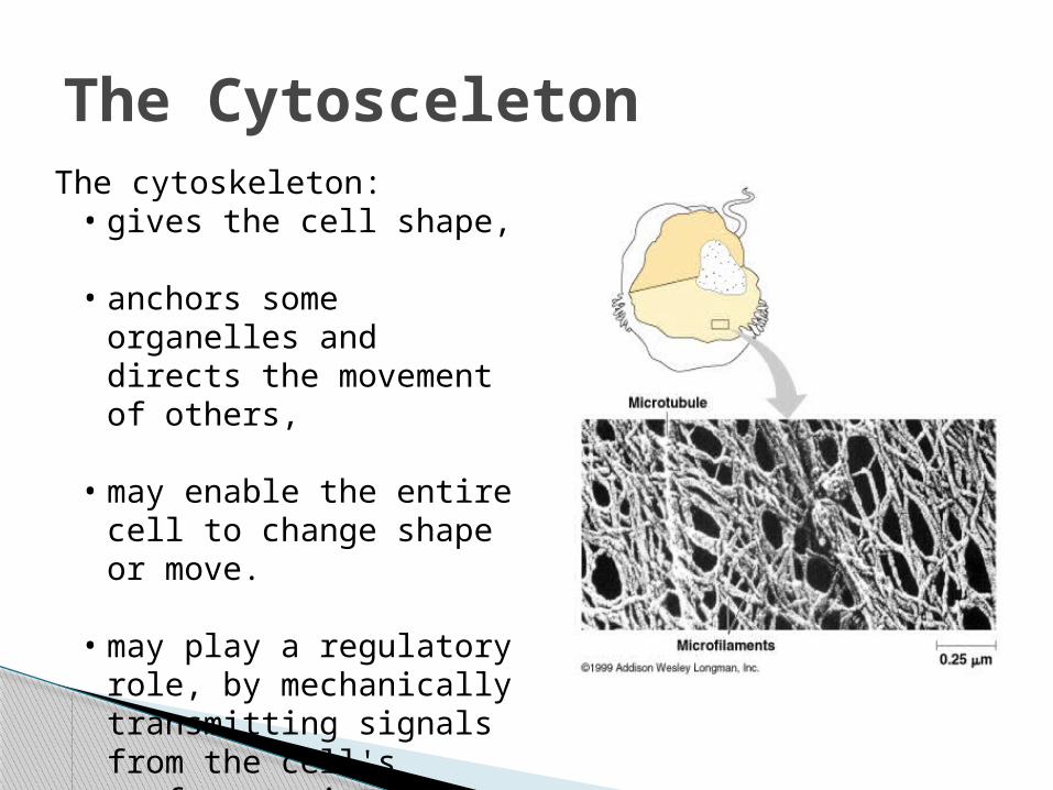
Motor molecules and the cytoskeleton
The microtubules and microfilaments interact with proteins called motor molecules.
Motor molecules change their shapes, moving back and forth something like microscopic legs.
ATP powers these conformational changes.

04/15/2023
Motor molecules and the cytoskeleton
(a) The motor molecule releases at its free end and then grips at a site further along a microtubule or microfilament.
For example, a sliding of neighboring microtubules moves cilia and flagella.

04/15/2023
Motor molecules and the cytoskeleton
In muscle cell contraction, motor molecules slide microfilaments rather than microtubules.
(b) Motor molecules can also attach to receptors on organelles such as vesicles and enable the organelles to "walk" along microtubules of the cytoskeleton.

04/15/2023
Motor molecules and the cytoskeleton
For example,
vesicles containing neurotransmitters migrate to the tips of axons,
the long extensions of nerve cells that release transmitter molecules as chemical signals to adjacent nerve cells.
04/15/2023
Motor molecules and the cytoskeleton
Kinesin moves organelles towards periphery (+),
Dinein towards the nucleus (-).

04/15/2023
Centrosome containing a pair of centrioles
An animal cell has a pair of centrioles within its centrosome,
the region near the nucleus where the cell's microtubules are initiated.
The centrioles, each about 250 nm (0.25 μm) in diameter, are arranged at right angles to each other, and each is made up of nine sets of three microtubules (TEM).

04/15/2023
Flagella and Cilia
Locomotive appendages that protrude from some cells.
A specialized arrangement of microtubules responsible for their beating

A comparison of the beating of flagella and cilia(a) A flagellum has a snakelike motion driving a cell in the same direction as the axis of the flagellum. Propulsion of a sperm cell is an example of flagellate locomotion (SEM).
04/15/2023
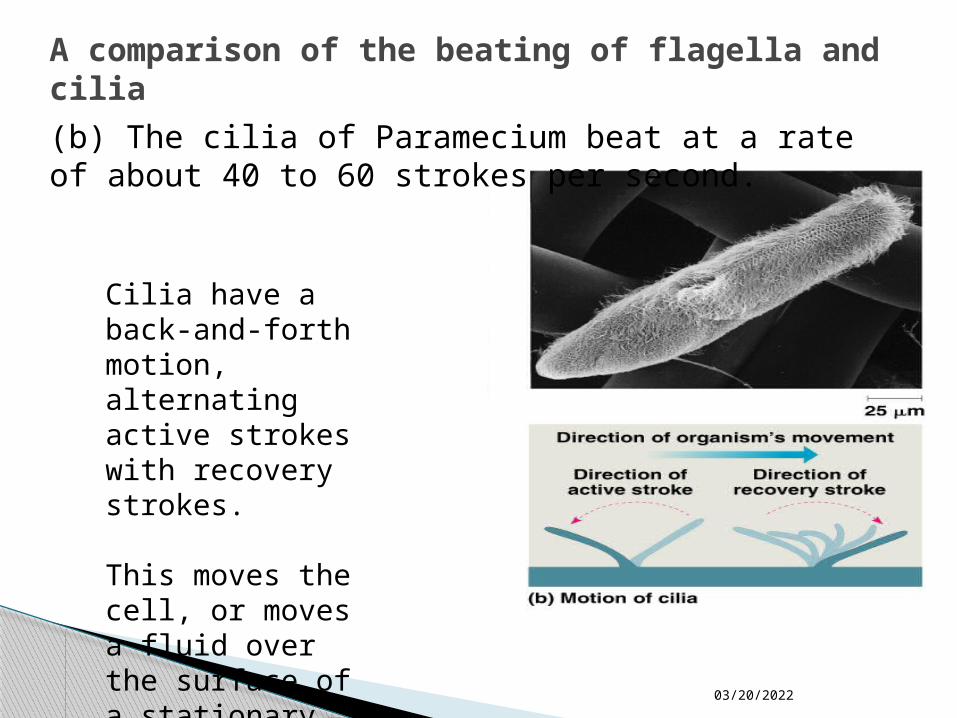
A comparison of the beating of flagella and cilia(b) The cilia of Paramecium beat at a rate of about 40 to 60 strokes per second.
Cilia have a back-and-forth motion, alternating active strokes with recovery strokes.
This moves the cell, or moves a fluid over the surface of a stationary cell.
04/15/2023
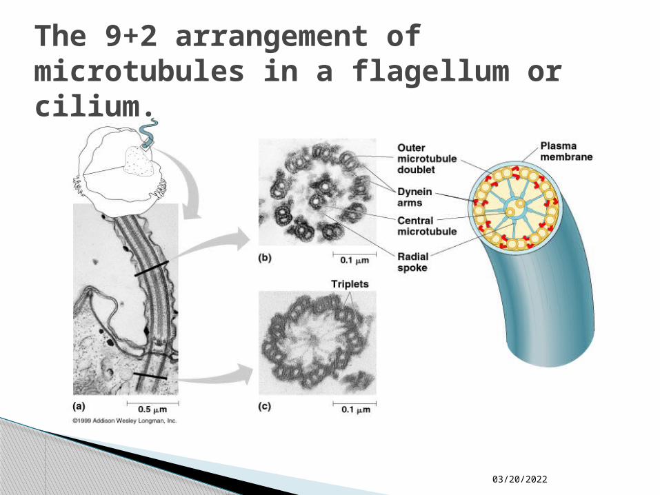
The 9+2 arrangement of microtubules in a flagellum or cilium.

04/15/2023
Ultrastructure
The nine doublets of the cilium extend into the basal body, where each doublet joins another microtubule to form the ring of nine triplets.
The two central microtubules of the cilium terminate above the basal body (TEM).
The basal body anchoring the cilium or flagellum to the cell has a ring of nine microtubule triplets.
04/15/2023
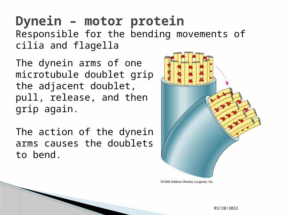
Dynein – motor protein
The dynein arms of one microtubule doublet grip the adjacent doublet, pull, release, and then grip again.
The action of the dynein arms causes the doublets to bend.
Responsible for the bending movements of cilia and flagella

Cilia from an epithelial cell in cross section

04/15/2023
Actin components of the cytoskeleton.
Microfilaments – actin filaments.They are built from molecules of a globular protein – actin.
A microfilament is a twisted double chain of actin subunits (7 nm in diameter)

04/15/2023
Role of microfilaments
Maintenance of cell shape (as a tension-bearing elements)
Changes in cell shape
Muscle contraction
Cytoplasmic streaming
Cell motility
Cell division – cleavage furrow formation

04/15/2023
A structural role of microfilaments
The surface area intestinal cell is increased by its many microvilli,
cellular extensions reinforced by bundles of microfilaments.
These actin filaments are anchored to a network of intermediate filaments

04/15/2023
Microfilaments and motility(a) In muscle cells, actin filaments (orange) lie parallel to thick myosin filaments (purple).
Myosin acts as a motor molecule.
The teamwork of many such sliding filaments enables the entire muscle cell to shorten.

04/15/2023
Microfilaments and motility
(b) In a crawling cell (ameboid movement), actin is organized into a network in the gel-like cortex (outer layer).
This contraction forces the interior fluid into the pseudopod, where the actin network has been weakened.
The pseudopod extends until the actin reassembles into a network.

04/15/2023
Microfilaments and motility(c) In cytoplasmic streaming, a layer of cytoplasm cycles around the cell, moving over a carpet of parallel actin filaments.
Myosin motors attached to organelles in the fluid cytosol may drive the streaming by interacting with the actin.

Role of intermediate filamentsFibrous proteins supercoiled into thicker cables (8-12 nm)
Depending on the cell type, it is presented by one of the several different proteins of the keratin family
Responsible for:maintenance of cell shape (tension-bearing elements)
anchorage of nucleus and certain other organelles
formation of nuclear lamina