Chapter 16 The Cytoskeleton. Cell stained with general protein stain shows the cytoskeleton.
Upload
janis-nelsonCategory
view
278download
2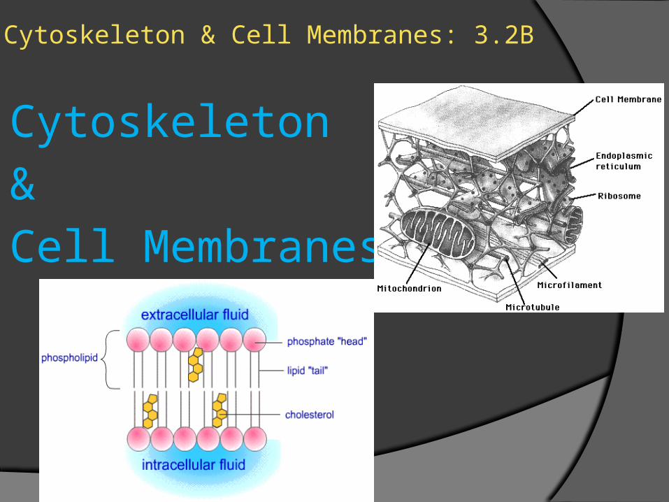
Cytoskeleton & Cell Membranes: 3.2B
Cytoskeleton & Cell Membranes
Cytoskeleton
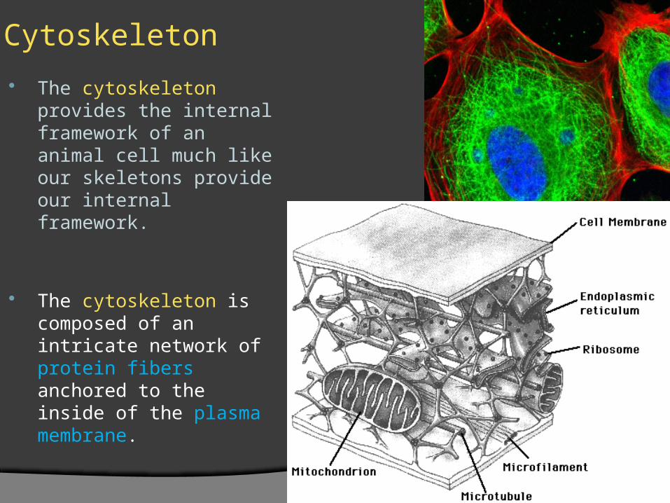
The cytoskeleton provides the internal framework of an animal cell much like our skeletons provide our internal framework.
The cytoskeleton is composed of an intricate network of protein fibers anchored to the inside of the plasma membrane.
Cytoskeleton
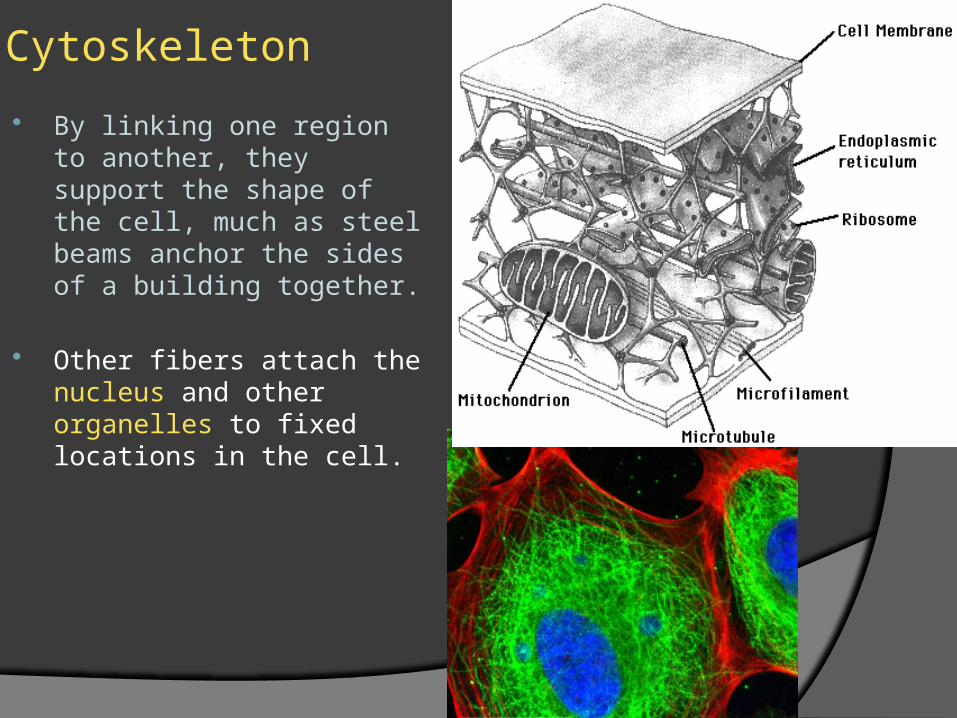
By linking one region to another, they support the shape of the cell, much as steel beams anchor the sides of a building together.
Other fibers attach the nucleus and other organelles to fixed locations in the cell.
Cytoskeleton
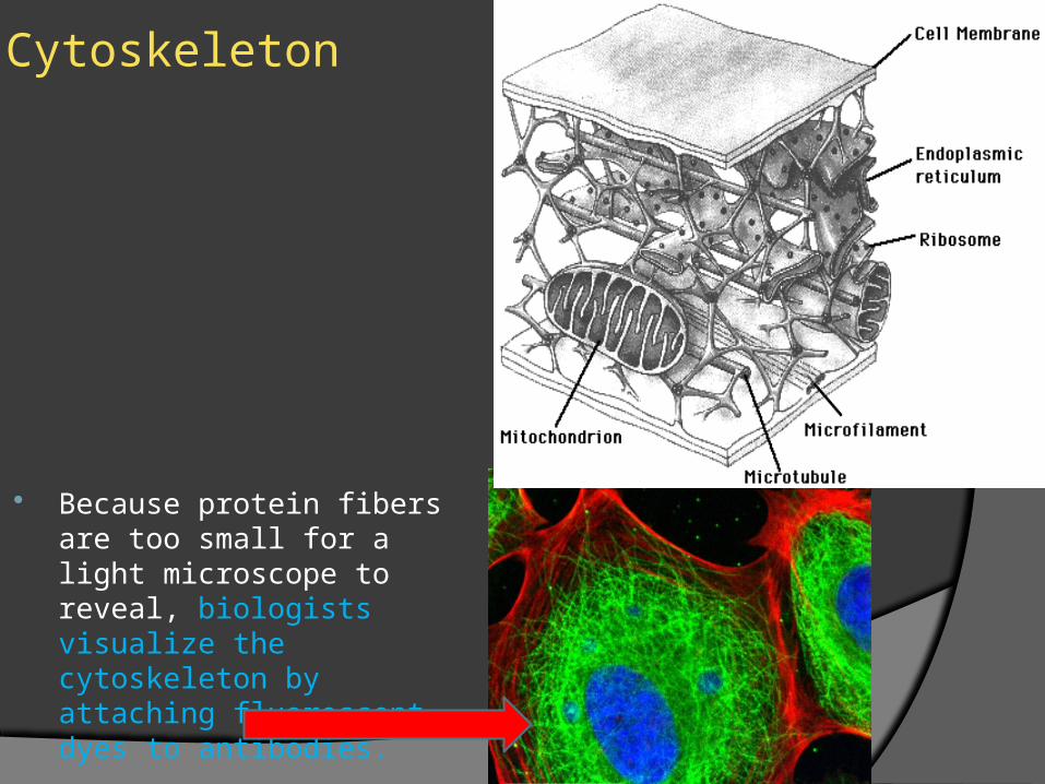
Because protein fibers are too small for a light microscope to reveal, biologists visualize the cytoskeleton by attaching fluorescent dyes to antibodies.
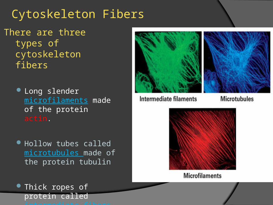
Cytoskeleton FibersThere are three types of
cytoskeleton fibers
Long slender microfilaments made of the protein actin.
Hollow tubes called microtubules made of the protein tubulin
Thick ropes of protein called intermediate fibers
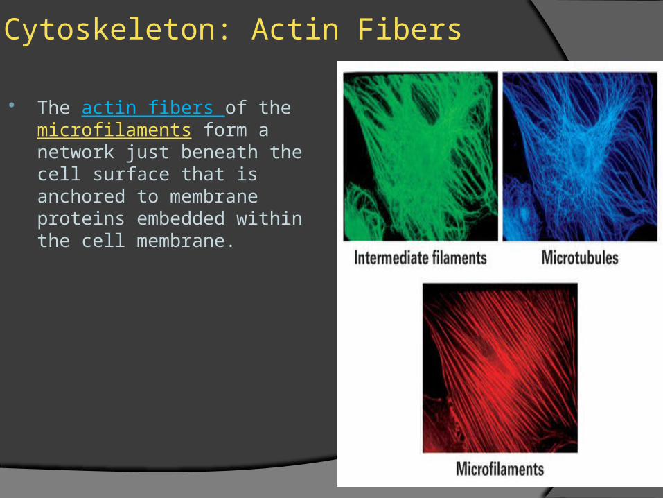
Cytoskeleton: Actin Fibers
The actin fibers of the microfilaments form a network just beneath the cell surface that is anchored to membrane proteins embedded within the cell membrane.
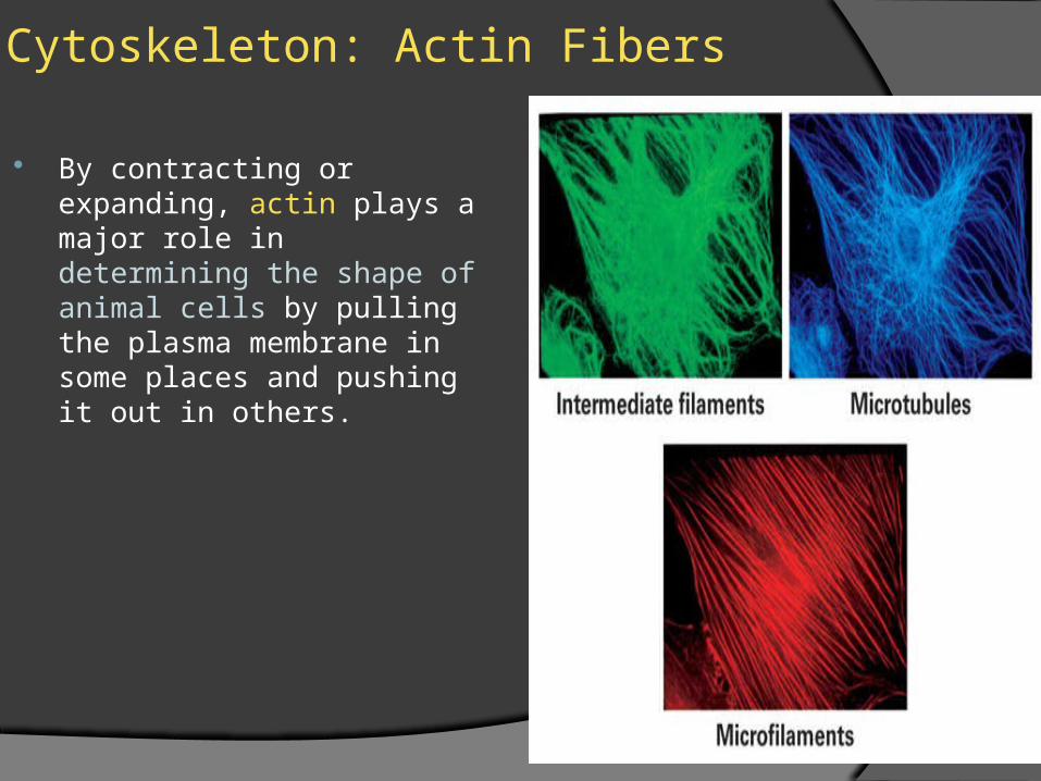
Cytoskeleton: Actin Fibers
By contracting or expanding, actin plays a major role in determining the shape of animal cells by pulling the plasma membrane in some places and pushing it out in others.
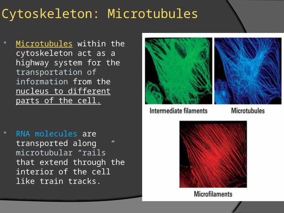
Cytoskeleton: Microtubules
Microtubules within the cytoskeleton act as a highway system for the transportation of information from the nucleus to different parts of the cell.
RNA molecules are transported along microtubular “rails” that extend through the interior of the cell like train tracks.
Cytoskeleton: Intermediate Fibers
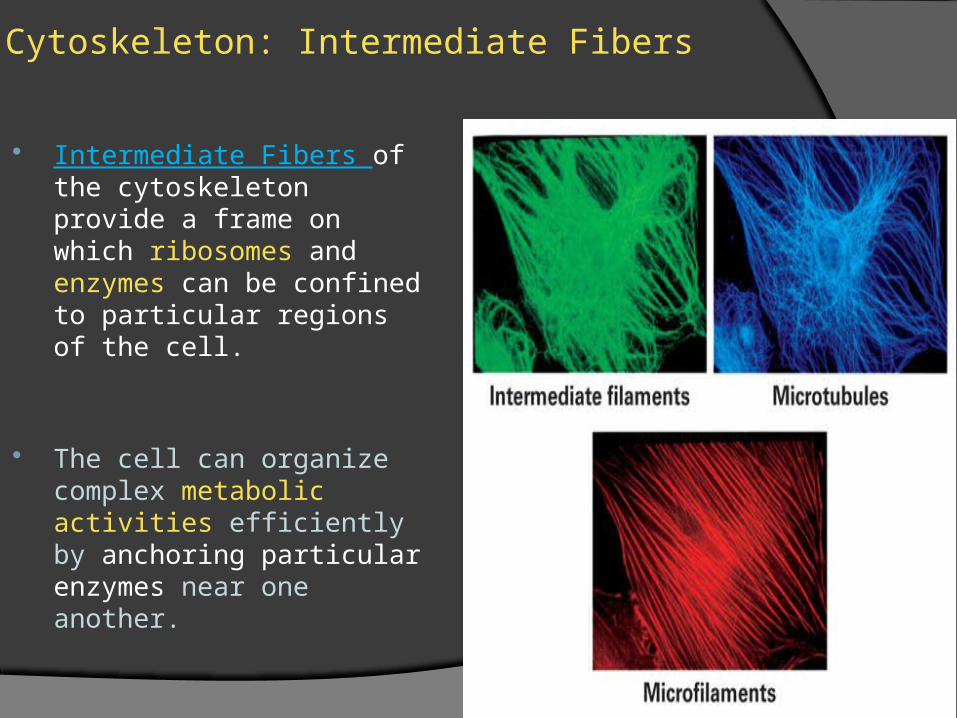
Intermediate Fibers of the cytoskeleton provide a frame on which ribosomes and enzymes can be confined to particular regions of the cell.
The cell can organize complex metabolic activities efficiently by anchoring particular enzymes near one another.
The Cell Membrane: Lipids
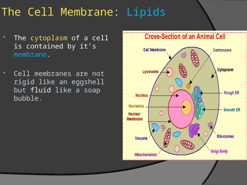
The cytoplasm of a cell is contained by it’s membrane.
Cell membranes are not rigid like an eggshell but fluid like a soap bubble.
The Cell Membrane: Lipids
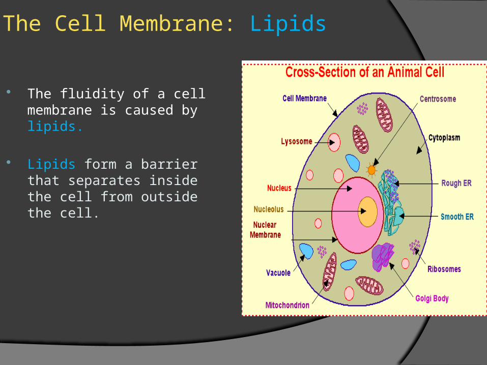
The fluidity of a cell membrane is caused by lipids.
Lipids form a barrier that separates inside the cell from outside the cell.
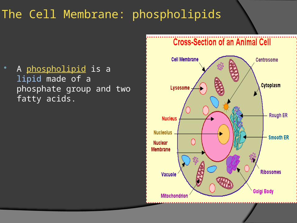
The Cell Membrane: phospholipids
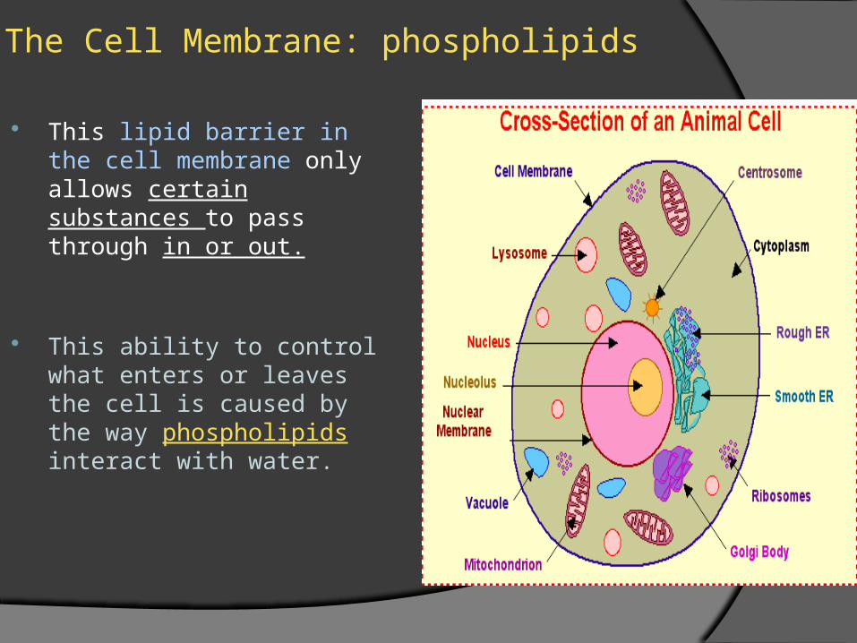
This lipid barrier in the cell membrane only allows certain substances to pass through in or out.
This ability to control what enters or leaves the cell is caused by the way phospholipids interact with water.
The Cell Membrane: phospholipids
A phospholipid is a lipid made of a phosphate group and two fatty acids.
The Cell Membrane:
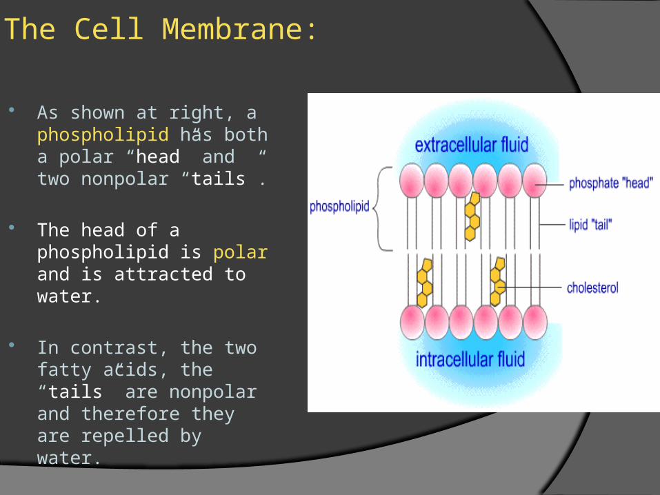
As shown at right, a phospholipid has both a polar “head” and two nonpolar “tails”.
The head of a phospholipid is polar and is attracted to water.
In contrast, the two fatty acids, the “tails” are nonpolar and therefore they are repelled by water.
The Cell Membrane
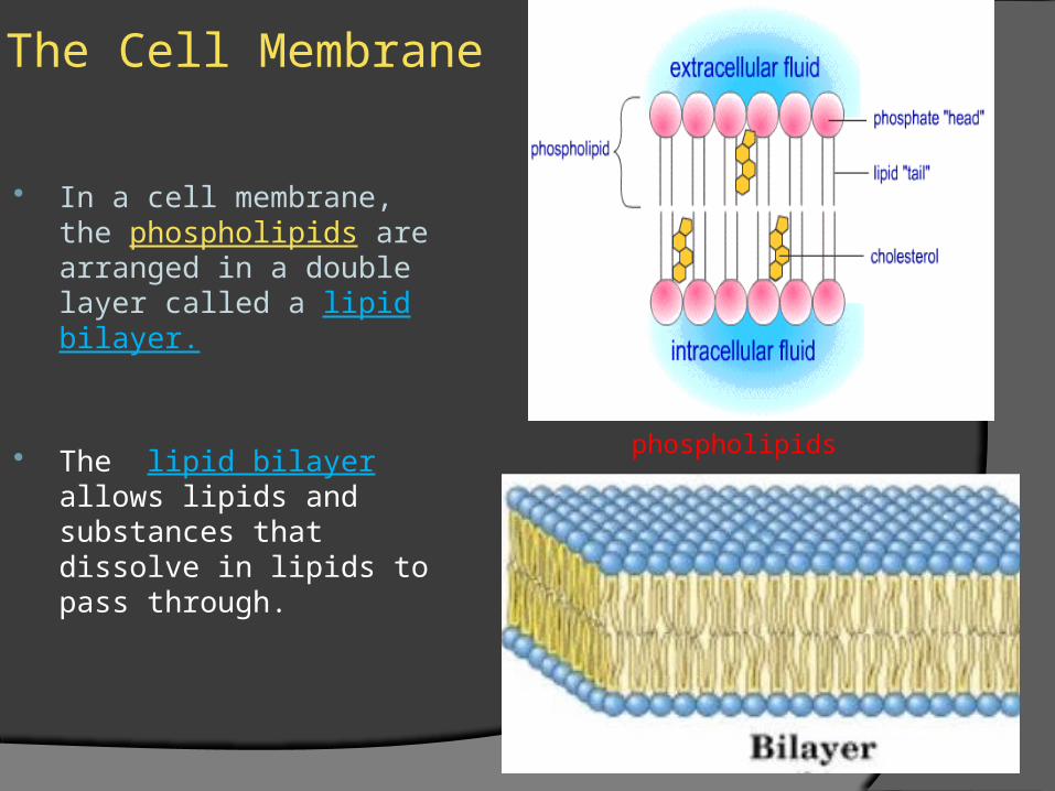
In a cell membrane, the phospholipids are arranged in a double layer called a lipid bilayer.
The lipid bilayer allows lipids and substances that dissolve in lipids to pass through.
phospholipids
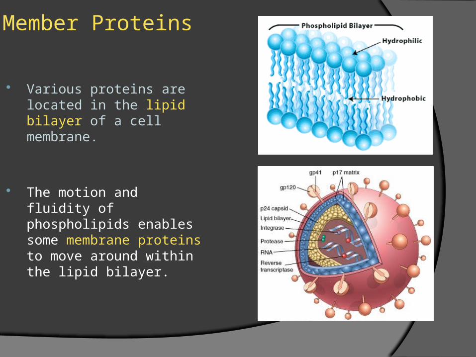
Member Proteins
Various proteins are located in the lipid bilayer of a cell membrane.
The motion and fluidity of phospholipids enables some membrane proteins to move around within the lipid bilayer.
Member Proteins
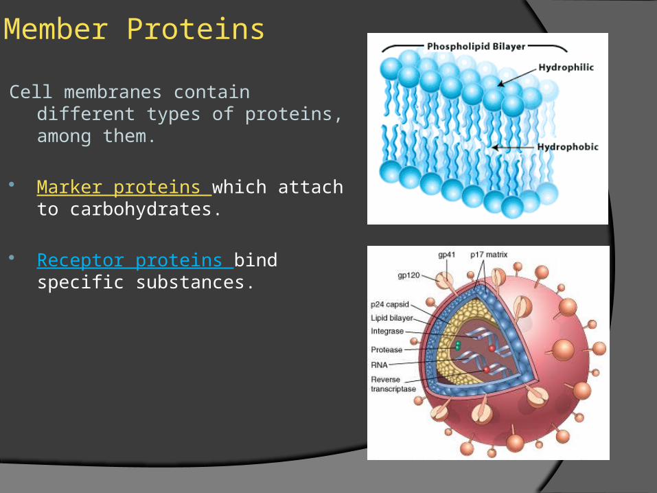
Cell membranes contain different types of proteins, among them.
Marker proteins which attach to carbohydrates.
Receptor proteins bind specific substances.
Member Proteins
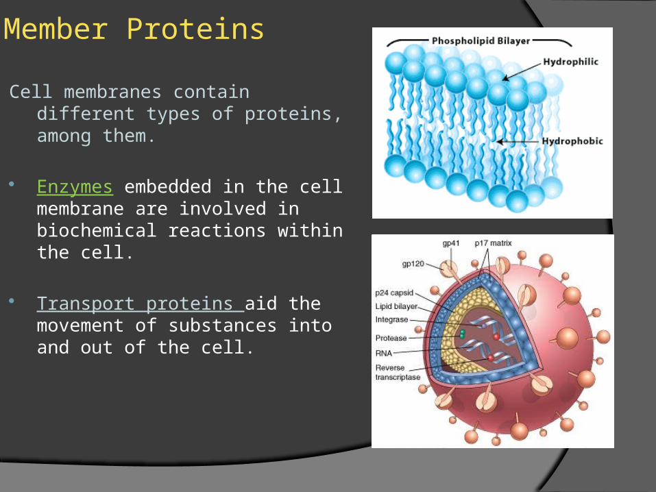
Cell membranes contain different types of proteins, among them.
Enzymes embedded in the cell membrane are involved in biochemical reactions within the cell.
Transport proteins aid the movement of substances into and out of the cell.