Cyanotype - The · PDF filehe cyanotype process (a print example is shown in ... proo˜ng...
19
The Atlas of Analytical Signatures of Photographic Processes CYANOTYPE Dusan C. Stulik | Art Kaplan
Transcript of Cyanotype - The · PDF filehe cyanotype process (a print example is shown in ... proo˜ng...

The
Atla
s of
Ana
lytic
al S
igna
ture
s of
Pho
togr
aphi
c Pr
oces
ses
CYANOTYPE
Dusan C. Stulik | Art Kaplan
2 The Atlas of Analytical Signatures of Photographic Processes The Getty Conservation Institute, © 2013 J. Paul Getty Trust
© 2013 J. Paul Getty Trust. All rights reserved.
The Getty Conservation Institute works internationally to advance conservation practice in the visual arts—broadly interpreted to include objects, collections, architecture, and sites. The GCI serves the conservation community through scientific research, education and training, model field projects, and the dissemination of the results of both its own work and the work of others in the field. In all its endeavors, the GCI focuses on the creation and delivery of knowledge that will benefit the professionals and organizations responsible for the conservation of the world’s cultural heritage.
The Getty Conservation Institute 1200 Getty Center Drive, Suite 700 Los Angeles, CA 90049-1684 United States Telephone: 310 440-7325 Fax: 310 440-7702 Email: [email protected] www.getty.edu/conservation
The Atlas of Analytical Signatures of Photographic Processes is intended for practicing photograph conservators and curators of collections who may need to identify more unusual photographs. The Atlas also aids individuals studying a photographer’s darkroom techniques or changes in these techniques brought on by new or different photographic technologies or by the outside influence of other photographers. For a complete list of photographic processes available as part of the Atlas and for more information on the Getty Conservation Institute’s research on the conservation of photographic materials, visit the GCI’s website at getty.edu/conservation.
ISBN number: 978-1-937433-08-6 (online resource)
Front cover: Cyanotype photograph, 1909. Photographer unknown.
Every effort has been made to contact the copyright holders of the photographs and illustrations in this work to obtain permission to publish. Any omissions will be corrected in future editions if the publisher is contacted in writing.
Historical Background 4
Identification: Cyanotypes 9
Important Variants of the Cyanotype Process 15
Interpretation Guide 18
CONTENTS
3 The Atlas of Analytical Signatures of Photographic Processes The Getty Conservation Institute, © 2013 J. Paul Getty Trust
4 CYANOTYPEThe Atlas of Analytical Signatures of Photographic Processes The Getty Conservation Institute, © 2013 J. Paul Getty Trust
CYANOTYPEEnglish: cyanotype, blueprint, ferro-prussiate process, blue processFrench: cyanotypeGerman: Cyanotypie, Zyanotypie, Eisenblaudruck, Blaupause
HISTORICAL BACKGROUND
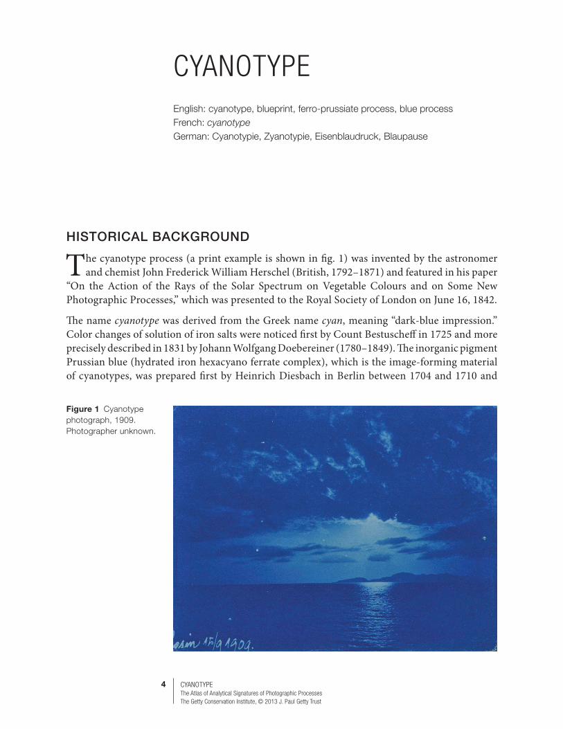
The cyanotype process (a print example is shown in fig. 1) was invented by the astronomer and chemist John Frederick William Herschel (British, 1792–1871) and featured in his paper
“On the Action of the Rays of the Solar Spectrum on Vegetable Colours and on Some New Photographic Processes,” which was presented to the Royal Society of London on June 16, 1842.
The name cyanotype was derived from the Greek name cyan, meaning “dark-blue impression.” Color changes of solution of iron salts were noticed first by Count Bestuscheff in 1725 and more precisely described in 1831 by Johann Wolfgang Doebereiner (1780–1849). The inorganic pigment Prussian blue (hydrated iron hexacyano ferrate complex), which is the image-forming material of cyanotypes, was prepared first by Heinrich Diesbach in Berlin between 1704 and 1710 and
Figure 1 Cyanotype photograph, 1909. Photographer unknown.

5 CYANOTYPEThe Atlas of Analytical Signatures of Photographic Processes The Getty Conservation Institute, © 2013 J. Paul Getty Trust
was used after about 1730 as a pigment in oil paintings and watercolors. Herschel experimented with the cyanotype process in the 1840s and inspired Anna Atkins, daughter of his friend Dr. John Children, to illustrate her botanical studies with cyanotype photograms. The three volumes of her book Photographs of British Algae: Cyanotype Impressions (1843–53) represent the earliest examples of books illustrated with photogenically produced images.
The cyanotype process was seldom used until the 1880s, when it became a cheap proofing process for collodion, dry gelatin plates, and gelatin roll film before the final printing, which used more expensive silver- or platinum-based photographic processes. From the 1870s until about the 1950s, when it was replaced by diazo-based reprographic processes, the cyanotype process and its variants were the primary processes used by engineers and architects to copy plans.
The first commercial cyanotype paper became available in 1872 in France, where it was produced by Marion et Cie in Paris under the name papier ferro-prussiate. Students at the Massachusetts Institute of Technology have been instructed in the use of cyanotype for reprographic purposes (blueprints) since 1875. The first commercial blueprint machine made in Switzerland was introduced into the United States at the Philadelphia Centennial Exposition of 1876. Sensitized cyanotype paper is still available today for student instruction and as an educational toy. Kits for making cyanotypes are also available.
The cyanotype process, together with a number of other, older photographic processes, was revived by contemporary photographers in the 1960s. The older processes were considered alternatives to the silver gelatin process using commercial photographic material. It is interesting to note that today, as digital photography replaces all applications of classic “chemical” photography, the silver gelatin process itself will soon be considered an alternative photographic process.
Commercial Cyanotype Material
French Satin Jr., Ideal, Eastman’s Blue Print, Opto, E.A. Ferroprussiate PaperEastman blueprint postcardSilkdown (cyanotype fabric)
Two types of commercial cyanotype material available during the early twentieth century are seen in figure 2. A historical timeline of the cyanotype process is given in figure 3.
Figure 2 Two types of blueprint photographic paper commercially available in the 20th century.

6 CYANOTYPEThe Atlas of Analytical Signatures of Photographic Processes The Getty Conservation Institute, © 2013 J. Paul Getty Trust
Process Description
The general principle of the cyanotype process is the photochemical reduction of iron (III) salts to iron (II) salt that reacts with potassium ferricyanide (red prussiate of iron), forming an intensely blue complex. The process has several simple steps:
1. Selected paper or other material is coated with a mixture of iron (III) salt (today mostly of ferric ammonium citrate) and potassium ferricyanide. The coating must be applied under dim light due to light sensitivity.
2. The resulting yellow-greenish layer of sensitized material is dried in the dark. 3. The dried, sensitized material is exposed under a negative or other partially or fully
opaque material (when creating photograms) to strong light (sun or an artificial UV light source), usually in a printing frame that assures good contact between the negative and the sensitized material.
4. Light exposure is aided by simultaneous exposure of a sensitometric wedge or timed based on exposure and development of a series of test samples.
5. The exposed material is transferred to a water bath to complete the formation of Prussian blue in areas exposed to light and to dissolve any unexposed mixture of sensitizing compounds. Full development of the blue image can be aided by the addition of a hydrogen peroxide solution to the bath. Otherwise, the blue image can be assessed only after the material is fully dried and oxidized by exposure to air.
Figure 3 Timeline of the cyanotype process.
1720 1760 1840 18801800 1920 1960 2000
Count Bestuscheff notices light sensitivity
of iron salt solution
J. Herschel invents cyanotype process
First commercial cyanotype paper
First commercial blueprint machine
M. Ware invents new cyanotype process
Blueprint process replaced by diazo process (also blue)
H. Pellet invents positive cyanotype process
Alternative photographic process community rediscovers cyanotype process
A. Atkins publishes Photographs of British Algae: Cyanotype Impressions;
�rst photographically illustrated book
Application of cyanotype printing for negative proo�ng and as a reprographic medium
1725 1842
1872
1876
1877
c. 1870–1945
1843–53
Ongoing from 1960s
c. 1950s 1994
7 CYANOTYPEThe Atlas of Analytical Signatures of Photographic Processes The Getty Conservation Institute, © 2013 J. Paul Getty Trust
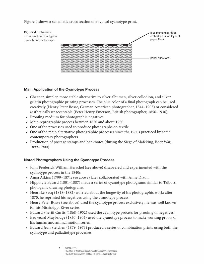
Figure 4 shows a schematic cross section of a typical cyanotype print.
Main Application of the Cyanotype Process
• Cheaper, simpler, more stable alternative to silver albumen, silver collodion, and silver gelatin photographic printing processes. The blue color of a final photograph can be used creatively (Henry Peter Bosse, German American photographer, 1844–1903) or considered aesthetically unacceptable (Peter Henry Emerson, British photographer, 1856–1936).
• Proofing medium for photographic negatives• Main reprographic process between 1870 and about 1950• One of the processes used to produce photographs on textile• One of the main alternative photographic processes since the 1960s practiced by some
contemporary photographers• Production of postage stamps and banknotes (during the Siege of Mafeking, Boer War,
1899–1900)
Noted Photographers Using the Cyanotype Process
• John Frederick William Herschel (see above) discovered and experimented with the cyanotype process in the 1840s.
• Anna Atkins (1799–1871; see above) later collaborated with Anne Dixon.• Hippolyte Bayard (1801–1887) made a series of cyanotype photograms similar to Talbot’s
photogenic drawing photograms.• Henri Le Secq (1818–1882) worried about the longevity of his photographic work; after
1870, he reprinted his negatives using the cyanotype process.• Henry Peter Bosse (see above) used the cyanotype process exclusively; he was well known
for his Mississippi River series.• Edward Sheriff Curtis (1868–1952) used the cyanotype process for proofing of negatives. • Eadweard Muybridge (1830–1904) used the cyanotype process to make working proofs of
his human and animal motion series.• Edward Jean Steichen (1879–1973) produced a series of combination prints using both the
cyanotype and palladiotype processes.
Figure 4 Schematic cross section of a typical cyanotype photograph.

8 CYANOTYPEThe Atlas of Analytical Signatures of Photographic Processes The Getty Conservation Institute, © 2013 J. Paul Getty Trust
Bibliography (by date)
Herschel, J. F. W. 1842. “On the Action of the Rays of the Solar Spectrum on Vegetable Colours and on Some New Photographic Processes.” Philosophical Transaction of the Royal Society of London 132: 181–214.
Brown, G. E. 1902. Ferric and Heliographic Processes. London: Dawbarn & Ward. Tennant, J. A. 1900 (January). Photo-Miniature 1(10): 481–514.Jones, B. E. 1911. Cassell’s Cyclopaedia of Photography. New York: Cassell, 66–68, 196, 397–98.Wall, E. J. 1926. The Dictionary of Photography and Reference Book for Amateur and Professional Photographers.
Edited by F. J. Mortimer. London: Iliffe & Sons, 187–92.Neblette, C. B. 1942. Photography: Its Principles and Practice (4th ed.). New York: Van Nostrand, 697–700.Eder, J. M. 1945. History of Photography. New York: Columbia University Press, 542–50.Eder, J. M. 1978. History of Photography. Translated by E. Epstean. New York: Dover, 177–78, 542–48, 549–50.Crawford, W. 1979. The Keepers of Light: A History and Working Guide to Early Photographic Processes. New
York: Morgan & Morgan, 67–68, 163–66.Wilson, J. L. 1990. “The Cyanotype.” In Technology and Art: The Birth and Early Years of Photography. Bath:
Historical Group, Royal Photographic Society. Schaaf, L. J. 1992. Out of the Shadows: Herschel, Talbot and the Invention of Photography. New Haven, CT: Yale
University Press. Nadeau, L. 1994. Encyclopedia of Printing, Photographic and Photomechanical Processes. Vols. 1 and 2.
Fredericton, New Brunswick, Canada: Atelier Luis Nadeau, 47–48, 80–81, 360, 415–17.Berrie, B. H. 1997. “Prussian Blue.” In Artist’s Pigments. Vol. 3. Edited by Elizabeth West Fitzhugh. Washington,
DC: National Gallery of Art, 191–217.Knodt, R., and Pollmeier, K. 1999. Verfahren der Fotografie. Essen: Museum Folkwang, 68.Ware, M. 1999. Cyanotype. Bradford, UK: Science Museum, London, and National Museum of Photography,
Film & Television. Lamb, A. R. 2003. Library of Professional Picture Framing. Vol. 6, Framing Photography. Akron, OH: Columbia
Publishing, 31.Cartier-Bresson, A. 2008. Le Vocabulaire Technique de la Photographie. Paris: Marval, 192.Kennel, S. 2009. In the Dark Room: An Illustrated Guide to Photographic Processes before the Digital Age.
Washington, DC: National Gallery of Art, 32–33.Lavédrine, B. 2009. Photographs of the Past: Process and Preservation. Los Angeles: The Getty Conservation
Institute, 150–55.Leyshon, W. E. 2012. Photographs from the 19th Century: A Process Identification Guide. Prescott, AZ:
Sharlot Hall Museum Archives, 15, 96. Accessed March 26, 2013. http://www.sharlot.org/archives/photographs/19th/book/index.html
Cyanotype-Related Patents
A. Baudesson and P. Houzeau, English Patent 2,526 (Oct. 13, 1864) Preparation of Prussian blue and treatment for toning of images to black, violet, and red colors
9 CYANOTYPEThe Atlas of Analytical Signatures of Photographic Processes The Getty Conservation Institute, © 2013 J. Paul Getty Trust
IDENTIFICATION: CYANOTYPES
Visual Signatures
Visual Characteristics
Visual identification of cyanotype photographs is rather simple. Once identified, there is often no further need for any microscopic or analytical investigation. Depending on the chemistry of the process and the paper substrate used for printing, cyanotype photographs can be found in many shades of blue, from light blue and gray blue to a bright and intense navy blue. The highlights of cyanotype photographs are light blue or white, and the dark areas are in various intensities of blue. When tilted and observed under a raking light, most cyanotype photographs exhibit a matte surface in both the blue and white areas of the image. A very slight sheen in some white areas, if any, can be attributed to sizing of the paper before printing. Some, namely modern, cyanotype images have signs of hand coating clearly visible in the image or hidden under a framing matte.
Microscopic Characteristics
Under a stereomicroscope, cyanotype photographs exhibit a typical one-layer structure that shows a mass of blue particles and particle clusters. These clusters coat fibers in the paper substrate that are clearly visible under high magnification (figs. 5a–5c). Some glossiness of paper fibers in white areas of the photograph can be seen under higher magnification. Its presence is attributed to the natural glossiness of cellulosic fibers and to the glossiness added due to the sizing of the paper substrate. No baryta layer is present when inspecting the edges and corners of cyanotype photographs.
Analytical Signatures
XRF
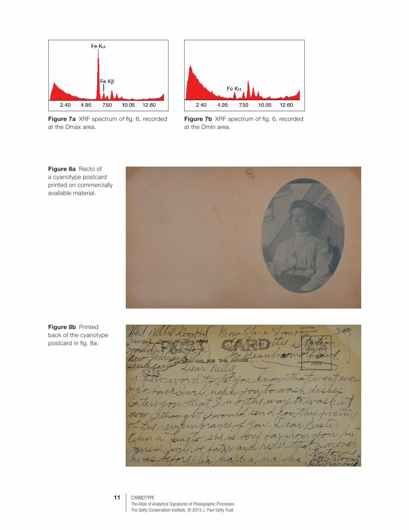
The elemental composition of untoned cyanotype photographs printed on clean paper substrates is rather simple. The key element that should be present in all cyanotype photographs is iron from the cyanotype blue complex pigment. The amount of iron should vary between the Dmax and Dmin areas. Figure 6 shows a modern cyanotype photograph printed on an archival paper substrate. Comparison of the XRF spectra recorded at the Dmax and Dmin areas of the image (figs. 7a, 7b) show different amounts of iron depending on how much of the cyanotype blue complex pigment is present. The XRF spectrum recorded at the Dmin area of the image shows a very low concentration of iron. This is typical even for a well-processed, cleared, and washed cyanotype image.
Some commercially available cyanotype printing paper was also available for making cyanotype postcards (figs. 8a, 8b). Figure 9 shows the XRF spectrum of the Dmax area of the postcard.
For postcard production, the cyanotype chemistry was coated onto a heavier paper stock. The small amount of lead recorded in the XRF spectrum is present in the paper substrate.
Note: The back of the cyanotype postcard (see fig. 8b) contains information that can help date the image even when there is no direct date on the postcard. The undivided back of the postcard

10 CYANOTYPEThe Atlas of Analytical Signatures of Photographic Processes The Getty Conservation Institute, © 2013 J. Paul Getty Trust
Figure 5a Photomicrograph of a cyanotype photograph at 10× magnification, showing blue particles and particle clusters.
Figure 5c Photomicrograph of the same photograph at 40× magnification, showing glossiness in the white areas.
Figure 5b Photomicrograph of the same photograph at 25× magnification, showing clearly visible fibers in the paper substrate.
Figure 6 Modern cyanotype photograph printed on archival paper substrate.
11 CYANOTYPEThe Atlas of Analytical Signatures of Photographic Processes The Getty Conservation Institute, © 2013 J. Paul Getty Trust
Figure 7a XRF spectrum of fig. 6, recorded at the Dmax area.
Figure 7b XRF spectrum of fig. 6, recorded at the Dmin area.
Figure 8a Recto of a cyanotype postcard printed on commercially available material.
Figure 8b Printed back of the cyanotype postcard in fig. 8a.
12 CYANOTYPEThe Atlas of Analytical Signatures of Photographic Processes The Getty Conservation Institute, © 2013 J. Paul Getty Trust
image was used in the United States until 1907. The address was to be written across the entire back of the postcard, and any message had to be incorporated into the image area on the front of the postcard. The area for the stamp on the back calls for a one-cent stamp, another visual clue in dating the image.
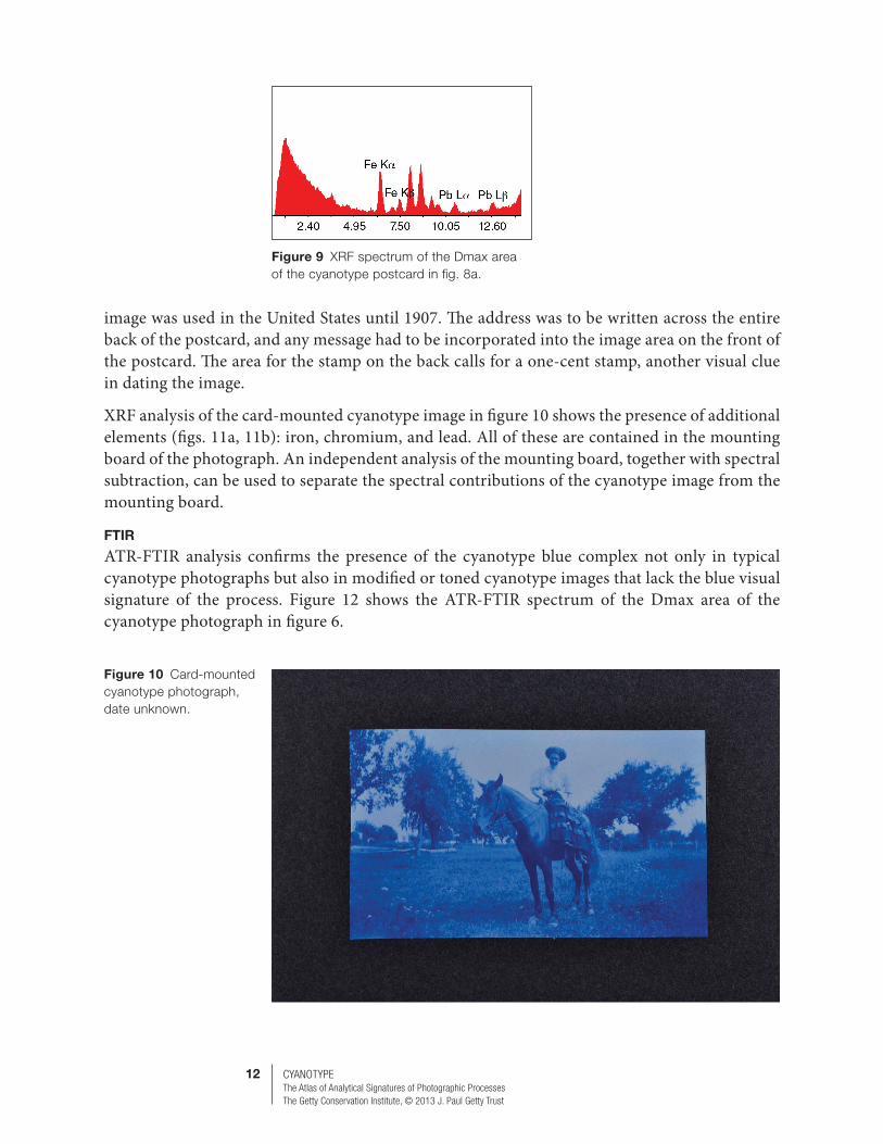
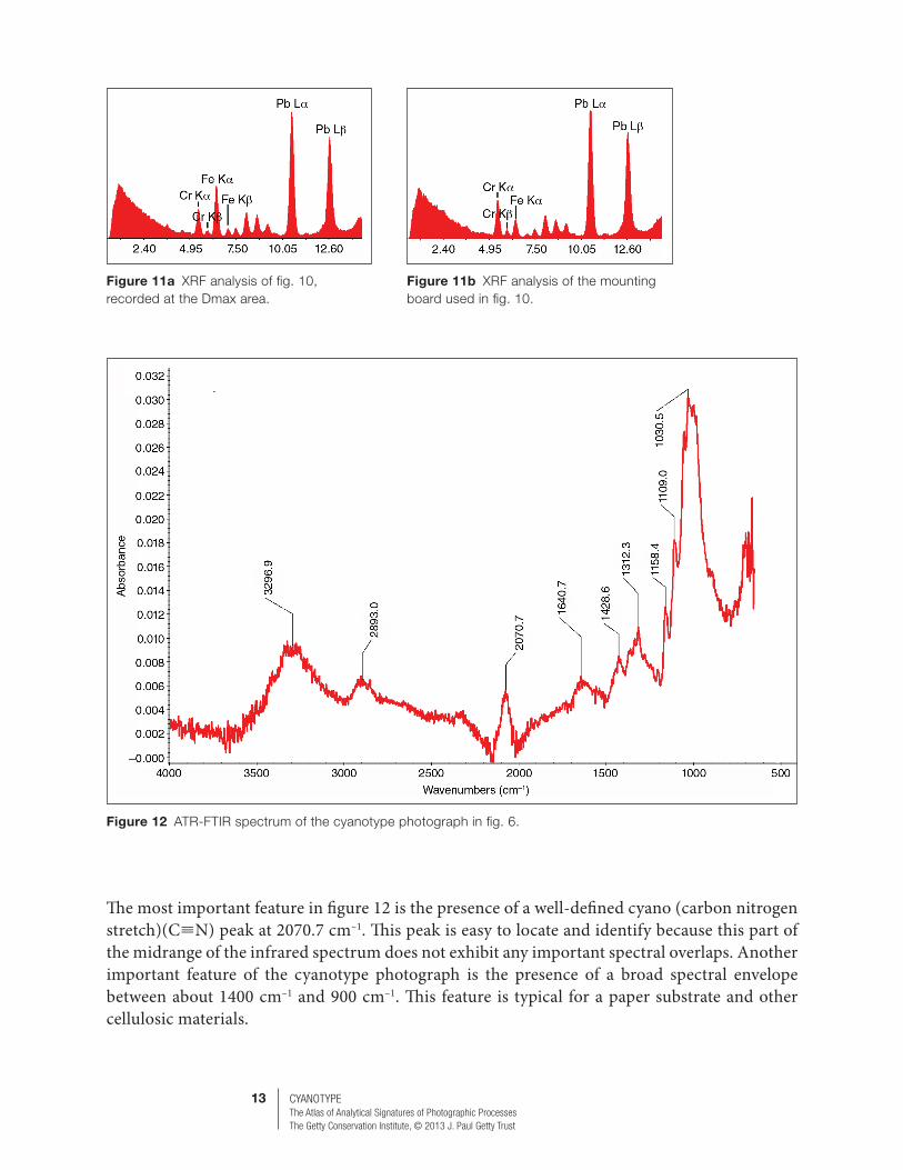
XRF analysis of the card-mounted cyanotype image in figure 10 shows the presence of additional elements (figs. 11a, 11b): iron, chromium, and lead. All of these are contained in the mounting board of the photograph. An independent analysis of the mounting board, together with spectral subtraction, can be used to separate the spectral contributions of the cyanotype image from the mounting board.
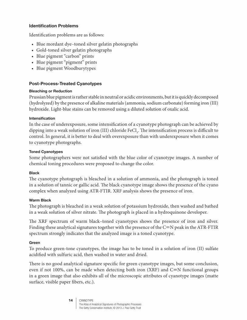
FTIR
ATR-FTIR analysis confirms the presence of the cyanotype blue complex not only in typical cyanotype photographs but also in modified or toned cyanotype images that lack the blue visual signature of the process. Figure 12 shows the ATR-FTIR spectrum of the Dmax area of the cyanotype photograph in figure 6.
Figure 9 XRF spectrum of the Dmax area of the cyanotype postcard in fig. 8a.
Figure 10 Card-mounted cyanotype photograph, date unknown.
13 CYANOTYPEThe Atlas of Analytical Signatures of Photographic Processes The Getty Conservation Institute, © 2013 J. Paul Getty Trust
The most important feature in figure 12 is the presence of a well-defined cyano (carbon nitrogen stretch)(C≡N) peak at 2070.7 cm–1. This peak is easy to locate and identify because this part of the midrange of the infrared spectrum does not exhibit any important spectral overlaps. Another important feature of the cyanotype photograph is the presence of a broad spectral envelope between about 1400 cm–1 and 900 cm–1. This feature is typical for a paper substrate and other cellulosic materials.
Figure 12 ATR-FTIR spectrum of the cyanotype photograph in fig. 6.
Figure 11a XRF analysis of fig. 10, recorded at the Dmax area.
Figure 11b XRF analysis of the mounting board used in fig. 10.
14 CYANOTYPEThe Atlas of Analytical Signatures of Photographic Processes The Getty Conservation Institute, © 2013 J. Paul Getty Trust
Identification Problems
Identification problems are as follows:
• Blue mordant dye–toned silver gelatin photographs• Gold-toned silver gelatin photographs• Blue pigment “carbon” prints• Blue pigment “pigment” prints• Blue pigment Woodburytypes
Post-Process-Treated Cyanotypes
Bleaching or Reduction
Prussian blue pigment is rather stable in neutral or acidic environments, but it is quickly decomposed (hydrolyzed) by the presence of alkaline materials (ammonia, sodium carbonate) forming iron (III) hydroxide. Light-blue stains can be removed using a diluted solution of oxalic acid.
Intensification
In the case of underexposure, some intensification of a cyanotype photograph can be achieved by dipping into a weak solution of iron (III) chloride FeCl3. The intensification process is difficult to control. In general, it is better to deal with overexposure than with underexposure when it comes to cyanotype photographs.
Toned Cyanotypes
Some photographers were not satisfied with the blue color of cyanotype images. A number of chemical toning procedures were proposed to change the color.
Black
The cyanotype photograph is bleached in a solution of ammonia, and the photograph is toned in a solution of tannic or gallic acid. The black cyanotype image shows the presence of the cyano complex when analyzed using ATR-FTIR. XRF analysis shows the presence of iron.
Warm Black
The photograph is bleached in a weak solution of potassium hydroxide, then washed and bathed in a weak solution of silver nitrate. The photograph is placed in a hydroquinone developer.
The XRF spectrum of warm black–toned cyanotypes shows the presence of iron and silver. Finding these analytical signatures together with the presence of the C≡N peak in the ATR-FTIR spectrum strongly indicates that the analyzed image is a toned cyanotype.
Green
To produce green-tone cyanotypes, the image has to be toned in a solution of iron (II) sulfate acidified with sulfuric acid, then washed in water and dried.
There is no good analytical signature specific for green cyanotype images, but some conclusion, even if not 100%, can be made when detecting both iron (XRF) and C≡N functional groups in a green image that also exhibits all of the microscopic attributes of cyanotype images (matte surface, visible paper fibers, etc.).
15 CYANOTYPEThe Atlas of Analytical Signatures of Photographic Processes The Getty Conservation Institute, © 2013 J. Paul Getty Trust
Bluish Lilac
Bluish lilac–toned cyanotype images are produced by bathing photographs in a dilute solution of borax or potassium oxalate, followed by toning the resulting image in a diluted solution of potassium sulfocyanide.
XRF analysis detects iron, and ATR-FTIR analysis of the Dmax areas of the image may show the presence of a weak spectral peak of the C≡N spectral group at around 2165 cm–1.
Violet Tones
Cyanotype photographs can be toned to violet tones by treating with a warm solution of lead acetate. XRF analysis of the resulting toned cyanotype image shows the presence of high concentrations of lead. The identification of the violet-toned cyanotype is confirmed by detecting the C≡N spectral group using ATR-FTIR analysis.
Mordant Toning of Cyanotypes
Mordant toning of cyanotype photographs was developed by John Mercer (1791–1866). Bleached cyanotypes were treated in a solution of organic dyes. XRF analysis of these types of toned images shows the presence of iron. ATR-FTIR analysis should detect the presence of the C≡N spectral peak but is usually not sensitive enough to detect the low concentration of organic dyes. Visual identification of the bright color images, in combination with the results of the XRF analysis (iron) and ATR-FTIR analysis (C≡N peak), leads to the proposal that the image might be a mordant-toned cyanotype.
IMPORTANT VARIANTS OF THE CYANOTYPE PROCESS
Pellet’s processThe new cyanotype (Ware)Cyanotype Rex (King)
Formula-Based Variants
At first John Frederick William Herschel experimented with the cyanotype process using only the low sensitivity of potassium ferricyanide. The process resulted in a weaker cyanotype image but required very long exposure times. A great number of different cyanotype formulas were described in the photographic and scientific literature. With the exception of formulas that include the addition of other inorganic materials (silver nitrate, potassium dichromate) or organic material (gelatin) to the basic cyanotype formula, there is no easy or proven way to relate the appearance, microstructure, and chemical composition of a cyanotype image to the different formulas that could have been used by an artist.
A major change in the basic cyanotype chemistry occurred after 1897, when the Austrian chemist Eduard Valenta introduced the green form of ammonium ferric citrate, which quickly replaced the brown form. Green ammonium ferric citrate was found to increase the light sensitivity of cyanotype material, but the final chemical composition of cyanotype images resulting from using either form of ammonium ferric citrate is identical.

16 CYANOTYPEThe Atlas of Analytical Signatures of Photographic Processes The Getty Conservation Institute, © 2013 J. Paul Getty Trust
Pellet’s Process (Positive Cyanotype)
Positive cyanotype was invented by Henri Pellet (French) in 1877.
Process Description
The basic idea for the direct positive cyanotype was developed early by Herschel, who conducted a series of initial experiments but did not achieve reproducible results. A more usable version of the process was invented and patented in 1877 by Henri Pellet.
Paper is coated with a mixture of solutions of gum arabic, ammonium ferric citrate, and ferric chloride. The dried and sensitized paper is exposed to strong light in contact with line drawings, continuous transparent images, or translucent positive images. In the areas exposed to light, photochemical reduction of iron (III) salts takes place. Only areas protected from light still retain some amounts of unreduced iron (III) that react with the developing solution of potassium ferrocyanide (yellow prussiate or ferro-prussiate), forming the Prussian blue pigment. The process was used from 1877 to the 1940s, though it was much more difficult and delicate than the basic cyanotype process.
Visual Characteristics
The visual characteristics of Pellet’s process include positive image when using positive transparency; negative image when exposing using a negative; blue lines on a white background when copying black-line drawings or plans.
Microscopic Characteristics
Pellet’s process is identical to cyanotype.
Analytical Signatures
XRF
Pellet’s process is identical to cyanotype.
FTIR
There is a theoretical possibility of detecting the presence of the gum arabic coating in blue areas of the image. However, the analytical signal of gum arabic is very weak, and it is in a spectral region highly prone to spectral interferences.
Identification Problems
Modern so-called blueprint reprographic copies that provide blue lines on a white background are actually based on the light sensitivity of diazo compounds.
The New Cyanotype (Ware)
The new cyanotype was invented by Mike Ware (British) in 1994.
Process Description
Faced with several shortcomings of the basic cyanotype process (long light exposure, short-term stability of mixed sensitizer, peptization of Prussian blue pigment during washing, and staining
17 CYANOTYPEThe Atlas of Analytical Signatures of Photographic Processes The Getty Conservation Institute, © 2013 J. Paul Getty Trust
of highlights), Mike Ware developed a new variant of the cyanotype process based on the light sensitivity of ammonium ferric oxalate and its reaction with ammonium ferricyanide that is produced during the preparation of the sensitizing solution. The major change in the chemistry of the new cyanotype process, in comparison with the basic cyanotype process that can be utilized for the identification of the new cyanotype photograph, is the frequently used addition of a solution of ammonium dichromate to the sensitizing solution.
Visual Characteristics
The new cyanotype is similar to a basic cyanotype.
Microscopic Characteristics
The new cyanotype is similar to a basic cyanotype.
Analytical Signatures
XRF
The presence of chromium can be an indication that Ware’s new cyanotype process was used.
FTIR
The new cyanotype is identical to the ATR-FTIR signature of the basic cyanotype process.
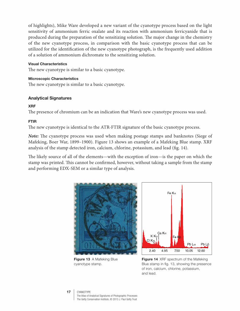
Note: The cyanotype process was used when making postage stamps and banknotes (Siege of Mafeking, Boer War, 1899–1900). Figure 13 shows an example of a Mafeking Blue stamp. XRF analysis of the stamp detected iron, calcium, chlorine, potassium, and lead (fig. 14).
The likely source of all of the elements—with the exception of iron—is the paper on which the stamp was printed. This cannot be confirmed, however, without taking a sample from the stamp and performing EDX-SEM or a similar type of analysis.
Figure 13 A Mafeking Blue cyanotype stamp.
Figure 14 XRF spectrum of the Mafeking Blue stamp in fig. 13, showing the presence of iron, calcium, chlorine, potassium, and lead.

18 CYANOTYPEThe Atlas of Analytical Signatures of Photographic Processes The Getty Conservation Institute, © 2013 J. Paul Getty Trust
INT
ER
PR
ETA
TIO
N G
UID
E
Tab
le 1
Sum
mar
y of
the
mai
n m
icro
scop
ic a
nd a
naly
tical
sig
natu
res
of c
yano
typ
e p
hoto
grap
hs a
nd s
ome
pro
cess
es c
omm
only
mis
iden
tified
as
cyan
otyp
es.
The
info
rmat
ion
bel
ow is
for
typ
ical
ver
sion
s of
eac
h p
roce
ss.
Exc
eptio
ns t
o ea
ch e
ntry
may
exi
st b
ut a
re r
are.
Cyan
otyp
e Pr
ints
Proc
ess
Imag
e St
ruct
ure
Surf
ace
Coat
ing
Pape
r Fi
bers
FeAg
Ba Ti
Othe
r In
orga
nics
Cellu
lose
Gela
tinCo
llodi
onCN
Othe
rOr
gani
csTo
nalit
yNo
tes
Cyan
otyp
ePh
otog
raph
– X
!X
!–
–(X
)*–
X !
––
X !
–
blue (light to dark)
Blue
tone
d ge
latin
Phot
ogra
ph(X
)–
XX
X–
––
X–
X(c
oatin
gs)
Blue
(gol
d)
tone
d ge
latin
Phot
ogra
ph(X
)–
–X
X–
Au–
X–
–(c
oatin
gs)
Unde
r spe
cial
co
nditi
ons
gold
m
ay p
rodu
ce a
bl
ue o
r red
tone
Blue
pig
men
t ca
rbon
Phot
ogra
ph(X
)(X
)(X
)–
––
Cr–
X–
(X)
–
Blue
pig
men
t W
oodb
uryt
ypes
w
ere
desc
ribed
in
liter
atur
e bu
t are
ra
re
Blue
pig
men
t gu
mPh
otog
raph
–X
(X)
––
–Cr
X–
–(X
)Gu
m
arab
ic
(low
)
For a
naly
tical
si
gnat
ures
of g
um
arab
ic, s
ee th
e Gu
m D
ichr
omat
e se
ctio
n (fo
rthco
min
g)
Blue
pig
men
t oi
lPh
otog
raph
–(X
)(X
)–
––
Cr(X
)X
–(X
)
Este
r bo
nds
(low
), in
ks
For a
naly
tical
si
gnat
ures
of o
il,
see
the
Oil s
ectio
n (fo
rthco
min
g)
Blue
pig
men
t br
omoi
lPh
otog
raph
––
(X)
–X
–Cr
, (Cu
)–
X–
(X)
Este
r bo
nds
(low
), in
ks
For a
naly
tical
si
gnat
ures
of
brom
oil,
see
the
Brom
oil s
ectio
n (fo
rthco
min
g)
Blue
ink
phot
o-m
echa
nica
l
Phot
o-m
echa
nica
l–
(X)*
*(X
)–
(X)
(X)
(Ca)
, (Sr
)(X
)–
(X)
(X)*
**–
X
Pres
ent
( )
May
be
pres
ent
(X)*
So
me
mod
ern
prin
ts o
n 20
th-
and
21st
-cen
tury
sub
stra
tes
cont
aini
ng T
iO2
–
Abse
nt
!
Key
sign
atur
e (X
)**
Pape
r fibe
rs a
re n
ot v
isib
le w
hen
a pa
per “
clay
” co
atin
g is
pre
sent
(X)*
**
Mos
t ear
ly b
lue
prin
ting
inks
con
tain
Pru
ssia
n bl
ue p
igm
ent