Cutmarked human remains bearing Neandertal features … vol87/PDF/On-Line_bassa... · Cutmarked...
34
JASs Reports Journal of Anthropological Sciences the JASs is published by the Istituto Italiano di Antropologia www.isita-org.com Vol. 87 (2009), pp. 153-185 Cutmarked human remains bearing Neandertal features and modern human remains associated with the Aurignacian at Les Rois Fernando V. Ramirez Rozzi 1 , Francesco d’Errico 2 , Marian Vanhaeren 3 , Pieter M. Grootes 4 , Bertrand Kerautret 5 & Véronique Dujardin 6 1) UPR 2147, Dynamique de l’Evolution Humaine, CNRS, 44 Rue de l’Amiral Mouchez, 75014 Paris, France e-mail: [email protected] 2) UMR 5199 PACEA, CNRS, Institut de Préhistoire et de Géologie du Quaternaire, Avenue des Facultés, 33405 Talence, France 3) UMR 7041 ArScAn, Ethnologie préhistorique, CNRS, 21 allée de l’université, F-92023 Nanterre, France 4) Leibniz Labor für Altersbestimmung und Isotopenforschung Universität Kiel, Max-Eyth-Strasse 11-13, 24118 Kiel, Germany 5) IUT St Dié - LORIA - ADAGIo Team, Campus Scientifique, B.P. 239, 54506 Vandoeuvre-Lès-Nancy cedex, France 6) DRAC Poitou-Charente, 102 Grand’Rue, 86020 Poitiers, France Summary - e view that Aurignacian technologies and their associated symbolic manifestations represent the archaeological proxy for the spread of Anatomically Modern Humans into Europe, is supported by few diagnostic human remains, including those from the Aurignacian site of Les Rois in south-western France. Here we reassess the taxonomic attribution of the human remains, their cultural affiliation, and provide five new radiocarbon dates for the site. Patterns of tooth growth along with the morphological and morphometric analysis of the human remains indicate that a juvenile mandible showing cutmarks presents some Neandertal features, whereas another mandible is attributed to Anatomically Modern Humans. Reappraisal of the archaeological sequence demonstrates that human remains derive from two layers dated to 28–30 kyr BP attributed to the Aurignacian, the only cultural tradition detected at the site. ree possible explanations may account for this unexpected evidence. e first one is that the Aurignacian was exclusively produced by AMH and that the child mandible from unit A2 represents evidence for consumption or, more likely, symbolic use of a Neandertal child by Aurignacian AMH. e second possible explanation is that Aurignacian technologies were produced at Les Rois by human groups bearing both AMH and Neandertal features. Human remains from Les Rois would be in this case the first evidence of a biological contact between the two human groups. e third possibility is that all human remains from Les Rois represent an AMH population with conserved plesiomorphic characters suggesting a larger variation in modern humans from the Upper Palaeolithic. Keywords - Contact, Upper Palaeolithic, Modern Human Variation, Tooth Morphology, Tooth Growth.
Transcript of Cutmarked human remains bearing Neandertal features … vol87/PDF/On-Line_bassa... · Cutmarked...
JASs ReportsJournal of Anthropological Sciences
the JASs is published by the Istituto Italiano di Antropologia www.isita-org.com
Vol. 87 (2009), pp. 153-185
Cutmarked human remains bearing Neandertal features and modern human remains associated with the Aurignacian at Les Rois
Fernando V. Ramirez Rozzi1, Francesco d’Errico2, Marian Vanhaeren3, Pieter M. Grootes4, Bertrand Kerautret5 & Véronique Dujardin6
1) UPR 2147, Dynamique de l’Evolution Humaine, CNRS, 44 Rue de l’Amiral Mouchez, 75014 Paris, Francee-mail: [email protected]
2) UMR 5199 PACEA, CNRS, Institut de Préhistoire et de Géologie du Quaternaire, Avenue des Facultés, 33405 Talence, France
3) UMR 7041 ArScAn, Ethnologie préhistorique, CNRS, 21 allée de l’université, F-92023 Nanterre, France
4) Leibniz Labor für Altersbestimmung und Isotopenforschung Universität Kiel, Max-Eyth-Strasse 11-13, 24118 Kiel, Germany
5) IUT St Dié - LORIA - ADAGIo Team, Campus Scientifique, B.P. 239, 54506 Vandoeuvre-Lès-Nancy cedex, France
6) DRAC Poitou-Charente, 102 Grand’Rue, 86020 Poitiers, France
Summary - � e view that Aurignacian technologies and their associated symbolic manifestations represent the archaeological proxy for the spread of Anatomically Modern Humans into Europe, is supported by few diagnostic human remains, including those from the Aurignacian site of Les Rois in south-western France. Here we reassess the taxonomic attribution of the human remains, their cultural affi liation, and provide fi ve new radiocarbon dates for the site. Patterns of tooth growth along with the morphological and morphometric analysis of the human remains indicate that a juvenile mandible showing cutmarks presents some Neandertal features, whereas another mandible is attributed to Anatomically Modern Humans. Reappraisal of the archaeological sequence demonstrates that human remains derive from two layers dated to 28–30 kyr BP attributed to the Aurignacian, the only cultural tradition detected at the site. � ree possible explanations may account for this unexpected evidence. � e fi rst one is that the Aurignacian was exclusively produced by AMH and that the child mandible from unit A2 represents evidence for consumption or, more likely, symbolic use of a Neandertal child by Aurignacian AMH. � e second possible explanation is that Aurignacian technologies were produced at Les Rois by human groups bearing both AMH and Neandertal features. Human remains from Les Rois would be in this case the fi rst evidence of a biological contact between the two human groups. � e third possibility is that all human remains from Les Rois represent an AMH population with conserved plesiomorphic characters suggesting a larger variation in modern humans from the Upper Palaeolithic.
Keywords - Contact, Upper Palaeolithic, Modern Human Variation, Tooth Morphology, Tooth Growth.

154 Aurignacian remains from Les Rois
Introduction
Biological and cultural interactions between Neandertals and Anatomically Modern Humans (AMH) in Europe during the Middle-to-Upper-Palaeolithic transition are the subjects of a lively debate (Bar-Yosef & Pilbeam, 2000; Churchill & Smith, 2001; Stringer, 2002; Zilhão & d’Errico, 2003; d’Errico, 2003; Conard et al., 2004; Zilhão, 2006; Finlayson et al., 2006; Gravina et al., 2006; Mellars 2004, 2006; Zilhão et al., 2006). Distinction between these two human groups has recently been emphasized by geometric morpho-metrics analysis of tooth morphology (Martinón-Torres et al., 2006; Gómez-Robles et al., 2007, 2008) as well as by the recognition of different evolutionary trajectories in brain expansion and cranial growth (Bruner et al., 2003; Ponce de León & Zollikofer, 2001; Ponce de León et al., 2008). However, the small number of human remains associated with archaeological material dated to this transition represents a major problem for testing hypotheses regarding the nature, extent, and chro-nology of relationships between the two human types. Furthermore, several human remains pre-viously assigned to the Early Aurignacian yielded recently younger radiocarbon dates and they have to be assigned to more recent periods (Conard et al., 2004; Zilhao & d’Errico, 2003; Svoboda et al., 2002) hence reducing to very few the diagnostic AMH remains associated with the first phase of this culture (Churchill & Smith, 2001; Svoboda et al., 2002). Other remains lack archaeological context (Churchill & Smith, 2001; Trinkaus et al., 1999). Taxonomic attribution for the makers of the other Early Upper Palaeolithic (EUP) technol-ogies is equally ambiguous. Widely accepted for the Châtelperronian, the only cultural tradition associated with diagnostic Neandertal remains (Lévêque & Vandermeersch, 1981; Hublin et al., 1996; Bailey & Hublin, 2005), the Neandertal authorship of other EUP technologies, even if plausible, remains undemonstrated. Here we reappraise the taxonomic diagnosis, cultural affili-ation, and chronological attribution of the human remains from Les Rois (Mouton & Joffroy, 1958), one of the few sites with Aurignacian artefacts and
human remains frequently cited to support the association of AMH with Aurignacian technolo-gies (Mellars, 2004, 2006; Churchill & Smith, 2001; Trinkaus et al., 1999).
Archaeological context
The Les Rois cave is located 2 km south of the village of Mouthier-sur-Boëme, Charente, France. The site was discovered in the late 1920s by the abbé Coiffard (1937), who collected a few human teeth, now lost, from inside the cave. Between 1930 and 1939, a test pit was dug at the entrance of the cave by a local amateur, Charles Potut. Little is known about this excava-tion, which remains unpublished. According to Jean Morel, who visited the site in August 1935 (Dujardin, 2000), Potut identified three layers that yielded Aurignacian lithic and bone artefacts comparable to those found at the nearby site of La Quina (Henry Martin, 1925, 1931). At the invi-tation of Potut, the deposit outside the cave was systematically excavated between 1948 and 1952 by Mouton and Joffroy (see Fig 1 in Mouton & Joffroy, 1958). The excavation extended over 45 m2 and reached a depth of between 1 and 2.50 m. Mouton & Joffroy (1958) recognised three main archaeological units below a humus layer containing few reworked artefacts (Fig. 1 Suppl. Mat.). Basal unit B, overlying the bedrock, yielded an exceptionally abundant faunal assem-blage, mostly composed of fractured reindeer mandibles and limb bones (more than 220 indi-viduals represented), which has been interpreted as an accumulation of butchery waste. Separated at places from unit B by a sterile layer, the overly-ing sub-unit A2β is dominated by reindeer and, to a lesser extent, fractured horse remains as also found in unit B. Following another sterile layer, called A2α, the top unit A1 provided less abun-dant faunal remains with reindeer and horse in equal proportions. Three hearths were identified in A2 and one in A1 (see Fig 5 in Mouton & Joffroy, 1958). The stone tool assemblage from unit B is dominated by carenated scrapers and end-scrapers on Aurignacian blades (Fig. 1).

www.isita-org.com
155F. V. Ramirez Rozzi et al.
Numerous lozenge-shaped antler spear points with elliptical cross-sections, characteristic of an advanced phase of the Aurignacian, were recov-ered from this unit, which also yielded a varied collection of personal ornaments. Stone tool types from units A1-2 are the same as in unit B with a gradual increase in busked burins and a reduction in the proportion of Aurignacian blades (Fig. 2). The bone industry is character-ised by lozenge-shaped points that are rectangu-lar-in-section. A single AMS 14C age of 28,715 ± 145 BP (Lyon-2171 OxA) obtained from burnt bone from unit B was available before the present study (Dujardin & Tymula, 2004).
Human remains
Vallois states that two human mandibles and 36 isolated teeth were discovered at Les Rois (Fig. 3) (Vallois, 1958). However, 37 teeth are described in his paper. Mandible A, from the basal unit B, consists of a right and left body broken on both sides at the level of the M2 socket and preserving right and left C, P3, M1, and dm2; incisors were lost post-mortem. Vallois (1958) attributes to this mandible an isolated lower LM2, R51 #6. Two other lower molars (R50#31 and R51#30) were found in the same layer. Mandible B, found in a fireplace from sub-unit A2β but showing no signs of burning, com-prises a portion of the alveolar margin from right I2 to right P4, with right P3 and P4 preserved, and shows a horizontal break below the tooth sockets. Isolated teeth come from the same unit. Vallois (1958) provides the tooth type assignation and, in most cases, the number and description of each tooth. We present here the list of isolated teeth after Vallois (1958):
four upper incisors: left I1 (R50 #45), -right I1 (without number), left I1 (without number), and left I (1?) (R50 #5),eight lower incisors: RI1 (R51 #12), LI1 -(R50 #13), LI2 (R51 #11), LI2 (R51 #17), LI1 (R50 #24), LI (without number), RI2 (#35), and I1 (#31),
two lower canines: RC (R40) and LC (A3 -#10),three lower premolars: LP3 (R51 #22), LP4 -(R51 #23), and LP3 (R50),two upper premolars: LP4 (R50) and RP4 -(R51 #29),ten lower molars: RM1 (R50 #40), LM2/3 -(R50 #3), RM2 (R50 #31), RM3 (R50 #9), RM1/2 (without number), LM1/2 (without number), LM1/2 (R51 #30), LM (without number), RM1 (R51 #14), RM (R51 #15),four upper molars: RM1/2 (#54), RM1/2 -(R53 P), RM3 (R50 #21A), and RM3 (R51 #16).one lower Ldm2 (R50). -
Two other isolated teeth, a lower RC and RI2, are attributed by him to mandible B, and a lower LM2 (R51 #6) to mandible A. This gives a total of 37 teeth. In the same paper, Vallois pro-vides a plan of 2m2 in which mandible B and nine isolated teeth were found (Vallois, 1958, Fig. 4). The lower incisors R51 #11, R51 #12, and R50 #13 were found 30 cm away from man-dible B, and assigned, after comparison with the anterior teeth from this mandible, to the same individual (Vallois, 1958). One additional heav-ily worn incisor from A2 has its root perforated in order to be used as a pendant.
Vallois identified primitive traits on both mandibles, particularly on mandible B, and also reported the presence of cut-marks on this speci-men (Mouton & Joffroy, 1958; Vallois, 1958). After Mouton & Joffroy’s work (1958), archae-ological remains from Les Rois were reviewed in two works focused on South-west France (Perpère, 1972; Leroy-Prost, 1985). The human remains from Les Rois have since been consid-ered as representing AMH from the Early Upper Palaeolithic of France (Gambier, 1989).
Methodology
The entire available anthropological and archaeological collections from Les Rois were reappraised in the framework of the present

156 Aurignacian remains from Les Rois
Fig.1 - Aurignacian artefacts from Les Rois unit B: a-f: fragmentary spear points made of antler, g: decorated antler, h-j: fragments of ivory sticks, k: preform of an ivory basket-bead, l: tubular bone bead, m: fragment of an antler decorated stick, n: pointed antler pendant, o-w: perforated horse incisor (o), fox canine (p), reindeer incisor (q), reindeer canine (r), hind canine (s), stag canine (t), bovid incisor (u), and wolf canine (v-w), x: perforated gastropod mould, y: perforated urchin, aa and jj: shouldered scrapers, bb: double end scraper, cc: retouched blade, dd: nosed end scraper, ee: carenated end scraper, ff-ii and kk: end scrapers on retouched blades, ll: dihedral burin.

www.isita-org.com
157F. V. Ramirez Rozzi et al.
Fig. 2 - Aurignacian artefacts from Les Rois unit A2 (top) and A1 (bottom). Top, a: lozenge-shaped antler spear point, b: fragment of perforated antler, c-d: bone awls, e: bone pin, f: perforated human tooth, g: carenated end scrapers, h: nosed end scraper, i: end scraper, j: burin on a retouched blade, k-l: end scrapers on blades, m-n: double end scrapers, o-p: burins-end scrapers. Bottom, a: fragment of a lozenge-shaped antler spear point, b: fragment of an antler stick, c: busqué burin, d: nosed end scraper, e-f: carenated end-scraper, g-h: end scrapers on retouched blades, i: end scraper-perforator, j-k: double end scrapers, l: retouched blades, m-n: end scrapers on Aurignacian “strangled” blades.

158 Aurignacian remains from Les Rois
study. Human remains are presented in Figure 3. The human and some faunal remains found by Joffroy and Mouton are housed at the Institut de Paléontologie Humaine (IPH), Paris; and the lithic and bone tools as well as the personal orna-ments are housed at the Musée d’Archéologie Nationale, Saint-Germain-en-Laye. We were una-ble to locate the remainder of the faunal remains analysed by Bouchud (1958), and we could not analyse four small samples of loose sediment preserved in sealed bottles housed at the Musée d’Archéologie Nationale. With the exception of the Mouton & Joffroy’s monograph (1958) and Vallois’ paper (1958), no other documents are available on the 1948- 1952 excavations.
The retouched tools, cores, debitage and bone tools were analysed for i) features character-istic of specific facies or chronocultural phases of the Aurignacian in order to narrow the cultural attribution of the three units, ii) pieces diagnos-tic of Middle- or other Upper Palaeolithic tech-nocomplexes in order to verify the monocultural nature of the human occupation. Reappraisal of the lithics was conducted using the criteria sug-gested by Pelegrin (1995), Bordes & Lenoble (2002), Bon (2002), Connet (2002), and sum-marized by Zilhão et al. (2006). Bone tools were analysed according to criteria suggested by Sonneville-Bordes (1966) and Liolios (1999). Personal ornament association was compared to that reported from the other Aurignacian sites (Vanhaeren & d’Errico, 2006). All human remains, bone tools and personal ornaments as well as a representative sample of the lithics from each unit were photographed.
Recent works have suggested that Neandertal and AMH teeth differ in their morphology (Bailey, 2002, 2004; Bailey & Lynch, 2005), as well as in the number and pattern of distribution of incremental lines in anterior teeth (Ramirez Rozzi & Bermudez de Castro, 2004; Guatelli-Steinberg et al., 2005). Although overlap exists between these two human groups in all variables, these traits can be used to suggest a probable attribution as either AMH or Neandertal in oth-erwise not-assignable isolated teeth and mandi-ble/maxilla fragments.
Bailey (2000, 2002, 2004), Bailey & Lynch (2005) and Bailey & Hublin (2006) have observed that some morphological dental characters are particularly diagnostic of Neandertals. These include the lingual tubercle in the second upper incisor, the asymmetric shape of the mandibular fourth premolar, the cusp relationships in maxil-lary molars, and the mid-trigonid crest - frequently present in Neandertal and frequently absent in AMH lower first molars. Other relevant dental traits are present in both groups but are expressed with greater frequency in Neandertals, such as the shovelling and lingual tubercles of upper incisors, the distal accessory ridge in lower canines, the mul-tiple lingual cusps, the mesiolingual groove and the crown asymmetry in lower third premolars, the distolingual cusp, the transverse crest in lower fourth premolars, the Cusp 6 in lower first molars, the Y pattern, the Cusp 6, the mid trigonid crest, and the anterior fovea in the second lower molars (Bailey & Hublin, 2006). These non-metric char-acters were recorded in teeth from Les Rois using the Arizona State University Dental Anthropology System (ASUDAS) (Turner et al., 1991). Those traits absent in ASUDAS were scored as outlined in Bailey (2002) and Bailey & Hublin, 2006. The presence/absence assessment of these dental charac-ters was established using the breakpoint suggested by Bailey (2002) and Bailey & Hublin (2006).
The occurrence of subvertical grooves was also recorded. Subvertical grooves on the wear facets of posterior teeth have been observed in modern and extinct human groups (Villa & Giacobini, 1995). Although high frequency of these grooves is observed in Australian aborigi-nes (Kaidonis et al., 1992), they show a higher occurrence in Neandertals than in any other hominid group (Perez-Perez et al., 2003; Villa & Giacobini, 1995).
Mesio-distal and bucca-lingual diameters of teeth from Les Rois were recorded with digital calipers and were used to establish the crown base area (CBA), which is a measure of tooth robus-ticity (Smith, 1989; Bermudez de Castro, 1993). Instead of the CBA, Bailey & Hublin (2006) employed the bucco-lingual breadth to compare Neandertals and modern humans from the Upper

www.isita-org.com
159F. V. Ramirez Rozzi et al.
Palaeolithic. Bucco-lingual diameter and CBA of teeth from Les Rois were compared with data from Neandertals and early Upper Palaeolithic modern humans (EUP). For Neandertals, the reference data are from Bailey & Hublin (2006) and Bermudez de Castro (1993). The Neandertal sample in Bailey & Hublin include individu-als attributed to the OIS 7-3 from Europe and Middle East. The Neandertal sample studied by Bermudez de Castro (1993) includes OIS 7-3 individuals found in Europe solely. Upper Palaeolithic Modern Humans data, kindly pro-vided by C. Verna, comes from bibliographic data for individuals associated to Aurignacian and Gravettian contexts (see Appendix 1). Bailey (2005) has suggested that root length provides additional taxonomic information that may be useful in diagnosing dental remains, in particu-lar for lower first incisor, lower fourth premo-lars, upper first incisors and upper canines. Root length have been measured in teeth from Les Rois and compared to values for Neandertals and ana-tomically modern humans. Root measurements were taken on the lingual aspect of anterior teeth and premolars, and on the mesial root of molars (Bailey, 2005; Bailey & Hublin, 2006).
Dental growth presents strong genetic basis and it can be characterized by incremental lines in enamel (Jernvall & Thesleff, 2000; Smith, 2008). One type of incremental line, the striae of Retzius, reaches the enamel surface and mani-fests itself as smooth grooves called perikymata, which have a modal periodicity of 8 or 9 days (range 6–12 days) in humans (Fitzgerald, 1998; Reid & Dean, 2000; Schwartz & Dean, 2001). A similar periodicity of 7-8 days was found in Neandertals (Dean et al., 2001; Macchiarelli et al., 2006; Smith et al., 2007).
Although the study of enamel microanatomy failed to establish a taxonomic attribution of Plio-Pleistocene hominid premolars and molars from Omo (Ramirez Rozzi, 1998), the pattern of perikymata distribution on anterior teeth has been shown to be different between Paranthropus and Australopithecus anterior teeth (Dean, 1987; Beynon & Dean, 1988; Dean et al., 2001). The study of incremental lines in enamel in the largest
sample of Neandertal and Upper Palaeolithic-Mesolithic AMH (UPAMH) anterior teeth to date has revealed significant differences between these two populations in the number and packing pat-tern of perikymata (Ramirez Rozzi & Bermudez de Castro, 2004). Indeed, significant differences in perikymata number are found in the last (cervi-cal) third or the last fourth of the crown depend-ing on tooth type (Ramirez Rozzi & Bermudez de Castro, 2004: 936). Differences in the perikymata packing pattern are also found when Neandertals are compared with modern human populations (Guatelli-Steinberg et al., 2007). The latter study, however, does not identify significant differences between Neandertal and some modern popula-tions in the total number of perikymata.
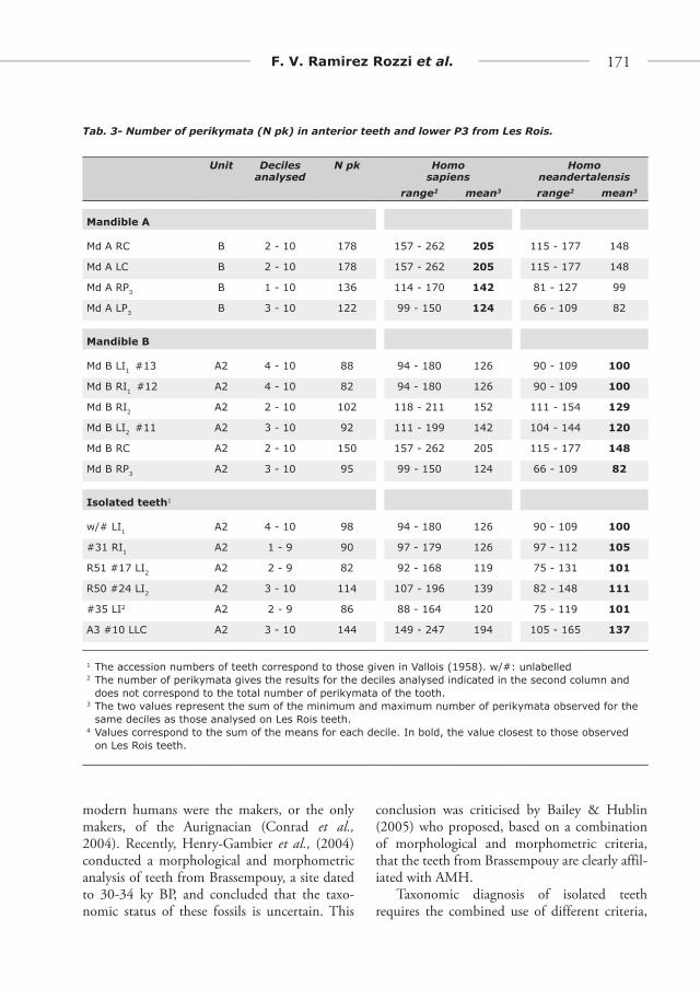
In the present work, we analyze the number and pattern of perikymata distribution on the anterior teeth as well as on the P3 from the two mandibles and isolated teeth from Les Rois. Results from a previous study of anterior teeth in Neandertals and Upper Palaeolithic-Mesolithic AMH (Ramirez Rozzi & Bermudez de Castro, 2004) are compared to those obtained from Les Rois. We expand on this approach to include information on P3. We add results for the P3 in Neandertals from Genay (n=2), Hortus (n=1), Zafarraya (n=1), Petit-Puymoyen (n=2), Sidron (n=3), Krapina (n=2), la Chapelle aux Saints (n=1), le Moustier (n=2), Circeo III (n=1), La Quina (n=2), and modern humans from the Upper Palaeolithic to historical times: Pataud (n=1), La Madeleine (n=1), Laugerie Basse (n=1), Lachaud 4 (n=1), Lespugues (n=2), Aurignac (n=1), historical times (n=6).
Wear prevented the study of perikymata in a lower canine (R40 w/# LRC), four fragmen-tary upper first incisors (R50 #45 LI1, w/# RI1, w/# RI1, R50 #5 LI1?), the lower isolated premo-lars (R51 #22 LP3, R50 LP3, R51 #23 LP4) and the perforated incisor from unit A2 at Les Rois. Environmental stress can produce enamel hypo-plasia affecting the enamel surface. Linear hypo-plasias represent a dysfunction in enamel organ function that may or may not be accompanied by an alteration in the distance between adjacent perikymata (Cunha et al., 2004). This alteration,

160 Aurignacian remains from Les Rois
when occurs, is locally placed and do not affect the entire hypoplastic enamel but is located at the limit of it and thus do not alter the number of perikymata neither the perikymata packing pattern. Teeth from Les Rois do not present hypoplasia. Only unworn teeth and teeth with a wear not higher than stage 3 (Hooper et al., 2004), i.e. a loss of ca 15% of crown height and exposure of a small area of dentine on the occlu-sal surface (Ramirez Rozzi & Sardi, 2007, Fig. 1), were selected for perikymata analysis.
Each tooth was individually cleaned prior to this study. Perikymata counts were made directly on the original specimens. The starting step of perikymata analysis consists of measuring the crown height, which was done using a Vernier micrometer eye-piece connected to a digital ocu-lar measure that was linked to a calculator-meter-printer RZD-DO (Leica). Values of buccal crown height of anterior teeth and P3 from Les Rois were divided into ten equal divisions (deciles) from the first formed enamel at the cusp to the last forming or cervical enamel as already described (Reid & Dean, 2000; Schwartz & Dean, 2001; Ramirez Rozzi & Bermudez de Castro, 2004).
Perikymata counts were made in each of the 10 divisions of the crown height. In order to test if perikymata number in the Les Rois teeth is closer to Neandertal or UPAMH teeth, each decile in each tooth class was compared with results from a sub-sample of teeth included in Ramirez Rozzi & Bermudez de Castro’s work (2004). This sub-sample is comprised of anterior teeth showing comparable degree of wear to that present in the Les Rois specimens. Ellipses of probability at 95% were constructed. Data were processed sta-tistically using StatView and Systat 9 software.
Cut-marks on mandible B were replicated by using the addition-curing silicone Coltene President putty and light body moulds. Epoxy resin replicas were made from each mould (Beynon, 1987) and observed with a Scanning Electron Microscope (SEM). The Les Rois faunal collection kept at the Institut de Paléontologie Humaine, mostly composed of reindeer mandi-bles, was also analyzed under the microscope to investigate possible anthropogenic modifications,
including the locations and orientations of cut-marks, which were recorded and photographed.
Radiocarbon Dating
Five faunal elements from units A and B, hold at the Musée d’Archéologie Nationale, were dated by accelerator mass spectrometer (AMS) at the Leibniz Laboratory. All remains showed cutmarks or traces of use as retouchers. Collagen extraction followed an extensive and rigorous pretreatment (Grootes et al., 2004) designed to remove soluble contaminants by chemical extraction and insoluble ones by filtering the collagen dissolved as gelatine, a commonly used method. Small samples of about 2 mg of bone were collected and their nitrogen concentration, determined by colorimetry as nitrate, was used to calculate the collagen concentration of the bone. These values estimate the collagen preservation in the bone and were used to calculate the mass of bone needed for AMS 14C dating, which mini-mized sampling damage.
The samples were checked and mechanically cleaned under the microscope. Surface contami-nation was removed with a scalpel; signs of glue or other contaminants were noted and avoided. Samples of crushed material (<3 mm) were first treated with acetone and rinsed with demineral-ised water to remove fatty coatings, which might make subsequent acid and alkali extractions less effective. A particle size of just below 3 mm was chosen to reduce diffusion pathways for the dem-ineralisation and extraction of the interior of the sample material, yet avoid fine dust, which would increase extraction losses. Because glue had been used on the reindeer mandible, KIA 25246, this sample was subjected to sequential soxhlet type serial extraction to remove organic contaminants (Bruhn et al., 2001). In sequence, sample KIA 25246 was extracted three times each with boiling tetrahydrofurane (THF), chloroform, petroleum-ether, acetone, and methanol and then rinsed with demineralized water. These solvents were chosen for their efficiency in removing a wide range of hydrophobic organic substances used in order of

www.isita-org.com
161F. V. Ramirez Rozzi et al.
Fig. 3 - Human remains from Les Rois. A. From top to bottom: frontal, right, left, occlusal, and basal views of mandible A from unit B. Note the chin is formed by a vertical keel along the symphysis becoming more prominent inferiorly to form the mental protuberance, and the associated mental fossae, both characteristic of H. sapiens (Schwartz & Tattersall, 2000). B. From left to right and top to bottom: occlusal, distal, frontal, lingual, mesial, and basal views of mandible B, loose right lateral incisor (LRI2) and canine (LRC) from this mandible from unit A2. The change in orientation of the mandibular surface at the canine level evokes a fl at or slightly arched anterior mandibular surface, characteristic of Neandertals (Schwartz & Tattersall, 2000). The fragmentary nature of the specimen, however, precludes a taxonomic diagnosis only on this basis. C. Mesial, lingual, buccal, and distal views of isolated lower incisors and canines from unit A2. Three isolated teeth (#11, #12, #13) are attributed to mandible B. D. Lateral, lingual, buccal, lateral, and occlusal views of a perforated human incisor from unit A2 (Continued on page 10).

162 Aurignacian remains from Les Rois
Fig. 3 (continued) - E. Mesial, lingual, buccal, and distal views of isolated upper incisors from unit A2. Vallois (1958) attribution of teeth to tooth classes was reviewed. R50 #24 is attributed here to a LI2 instead than to a LI1 and #35 to a LI2 instead than to a RI2. F. Occlusal (buccal face up), mesial, lingual, buccal, and distal views of isolated premolars.

www.isita-org.com
163F. V. Ramirez Rozzi et al.
Fig. 3 (continued) - G. Idem molars. # refers to the number given by Vallois (1958); w/#: without number. Three molars described by Vallois were not identifi ed among the remnant teeth labelled as ‘Les Rois’ (see text).

164 Aurignacian remains from Les Rois
decreasing hydrophobicity, also effectively remov-ing previous extraction solvents.
The samples were subsequently demineralised in HCl (ca. 1 %). Initially ca. 10 ml 1 % HCl is added. When the pH shows that most of the acid has reacted, concentrated acid is added to bring the solution back to ca. 1 % (pH< 1) until the pH stays below 1. Then the solution is siphoned off and the sample is washed repeatedly with deion-ized water until the pH is above four. This removes not only carbonates, salts, and the extracting acid, but also the water/acid soluble organic compo-nents designated as the fulvic acid fraction. As these are water soluble, they are mobile in the soil and are thus likely contaminants.
To remove mobile humic acids, the dem-ineralised bone material was treated with 1 wt% NaOH (at 20°C, for 1 h) and subsequently washed repeatedly with demineralised water until the pH is below 9. The sample is then again treated with 1 % HCl (20°C, 1 h) and washed to remove atmospheric CO2, which may have dis-solved into the alkali solution.
The preferred dating material, bone collagen, was dissolved overnight as gelatine in demineral-ised water at 85 °C and pH = 3. The non-soluble fraction, including insoluble bone protein and possible contamination, was filtered through a 0.45 µm pore silver filter, which had been pre-cleaned by bake-out at 900°C. The gelatine solu-tion was freeze dried, and the gelatin was com-busted as the “bone sample”. In addition, we also dated the gelatinization-insoluble fraction on the filter (bone residue) to obtain an indica-tion of the presence or absence of non-collagen-ous contaminants in the bone sample.
The com bustion to CO2 of all fractions was performed at 900°C in a closed pre-combusted quartz tube together with precombusted CuO and silver wool. The sample CO2 was reduced with H2 over Fe powder as catalyst, and the resulting carbon/iron mixture was pressed into a pellet in the target holder. CO2 to yield 1 mg of carbon was reduced on 2 mg Fe. The CO2 of all the small bone was reduced on 1 mg Fe to approach a C:Fe weight ratio of 1 to 2 for these smaller-size fractions.
The above protocol is effective in producing reliable ages for most bone samples because the process deals sequentially with particulates, con-servation chemicals and hydrophobic compounds, carbonates, fulvic acids, humic acids, and finally non-collagen particulate organic matter.
A two-fraction AMS measurement of a bone sample, which not only reveals the presence of contamination but also gives an indication of its severity, is very useful. If one makes a mass bal-ance calculation of the amount of contamination involved, one may estimate how likely it is that the measured age is significantly influenced.
A 13C/12C ratio was obtained during the AMS measurement of the 14C/12C ratio. This δ13C value does not have the same accuracy as traditional sta-ble isotope ratio analysis (SIRA), but is suitable for the 13C fractionation correction and provides information on collagen composition and the presence of glues and other foreign compounds.
Results
ArchaeologyAnalysis of the lithic assemblages, including
pieces with no stratigraphic assignment or com-ing from the very base of the sequence, confirms the excavators’ diagnosis that all the lithics can be attributed to the Aurignacian. Mousterian and Châtelperronian tools, debitage, and cores are absent. The highly selective nature of the collection makes it difficult to propose a more precise cultural attribution. Retouched bladelets (Font-Yves, Dufour of the Dufour, and Roc-de-Combe subtypes,) characteristic of the various facies/phases of the Aurignacian are absent in the assemblage, which seems incompatible with the relative abundance in all units of carinated “scrap-ers” from which suitable blanks were certainly detached. Debitage is almost entirely restricted to large unbroken blades, which is inconsistent with the numerous recovered flake cores. In spite of these limitations, the presence of an Archaic Aurignacian (Bon, 2000) can be excluded consid-ering the absence of unipolar prismatic bladelet cores. The abundance of large retouched blades

www.isita-org.com
165F. V. Ramirez Rozzi et al.
with a curved profile, particularly in unit B, and the increase of the busked burins and nosed end-scrapers in unit A supports a tentative attribution of the two main units, B and A, to the Early and Evolved Aurignacian, respectively. However, the richness, in all units, of lozenge-shaped antler points, characteristic of the Evolved Aurignacian, and the absence of split base antler points in unit B, seem to indicate that the site does not record the earliest phase of the Early Aurignacian. The analysis of the personal ornaments (Figs. 1 and 2) identifies cultural links with northern Aurignacian groups. Three out of the thirteen ornament types from unit B (perforated reindeer canine and inci-sor, gastropod mould) are found only at Les Rois, five others (perforated hyena canine, human tooth, urchin, pointed antler pendant, ivory basket bead) are found at other sites from southwestern France. Seven other types (perforated red deer, fox, wolf canines, horse and bovid incisors, tubular bone bead, antler diadem) are common at both sites from this last region, and in northern Europe. The only personal ornament from unit A, a perforated human incisor, corresponds to a type whose distri-bution is restricted to the south-western France. Apart from the perforated red deer canine, which is the most ubiquitous Aurignacian ornament, none of the ornament types found at Les Rois occur at Mediterranean sites.
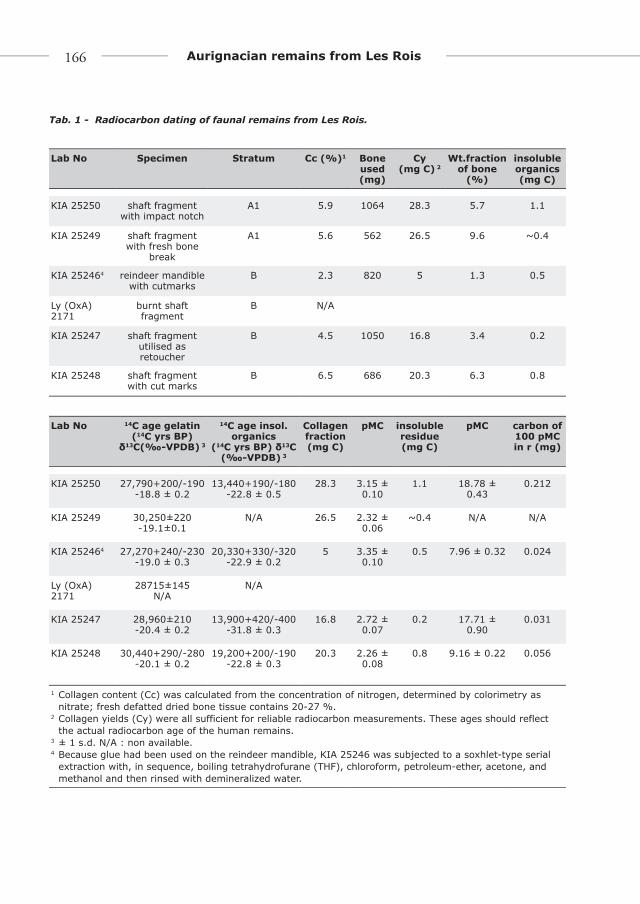
ChronologyThe collagen content for each sample from
Les Rois is shown in Table 1. The bone organic material has thus largely disappeared, but the remaining concentrations are, in our experience, sufficient for precise radiocarbon ages.
Collagen extracted from the bones provides dates ranging between ca. 27.3 kyr and 30.4 14C kyr BP (1 kyr = 1,000 years) for both units. Supplemental dating of the organic bone residue, remaining after the extraction of the collagen as dissolved gelatin, yielded ages between 13.4 and 20.3 14C kyr BP. This shows the –not unexpected- presence of younger contaminants. The age dif-ferences between the collagen and the insoluble residue fractions are large but unfortunately not out of the ordinary for old bones. Yet, the
sensitivity to young contamination for the Les Rois samples is not as large as for very old sam-ples near 40 kyr, because the 14C concentrations are still in the range of 2 to 3 percent of modern carbon (Tab. 1). As can be seen, the amount of carbon in the insoluble residues (calculated from the CO2 pressure after combustion) was much smaller than the amount of carbon in the col-lagen fractions (from 1 % to 10 % (KIA 25246) of the collagen fraction). The chemical extrac-tion of the collagen after the acid-alkali-acid pre-treatment—as gelatin in solution through a silver filter—aims to prepare a pure gelatin sample and concentrate possible remaining non-soluble contaminants in the insoluble fraction. Table 1 shows the amount of young (100 percent modern carbon (pMC)) contaminant needed to bring the 14C concentration of the rest fraction from that of the collagen/gelatin to its measured value. As expected little is needed (24 to 212 µg). The question is how effectively the 0.45 µm silver-filter eliminates non-soluble contaminants (soluble ones were dealt with before in the acid-alkali-acid extraction and washing).
Considering a scenario where 10 % of the amount of contamination concentrated in the insoluble fraction would have passed the filter and remained in the collagen (which is unlikely), the “real” age of the collagen fraction would be from 60 yr to 190 yr older –corresponds with 0.3σ and 1.0σ for KIA 25247 and KIA 25250 respectively – than reported, well within the ± 2σ uncertainty range of the measurement (95.4 % confidence probability). Based on these con-siderations we are confident that the residue ages (and masses of that fraction) indicate a fairly light contamination and thus support the reliability of the ages obtained for the collagen.
The δ13C values of the collagen (Tab. 1) are in the normal range and do not indicate contamina-tion. The insoluble fractions are somewhat more negative, which could indicate some minor influ-ence of contaminating plant material consistent with Table 1. The small insoluble fraction of KIA 25247 shows a quite different δ13C and must be mostly contamination. Considering the calculation of the amount of modern contaminant needed to
166 Aurignacian remains from Les Rois
Lab No Specimen Stratum Cc (%)1 Bone used (mg)
Cy (mg C) 2
Wt.fraction of bone
(%)
insoluble organics (mg C)
KIA 25250 shaft fragment with impact notch
A1 5.9 1064 28.3 5.7 1.1
KIA 25249 shaft fragment with fresh bone
break
A1 5.6 562 26.5 9.6 ~0.4
KIA 252464 reindeer mandible with cutmarks
B 2.3 820 5 1.3 0.5
Ly (OxA) 2171
burnt shaft fragment
B N/A
KIA 25247 shaft fragment utilised as retoucher
B 4.5 1050 16.8 3.4 0.2
KIA 25248 shaft fragment with cut marks
B 6.5 686 20.3 6.3 0.8
Lab No 14C age gelatin (14C yrs BP)
δ13C(‰-VPDB) 3
14C age insol. organics
(14C yrs BP) δ13C(‰-VPDB) 3
Collagen fraction (mg C)
pMC insoluble residue (mg C)
pMC carbon of 100 pMC in r (mg)
KIA 25250 27,790+200/-190 -18.8 ± 0.2
13,440+190/-180 -22.8 ± 0.5
28.3 3.15 ± 0.10
1.1 18.78 ± 0.43
0.212
KIA 25249 30,250±220 -19.1±0.1
N/A 26.5 2.32 ± 0.06
~0.4 N/A N/A
KIA 252464 27,270+240/-230 -19.0 ± 0.3
20,330+330/-320-22.9 ± 0.2
5 3.35 ± 0.10
0.5 7.96 ± 0.32 0.024
Ly (OxA) 2171
28715±145 N/A
N/A
KIA 25247 28,960±210 -20.4 ± 0.2
13,900+420/-400-31.8 ± 0.3
16.8 2.72 ± 0.07
0.2 17.71 ± 0.90
0.031
KIA 25248 30,440+290/-280 -20.1 ± 0.2
19,200+200/-190-22.8 ± 0.3
20.3 2.26 ± 0.08
0.8 9.16 ± 0.22 0.056
1 Collagen content (Cc) was calculated from the concentration of nitrogen, determined by colorimetry as nitrate; fresh defatted dried bone tissue contains 20-27 %.
2 Collagen yields (Cy) were all sufficient for reliable radiocarbon measurements. These ages should reflect the actual radiocarbon age of the human remains.
3 ± 1 s.d. N/A : non available.4 Because glue had been used on the reindeer mandible, KIA 25246 was subjected to a soxhlet-type serial
extraction with, in sequence, boiling tetrahydrofurane (THF), chloroform, petroleum-ether, acetone, and methanol and then rinsed with demineralized water.
Tab. 1 - Radiocarbon dating of faunal remains from Les Rois.

www.isita-org.com
167F. V. Ramirez Rozzi et al.
produce the observed age discrepancies, the non-collagen material in the insoluble fraction must be composed primarily of old plant material.
Proof that contamination has been fully removed is, unfortunately, difficult if not impos-sible to provide and an earlier age for these units cannot be ruled out.
Human remainsMatching isolated teeth from the Les Rois
collection with those listed by Vallois is made difficult by the fact that some catalogue num-bers have rubbed off and the same numbers are present on more than one tooth. We were able to identify incisors, canines, premolars and upper molars with the help of Vallois description. Eight out of eleven isolated lower molars were identi-fied in the same way. A degree of uncertainty remains about the others. Surprisingly, four pre-viously unidentified teeth were found in the box containing the Les Rois remains. These speci-mens do no appear in Vallois original reports and thus were excluded from the analysis.
Vallois (1958) assigned the isolated lower RM2 R51 #6, found by Mouton and Joffroy in reworked layers, to mandible A. The alveolar socket of this tooth is reduced to a small area of the mesial wall, which makes this matching questionable. Four isolated lower teeth, two premolars and two molars, were found close to mandible B and attributed to the same individ-ual by Vallois (1958). The two premolars (R51 #22 LP3, R51 #23 LP4) were found in the square adjacent to the one that has yielded mandible B. Morphological comparison indicates that they certainly derive from this mandible. Both P3 have a similar asymmetric outline shape and a transverse crest. In both P4, the metaconid has a mesial position when compared to the protoco-nid and both show a small distolingual cusp.
The two molars found in the same square as mandible B are R51 #14 RM1 and R51 #15 LRM. R51 #14 RM1 cannot be assigned to man-dible B since the root of the lower RM1 does not fit the alveolar wall and the proximal wear facet does not correspond to that observed on the lower P4 in the mandible. The lower RM (R51
#15), described as a tooth germ by Vallois (1958) could not be located.
Vallois (1958) attribution of teeth to tooth classes was reviewed. R50 #24 is attributed here to a LI2 instead than to a LI1 and #35 to a LI2 instead than to a RI2.
Inspection of dental morphological charac-ters reveals the following aspects:
in the upper incisors, the shovel shape is -absent or, as in R50 #5, does not reach the breakpoint; a right I1 (w/#) is the only up-per incisor that presents a lingual tubercle.R40 w/# LRC and the canine of mandible -B show a distal accessory ridge not present in A3 #10 LLC neither in the canine of mandible Alower third premolars do not present multi- -ple lingual cusps nor mesiolingual grooves. Crown asymmetry cannot be assessed con-fi dently, but it is diffi cult to assign a sym-metric crown shape to P3 of mandible B and R51 #22;the transverse crest is absent in lower P4; -the mesial position of the metaconid rela-tive to the protoconid in the P4 of mandi-ble B and R51 #23 creates a asymmetrical crown shape in occlusal view;the Cusp 6 is absent in the isolated lower fi rst -molars but present in the right M1 of mandi-ble A (the left M1 is broken distally); the mid-trigonid crest is absent but in one tooth (R50 #40) the anterior fovea is well developed; the Cusp 6 and the mid-trigonid crest are -absent in lower second molars; the anterior fovea is only present in R51 #6, a right M2; the Y pattern is observed in three isolated lower M2 (R50 #3 and the two without number), however the attribution of these teeth as M2 is uncertain.subvertical grooves on wear facets were ob- -served on the mesial face of the LP4 w/#, RM2 R51 #6, and R50 #31. � eir absence in the other molars, all unworn or with a low degree of wear, is not signifi cant considering that these features only occur on teeth show-ing an advanced degree of wear.

168 Aurignacian remains from Les Rois
Anterior teeth diameters (ATD) and crown base area (CBA) for Les Rois teeth and for Neandertal and AMH from the early Upper Palaeolithic (EUP) are presented in Appendix 1. The range of variation in ATD and CBA differ between Neandertals and EUP while showing large overlaps. The mesio-distal diameter in teeth from Les Rois falls in the range of both popula-tions (Appendix 1). CBA of teeth from mandi-ble A are systematically closer to EUP than to Neandertal averages. In contrast CBA of incisors and P4 from mandible B are closer to Neandertal than to EUP means. In isolated incisor and the lower P3 (#22) and P4 (#23), CBA is also closer to Neandertal than EUP means. In six molars, CBA is closer to EUP. In three others the oppo-site is observed.
The bucco-lingual diameter (BL), a vari-able considered particularly diagnostic by Bailey & Hublin (2006), reveals an interesting dif-ference between the two mandibles. Values for mandible A fall in the range of both popula-tions (Neandertals and AMH from the Upper Palaeolithic [UPAMH]) but are, with the excep-tion of the right canine, closer to mean values observed in UPAMH (Appendix 1). BL values for the first incisors from mandible B fall outside the individual range of UPAMH and is, in all anterior teeth, closer to Neandertal means. In iso-lated anterior teeth and premolars, BL is closer to values recorded on UPAMH than in Neandertals; in molars, some are closer to Neandertals, other to UPAMH values (Appendix 1).
Root length cannot be measured on teeth from mandible A. In mandible B (Tab. 2), root length in incisors lays outside or close to the lower limit of root length variation in Neandertals, and inside the range observed in Upper Palaeolithic AMH. Root length of lower C from mandible B is close to Neandertal average value and at the limit of AMH variation. That of the lower P4 in mandible B falls in the variation in both ref-erence populations but is closer to the average of AMH. Root length in isolated anterior teeth falls outside Neandertal and fit well in AMH variation. In isolated premolars attributed to mandible B (R51 #22, R51 # 23) root length is
closer to Neandertal average values; in premo-lars and molar R50 #40, values are also close to Neandertals’ and not comprised in UPAMH variation (Tab. 2). R50 w/# LP4 show an inter-mediate value falling outside Neandertal and UPAMH variation.
Comparison between Neandertal and UPAMH teeth with low degree of wear shows that significant difference (Mann-Whitney U-test) exist in the number of perikymata in the last deciles (Tab. 1 Suppl. Mat.). Ramirez-Rozzi & Bermudez de Castro (2004) found significant differences between the two groups in the last four deciles of the I1, while in the subsample analyzed here, significant differences were found only in the last two deciles. Crown height reconstruction in worn teeth included in Ramirez Rozzi & Bermudez de Castro’s work (2004) can be responsible for this discrepancy. However, it could also, and most likely, result from a type II error in the statistical analyses. Indeed, only three Neandertal teeth are used for comparison. It is worth noting that the greater the number of teeth compared, the greater the number of deciles showing significant differ-ences in perikymata number between the two human groups.
The number of perikymata in canines and P3 from mandible A fall within AMH, and out-side Neandertal, variation (Tab. 3). In contrast, incisors, and P3 from mandible B provide values closer to those in Neandertals. The number of perikymata, which is lower than the Neandertal averages, is incompatible with an attribution to UPAMH. The lower canine from this same mandible falls well within the Neandertal val-ues, and below the UPAMH variation. All iso-lated incisors and canine from unit A2, coming from at least two individuals, have a number of perikymata well in the range of Neandertals or even lower and, only in two cases, at the limit of UPAMH variation. In decile by decile compari-son, the same distinction is observed. Isolated teeth and teeth from mandible B correspond more closely to Neandertal distribution, whereas teeth from mandible A are closer to UPAMH distribution (Figs. 2 Suppl. Mat). When ellipses

www.isita-org.com
169F. V. Ramirez Rozzi et al.
of probability at P=0.95 are generated for each tooth type and decile, the number of perikymata in isolated teeth and in teeth from mandible B are included in the Neandertal ellipse, whereas values for teeth from mandible A accommodate in the UPAMH ellipse (Figs. 3 Suppl. Mat). The perikymata packing pattern in incisors from mandible B matches closely that observed in Neandertals, characterised by a homogenous
distribution of perikymata through deciles (Guatelli-Steinberg et al., 2007). In canines and P3, the perikymata packing pattern does not reveal significant differences between mandible A and mandible B (Fig. 4).
Morphologically, mandible B shows a change in the orientation of the mandibular surface at the canine level evoking a flat or slightly arched anterior mandibular surface, a feature described
Tooth Rois UPAMH* Neandertal*
I1 Md B right 14.1 13.3 (11.7-14.1) 16.1 (15.7-16.6)
Md B left 12.9 13.3 (11.7-14.1) 16.1 (15.7-16.6)
I2 Md B right 15.2 15.0 (13.6-16.3) 17.1 (15.3-17.9)
Md B left 15.2 15.0 (13.6-16.3) 17.1 (15.3-17.9)
C Md B right 18.5 16.2 (13.1-19.0) 19.4 (16.3-23.2)
P4 Md B right 16.1 15.0 (12.6-17.1) 18.7 (14.5-22.6)
I1 R50 #45 13.6 12.5 (10.4-15.2) 17.5 (15.7-19.7)
w/# 11.7 12.5 (10.4-15.2) 17.5 (15.7-19.7)
P4 R50 w/# 13.7 11.9 (10.5-13.3) 17.6 (16.2-19.0)
R51 #29 13.3 11.9 (10.5-13.3) 17.6 (16.2-19.0)
I2 R50 #24 14.8 15.0 (13.6-16.3) 17.1 (15.3-17.9)
R51 #17 14.6 15.0 (13.6-16.3) 17.1 (15.3-17.9)
C R40 w/# 16.9 16.2 (13.1-19.0) 19.4 (16.3-23.2)
P3 R51 #22 16.4 13.7 (11.4-16.3) 16.6 (14.5-18.1)
P4 R51 #23 17.0 15.0 (12.6-17.1) 18.7 (14.5-22.6)
M1 R50 #40 15.6 13.2 (11.6-14.0) 14.3 (12.2-16.8)
M2 R50 #3 16.9 13.7 (11.3-16.8) 15.3 (14.3-16.5)
R51 #6 16.9 13.7 (11.3-16.8) 15.3 (14.3-16.5)
Measurements in mm. * averages and ranges from Bailey 2005. In bold, the average value closest to result for Les Rois tooth.
The root length of the P4 R50 is closer to the average of UPAMH than to that of Neandertal, but it is not marked in bold because it falls outside the UPAMH variation.
Tab. 2 - Root length in teeth from Les Rois compared to those in Neandertals and anatomically mod-ern humans from the Upper Palaeolithic.

170 Aurignacian remains from Les Rois
in Neandertal mandibles (Schwartz & Tattersall, 2000), while mandible A presents a typical AMH chin formed by a vertical keel along the symphy-sis becoming more prominent inferiorly to form the mental protuberance, and the associated mental fossae (Schwartz & Tattersall, 2000).
CutmarksMicroscopic analysis confirms the presence
of cutmarks on mandible B and their absence on mandible A. Cutmarks on mandible B con-sist of three parallel striations located on the lin-gual aspect, below the right lateral canine and P3 (Fig. 5 and Fig. 4 Suppl. Mat.). Two of them bear diagnostic features of flint cutting-edge generated marks in form of v-shaped cross sections, “barbs” and, in one case, a typical splitting (Fisher 1995).
The faunal sample available for comparison is composed of 48 fragmentary reindeer mandi-bles. All show fresh bone fractures along the man-dibular canal, suggesting deliberate smashing for marrow extraction, a practice repeatedly observed ethnographically (Binford, 1978). Twenty-five mandibles (52%) bear percussion marks located on the vestibular aspect midway between cheek teeth and the mandible base (Fig. 4 Suppl. Mat.). Eleven mandibles (23%) display cutmarks on their lingual aspect located below the premolars and oriented obliquely with respect to the mandi-ble base. A single specimen has cutmarks on the vestibular aspect below the M1 and M2.
Discussion
Human remains associated with Aurignacian contexts are rare. With the exception of Mladeč, which includes cranial, dental, and postcranial elements (Wild et al., 2005) dating to ca. 31.000 BP, the remaining human fossils securely associ-ated with the Aurignacian are, for the most part, undiagnostic, poorly dated, or both (Churchill & Smith, 2000). Les Rois is one of the few sites where the association of the human remains with the Aurignacian is unambiguous. This situation has led some authors to suggest that there are no definite bases to conclude that anatomically
Fig. 4 - Perikymata packing pattern in ante-rior teeth from Les Rois compared to the range (grey areas) and the mean of the same charac-ter in (a sub-sample of) UPAMH (solid line) and Neandertals (dotted line) teeth showing com-parable degree of tooth wear to those from Les Rois. Overlap exists between the two human groups, however values distribution are clearly different in Neandertals and UPAMH in cervi-cal deciles (see Table 1 Suppl. Mat., Fig 2 & 3 Suppl. Mat.).
www.isita-org.com
171F. V. Ramirez Rozzi et al.
modern humans were the makers, or the only makers, of the Aurignacian (Conrad et al., 2004). Recently, Henry-Gambier et al., (2004) conducted a morphological and morphometric analysis of teeth from Brassempouy, a site dated to 30-34 ky BP, and concluded that the taxo-nomic status of these fossils is uncertain. This
conclusion was criticised by Bailey & Hublin (2005) who proposed, based on a combination of morphological and morphometric criteria, that the teeth from Brassempouy are clearly affil-iated with AMH.
Taxonomic diagnosis of isolated teeth requires the combined use of different criteria,
Tab. 3- Number of perikymata (N pk) in anterior teeth and lower P3 from Les Rois.
Unit Decilesanalysed
N pk Homo sapiens
Homoneandertalensis
range2 mean3 range2 mean3
Mandible A
Md A RC B 2 - 10 178 157 - 262 205 115 - 177 148
Md A LC B 2 - 10 178 157 - 262 205 115 - 177 148
Md A RP3 B 1 - 10 136 114 - 170 142 81 - 127 99
Md A LP3 B 3 - 10 122 99 - 150 124 66 - 109 82
Mandible B
Md B LI1 #13 A2 4 - 10 88 94 - 180 126 90 - 109 100
Md B RI1 #12 A2 4 - 10 82 94 - 180 126 90 - 109 100
Md B RI2 A2 2 - 10 102 118 - 211 152 111 - 154 129
Md B LI2 #11 A2 3 - 10 92 111 - 199 142 104 - 144 120
Md B RC A2 2 - 10 150 157 - 262 205 115 - 177 148
Md B RP3 A2 3 - 10 95 99 - 150 124 66 - 109 82
Isolated teeth1
w/# LI1 A2 4 - 10 98 94 - 180 126 90 - 109 100
#31 RI1 A2 1 - 9 90 97 - 179 126 97 - 112 105
R51 #17 LI2 A2 2 - 9 82 92 - 168 119 75 - 131 101
R50 #24 LI2 A2 3 - 10 114 107 - 196 139 82 - 148 111
#35 LI2 A2 2 - 9 86 88 - 164 120 75 - 119 101
A3 #10 LLC A2 3 - 10 144 149 - 247 194 105 - 165 137
1 The accession numbers of teeth correspond to those given in Vallois (1958). w/#: unlabelled 2 The number of perikymata gives the results for the deciles analysed indicated in the second column and
does not correspond to the total number of perikymata of the tooth.3 The two values represent the sum of the minimum and maximum number of perikymata observed for the
same deciles as those analysed on Les Rois teeth.4 Values correspond to the sum of the means for each decile. In bold, the value closest to those observed
on Les Rois teeth.

172 Aurignacian remains from Les Rois
the pertinence of which ultimately resides in the robustness of reference data. Here we have followed this path and recorded morphologi-cal, morphometric and tooth growth informa-tion on isolated teeth and on teeth in mandi-bles from Les Rois. Morphologically, teeth from Les Rois are closer to AMH than to Neandertal teeth. However, non-metrical traits common in Neandertals are recorded: the distal accessory ridge on mandible B canine (67% in Neandertals,
27% in AMH) and the anterior fovea in R51 #6 RM2 (88% in Neandertals, 53% in AMH) which presents subvertical grooves. Two of the most diagnostic non-metric traits of Neandertal teeth are the asymmetric shape of the mandib-ular fourth premolar and the presence of the mid-trigonid crest in lower first molar (Bailey, 2002; Bailey & Hublin, 2005). The asymmetric shape of the mandibular fourth premolar can be accompanied with a well-developed, mesially-
Fig. 5 - A. Lingual aspect of Les Rois mandible B from unit A2 with cutmarks produced by multiple strokes of a sharp stone tool. B-C. Cutmarks on the lingual aspect of reindeer mandible fragments from the same unit. Rectangles on the left identify areas magnifi ed on the right.

www.isita-org.com
173F. V. Ramirez Rozzi et al.
placed lingual cusp (metaconid) and an unin-terrupted crest joining the metaconid to the buccal (protoconid) cusp. Approximately 35% of Neandertals possess at least two P4 traits, a combination observed in only 2.4% of modern humans (Bailey, 2002, p. 152). A well developed mid-trigonid crest in lower first molar shows high frequency in Neandertals (98%) and low fre-quency in AMH (0%) (Bailey, 2002). The cusps of the P4 from Les Rois mandible B and #23 are clearly separated by the mesio-distal groove, but the mesial position of the metaconid relative to the protoconid creates a non-symmetrical shape in occlusal view, which recalls the Neandertal morphology. In R50 #40, a lower M1, the ante-rior fovea is limited distally by a bridge between the two mesial cusps but the protoconid ridge does not contact the metaconid ridge, a condi-tion which seems to be below the breakpoint assigned by Bailey (2002) to attribute this tooth to Neandertals.
Teeth from mandible B as well as isolated incisors, canine and P3 from Les Rois show a par-ticular perikymata packing pattern. Such a pat-tern fits better with Neandertal variation range than it does for that of UPAMH range (Ramirez Rozzi & Bermudez de Castro, 2004). Although it is now clear that overlap in the number of perikymata in Neandertal and modern human teeth exists (Ramírez Rozzi & Bermúdez de Castro, 2004; Ramírez Rozzi, 2005; Guatelli-Steinberg et al., 2005) perikymata number can be used as a good probabilistic criterion to dis-tinguish between distinct human populations. The recorded differences for perikymata number between Neandertals and UPAMH may reflect population affiliation.
Both Ramirez Rozzi & Bermudez de Castro (2004), and Guatelli-Steinberg et al. (2007) agree that perikymata distributions are different between Neandertals and AMH. Neandertals are characterised by a homogenous distribution of perikymata i.e., the number of perikymata does not change abruptly from one decile to another. This is the pattern observed for the incisors from mandible B and some isolated anterior teeth
(w/# LI1, #31 RI1, R51 #17 LI2, R50 #24 LI2, A3 #10 LC, #35 LI2).
In summary, three teeth from mandible A (C and right and left P3) and four isolated teeth (R50 #5 LI1, R50 LP3, #54 RM1/2 and R51 #16 RM3) are attributed to a taxon (UPAMH) by con-verging independent observations resulting from the application of a variety of methods. Results indicate a clear affinity of mandible A with AMH from the Upper Palaeolithic. Differently, in man-dible B, BL diameter in anterior teeth and in P4 as well as perikymata numbers and CBA in incisor suggest an attribution of this mandible to Neandertals. Root length would indicate an affin-ity of mandible B with UPAMH, however it is worth indicating that the reference data on root length that we have used (Bailey, 2005) is based on a reduced sample and include Les Rois teeth in the UPAMH sample. If teeth from Les Rois were excluded, the upper limit of the variation in AMH would have been lower and Les Rois teeth would lie outside the individual range of AMH.
Perikymata numbers and CBA place isolated anterior teeth with Neandertals. Non-metric traits, BL and, where possible, root length sug-gest affinity with UPAMH. The same is true for isolated premolars, excepted R51 #23 – inter-preted as coming from mandible B - in which BL is close to UPAMH, the other traits to Neandertals. In general isolated molars fit well UPAMH variation. However, R50 #40 RM1 has a long “neandertal” root and an undiagnostic BL. The same is valid for R51 #6 RM2, which exhib-its an anterior fovea and subvertical grooves.
Providing a univocal interpretation for the presence of cutmarks on mandible B is not an easy task. Secondary burial practices and canni-balism are the two alternative explanations tra-ditionally proposed to account for modifications on prehistoric human bones. When insightfully argued, the latter is based on the demonstration that faunal and hominin remains were subjected to similar treatment and, ideally, that contempo-rary mortuary practices resulted in modifications on bone significantly different from those inter-preted as the product of alimentary consump-tion. This requires data on depositional context,

174 Aurignacian remains from Les Rois
a fairly large sample of animal and human remains, including post-cranial, and information on local mortuary practices (Villa et al., 1986; White, 1992). A consistent number of reindeer mandibles from Mouton and Joffroy’s excavation show cut-marks located and oriented similarly to those recorded on mandible B. Considering their location and orientation, these cut-marks may have resulted from slicing through the gen-iohyoid muscle to remove the tongue. Mandible B and associated teeth were apparently found in a fireplace located close to a pavement of burnt pebbles, but this is all we know about their depo-sitional context. Similarly located and oriented cutmarks are observed on the juvenile Neandertal mandible from the Mousterian site of Moula-Guercy (France) and interpreted, like those on the other human remains found at this site, as evidence for cannibalism (Defleur et al., 1999). In our case, however, contextual pieces of infor-mation needed to favour the cannibalistic inter-pretation are missing. Three other reasons make Les Rois evidence ambiguous. Firstly, cranial bones are the less appropriate remains to assess the consumption hypothesis because skinning of the skull and removal of underlying muscles is common in practices of trophy keeping and secondary burials (Pickering, 1989; Villa, 1992). Secondly, no convincing Aurignacian primary burials are known (Churchill & Smith, 2000) that may suggest the existence of mortuary prac-tices distinct and contemporary with cannibal-istic practices. Thirdly, a number of cranial and mandible fragments from Upper Palaeolithic sites (Saint-Germain-la-Rivière, Isturitz, Bedeilhac, Placard, etc.) reveal modifications in the form of perforations, engravings, and scraping, indi-cating that they were given special treatment (Le Mort 1981; Buisson & Gambier 1991). Four Aurignacian sites (Vanhaeren & d’Errico, 2006), including Les Rois, have yielded perfo-rated human teeth, which confirms the interest in using human bone, and teeth in particular, by Aurignacians, for symbolic purposes. Fractures of the maxilla compatible with it being broken with intent to extract teeth and processing marks on the extracted teeth to be used as personal
ornaments are reported from the Aurignacian site of Brassempuy (Haenry-Gambier et al., 2004). The only known other Aurignacian youth man-dible with cut-marks, found at Fontechevade (Gambier, 2000), occurs in isolation. This fits the pattern seen at Les Rois and Brassempouy. In summary, although the possibility that the young individual bearing Neandertal features was con-sumed cannot be discarded, available data on the treatment and symbolic use of human remains during the Aurignacian do not appear to support this interpretation.
Conclusion
Reappraisal of the Les Rois sequence indi-cates that Aurignacians must be considered solely responsible for the accumulation of the archaeological and human remains dated to 28–30 kyr BP. Morphological and morphomet-ric analyses, and number and packing of periky-mata on Les Rois teeth indicate UPAMH affini-ties for the juvenile mandible A from the lowest unit B. Taxonomic assignation of isolated teeth remains uncertain although most of them con-form well to UPAMH variability. As regards the juvenile mandible B from overlying unit A2, the taxonomic assignation remains also uncertain. Indeed, most of morphological features of teeth suggest an attribution to EUP, however dental size and perikymata packing pattern indicate a Neandertal affinity for the mandible B.
Three possibilities may account for this unexpected evidence. The first one is that the Aurignacian was exclusively produced by AMH and that the child mandible from unit A2 repre-sents evidence for consumption and/or symbolic use of a Neandertal child by Aurignacian AMH. The second possibility is that Aurignacian tech-nologies were produced at Les Rois by human groups bearing both AMH and Neandertal fea-tures. Human remains from Les Rois would be in this case the first evidence of a biological contact between the two human groups. The third pos-sibility is that all human remains from Les Rois represent an AMH population with conserved

www.isita-org.com
175F. V. Ramirez Rozzi et al.
plesiomorphic characters suggesting a larger variation in modern humans from the Upper Palaeolithic.
The first possibility implies that Neandertals communities bearing Mousterian or “transi-tional” cultures, such as the Châtelperronian in France, persisted in the region for several mil-lennia after the arrival of Aurignacian AMHs. This scenario has been proposed to explain the age of ca. 29-28 ka obtained for two Neandertal remains from level G1 of Vindija, Croatia (Smith et al., 1999), the dates of ca. 24–30 ka for the Mousterian level IV at Gorham’s Cave, Gibraltar (Finlayson et al., 2006), and the cultural innovations associated with Châtelperronian Neandertals (Mellars, 1999, 2005; Gravina et al., 2005). However, these claims are now dismissed or considered uncertain for a number of reasons. The Vindija Neandertals have been re-dated to 32–33 ka and possibly earlier (Higham et al., 2005). A reappraisal of the Gorham’s Cave data suggests that the most parsimonious explanation for layer IV and associated 14C determinations is that they represent a Middle Palaeolithic occupa-tion up to, but not beyond, ca. 32–30 ka (Zilhao & Pettitt, 2006). A revision of purported inter-stratifications of Aurignacian/Châtelperronian layers, such as those at Grotte des Fées, and avail-able 14C ages indicates that the Châtelperronian is significantly earlier than the Aurignacian and may be seen as a largely independent Neandertal cultural development (d’Errico et al., 1998; Zilhao & d’Errico, 1999; Zilhao et al., 2006).
As far as the archaeological record of south-western France is concerned, no Châtelperronian or Late Mousterian sites have produced reli-able 14C ages younger than 35 ka (d’Errico & Sanchez Goñi, 2003). Even though the 14C dates obtained from Les Rois may represent an under-estimation of the real age of the site, a gap of at least 2,000 to 3,000 years remains between the latest recorded presence of the Mousterian or the Châtelperronian in the region and the chrono-logical attribution of the Les Rois layer that yielded the child’s mandible bearing Neandertal features, thereby contradicting our second pro-posed suggestion.
Considering the age (ca. 30–28 ka), the cultural attribution of the archaeological layers (ancient Aurignacian with no split base points) and the apparent admixture of Neandertal and UPAMH characters in some human remains, the second possibility implies that a certain degree of cultural and biological exchange did occur between the two populations in order for individuals of an Aurignacian community to inherit Neandertal traits. A Neandertal genetic and cultural contribution to Europe earliest modern human societies has been proposed repeatedly (see Zilhão 2006 for a synthesis), and human remains such as those from Lagar Velho (Duarte et al., 1999; Trinkaus & Zilhâo, 2002), Mladeč (Wolpoff, 1999; Wild et al., 2005), and Oase (Trinkaus et al., 2003; Soficaru et al., 2006) have been interpreted as bearing inherited Neandertal features. The mtDNA sequences obtained thus far from a dozen Neandertal spec-imens lie outside the range of variation of mod-ern Europeans (Krings et al., 1997; Lalueza-Fox et al., 2005; Ovchinnikov et al., 2000; Schmitz et al., 2002; Serre et al., 2004), suggesting that Neandertals did not contribute significantly to the present mtDNA gene pool. Also, recent work on nuclear DNA suggests that Neandertal and AMH lineages split around 500 ka (Noonan et al., 2006; Green et al., 2006). These results, however, do not exclude the possibility of gene flow from modern humans into Neandertals or a genetic Neandertal input to the gene pool of early modern colonisers, later eliminated by bottleneck and replacement events. No con-sensus exists, however, on what potential rate of admixture between the two populations is compatible with the available paleogenetic data. Templeton (2002, 2005) and Serre (Serre et al., 2004) accepts the possibility of up to 10% admixture, but the majority of authors exclude an interbreeding rate higher than 1% (Krings et al., 2000; Ovchinnikov et al., 2000; Caramelli et al., 2003; Currat & Excoffier, 2004). In the current state of affairs, the possibility that mak-ers of the Aurignacian bore Neandertal features is compatible with, but not supported by, the available genetic evidence.
176 Aurignacian remains from Les Rois
The interpretation that Neandertal features in AMHUP result for a some degree of biologi-cal contact between these two human groups have been challenged by a number of authors who suggest that features interpreted as evi-dence of admixture, are, in reality, plesiomor-phic features, or that the anatomical traits have been studied without paying adequate atten-tion to their developmental context (Tattersal & Schwartz, 1999; see Trinkaus, 2006). It is possible that morphological features in human remains from Les Rois with values outside of the AMHUP range of variation result by the reten-tion of plesiomorphic characters (third possibil-ity). Works based on external cranial and dental features suggest that modern humans appear in Africa between 150 000 and 200 000 years ago by morphological transition from a more primitive form, i.e. H. heidelbergensis/H. ergaster (Hublin 2001; McDougall et al., 2005). These Homo species present large tooth size, which is thus the plesiomorphic condition of Homo sapiens. This latter species has since experienced a trend towards tooth size reduction, although this is not a universal trend as some dimensions in modern human teeth from the Middle Stone Age are reduced, whereas others are not and approach values reported for Neandertals (Grine et al., 2002). The pattern observed in teeth from Les Rois in which typical characteristics of later AMHUP are accompanied by more robust fea-tures exemplifies this scenario. Further, big tooth dimensions have been reported for the earliest modern human remains in Europe (Trinkaus et al., 2003a,b). It is possible that the first modern humans in Europe still preserved plesiomorphic features and that a general reduction in tooth size appeared later in time, leading to a clearer separa-tion from the more robust Neandertal teeth.
In contrast with morphological features, aspects of dental development could not be explained by a plesiomorphic retention. Smith et al., (2007) have suggested that the modern humans pattern on dental growth appeared 200 000 years ago and that this pattern differs from that of Neandertals. It is worth to note that the perikymata packing pat-tern in Neandertals differs from that of AMHUP
and also from that of H. heidelbergensis (Ramirez Rozzi & Bermudez de Castro, 2004). Despite the known geographical, ecological, and growth pat-terns diversity observed in recent modern human populations, tooth structure and dental growth seem to have remained constant during the last 60 000 years of the Palaeolithic (Smith et al., 2006). Therefore, it is difficult to interpret aspects of the dental developmental (e.g. perikymta pack-ing pattern) in teeth from Les Rois as a retention of the plesiomorphic condition, suggesting that they have to be considered most likely as charac-teristics of a particular population.
Given the paucity of human remains from the early Upper Palaeolithic and the relative antiquity of the excavations, which were not conducted to modern standards, it is difficult to reach a definite conclusion. One of the main goals of the new excavations that we recently ini-tiated at Les Rois is to recover diagnostic human remains in well-defined cultural contexts in order to better characterise the skeletal morphology of the inhabitants of south-western France during the accumulation of the two Aurignacian layers.
Acknowledgements
Special thanks are given to C. Verna for sharing her database on Upper Paleolithic individuals and for her suggestions on previous version of this paper. We are grateful to D. Grimaud-Hervé, M. Patou-Mathis, and C. Schwab for providing access to the Les Rois material curated at the Institut de Paléontologie Humaine, Paris and the Musée des Antiquités Nationales, Saint-Germain-en-Laye, M.-J. Nadeau for discussion and preparation of the dated samples, B. Pinilla-Pérez, A. Pérez-Pérez, S. Largueze, D. Fouchier, R. Lacruz, and M. Tersis for their assistance. We also thank E. Trinkaus for fruitful comments on a previous manuscript, and W. Banks for careful editing of the final manuscript. This work was funded by the Origin of Man, Lan-guage and Languages program of the European Sci-ence Foundation, the French Ministry of Research, and a post-doctoral grant of the Centre National de la Recherche Scientifique.

www.isita-org.com
177F. V. Ramirez Rozzi et al.
References
Bailey S.E. 2000. Dental morphological affinities among late Pleistocene and recent humans. Dent. Anthropol., 14:1–8
Bailey S.E. 2002. A closer look at Neandertal postcanine dental morphology, I, the mandib-ular dentition. New Anatomist, 269:148-156.
Bailey S.E. 2004. A morphometric analysis of maxillary molar crowns of Middle-Late Pleis-tocene hominids. J. Hum. Evol., 47:183-198.
Bailey S.E. 2005. Diagnostic dental differences between Neandertals and Upper Paleolithic modern humans: Getting to the root of the matter. In E. Zadzinska (ed): Current Trends in Dental Morphology Research, pp. 201-210. University of Lodz, Lodz, Poland.
Bailey S.E. & Lynch J.M. 2005. Diagnostic dif-ferences in mandibular P4 shape between Ne-andertals and anatomically modern humans. Am. J. Phys. Anthropol., 126:268-277.
Bailey S.E. & Hublin J.J. 2005. Who made the early Aurignacian? A reconsideration of the Brassempouy dental remains. Bull. Mém. Soc. Anthropol. Paris, 17:115-121.
Bailey S.E. & Hublin J.J. 2006. Dental remains from Grotte du Renne at Arcy-sur-Cure (Yon-ne). J. Hum. Evol., 50:485-508.
Bar-Yosef O. 2002. The Upper Paleolithic revo-lution. Annu. Rev. Anthropol., 31:363-393.
Bar-Yosef O. & Pilbeam, D. (eds) 2000. The Geography of Neandertals and Modern Humans in Europe and the Greater Mediterranean. Pea-body Museum, Harvard Univ., Cambridge, Massachusetts.
Bermúdez de Castro J.M. 1993. The Atapuerca dental remains. New evidence (1987-1991 ex-cavations) and interpretations. J. Hum. Evol., 24:339-371.
Beynon A.D. 1987. Replication technique for studying microstructure in fossil enamel. Scan-ning Microsc., 1:663-669.
Beynon A.D. & Dean M.C. 1988. Distinct den-tal development patterns in early fossil homin-ids. Nature, 335:509-514.
Billy G. 1975. Étude Anthropologique des Rest-es Humains de l’Abri Pataud. In H.L. Movius
(ed): Excavation of the Abri Pataud, les Eyzies (Dordogne), pp. 201-261. Peabody Museum of Archaeology and Ethnology, Harvard Univer-sity, Cambridge, Massachusetts.
Binford L.R. 1978. Nunamiut ethnoarchaeology. Academic Press, New York.
Bon F. 2002. L’Aurignacien entre Mer et Océan. Réflexion Sur l’Unité des Phases Anciennes de l’Aurignacien dans le Sud de la France. Mémoire 29, Société Préhistorique Française, Paris.
Bordes J.-G. & Lenoble A. 2002. La lamelle Caminade: un nouvel outil lithique aurigna-cien? (The Caminade bladelet : a new Au-rignacian lithic tool?). Bulletin de la Société Préhistorique Française, 99:735-749.
Bouchud in Mouton P. & Joffroy R. 1958. Le gisement aurignacien des Rois à Mouthiers (Cha-rente). Suppl. à Gallia Préhistoire IX, CNRS.
Bruhn F., Duhr A., Grootes P.M., Mintrop A. & Nadeau M.-J. 2001. Chemical removal of conservation substances by “Soxhlet”-type ex-traction. Radiocarbon, 43:229-237.
Bruner E., Manzi G. & Arsuaga J. L. 2003. En-cephalization and allometric trajectories in the genus Homo: evidence from the Neandertal and modern lineages. Proc. Natl. Acad. Sci. U.S.A., 100:15335-15340.
Buisson D. & Gambier D. 1991. Façonnage et gravures sur des os humains d’Isturitz (Pyre-nées-Atlantiques). Bulletin de la Société Préhis-torique Française, 88:172-177.
Caramelli D., Lalueza-Fox C., Vernesi C., Lari M., Casoli A., Mallegni F., Chiarelli B., Du-panloup I., Bertranpetit J., Barbujani G., Bertorelle G. et al., 2003. Evidence for a ge-netic discontinuity between Neandertals and 24,000-year-old anatomically modern Euro-peans. Proc. Natl. Acad. Sci. U.S.A., 100:6593–6597.
Churchill S. E. & Smith F. H. 2001. Makers of the Early Aurignacian of Europe. Yearb. Phys. Anthropol., 43:61–115.
Coiffard J. 1937. L’Aurignacien en Charente. Bulletin et Mémoire de la Societé de Archèologi-que et Historique de Charente, 113-128.
Conard N. J., Grootes P. M. & Smith F. H. 2004. Unexpectedly recent dates for human remains

178 Aurignacian remains from Les Rois
from Vogelherd. Nature, 430:198-201.Connet N. 2002. Le Châtelperronien: Réflexions
sur l’unité et l’identite techno-économique de l’industrie. Ph.D. dissertation. University of Lille I, France.
Cunha E., Ramirez Rozzi F., Bermúdez de Castro J.M., Martinón-Torres M., Wasterlain S.N. & Sarmiento S. 2004. A new look into enamel hypoplasias: evidence from the Sima de los Huesos Middle Pleistocene site. Am. J. Phys. Anthropol., 125:220-231.
Currat M. & Excoffier L., 2004. Modern hu-mans did not admix with Neandertals during their range expansion into Europe. PLoS Biol., 2:2264–2274.
d’Errico F. 2003. The invisible frontier. A multi-ple species model for the origin of behavioural modernity. Evol. Anthopol., 12:188-202.
d’Errico F. & Sanchez Goñi M. F. 2003. Ne-andertal extinction and the millennial scale climatic variability of OIS 3. Quat. Sc. Rev., 22:769-788.
d’Errico F., Zilhao J., Baffier,D. , Julien M. & Pelegrin, J. 1998. Neandertal acculturation in western Europe? a critical review of the evi-dence and its interpretation. Curr. Anthropol., 39:1-44.
Dean M.C. 1987. The dental developmental sta-tus of six East African juvenile fossil hominids. J. Hum. Evol., 16:197-213.
Dean M.C., Leakey M.G., Reid D., Schrenk F., Schwartz G., Stringer C. & Walker A. 2001. Growth processes in teeth distinguish modern humans from Homo erectus and earlier hom-inins. Nature, 414:628-631.
Defleur A., White T., Valensi P., Slimak L. & Crégut-Bonnoure E. 1999. Neandertal can-nibalism at Moula-Guercy, Ardèche, France. Science, 286:128-131.
Duarte C., Maurício J., Pettitt P., Souto P., Trinkaus E., van der Plicht H. & Zilhão J. 1999. The early Upper Paleolithic human skel-eton from the Abrigo do Lagar Velho (Portu-gal) and modern human emergence in Iberia. Proc. Natl. Acad. Sci. U.S.A., 96:7604–7609.
Dujardin V. & Tymula S. 2004. Relecture chro-nologique de sites anciennement fouillés :
datation des sites paléolithiques et épipaléo-lithiques de Poitou-Charentes. 129e Con-grès des Travaux historiques et scientifiques, Besançon. Bulletin de la Société Préhistorique Française, 102:771-88.
Dujardin V. 2000. Une excursion en Charente préhistorique en 1935… D’après les cahiers de Jean Morel. Bulletin et Mémoire de la Societé de Archèologique et Historique de Charente, 1:35-48.
Finlayson C., Pacheco F.G., Rodríguez-Vidal J., Fa D.A., Gutierrez López J.M., Pérez A.S., Finlayson G., Allue E., Baena Preysler J., Cáceres I., Carrión J.S., Fernández Jalvo Y., Gleed-Owen C.P., Jimenez Espejo F.J., Ló-pez P., López Sáez J.A., Riquelme Cantal J.A., Sánchez Marco A., Giles Guzman F., Brown K., Fuentes N., Valarino C.A., Villalpando A., Stringer C.B., Martinez Ruiz F. & Saka-moto T., 2006. Late survival of Neandertals at the southernmost extreme of Europe. Nature, 443:850-853.
Fisher J.W. 1995. Bone surface modifications in zooarchaeology. J. Arch. Met. & Th., 2:7-67.
Fitzgerald C. M. 1998. Do enamel microstruc-tures have regular time dependency? Con-clusions from the literature and a large-scale study. J. Hum. Evol., 35:371-386.
Frayer D.W. 1977. Metric dental change in the European Upper Paleolithic and Mesolithic. Am. J. Phys. Anthropol., 46:109-120.
Frayer D.W. 1978. Evolution of the Dentition in Upper Paleolithic and Mesolithic Europe. Pub-lications in Anthropology, 10. University of Kansas, Lawrence, Kansas.
Gambier D. 1989. Fossil Hominids from the Early Upper Paleolithic (Aurignacian) of France. In P. Mellars, C. B. Stringer (eds): The Human Revolution: behavioural and biological perspectives on the Origins of Modern Humans, pp. 298-320. Princeton University Press.
Gambier D. 2000. Aurignacian children and mortuary practice in Western Europe. Anthro-pologie, 38:5-21.
Gómez-Robles A., Martinón-Torres M., Bermú-dez de Castro J. M., Margvelashvili A., Bastir M., Arsuaga J. L., Pérez-Pérez A., Estebaranz

www.isita-org.com
179F. V. Ramirez Rozzi et al.
F. & Martinez L. 2007. A geometric morpho-metric analysis of hominin upper first molar shape. J. Hum. Evol., 53:272-285.
Gómez-Robles A., Martinón-Torres M., Bermú-dez de Castro J. M., Prado L., Sarmiento S. & Arsuaga J. L. 2008. Geometric morphomet-ric analysis of the crown morphology of the lower first premolar of hominins, with special attention to Pleistocene Homo. J. Hum. Evol., 55:627-638.
Gravina B., Mellars P.A. & Bronk Ramsey C., 2005. Radiocarbon dating of interstratied Neandertal and early modern human occupa-tions at the Chatelperronian type-site. Nature, 438:51–56.
Green R.E., Krause J., Ptak S.E., Briggs A.W., Ronan M.T., Simons J.F., Du L., Egholm M., Rothberg J.M., Paunovic & M., Paäbo S. 2006. Analysis of one million base pairs of Ne-andertal DNA. Nature, 444:330-336.
Grine F.E. & Henshilwood C.S. 2002 Addition-al human remains from Blombos Cave, South Africa: (1999-2000 excavations). J. Hum. Evol., 42:293-302.
Grootes P.M., Nadeau M.-J. & Rieck A. 2004. 14C-AMS at the Leibniz-Labor: Radiometric dating and isotope research. Nucl. Instr. Meth., B223-224:55-61.
Guatelli-Steinberg D. & Reid D.J. 2008. What molars contribute to an emerging understand-ing of lateral enamel formation in Neandertals vs. modern humans? J. Hum. Evol., 54:236-250.
Guatelli-Steinberg D., Reid D.J., Bishop T.A. & Larsen C.S. 2005. Anterior tooth growth periods in Neandertals were comparable to those of modern humans. Proc. Natl. Acad. Sc. U.S.A., 102:14197-14202.
Guatelli-Steinberg D., Reid D.J. & Bishop T.A. 2007. Did the lateral enamel of Neandertal anterior teeth grow differently from that of modern humans? J. Hum. Evol., 52:72-84.
Henri-Martin L. 1925. La station aurignacienne de la Quina, Charente. Bull. Mém. Soc. An-thropol. Paris, 6:10-17
Henri-Martin L. 1931. La station aurignacienne de la Quina. Bulletin et Mémoire de la Societé
de Archèologique et Historique de Charente pour l’année 1930, t. XX, 77 p., 8 Fig., 24 pl. h.t.
Henry-Gambier D., Maureille B. & White R. 2004. Vestiges humains des niveaux de l’Auri-gnacien ancien du site de Brassempouy (Lan-des). Bull. Mém. Soc. Anthropol. Paris n.s., 6:49-87.
Higham T., Ramsey C.B., Karavanic I., Smith F.H. & Trinkaus E. 2006. Revised direct ra-diocarbon dating of the Vindija G1 Upper Paleolithic Neandertals. Proc. Natl. Acad. Sc. U.S.A., 103:553–557.
Hillson S. & Trinkaus E. 2002. Comparative dental crown metrics. In J. Zilhão, E. Trinkaus (eds): Portrait of the Artist as a Child. The Gravettian Human Skeleton from the Abrigo do Lagar Velho and its Archeological Context, pp. 355-364. Trabalhos de Arqueologia, 22. Insti-tuto Português de Arqueologia, Lisbonne.
Hooper S.M., Meredith N., Jagger D.C. 2004. The development of a new index for measu-rement of incisal/occlusal tooth wear. J. Oral Rehabilitation, 31:206-212.
Hublin J.J. 2001. Northwestern African Middle Pleistocene hominids and their bearing on the emergence of Homo sapiens. In L. Barham, K. Robson-Brown (eds): Human Roots, pp. 99–121. Western Academic and Specialist Press, Bristol.
Hublin J.J., Spoor F., Braun M., Zonneveld F. & Condemi S. 1996. A late Neandertal associ-ated with Upper Palaeolithic artefacts. Nature, 381:224–226.
Jelínek J., Pelisek J. & Valoch K. 1959. Der fos-sile Mensch Brno II. Anthropos, 9, (n.s. 1): 7-30.
Jernvall J. & Thesleff I. 2000. Reiterative signal-ling and patterning in mammalian tooth mor-phogenesis. Mech. Dev., 92:19–29.
Kaidonis J.A., Townsend G.C. & Richards L.C. 1992. Brief communication: Interproximal tooth wear: a new observation. Am. J. Phys. Anthropol., 88:105-107.
Krings M., Capelli C., Tschentscher F., Geisert H, Meyer S. et al., 2000. A view of Neandertal genetic diversity. Nat. Genet., 26:144–146.
Krings M., Stone A., Schmitz R.W., Krainitzki

180 Aurignacian remains from Les Rois
H., Stoneking M. & Paabo, S. 1997. Neander-tal DNA sequences and the origin of modern humans. Cell, 90:19-30.
Lalueza-Fox C., Sampietro M.L., Caramelli D., Puder Y., Lari M., Calafell F., Martinez-Maza C., Bastir M., Fortea J., de la Rasilla M., Ber-tranpetit & J., Rosas A. 2005. Neandertal ev-olutionary genetics; mitochondrial DNA data from the Iberian peninsula. Mol. Biol. Evol, 22:1077-1081.
Le Mort F. 1981. Degradation artificielles sur des os humains du Paléolithique. Unpublished PhD. Université Pierre and Marie Curie.
Leroy-Prost C. 1985. L’industrie osseuse aurigna-cienne; essai regional de classification: Poitou, Charente, Périgord. Gallia Préhistoire, 18:65-156.
Lévêque E. & Vandermeersch B., 1981. Le Néandertalien de Saint-Césaire. La Recherche, 12:242-244.
Liolios D. 1999. Variabilité et caractéristiques du travail des matières osseuses au début de l’Aurignacien: approche technologique et économique. PhD Dissertation, University Pa-ris X , Nanterre.
Macchiarelli R., Bondioli L., Debénath A., Ma-zurier A., Tournepiche J.-F., Birch W. & Dean C. 2006. How Neanderthal molar teeth grew. Nature, 444:748–751.
Martinón-Torres M., Bastir M., Bermúdez de Castro J. M., Gómez A., Sarmiento S., Mue-la A. & Arsuaga J. L. 2006. Hominin lower second premolar morphology: evolutionary inferences through geometric morphometric analysis. J. Hum. Evol., 50:523-533.
Matiegka J. 1934. L’Homme fossile de Předmostí en Moravie (Tchécoslovaquie). I. Les crânes. Académie Tchèque des Sciences et des Arts, Prague.
McDougall I., Brown F. & Fleagle J.G. 2005. Stratigraphic placement and age of mod-ern humans from Kibish, Ethiopia. Nature, 433:733–736.
Mellars P.A. 1999. The Neandertal problem con-tinued. Curr. Anthropol., 40:341-364.
Mellars P.A. 2005. The impossible coincidence: a single-species model for the origins of modern
human behavior in Europe. Evol Anthropol., 14:12–27.
Mellars P.A. 2004. Neandertals and the mod-ern human colonisation of Europe. Nature, 432:461-465.
Mouton P. & Joffroy R. 1958. Le gisement aurig-nacien des Rois à Mouthiers (Charente). Suppl. à Gallia Préhistoire IX, CNRS.
Noonan J.P., Coop G., Kudaravalli S., Smith D., Krause J., Alessi J., Chen F., Platt D., Pääbo S., Pritchard J.K. & Rubin E.M. 2006. Sequenc-ing and Analysis of Neandertal Genomic DNA. Science, 314:1113-1118.
Ovchinnikov I..V., Götherström A, Romanova G.P., Kharitonov V. M., Liden K. et al., 2000. Molecular analysis of Neandertal DNA from the northern Caucasus. Nature, 404:490–493.
Pelegrin J. 1995. Technologie lithique: le Châtelp-erronien de Roc-de Combe (Lot) et de la Côte (Dordogne). Centre National de la Recherche Scientifique, Paris.
Pérez-Pérez A., Espurz V., Bermudez de Castro J.M., De Lumley M.A. & Turbón D. 2003. Non-occlusal dental microwear variability in a simple of Middle and Late Pleistocene human populations from Europe and the Near East. J. Hum. Evol., 44:497-513.
Perpère M. 1972. Remarques sur l’Aurignacien en Poitou-Charente. L’Anthropologie, 76:387-425.
Pickering M.P. 1989. Food for thought: an alter-native to “Cannibalim in the Neolithic. Aus-tralian Archaeology, 28:35-39.
Ponce de León M. S., Golovanova L., Doron-ichev V., Romanova G., Akazawa T., Kondo O., Ishida H. & Zollikofer C. P. E. 2008. Ne-anderthal brain size at birth provides insights into the evolution of human life history. Proc. Natl. Acad. Sci. U.S.A., 105:13764-13768.
Ponce de León M. S. & Zollikofer C. P. E. 2001 Neanderthal cranial ontogeny and its impli-cations for late hominid diversity. Nature, 412:534–538.
Ramírez Rozzi F.V. 1998. Can enamel microstruc-ture be used to establish the presence of differ-ent species of Plio-Pleistocene hominids from Omo, Ethiopia? J. Hum. Evol., 35:543-576.

www.isita-org.com
181F. V. Ramirez Rozzi et al.
Ramírez Rozzi F.V. 2005. Age au décès de l’en-fant néandertalien de l’Hortus. Bull. Mém. Soc. Anthropol. Paris, 17:47-55.
Ramírez Rozzi F.V. & Bermudez de Castro J. M. 2004. Surprisingly rapid growth in Neander-tals. Nature, 428:936-939.
Ramírez Rozzi F.V. & Sardi, M. 2007. Crown-formation time in Neandertal anterior teeth revisited. J. Hum. Evol., 53:108-113.
Reid D.J. & Dean M.C. 2000. Brief commu-nication: The timing of linear hypoplasia on human anterior teeth. Am. J. Phys. Anthropol., 113:135-139.
Reid D.J., Guatelli-Steinberg D & Walton P. 2008. Variation in modern human premolar enamel formation times: Implications for Ne-andertals. J. Hum. Evol., 54:225-235.
Schmitz R.W., Serre D., Bonani G., Feine S., Hillgruber F., Krainitzki H., Paabo S. & Smith F.H. 2002. The Neandertal type site revisited: Interdisciplinary investigations of skeletal re-mains from the Neander Valley, Germany. Proc. Natl. Acad. Sci. U.S.A., 99:13342-13347.
Schwartz G.T. & Dean M.C. 2001. The ontog-eny of canine dimorphism in extant homi-noids. Am. J. Phys. Anthropol., 115:269-283.
Schwartz J.H. & Tattersall I. 2000. The human chin revisited: what is it and who has it? J. Hum. Evol., 38:367-409.
Serre D., Langaney A., Chech M., Teschler-Nicola M., Paunović M., Mennecier P., Hofreiter M., Possnert G. & Pääbo S. 2004. No evidence of Neandertal mtDNA contribution to early mod-ern humans. PLoS Biol., 2:313–317.
Sládek V., Trinkaus E., Hillson S.W. & Holliday T.W. 2000. The People of the pavlovian. Skeletal Catalogue and Osteometrics of the Gravettian Fossil Hominids from Dolní Vestoniče and Pav-lov. The Dolní Vestoniče Studies, 5. J. Svoboda, Institute of Archaeology, Academy of Sciences of the Czech Republic, Brno.
Smith B.H. 1991. Dental development and the evolution of life history in Hominidae. Am. J. Phys. Anthropol., 86:157-174.
Smith F.H., Trinkaus E., Pettitt P.B., Karavanic I. & Paunovic M. 1999. Direct radiocarbon dates for Vindija G1 and Velika Pećina Late
Pleistocene hominid remains. Proc. Natl. Acad. Sci. U.S.A., 96:12281-12286.
Smith P. 1989. Dental evidence for phylogenetic relationships of middle Palaeolithic hominids. In M. Otte (ed): L’Homme de Neandertal, vol. 7 L’extinction, pp. 111-120. Etudes et Recher-ches Archéologiques de l’Université de Liège.
Smith T. 2008. Incremental dental development: methods and applications in hominid evolu-tionary studies. J. Hum. Evol., 54:205-224.
Smith T. Olejniczak A.J., Tafforeau P., Reid D.J., Grine F.E. & Hublin J.-J. 2006. Molar crown thickness, volume, and development in South African Middle Stone Age humans. South Af-rican Journal of Science, 102:1-5.
Smith T.M., Tafforeau P.T., Reid D.J., Grün R., Eggins S., Boutakiout M. & Hublin J.-J. 2007. Earliest evidence of modern human life history in North African early Homo sapiens. Proc. Natl. Acad. Sci. U.S.A., 104:6128–6133.
Smith T.M., Toussaint M., Reid D.J., Olejniczak J. & Hublin J.-J. 2007. Rapid dental develop-ment in a Middle Paleolithic Belgian Neander-tal. Proc. Natl. Acad. Sci. U.S.A., 104:20220-20225.
Soficaru A., Doboş A. & Trinkaus E., 2006. Ear-ly modern humans from the Peteşra Muierii, Baia de Fier, Romania. Proc. Natl. Acad. Sci. U.S.A., 103:17196-17201.
Sonneville-Bordes D. 1966. L’évolution du Paléolithique supérieur en Europe Occidentale et sa signification. Bulletin de la Societé Préhis-torique Française, 63:3-34.
Stringer C. 2002. Modern human origins: progress and prospects. Phil. Trans. R. Soc. Lond. B, 357:563–579.
Svoboda J., van der Plicht J. & Kuzelka V. 2002. Upper Palaeolithic and Mesolithic human fossil from Moravia and Bohemia (Czech Republic): some new 14C dates. Antiquity, 76:957-962.
Tattersall I. & Schwartz J.H. 1999. Hominids and hybrids: The place of Neandertals in hu-man evolution. Proc. Natl. Acad. Sci. U.S.A., 96:7117-7119.
Templeton A.R. 2002. Out of Africa again and again. Nature, 416:45-50.
Templeton A.R. 2005. Haplotype trees and

182 Aurignacian remains from Les Rois
modern human origins. Yearb. Phys. An-thropol., 48:33-59.
Trinkaus E. 2005. Early modern humans. Annu. Rev. Anthropol., 34:207–230.
Trinkaus E. 2006. Modern human versus Nean-dertal evolutionary distinctiveness. Curr. An-thropol., 47:597-620.
Trinkaus E., Moldovan O., Milota S., Bîlgar A., Sarcina L., Athreya S., Bailey S.E., Rodrigo R., Gherase M., Higham T., Bronk Ramsey C. & van der Plicht J. 2003a. An early modern hu-man from the Peştera cu Oase , Romania . Proc. Natl. Acad. Sci. U.S.A., 100:11231-11236.
Trinkaus, E., Milota Ş., Rodrigo R., Mircea G. & Moldovan, O. 2003b. Early modern hu-man cranial remains from the Peştera cu Oase, Romania. J. Hum. Evol., 45:245-253.
Turner II C.G., Nichol C.R. & Scott G.R., 1991. Scoring procedures for key morphological traits of the permanent dentition: the Arizona State University Dental Anthropology System. In M. Kelley & C. S. Larsen (eds): Advances in Dental Anthropology, pp. 13-31. Wiley Liss, New York.
Vallois H. V. 1958. Les restes humains d’âge aurignacien de la grotte des Rois, Charente. Bull. Mém. Soc. Anthropol. Paris, 9, série X:138-159.
Vanhaeren M. & d’Errico F. 2006. Aurigna-cian ethno-linguistic geography of Europe re-vealed by personal ornaments. J. Archaeol. Sci., 33:1105-1128.
Verneau R. 1906. Les grottes de Grimaldi, Baoussé-Roussé. Anthropologie, II. Monaco.
Villa G. & Giacobini G. 1995. Subvertical grooves of interproximal facets in Neandertal posterior teeth. Am. J. Phys. Anthropol., 96:51-62.
Villa P. 1992. Cannibalism in prehistoric Europe. Evol. Anthropol., 1:93-104.
Villa P. C. Bouville, Courtin J., Helmer D., Ma-hieu E., Shipman P., Belluomini G. & Branca M. 1986. Cannibalism in the Neolithic. Sci-ence, 233:431-437.
Voelker A.H.L., Grootes P.M., Nadeau M.-J. &
Sarnthein M. 2000. Radiocarbon levels in the Iceland Sea from 25-53 kyr and their link to the earth´s magnetic field intensity. Radiocar-bon, 42:437-452.
White T. 1992. Prehistoric cannibalism at Mancos 5MTUMR-2346. Princeton Univ. Press, Prin-ceton.
Wild E.M., Teschler-Nicola M., Kutschera W., Steier P., Trinkaus E. & Wanek W. 2005. Di-rect dating of Early Upper Palaeolithic human remains from Mladeč. Nature, 435:332-335.
Wolpoff M.H. 1999. Paleoanthropology. Mc-Graw-Hill, New York.
Wolpoff M.H. 1979. The Krapina Dental Re-mains. Am. J. Phys. Anthropol., 50:67-113.
Zilhão J. 2006. Neandertals and moderns mixed, and it matters. Evol. Anthropol., 15:183-195.
Zilhão J. & d’Errico, F. (eds.) 2003. The chronol-ogy of the Aurignacian and of the transitional technocomplexes. Dating, stratigraphies, cultural implications. Portuguese Institute of Archaeol-ogy (IPA), Trabalhos de Arqueologia, Lisbon.
Zilhão J. & d’Errico F. 1999. The chronology and taphonomy of the earliest Aurignacian and its implications for the understanding of Neander-tal extinction. J. World Prehist., 13:1-68.
Zilhão J., d’Errico F., Bordes J.-G., Lenoble A., Texier J.-P. & Rigaud J.-P. 2006. Analysis of Aurignacian interstratification at the Châtelp-erronian-type site and implications for the be-havioral modernity of Neandertals. Proc. Natl. Acad. Sci. U.S.A., 103:12643-12648.
Zilhão J. & Pettitt P. 2006. On the new dates for Gorham’s Cave and the late survival of Iberian Neandertals. Before Farming 3, article 3:1-9.
Zilhão J. & Trinkaus, E. (eds) 2002. Portrait of the Artist as a Child. The Gravettian Human Skeleton from the Abrigo do Lagar Velho and its Archeological Context. Trabalhos de Arqueolo-gia Vol. 22. Lisbon.
Associate Editor, Emiliano Bruner

www.isita-org.com
183F. V. Ramirez Rozzi et al.
Les Rois diameters CBA EUP1 H. neandertalensis2 UPAMH3
BL diam x(range)
Neand3
BL diam x(range)diameters
range CBA x range
diameters range
CBA x range
Mandible A
Md A RC MDBL
6.98.6
59.3 6.1-8.07.8-10
63.647.6-80.0
6.8-8.87.5-10.3
71.654.4-90.1
8.2(7.7-9.5)
8.5(5.6-9.8)
Md A LC MDBL
6.78.1
54.3 6.8-8.38.0-9.8
66.557.3-78.4
6.8-8.87.5-10.3
71.654.4-90.1
8.2(7.7-9.5)
8.5(5.6-9.8)
Md A RP3 MDBL
7.28.4
60.5 6.0-8.07.7-9.0
61.246.2-68.0
7.0-9.07.3-10.3
70.951.8-91.7
8.4* 9.0 °(7.3-10.3)
Md A LP3 MDBL
7.18.5
60.4 6.0-8.07.7-9.5
60.146.8-66.5
7.0-9.07.3-10.3
70.951.8-91.7
8.5* 9.0 °(7.3-10.3)
Md A Rdm2 MDBL
-9.5
10.8(9.8-11.9)
10.8(9.7-11.8)
Md A RM1 MDBL
11.310.5
118.6 10.3-1310.0-12.5
131.0103.0-150.2
10.0-13.69.6-12.9
127.4102.7-174.8
10.8(9.8-11.9)
10.8(9.7-11.8)
Md A LM1 MDBL
-10.3
10.3-1310.0-12.5
131.0103.0-150.2
10.0-13.69.6-12.9
127.4102.7-174.8
8.2(7.7-9.5)
8.5(5.6-9.8)
Mandible B
Md B LI1 (R50 #13)
MDBL
6.47.0
44.8 3.7-6.55.4-6.9
30.921.5-42.2
5.1-6.36.8-8.2
43.136.2-51.3
6.2* 7.4 °(6.8-8.2)
Md B RI1 (R51 #12)
MDBL
6.37.0
44.1 3.7-6.15.4-6.9
30.620.9-42.1
5.1-6.36.8-8.2
43.136.2-51.3
6.2* 7.4 °(6.8-8.2)
Md B RI2 MDBL
-7.2
5.0-7.36.0-7.5
42.433.6-52.6
6.1-7.56.7-9.0
51.740.8-65.6
6.7(6.0-7.5)
7.5(6.0-8.0)
Md B LI2 (R51 #11)
MDBL
7.17.3
51.8 4.1-7.26.6-7.6
42.427.1-51.1
6.1-7.56.7-9.0
51.740.8-65.6
6.7(6.0-7.5)
7.5(6.0-8.0)
Md B RC MDBL
7.38.9
65.0 6.1-8.07.8-10
63.647.6-80.0
6.8-8.87.5-10.3
71.654.4-90.1
8.2(7.7-9.5)
8.5(5.6-9.8)
Md B RP3 MDBL
7.48.3
61.4 6.0-8.07.7-9.0
61.246.2-68.0
7.0-9.07.3-10.3
70.951.8-91.7
8.4* 9.0 °(7.3-10.3)
Md B RP4 MDBL
7.68.8
66.9 6.6-8.57.8-9.5
65.054.4-78.8
6.9-9.47.1-10.5
69.550.4-86.6
8.3(7.1-9.2)
8.9(7.6-10.5)
Isolated teeth1
R50 #45 LI1
MDBL
10.27.4
75.5 7.7-10.07.0- 8.3
69.960.0-81.0
8.2-12.07.4-9.7
81.663.3-105.7
7.6* 8.6 °(7.4-9.7)
w/# RI1 MDBL
10.6-
7.9-10.07.1-8.2
69.960.0-81.0
8.2-12.07.4-9.7
81.663.3-105.7
Appendix 1- Tooth dimensions in teeth from Les Rois.

184 Aurignacian remains from Les Rois
Les Rois diameters CBA EUP1 H. neandertalensis2 UPAMH3
BL diam x(range)
Neand3
BL diam x(range)diameters
range CBA x range
diameters range
CBA x range
Isolated teeth1 (Continued)w/# RI1 MD
BL10.67.7
81.6 7.9-10.07.1-8.2
69.960.0-81.0
8.2-12.07.4-9.7
81.663.3-105.7
7.7* 8.6 °(7.4-9.7)
R50 #5 LI1 (?)
MDBL
9.57.3
69.4 7.7-10.07.0- 8.3
69.960.0-81.0
8.2-12.07.4-9.7
81.663.3-105.7
7.7* 8.6 °(7.4-9.7)
w/# LI1 MDBL
6.16.9
42.1 3.7-6.55.4-6.9
30.921.5-42.2
5.1-6.36.8-8.2
43.136.2-51.3
6.2* 7.4 °(6.8-8.2)
#31 RI1 MDBL
6.36.5
41.0 3.7-6.15.4-6.9
30.620.9-42.1
5.1-6.36.8-8.2
43.136.2-51.3
6.2* 7.4 °(6.8-8.2)
#35 LI2 MDBL
6.9-
5.8-8.86.3-8.1
51.541.7-66.0
6.3-9.37.7-9.9
68.051.0-81.4
R51 #17 LI2
MDBL
6.97.0
48.3 4.1-7.26.6-7.6
42.427.1-51.1
6.1-7.56.7-9.0
51.740.8-65.6
6.7(6.0-7.5)
7.5(6.0-8.0)
R50 #24 LI2
MDBL
7.16.9
49.0 4.1-7.26.6-7.6
42.427.1-51.1
6.1-7.56.7-9.0
51.740.8-65.6
6.7(6.0-7.5)
7.5(6.0-8.0)
A3 #10 LLC MDBL
6.9-
5.8-8.38.0-9.8
66.557.3-78.4
6.8-8.87.5-10.3
71.654.4-90.1
R40 w/# LRC
MDBL
-9.0
6.1-8.07.8-10
63.647.6-80.0
6.8-8.87.5-10.3
71.654.4-90.1
8.2(7.7-9.5)
8.5(5.6-9.8)
R51 #22 LP3
MDBL
8.27.9
64.8 6.0-8.07.7-9.5
60.146.8-66.5
7.0-9.07.3-10.3
70.951.8-91.7
8.5* 9.0 °(7.3-10.3)
R50 LP3 MDBL
7.38.4
61.3 6.0-8.07.7-9.5
60.146.8-66.5
7.0-9.07.3-10.3
70.951.8-91.7
8.5* 9.0 °(7.3-10.3)
R51 #29 RP4
MDBL
6.49.1
58.2 5.9-7.98.8-11.2
69.454.7-88.5
6.4-8.89.0-11.7
73.658.5-102.9
9.6(8.8-10.5)
10.3(8.2-11.3)
R50 LP4 MDBL
6.39.3
58.6 5.9-7.98.8-11.2
69.454.7-88.5
6.4-8.89.0-11.7
73.658.5-102.9
9.6(8.8-10.5)
10.3(8.2-11.3)
R51 #23 LP4
MDBL
7.98.7
68.7 6.6-8.57.8-9.5
65.051.2-75.5
6.9-9.47.1-10.5
69.550.4-86.6
8.3(7.1-9.2)
8.9(7.6-10.5)
#54 RM1/2 MDBL
10.611.8
125.1 9.2-12.011.0-14.0
131.2102.1-165.2
10.0-13.411.1-14.2
138.8113.2-185.3
12.1*- 12.2 °(11.1-14.2)
R53 P RM1/2 MDBL
10.9-
9.2-12.011.0-14.0
131.2102.1-165.2
10.0-13.411.1-14.2
138.8113.2-185.3
R50 #21A RM3
MDBL
8.712.3
107.0 8.3-11.19.2-13.2
113.278.2-145.3
8.3-11.410.0-14.0
121.784.7-148.5
11.6* 12.2 °(10-14)
R51 #16 RM3
MDBL
8.911.2
99.7 8.3-11.19.2-13.2
113.278.2-145.3
8.3-11.410.0-14.0
121.784.7-148.5
11.6* 12.2 °(10-14)
R50 #40 RM1
MDBL
12.410.9
135.2 10.3-1310.0-12.5
131.0103.0-150.2
10.0-13.69.6-12.9
127.4102.7-174.8
10.8(9.8-11.9)
10.8(9.7-11.8)
R50 #3 LM2/3
MDBL
11.310.1
114.1 9.2-12.89.8-12.0
125.192.0-150.0
10.5-14.09.6-12.4
130.8102.7-172.4
10.6(9.8-12.3)
10.8(9.9-12.1)
Appendix 1- Tooth dimensions in teeth from Les Rois (continued).

www.isita-org.com
185F. V. Ramirez Rozzi et al.
Les Rois diameters CBA EUP1 H. neandertalensis2 UPAMH3
BL diam x(range)
Neand3
BL diam x(range)diameters
range CBA x range
diameters range
CBA x range
Isolated teeth1 (Continued)R50 #31 RM2
MDBL
-10.0
9.2-12.89.8-12.0
125.192.0-150.0
10.5-14.09.6-12.4
130.8102.7-172.4
10.6(9.8-12.3)
10.8(9.9-12.1)
w/# RM1/2 MDBL
11.010.8
118.8 10.3-1310.0-12.5
131.0103.0-150.2
10.0-13.69.6-12.9
127.4102.7-174.8
10.8(9.8-11.9)
10.8(9.7-11.8)
w/# LM1/2 MDBL
11.211.0
123.2 10.3-1310.0-12.5
131.0103.0-150.2
10.0-13.69.6-12.9
127.4102.7-174.8
10.8(9.8-11.9)
10.8(9.7-11.8)
w/# LM MDBL
11.810.6
125.1
R51 #14 RM1
MDBL
11.910.1
120.2 10.3-1310.0-12.5
131.0103.0-150.2
10.0-13.69.6-12.9
127.4102.7-174.8
10.8(9.8-11.9)
10.8(9.7-11.8)
R51 #6 RM2
MDBL
11.410.9
124.3 9.2-12.89.8-12.0
125.192.0-150.0
10.5-14.09.6-12.4
130.8102.7-172.4
10.6(9.8-12.3)
10.8(9.9-12.1)
R50 Ldm2 MDBL
11.69.4
109.0
1 Tooth dimensions range and CBA in early Upper Palaeolithic humans (EUP) from Abri Pataud, Brno, Dolní Vestoniče, Grotte des enfants, Mladeč, Pavlov, Předmostí and Brassempouy-grotte des hyènes (Billy 1975, Frayer 1977, 1978, Hillson & Trinkaus 2002, Henry-Gambier et al., 2004, Jelinek et al., 1959, Matiegka 1934, Sládek et al., 2000, Verneau 1906) (Data provided by C. Verna). Values for EUP were obtained sep-arately for left and right antimers.
2 Tooth dimensions and CBA in Neandertals (Bermudez de Castro 1993). CBA: crown base area (MD x BL). In bold, the CBA closest to Les Rois value.
3 Bucco-lingual diameter (BL diam) from Bailey & Hublin (2006), excepted ° from Bermúdez de Castro (1993) and * average from our results EUP. In bold, the average value closest to the measure in teeth from Les Rois.
Appendix 1- Tooth dimensions in teeth from Les Rois (continued).