Curriculum: Phase 1/ Semester 2/ TOB/ Session 3,Lecture 1 ...
45
Curriculum: Phase 1/ Semester 2/ TOB/ Session 3,Lecture 1 Lecturer: teacher -1- Dr. Hameda Abdul-AI Mahdi Degrees: MSc/Ph.D. Histology Email: [email protected] Tissue of the body
Transcript of Curriculum: Phase 1/ Semester 2/ TOB/ Session 3,Lecture 1 ...
Curriculum: Phase 1/ Semester 2/ TOB/
Session 3,Lecture 1
Lecturer: teacher -1- Dr. Hameda Abdul-AI
Mahdi
Degrees: MSc/Ph.D. Histology
Email: [email protected]
Tissue of the body

• Histology Textbooks ‘Basic Histology’,
Junqueira,13 th Edition.
• ‘Colour Atlas of Histology’ Gartner and Hiatt
Selected references
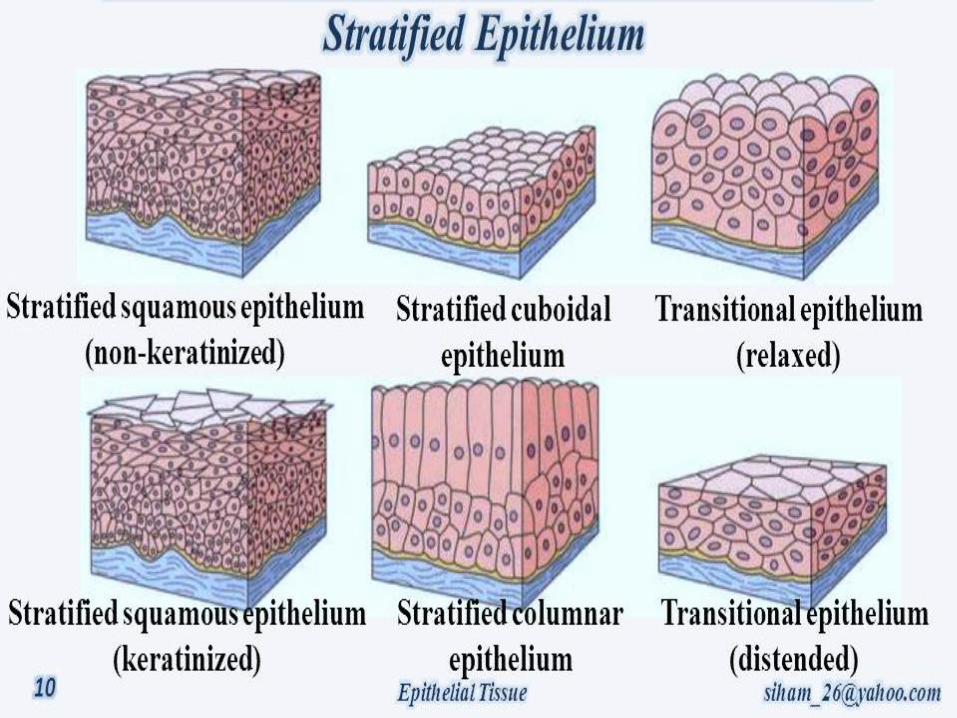
Lecture 1 Stratified Epithelium
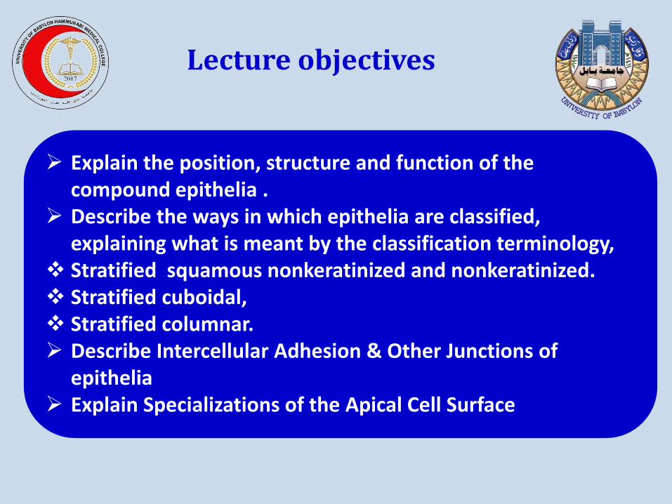
Explain the position, structure and function of the compound epithelia .
Describe the ways in which epithelia are classified, explaining what is meant by the classification terminology,
Stratified squamous nonkeratinized and nonkeratinized. Stratified cuboidal, Stratified columnar. Describe Intercellular Adhesion & Other Junctions of
epithelia Explain Specializations of the Apical Cell Surface
Lecture objectives
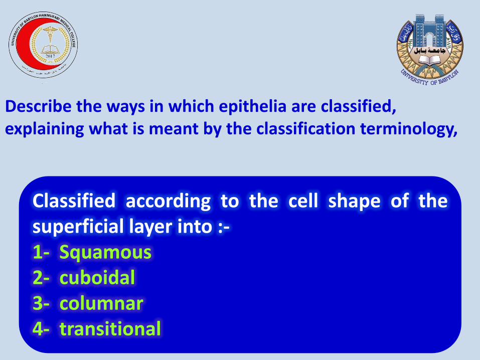
Classified according to the cell shape of the superficial layer into :- 1- Squamous 2- cuboidal 3- columnar 4- transitional
Describe the ways in which epithelia are classified, explaining what is meant by the classification terminology,
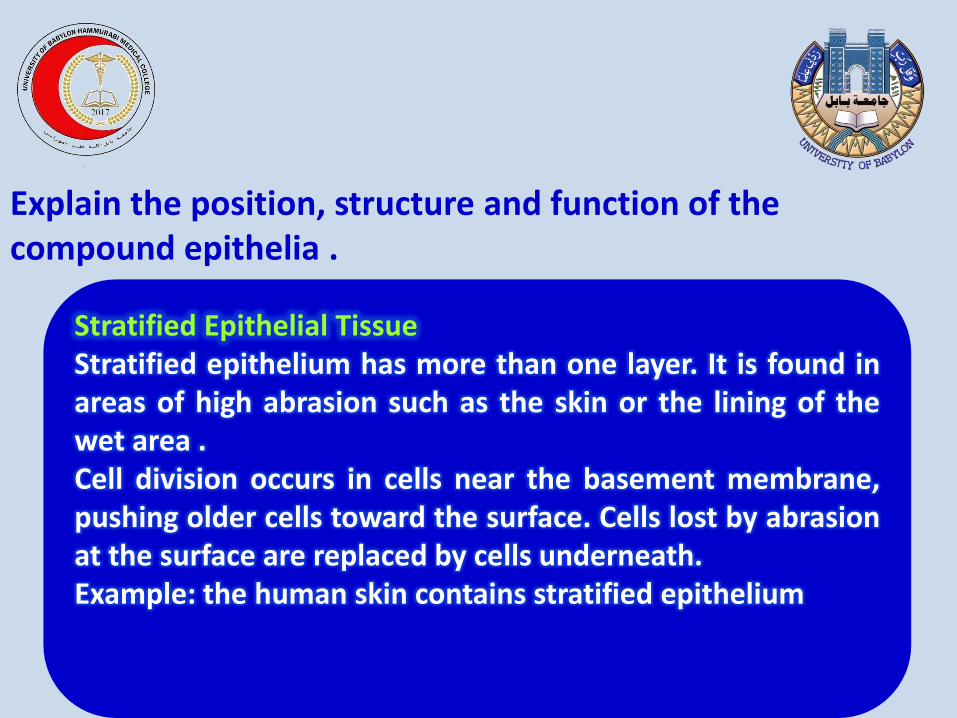
Stratified Epithelial Tissue Stratified epithelium has more than one layer. It is found in areas of high abrasion such as the skin or the lining of the wet area . Cell division occurs in cells near the basement membrane, pushing older cells toward the surface. Cells lost by abrasion at the surface are replaced by cells underneath. Example: the human skin contains stratified epithelium
Explain the position, structure and function of the compound epithelia .
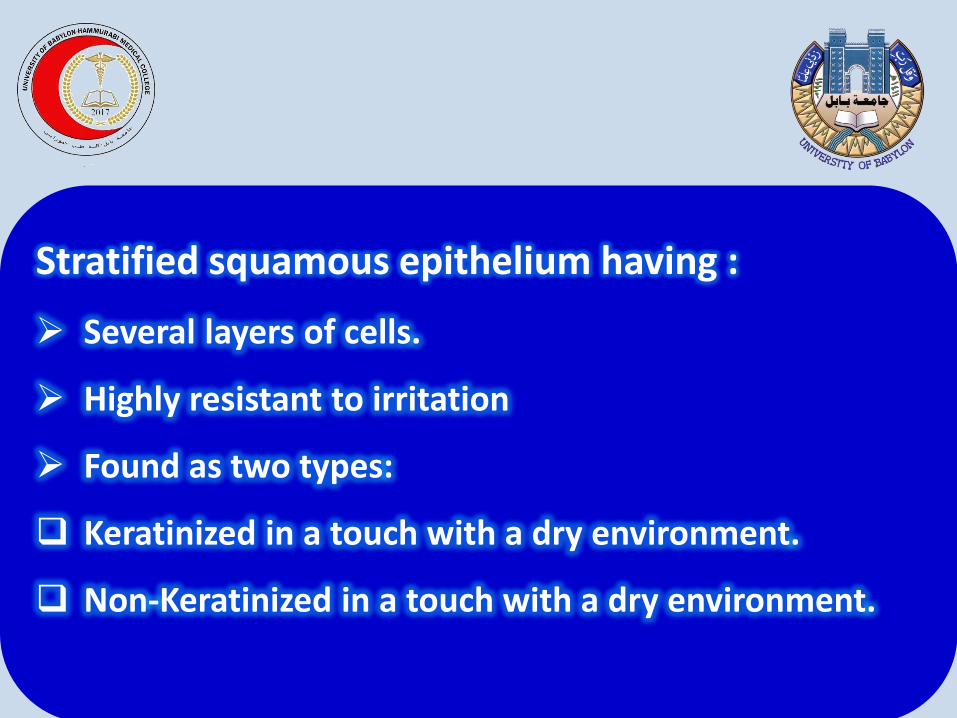
Stratified squamous epithelium having :
Several layers of cells.
Highly resistant to irritation
Found as two types:
Keratinized in a touch with a dry environment.
Non-Keratinized in a touch with a dry environment.
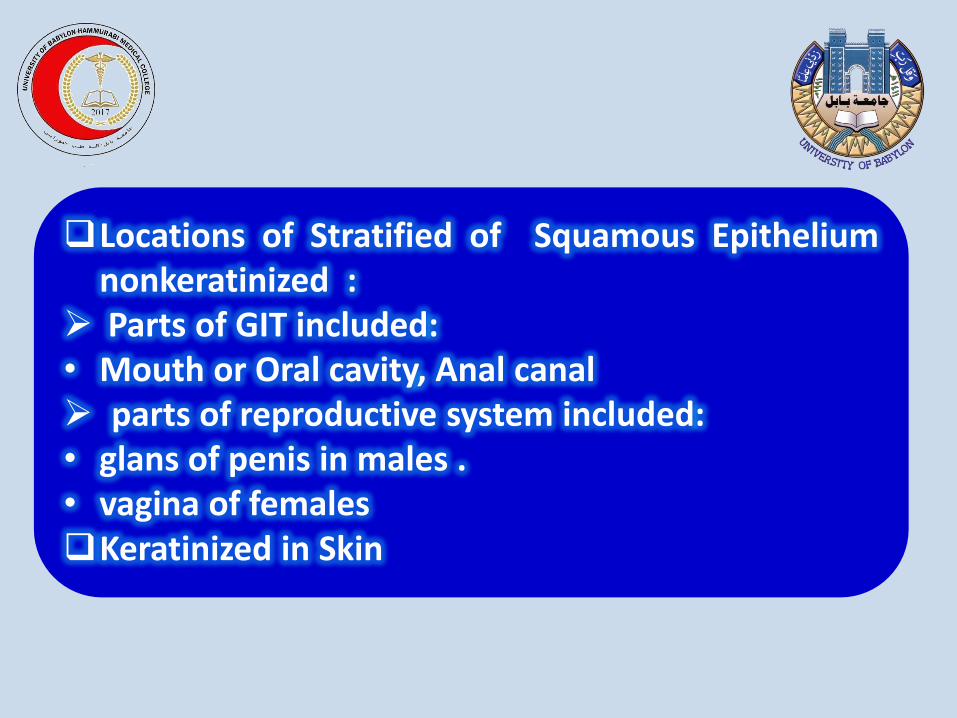
Locations of Stratified of Squamous Epithelium nonkeratinized :
Parts of GIT included: • Mouth or Oral cavity, Anal canal parts of reproductive system included: • glans of penis in males . • vagina of females Keratinized in Skin
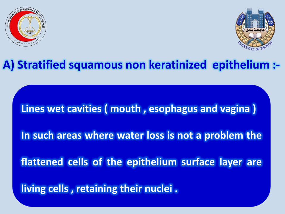
Lines wet cavities ( mouth , esophagus and vagina )
In such areas where water loss is not a problem the
flattened cells of the epithelium surface layer are
living cells , retaining their nuclei .
A) Stratified squamous non keratinized epithelium :-
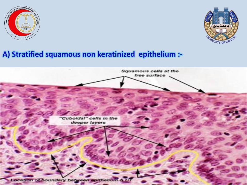
A) Stratified squamous non keratinized epithelium :-
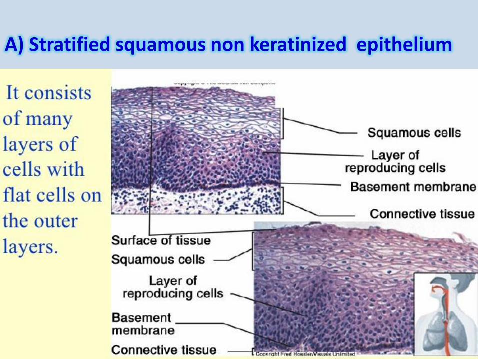
A) Stratified squamous non keratinized epithelium
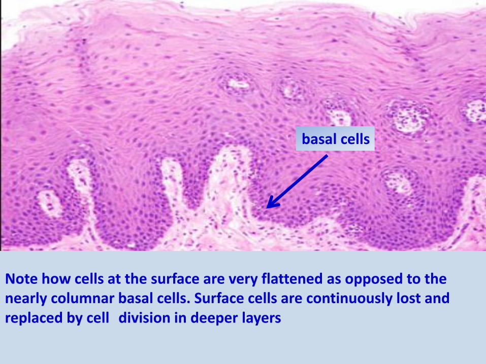
Note how cells at the surface are very flattened as opposed to the nearly columnar basal cells. Surface cells are continuously lost and replaced by cell division in deeper layers
basal cells

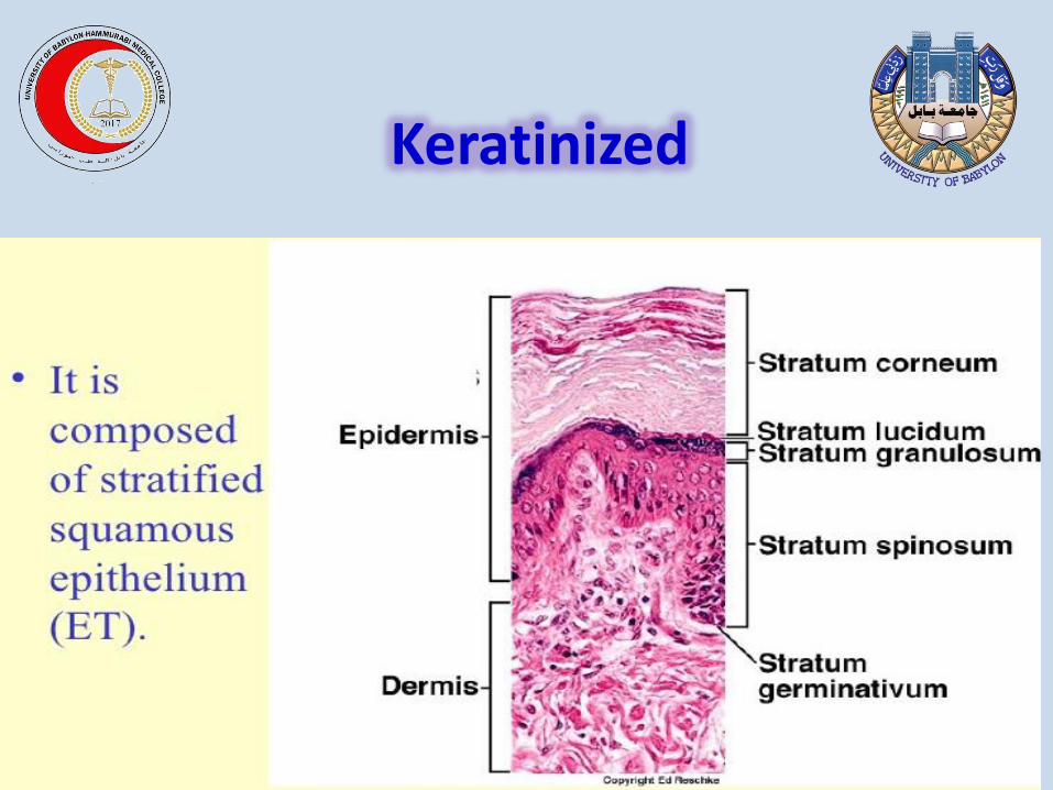
The cells become irregular in shape and flatten as they accumulate keratin in the process of keratinization and are moved progressively closer to the surface where they become thin , metabolically inactive squames of keratin lacking nuclei this surface layer of cells helps protect against water loss across this epithelium
B-Stratified squamous keratinized epithelium
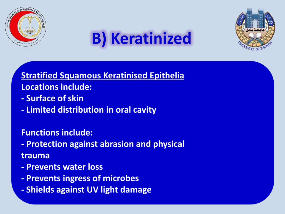
Stratified Squamous Keratinised Epithelia Locations include: - Surface of skin - Limited distribution in oral cavity Functions include: - Protection against abrasion and physical trauma - Prevents water loss - Prevents ingress of microbes - Shields against UV light damage
B) Keratinized
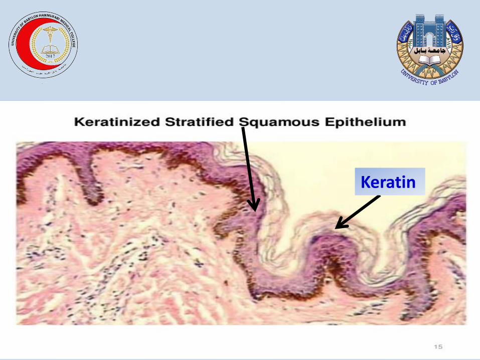
Keratin
Keratinized
Keratin
Stratified Squamous Keratinised Epithelia
consists of several layers of cells in which the top
layer is cube-shaped.. Function is mainly protective.
Location:-
Testis tubules; vesicular (Graafian) follicles of ovary.
Ducts of sweat glands; sebaceous glands
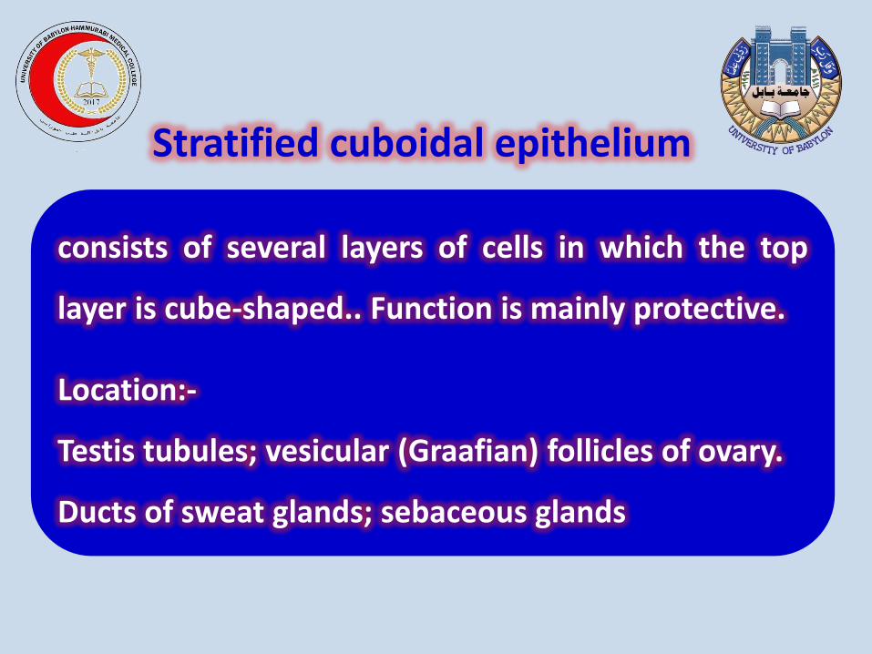
Stratified cuboidal epithelium
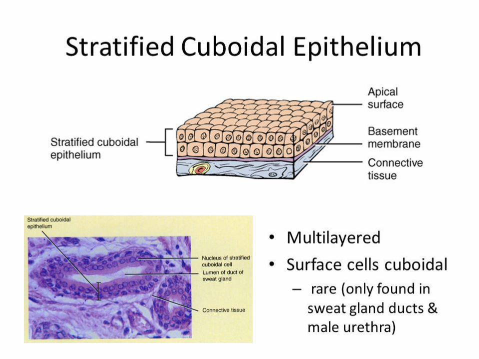
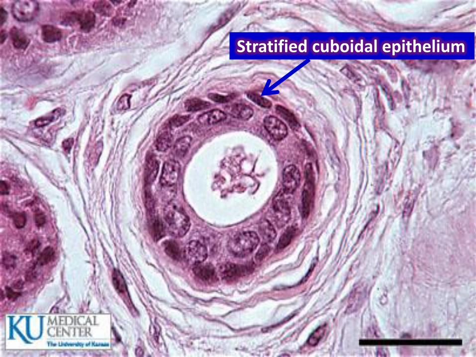
Stratified cuboidal epithelium
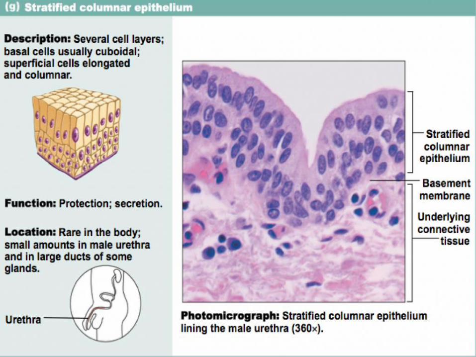
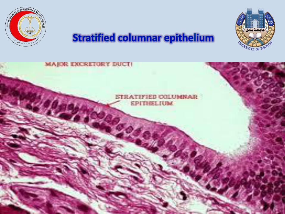
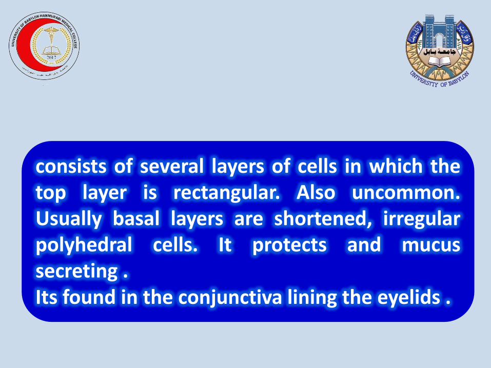
consists of several layers of cells in which the top layer is rectangular. Also uncommon. Usually basal layers are shortened, irregular polyhedral cells. It protects and mucus secreting . Its found in the conjunctiva lining the eyelids .
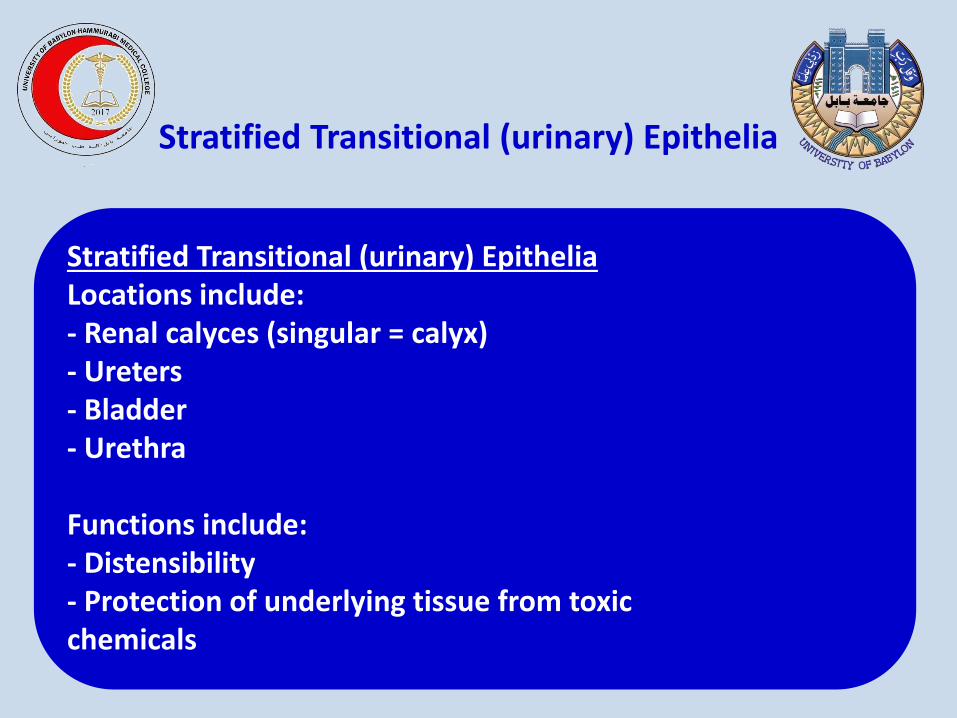
Stratified Transitional (urinary) Epithelia Locations include: - Renal calyces (singular = calyx) - Ureters - Bladder - Urethra Functions include: - Distensibility - Protection of underlying tissue from toxic chemicals
Stratified Transitional (urinary) Epithelia
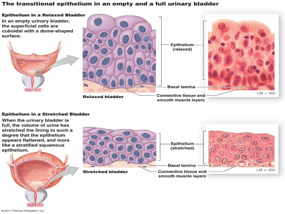
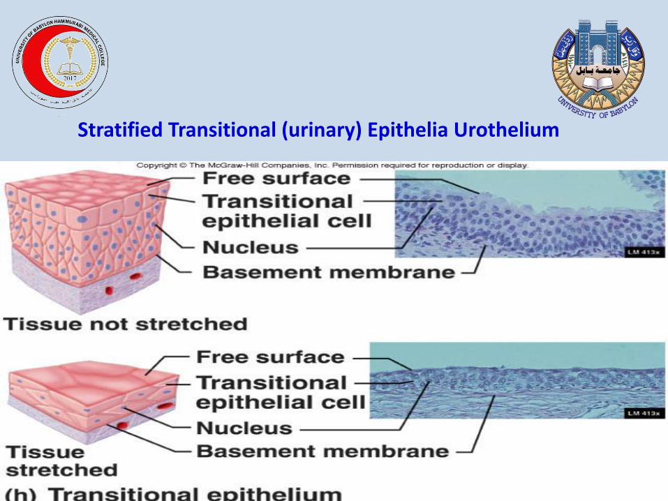
Stratified Transitional (urinary) Epithelia Urothelium
Explain Specializations of the Apical Cell Surface
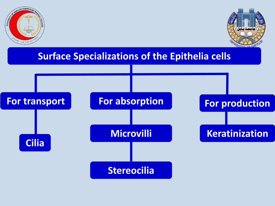
Surface Specializations of the Epithelia cells
For production For absorption For transport
Stereocilia
Keratinization Microvilli Cilia
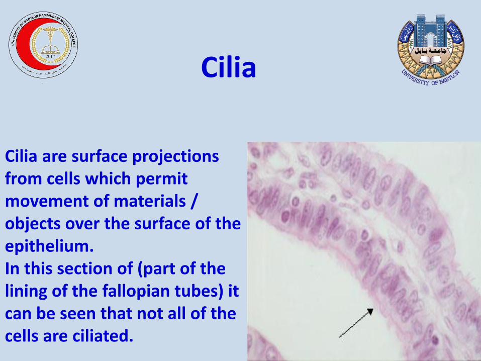
Cilia are surface projections from cells which permit movement of materials / objects over the surface of the epithelium. In this section of (part of the lining of the fallopian tubes) it can be seen that not all of the cells are ciliated.
Cilia
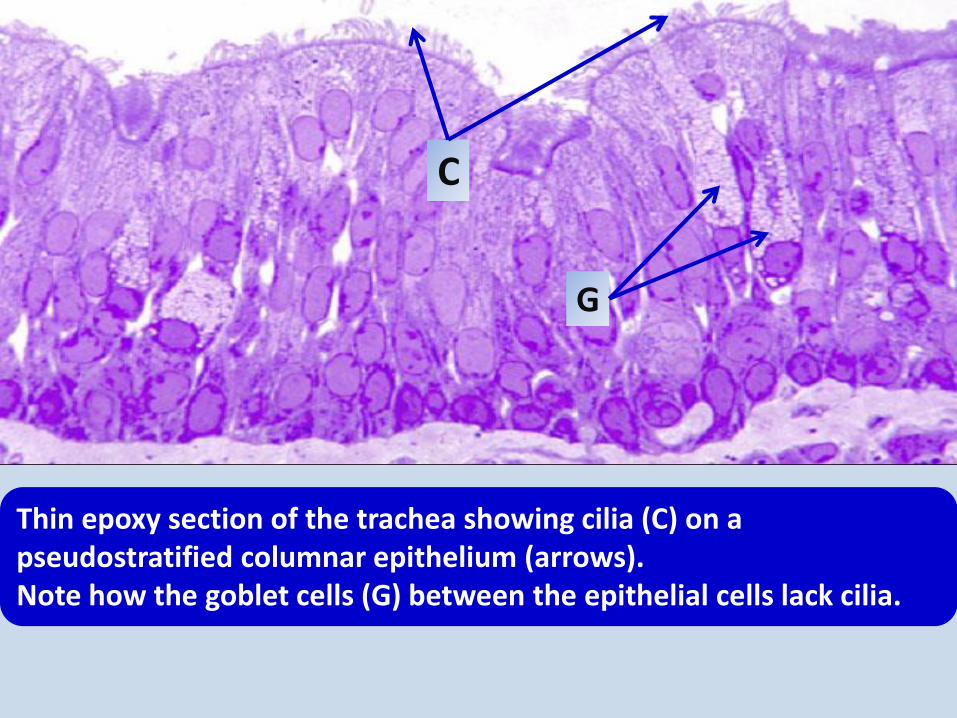
Thin epoxy section of the trachea showing cilia (C) on a pseudostratified columnar epithelium (arrows). Note how the goblet cells (G) between the epithelial cells lack cilia.
G
C
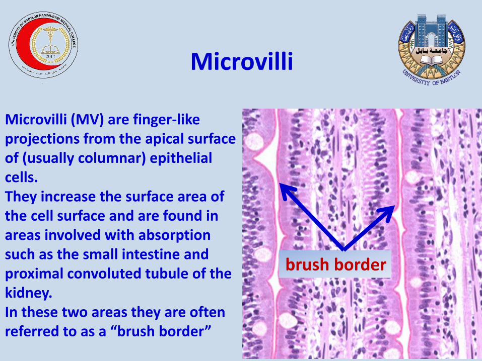
Microvilli (MV) are finger-like projections from the apical surface of (usually columnar) epithelial cells. They increase the surface area of the cell surface and are found in areas involved with absorption such as the small intestine and proximal convoluted tubule of the kidney. In these two areas they are often referred to as a “brush border”
brush border
Microvilli
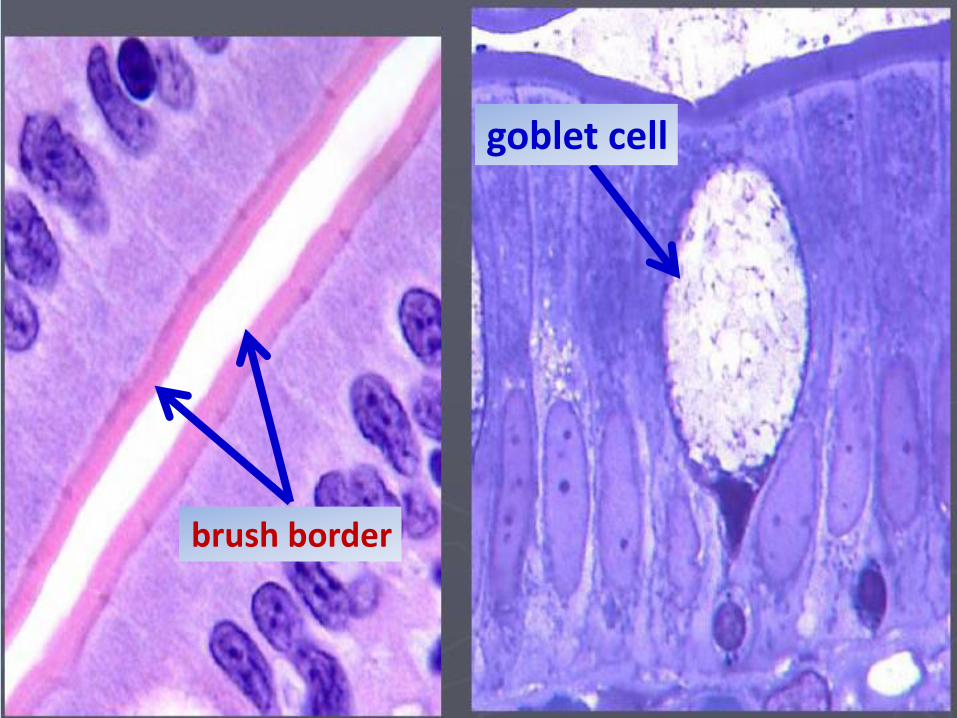
brush border
goblet cell
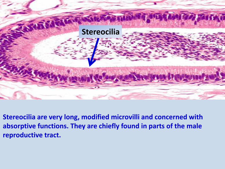
Stereocilia are very long, modified microvilli and concerned with absorptive functions. They are chiefly found in parts of the male reproductive tract.
Stereocilia
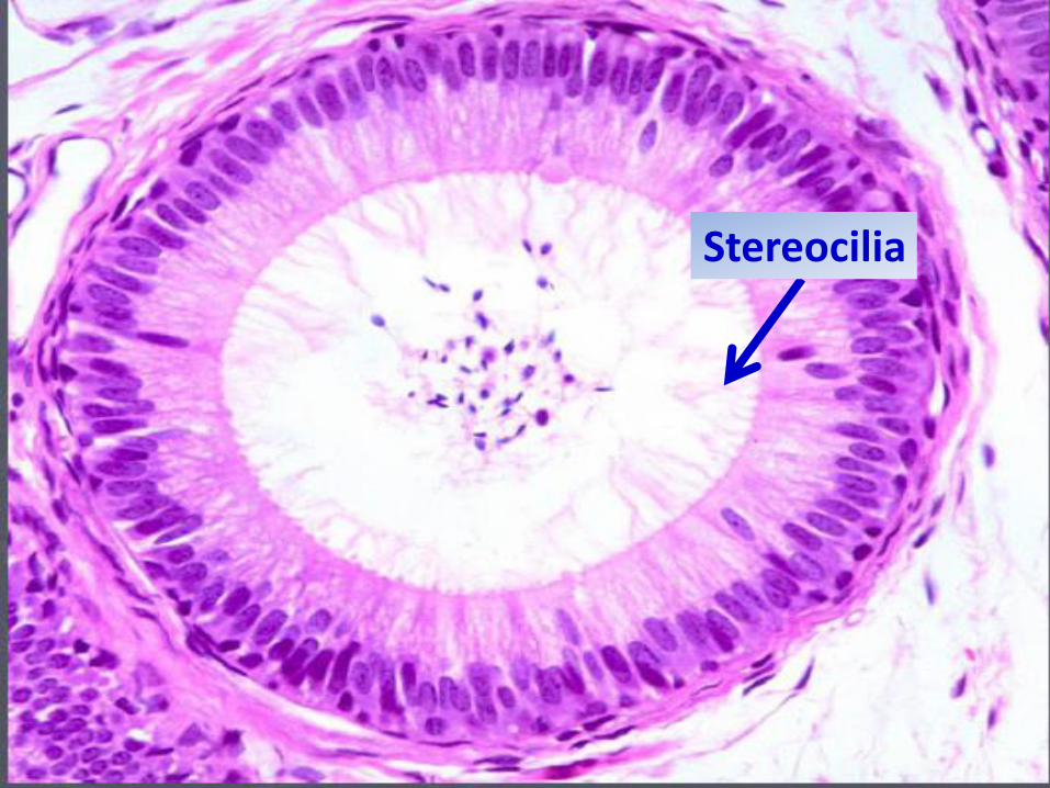
Stereocilia
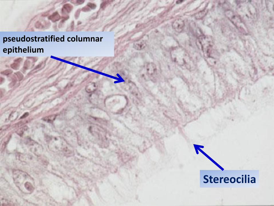
Stereocilia
pseudostratified columnar epithelium

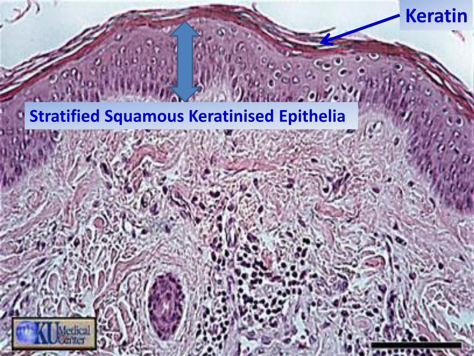
Characteristically found in the skin, this adaptation is for protection. The thickness of the keratin layer varies (depth of it here indicated by two arrows) in different sites. It is thickest in the sole of the foot and thinnest on the outer surface of the lip.
Keratinisation
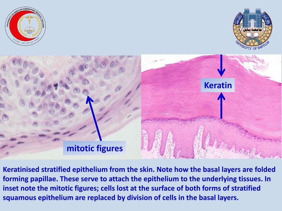
Keratinised stratified epithelium from the skin. Note how the basal layers are folded forming papillae. These serve to attach the epithelium to the underlying tissues. In inset note the mitotic figures; cells lost at the surface of both forms of stratified squamous epithelium are replaced by division of cells in the basal layers.
Keratin
mitotic figures
consists of several layers of cells in which the top layer is rectangular. Also uncommon. Usually basal layers are shortened, irregular polyhedral cells. It protects and mucus secreting . Its found in the conjunctiva lining the eyelids .
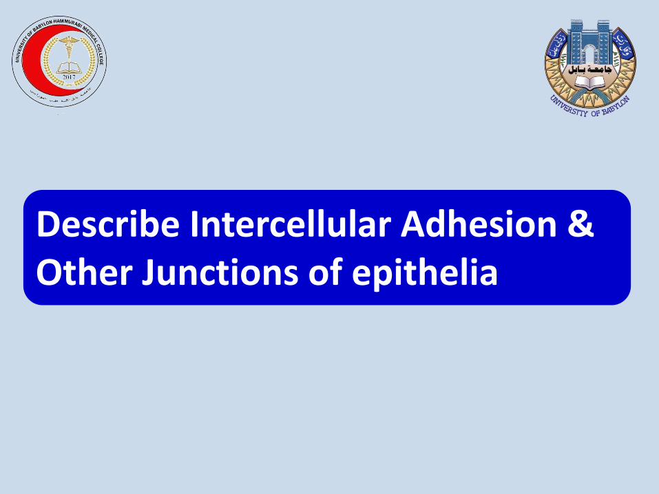
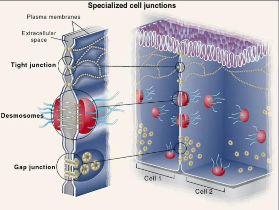
Describe Intercellular Adhesion & Other Junctions of epithelia
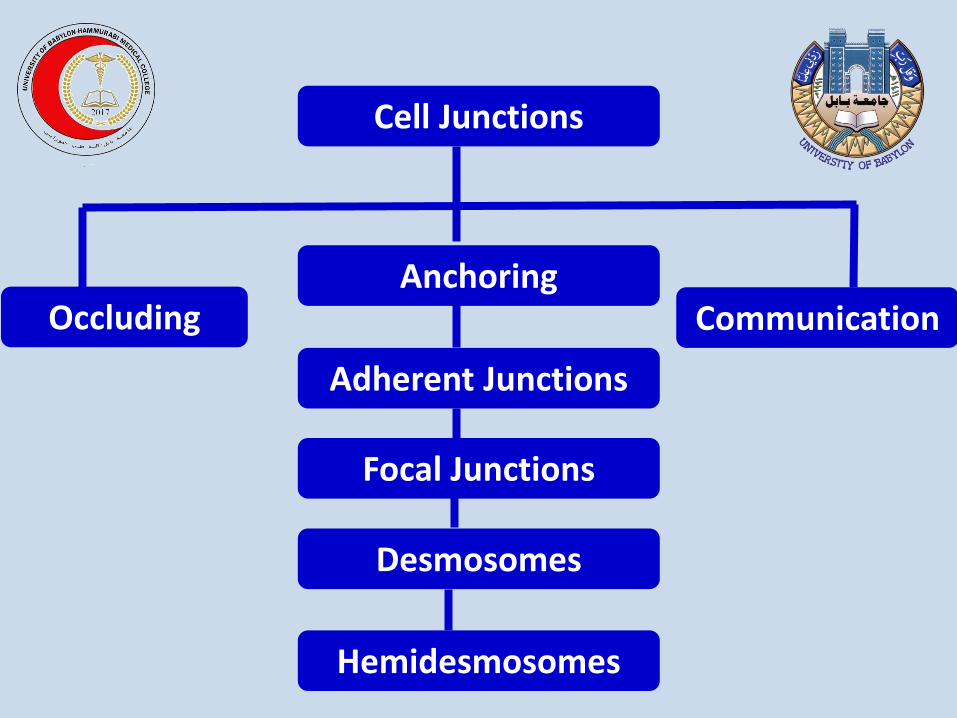
Cell Junctions
Communication Anchoring
Occluding
Desmosomes
Focal Junctions
Adherent Junctions
Hemidesmosomes
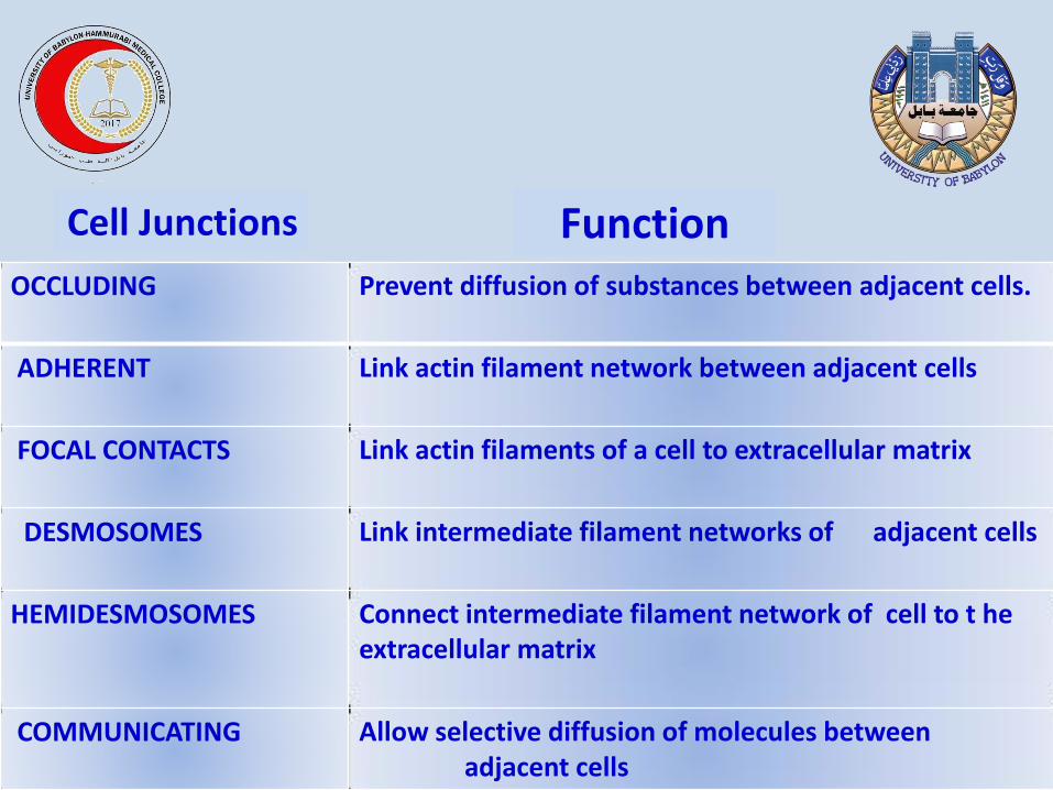
Prevent diffusion of substances between adjacent cells.
OCCLUDING
Link actin filament network between adjacent cells
ADHERENT
Link actin filaments of a cell to extracellular matrix
FOCAL CONTACTS
Link intermediate filament networks of adjacent cells
DESMOSOMES
Connect intermediate filament network of cell to t he extracellular matrix
HEMIDESMOSOMES
Allow selective diffusion of molecules between adjacent cells
COMMUNICATING
Cell Junctions Function
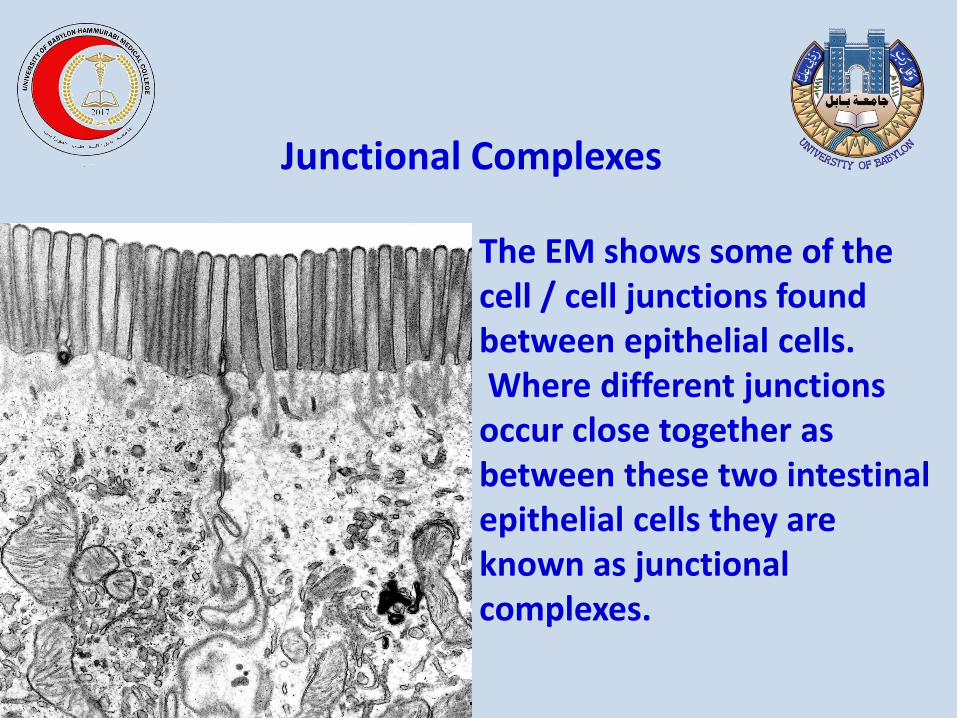
The EM shows some of the cell / cell junctions found between epithelial cells. Where different junctions occur close together as between these two intestinal epithelial cells they are known as junctional complexes.
Junctional Complexes
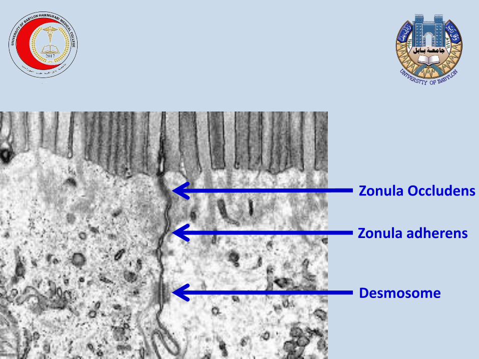
Zonula Occludens
Zonula adherens
Desmosome