Current controversies in emergency room CT: could … controversies in emergency room CT: ... in...
13
591 Clin. Pract. (2014) 11(6), 591–603 ISSN 2044-9038 part of Clinical Perspective 10.2217/CPR.14.59 © 2014 Future Medicine Ltd T: cal Practice points • Both pre- and in-hospital care for the trauma patient is organized worldwide according to the principles of ATLS ® (Advanced Trauma Life Support). It follows the central paradigm: treat first that kills first. • At the emergency department, standard radiological evaluation entails x-rays of the chest and pelvis and focused assessment with sonography for trauma ultrasound, with additional selective computed tomography (CT) scanning, if necessary. • The rationale for early total-body CT (TBCT) scanning is to detect all important injuries and begin treatment without missing injuries and losing time, thus reducing mortality. • TBCT scanning can lead to early detection of life threatening injuries, but may expose trauma patients to significant radiation. Only a few studies exist on cost–effectiveness and unsuspected findings, therefore, these topics remain unclear. • Pooled analysis of studies comparing TBCT with selective CT scanning show a trend, but no significant difference, in mortality. However, TBCT results in improvement of emergency department time and changes in treatment. • For a more definite statement, firm clinical outcome data from randomized controlled trials are needed, and are expected from the prospective REACT-2 trial in the coming years. • To reduce time delays, a trauma patient should be scanned in the emergency department. • For justification and optimization of CT scanning, and direct and complete interpretation of the radiological findings, an emergency department radiologist should be present at the trauma bay. In trauma patients who have sustained multiple injuries, every second counts: time is life. Immediate total-body computed tomography (TBCT) can give quick and complete radiological diagnoses for these critically ill patients. Hence, direct scanning can result in increased survival and a reduction in emergency department time. Several, mostly retrospective studies, have shown the potential of TBCT scanning in trauma patients. In these studies, TBCT appears to be associated with reduced evaluation times and decreased mortality. On the other hand, concerns have risen concerning the amount of radiation exposure in this overall relatively young population. Therefore, the need for prospective studies has been stressed recently by several authors. If immediate TBCT scanning is found to be the best imaging strategy in severely injured trauma patients, it could replace conventional imaging with selective CT in this specific group. Keywords: adults • computed tomography • emergency radiology • mortality • radiation exposure • trauma • whole body scanning Introduction In recent years, the role of emergency radiol- ogy in trauma has become more and more prominent [1,2] . Owing to the improvements in computed tomography (CT) technol- ogy, trauma CT scanning has changed from what was once called a “donut of death” for severely injured patients into an invalu- Current controversies in emergency room CT: could trauma total-body CT scanning improve clinical outcome? Ludo F Beenen , Accident & Emergency Radiology, Department of Radiology, Academic Medical Center, Amsterdam, Meibergdreef 9, 1105 AZ Amsterdam, The Netherlands [email protected]
Transcript of Current controversies in emergency room CT: could … controversies in emergency room CT: ... in...
591Clin. Pract. (2014) 11(6), 591–603 ISSN 2044-9038
part of
Clinical Perspective
10.2217/CPR.14.59 © 2014 Future Medicine Ltd
Clin. Pract.
10.2217/CPR.14.59
Clinical Perspective
BeenenCurrent controversies in emergency room CT:
could trauma TBCT scanning improve clinical outcome?
11
6
2014
Practice points
• Both pre- and in-hospital care for the trauma patient is organized worldwide according to the principles of ATLS® (Advanced Trauma Life Support). It follows the central paradigm: treat first that kills first.
• At the emergency department, standard radiological evaluation entails x-rays of the chest and pelvis and focused assessment with sonography for trauma ultrasound, with additional selective computed tomography (CT) scanning, if necessary.
• The rationale for early total-body CT (TBCT) scanning is to detect all important injuries and begin treatment without missing injuries and losing time, thus reducing mortality.
• TBCT scanning can lead to early detection of life threatening injuries, but may expose trauma patients to significant radiation. Only a few studies exist on cost–effectiveness and unsuspected findings, therefore, these topics remain unclear.
• Pooled analysis of studies comparing TBCT with selective CT scanning show a trend, but no significant difference, in mortality. However, TBCT results in improvement of emergency department time and changes in treatment.
• For a more definite statement, firm clinical outcome data from randomized controlled trials are needed, and are expected from the prospective REACT-2 trial in the coming years.
• To reduce time delays, a trauma patient should be scanned in the emergency department.• For justification and optimization of CT scanning, and direct and complete interpretation
of the radiological findings, an emergency department radiologist should be present at the trauma bay.
In trauma patients who have sustained multiple injuries, every second counts: time is life. Immediate total-body computed tomography (TBCT) can give quick and complete radiological diagnoses for these critically ill patients. Hence, direct scanning can result in increased survival and a reduction in emergency department time. Several, mostly retrospective studies, have shown the potential of TBCT scanning in trauma patients. In these studies, TBCT appears to be associated with reduced evaluation times and decreased mortality. On the other hand, concerns have risen concerning the amount of radiation exposure in this overall relatively young population. Therefore, the need for prospective studies has been stressed recently by several authors. If immediate TBCT scanning is found to be the best imaging strategy in severely injured trauma patients, it could replace conventional imaging with selective CT in this specific group.
Keywords: adults • computed tomography • emergency radiology • mortality • radiation exposure • trauma • whole body scanning
IntroductionIn recent years, the role of emergency radiol-ogy in trauma has become more and more prominent [1,2]. Owing to the improvements
in computed tomography (CT) technol-ogy, trauma CT scanning has changed from what was once called a “donut of death” for severely injured patients into an invalu-
Current controversies in emergency room CT: could trauma total-body CT scanning improve clinical outcome?
Ludo F Beenen,
Accident & Emergency Radiology,
Department of Radiology, Academic
Medical Center, Amsterdam,
Meibergdreef 9, 1105 AZ Amsterdam,
The Netherlands
592 Clin. Pract. (2014) 11(6) future science group
Clinical Perspective Beenen
able “circle of life”. But what is the role for radiology, or more specifically emergency radiology, and total body scanning in trauma? For this perspective paper, all relevant publications on trauma and radiology/CT were searched. Databases of PubMed, Embase, Web of Science and Cochrane Library were screened for papers published until December 2013, using the terms “trauma” or “injury”, in combination with “Total-Body CT (TBCT)” or “Whole Body CT” or “Full Body CT”, or “panscan”. In addition, reference lists of selected articles were evaluated for additional references. In this way, the many different aspect of TBCT scanning will be discussed. We will begin with what happens to a trauma patient from the moment of the accident, until after admission, when every minute counts.
Where do the paramedics take the trauma patient?At the trauma scene, the paramedics will perform an ATLS® (Advanced Trauma Life Support) based care on the trauma patient, which will be described in more detail later. The decision of the paramedics at the scene is to “stay and play” or “scoop and run”. That is, will they institute some care at the trauma scene, which will cost time and where resources are limited. After a short evaluation and stabilization, the patient will be transported to a hospital, either by ambulance or helicopter. However, should transport be to the nearest hospital, even if that is not accustomed to trauma care, without direct availability of all necessary specialists and no dedicated intensive care unit (ICU)? Or should they take more time and take the trauma patient to a dedicated major trauma center [3]?
Regionalization of trauma care has been a slow, but steady process over recent years [4]. Concentration of severely injured patients in trauma centers is associated with better outcomes [5,6]. Regular contact to low fre-quency, highly complex problems is best handled by a limited number of hospitals, thus increasing expo-sure and hence experience. TBCT scanning with its specific demands will also be part of the standard armamentarium. Thus, a settled trauma system with better handling for complications will lead to reduced mortality and morbidity [7]. A major trauma center has proven to save lives when having >250 trauma patients per year. For hemodynamically unstable patients delay to interventional radiologic procedures independently is associated with a twofold higher risk in mortality [8]. Therefore, also interventional radiology should be available on a 24/7 basis.
Although the sensitivity for pre-hospital triage is high, in daily practice both overtriage and undertri-age occurs [9]. Therefore, every emergency department
(ED) should be prepared to receive severe trauma patients, and have updated trauma protocols. When a patient has been undertriaged and brought to a local hospital without experience in total body trauma CT scanning, the risk exists that this may not be performed appropriately. As trauma is not a static but a dynamic process, the admitted patient could deteriorate unex-pectedly in hospital, and the severity of injuries could be more serious than the physicians ordinarily see. Of note, in cases of a mass casualty, every hospital is obliged to contribute to its capabilities.
During transport the paramedics will evaluate the clinical condition of the patient, specifically the vital parameters, such as blood pressure, heart rate and Glasgow Coma Score. It is paramount that this essen-tial information is transmitted to the receiving hospi-tal, so that they can prepare the whole trauma team (surgery, emergency, anesthesia, radiology) and take appropriate measures.
What happens after admission at the ED?The patient will be handed over by the paramedics to the trauma team. Worldwide trauma patient care is organized according to the principles of ATLS, a well-established method originating in 1976 for the systematic care on trauma patients, both in the field as well as in the hospital [10]. Due to this common ATLS language, communication and collaboration of all professionals involved is facilitated.
The basic principles are to first adress the injuries that are the most endangering, i.e. “treat first that kills first”. Therefore, a stepwise approach is used, performed in strict order. A systematic evaluation and treatment is started according to the “ABCDE”:
• A: Airway
• B: Breathing
• C: Circulation
• D: Disability
• E: Exposure and Environment
Standard radiological evaluation mostly entails x-rays of the chest and pelvis, and focused assessment with sonography for trauma (FAST) ultrasound [11,12]. Therefore, pneumothoraxes as well as injuries with large blood loss (hematothorax, pelvic ring fracture) can be recognized. In FAST, free intraperitoneal fluid (blood in trauma) can be detected irrespective of the hemo-dynamic status of the patient, although sensitivity for parenchymal injury is not as good [13]. Alternatively, a LODOX-Statscan system can produce a complete radio-graphic display of the whole body of a trauma patient
www.futuremedicine.com 593future science group
Current controversies in emergency room CT: could trauma TBCT scanning improve clinical outcome? Clinical Perspective
[14]. However, unlike CT, it cannot give information about parenchymal organ injuries, therefore, it never gained much popularity and nowadays is surpassed by CT and TBCT.
In a second systematic survey of the patient from head to toe, other injuries of the extremities are evalu-ated. According to the ATLS guidelines, CT can be used as an adjunct to conventional radiology or physi-cal examination to more clearly depict the location and severity of injuries. It can also be valuable in exclud-ing pathology and, as such, can have the patient safely discharged from the ED. Therefore, not only for high energy trauma but also for low energy trauma, patients can benefit from the use of CT. Compared with conven-tional radiography and ultrasound, CT yields new and additional findings in polytrauma [15]. These new find-ings primarily involve the head; additional findings are found in the chest, pelvis and spine, and are potentially life threatening in nature.
When a CT is performed, different set ups and pro-tocols can be chosen, depending on indications, local circumstances, logistics and preferences. As a member of the multidisciplinary team, the radiologist must decide which is the most adequate technique and protocol for each situation after taking in consideration the techno-logical resources available [16,17].
How reliable is physical examination?Can all injuries be evaluated with good accuracy and consequently safely excluded, thus avoiding radiologi-cal exams? The answer is no. Physical examination in patients with blunt abdominal trauma is useful but not completely reliable [18]. This was recently confirmed in a prospective study including 400 high-acuity patients [19]. Although bedside examination by emergency physi-cians could adequately attribute a very low probability of injury in the head, cervical spine, chest, abdomen/pel-vis and thoracic/lumbar spine injuries, overall diagnos-tic accuracy was low. Therefore, in these patients, they advised TBCT to prevent injuries being missed.
The severity of trauma is assessed using the Injury Severity Score (ISS), ranging from 0–75, which corre-lates with mortality, morbidity and hospitalization time after trauma. A major trauma (or polytrauma) is defined as an ISS being greater than 15 [20].
One major problem is that not all trauma patients are cooperative, or in severe trauma cases are unconscious or intubated, resulting in limited communication pos-sibilities. As the context, such as mechanism of injury, is important and unreliable when not verified, this also hampers interpretation. The right context can some-times put findings into another perspective; from unno-ticed and irrelevant, to potentially life endangering. This applies to both physical and radiological examinations.
Why do we need CT & what is the rationale for early CT?Historically, the timing of trauma deaths has been described using a trimodal mortality model (immedi-ate deaths, early deaths and late deaths) [21,22]. Imme-diate deaths occur within minutes of the accident, mostly at the scene or shortly after arrival in hospital, due to non-survivable injuries and account for 50% of all mortality. The second peak are the early deaths, within hours of arrival in the hospital. Late deaths occur days to weeks after the injury. As trauma sys-tems mature and ICU care improves, a decline in late deaths and hence a shift toward a bimodal distribution has emerged.
Hence, the main focus for controlling mortality in trauma lies in the prevention of early deaths. The first 24 h after trauma are the deadliest for trauma patients. Primary and secondary CNS injuries are the leading causes of death [23,24]. Next, almost a third of victims die of hemorrhage. For hypotensive trauma patients needing emergency laparotomy, mortality increases 1% for every 3 min in the ED [25].
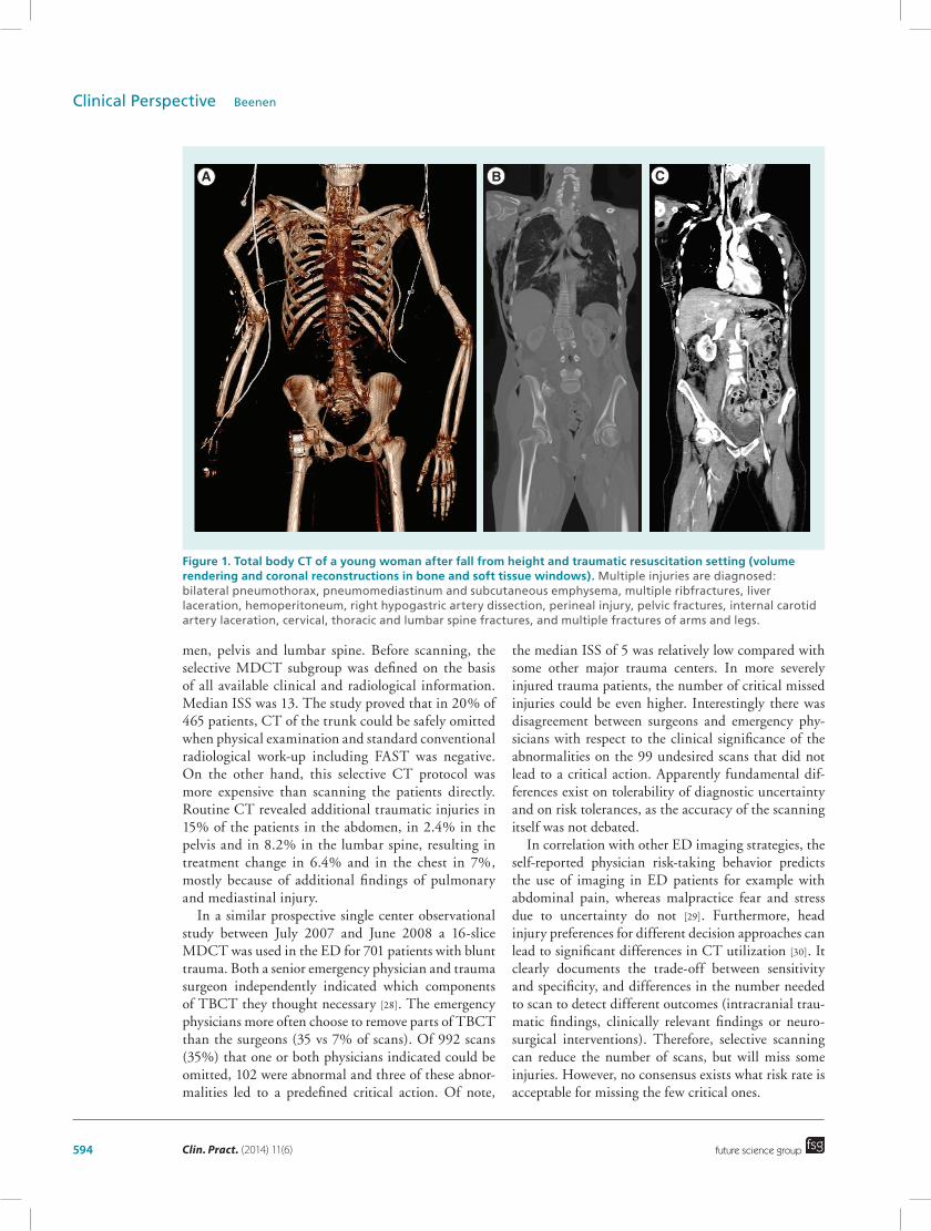
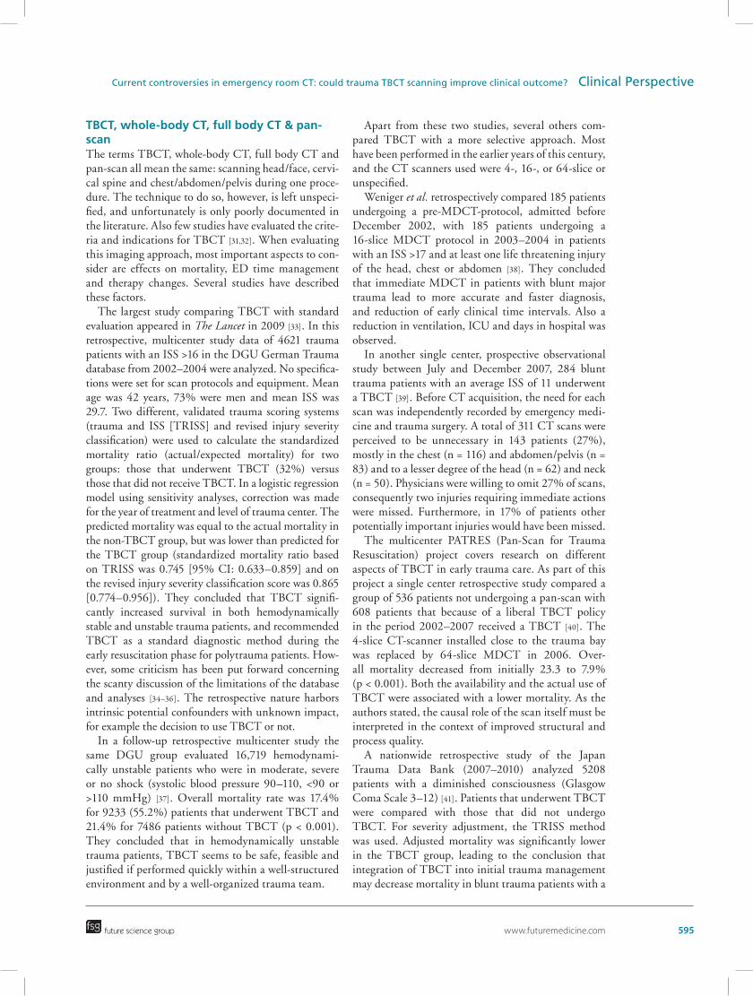
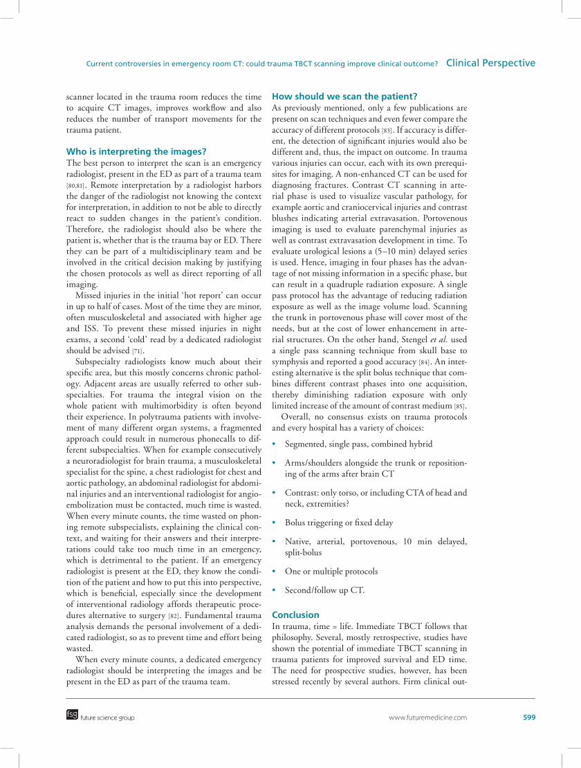
Therefore, the ratio for early CT lies in the early detection of these life-threatening injuries and asso-ciated possibility of starting treatment early. CT for trauma has a reported high accuracy for a wide variety of injuries, low missed injury rate, it is quick, available everywhere and can prevent unnecessary surgeries. Per-forming direct TBCT means surpassing conventional radiography with its lower sensitivity and could spare delay times up to 30 min. Detection of hematoma logically targets on stop the bleeding (“C” problem), but gathers no information on brain injury (“D” prob-lem), therefore, only after correcting circulation is an evaluation of the brain started, which could be too late and the damage already irreversible. The most logical approach is to instantly be informed on all potentially life threatening injuries, to make the right treatment decision and start this as soon as possible (Figure 1).
Do we need to scan all? Do we need selective CT or TBCT?Compared with a trauma protocol with selective CT after high-energy trauma, a routine CT of chest-abdo-men-pelvis finds substantially more clinically relevant diagnoses, even in patients with an unsuspicious clini-cal examination and normal radiography and FAST [26,27]. This prospective TRACT cohort study from May 2005 to November 2006 used a 16-slice mul-tidetector CT (MDCT) scanner that was located in a room adjacent to the trauma resuscitation room. All patients underwent physical examination, FAST and conventional radiography of the pelvis and lum-bar spine, and subsequently routine CT of the abdo-
594 Clin. Pract. (2014) 11(6)
Figure 1. Total body CT of a young woman after fall from height and traumatic resuscitation setting (volume rendering and coronal reconstructions in bone and soft tissue windows). Multiple injuries are diagnosed: bilateral pneumothorax, pneumomediastinum and subcutaneous emphysema, multiple ribfractures, liver laceration, hemoperitoneum, right hypogastric artery dissection, perineal injury, pelvic fractures, internal carotid artery laceration, cervical, thoracic and lumbar spine fractures, and multiple fractures of arms and legs.
future science group
Clinical Perspective Beenen
men, pelvis and lumbar spine. Before scanning, the selective MDCT subgroup was defined on the basis of all available clinical and radiological information. Median ISS was 13. The study proved that in 20% of 465 patients, CT of the trunk could be safely omitted when physical examination and standard conventional radiological work-up including FAST was negative. On the other hand, this selective CT protocol was more expensive than scanning the patients directly. Routine CT revealed additional traumatic injuries in 15% of the patients in the abdomen, in 2.4% in the pelvis and in 8.2% in the lumbar spine, resulting in treatment change in 6.4% and in the chest in 7%, mostly because of additional findings of pulmonary and mediastinal injury.
In a similar prospective single center observational study between July 2007 and June 2008 a 16-slice MDCT was used in the ED for 701 patients with blunt trauma. Both a senior emergency physician and trauma surgeon independently indicated which components of TBCT they thought necessary [28]. The emergency physicians more often choose to remove parts of TBCT than the surgeons (35 vs 7% of scans). Of 992 scans (35%) that one or both physicians indicated could be omitted, 102 were abnormal and three of these abnor-malities led to a predefined critical action. Of note,
the median ISS of 5 was relatively low compared with some other major trauma centers. In more severely injured trauma patients, the number of critical missed injuries could be even higher. Interestingly there was disagreement between surgeons and emergency phy-sicians with respect to the clinical significance of the abnormalities on the 99 undesired scans that did not lead to a critical action. Apparently fundamental dif-ferences exist on tolerability of diagnostic uncertainty and on risk tolerances, as the accuracy of the scanning itself was not debated.
In correlation with other ED imaging strategies, the self-reported physician risk-taking behavior predicts the use of imaging in ED patients for example with abdominal pain, whereas malpractice fear and stress due to uncertainty do not [29]. Furthermore, head injury preferences for different decision approaches can lead to significant differences in CT utilization [30]. It clearly documents the trade-off between sensitivity and specificity, and differences in the number needed to scan to detect different outcomes (intracranial trau-matic findings, clinically relevant findings or neuro-surgical interventions). Therefore, selective scanning can reduce the number of scans, but will miss some injuries. However, no consensus exists what risk rate is acceptable for missing the few critical ones.
A B C
www.futuremedicine.com 595future science group
Current controversies in emergency room CT: could trauma TBCT scanning improve clinical outcome? Clinical Perspective
TBCT, whole-body CT, full body CT & pan-scanThe terms TBCT, whole-body CT, full body CT and pan-scan all mean the same: scanning head/face, cervi-cal spine and chest/abdomen/pelvis during one proce-dure. The technique to do so, however, is left unspeci-fied, and unfortunately is only poorly documented in the literature. Also few studies have evaluated the crite-ria and indications for TBCT [31,32]. When evaluating this imaging approach, most important aspects to con-sider are effects on mortality, ED time management and therapy changes. Several studies have described these factors.
The largest study comparing TBCT with standard evaluation appeared in The Lancet in 2009 [33]. In this retrospective, multicenter study data of 4621 trauma patients with an ISS >16 in the DGU German Trauma database from 2002–2004 were analyzed. No specifica-tions were set for scan protocols and equipment. Mean age was 42 years, 73% were men and mean ISS was 29.7. Two different, validated trauma scoring systems (trauma and ISS [TRISS] and revised injury severity classification) were used to calculate the standardized mortality ratio (actual/expected mortality) for two groups: those that underwent TBCT (32%) versus those that did not receive TBCT. In a logistic regression model using sensitivity analyses, correction was made for the year of treatment and level of trauma center. The predicted mortality was equal to the actual mortality in the non-TBCT group, but was lower than predicted for the TBCT group (standardized mortality ratio based on TRISS was 0.745 [95% CI: 0.633–0.859] and on the revised injury severity classification score was 0.865 [0.774–0.956]). They concluded that TBCT signifi-cantly increased survival in both hemodynamically stable and unstable trauma patients, and recommended TBCT as a standard diagnostic method during the early resuscitation phase for polytrauma patients. How-ever, some criticism has been put forward concerning the scanty discussion of the limitations of the database and analyses [34–36]. The retrospective nature harbors intrinsic potential confounders with unknown impact, for example the decision to use TBCT or not.
In a follow-up retrospective multicenter study the same DGU group evaluated 16,719 hemodynami-cally unstable patients who were in moderate, severe or no shock (systolic blood pressure 90–110, <90 or >110 mmHg) [37]. Overall mortality rate was 17.4% for 9233 (55.2%) patients that underwent TBCT and 21.4% for 7486 patients without TBCT (p < 0.001). They concluded that in hemodynamically unstable trauma patients, TBCT seems to be safe, feasible and justified if performed quickly within a well-structured environment and by a well-organized trauma team.
Apart from these two studies, several others com-pared TBCT with a more selective approach. Most have been performed in the earlier years of this century, and the CT scanners used were 4-, 16-, or 64-slice or unspecified.
Weniger et al. retrospectively compared 185 patients undergoing a pre-MDCT-protocol, admitted before December 2002, with 185 patients undergoing a 16-slice MDCT protocol in 2003–2004 in patients with an ISS >17 and at least one life threatening injury of the head, chest or abdomen [38]. They concluded that immediate MDCT in patients with blunt major trauma lead to more accurate and faster diagnosis, and reduction of early clinical time intervals. Also a reduction in ventilation, ICU and days in hospital was observed.
In another single center, prospective observational study between July and December 2007, 284 blunt trauma patients with an average ISS of 11 underwent a TBCT [39]. Before CT acquisition, the need for each scan was independently recorded by emergency medi-cine and trauma surgery. A total of 311 CT scans were perceived to be unnecessary in 143 patients (27%), mostly in the chest (n = 116) and abdomen/pelvis (n = 83) and to a lesser degree of the head (n = 62) and neck (n = 50). Physicians were willing to omit 27% of scans, consequently two injuries requiring immediate actions were missed. Furthermore, in 17% of patients other potentially important injuries would have been missed.
The multicenter PATRES (Pan-Scan for Trauma Resuscitation) project covers research on different aspects of TBCT in early trauma care. As part of this project a single center retrospective study compared a group of 536 patients not undergoing a pan-scan with 608 patients that because of a liberal TBCT policy in the period 2002–2007 received a TBCT [40]. The 4-slice CT-scanner installed close to the trauma bay was replaced by 64-slice MDCT in 2006. Over-all mortality decreased from initially 23.3 to 7.9% (p < 0.001). Both the availability and the actual use of TBCT were associated with a lower mortality. As the authors stated, the causal role of the scan itself must be interpreted in the context of improved structural and process quality.
A nationwide retrospective study of the Japan Trauma Data Bank (2007–2010) analyzed 5208 patients with a diminished consciousness (Glasgow Coma Scale 3–12) [41]. Patients that underwent TBCT were compared with those that did not undergo TBCT. For severity adjustment, the TRISS method was used. Adjusted mortality was significantly lower in the TBCT group, leading to the conclusion that integration of TBCT into initial trauma management may decrease mortality in blunt trauma patients with a
596 Clin. Pract. (2014) 11(6) future science group
Clinical Perspective Beenen
Glasgow Coma Scale score of 3 to 12.In 2012 results of the FIRST study were published
[42]. In this prospective, multicenter cohort study 1696 out of 1950 (87%) consecutive severe blunt trauma patients underwent total body CT. Recruitment was between December 2004 and March 2007. No limita-tions on scanner type were given. The main outcome was the vital status at 30 days or at ICU discharge if discharge occurred within the first 30 days. Survival significantly improved for TBCT patients related to the TRISS predicted survival as compared with selec-tive CT. Diagnostic TBCT was associated with a significant reduction in 30-day mortality.
Time requirements for resuscitation, diagnostic work-up and transfer to definitive treatment were analyzed in an observational study with retrospective analysis after changing from the Trauma-Protocol in 2004 [43]. A mobile 16-slice ‘sliding gantry’ MDCT in the trauma resuscitation suite was used and 82 patients with suspected multiple trauma treated with the MDCT-Trauma-Protocol in 2004 versus 79 patients with the Conventional-Trauma-Protocol in 2002 were analyzed. Patient care could be improved with the TBCT approach as it shortened the time interval from arrival in the ED to obtaining a final diagnosis and management plan in trauma patients.
Does TBCT reduce mortality in severe trauma patients?To address this question and other benefits of TBCT, three systematic reviews and meta-analyses have been published, the most recent in 2013 [44]. Only studies that directly compared TBCT with a conventional approach with selective scanning were selected (most of the abovementioned studies) [33,38,40,42,43]. Both effects on mortality and time management were ana-lyzed. This systematic review and meta-analysis could provide Oxford grade level 2b evidence. Unfortu-nately, although indicative results of improved sur-vival, most had a suboptimal design to definitively prove that TBCT results in reduced mortality in blunt high-energy trauma patients. All were non-random-ized cohort studies and were prone to several sources of bias (selection for non-randomized, performance and detection to non blinding, attrition due to ret-rospective nature). Of note, traumatic brain injury is known to be the single largest contributor to trauma mortality [21,22]. Yet, no dedicated analysis on this specific subgroup has been presented in these papers, thereby limiting comparison and bias evaluation. In the presented papers, baseline characteristics of differ-ent items on traumatic brain injury are displayed. In some, a significant lower number of severe traumatic brain injury was included in the TBCT group [33,40].
As a steady decline in trauma mortality is generally seen worldwide, temporal comparisons can be major confounders. The meta-analysis showed that in 8180 patients of five pooled studies, no significant difference on mortality between groups was found (pooled OR: 0.68; 95% CI 0.43–1.09, p = 0.11). However, in 6073 patients in four studies, a TBCT resulted in improved ED time intervals when compared with selective CT. Owing to a significant reduction in the time spent in the ED when patients underwent TBCT (pooled effect size of weighted mean difference = -32.39 min; 95% CI: -51.78 to -13.00; p = 0.001), therapy in life threatening situations can be started sooner.
In an effort to display the whole picture, a recent systematic review did not supply a meta-analysis, but instead focused on the benefits (shorter time to diagno-sis, treatment, stay, mortality, efficient diagnostics, fewer missed injuries, potential lower total radiation exposure) and harms (delayed time to immediate intervention, increased radiation exposure) of TBCT [45]. Although more benefits than harms were named, the authors critically questioned whether the reported benefits in mortality truly represents an improved survival due to substantiated management changes, or whether this is the result of hospital- or individual-level confounders.
To circumvent the weaknesses intrinsically associ-ated with the used methodology in the reported stud-ies, there is a need for high-quality studies and for the best and most solid scientific proof. Therefore, randomized controlled trials are needed.
Prospective international multicenter randomized clinical trialThe REACT-2 trial is an international, multicenter randomized clinical trial that is evaluating the value of immediate TBCT scanning during the primary survey of severely injured trauma patients [46]. All participat-ing trauma centers have a MDCT scanner located in the trauma room or at the ED. Patients are random-ized between direct TBCT scan (unenhanced head and neck followed by repositioning of the arms with contrast-enhanced imaging of chest-abdomen-pelvis) versus ATLS-based conventional trauma imaging pro-tocols with supplemental selective CT scanning. Inclu-sion started April 2011 and ended December 2013, with follow-up of 1 year. The primary outcome is in-hospital mortality and secondary outcomes are differ-ences in mortality and morbidity during the first year, several work-up time intervals at the ED, radiation exposure, quality of life and cost–effectiveness.
What about radiation exposure?When every minute counts, there is no time to think about radiation exposure. It has to be done earlier, by
www.futuremedicine.com 597future science group
Current controversies in emergency room CT: could trauma TBCT scanning improve clinical outcome? Clinical Perspective
setting up the right protocols by dedicated radiolo-gists in good collaboration with specialized radiologi-cal technicians. All trauma team members should be familiar with the protocols; protocols should be easily accessible and clear to interpret.
If during trauma evaluation inappropriate use of CT scanning has occurred, a correction of this procedure should be undertaken by the responsible radiologist at a later stage.
In general, trauma patients are relatively young, mostly males between 20–50 years. In recent years, an increasing number of publications on radiation expo-sure and trauma have appeared, not only for different parts of the body, but also for total body scanning [47,48]. Critics to a liberal use of scanning are emphasizing the unnecessary radiation exposure without any impact on mortality, missed injuries or length of stay [49].
After the introduction of a 64-slice TBCT proto-col in 2007, an increasing number of trauma patients received a radiation dose >20 mSv, regardless of age or injury severity [50]. In another study from 2006, cumu-lative effective dose was 14.6 mSv per trauma patient [51]. CT scans accounted for only 21% of radiologic studies but for 93% of the cumulative effective dose. Patients with ISS >9 and longer length of stay had a higher cumulative effective dose rate.
Radiation during admission as well as during the complete hospital period has been studied. Although TBCT can increase the CT-induced trauma room radiation dose compared with selective CT scanning, the total overall effective dose during hospital admis-sion was not significantly different before and after the introduction of a dedicated TBCT protocol (20 vs 24 mSv, p = 0.509) [52].
As trauma patients are exposed to significant radia-tion doses from diagnostic imaging, resulting in a small but measurable cancer risk, unnecessary CT scans should be avoided [53]. Diagnostic benefit always needs to be weighed against risk of the cumulative radiation dose [54]. The risk of mortality from trauma was cal-culated to be six times higher than the estimated risk of radiation-induced cancer mortality in intermediate level trauma patients [55]. As mortality is greatest in older trauma patients, a more liberal attitude toward CT-scanning in these patients could be of advantage. Efforts to reduce radiation exposure to trauma patients should certainly focus on young patients with minor injuries, for example, by standard usage of commonly available techniques for dose reduction, such as auto-mated exposure control. Iterative reconstructions are considered a valuable quality improvement technique in comparison to the commonly used filtered back pro-jection. Depending on preferences one can choose for either improving image quality for a constant radia-
tion exposure or reduction of radiation exposure, while maintaining the same image quality [56,57]. A major downside of this technique, however, is the intrinsic higher time consumption, which is unwanted in major trauma patients.
In addition, some specific trauma related issues can be mentioned. By limiting redundant imaging in over-lap zones, TBCT reduces radiation exposure compared with a segmented approach while covering the same range [58]. Scanning with the arms alongside the body can result in a 45% increased radiation exposure and a decrease in image quality compared with scanning patients with a cranial position of the arms [59]. Scan-ning with one arm cranial and one caudal (superman configuration) results in in-between exposure and image quality. Thus, effort should be made to position the arms above the shoulder when possible.
Some bearing devices can increase radiation dose and affect image quality. For the best balance there-fore preferably aluminum and metal-free devices are chosen. Nowadays, with new hardware, software and individual placement algorithms, significant dose sav-ings to a radiation level of 10 mSv can be reached [60].
As with the newest high-end CT-scanners, in some cases, radiation doses are approaching ranges of con-ventional radiography, which could potentially chal-lenge that modality. There may even be a paradigm shift, challenging the early ATLS resuscitation phase by using CT as the primary screening tool.
Specific populations: children, pregnant women, elderly & obese patientsChildren are more sensitive to radiation compared with adults, therefore, the younger the patient at the time of exposure, the higher the radiation dose to the organs [61]. Higher organ radiation doses are associated with an increased risk for cancer in this population. The majority of radiation exposure to pediatric trauma patients is secondary to CT [62]. Scan protocols should be adapted to specific age categories. In trauma, the mean whole body effective dose is 17.4 mSv [63]. How-ever, no survival benefit of TBCT in pediatric trauma has been established. Therefore, the indiscriminate use of CT in the evaluation of the pediatric trauma patient is not warranted.
In a step-up imaging strategy for pediatric patients with blunt abdominal trauma, decisions for observa-tion or intervention depends on the vital parameters of the patient in combination with the evaluation of free fluid at FAST and the findings on CT (performed only on indication) [64]. This step-up imaging strategy has a high sensitivity and a high negative predictive value for ruling out abdominal injury without missing clinically relevant injuries.
598 Clin. Pract. (2014) 11(6) future science group
Clinical Perspective Beenen
Trauma is the leading cause of non-obstetric maternal mortality and a significant cause of fetal loss. Ultrasound can be a good screening alterna-tive, however, even in pregnant women with severe trauma, CT imaging is of importance but must be used wisely. Of course the risks of radiation exposure should be weighted individually against the benefits, and most are small compared with the possible effects of missed injuries. The pregnant trauma patient requires imaging tests to diagnose maternal injuries and diagnostic tests to evaluate the viability of her pregnancy [65,66].
Older patients have an increased mortality in trauma, even without physiological derangement on admission. Exposure to radiation is less of an issue in this age group, so active diagnosis and treatment should not be discouraged. By doing so, in older patients, sig-nificant survival rates can achieved [67].
Obesity is an increasing problem in the modern world. In these patients, higher rates of injuries to torso and proximal upper extremities are found [68], and even more so in older patients. For these imaging chal-lenges, specific adjustments must be made in diagnos-tic studies, including optimizing scanning parameters, using antiscatter grids with tight collimation, iterative reconstruction and artifact reduction techniques [69].
Disadvantages of TBCTIncluding the aforementioned radiation exposure, there are other disadvantages to TBCT: economics/cost–effectiveness and unsuspected findings/inci-dentalomas. Injuries can be missed because of image overload, lack of context or over reliance on TBCT findings [70]. Furthermore, injuries are not always immediately apparent, as imaging is only a snapshot in time. Therefore, correlation with clinical findings by implementing tertiary and quarterly survey is fun-damental, as well as studying imaging reports [71] or an itemized structured reporting.
In 45% of trauma patients that underwent TBCT scanning an incidental finding is found [72]. Of these, 7% are category I findings (potentially severe condi-tion, further diagnostic work-up required), 24% cat-egory II findings (diagnostic work-up dependent on patients’ symptoms) and 69% are category III inciden-tal findings (findings of minor concern, no diagnostic work-up required). Both additional radiologic imag-ing and further invasive work-up or treatment could, therefore, be needed.
CostsScanning a complete body can theoretically cause an increase in total costs. However, in a recent study, changing to a liberal mechanism-driven scanning
protocol not only resulted in change of treatment in 74% of patients, including prevention of unnecessary admission, but also saved £93,000 [73]. During a 1-year observation period, 336 of 452 trauma patients (74%) had a positive scan. However, due to the chosen set up, it remains unclear if observation of the other 116 would have lead to additional imaging or not. If so, this would have added to even higher costs.
Is more needed?Just moving the patient in the scanner will not save a life. There are some crucial elements that are often overlooked, but are essential for good trauma care, and even more so for TBCT. These are: place, technique and interpretation of the scan.
Where should we scan the patient?Should the patient go to a CT scan in the radiology department, which is often on another floor? Do we need a scanner in the ED, or is a remote scanner in the radiology department fulfilling all demands?
There are some disadvantages to sending the trauma patient to the radiological department. First, transport itself harbors the danger of complications. For criti-cal patients, intra-hospital transport is associated with a higher risk for complications, such as pneumotho-rax, atelectasis or displacement of lines and tubes [74]. In addition, they have to wait until a CT scanner is available, costing even more critical minutes. When the condition of the patient deteriorates during scan-ning, who can care for the patient or are there suffi-cient resuscitation possibilities available? This could be especially dangerous if only radiological technicians and ED nurses are present.
The prospective REACT-1 study compared the impact of having a CT in the trauma room versus the radiology department in 1124 patients in two Dutch level-1 trauma hospitals [75]. In this randomized con-trolled trial there was a reduction in the number of patient transfers and transports, and a significant reduction in time to first CT imaging (13 min shorter, p < 0.001) was recorded for the trauma room CT set-ting. Although not statistically significant, it also favored a reduction in mortality and out-of-hospital days.
Therefore, the best option is a CT scanner in the ED or, even better, a CT scanner in the trauma bay [76–78]. Choosing the optimal planning and distribu-tion strategies depend on the number and location of available CT scanners, along with number of trauma, urgent and regular patients [79]. With a sliding gantry solution (The Amsterdam Trauma Workflow concept with one CT scanner servicing two rooms), one room is always available for the highest emergency. A CT
www.futuremedicine.com 599future science group
Current controversies in emergency room CT: could trauma TBCT scanning improve clinical outcome? Clinical Perspective
scanner located in the trauma room reduces the time to acquire CT images, improves workflow and also reduces the number of transport movements for the trauma patient.
Who is interpreting the images?The best person to interpret the scan is an emergency radiologist, present in the ED as part of a trauma team [80,81]. Remote interpretation by a radiologist harbors the danger of the radiologist not knowing the context for interpretation, in addition to not be able to directly react to sudden changes in the patient’s condition. Therefore, the radiologist should also be where the patient is, whether that is the trauma bay or ED. There they can be part of a multidisciplinary team and be involved in the critical decision making by justifying the chosen protocols as well as direct reporting of all imaging.
Missed injuries in the initial ‘hot report’ can occur in up to half of cases. Most of the time they are minor, often musculoskeletal and associated with higher age and ISS. To prevent these missed injuries in night exams, a second ‘cold’ read by a dedicated radiologist should be advised [71].
Subspecialty radiologists know much about their specific area, but this mostly concerns chronic pathol-ogy. Adjacent areas are usually referred to other sub-specialties. For trauma the integral vision on the whole patient with multimorbidity is often beyond their experience. In polytrauma patients with involve-ment of many different organ systems, a fragmented approach could result in numerous phonecalls to dif-ferent subspecialties. When for example consecutively a neuroradiologist for brain trauma, a musculoskeletal specialist for the spine, a chest radiologist for chest and aortic pathology, an abdominal radiologist for abdomi-nal injuries and an interventional radiologist for angio-embolization must be contacted, much time is wasted. When every minute counts, the time wasted on phon-ing remote subspecialists, explaining the clinical con-text, and waiting for their answers and their interpre-tations could take too much time in an emergency, which is detrimental to the patient. If an emergency radiologist is present at the ED, they know the condi-tion of the patient and how to put this into perspective, which is beneficial, especially since the development of interventional radiology affords therapeutic proce-dures alternative to surgery [82]. Fundamental trauma analysis demands the personal involvement of a dedi-cated radiologist, so as to prevent time and effort being wasted.
When every minute counts, a dedicated emergency radiologist should be interpreting the images and be present in the ED as part of the trauma team.
How should we scan the patient?As previously mentioned, only a few publications are present on scan techniques and even fewer compare the accuracy of different protocols [83]. If accuracy is differ-ent, the detection of significant injuries would also be different and, thus, the impact on outcome. In trauma various injuries can occur, each with its own prerequi-sites for imaging. A non-enhanced CT can be used for diagnosing fractures. Contrast CT scanning in arte-rial phase is used to visualize vascular pathology, for example aortic and craniocervical injuries and contrast blushes indicating arterial extravasation. Portovenous imaging is used to evaluate parenchymal injuries as well as contrast extravasation development in time. To evaluate urological lesions a (5–10 min) delayed series is used. Hence, imaging in four phases has the advan-tage of not missing information in a specific phase, but can result in a quadruple radiation exposure. A single pass protocol has the advantage of reducing radiation exposure as well as the image volume load. Scanning the trunk in portovenous phase will cover most of the needs, but at the cost of lower enhancement in arte-rial structures. On the other hand, Stengel et al. used a single pass scanning technique from skull base to symphysis and reported a good accuracy [84]. An inter-esting alternative is the split bolus technique that com-bines different contrast phases into one acquisition, thereby diminishing radiation exposure with only limited increase of the amount of contrast medium [85].
Overall, no consensus exists on trauma protocols and every hospital has a variety of choices:
• Segmented, single pass, combined hybrid
• Arms/shoulders alongside the trunk or reposition-ing of the arms after brain CT
• Contrast: only torso, or including CTA of head and neck, extremities?
• Bolus triggering or fixed delay
• Native, arterial, portovenous, 10 min delayed, split-bolus
• One or multiple protocols
• Second/follow up CT.
ConclusionIn trauma, time = life. Immediate TBCT follows that philosophy. Several, mostly retrospective, studies have shown the potential of immediate TBCT scanning in trauma patients for improved survival and ED time. The need for prospective studies, however, has been stressed recently by several authors. Firm clinical out-
600 Clin. Pract. (2014) 11(6) future science group
Clinical Perspective Beenen
come data are expected to emerge in the near future as the international REACT-2 trial is set up to answer the questions raised, studying not only at survival, but also at quality of life, cost–effectiveness and radia-tion burden. If immediate TBCT scanning is found to be the best imaging strategy in severely injured trauma patients, it could replace conventional imaging supplemented with CT in this specific group.
Future perspectiveIn the next 5–10 years, TBCT scanning will be the standard examination, particularly in patients with severe trauma, but also in low risk patients. Process-ing and post-processing techniques will continuously develop in such a way that radiation exposure will be
at levels of conventional radiography and, therefore, no longer a big issue. For the severely injured, CT scan-ning will move in forefront and become mainstay of trauma care. Its use as a screening tool could change the “ABCDE” to “AB CT”.
Financial & competing interests disclosureThe author has no relevant affiliations or financial involvement
with any organization or entity with a financial interest in or fi-
nancial conflict with the subject matter or materials discussed
in the manuscript. This includes employment, consultancies,
honoraria, stock ownership or options, expert testimony,
grants or patents received or pending, or royalties.
No writing assistance was utilized in the production of this
manuscript.
ReferencesPapers of special note have been highlighted as: • of interest; •• of considerable interest
1 Beck JJW. Trauma imaging in and out of conflict: a review of the evidence. Radiography 18(4), 292–295 (2012).
2 Rao VM, Levin DC, Parker L, Frangos AJ, Sunshine JH. Trends in utilization rates of the various imaging modalities in emergency departments: nationwide Medicare data from 2000 to 2008. J. Am. Coll. Radiol. 8(10), 706–709 (2011).
3 Haas B, Stukel TA, Gomez D et al. The mortality benefit of direct trauma center transport in a regional trauma system: a population-based analysis. J. Trauma Acute Care Surg. 72(6), 1510–1517 (2012).
4 Harvey JJ, West AT. The right scan, for the right patient, at the right time: the reorganization of major trauma service provision in England and its implications for radiologists. Clin. Radiol. 68(9), 871–886 (2013).
•• Niceoverviewondifferentaspectofradiologyintraumacare.
5 Chiara O, Cimbanassi S. Organized trauma care: does volume matter and do trauma centers save lives? Curr. Opin. Crit. Care 9(6), 510–514 (2003).
6 Demetriades D, Martin M, Salim A et al. Relationship between American College of Surgeons trauma center designation and mortality in patients with severe trauma (injury severity score > 15). J. Am. Coll. Surg. 202(2), 212–215 (2006).
7 Celso B, Tepas J, Langland-Orban B et al. A systematic review and meta-analysis comparing outcome of severely injured patients treated in trauma centers following the establishment of trauma systems. J. Trauma 60(2), 371–378 (2006).
8 Howell GM, Peitzman AB, Nirula R et al. Delay to therapeutic interventional radiology postinjury: time is of the essence. J. Trauma 68(6), 1296–1300 (2010).
9 Cox S, Smith K, Currell A, Harriss L, Barger B, Cameron P. Differentiation of confirmed major trauma patients and potential major trauma patients using pre-hospital trauma triage criteria. Injury 42(9), 889–895 (2011).
10 Kortbeek JB, Al Turki SA, Ali J et al. Advanced trauma life support, 8th edition, the evidence for change. J. Trauma 64(6), 1638–1650 (2008).
11 Kool DR, Blickman JG. Advanced trauma life support. ABCDE from a radiological point of view. Emerg. Radiol. 14(3), 135–141 (2007).
12 Kloth JK, Kauczor HU, Hosch W. Imaging in the emergency room. Med. Klin. Intensivmed. Notfmed. 106(2), 82–88 (2011).
13 Korner M, Krotz MM, Degenhart C, Pfeifer KJ, Reiser MF, Linsenmaier U. Current role of emergency US in patients with major trauma. Radiographics 28(1), 225–242 (2008).
14 Boffard KD, Goosen J, Plani F, Degiannis E, Potgieter H. The use of low dosage x-ray (Lodox/Statscan) in major trauma: comparison between low dose x-ray and conventional x-ray techniques. J. Trauma 60(6), 1175–1181 (2006).
15 Maurer MH, Knopke S, Schroder RJ. Added diagnostic benefit of 16-row whole-body spiral CT in patients with multiple trauma differentiated by region and injury severity according to the ATLS concept. Rofo 180(12), 1117–1123 (2008).
16 Marti De Gracia M, Artigas Martin JM, Vicente Bartulos A, Carreras Aja M. Radiological management of patients with multiple trauma: history and current practice. Radiologia 52(2), 105–114 (2010).
17 Choy G, Novelline RA. Past, present, and future of emergency radiology. Can. Assoc. Radiol. J. 64(2), 85–89 (2013).
18 Schurink GW, Bode PJ, van Luijt PA, van Vugt AB. The value of physical examination in the diagnosis of patients with blunt abdominal trauma: a retrospective study. Injury 28(4), 261–265 (1997).
19 Smith CB, Barrett TW, Berger CL, Zhou C, Thurman RJ, Wrenn KD. Prediction of blunt traumatic injury in high-acuity patients: bedside examination vs computed tomography. Am. J. Emerg. Med. 29(1), 1–10 (2011).
20 Baker SP, O’Neill B, Haddon W Jr, Long WB. The injury severity score: a method for describing patients with multiple injuries and evaluating emergency care. J. Trauma 14(3), 187–196 (1974).

www.futuremedicine.com 601future science group
Current controversies in emergency room CT: could trauma TBCT scanning improve clinical outcome? Clinical Perspective
21 de Knegt C, Meylaerts SA, Leenen LP. Applicability of the trimodal distribution of trauma deaths in a level I trauma centre in the Netherlands with a population of mainly blunt trauma. Injury 39(9), 993–1000 (2008).
22 Sobrino J, Shafi S. Timing and causes of death after injuries. Proc. (Bayl. Univ. Med. Cent). 26(2), 120–123 (2013).
23 Kleber C, Giesecke MT, Tsokos M et al. Overall distribution of trauma-related deaths in Berlin 2010: advancement or stagnation of German trauma management? World J. Surg. 36(9), 2125–2130 (2012).
24 Acosta JA, Yang JC, Winchell RJ et al. Lethal injuries and time to death in a level I trauma center. J. Am. Coll. Surg. 186(5), 528–533 (1998).
25 Clarke JR, Trooskin SZ, Doshi PJ, Greenwald L, Mode CJ. Time to laparotomy for intra-abdominal bleeding from trauma does affect survival for delays up to 90 minutes. J. Trauma 52(3), 420–425 (2002).
26 Brink M, Deunk J, Dekker HM et al. Added value of routine chest MDCT after blunt trauma: evaluation of additional findings and impact on patient management. Am. J. Roentgenol. 190(6), 1591–1598 (2008).
27 Deunk J, Brink M, Dekker HM et al. Routine versus selective computed tomography of the abdomen, pelvis, and lumbar spine in blunt trauma: a prospective evaluation. J. Trauma 66(4), 1108–1117 (2009).
28 Gupta M, Schriger DL, Hiatt JR et al. Selective use of computed tomography compared with routine whole body imaging in patients with blunt trauma. Ann. Emerg. Med. 58(5), 407–416.e15 (2011).
29 Pines JM, Hollander JE, Isserman JA et al. The association between physician risk tolerance and imaging use in abdominal pain. Am. J. Emerg. Med. 27(5), 552–557 (2009).
30 Smits M, Dippel DW, de Haan GG et al. Minor head injury: guidelines for the use of CT-a multicenter validation study. Radiology 245(3), 831–838 (2007).
31 Babaud J, Ridereau-Zins C, Bouhours G et al. Benefit of the Vittel criteria to determine the need for whole body scanning in a severe trauma patient. Diagn. Interv. Imaging 93(5), 371–379 (2012).
32 Hsiao KH, Dinh MM, McNamara KP et al. Whole-body computed tomography in the initial assessment of trauma patients: is there optimal criteria for patient selection? Emerg. Med. Australas. 25(2), 182–191 (2013).
33 Huber-Wagner S, Lefering R, Qvick LM et al. Effect of whole-body CT during trauma resuscitation on survival: a retrospective, multicentre study. Lancet 373(9673), 1455–1461 (2009).
•• Largestandmostinfluentialstudyontotal-bodycomputedtomographyusingretrospectiveanalysisoftheGermanTraumaDatabaseregistry.
34 Giannoudis PV. Effect on survival of whole-body CT during trauma resuscitation. Lancet 374(9685) 197 (2009).
35 Saltzherr TP, Goslings JC, Multidisciplinary REACT 2 study group. Effect on survival of whole-body CT during trauma resuscitation. Lancet 374(9685), 198–199 (2009).
36 Stengel D, Frank M, Matthes G et al. Primary pan-computed tomography for blunt multiple trauma: can the whole be
better than its parts?. Injury 40(Suppl. 4), S36–S46 (2009).
37 Huber-Wagner S, Biberthaler P, Häberle S et al. Whole-body CT in haemodynamically unstable severely injured patients – a retrospective, multicentre study. PLoS ONE 8(7), e68880 (2013).
38 Weninger P, Mauritz W, Fridrich P et al. Emergency room management of patients with blunt major trauma: evaluation of the multislice computed tomography protocol exemplified by an urban trauma center. J. Trauma 62(3), 584–591 (2007).
39 Tillou A, Gupta M, Baraff LJ et al. Is the use of pan-computed tomography for blunt trauma justified? A prospective evaluation. J. Trauma 67(4), 779–787 (2009).
40 Hutter M, Woltmann A, Hierholzer C, Gartner C, Buhren V, Stengel D. Association between a single-pass whole-body computed tomography policy and survival after blunt major trauma: a retrospective cohort study. Scand. J. Trauma Resusc. Emerg. Med. 19, 73 (2011).
41 Kimura A, Tanaka N. Whole-body computed tomography is associated with decreased mortality in blunt trauma patients with moderate-to-severe consciousness disturbance: a multicenter, retrospective study. J. Trauma Acute Care Surg. 75(2), 202–206 (2013).
42 Yeguiayan JM, Yap A, Freysz M et al. Impact of whole-body computed tomography on mortality and surgical management of severe blunt trauma. Crit. Care 16(3), R101 (2012).
43 Wurmb TE, Fruhwald P, Hopfner W et al. Whole-body multislice computed tomography as the first line diagnostic tool in patients with multiple injuries: the focus on time. J. Trauma 66(3), 658–665 (2009).
44 Healy DA, Hegarty A, Feeley I, Clarke-Moloney M, Grace PA, Walsh SR. Systematic review and meta-analysis of routine total body CT compared with selective CT in trauma patients. Emerg. Med. J. 31(2), 101–108 (2014).
•• Detailedanalysisonthescientificevidencefortheuseoftotal-bodycomputedtomographyscanning.
45 Surendran A, Mori A, Varma DK, Gruen RL. Systematic review of the benefits and harms of whole-body computed tomography in the early management of multitrauma patients: are we getting the whole picture? J. Trauma Acute Care Surg. 76(4), 1122–1130 (2014).
46 Sierink JC, Saltzherr TP, Beenen LF et al. A multicenter, randomized controlled trial of immediate total-body CT scanning in trauma patients (REACT-2). BMC Emerg. Med. 12, 4 (2012).
•• Muchawaitedinternationalprospectiverandomizedcontrolledtrialthatcomparesimmediatetotal-bodycomputedtomographyscanningwithstandardradiologicalwork-upincludingselectivecomputedtomographyscanning.
47 Yu L, Liu X, Leng S et al. Radiation dose reduction in computed tomography: techniques and future perspective. Imaging Med. 1(1), 65–84 (2009).
48 Romano S, Romano L. Utilization patterns of multidetector computed tomography in elective and emergency conditions: indications, exposure risk, and diagnostic gain. Semin. Ultrasound CT MR 31(1), 53–56 (2010).

602 Clin. Pract. (2014) 11(6) future science group
Clinical Perspective Beenen
49 Inaba K, Branco BC, Lim G et al. The increasing burden of radiation exposure in the management of trauma patients. J. Trauma 70(6) 1366–1370 (2011).
50 Asha S, Curtis KA, Grant N et al. Comparison of radiation exposure of trauma patients from diagnostic radiology procedures before and after the introduction of a panscan protocol. Emerg. Med. Australas. 24 (1), 43–51 (2012).
51 Sharma OP, Oswanski MF, Sidhu R et al. Analysis of radiation exposure in trauma patients at a level I trauma center. J. Emerg. Med. 41(6), 640–648 (2011).
52 Sierink JC, Saltzherr TP, Wirtz MR, Streekstra GJ, Beenen LF, Goslings JC. Radiation exposure before and after the introduction of a dedicated total-body CT protocol in multitrauma patients. Emerg. Radiol. 20(6), 507–512 (2013).
53 Tien HC, Tremblay LN, Rizoli SB et al. Radiation exposure from diagnostic imaging in severely injured trauma patients. J. Trauma 62(1), 151–156 (2007).
54 Rohner DJ, Bennett S, Samaratunga C et al. Cumulative total effective whole body radiation dose in critically-ill patients. Chest 144(5), 1481–1486 (2013).
55 Laack TA, Thompson KM, Kofler JM, Bellolio MF, Sawyer MD, Laack NN. Comparison of trauma mortality and estimated cancer mortality from computed tomography during initial evaluation of intermediate-risk trauma patients. J. Trauma 70(6), 1362–1365 (2011).
56 Martinsen AC, Saether HK, Hol PK, Olsen DR, Skaane P. Iterative reconstruction reduces abdominal CT dose. Eur. J. Radiol. 81(7), 1483–1487 (2012).
57 Noel PB, Fingerle AA, Renger B et al. Initial performance characterization of a clinical noise-suppressing reconstruction algorithm for MDCT. Am. J. Roentgenol. 197(6), 1404–1409 (2011).
58 Ptak T, Rhea JT, Novelline RA. Radiation dose is reduced with a single-pass whole-body multi-detector row CT trauma protocol compared with a conventional segmented method: initial experience. Radiology 229(3), 902–905 (2003).
59 Brink M, de Lange F, Oostveen LJ et al. Arm raising at exposure-controlled multidetector trauma CT of thoracoabdominal region: higher image quality, lower radiation dose. Radiology 249(2), 661–670 (2008).
60 Loewenhardt B, Buhl M, Gries A et al. Radiation exposure in whole-body computed tomography of multiple trauma patients: bearing devices and patient positioning. Injury 43(1), 67–72 (2012).
61 Munk RD, Strohm PC, Saueressig U et al. Effective dose estimation in whole-body multislice CT in paediatric trauma patients. Pediatr. Radiol. 39(3), 245–252 (2009).
62 Kharbanda AB, Flood A, Blumberg K, Kreykes NS. Analysis of radiation exposure among pediatric trauma patients at national trauma centers. J. Trauma Acute Care Surg. 74(3), 907–911 (2013).
63 Mueller DL, Hatab M, Al-Senan R et al. Pediatric radiation exposure during the initial evaluation for blunt trauma. J. Trauma 70(3), 724–731 (2011).
64 van Schuppen J, Olthof DC, Wilde JC, Beenen LFM, van Rijn RR, Goslings JC. Diagnostic accuracy of a step-up
imaging strategy in pediatric patients with blunt abdominal trauma. Eur. J. Radiol. 83(1), 206–211 (2014).
65 Puri A, Khadem P, Ahmed S, Yadav P, Al-Dulaimy K. Imaging of trauma in a pregnant patient. Semin. Ultrasound CT MR 33(1), 37–45 (2012).
66 Sadro C, Bernstein MP, Kanal KM. Imaging of trauma: part 2, abdominal trauma and pregnancy – a radiologist’s guide to doing what is best for the mother and baby. Am. J. Roentgenol. 199(6), 1207–1219 (2012).
67 Giannoudis PV, Harwood PJ, Court-Brown C, Pape HC. Severe and multiple trauma in older patients, incidence and mortality. Injury 40(4), 362–367 (2009).
68 Evans DC, Stawicki SP, Davido HT, Eiferman D. Obesity in trauma patients: correlations of body mass index with outcomes, injury patterns, and complications. Am. Surg. 77(8), 1003–1008 (2011).
69 Modica MJ, Kanal KM, Gunn ML. The obese emergency patient: imaging challenges and solutions. Radiographics 31(3), 811–823 (2011).
70 Geyer LL, Körner M, Linsenmaier U et al. Incidence of delayed and missed diagnoses in whole-body multidetector CT in patients with multiple injuries after trauma. Acta Radiol. 54(5), 592–598 (2013).
71 Agostini C, Durieux M, Milot L et al. Value of double reading of whole body CT in polytrauma patients. J. Radiol. 89 (3 Pt 1), 325–330 (2008).
72 Sierink JC, Saltzherr TP, Russchen MJ et al. Incidental findings on total-body CT scans in trauma patients. Injury 45(5), 840–844 (2014).
73 Majeed M, Yeo D, Kayani J. Is whole body CT safe and cost effective in managing MT (Major Trauma) patients in the emergency department (ED)? J. Emerg. Med. 43(5), 939 (2012).
74 Schwebel C, Clec’h C, Magne S et al. Safety of intrahospital transport in ventilated critically ill patients: a multicenter cohort study. Crit. Care Med. 41(8), 1919–1928 (2013).
75 Saltzherr TP, Bakker FC, Beenen LF, Dijkgraaf MG, Reitsma JB, Goslings JC; REACT Study Group. Randomized clinical trial comparing the effect of computed tomography in the trauma room versus the radiology department on injury outcomes. Br. J. Surg. 99(Suppl. 1), 105–113 (2012).
76 Gralla J, Spycher F, Pignolet C, Ozdoba C, Vock P, Hoppe H. Evaluation of a 16-MDCT scanner in an emergency department: initial clinical experience and workflow analysis. Am. J. Roentgenol. 185(1), 232–238 (2005).
77 Linsenmaier U, Krotz M, Hauser H et al. Whole-body computed tomography in polytrauma: techniques and management. Eur. Radiol. 12(7), 1728–1740 (2002).
78 Wurmb T, Fruhwald P, Brederlau J et al. The Wurzburg polytrauma algorithm. Concept and first results of a sliding-gantry-based computer tomography diagnostic system. Anaesthesist 54(8) 763–768, 770–772 (2005).
79 Fung Kon Jin PHP, Dijkgraaf MGW, Alons CL et al. Improving CT scan capabilities with a new trauma workflow concept: Simulation of hospital logistics using different CT scanner scenarios. Eur. J. Radiol. 80(2), 504–509 (2011).

www.futuremedicine.com 603future science group
Current controversies in emergency room CT: could trauma TBCT scanning improve clinical outcome? Clinical Perspective
80 Pinto F, Bode PJ, Tonerini M, Orsitto E. The role of the radiologist in the management of politrauma patients. Eur. J. Radiol. 59(3), 315–316 (2006).
81 Schueller G. The role of the radiologist: when images save lives. Imaging Med. 2(3), 249–250 (2010).
82 Wintermark M, Poletti PA, Becker CD, Schnyder P. Traumatic injuries: organization and ergonomics of imaging in the emergency environment. Eur. Radiol. 12(5), 959–968 (2002).
83 Fanucci E, Fiaschetti V, Rotili A, Floris R, Simonetti G. Whole body 16-row multislice CT in emergency room: effects of different protocols on scanning time, image
quality and radiation exposure. Emerg. Radiol. 13(5), 251–257(2007).
84 Stengel D, Ottersbach C, Matthes G et al. Accuracy of single-pass whole-body computed tomography for detection of injuries in patients with major blunt trauma. CMAJ 184(8), 869–76 (2012).
85 Beenen LFM, Sierink JC, Kolkman S et al. Split bolus technique in polytrauma: a prospective study on scan protocols for trauma analysis. Acta Radiologica doi:10.1177/0284185114539319 (2014) (Epub ahead of print).