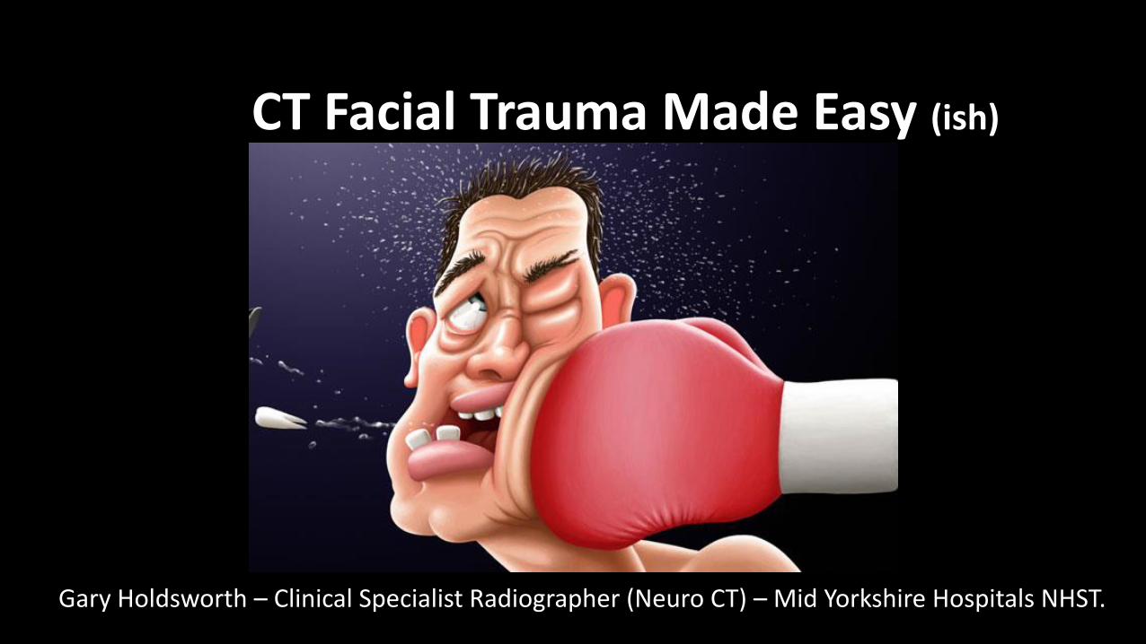
(Some) CT Facial Trauma Made Easy (ish)
122
Gary Holdsworth – Clinical Specialist Radiographer (Neuro CT) – Mid Yorkshire Hospitals NHST. (Some) CT Facial Trauma Made Easy (ish)
Transcript of (Some) CT Facial Trauma Made Easy (ish)
Gary Holdsworth – Clinical Specialist Radiographer (Neuro CT) – Mid Yorkshire Hospitals NHST.
(Some) CT Facial Trauma Made Easy (ish)

Facial fractures are commonly caused by blunt or penetrating trauma sustained during RTC, assaults, and falls.

Facial fractures are commonly caused by blunt or penetrating trauma sustained during RTC, assaults, and falls.
Right 28%, Left 36%, Midline 36%Bilateral fractures 19%One fracture pattern 52%Panfacial injury 1%
Mundinger et al. J Craniomaxillofac Surg 2014 (n=8127)

Facial Fractures
Upper Face: frontal, superior orbit 11%
Mundinger et al. J Craniomaxillofac Surg 2014

Facial Fractures
Upper Face: frontal, superior orbit
Mid Face: rest of orbit, nasal, zygoma, Le Fort, maxillary sinus, dentoalveolar, NOE, ZMC
11%
70%
Mundinger et al. J Craniomaxillofac Surg 2014

Facial Fractures
Upper Face: frontal, superior orbit
Mid Face: rest of orbit, nasal, zygoma, Le Fort, maxillary sinus, dentoalveolar, NOE, ZMC
Lower Face: mandible
11%
70%
19%Mundinger et al. J Craniomaxillofac Surg 2014

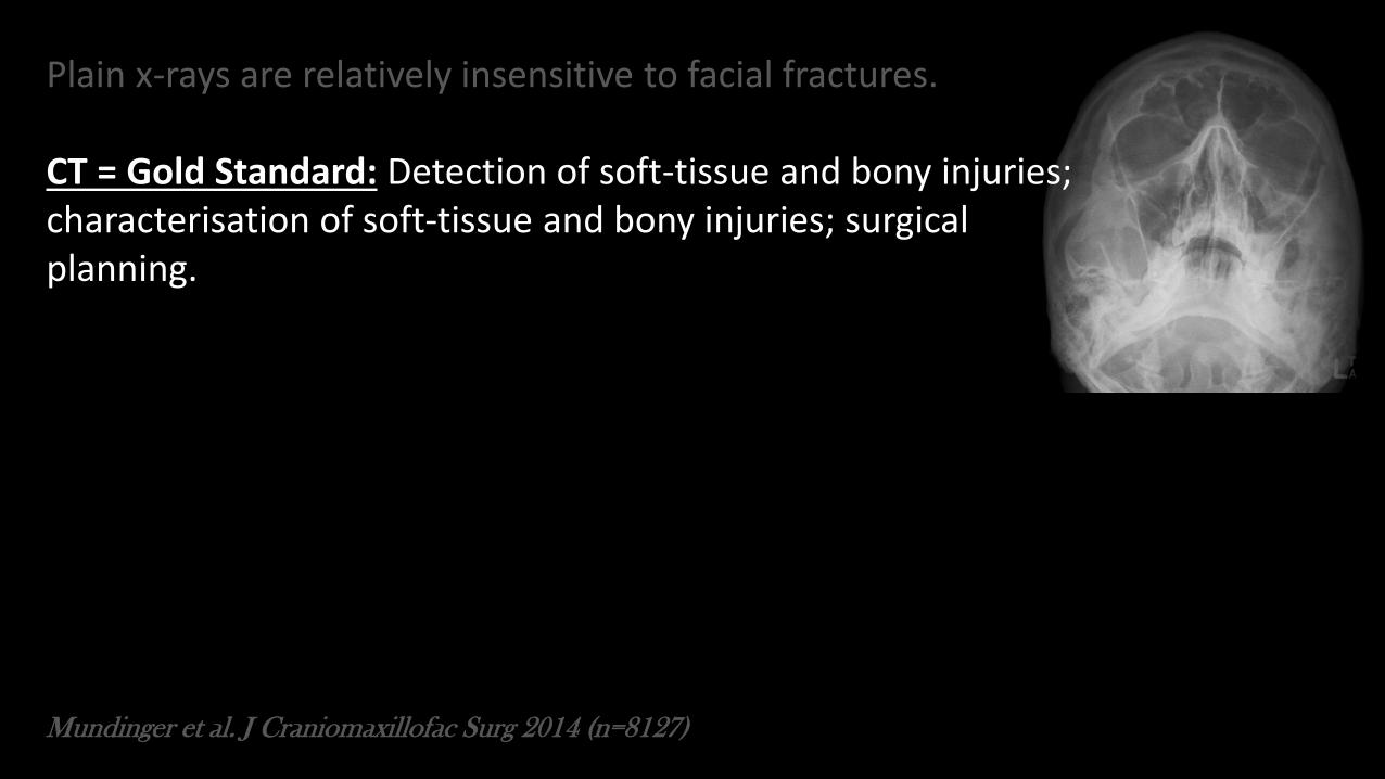
Plain x-rays are relatively insensitive to facial fractures.
Mundinger et al. J Craniomaxillofac Surg 2014 (n=8127)
Plain x-rays are relatively insensitive to facial fractures.
CT = Gold Standard: Detection of soft-tissue and bony injuries; characterisation of soft-tissue and bony injuries; surgical planning.
Mundinger et al. J Craniomaxillofac Surg 2014 (n=8127)

Plain x-rays are relatively insensitive to facial fractures.
CT = Gold Standard: Detection of soft-tissue and bony injuries; characterisation of soft-tissue and bony injuries; surgical planning.
With the high definition of CT even small fractures of the facial skeleton can be visualized. In complex midface injuries, it can be difficult to know which fractures are important to point out to the surgeon.
Mundinger et al. J Craniomaxillofac Surg 2014 (n=8127)
Plain x-rays are relatively insensitive to facial fractures.
CT = Gold Standard: Detection of soft-tissue and bony injuries; characterisation of soft-tissue and bony injuries; surgical planning.
With the high definition of CT even small fractures of the facial skeleton can be visualized. In complex midface injuries, it can be difficult to know which fractures are important to point out to the surgeon.
An understanding of the anatomically relevant and surgically accessible craniofacial buttresses is critical for management of these injuries.
Mundinger et al. J Craniomaxillofac Surg 2014 (n=8127)

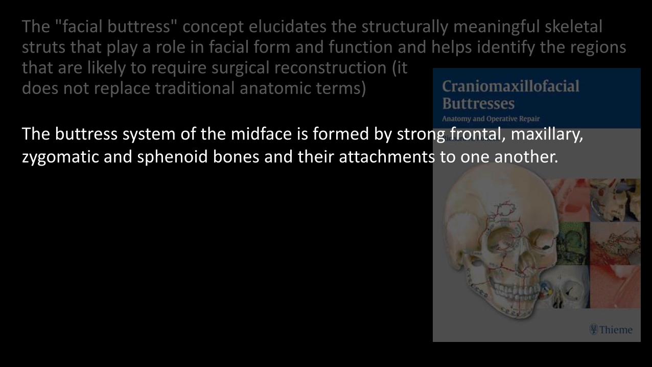
The "facial buttress" concept elucidates the structurally meaningful skeletal struts that play a role in facial form and function and helps identify the regions that are likely to require surgical reconstruction (it does not replace traditional anatomic terms)
The "facial buttress" concept elucidates the structurally meaningful skeletal struts that play a role in facial form and function and helps identify the regions that are likely to require surgical reconstruction (it does not replace traditional anatomic terms)
The buttress system of the midface is formed by strong frontal, maxillary, zygomatic and sphenoid bones and their attachments to one another.
Buttresses represent areas of relative increased bone thickness that support the functional units of the face (muscles, eyes, dental occlusion, airway) in an optimal relation; they define the form of the face and have sufficient bone thickness to accommodate metal screw fixation.
4 Vertical Buttresses (3 face; 1 mandible) 5* Horizontal Buttresses (3 face; 2 mandible)

The vertical facial buttresses
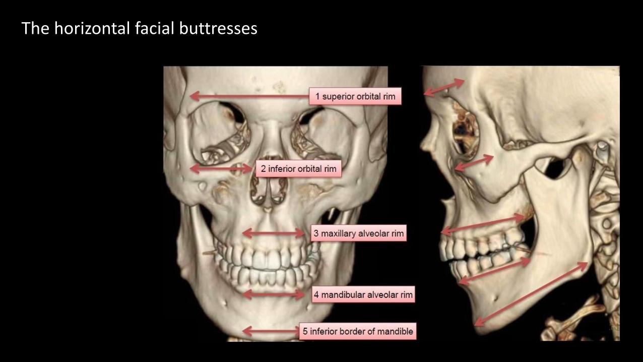
The horizontal facial buttresses

IMPORTANT!Do not get distracted by facial injuries when reporting cranial trauma:

IMPORTANT!Do not get distracted by facial injuries when reporting cranial trauma:
airway can be compromised

IMPORTANT!Do not get distracted by facial injuries when reporting cranial trauma:
airway can be compromisedconcomitant skull-base fracture in 8% of casesconcomitant c-spine fracture in 7%

IMPORTANT!Do not get distracted by facial injuries when reporting cranial trauma:
airway can be compromisedconcomitant skull-base fracture in 8% of casesconcomitant c-spine fracture in 7%
Airway OK?Focus upon Brain & Skull first.Check craniocervical junction.

IMPORTANT!Do not get distracted by facial injuries when reporting cranial trauma:
airway can be compromisedconcomitant skull-base fracture in 8% of casesconcomitant c-spine fracture in 7%
Airway OK?Focus upon Brain & Skull first.Check craniocervical junction.
Then move to the face …

CT ‘Clear Sinus Sign’: “Absence of paranasal sinus fluid after facial trauma is a highly reliable criterion to exclude acute fractures involving the paranasal sinus walls” Lambert DM et al. J Oral Maxillofac Sug 1997;55:1207-1210

CT ‘Clear Sinus Sign’: “Absence of paranasal sinus fluid after facial trauma is a highly reliable criterion to exclude acute fractures involving the paranasal sinus walls” Lambert DM et al. J Oral Maxillofac Sug 1997;55:1207
Critical Facial Injuries.
Airway compromise: Flail MandibleNasal septal haematoma
Vision: Retro-bulbar haemorrhageOrbital apex fractureGlobe injuries
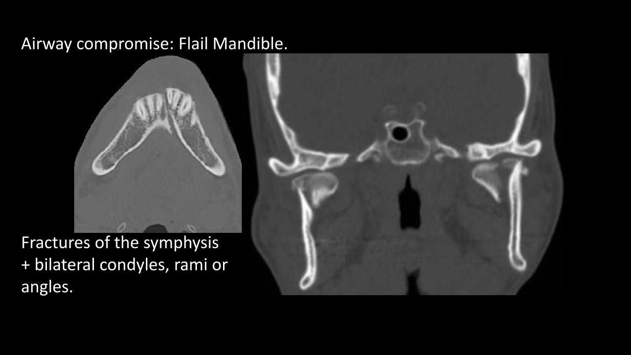
Airway compromise: Flail Mandible.
Fractures of the symphysis+ bilateral condyles, rami orangles.

Airway compromise: Flail Mandible.
Fractures of the symphysis+ bilateral condyles, rami orangles. Can potentiallycompromise airway; concomitant pharyngeal haematoma. Tongue position not maintained.

Airway compromise: Nasal Septal Haematoma.
Potentially compromise nasal airway; life-threatening epistaxis.

Airway compromise: Nasal Septal Haematoma.
Potentially compromise nasal airway; life-threatening epistaxis.
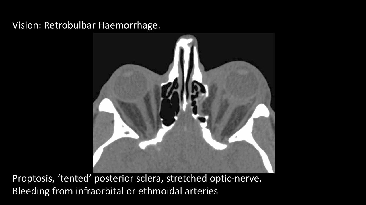
Vision: Retrobulbar Haemorrhage.
Proptosis, ‘tented’ posterior sclera, stretched optic-nerve. Bleeding from infraorbital or ethmoidal arteries

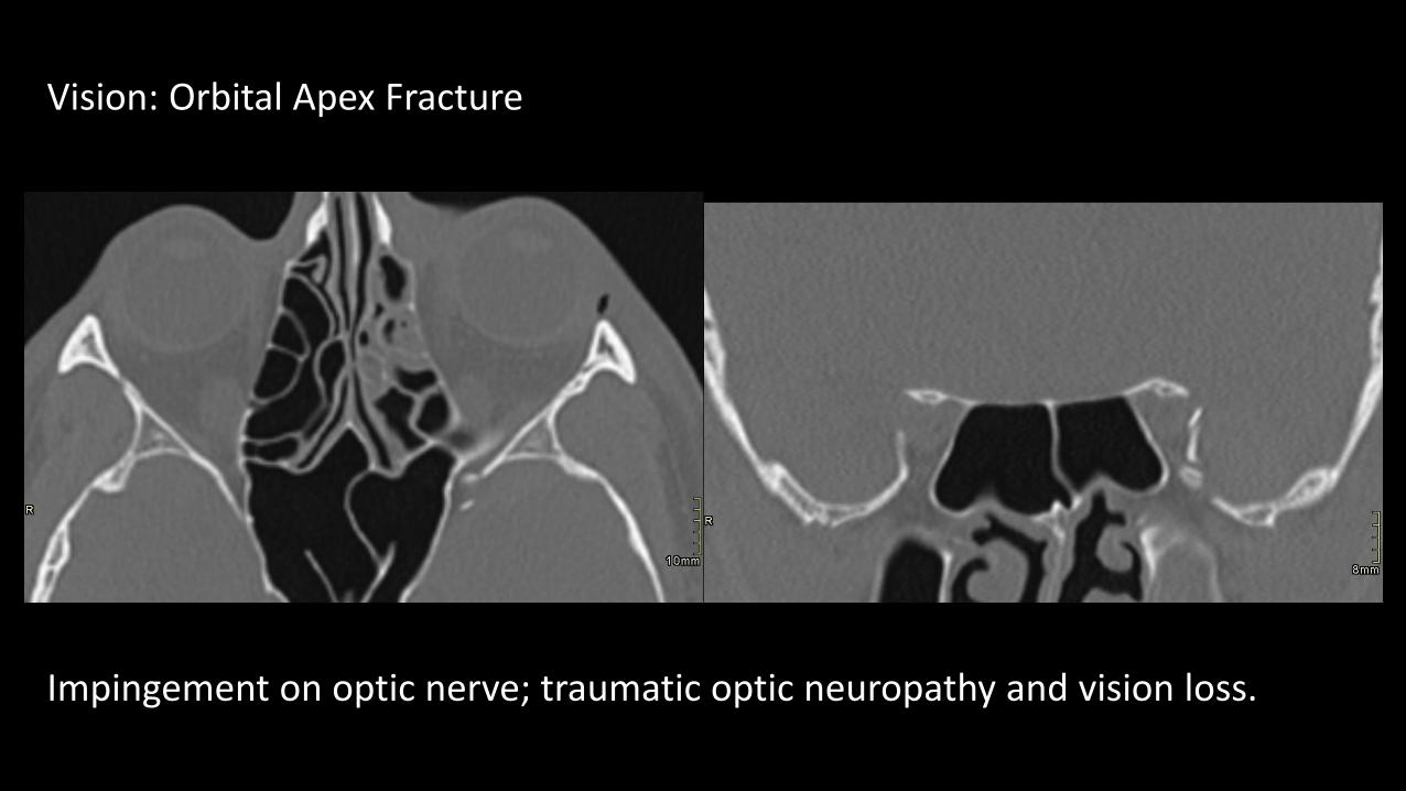
Vision: Orbital Apex Fracture
Impingement on optic nerve; traumatic optic neuropathy and vision loss.
Vision: Orbital Apex Fracture
Impingement on optic nerve; traumatic optic neuropathy and vision loss.
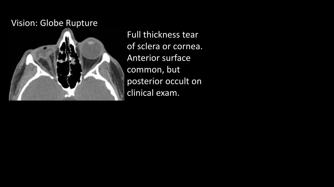
Vision: Globe RuptureFull thickness tearof sclera or cornea.Anterior surfacecommon, butposterior occult onclinical exam.

Vision: Globe RuptureFull thickness tearof sclera or cornea.Anterior surfacecommon, butposterior occult onclinical exam.
Intra-ocular FB’s? ‘Flat-tire’ signScleral discontinuity

Vision: Globe RuptureFull thickness tearof sclera or cornea.Anterior surfacecommon, butposterior occult onclinical exam.
Intra-ocular FB’s? ‘Flat-tire’ signScleral discontinuityIntra-ocular air

Vision: Globe RuptureFull thickness tearof sclera or cornea.Anterior surfacecommon, butposterior occult onclinical exam.
Intra-ocular FB’s? ‘Flat-tire’ signScleral discontinuityIntra-ocular air
Lens dislocation.Acute lens oedema(30 HU lower than normal side) = Traumatic Cataract
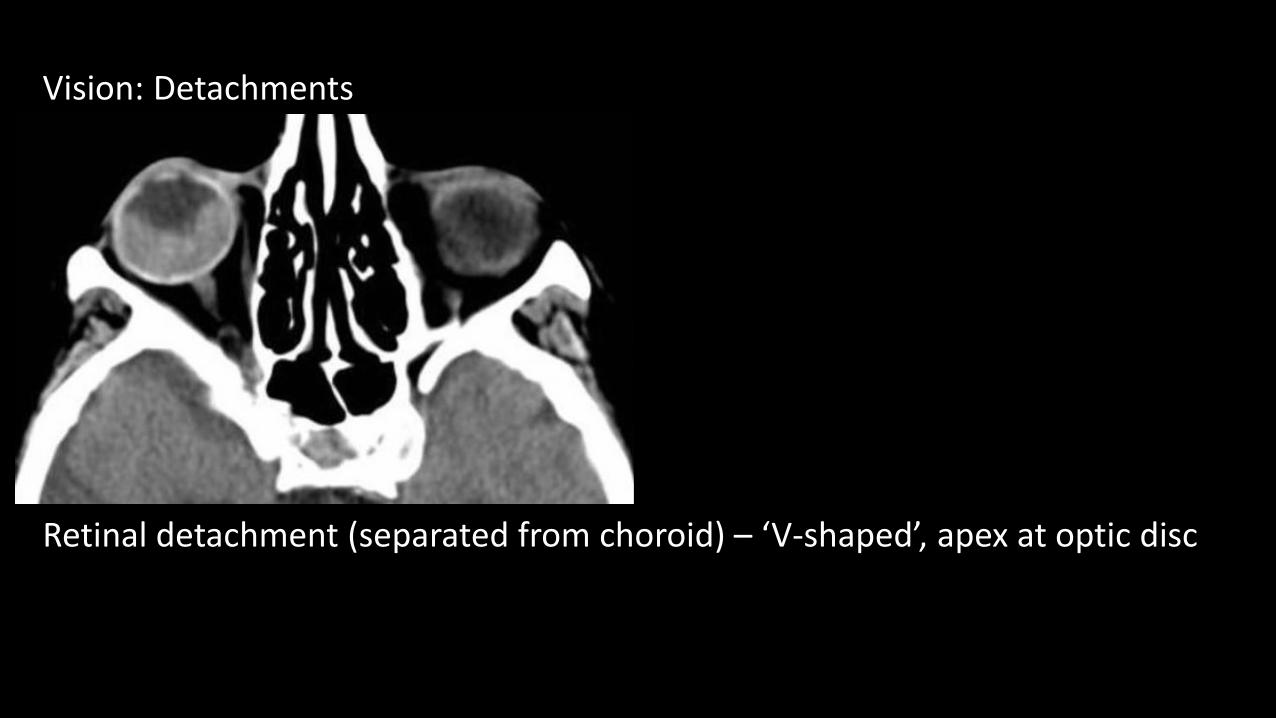
Vision: Detachments
Retinal detachment (separated from choroid) – ‘V-shaped’, apex at optic disc

Vision: Detachments
Retinal detachment (separated from choroid) – ‘V-shaped’, apex at optic discChoroidal detachment (separated from sclera) – ‘lens-shaped’

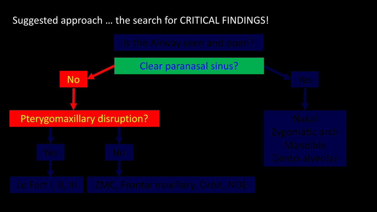
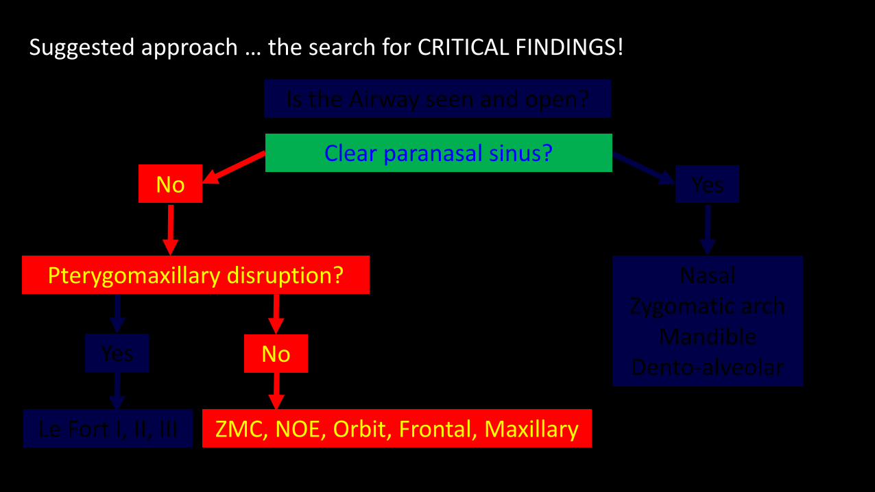
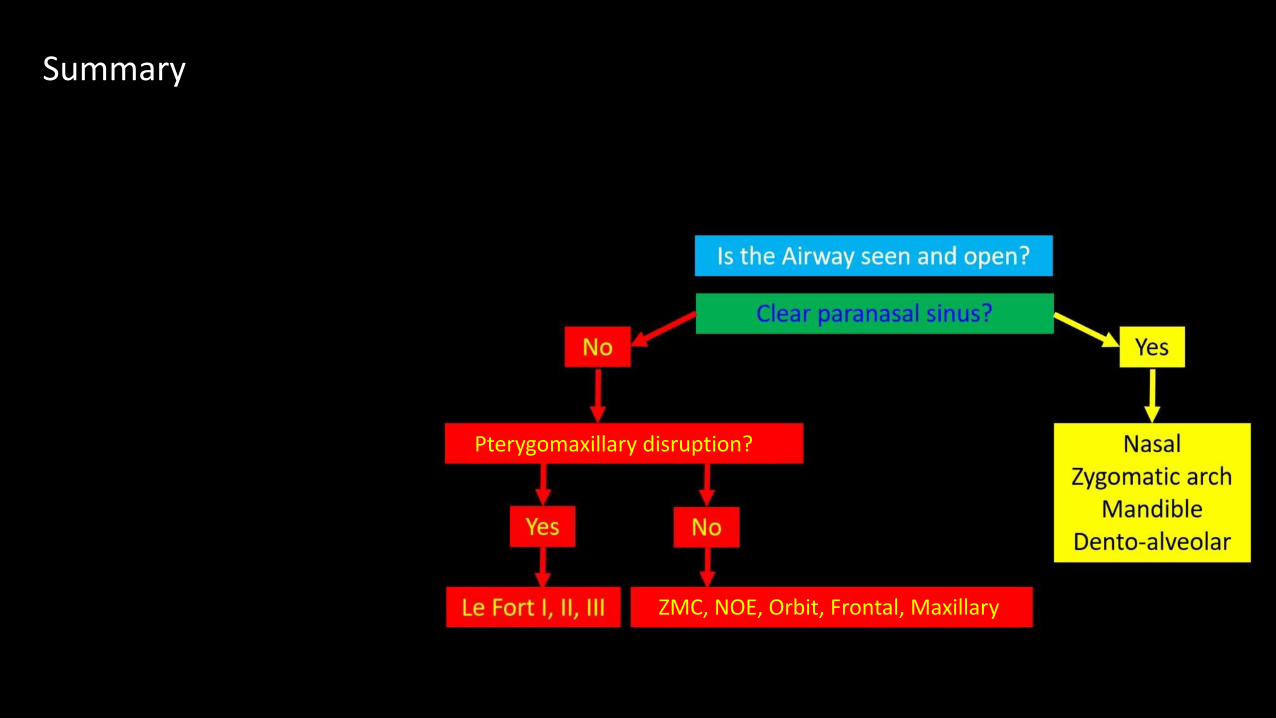
Suggested approach … the search for CRITICAL FINDINGS!
Is the Airway seen and open?
Clear paranasal sinus?No Yes
Pterygomaxillary disruption?
No
Le Fort I, II, III
Yes
ZMC, Frontal maxillary, Orbit, NOE
NasalZygomatic arch
MandibleDento-alveolar

Suggested approach … the search for CRITICAL FINDINGS!
Is the Airway seen and open?
No Yes
Pterygomaxillary disruption?
No
Le Fort I, II, III
Yes
ZMC, Frontal maxillary, Orbit, NOE
NasalZygomatic arch
MandibleDento-alveolar
Clear paranasal sinus?

Suggested approach … the search for CRITICAL FINDINGS!
Is the Airway seen and open?
No Yes
Pterygomaxillary disruption?
No
Le Fort I, II, III
Yes
ZMC, Frontal maxillary, Orbit, NOE
NasalZygomatic arch
MandibleDento-alveolar
Clear paranasal sinus?

Nasal FractureUnilateral vs bilateral, simple vs comminuted; if comminuted, telescoping or depression?Septum involved?Haematoma?

Nasal FractureUnilateral vs bilateral, simple vs comminuted; if comminuted, telescoping or depression?Septum involved?Haematoma?
Frontal Process of Maxilla FracturePart of a more complex fracture?

Nasal FractureUnilateral vs bilateral, simple vs comminuted; if comminuted, telescoping or depression?Septum involved?Haematoma?
Frontal Process of Maxilla FracturePart of a more complex fracture?
Zygomatic Arch FractureThree fracture lines,depressed middlefragment. Limit motionof mandible by impinging
on coronoid process or masseter origins

Dento-alveolar FractureAny portion of thealveolar process.Malaligned and displacedtooth.Tooth injuries: luxation, subluxation, avulsion, and fracture.
Suggested approach … the search for CRITICAL FINDINGS!
Is the Airway seen and open?
No Yes
NoYes
NasalZygomatic arch
MandibleDento-alveolar
Le Fort I, II, III ZMC, Frontal maxillary, Orbit, NOE
Clear paranasal sinus?
Pterygomaxillary disruption?

Suggested approach … the search for CRITICAL FINDINGS!
Is the Airway seen and open?
No Yes
Pterygomaxillary disruption?
No
Le Fort I, II, III
Yes
NasalZygomatic arch
MandibleDento-alveolar
ZMC, Frontal maxillary, Orbit, NOE
Clear paranasal sinus?

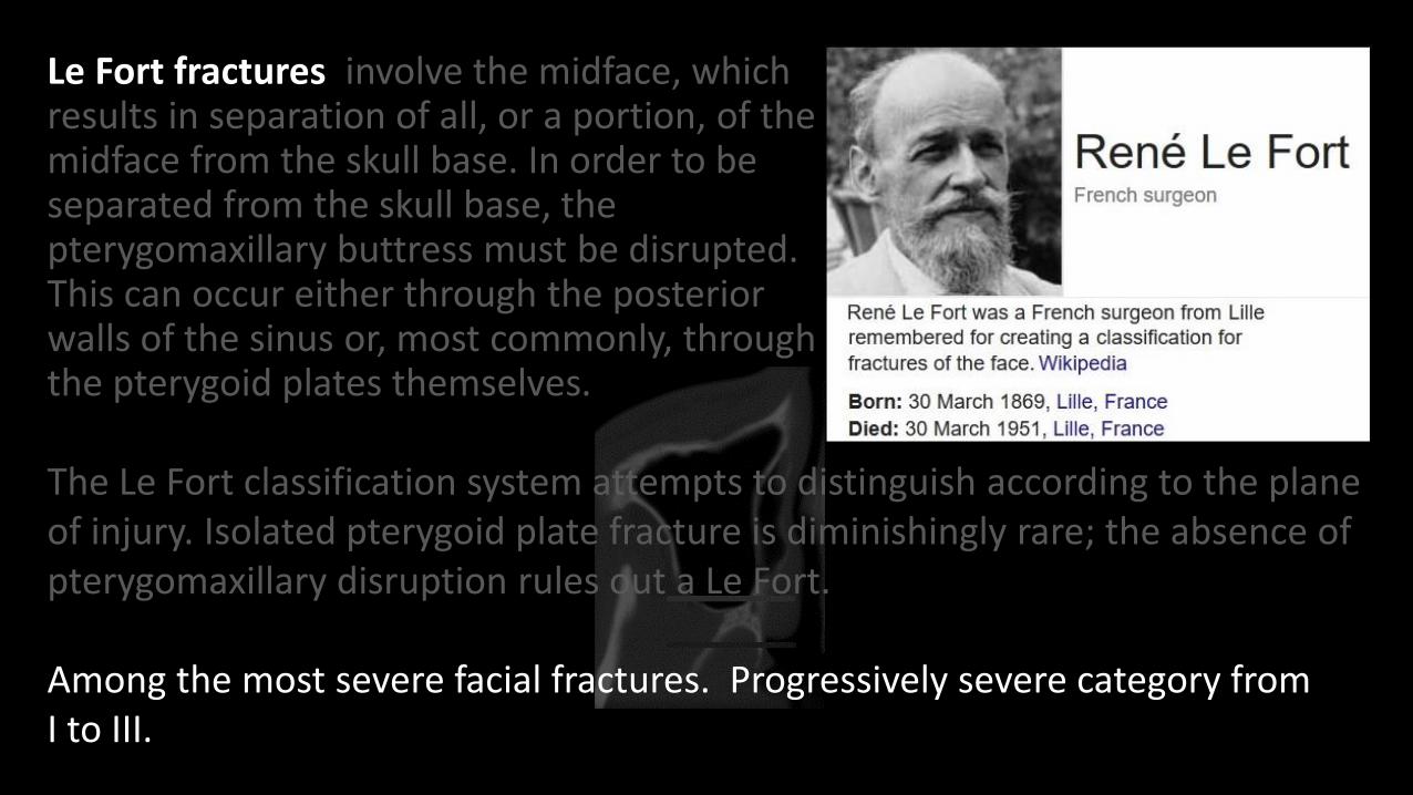
Le Fort fractures involve the midface, which results in separation of all, or a portion, of the midface from the skull base.
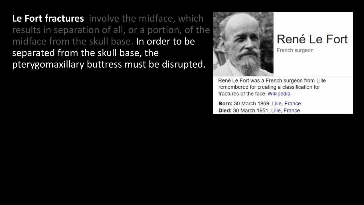
Le Fort fractures involve the midface, which results in separation of all, or a portion, of the midface from the skull base. In order to be separated from the skull base, the pterygomaxillary buttress must be disrupted.

Le Fort fractures involve the midface, which results in separation of all, or a portion, of the midface from the skull base. In order to be separated from the skull base, the pterygomaxillary buttress must be disrupted. This can occur either through the posterior walls of the sinus or, most commonly, through the pterygoid plates themselves.

Le Fort fractures involve the midface, which results in separation of all, or a portion, of the midface from the skull base. In order to be separated from the skull base, the pterygomaxillary buttress must be disrupted. This can occur either through the posterior walls of the sinus or, most commonly, through the pterygoid plates themselves.
The Le Fort classification system attempts to distinguish according to the plane of injury. Isolated pterygoid plate fracture is diminishingly rare; the absence of pterygomaxillary disruption rules out a Le Fort.
Among the most severe facial fractures. Progressively severe category from I to III.

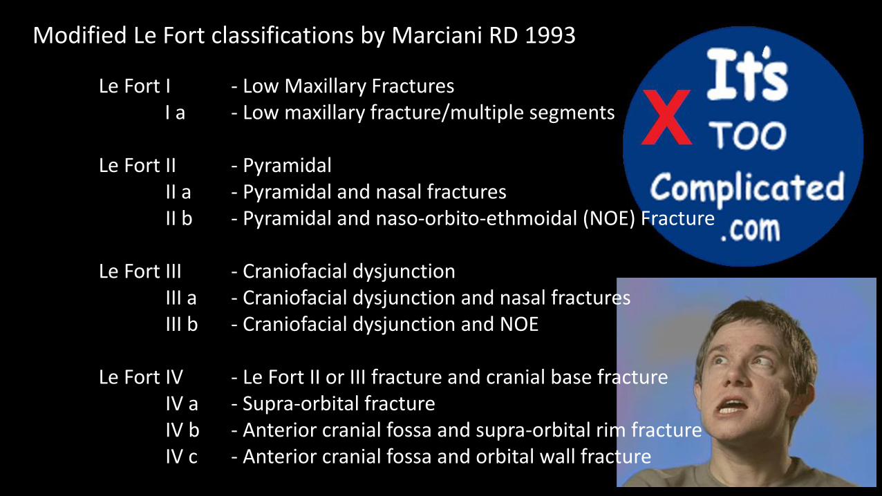
Modified Le Fort classifications by Marciani RD 1993
Le Fort I - Low Maxillary FracturesLe Fort I a - Low maxillary fracture/multiple segments
Le Fort II - PyramidalLe Fort II a - Pyramidal and nasal fracturesLe Fort II b - Pyramidal and naso-orbito-ethmoidal (NOE) Fracture
Le Fort III - Craniofacial dysjunctionLe Fort III a - Craniofacial dysjunction and nasal fracturesLe Fort III b - Craniofacial dysjunction and NOE
Le Fort IV - Le Fort II or III fracture and cranial base fractureLe Fort IV a - Supra-orbital fractureLe Fort IV b - Anterior cranial fossa and supra-orbital rim fractureLe Fort IV c - Anterior cranial fossa and orbital wall fracture

Le Fort fractures

Le Fort fractures
a floating palate a floating maxilla a floating face
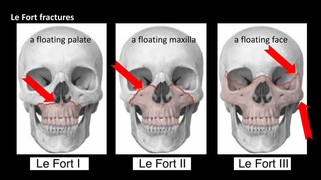
Le Fort fractures
a floating palate a floating maxilla a floating face

Le Fort fractures

Le Fort fractures

Le Fort fractures
The hard palate is an important posterior extension of the lower transverse buttress of the Maxilla (maxillary alveolar rim). A displaced unilateral Le Fort fracture is possible only if the palate is fractured sagittally or parasagittally.

Le Fort fractures
Le Fort fractures
Multiplanar CT: axial, coronal and sagittal images – need to work on all 3!

Le Fort fractures
Multiplanar CT: axial, coronal and sagittal images – need to work on all 3!
Is there a fracture of the pterygomaxillary buttress? Yes –> likely Le Fort
Le Fort fractures
Multiplanar CT: axial, coronal and sagittal images – need to work on all 3!
Is there a fracture of the pterygomaxillary buttress? Yes –> likely Le Fort
Is the anterolateral margin of the nasal fossa fractured? Yes –> Type 1 fracture

Le Fort fractures
Multiplanar CT: axial, coronal and sagittal images – need to work on all 3!
Is there a fracture of the pterygomaxillary buttress? Yes –> likely Le Fort
Is the anterolateral margin of the nasal fossa fractured? Yes –> Type 1 fracture
Is the infraorbital rim fractured? Yes –> Type 2 fracture
Is the lateral orbital wall and zygomatic arch fractured? Yes –> Type 3 fracture
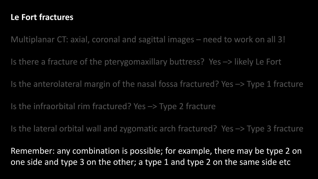
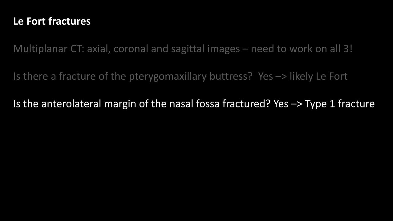
Remember: any combination is possible; for example, there may be type 2 on one side and type 3 on the other; a type 1 and type 2 on the same side etc

Le Fort fractures
Le Fort I – transverse fracture of inferior maxillae (all walls of the maxillary sinus except the superior wall/roof), anterolateral margins of the nasal fossa, nasal septum.
Le Fort fractures
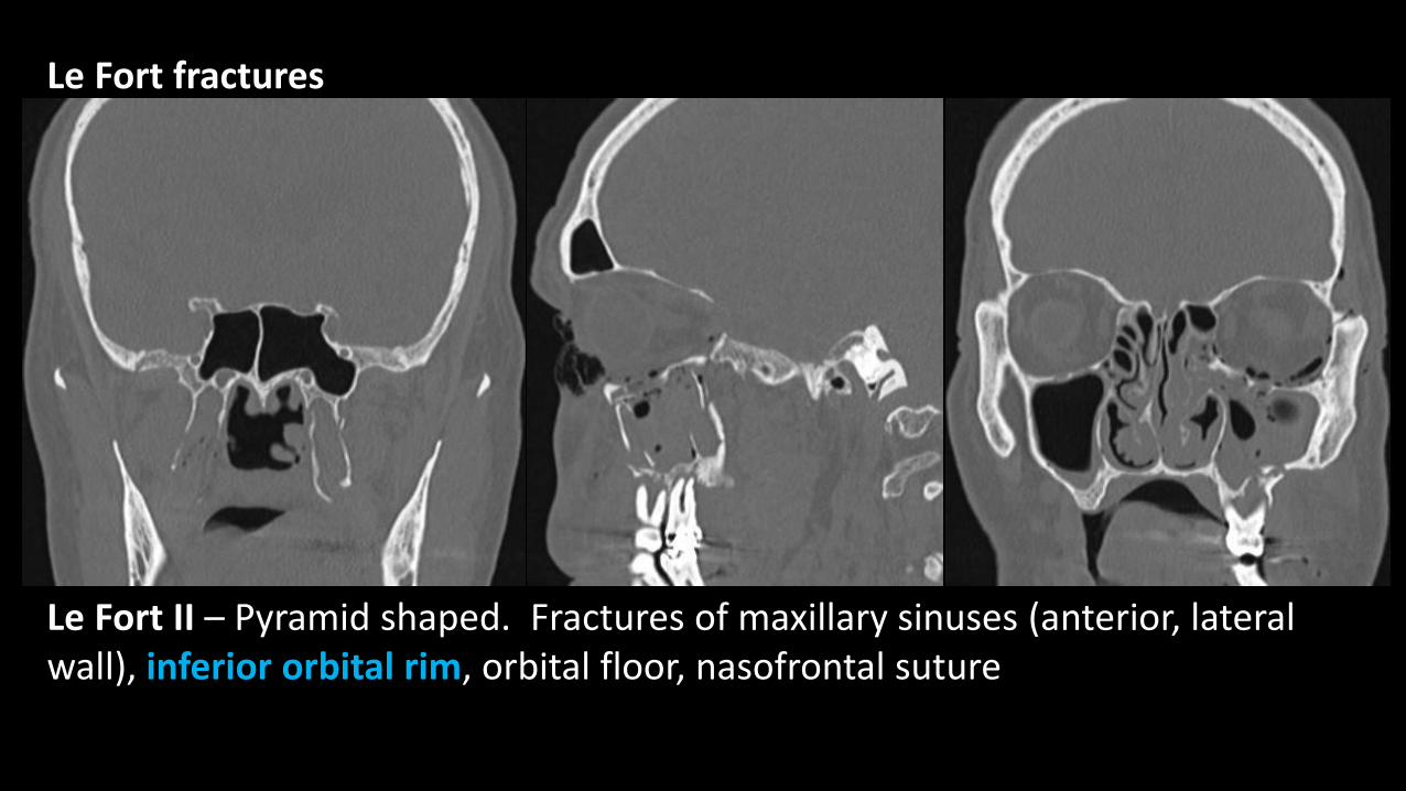
Le Fort II – Pyramid shaped. Fractures of maxillary sinuses (anterior, lateral wall), inferior orbital rim, orbital floor, nasofrontal suture

Le Fort fractures
Le Fort III – Fractures of the nasofrontal suture, maxillofrontal suture, lateral orbital wall and zygomatic arch/zygomaticofrontal suture

Le Fort fractures
Le Fort I, II & III

Le Fort fractures
Le Fort I, II & III
The hard palate is an important posterior extension of the lower transverse buttress of the Maxilla (maxillary alveolar rim). A displaced unilateral Le Fort fracture is possible only if the palate is fractured sagittally or parasagittally.
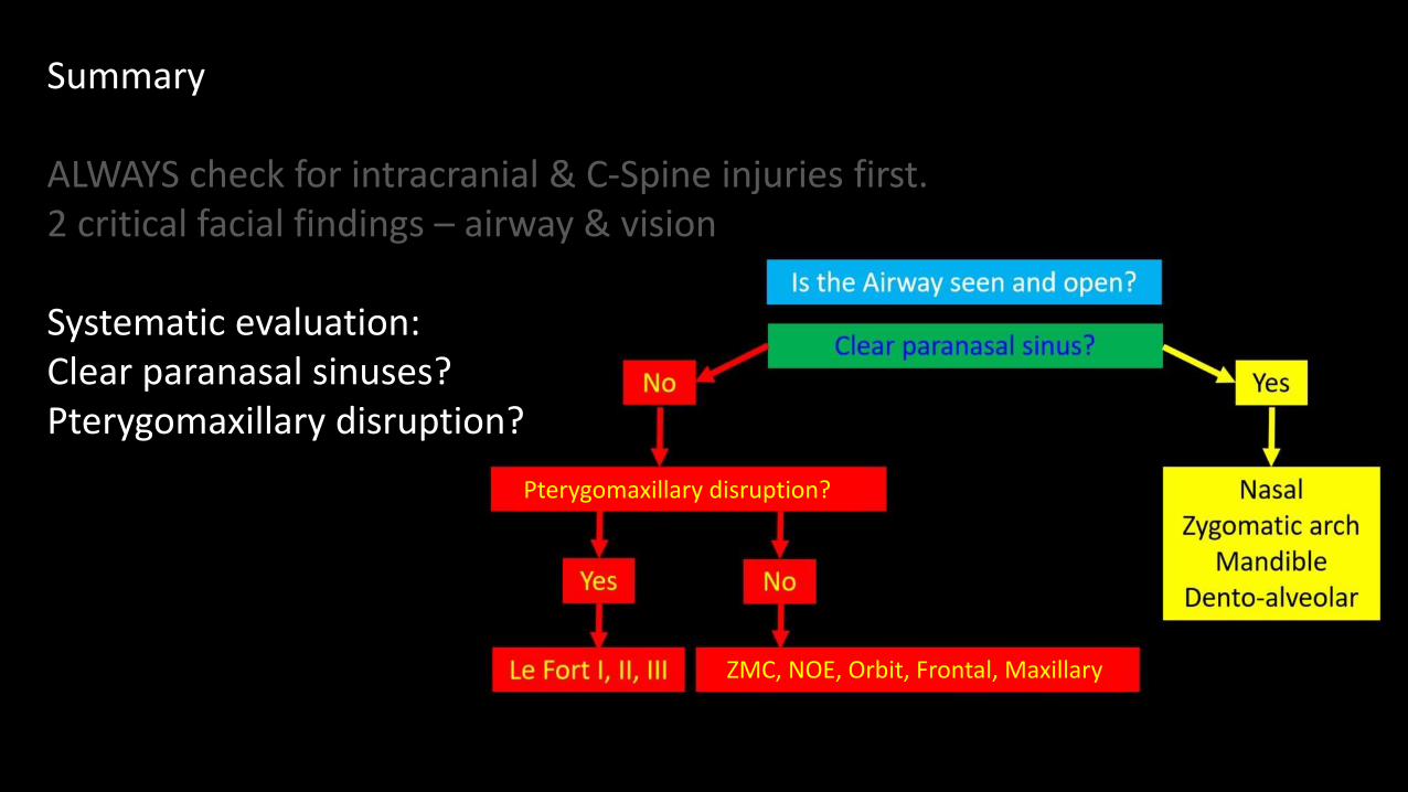
Suggested approach … the search for CRITICAL FINDINGS!
Is the Airway seen and open?
No Yes
No
Le Fort I, II, III
Yes
ZMC, NOE, Orbit, Frontal, Maxillary
NasalZygomatic arch
MandibleDento-alveolar
Clear paranasal sinus?
Pterygomaxillary disruption?
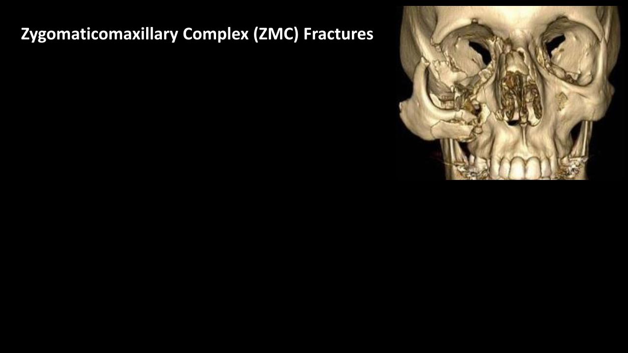
Zygomaticomaxillary Complex (ZMC) Fractures

Zygomaticomaxillary Complex (ZMC) Fractures
4 principle fracture lines: lateral orbital rim, zygomatic arch, zygomaticomaxillary buttress,inferior orbital rim.

Zygomaticomaxillary Complex (ZMC) Fractures
4 principle fracture lines: lateral orbital rim, zygomatic arch, zygomaticomaxillary buttress, inferior orbital rim. They are the 2nd most common facial bone fracture after the nasal bones, and are also known as a tripod, tetrapod, quadripod, malar or trimalar fracture.

Zygomaticomaxillary Complex (ZMC) Fractures
Results from a direct blow to the malar eminence with distinct fracture components that disrupt the anchoring of the zygoma.

Zygomaticomaxillary Complex (ZMC) Fractures
Results from a direct blow to the malar eminence with distinct fracture components that disrupt the anchoring of the zygoma. Additionally, the fracture components may result in impingement of the temporalis muscle = trismus;

Zygomaticomaxillary Complex (ZMC) Fractures
Results from a direct blow to the malar eminence with distinct fracture components that disrupt the anchoring of the zygoma. Additionally, the fracture components may result in impingement of the temporalis muscle = trismus; may compromise the infraorbital foramen &/or nerve resulting in hypo-aesthesia within its sensory distribution.
N.B. 2 x orbital rims are fractured: Orbital volume? Globe? Nerve? Extra ocular muscles? Orbital apex?

Naso-orbitoethmoid (NOE) fractures
Comminution of both naso-maxillary buttresses results in fractures involving the nasal bones and septum, ethmoid sinuses, and medial orbital walls.

Naso-orbitoethmoid (NOE) fractures (also known as orbito-ethmoid or naso-ethmoidal complex fractures) are fractures which involve the central upper midface.

Naso-orbitoethmoid (NOE) fractures (also known as orbito-ethmoid or naso-ethmoidal complex fractures) are fractures which involve the central upper midface.
NOE fractures are caused by a high-impact force applied anteriorly to the nose and transmitted posteriorly through the ethmoid bone.

Naso-orbitoethmoid (NOE) fractures (also known as orbito-ethmoid or naso-ethmoidal complex fractures) are fractures which involve the central upper midface.
NOE fractures are caused by a high-impact force applied anteriorly to the nose and transmitted posteriorly through the ethmoid bone.
Telecanthus secondary to medial canthal tendon injury (Markowitz & Manson
classification system = whether this tendon is disrupted or not) Reports should try to comment on the degree of comminution of the nasomaxillary buttress, specifically in the region of the lacrimal fossa, where the medial canthus attaches.

Naso-orbitoethmoid (NOE) fractures (also known as orbito-ethmoid or naso-ethmoidal complex fractures) are fractures which involve the central upper midface.
NOE fractures are caused by a high-impact force applied anteriorly to the nose and transmitted posteriorly through the ethmoid bone.
Telecanthus secondary to medial canthal tendon injury (Markowitz & Manson
classification system = whether this tendon is disrupted or not) Nasofrontal duct disruption and subsequent frontal mucocoele formation

Nasofrontal duct injury is suggested if base of frontal sinus is fractured &/or the anterior ethmoid complex. ?Fragments in the nasofrontal outflow tract.Surgical obliteration of the frontal sinus might be needed to prevent mucocele formation.
Ravinda VM et al. Surg Neurol Int
2015; 6:141
&
Harris L et al. Radiology 1987;
165:195

Naso-orbitoethmoid (NOE) fractures (also known as orbito-ethmoid or naso-ethmoidal complex fractures) are fractures which involve the central upper midface.
NOE fractures are caused by a high-impact force applied anteriorly to the nose and transmitted posteriorly through the ethmoid bone.
Telecanthus secondary to medial canthal tendon injury (Markowitz & Manson
classification system = whether this tendon is disrupted or not) Nasofrontal duct disruption and subsequent frontal mucocoele formationOrbital injuries and exophthalmos due to reduced intra-orbital volume

Naso-orbitoethmoid (NOE) fractures (also known as orbito-ethmoid or naso-ethmoidal complex fractures) are fractures which involve the central upper midface.
NOE fractures are caused by a high-impact force applied anteriorly to the nose and transmitted posteriorly through the ethmoid bone.
Telecanthus secondary to medial canthal tendon injury (Markowitz & Manson
classification system = whether this tendon is disrupted or not) Nasofrontal duct disruption and subsequent frontal mucocoele formationOrbital injuries and exophthalmos due to reduced intra-orbital volumeCSF rhinorrhoea due to fracture through the cribriform plate


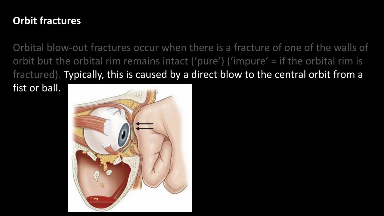
Orbit fractures
Orbital blow-out fractures occur when there is a fracture of one of the walls of orbit but the orbital rim remains intact (‘pure’) (‘impure’ = if the orbital rim is fractured).
Orbit fractures
Orbital blow-out fractures occur when there is a fracture of one of the walls of orbit but the orbital rim remains intact (‘pure’) (‘impure’ = if the orbital rim is fractured). Typically, this is caused by a direct blow to the central orbit from a fist or ball.

Orbit fractures
Orbital blow-out fractures occur when there is a fracture of one of the walls of orbit but the orbital rim remains intact (‘pure’) (‘impure’ = if the orbital rim is fractured). Typically, this is caused by a direct blow to the central orbit from a fist or ball.
Resulting from a sudden increase in the intra-orbital pressure which decompresses by fracturing one or more of the bounding walls of the orbit.
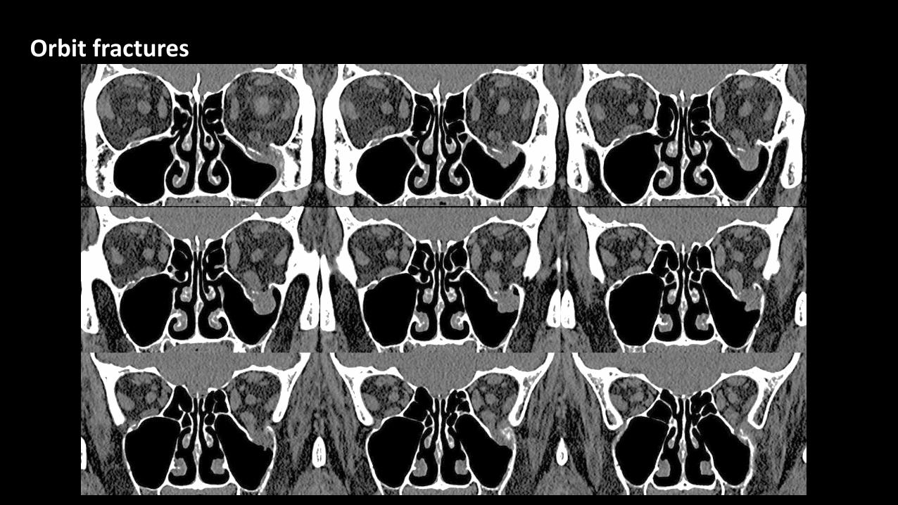
Blow-out fractures can occur through one or more of the walls of the orbit:Common: inferior (floor) > medial wall (lamina papyracea)Rare: superior (roof)Never?: lateral wall (?)

Orbit fractures
Inferior blow-out fractures are the most common. Orbital fat prolapses into the maxillary sinus and may be joined by prolapse of the inferior rectus muscle.

Orbit fractures
Inferior blow-out fractures are the most common. Orbital fat prolapses into the maxillary sinus and may be joined by prolapse of the inferior rectus muscle.
In the young, the fracture may spring back into place (known as a ‘trapdoor fracture’).
Inferior rectus ‘Missing’ inferior rectus
Yano H, Minagawa T, Masuda K & Hirano A. Urgent rescue of “missing rectus” in blowout fracture. Journal of
Plastic, Reconstructive & Aesthetic Surgery (JPRAS). 2009: 62 (9); 301-304

Orbit fractures
Inferior blow-out fractures are the most common. Orbital fat prolapses into the maxillary sinus and may be joined by prolapse of the inferior rectus muscle.
In the young, the fracture may spring back into place (known as a ‘trapdoor fracture’). Most fractures occur in the floor posterior and medial to the infraorbital groove.
In approximately 50% of cases, inferior blow-out fractures are associated with fractures of the medial wall
Inferior rectus ‘Missing’ inferior rectus
Yano H, Minagawa T, Masuda K & Hirano A. Urgent rescue of “missing rectus” in blowout fracture. Journal of
Plastic, Reconstructive & Aesthetic Surgery (JPRAS). 2009: 62 (9); 301-304

Orbit fractures
Inferior blow-out fractures

Orbit fractures
Medial blow-out fractures are the second most common type, occurring through the lamina papyracea. Orbital fat and the medial rectus muscle may prolapse into the ethmoid air cells.

Orbit fractures
Medial blow-out fractures are the second most common type, occurring through the lamina papyracea. Orbital fat and the medial rectus muscle may prolapse into the ethmoid air cells.

Orbit fractures
Pure superior blow-out fractures (i.e. those without an associated orbital rim fracture) are uncommon.

Orbit fractures
Pure superior blow-out fractures (i.e. those without an associated orbital rim fracture) are uncommon. They are usually seen in patients with pneumatisation of the orbital roof.

Orbit fractures
Pure superior blow-out fractures (i.e. those without an associated orbital rim fracture) are uncommon. They are usually seen in patients with pneumatisation of the orbital roof.
Fractures may only involve the sinus, the anterior cranial fossa (less common), or both sinus and anterior cranial fossa. In the latter, CSF leaks and meningitis may occur.
Very easily missed on axial images

Orbit fractures

Orbit fractures
In addition to evaluating the location and extent of the fracture, other features that need to be assessed and commented on include:

Orbit fractures
In addition to evaluating the location and extent of the fracture, other features that need to be assessed and commented on include:
presence of intra-orbital haemorrhage - may result in stretching or compression of the optic nerve

Orbit fractures
In addition to evaluating the location and extent of the fracture, other features that need to be assessed and commented on include:
presence of intra-orbital haemorrhage - may result in stretching or compression of the optic nerve
globe injury / rupture

Orbit fractures
In addition to evaluating the location and extent of the fracture, other features that need to be assessed and commented on include:
presence of intra-orbital haemorrhage - may result in stretching or compression of the optic nerve
globe injury / rupture
prolapse of orbital fat

Orbit fractures
In addition to evaluating the location and extent of the fracture, other features that need to be assessed and commented on include:
presence of intra-orbital haemorrhage - may result in stretching or compression of the optic nerve
globe injury / rupture
prolapse of orbital fat
extraocular muscle entrapment - suspected if there is a change in the shape &/or angle of the muscle
Orbit fractures

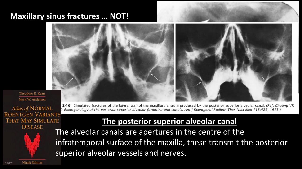
Maxillary sinus fractures … NOT!
The posterior superior alveolar canalThe alveolar canals are apertures in the centre of the infratemporal surface of the maxilla, these transmit the posterior superior alveolar vessels and nerves.
Maxillary sinus fractures … NOT!
The posterior superior alveolar canalThe alveolar canals are apertures in the centre of the infratemporal surface of the maxilla, these transmit the posterior superior alveolar vessels and nerves.

Maxillary sinus fractures … NOT!
Summary
Pterygomaxillary disruption?
ZMC, NOE, Orbit, Frontal, Maxillary

Summary
ALWAYS check for intracranial & C-Spine injuries first.2 critical facial findings – airway & vision
Pterygomaxillary disruption?
ZMC, NOE, Orbit, Frontal, Maxillary
Summary
ALWAYS check for intracranial & C-Spine injuries first.2 critical facial findings – airway & vision
Systematic evaluation:Clear paranasal sinuses?Pterygomaxillary disruption?
Pterygomaxillary disruption?
ZMC, NOE, Orbit, Frontal, Maxillary

Pterygomaxillary disruption?
Summary
ALWAYS check for intracranial & C-Spine injuries first.2 critical facial findings – airway & vision
Systematic evaluation:Clear paranasal sinuses?Pterygomaxillary disruption?
Try to fit all fractures into1 or 2 patterns (but don’tworry if you can’t)
Look for potential soft-tissue complications.
ZMC, NOE, Orbit, Frontal, Maxillary

The End?
Any Questions??
The End?
Any Questions??
Thank you for your attention.
Acknowledgements/References:
1) Hopper RA, Salemy S & Sze RW. Diagnosis of Midface fractures with CT: What the surgeon needs to know. Radiographics. May-June 2006.
2) https://radiopaedia.org/articles/facial-fractures; https://radiopaedia.org/articles/nasal-bone-fracture
3) https://radiopaedia.org/articles/le-fort-fracture-classification; https://radiopaedia.org/articles/zygomaticomaxillary-complex-fracture-1
4) https://radiopaedia.org/articles/naso-orbitoethmoid-noe-complex-fracture;
5) https://radiopaedia.org/articles/orbital-blow-out-fracture
6) Rathachai Kaewlai MD. Division of Emergency Radiology, Ramathibodi Hospital, Bangkok, Thailand. 25 Sep 2016
(available via: https://www.slideshare.net/TeleradiologySolutio/20160925-ser-drrathachai-kaewlai?qid=d60b52e2-a877-4014921c0ad7c3c605fb&v
=&b=&from_search=19)