Curcumin Attenuates Acute Graft-versus-Host … Attenuates Acute Graft-versus-Host Disease Severity...
10
Curcumin Attenuates Acute Graft-versus-Host Disease Severity via In Vivo Regulations on Th1, Th17 and Regulatory T Cells Min-Jung Park 1. , Su-Jin Moon 2. , Sung-Hee Lee 1 , Eun-Ji Yang 1 , Jun-Ki Min 2 , Seok-Goo Cho 3 , Chul- Woo Yang 4 , Sung-Hwan Park 1 , Ho-Youn Kim 1 , Mi-La Cho 1 * 1 The Rheumatism Research Center, Catholic Research Institute of Medical Science, The Catholic University of Korea, Seoul, South Korea, 2 Division of Rheumatology, Department of Internal Medicine, College of Medicine, The Catholic University of Korea, Seoul, South Korea, 3 Catholic Blood and Marrow Transplantation Center, College of Medicine, The Catholic University of Korea, Seoul, South Korea, 4 Transplant Research Center, Seoul St. Mary’s Hospital, College of Medicine, The Catholic University of Korea, Seoul, South Korea Abstract Background: In this study we examined the in vivo and in vitro effects and mechanisms of action of curcumin on the development of acute graft-versus-host disease (GVHD) using a murine model. Methodology/Principal Findings: Mixed lymphocyte reactions were used to determine the in vitro effects of curcumin. Treatment with curcumin attenuated alloreactive T cell proliferation and inhibited the production of interferon (IFN)-c and interleukin (IL)-17. In a murine acute GVHD model, transplantation of curcumin-treated allogeneic splenocytes into irradiated recipient mice significantly reduced the clinical severity scores of acute GVHD manifested in the liver, skin, colon and lung as compared with animals receiving vehicle-treated splenocytes. c-Fos and c-Jun expression levels in the skin and intestine, which are major target organs, were analyzed using immunohistochemical staining. Expression of both proteins was reduced in epithelial tissues of skin and intestine from curcumin-treated GVHD animals. The IFN-c-expressing CD4 + splenocytes and IFN-c-expressing lymph node cells were dramatically decreased in curcumin-treated mice. In contrast, CD4 + Foxp3 + splenocytes were increased in the curcumin-treated acute GVHD animals. Flow cytometric analysis revealed that animals transplanted with curcumin-treated allogeneic splenocytes showed increased populations of CD4 + regulatory T cells (Tregs) as well as CD8 + Treg cells, compared to animals administered vehicle-treated splenocytes. Curcumin-treated acute GVHD animals could have a change in B cell subpopulations. Conclusion/Significance: In the present study, we investigated the efficacy and mechanism of action of curcumin treatment against acute GVHD. The acute GVHD mice administered with curcumin-treated splenocytes showed significantly reduced severity of acute GVHD. Curcumin exerted in vivo preventive effects on acute GVHD by reciprocal regulation of T helper 1 (Th1) and Treg (both CD4 + and CD8 + Treg) cell lineages as well as B cell homeostasis. Citation: Park M-J, Moon S-J, Lee S-H, Yang E-J, Min J-K, et al. (2013) Curcumin Attenuates Acute Graft-versus-Host Disease Severity via In Vivo Regulations on Th1, Th17 and Regulatory T Cells. PLoS ONE 8(6): e67171. doi:10.1371/journal.pone.0067171 Editor: Markus M. Heimesaat, Charite ´, Campus Benjamin Franklin, Germany Received November 14, 2012; Accepted May 16, 2013; Published June 20, 2013 Copyright: ß 2013 Park et al. This is an open-access article distributed under the terms of the Creative Commons Attribution License, which permits unrestricted use, distribution, and reproduction in any medium, provided the original author and source are credited. Funding: This work was supported by a grant (A092258) from the Korea Healthcare Technology R&D Project, Ministry for Health, Welfare, and Family Affairs, Republic of Korea. The funders had no role in study design, data collection and analysis, decision to publish, or preparation of the manuscript. Competing Interests: The authors have declared that no competing interests exist * E-mail: [email protected] . These authors contributed equally to this work. Introduction Allogenic hematopoietic stem cell transplantation (HSCT) is the only curative therapy with proven efficacy for the management of many hematologic malignancies and other life-threatening hema- tological diseases. However, the development of graft-versus-host disease (GVHD), which is the main complication of HSCT, is a significant obstacle of allogenic HSCT [1]. Acute GVHD mainly affects the skin, gastrointestinal tract, liver, and lung. The development of GVHD requires escalated and prolonged immu- nosuppressive therapy with increased risk of infectious complica- tions. Ultimately, GVHD increases the risk of fatal morbidities and moralities in HSCT recipients. Although successive improvements in GVHD prevention have been achieved, complete protection from acute GVHD remains elusive. Acute GVHD (grades II–IV) occurs in 30–60% of patents after allogenic HSCT from human leukocyte antigen (HLA)-identical sibling donors [2]. Following the development of GVHD, complete remission has been observed in only 30 to 50% of patients with acute GVHD [3,4]. Knowledge of the immunobiology underlying GVHD has advanced by virtue of immunology research in animal models, as well as clinical observations. GVHD occurs as a result of T cell activation followed by alloreactive T cell expansion and differen- tiation [5]. Acute GVHD is considered a process driven mainly by T helper 1 (Th1) and Th17 type immune responses. Th1 cell- associated cytokines involved in acute GVHD include interferon PLOS ONE | www.plosone.org 1 June 2013 | Volume 8 | Issue 6 | e67171
Transcript of Curcumin Attenuates Acute Graft-versus-Host … Attenuates Acute Graft-versus-Host Disease Severity...

Curcumin Attenuates Acute Graft-versus-Host DiseaseSeverity via In Vivo Regulations on Th1, Th17 andRegulatory T CellsMin-Jung Park1., Su-Jin Moon2., Sung-Hee Lee1, Eun-Ji Yang1, Jun-Ki Min2, Seok-Goo Cho3, Chul-
Woo Yang4, Sung-Hwan Park1, Ho-Youn Kim1, Mi-La Cho1*
1 The Rheumatism Research Center, Catholic Research Institute of Medical Science, The Catholic University of Korea, Seoul, South Korea, 2 Division of Rheumatology,
Department of Internal Medicine, College of Medicine, The Catholic University of Korea, Seoul, South Korea, 3 Catholic Blood and Marrow Transplantation Center, College
of Medicine, The Catholic University of Korea, Seoul, South Korea, 4 Transplant Research Center, Seoul St. Mary’s Hospital, College of Medicine, The Catholic University of
Korea, Seoul, South Korea
Abstract
Background: In this study we examined the in vivo and in vitro effects and mechanisms of action of curcumin on thedevelopment of acute graft-versus-host disease (GVHD) using a murine model.
Methodology/Principal Findings: Mixed lymphocyte reactions were used to determine the in vitro effects of curcumin.Treatment with curcumin attenuated alloreactive T cell proliferation and inhibited the production of interferon (IFN)-c andinterleukin (IL)-17. In a murine acute GVHD model, transplantation of curcumin-treated allogeneic splenocytes intoirradiated recipient mice significantly reduced the clinical severity scores of acute GVHD manifested in the liver, skin, colonand lung as compared with animals receiving vehicle-treated splenocytes. c-Fos and c-Jun expression levels in the skin andintestine, which are major target organs, were analyzed using immunohistochemical staining. Expression of both proteinswas reduced in epithelial tissues of skin and intestine from curcumin-treated GVHD animals. The IFN-c-expressing CD4+
splenocytes and IFN-c-expressing lymph node cells were dramatically decreased in curcumin-treated mice. In contrast,CD4+Foxp3+ splenocytes were increased in the curcumin-treated acute GVHD animals. Flow cytometric analysis revealedthat animals transplanted with curcumin-treated allogeneic splenocytes showed increased populations of CD4+ regulatory Tcells (Tregs) as well as CD8+ Treg cells, compared to animals administered vehicle-treated splenocytes. Curcumin-treatedacute GVHD animals could have a change in B cell subpopulations.
Conclusion/Significance: In the present study, we investigated the efficacy and mechanism of action of curcumin treatmentagainst acute GVHD. The acute GVHD mice administered with curcumin-treated splenocytes showed significantly reducedseverity of acute GVHD. Curcumin exerted in vivo preventive effects on acute GVHD by reciprocal regulation of T helper 1(Th1) and Treg (both CD4+ and CD8+ Treg) cell lineages as well as B cell homeostasis.
Citation: Park M-J, Moon S-J, Lee S-H, Yang E-J, Min J-K, et al. (2013) Curcumin Attenuates Acute Graft-versus-Host Disease Severity via In Vivo Regulations onTh1, Th17 and Regulatory T Cells. PLoS ONE 8(6): e67171. doi:10.1371/journal.pone.0067171
Editor: Markus M. Heimesaat, Charite, Campus Benjamin Franklin, Germany
Received November 14, 2012; Accepted May 16, 2013; Published June 20, 2013
Copyright: � 2013 Park et al. This is an open-access article distributed under the terms of the Creative Commons Attribution License, which permits unrestricteduse, distribution, and reproduction in any medium, provided the original author and source are credited.
Funding: This work was supported by a grant (A092258) from the Korea Healthcare Technology R&D Project, Ministry for Health, Welfare, and Family Affairs,Republic of Korea. The funders had no role in study design, data collection and analysis, decision to publish, or preparation of the manuscript.
Competing Interests: The authors have declared that no competing interests exist
* E-mail: [email protected]
. These authors contributed equally to this work.
Introduction
Allogenic hematopoietic stem cell transplantation (HSCT) is the
only curative therapy with proven efficacy for the management of
many hematologic malignancies and other life-threatening hema-
tological diseases. However, the development of graft-versus-host
disease (GVHD), which is the main complication of HSCT, is a
significant obstacle of allogenic HSCT [1]. Acute GVHD mainly
affects the skin, gastrointestinal tract, liver, and lung. The
development of GVHD requires escalated and prolonged immu-
nosuppressive therapy with increased risk of infectious complica-
tions. Ultimately, GVHD increases the risk of fatal morbidities and
moralities in HSCT recipients. Although successive improvements
in GVHD prevention have been achieved, complete protection
from acute GVHD remains elusive. Acute GVHD (grades II–IV)
occurs in 30–60% of patents after allogenic HSCT from human
leukocyte antigen (HLA)-identical sibling donors [2]. Following
the development of GVHD, complete remission has been
observed in only 30 to 50% of patients with acute GVHD [3,4].
Knowledge of the immunobiology underlying GVHD has
advanced by virtue of immunology research in animal models, as
well as clinical observations. GVHD occurs as a result of T cell
activation followed by alloreactive T cell expansion and differen-
tiation [5]. Acute GVHD is considered a process driven mainly by
T helper 1 (Th1) and Th17 type immune responses. Th1 cell-
associated cytokines involved in acute GVHD include interferon
PLOS ONE | www.plosone.org 1 June 2013 | Volume 8 | Issue 6 | e67171

(IFN)-c, interleukin (IL)-1, IL-6, and tumor necrosis factor (TNF)-
a [6,7]. Th17 cells are IL-17 producing T helper cells that are a
lineage of CD4+ effector T cells distinct from the Th1 and Th2 cell
lineages. Th17 cells were found to have a direct role in the
development of GVHD [8]. Adoptive transfer of in vitro-differen-
tiated Th17 cells is capable of inducing lethal acute GVHD [9].
Acting opposite of Th1 and Th17 cells, there are regulatory T
(Treg) cells. Several observational studies have shown that a
decreased population of circulating Treg cells was observed in
patients that developed acute GVHD, compared to those without
acute GVHD [10,11]. Parallel to those findings, Treg cell
expansion has been shown to be capable of reducing the severity
of acute GVHD in murine acute GVHD models [12,13].
Although there has been great advances in understanding the
pathophysiology of GVHD, current GVHD prophylaxis and
treatment are still based on non-specific immunosuppressive drug
therapy [14].
Curcumin is a naturally occurring polyphenolic phytochemical
that is derived from the root of the turmeric plant Curcuna longa and
is responsible for the spicy taste of curries [15]. It is a nontoxic
food that is used as a coloring agent and remains a vital ingredient
of traditional medicine in India and China. More recently,
antioxidant, anti-inflammatory, anti-microbial, and anti-carcino-
genic properties of curcumin have been identified [16–20]. A small
number of experiments revealed that curcumin treatment
attenuated autoimmune diseases by downregulating IL-17 pro-
duction [21] and shifting from Th1 to Th2 type responses [22].
However, its application in GVHD models has not been tested to
date.
In the present study, we investigated the in vivo effect of
curcumin in a murine model of acute GVHD. The acute GVHD
model was developed by bone marrow transplantation, supple-
mented with varying numbers and different types of donor
lymphocytes, into irradiated allogenic recipients that differ from
the donors by major histocompatibility complex (MHC) class.
Materials and Methods
MiceC57BL/6 (B6; H-2kb), and BALB/c (H-2kd) mice, 8–10 weeks
old, were purchased from OrientBio (Sungnam, Korea). The mice
were maintained under specific pathogen-free conditions in an
animal facility with controlled humidity (5565%), light (12 h/
12 h light/dark), and temperature (2261uC). The air in the facility
was passed through a HEPA filter system designed to exclude
bacteria and viruses. Animals were fed mouse chow and tap water
ad libitum. The protocols used in this study were approved by the
Animal Care and Use Committee of The Catholic University of
Korea.
BMT Model and Histopathology ScoringRecipients (BALB/c) mice were injected intraveneously (i.v.)
with 56106 total bone marrow cells from donor mice after lethal
irradiation with 800 cGy. To induce acute GVHD, splenocytes
were isolated from donor mice. Splenocytes (16107) from MHC
major and minor antigen-disparate B6 donors were incubated with
20 nM cucurmin or control vehicle (DMSO) for 1 h at 37uCbefore adoptive transfer into recipient mice. To create GVHD–
negative controls, 56106 T cell–depleted (TCD) bone marrow
cells were transplanted into irradiated BALB/c recipient mice. All
experiments were performed at least three times with six mice per
group. Survival after BMT was monitored daily, and the degree of
clinical GVHD was assessed using a scoring system that summed
changes in five clinical parameters: weight loss, posture, activity, fir
texture, and skin integrity. Mice were sacrificed on day 35 after
BMT for blinded histopathological analysis of GVHD target
tissues (skin, liver, and small and large intestine) [23]. Organs were
harvested, cryo-embedded, and subsequently sectioned. Tissue
sections were fixed in 10% buffered formalin and stained with
hematoxylin and eosin (H&E) for histological examination.
MLR Culture in vitroSplenocytes derived from BALB/c mice were used as stimulator
cells and those from B6 were used as responder cells in MLR
assays. Spleen cells were removed using ammonium-chloride-
potassium lysis buffer, washed, and resuspended in complete
culture medium (RPMI 1640 medium supplemented with 10%
heat-inactivated fetal calf serum, 1 mM sodium pyruvate,
56105 M 2-ME, 20 mM HEPES, and antibiotics [100 U/mL
penicillin, 100 ug/mL streptomycin]). Aliquots of 26105 CD4+ T
cells (responders) were cultured with 26105 irradiated (2500 cGy)
antigen-presenting cells (APC) in 96-well plates containing 200
of complete medium at 37uC in a humidified 5% CO2
atmosphere, pulsed with 1 mCi of [3H]-TdR (NEN Life Science
Products Inc., Boston, MA, USA) for 18 h before harvesting, and
counted using an automated harvester (PHD Cell Harvester;
Cambridge Technology, Inc., Cambridge, MA, USA). Results are
expressed as the mean counts per minute (cpm) of triplicate
samples 6 standard deviation (SD). The stimulation index was
calculated by comparing the anti-stimulator response with the
anti-self response.
Flow CytometryMononuclear cells were immunostained with various combina-
tions of fluorescence-conjugated antibodies against CD4, CD25,
Foxp3, IFN-c, IL-4, IL-17, B220, IgD, IgM, CD95, GL-7, and
streptavidin. These cells were also intracellularly stained with
antibodies against IL-4 (BD Biosciences, San Jose, CA, USA), IL-
10 (Biolegend, San Diego, CA, USA), IL-17, and Foxp3
(eBioscience, San Diego, CA, USA). Before intracellular staining,
the cells were restimulated for 4 h with 25 ng/mL phorbol 12-
myristate 13-acetate (PMA) and 250 ng/mL ionomycin in the
presence of GolgiSTOP (BD Biosciences). Intracellular staining
was conducted using an intracellular staining kit (eBioscience)
according to the manufacturer’s protocol. Flow cytometric analysis
was performed using a FACSCalibur cytometer (BD Biosciences).
Measurement of Cytokines and IgG TitersThe concentrations of IFN-c and IL-17 in cell culture
supernatants and serum were measured using a sandwich ELISA
(Duoset; R&D Systems, Lille, France). Serum levels of IgG, IgG1
and IgG2a antibodies were measured using a commercially
available ELISA kit (Bethyl Laboratories, Montgomery, TX,
USA).
ImmunohistochemistryImmunohistochemistry was performed using the Vectastain
ABC kit (Vector Laboratories, Burlingame, CA, USA). Tissues
were first incubated with the primary anti-C-jun and anti-C-fos
antibodies overnight at 4uC. The primary antibody was detected
with a biotinylated secondary linking antibody, followed by
incubation with a streptavidin–peroxidase complex for 1 h. The
final color product was developed using DAB chromogen (DAKO,
Carpinteria, CA, USA).
Therapeutic Efficacy of Curcumin in Acute GVHD
PLOS ONE | www.plosone.org 2 June 2013 | Volume 8 | Issue 6 | e67171

Confocal StainingSpleen tissue was obtained 14 days after BMT and was snap-
frozen in liquid nitrogen and stored at 280uC. Tissue cryosections
(7-mm thick) were fixed in 4% paraformaldehyde and stained using
PE-labeled anti-IFN-c, IL-4, IL-17, or Foxp3 antibody
(eBioscience) and FITC-labeled anti-CD4 antibody and APC-
labeled anti CD25 antibody (Biolegend). After incubation over-
night at 4uC, stained sections were analyzed using a confocal
microscope (LSM 510 Meta; Zeiss, Gottingen, Germany).
CD4+IFN-c+, CD4+IL-4+, CD4+IL-17+, CD4+CD25+Foxp3+ T
cells were enumerated visually at higher magnification (projected
on a screen) by four individuals.
Statistical AnalysisComparison of numerical data between three groups was
performed with nonparametric Mann-Whitney tests. Statistical
analysis was performed using SPSS 10.0 for Windows (SPSS,
Chicago, IL, USA). P values ,0.05 were considered significant.
Data are presented as the mean 6 SD.
Results
Curcumin Modulates Alloreative T Cell Responses in vitroTo assess the effects of curcumin on the proliferative capacity of
donor CD4+ T cells in response to alloantigens following
transplantation, T cell alloreactivity was measured by [3H]-
thymidine incorporation to assess T cell proliferation in mixed
lymphocyte reactions (MLR). Proliferative responses to BALB/c T
cells (allogeneic stimulator) was observed in C57BL/6 (B6) T cells
(responder cells). Treatment with curcumin inhibited T cell
alloreactivity in a dose-dependent manner (Fig. 1A). To ascertain
whether or not the inhibitory effect on alloreactive T cell responses
by curcumin was associated with the apoptosis induction.
Annexin-V and propidium iodide (PI) double staining were
performed and analyzed using flow cytometery (Fig. S1B). In
addition, we performed MTT assays to determine whether
curcumin treatment affects cell viability (Fig. S1A). The results
indicated that the treatment with curcumin and DSMO at various
concentrations did not induce apoptosis or affect cell viability.
IFN-c and IL-17 concentrations were then measured using an
enzyme-linked immunosorbent assay (ELISA). Curcumin treat-
ment significantly reduced IFN-c and IL-17 level in culture
supernatants (Fig. 1B). To determine whether in vitro curcumin
treatment can regulate the Th1 and Th2 balance, B6 splenic T
cells incubated with irradiated B6 splenic T cells (syngeneic
stimulator) or BALB/c splenic T cells (allogeneic stimulator) in the
absence of presence of curcumin (2.5 mM) were analyzed by
intracellular staining for IL-4, IFN-c, IL-17, and Foxp3 (Fig. 1C).
While the Th1 cell population (IFN-c+IL-42 T cells) tended to
decrease upon the curcumin treatment, the Th2 cell population
(IFN-c2IL-4+ T cells) tended to increase (statistically insignificant).
Similarly, curcumin treatment generated reciprocal changes in
Th17 and Treg cell differentiation, although the changes were not
statistically significant (Fig. 1C).
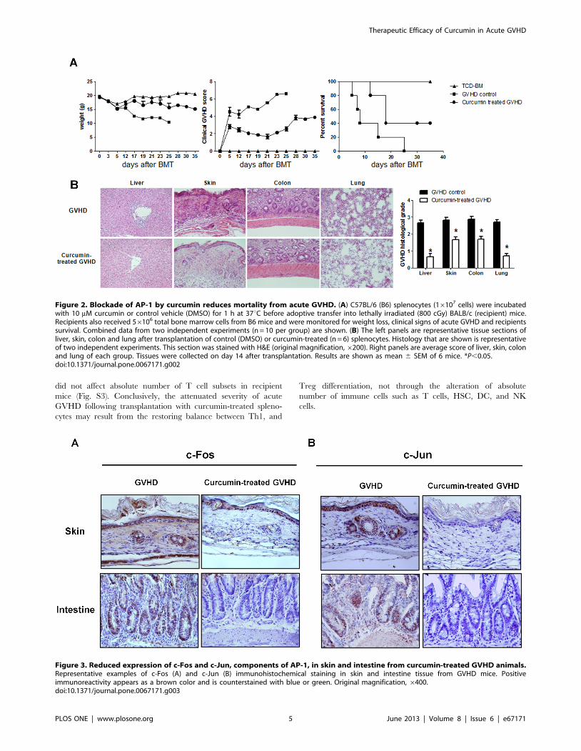
Transfer of Curcumin-treated Splenocytes Protects Micefrom Acute GVHD after Bone Marrow Transplant
Because curcumin significantly suppressed alloreactivity in vitro,
we next assessed its effects in vivo using a murine model of acute
GVHD. Severe acute GVHD occurred in all BALB/c (H-2kd)
recipient mice undergoing bone marrow transplantation (BMT)
and infusion of donor (B6 mice, H-2kb) splenocytes that were
cultured with or without curcumin (10 mM). Using this strategy,
we found that acute GVHD animals transplanted with curcumin-
treated splenocytes showed attenuated weight loss, less severe
clinical scores of acute GVHD, and significantly delayed acute
GVHD lethality in recipient mice, compared to the vehicle-treated
group (Fig. 2A). Liver, skin, colon and lung are the target organs
of acute GVHD. Mice from each treatment group were sacrificed
on day 14 post-BMT after GVHD induction. To determine the
protective effects of curcumin on the development of acute
GVHD, we evaluated tissue pathology in liver, skin, colon and
lung. As shown in Fig. 2B, moderate to severe acute GVHD was
noted in those organs of vehicle-treated animals. Curcumin
treatment on donor splenocytes significantly improved pathologic
severity scores in the liver, skin, colon and lung. To ascertain
whether the protective functions of curcumin work by inhibiting
the function of nuclear activator protein-1 (AP-1), the expression
of its components, c-Fos and c-Jun, in the skin and intestine were
assessed by immunohistochemical staining. Expression of both
proteins in epithelial tissues of skin and intestine was significantly
reduced in curcumin-treated acute GVHD animals (Fig. 3A, B).
Curcumin Acts in the Acute GVHD Model via ReciprocalRegulation of Th1 and Treg Cells
To investigate the in vivo mechanism of curcumin in the acute
GVHD murine model, the numbers of CD4+IFN-c+, CD4+IL-
4+, CD4+IL-17+, and CD4+CD25+Foxp3+ T cells in spleens
isolated from each group were counted using confocal staining.
The numbers of CD4+IFN-c+ and CD4+IL-17+ T cells in
spleens were decreased in curcumin-treated GVHD animals as
compared with the vehicle-treated group. On the other hand,
the numbers of CD4+IL-4+ and CD4+CD25+Foxp3+ splenocytes
were increased in the curcumin-treated group, although the
differences between the two groups were modest (Fig. 4A).
Fourteen days after BMT, lymph node cells isolated from each
group were analyzed for the expression of IL-4, IL-17, and
IFN-c. The number of lymph node cells expressing IFN-c was
significantly decreased in the curcumin-treated group, whereas
IL-4- and IL-17-expressing lymph node cells did not decrease,
but rather increased moderately (Fig. 4B). To determine
whether treatment with curcumin affected the number of
circulating regulatory T cells in vivo, CD4+ and CD8+ spleno-
cytes isolated from each group were analyzed for CD25 and
Foxp3 expression. Interestingly, the CD25+Foxp3+ subset of
CD4+ splenocytes was increased upon treatment with curcumin.
In addition, the percentage of CD8+CD25+Foxp3+ cells also
increased in the curcumin-treated group (Fig. 4C). Taken
together, curcumin treatment on donor splenocytes significantly
inhibited IFN-c expression and reciprocally expanded Treg
population in recipient mice. On the other hand, the present
study showed that effects of curcumin on IL-4 and IL-17
expressions demonstrated conflicting results in splenocytes and
lymph node cells. Next, we determined to identify whether or
not curcumin treatment affects cell populations of hematopoietic
stem cell (HSC), dendritic cell (DC), and natural killer cells (NK
cells) in recipient mice after BMT. There was no significant
difference in HSC (c-kit or CD34-expressing cells) and DC
(CD11c-expressing cells) population in spleens and bone
marrows that were isolated from mice of each group. Although
the difference was marginal, NK cell (NK1.1-expressing cells)
population in spleens and bone marrows tended to decrease in
curcumin-treated group, compared to that of vehicle-treated
mice (Fig. S2). Based on the flow cytometry data that showed
the majority of CD4+ T cells and whole splenocytes expressed
H-2kb but not H-2kd, those cells from mice transplanted with
curcumin- and vehicle-treated splenocytes almost originated
from donor cells. And curcumin treatment on donor splenocytes
Therapeutic Efficacy of Curcumin in Acute GVHD
PLOS ONE | www.plosone.org 3 June 2013 | Volume 8 | Issue 6 | e67171

Figure 1. Curcumin inhibits alloreactive T cell responses and that is associated with downregulation of IFN-c and IL-17. (A) A total of105 RBC-lysed C57BL/6 (B6) splenic T cells (responders) were incubated with 105 irradiated RBC-lysed B6 splenic T cells (syngenic stimulators) or BALB/c splenic T cells (allogeneic stimulators) in an MLR. Curcumin was added on day 0, and T cell proliferation was measured by 3H-thymidineincorporation in each group. (B) The concentrations of IFN-c and IL-17 in culture supernatants (A) were measured by ELISA. Data are shown as mean6 SD from at least 3 independent experiments. *P,0.05, **P,0.01, ***P,0.001. (C) A total of 105 RBC-lysed B6 splenic T cells (responders) wereincubated with 105 irradiated and RBC-lysed B6 T cells (syngenic stimulators) or BALB/c splenic T cells (allogenic stimulators) in an MLR. Curcumin(2.5 mM) was added on day 0, and cells were harvested on day 4. Then, intracellular staining for IL-4, IFN-c, IL-17, and Foxp3 in isolated CD4+ T cellswas performed and analyzed by flow cytometric analysis. The flow cytometry data that are shown in (C) is representative of three independentexperiments. Bars are shown as mean 6 SD from at least 3 independent experiments.doi:10.1371/journal.pone.0067171.g001
Therapeutic Efficacy of Curcumin in Acute GVHD
PLOS ONE | www.plosone.org 4 June 2013 | Volume 8 | Issue 6 | e67171
did not affect absolute number of T cell subsets in recipient
mice (Fig. S3). Conclusively, the attenuated severity of acute
GVHD following transplantation with curcumin-treated spleno-
cytes may result from the restoring balance between Th1, and
Treg differentiation, not through the alteration of absolute
number of immune cells such as T cells, HSC, DC, and NK
cells.
Figure 2. Blockade of AP-1 by curcumin reduces mortality from acute GVHD. (A) C57BL/6 (B6) splenocytes (16107 cells) were incubatedwith 10 mM curcumin or control vehicle (DMSO) for 1 h at 37uC before adoptive transfer into lethally irradiated (800 cGy) BALB/c (recipient) mice.Recipients also received 56106 total bone marrow cells from B6 mice and were monitored for weight loss, clinical signs of acute GVHD and recipientssurvival. Combined data from two independent experiments (n = 10 per group) are shown. (B) The left panels are representative tissue sections ofliver, skin, colon and lung after transplantation of control (DMSO) or curcumin-treated (n = 6) splenocytes. Histology that are shown is representativeof two independent experiments. This section was stained with H&E (original magnification,6200). Right panels are average score of liver, skin, colonand lung of each group. Tissues were collected on day 14 after transplantation. Results are shown as mean 6 SEM of 6 mice. *P,0.05.doi:10.1371/journal.pone.0067171.g002
Figure 3. Reduced expression of c-Fos and c-Jun, components of AP-1, in skin and intestine from curcumin-treated GVHD animals.Representative examples of c-Fos (A) and c-Jun (B) immunohistochemical staining in skin and intestine tissue from GVHD mice. Positiveimmunoreactivity appears as a brown color and is counterstained with blue or green. Original magnification, 6400.doi:10.1371/journal.pone.0067171.g003
Therapeutic Efficacy of Curcumin in Acute GVHD
PLOS ONE | www.plosone.org 5 June 2013 | Volume 8 | Issue 6 | e67171

Figure 4. Analysis of CD4+ T helper cells in curcumin-treated GVHD mice. (A) C57BL/6 (B6) splenocytes (16107 cells) were incubated with10 mM curcumin or control vehicle (DMSO) for 1 h at 37uC before adoptive transfer into lethally irradiated (800 cGy) BALB/c mice. Recipient BALB/cmice also received 56106 total bone marrow cells from B6 mice. Intracellular cytokines were determined in the splenocytes of each group and wereanalyzed by confocal microscopy on day 14 after BMT. CD4+IFN-c+, CD4+IL-4+, CD4+IL-17+, CD4+CD25+Foxp3+ T cells were enumerated visually at
Therapeutic Efficacy of Curcumin in Acute GVHD
PLOS ONE | www.plosone.org 6 June 2013 | Volume 8 | Issue 6 | e67171
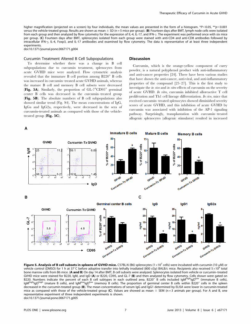
Curcumin Treatment Altered B Cell SubpopulationsTo determine whether there was a change in B cell
subpopulations due to curcumin treatment, splenocytes from
acute GVHD mice were analyzed. Flow cytometric analysis
revealed that the immature B cell portion among B220+ B cells
was increased in curcumin–treated acute GVHD animals, whereas
the mature B cell and memory B cell subsets were decreased
(Fig. 5A). Similarly, the proportion of GL-7+CD95+ germinal
center B cells was decreased in the curcumin–treated group
(Fig. 5B). The absolute numbers of B cell subpopulations also
showed similar trend (Fig. S4). The mean concentrations of IgG,
IgGa and IgG2a, respectively, were decreased in the sera of
curcumin-treated animals as compared with those of the vehicle-
treated group (Fig. 5C).
Discussion
Curcumin, which is the orange-yellow component of curry
powder, is a natural polyphenol product with anti-inflammatory
and anti-cancer properties [24]. There have been various studies
that have shown the anti-cancer, anti-viral, and anti-inflammatory
properties of the compound [25–27]. This is the first study to
investigate the in vivo and in vitro effects of curcumin on the severity
of acute GVHD. In vitro, curcumin inhibited alloreactive T cell
proliferation and Th1 cell lineage differentiation. In vivo, mice that
received curcumin–treated splenocytes showed diminished severity
scores of acute GVHD, and this inhibition of acute GVHD by
curcumin was associated with inhibition of the AP-1 signaling
pathway. Surprisingly, transplantation with curcumin–treated
allogenic splenocytes (allogenic stimulator) resulted in increased
higher magnification (projected on a screen) by four individuals, the mean values are presented in the form of a histogram. *P,0.05, **p,0.001versus the vehicle-treated group. Results are shown as mean 6 SD (n = 5 mice per group). (B) Fourteen days after BMT, lymph node cells were isolatedfrom each group and then analyzed by flow cytometry for the expression of IL-4, IL-17, and IFN-c. The experiment was performed once with six miceper group. (C) Fourteen days after BMT, splenocytes isolated from each group were stained with anti-CD4 and anti-CD8 antibodies followed byintracellular IFN-c, IL-4, Foxp3, and IL-17 antibodies and examined by flow cytometry. The data is representative of at least three independentexperiments.doi:10.1371/journal.pone.0067171.g004
Figure 5. Analysis of B cell subsets in spleens of GVHD mice. C57BL/6 (B6) splenocytes (16107 cells) were incubated with curcumin (10 mM) orvehicle control (DMSO) for 1 h at 37uC before adoptive transfer into lethally irradiated (800 cGy) BALB/c mice. Recipients also received 56106 totalbone marrow cells from B6 mice. (A and B) On day 14 after BMT, B cell subsets were analyzed. Splenocytes isolated from vehicle or curcumin–treatedGVHD mice were stained for B220, IgM, and IgD (A) or B220, CD95, and GL-7 (B) and then analyzed by flow cytometry. Cells shown were gated onB220. Numbers indicate the percent of each B cell subtypes in each outlined area. B220+ B cells included IgMhighIgDlow (immature B cells),IgMhighIgDhigh (mature B cells), and IgMlowIgDlow (memory B cells). The proportion of germinal center B cells within B220+ cells in the spleendecreased in the curcumin-treated group (B). The mean concentrations of serum IgG and IgG1 determined by ELISA were lower in curcumin-treatedmice as compared with those of the vehicle-treated group (C). Values are showed as mean 6 SEM (n = 3 animals per group). For A and B, onerepresentative experiment of three independent experiments is shown.doi:10.1371/journal.pone.0067171.g005
Therapeutic Efficacy of Curcumin in Acute GVHD
PLOS ONE | www.plosone.org 7 June 2013 | Volume 8 | Issue 6 | e67171
populations of CD4+ Treg cells, as well as CD8+ Treg cells in
recipient mice, compared to those of mice transplanted with
vehicle-treated splenocytes.
Along with HLA incompatibility, the intensity of conditioning
therapy is known to be a risk factor for the development of acute
GVHD [28]. Unfortunately, acute GVHD does develop despite
the administration of prophylactic agents, such as calcineurin
inhibitors and methotrexate. Upon the occurrence of acute
GVHD after HSCT, many patients should take immunosuppres-
sive agents despite the increased risk of severe infection and many
other adverse events. Our present study suggests the therapeutic
potential of curcumin, which has been used safely for a long time.
Although the pathophysiology of acute GVHD is complex, it
develops due to donor T cell responses to host alloantigens
expressed by host antigen-presenting cells and subsequent
dysregulation of inflammatory cytokine cascades [5,29]. Classical-
ly, acute GVHD is also considered to be predominantly related to
Th1 responses [30]. However, recent scientific investigations have
discovered the possible role of Treg and Th17 cells in the
development of GVHD [31]. The results obtained from our
present study correspond with other previous reports that showed
a shift from Th1 to Th2 responses by curcumin and its reciprocal
effects on Th17/Treg cells [21,32,33]. In the current study, the
increased populations of CD8+ Treg cells along with CD4+ Treg
cells by curcumin treatment were associated with attenuated acute
GVHD severity in a murine model. The novelty of our study was
the finding of increased CD8+ Treg cells by curcumin treatment.
Treg cells are known to have suppressive effects on autoreactive
lymphocytes and to control innate and adaptive immunity [34].
Removal of Treg cells from the donor graft dramatically
accelerated GVHD in an experimental GVHD model [35].
Conversely, ongoing GVHD was ameliorated by infusion with
donor or host Treg cells [36,37]. Although the beneficial effects of
Tregs in human GVHD were uncertain up until now, the finding
that peripheral blood from patients with GVHD demonstrated
reduced numbers of Foxp3+CD4+CD25+ T cells suggested the
potential benefits of the clinical application of Treg cells [38,39].
Accumulating evidence from experimental animal studies suggest
that the adoptive transfer of Tregs is a potential strategy to
suppress or prevent human GVHD. However, the relative scarcity
of circulating Tregs and the difficulty in isolating pure Treg cells
remain critical obstacles to carrying out this promising strategy. If
curcumin induces the expansion of the Treg population in
humans, the compound could be an adjunctive therapy in
allogenic HSCT. However, there are controversies that surround
the effects of curcumin on the number and immunomodulatory
function of Treg cells. Zhao et al. recently showed that curcumin
inhibits the immunosuppressive activity of Treg cells in vitro [40].
In that study, Foxp3, a critical regulator of Treg cell development
and function, was downregulated by curcumin treatment.
Conversely, one recent study revealed the induced differentiation
of the Treg lineage by curcumin-treated dendritic cells [33].
Curcumin was revealed to enact its immunomodulatory effect
through the inhibition of several transcriptional factors, including
AP-1 signaling [41]. In the present study, the inhibitory effect on
acute GVHD by curcumin was associated with attenuated AP-1
activity in skin and intestine. Skin and gut epithelial tissues induce
class II HLA, consequently promoting specific targeting during
acute GVHD [42,43]. Skin keratinocytes expressing endogenous
tissue antigens can directly prime naıve T cells [44], contributing the
development of skin GVHD. In gut GVHD, intestinal epithelial
cells are preferential target cells damaged by infiltrating donor T
lymphocytes [45]. In our present study, the inhibitory effects of
curcumin on the development of GVHD were associated with
attenuated expressions of c-Fos/c-Jun in the epithelial tissues of skin
(including keratinocytes) and intestine, suggesting that decreased
AP-1 signaling in skin keratinocytes and intestinal epithelial cells
may at least contribute to the attenuated severity of acute GVHD in
animal models. Within our knowledge, little is known about the
pathobiological roles of AP-1 signaling in GVHD development.
Although acute GVHD is considered to be mediated mainly by
donor T cells, recent animal studies suggest that B cells might also
play an important role in the biology of GVHD [46]. The specific
mechanisms of B cells involved in the development of acute
GVHD remains in large part unknown until now. Nevertheless,
some circumstantial evidence for the pathological role of B cells in
acute GVHD comes from clinical reports demonstrating that anti-
B cell therapy such as rituximab reduced the incidence and
severity of acute GVHD [47]. In line with previous studies, our
study also showed that the preventive effect of curcumin on acute
GVHD might be associated with a change of B cell homeostasis.
In the present study, curcumin-treatment on donor splenocytes
did not affect the absolute number of CD4+, CD8+ T cells, B cells,
and other immune cells including NK cells, HSCs, and DCs in
recipients. Our study showed that the inhibitory effects of
curcumin on the development of acute GVHD after BMT were
associated with altered subset of T and B cells, not associated with
absolute number of immune cells. On the other hand, previous
studies that identified anti-cancer effects of curcumin have showed
its beneficial effect was achieved through induction of T cell
apoptosis even in normal T cells via increasing endoplasmic
reticulum stress [48–50]. Although there is less research on
anticancer effect of curcumin on B cells, curcumin can selectively
induce apoptosis of B lymphoma cells [51] and showed anti-
inflammatory effects through repressing B call-activating factor
belonging to the TNF family [52,53]. To our knowledge, this is the
first study that shows immunomodulatory effects of curcumin
in vivo treatment through the altered subpopulations of B cells, not
affecting cell viability.
In conclusion, the present report showed that curcumin
inhibited alloreactive T cell responses and IFN-c and IL-17
production in vitro. Transplantation of curcumin-treated spleno-
cytes attenuated acute GVHD severity and shifted responses from
Th1 to Th2 in vivo. Interestingly, transplantation with curcumin-
treated splenocytes resulted in the expansion of CD4+ as well as
CD8+ Tregs in vivo. Thus, our present observations reveal a
promising strategy to prevent lethal acute GVHD through the
expansion of Treg cells. Recipient mice transplanted with
curcumin-treated splenocytes showed altered B-cell subpopulation,
increased immature B cells and reciprocally decreased mature-
and memory B cells, compared with those transplanted with
vehicle-treated splenocytes.
Supporting Information
Figure S1 The inhibitory effect of curcumin on allor-eactive T cell responses is not associated with apoptosisinduction or decreased cell viability. (A) Cell apoptosis
analyzed by flow cytometry. The lower left Annexin-V2/
propidium iodide (PI)– represents normal healthy cells. The lower
right Annexin-V+/PI– and upper right Annxin-V+/PI+ quadrant
represent early and later apoptotic cells, respectively. The upper
left quadrant, Annexin-V2/PI+ represent necrotic cells. (B) Cell
viability as evaluated with the MTT assay. Values of MTT assay
on cell viability after the different treatment with curcumin or
DMSO (diluent). Bars are shown as means 6 SEM from at least 3
independent experiments.
(TIF)
Therapeutic Efficacy of Curcumin in Acute GVHD
PLOS ONE | www.plosone.org 8 June 2013 | Volume 8 | Issue 6 | e67171
Figure S2 Effect of hematopoietic stem cell and otherimmune cell by curcumin. (A) CD34- or c-Kit-expressing
hematopoietic stem cell, (B) CD11c-expressing dendritic cells, and
(C) NK1.1-expressing natural killer cell populations among
splenocytes and bone marrow cells were analyzed by flow
cytomertry.
(TIF)
Figure S3 Analysis of immune reconstitution afterBMT. (A) Splenocytes and CD4+ T cells of BMT mice
tranaplanted with vehicle- and curcumin-treated splenocytes
originate from donor cells expressing H-2kb. (B) Absolute number
of CD4+ and CD8+ T cells were similar between mice transplanted
with vehicle- and curcumin-treated splenocytes.
(TIF)
Figure S4 Analysis of B cell subset after BMT. Absolute
number of B cell subpopulation among B220+ B cells were shown
in BMT mice and were compared between vehicle- and curcumin-
treated groups.
(TIF)
Author Contributions
Conceived and designed the experiments: MLC CWY HYK. Performed
the experiments: MJP SHL EJY JKM. Analyzed the data: MJP SGC SHP.
Contributed reagents/materials/analysis tools: SGC. Wrote the paper:
MJP SJM. Commented and reviewed the manuscript: SGC CWY SHP
HYK MLC.
References
1. Pasquini MC, Wang Z, Horowitz MM, Gale RP (2010) 2010 report from the
Center for International Blood and Marrow Transplant Research (CIBMTR):
current uses and outcomes of hematopoietic cell transplants for blood and bone
marrow disorders. Clin Transpl: 87–105.
2. Bolanos-Meade J (2006) Update on the management of acute graft-versus-host
disease. Curr Opin Oncol 18: 120–125.
3. MacMillan ML, Weisdorf DJ, Wagner JE, DeFor TE, Burns LJ, et al. (2002)
Response of 443 patients to steroids as primary therapy for acute graft-versus-
host disease: comparison of grading systems. Biol Blood Marrow Transplant 8:
387–394.
4. Martin PJ, Schoch G, Fisher L, Byers V, Anasetti C, et al. (1990) A retrospective
analysis of therapy for acute graft-versus-host disease: initial treatment. Blood 76:
1464–1472.
5. Ferrara JL, Levine JE, Reddy P, Holler E (2009) Graft-versus-host disease.
Lancet 373: 1550–1561.
6. Lu Y, Waller EK (2009) Dichotomous role of interferon-gamma in allogeneic
bone marrow transplant. Biol Blood Marrow Transplant 15: 1347–1353.
7. Cooke KR, Hill GR, Crawford JM, Bungard D, Brinson YS, et al. (1998) Tumor
necrosis factor- alpha production to lipopolysaccharide stimulation by donor
cells predicts the severity of experimental acute graft-versus-host disease. J Clin
Invest 102: 1882–1891.
8. Blazar BR, Murphy WJ, Abedi M (2012) Advances in graft-versus-host disease
biology and therapy. Nat Rev Immunol 12: 443–458.
9. Carlson MJ, West ML, Coghill JM, Panoskaltsis-Mortari A, Blazar BR, et al.
(2009) In vitro-differentiated TH17 cells mediate lethal acute graft-versus-host
disease with severe cutaneous and pulmonary pathologic manifestations. Blood
113: 1365–1374.
10. Ukena SN, Grosse J, Mischak-Weissinger E, Buchholz S, Stadler M, et al. (2011)
Acute but not chronic graft-versus-host disease is associated with a reduction of
circulating CD4(+)CD25 (high)CD127 (low/2) regulatory T cells. Ann Hematol
90: 213–218.
11. Magenau JM, Qin X, Tawara I, Rogers CE, Kitko C, et al. (2010) Frequency of
CD4(+)CD25(hi)FOXP3(+) regulatory T cells has diagnostic and prognostic
value as a biomarker for acute graft-versus-host-disease. Biol Blood Marrow
Transplant 16: 907–914.
12. Shin HJ, Baker J, Leveson-Gower DB, Smith AT, Sega EI, et al. (2011)
Rapamycin and IL-2 reduce lethal acute graft-versus-host disease associated with
increased expansion of donor type CD4+CD25+Foxp3+ regulatory T cells.
Blood 118: 2342–2350.
13. Duramad O, Laysang A, Li J, Ishii Y, Namikawa R (2011) Pharmacologic
expansion of donor-derived, naturally occurring CD4(+)Foxp3(+) regulatory T
cells reduces acute graft-versus-host disease lethality without abrogating the
graft-versus-leukemia effect in murine models. Biol Blood Marrow Transplant
17: 1154–1168.
14. Kroger N, Zabelina T, Kruger W, Renges H, Stute N, et al. (2002) In vivo T cell
depletion with pretransplant anti-thymocyte globulin reduces graft-versus-host
disease without increasing relapse in good risk myeloid leukemia patients after
stem cell transplantation from matched related donors. Bone Marrow
Transplant 29: 683–689.
15. Ali T, Shakir F, Morton J (2012) Curcumin and inflammatory bowel disease:
biological mechanisms and clinical implication. Digestion 85: 249–255.
16. Aggarwal BB, Harikumar KB (2009) Potential therapeutic effects of curcumin,
the anti-inflammatory agent, against neurodegenerative, cardiovascular, pulmo-
nary, metabolic, autoimmune and neoplastic diseases. Int J Biochem Cell Biol
41: 40–59.
17. Ruby AJ, Kuttan G, Babu KD, Rajasekharan KN, Kuttan R (1995) Anti-
tumour and antioxidant activity of natural curcuminoids. Cancer Lett 94: 79–83.
18. Ramsewak RS, DeWitt DL, Nair MG (2000) Cytotoxicity, antioxidant and anti-
inflammatory activities of curcumins I-III from Curcuma longa. Phytomedicine
7: 303–308.
19. Apisariyakul A, Vanittanakom N, Buddhasukh D (1995) Antifungal activity of
turmeric oil extracted from Curcuma longa (Zingiberaceae). J Ethnopharmacol
49: 163–169.
20. Negi PS, Jayaprakasha GK, Jagan Mohan Rao L, Sakariah KK (1999)
Antibacterial activity of turmeric oil: a byproduct from curcumin manufacture.
J Agric Food Chem 47: 4297–4300.
21. Xie L, Li XK, Funeshima-Fuji N, Kimura H, Matsumoto Y, et al. (2009)
Amelioration of experimental autoimmune encephalomyelitis by curcumin
treatment through inhibition of IL-17 production. Int Immunopharmacol 9:
575–581.
22. Zhang M, Deng CS, Zheng JJ, Xia J (2006) Curcumin regulated shift from Th1
to Th2 in trinitrobenzene sulphonic acid-induced chronic colitis. Acta
Pharmacol Sin 27: 1071–1077.
23. Fukui J, Inaba M, Ueda Y, Miyake T, Hosaka N, et al. (2007) Prevention of
graft-versus-host disease by intra-bone marrow injection of donor T cells. Stem
Cells 25: 1595–1601.
24. Zhou H, Beevers CS, Huang S (2011) The targets of curcumin. Curr Drug
Targets 12: 332–347.
25. Baumeister P, Reiter M, Harreus U (2012) Curcumin and other polyphenolic
compounds in head and neck cancer chemoprevention. Oxid Med Cell Longev
2012: 902716.
26. Zandi K, Ramedani E, Mohammadi K, Tajbakhsh S, Deilami I, et al. (2010)
Evaluation of antiviral activities of curcumin derivatives against HSV-1 in Vero
cell line. Nat Prod Commun 5: 1935–1938.
27. Xiao X, Yang M, Sun D, Sun S (2012) Curcumin protects against sepsis-induced
acute lung injury in rats. J Surg Res 176: e31–39.
28. Mielcarek M, Martin PJ, Leisenring W, Flowers ME, Maloney DG, et al. (2003)
Graft-versus-host disease after nonmyeloablative versus conventional hemato-
poietic stem cell transplantation. Blood 102: 756–762.
29. Shlomchik WD, Couzens MS, Tang CB, McNiff J, Robert ME, et al. (1999)
Prevention of graft versus host disease by inactivation of host antigen-presenting
cells. Science 285: 412–415.
30. Krenger W, Ferrara JL (1996) Graft-versus-host disease and the Th1/Th2
paradigm. Immunol Res 15: 50–73.
31. Teshima T, Maeda Y, Ozaki K (2011) Regulatory T cells and IL-17-producing
cells in graft-versus-host disease. Immunotherapy 3: 833–852.
32. Kang BY, Song YJ, Kim KM, Choe YK, Hwang SY, et al. (1999) Curcumin
inhibits Th1 cytokine profile in CD4+ T cells by suppressing interleukin-12
production in macrophages. Br J Pharmacol 128: 380–384.
33. Cong Y, Wang L, Konrad A, Schoeb T, Elson CO (2009) Curcumin induces the
tolerogenic dendritic cell that promotes differentiation of intestine-protective
regulatory T cells. Eur J Immunol 39: 3134–3146.
34. Janssens W, Carlier V, Wu B, VanderElst L, Jacquemin MG, et al. (2003)
CD4+CD25+ T cells lyse antigen-presenting B cells by Fas-Fas ligand
interaction in an epitope-specific manner. J Immunol 171: 4604–4612.
35. Cohen JL, Trenado A, Vasey D, Klatzmann D, Salomon BL (2002)
CD4(+)CD25(+) immunoregulatory T Cells: new therapeutics for graft-versus-
host disease. J Exp Med 196: 401–406.
36. Zhao D, Zhang C, Yi T, Lin CL, Todorov I, et al. (2008) In vivo-activated
CD103+CD4+ regulatory T cells ameliorate ongoing chronic graft-versus-host
disease. Blood 112: 2129–2138.
37. Anderson BE, McNiff JM, Matte C, Athanasiadis I, Shlomchik WD, et al. (2004)
Recipient CD4+ T cells that survive irradiation regulate chronic graft-versus-
host disease. Blood 104: 1565–1573.
38. Miura Y, Thoburn CJ, Bright EC, Phelps ML, Shin T, et al. (2004) Association
of Foxp3 regulatory gene expression with graft-versus-host disease. Blood 104:
2187–2193.
39. Zorn E, Kim HT, Lee SJ, Floyd BH, Litsa D, et al. (2005) Reduced frequency of
FOXP3+ CD4+CD25+ regulatory T cells in patients with chronic graft-versus-
host disease. Blood 106: 2903–2911.
Therapeutic Efficacy of Curcumin in Acute GVHD
PLOS ONE | www.plosone.org 9 June 2013 | Volume 8 | Issue 6 | e67171
40. Zhao GJ, Lu ZQ, Tang LM, Wu ZS, Wang DW, et al. (2012) Curcumin inhibits
suppressive capacity of naturally occurring CD4+CD25+ regulatory T cells inmice in vitro. Int Immunopharmacol 14: 99–106.
41. Dhandapani KM, Mahesh VB, Brann DW (2007) Curcumin suppresses growth
and chemoresistance of human glioblastoma cells via AP-1 and NFkappaBtranscription factors. J Neurochem 102: 522–538.
42. Sviland L, Pearson AD, Eastham EJ, Green MA, Hamilton PJ, et al. (1988) ClassII antigen expression by keratinocytes and enterocytes–an early feature of graft-
versus-host-disease. Transplantation 46: 402–406.
43. Bland PW, Whiting CV (1992) Induction of MHC class II gene products in ratintestinal epithelium during graft-versus-host disease and effects on the immune
function of the epithelium. Immunology 75: 366–371.44. Kim BS, Miyagawa F, Cho YH, Bennett CL, Clausen BE, et al. (2009)
Keratinocytes function as accessory cells for presentation of endogenous antigenexpressed in the epidermis. J Invest Dermatol 129: 2805–2817.
45. Schattenfroh NC, Hoffman RA, McCarthy SA, Simmons RL (1995) Phenotypic
analysis of donor cells infiltrating the small intestinal epithelium and spleenduring graft-versus-host disease. Transplantation 59: 268–273.
46. Young JS, Wu T, Chen Y, Zhao D, Liu H, et al. (2012) Donor B cells intransplants augment clonal expansion and survival of pathogenic CD4+ T cells
that mediate autoimmune-like chronic graft-versus-host disease. J Immunol 189:
222–233.
47. Khouri IF, McLaughlin P, Saliba RM, Hosing C, Korbling M, et al. (2008)
Eight-year experience with allogeneic stem cell transplantation for relapsedfollicular lymphoma after nonmyeloablative conditioning with fludarabine,
cyclophosphamide, and rituximab. Blood 111: 5530–5536.
48. Korwek Z, Bielak-Zmijewska A, Mosieniak G, Alster O, Moreno-Villanueva M,et al. (2013) DNA damage-independent apoptosis induced by curcumin in
normal resting human T cells and leukaemic Jurkat cells. Mutagenesis.49. Alizadeh AM, Khaniki M, Azizian S, Mohaghgheghi MA, Sadeghizadeh M, et
al. (2012) Chemoprevention of azoxymethane-initiated colon cancer in rat by
using a novel polymeric nanocarrier-curcumin. Eur J Pharmacol 689: 226–232.50. Vishvakarma NK, Kumar A, Singh SM (2011) Role of curcumin-dependent
modulation of tumor microenvironment of a murine T cell lymphoma in alteredregulation of tumor cell survival. Toxicol Appl Pharmacol 252: 298–306.
51. Han SS, Chung ST, Robertson DA, Ranjan D, Bondada S (1999) Curcumincauses the growth arrest and apoptosis of B cell lymphoma by downregulation of
egr-1, c-myc, bcl-XL, NF-kappa B, and p53. Clin Immunol 93: 152–161.
52. Hayun R, Okun E, Berrebi A, Shvidel L, Bassous L, et al. (2009) Rapamycinand curcumin induce apoptosis in primary resting B chronic lymphocytic
leukemia cells. Leuk Lymphoma 50: 625–632.53. Huang G, Yang Y, Xu Z, Zhou P, Gong W, et al. (2011) Downregulation of B
lymphocyte stimulator expression by curcumin in B lymphocyte via suppressing
nuclear translocation of NF-kappaB. Eur J Pharmacol 650: 451–457.
Therapeutic Efficacy of Curcumin in Acute GVHD
PLOS ONE | www.plosone.org 10 June 2013 | Volume 8 | Issue 6 | e67171