Culture, characterization and differentiation of neural precursors from the central … ·...
8
Pesq. Vet. Bras. 36(Supl.1):71-78, junho 2016 DOI: 10.1590/S0100-736X2016001300011 71 RESUMO.- [Cultura, caracterização e diferenciação de precursores neurais do sistema nervoso central de porquinho-da-índia (Cavia porcellus Linnaeus, 1758).] Áreas potencialmente neurogênicas foram identificadas por incorporação de bromodeoxiuridina (BrdU) na zona subventricular (SVZ) dos ventrículos laterais, hipocampo e bulbos olfatórios de cobaias neonatos. Precursores neu- rais provenientes da SVZ foram cultivados em suspensão, resultando na geração de neuroesferas (NSFs), que quando dissociadas foram capazes de proliferar e gerar novas NSFs. Quando cultivadas na ausência de fatores de crescimento, as células provenientes de NSFs dissociadas apresentaram evi- dências de diferenciação neuronal, dando origem a células da linhagem neural. Citometria de fluxo em células das NSFs após a diferenciação revelou aproximadamente 13,3% posi- tivas para nestina, 5,5% positivas para Beta-III-tubulina, 9% positivas para GFAP e 7,8% positivas para mGalC. Testes de funcionalidade pela mensuração de influxo de cálcio após estímulo com ácido gama amino butírico (GABA) e glutama- to revelaram a estimulação de células diferenciadas, um in- dicador de função neuronal. A capacidade de células da SVZ de fetos de cobaias originarem células neurais funcionais in vitro é promissora para a pesquisa e eventual uso terapêuti- co de células tronco em disordens do sistema nervoso. TERMOS DE INDEXAÇÃO: Cobaios, células-tronco neurais, zona subventricular, cultura, caracterização in vitro. INTRODUCTION Neural stem cells (NSCs) are defined by two functional pro- perties: (1) self renewal, the capacity to generate identical progeny and (2) multipotentiality, the capacity to generate Culture, characterization and differentiation of neural precursors from the central nervous system of guinea pigs (Cavia porcellus Linnaeus, 1758) 1 Erika Toledo da Fonseca 2,4 *, Layla Testa Galindo 3 , Marimélia A. Porcionatto 3 and Maria Angélica Miglino 4 ABSTRACT.- Fonseca E.T., Galindo L.T., Porcionatto M.A. & Miglino M.A. 2016. Culture, charac- terization and differentiation of neural precursors from the central nervous system of guinea pigs (Cavia porcellus Linnaeus, 1758). Pesquisa Veterinária Brasileira 36(Supl.1):71- 78. Escola de Medicina Veterinária e Zootecnia, Universidade Federal do Tocantins, BR-153 Km 112, Rural, Araguaína, Tocantins, TO 77804-970, Brazil. E-mail: [email protected] Potentially neurogenic areas were initially identified by incorporation of bromodeoxyu- ridine (BrdU) in cells underlying the subventricular zone (SVZ) of the lateral ventricles wall, hippocampus and olfactory bulbs of newborn guinea pigs. Neural precursors from the SVZ were cultured in suspension, generating neurospheres (NSFs), which, upon dis- sociation were able to generate new NSFs. Upon culture in the absence of growth factors, cells dissociated from NSFs displayed evidence for neural differentiation, giving rise to cells from neural lineage. Flow cytometry analysis for of NSFs-derived cells after differentiation revealed approximately 13.3% nestin positive, 5.5% Beta-III-tubulin positive, 9% GFAP po- sitive and 7.8% mGalC positive. Functional assays by measurement of calcium influx upon gamma butiric amino acid (GABA) and glutamate stimuli, revealed stimulation in differen- tiated cells, an indicator of neuronal differentiation. The ability of guinea pig SVZ cells to originate functional neurons in vitro is promising for research and towards a future use of neural stem cells in the therapy of neurological disorders. INDEX TERMS: Guinea pigs, neural stem cells, subventricular zone, culture, in vitro characterization. 1 Received on June 16, 2015. Accepted for publication on April 1, 2016. 2 Escola de Medicina Veterinária e Zootecnia, Universidade Federal do Tocantins (UFT), BR-153 Km 112, Rural, Araguaína, Tocantins, TO 77804- 970, Brazil. *Corresponding author: [email protected] 3 Departamento de Bioquímica, Universidade Federal de São Paulo (Uni- fesp), Rua Pedro de Toledo 669, São Paulo, SP 04039-030. Brazil. 4 Departamento de Cirurgia, Faculdade de Medicina Veterinária e Zoo- tecnia, Universidade de São Paulo (USP), Av. Prof. Orlando Marques de Pai- va 87, São Paulo, SP 05508-270, Brazil.
Transcript of Culture, characterization and differentiation of neural precursors from the central … ·...

Pesq. Vet. Bras. 36(Supl.1):71-78, junho 2016DOI: 10.1590/S0100-736X2016001300011
71
RESUMO.- [Cultura, caracterização e diferenciação de precursores neurais do sistema nervoso central de porquinho-da-índia (Cavia porcellus Linnaeus, 1758).] Áreas potencialmente neurogênicas foram identificadas por incorporação de bromodeoxiuridina (BrdU) na zona subventricular (SVZ) dos ventrículos laterais, hipocampo e bulbos olfatórios de cobaias neonatos. Precursores neu-rais provenientes da SVZ foram cultivados em suspensão, resultando na geração de neuroesferas (NSFs), que quando dissociadas foram capazes de proliferar e gerar novas NSFs. Quando cultivadas na ausência de fatores de crescimento, as
células provenientes de NSFs dissociadas apresentaram evi-dências de diferenciação neuronal, dando origem a células da linhagem neural. Citometria de fluxo em células das NSFs após a diferenciação revelou aproximadamente 13,3% posi-tivas para nestina, 5,5% positivas para Beta-III-tubulina, 9% positivas para GFAP e 7,8% positivas para mGalC. Testes de funcionalidade pela mensuração de influxo de cálcio após estímulo com ácido gama amino butírico (GABA) e glutama-to revelaram a estimulação de células diferenciadas, um in-dicador de função neuronal. A capacidade de células da SVZ de fetos de cobaias originarem células neurais funcionais in vitro é promissora para a pesquisa e eventual uso terapêuti-co de células tronco em disordens do sistema nervoso.TERMOS DE INDEXAÇÃO: Cobaios, células-tronco neurais, zona subventricular, cultura, caracterização in vitro.
INTRODUCTIONNeural stem cells (NSCs) are defined by two functional pro-perties: (1) self renewal, the capacity to generate identical progeny and (2) multipotentiality, the capacity to generate
Culture, characterization and differentiation of neural precursors from the central nervous system of guinea pigs
(Cavia porcellus Linnaeus, 1758)1
Erika Toledo da Fonseca2,4*, Layla Testa Galindo3, Marimélia A. Porcionatto3
and Maria Angélica Miglino4
ABSTRACT.- Fonseca E.T., Galindo L.T., Porcionatto M.A. & Miglino M.A. 2016. Culture, charac-terization and differentiation of neural precursors from the central nervous system of guinea pigs (Cavia porcellus Linnaeus, 1758). Pesquisa Veterinária Brasileira 36(Supl.1):71-78. Escola de Medicina Veterinária e Zootecnia, Universidade Federal do Tocantins, BR-153 Km 112, Rural, Araguaína, Tocantins, TO 77804-970, Brazil. E-mail: [email protected]
Potentially neurogenic areas were initially identified by incorporation of bromodeoxyu-ridine (BrdU) in cells underlying the subventricular zone (SVZ) of the lateral ventricles wall, hippocampus and olfactory bulbs of newborn guinea pigs. Neural precursors from the SVZ were cultured in suspension, generating neurospheres (NSFs), which, upon dis-sociation were able to generate new NSFs. Upon culture in the absence of growth factors, cells dissociated from NSFs displayed evidence for neural differentiation, giving rise to cells from neural lineage. Flow cytometry analysis for of NSFs-derived cells after differentiation revealed approximately 13.3% nestin positive, 5.5% Beta-III-tubulin positive, 9% GFAP po-sitive and 7.8% mGalC positive. Functional assays by measurement of calcium influx upon gamma butiric amino acid (GABA) and glutamate stimuli, revealed stimulation in differen-tiated cells, an indicator of neuronal differentiation. The ability of guinea pig SVZ cells to originate functional neurons in vitro is promising for research and towards a future use of neural stem cells in the therapy of neurological disorders.INDEX TERMS: Guinea pigs, neural stem cells, subventricular zone, culture, in vitro characterization.
1 Received on June 16, 2015.Accepted for publication on April 1, 2016.
2 Escola de Medicina Veterinária e Zootecnia, Universidade Federal do Tocantins (UFT), BR-153 Km 112, Rural, Araguaína, Tocantins, TO 77804-970, Brazil. *Corresponding author: [email protected]
3 Departamento de Bioquímica, Universidade Federal de São Paulo (Uni-fesp), Rua Pedro de Toledo 669, São Paulo, SP 04039-030. Brazil.
4 Departamento de Cirurgia, Faculdade de Medicina Veterinária e Zoo-tecnia, Universidade de São Paulo (USP), Av. Prof. Orlando Marques de Pai-va 87, São Paulo, SP 05508-270, Brazil.
Pesq. Vet. Bras. 36(Supl.1):71-78, junho 2016
72 Erika Toledo da Fonseca et al.
the main neural cells, e.g. neurons, astrocytes and oligo-dendrocytes (Conti et al. 2006). The potential applications of NSCs in degenerative and traumatic diseases therapy in the central nervous system (CNS) have called tremendous scientific interest.
During vertebrate embryonic development, CNS cells may be originated from virtually all regions of the neuro-epithelium. Two well characterized neurogenic regions are the dentate gyrus (DG) of the hippocampus (Altman & Das 1967, Lacbawan & Muenke 2002) and the subventricular zone (SVZ) of the lateral ventricles (Götz & Huttner 2005). The SVZ is a thin layer in the wall of the lateral ventricles that contains neural progenitor cells able to generate neu-rons and glial cells (Alvarez-Buylla & Lois 1995).
García-Verdugo et al. (1998) highlighted the main im-munostaining properties of the cell types found in the CNS: nestin antibodies react with embryonic neuroepithelial cells and, thus, nestin has been suggested as the marker for NSCs. In the adult SVZ, nestin is expressed in multiple cell types, including ependymal cells. Vimentin and GFAP glial markers are present in astrocytes, yet apparently not in neurons. PSA-NCAM antibodies and neuron-specific Beta--III-tubulin antibodies (Tuj1 for example) stain only neu-rons. The pattern of recognition by cellular marker-specific antibodies suggests that precursor cells are the most im-mature since they express only nestin and do not express other markers of differentiation. The most used markers for neural precursors and differentiated cells include inclu-de Nestin, Sox2 (Schwartz et al. 2003), GFAP, Beta-III-tubu-lin (Buddensiek et al. 2010, Piper et al. 2001, Schwartz et al. 2003), MAP2ab (Buddensiek et al. 2010; Schwartz et al. 2003), Vimentin, NeuN, (Schwartz et al. 2003), mGalC (Pi-per et al. 2001, Buddensiek et al. 2010) and O4 (Piper et al. 2001, Schwartz et al. 2003).
Neural regeneration should be considered in three main applications: (1) regeneration of interrupted/disrupted neuronal axons; (2) replacement of damaged neural cells and (3) rescue of neural functions (Okano 2010). The ob-servation that all cells of the CNS are derived from NSCs originated in different regions of the brain has stimulated the scientific community for mapping and characterizing neurogenic areas and investigating the aplicability of NSCs in the therapy of neurological disorders as multiple sclero-sis (Manganas and Maletic-Savatic, 2005), Parkinson’s di-sease, stroke, spinal trauma (Jandial et al., 2007) and many lesions/diseases of the nervous system (Mansergh et al. 2004).
Although adult neurogenesis is an event that occurs in several animal species, the rate of production of neural cells exhibits notable differences among species. Interes-tingly, the pattern of development of the DG in guinea pigs is more similar to that of humans than other species (Guidi et al. 2005). In addition, guinea pigs display an extensive pre-birth development of the brain leading to a full forma-tion of the volumous cerebral hemispheres (Potter & Brue-ck 1958).
Therefore, this study aimed to culture, to characterize and to differentiate neural precursors obtained from the SVZ of guinea pigs fetuses.
MATERIALS AND METHODSTwenty one fetuses and two neonates from nine pregnant guinea pig females from a particular breeder were used. All experimen-tal procedures were performed under veterinary supervision, ac-cording to recommendations by the National Council for Control of Animal Experimentation from Brazil, under license number 1999/2010 from the University of São Paulo Ethics Committee on Animal Experimentation.
Female guinea pigs near the end of pregnancy were sedated by intramuscular administration of azaperone (4mg/kg) followed by administration of cloridrate of ketamine (20mg/kg) and xilazine (1.5mg/kg). Fetuses were aseptically removed upon laparotomy.
BrdU staining. For BrdU staining, two one-day-old guinea pigs received intraperitonial administrations of 5-bromo-2’de-oxyuridine (BrdU; 50mg/kg) every 12h during 2 days. Twelve hours after the last administration they were sedated by intra-muscular administration of cloridrate of ketamine (20mg/kg) and xilazine (1.5mg/kg) and perfused with 4% parafolmaldey-de (PFA). Brain was removed from the skull, sectioned along the midline and remains PFA in PBS for 4h at 4oC. After washing, the tissue was dehydrated in 30% sacarose at 4oC overnight, followed by inclusion in Tissue Tek - OCT (FK Biotec) and frozen. 20µm sections were cut in a criostat, layed on glass slides and stored at -20oC. Slides were incubated with 0.1% Triton X-100 in PBS at RT for 10 min followed by washing with 0.1 M TBS pH 7.4. Block-ing was performed with 5% FBS and 0.1% Triton X-100 for 1h at RT. Then slides were incubated with an anti-BrdU antibody (rat, Abcam, 1:100) in 0.1 % triton X-100 in TRIS-borate buffer for 30 min. After washings in PBS and H2O, sections were incubated with a secondary antibody (anti-rat IgG, Alexa488 conjugated, Abcam) for additional 30 min. For nuclei visualization, slides were stained with DAPI (4’-6-diamidino-2-fenilindol, Molecular Probes) for 10 min. Then, slides were washed, mounted and examined in a con-focal scope (Zeiss® model LSM510).
Cell cultures. Fetal brains were removed and placed on Petri dishes containing high glucose DMEM (4.5g/l) (Dulbecco’s Mod-ified Eagle Medium, Sigma). Fragments obtained from the SVZ were transferred to tubes containing 1 ml of high glucose DMEM (Sigma). After tissue sedimentation, the DMEMwas removed and tissue fragments were incubated with 1ml TrypLEtm (GibcoBRL; 0.1%), EDTA (10mM), pH 6.3 for 5 min at 37°C. TrypLEtm was in-activated with fetal bovine serum (FBS, Cultilab) and the material was homogeneized and centrifuged at 400xg for 5 min. Superna-tant was removed and the pellet ressuspended in culture medium as described below. Tissues were dissociated by multiple passages under high pressure through pipete tips of 1000µl to 200µl until a cell suspension was obtained. The cell suspension was filtered through a 40µm filter (BD) and placed on 75cm2 culture dishes previously treated with Poly-Hema (Poli [2-hidroxyetil metacri-late], Sigma). Culture medium consisted of high glucose DMEM (Sigma; 70%), HAM’S F12 (LGC Biotecnologia; 30%), penicilin/streptomycin (Gibco BRL; 1%), supplement B27 (Gibco BRL; 2%), EGF (Sigma; 20ng/ml), FGF (R&D, 20ng/ml), heparin (Sigma; 5g/ml), L-Glutamine (Sigma; 2mM). 50% of Culture medium was re-placed every 4 to 5 days until the formation of the NSFs. NSFs Dis-sociation in individual cells was achieved by TrypLEtm incubation for 5 min at 37 ºC, followed by gentle pipeting the cell suspension until complete dissociation.
Differentiation. NSFs were harvested and transferred to laminin and poly-L-lisine coated sterile coverslips to allow cell adhesion in 24-well plates. The culture medium consisted of high glucose DMEM (Sigma; 70%), HAM’S F12 (LGC Biotecnologia; 30%), penicillin/streptomycin (GibcoBRL; 1%), suplement B27 (Gibco BRL; 2%). After 7 days, culture medium was removed and
Pesq. Vet. Bras. 36(Supl.1):71-78, junho 2016
73Culture, characterization and differentiation of neural precursors from the central nervous system of guinea pigs
cells were fixed in 4% PFA for 20 min at room temperature (RT). Then PFA was removed, cells were washed three times with 0.1 M TBS and immunofluorescence and cytometry analysis were per-formed.
Immunofluorescence for neuronal markers. NSCs as NSFs or single cells were cultured in 24-well plates covered by laminin and poli-L-lisine-treated coverslips at 37°C for 7 days. Then, cells were fixed in 4% PFA for 20 min at RT, washed three times with 0.1 M TBS and incubated with 0.1% Triton X-100 in PBS at RT for 10 min followed by washing with 0.1 M TBS pH 7.4. Blocking was performed with 5% FBS and 0.1% Triton X-100 for 1h at RT. Slides were incubated with each of the following primary antibod-ies overnight at 4°C: anti-Nestin (rabbit, Sigma, 1:10), anti-GFAP (rabbit, Abcam, 1:50), anti-galactocerebroside (mGalC) (mouse, Millipore, 5µg) and anti- β-III Tubulin (mouse, Abcam, 1:50). Slides were washed twice with 0.1 M TBS and incubated with the secondary antibodies for an additional hour at 37°C: anti-rabbit IgG TEXAS RED conjugated (Santa Cruz Biotechnology, 1:300), anti-rabbit IgG FITC-conjugated (Sigma, 1:200), anti-mouse IgG FITC-conjugated (Santa Cruz Biotechonoly, 1:200). Slides were then washed twice in 0.1 M TBS, mounted with Vectashield (Vec-tor Laboratories) and examined in an Eclipse E600 ultraviolet mi-croscope (Nikon).
Flow cytometry. NSFs were cultured in Petri dishes treated with poly-L-lisin and laminin as described above in the same cul-ture medium used for cell differentiation and cells were cultured for 10 days. After this period, culture medium was removed, cells were dissociated as described above the supernatant discarded. cell pellet was washed with PBS and permeabilized with 0.1% Triton X-100 in PBS for 6 min at RT, followed by PBS washing and incubationfor 1h at RT with the following primary antibodies, in-dividually: polyclonal anti-Nestin (rabbit, Sigma, 1:10), polyclonal anti-GFAP (rabbit, Abcam, 1:50), monoclonal anti-galactocerebro-side (mGalC) (mouse, Millipore, 5µg) and monoclonal anti- β-III Tubulin (mouse, Abcam, 1:50). Then the cells were washed with PBS and incubated for an additional hour with the respective sec-ondary antibodies: anti-rabbit IgG TEXAS RED conjugated (San-ta Cruz Biotechnology, 1:300), anti-rabbit IgG FITC-conjugated (Sigma, 1:200), anti-mouse IgG FITC-conjugated (Santa Cruz Bio-techonoly, 1:200). Cells were then fixed in 1% PFA in PBS con-taining 1% BSA for 15 min at RT, followed by PBS washing and ressuspension in 1mL PBS. Analysis were performed in a flow cytometer (Attune® Acoustic Focusing Cytometer, Applied Biosys-tem) using the Attune Cytometric Software V1.2.5, with acquisi-tion of, at least 5.000 events. The flow cytometer was calibrated
with the following controls: cell suspension without antibody; cell suspension incubated with Alexa488 secondary antibody only and cell suspension incubated with Alexa647 secondary antibody only. Parameters were adjusted to remove any contribution by unla-belled cells.
Modulation of intracellular Ca2+. Intracellular Ca2+ influx as-say was performed to investigate the functionality of NSCs sub-mitted to differentiation. NSFs were cultured for 10 days over poly-l-lysin and laminin-coated coverslips in 24-well plates in the same medium used for differentiation. Culture medium was re-placed every three days. Then, culture medium was removed and cells incubated with buffer A (116mM NaCl, 5.4mM KCl, 0.8mM MgSO4, 5.5mM D-Glucose, 50mM MOPS, 1mM CaCl2, pH 7.2) sup-plemented with 5µM Fluo-3 AM (Molecular Probes, Caslsbad, CA) at RT for 30 min. Cells were washed twice with buffer A. The neu-rotransmitters gama-aminobutiric acid (GABA) (Sigma, 5nM) and glutamate (Sigma, 5mM) were added, separatedly, to the medium. The drug Thapsigargin (THG) (Sigma, 2nM) was used as positive control. After addition of each neurotransmitter or THG, fluores-cence was observed and images were captured in a fluorescence Carl Zeiss AxioObserver Z1 microscope.
RESULTSIdentification of neurogenic areas
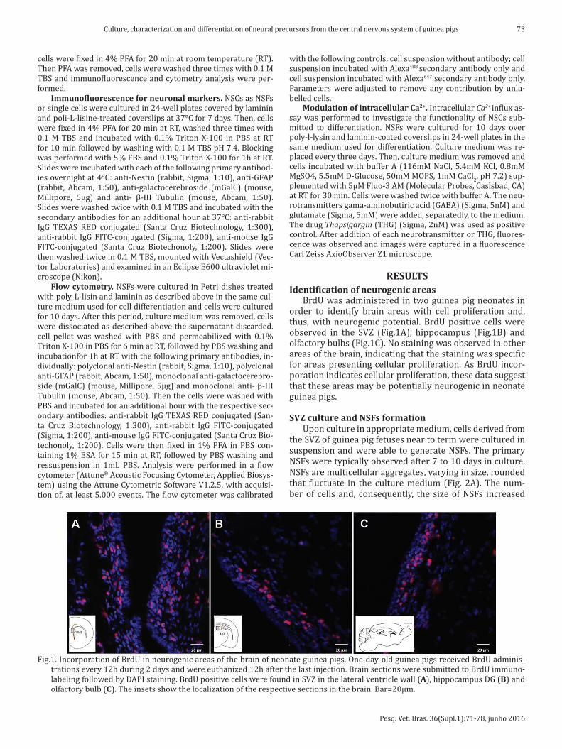
BrdU was administered in two guinea pig neonates in order to identify brain areas with cell proliferation and, thus, with neurogenic potential. BrdU positive cells were observed in the SVZ (Fig.1A), hippocampus (Fig.1B) and olfactory bulbs (Fig.1C). No staining was observed in other areas of the brain, indicating that the staining was specific for areas presenting cellular proliferation. As BrdU incor-poration indicates cellular proliferation, these data suggest that these areas may be potentially neurogenic in neonate guinea pigs.
SVZ culture and NSFs formationUpon culture in appropriate medium, cells derived from
the SVZ of guinea pig fetuses near to term were cultured in suspension and were able to generate NSFs. The primary NSFs were typically observed after 7 to 10 days in culture. NSFs are multicellular aggregates, varying in size, rounded that fluctuate in the culture medium (Fig. 2A). The num-ber of cells and, consequently, the size of NSFs increased
Fig.1. Incorporation of BrdU in neurogenic areas of the brain of neonate guinea pigs. One-day-old guinea pigs received BrdU adminis-trations every 12h during 2 days and were euthanized 12h after the last injection. Brain sections were submitted to BrdU immuno-labeling followed by DAPI staining. BrdU positive cells were found in SVZ in the lateral ventricle wall (A), hippocampus DG (B) and olfactory bulb (C). The insets show the localization of the respective sections in the brain. Bar=20µm.

Pesq. Vet. Bras. 36(Supl.1):71-78, junho 2016
74 Erika Toledo da Fonseca et al.
Fig.3. NSFs cells cultivated under differentiation conditions. (A) After differentiation, some cells kept the undifferentiated state and were positive for nestin. (B) Some cells were positive for Beta-III-tubulin, (C) GFAP and (D) mGalC, indicating neuron, astrocyte and oligo-dendrocyte phenotypes respectively. Nuclei were stained with DAPI (blue). Bar=20µm.
Fig.2. (A) Neurospheres derived from guinea pig fetuses SVZ. After nine days in culture NSFs can be visualized with variable diameters and there is still the presence of individual cells (arrows). Bar=500µm; (B) and (C) Differentiation assays of cells derived from the guinea pig SVZ cultured as NSFs. (B) Two days of differentiation. Note the presence of adherent and morphologically different cells in the periphery of NSFs. Magnification 10x. (C) Seven days of differentiation. Many cells morphologically different and adherent to the flask are observed. Magnification 20x. (D) Control cultures. The original NSF morphology is maintained. Magnification 10x.
Pesq. Vet. Bras. 36(Supl.1):71-78, junho 2016
75Culture, characterization and differentiation of neural precursors from the central nervous system of guinea pigs
with time in culture. In addition to NSFs, numerous indivi-dualized cells, with irregular surface, refringent and non--adherent were also observed. After subculture, either with or without dissociation, NSFs-derived cells proliferated originating new and abundant NSFs. Typically, some NSFs cultures were subcultured up to 6 to 7 times, every appro-ximately 7 days, reaching up to 60-70 days in culture. NSFs were successfully frozen and thawed, and they kept their proliferation ability (data not shown). Thus, these results demonstrate the proliferation ability of the cells derived from guinea pig SVZ and also show the potential of these cells to generate NSFs in vitro – a characteristic of neural precursors (Reynold & Weiss 1992).
Differentiation and neural markersFor differentiation assays, NSFs were cultured in gro-
wth factors free media over poly-l-lysin and laminin-coa-ted coverslips. Two days after differentiation stimuli, NSFs adhered to the surface and some cells started to migrate out the NSFs. A number of these cells sneaking off of the NSFs showed changed morphology (Fig.2B). After 7 days in culture, the identification of NSFs was no longer possi-ble (Fig.2C). In contrast, NSFs grown in medium containing growth factors maintained their morphology and prolife-ration potential and did not display signs of differentiation (Fig.2D).
NSFs-derived cells induced to differentiation were po-sitive for nestin (Fig.3A), Beta-III-tubulin (Fig.3B), GFAP (Fig.3C) and mGalC (Fig.3D). Nestin staining was obser-ved in cells composing the NSFs and individualized cells as well. Many GFAP-positive cells were identified within NSFs. mGalC and Beta-III-tubulin-positive cells were better ob-served among cells detached from the NSFs than in cells inside the NSFs. These results show that NSFs induced to differentiate are composed by a mixed population of cells displaying markers of the main differentiated cells of the nervous system and markers of neural stem cells as well.
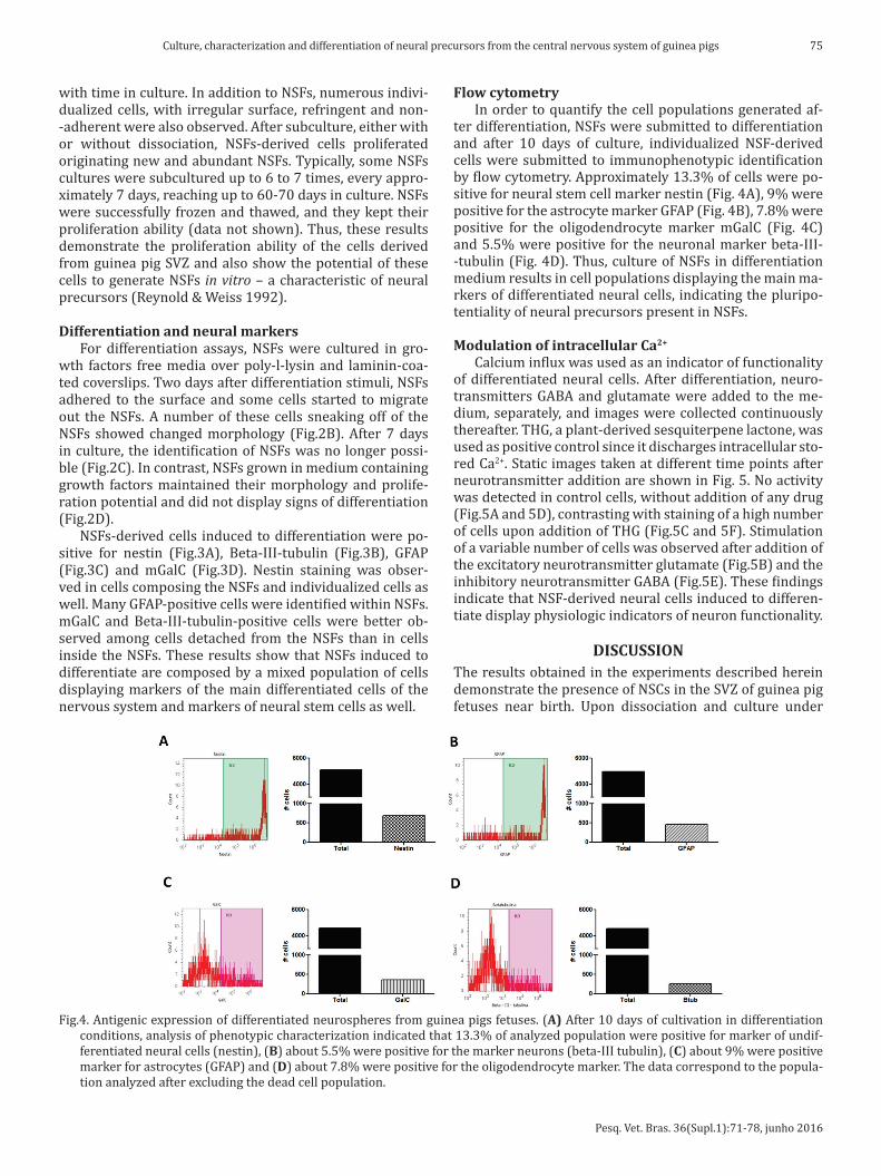
Flow cytometryIn order to quantify the cell populations generated af-
ter differentiation, NSFs were submitted to differentiation and after 10 days of culture, individualized NSF-derived cells were submitted to immunophenotypic identification by flow cytometry. Approximately 13.3% of cells were po-sitive for neural stem cell marker nestin (Fig. 4A), 9% were positive for the astrocyte marker GFAP (Fig. 4B), 7.8% were positive for the oligodendrocyte marker mGalC (Fig. 4C) and 5.5% were positive for the neuronal marker beta-III--tubulin (Fig. 4D). Thus, culture of NSFs in differentiation medium results in cell populations displaying the main ma-rkers of differentiated neural cells, indicating the pluripo-tentiality of neural precursors present in NSFs.
Modulation of intracellular Ca2+
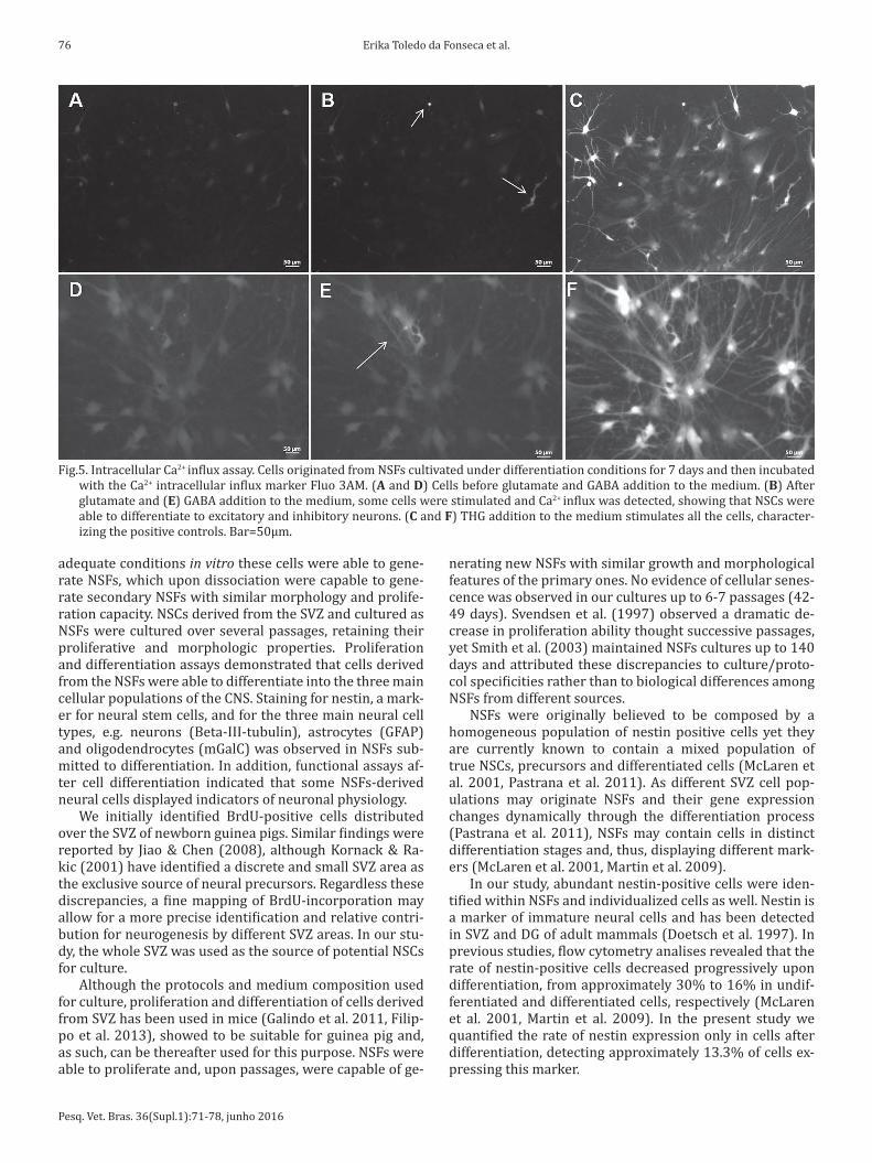
Calcium influx was used as an indicator of functionality of differentiated neural cells. After differentiation, neuro-transmitters GABA and glutamate were added to the me-dium, separately, and images were collected continuously thereafter. THG, a plant-derived sesquiterpene lactone, was used as positive control since it discharges intracellular sto-red Ca2+. Static images taken at different time points after neurotransmitter addition are shown in Fig. 5. No activity was detected in control cells, without addition of any drug (Fig.5A and 5D), contrasting with staining of a high number of cells upon addition of THG (Fig.5C and 5F). Stimulation of a variable number of cells was observed after addition of the excitatory neurotransmitter glutamate (Fig.5B) and the inhibitory neurotransmitter GABA (Fig.5E). These findings indicate that NSF-derived neural cells induced to differen-tiate display physiologic indicators of neuron functionality.
DISCUSSIONThe results obtained in the experiments described herein demonstrate the presence of NSCs in the SVZ of guinea pig fetuses near birth. Upon dissociation and culture under
Fig.4. Antigenic expression of differentiated neurospheres from guinea pigs fetuses. (A) After 10 days of cultivation in differentiation conditions, analysis of phenotypic characterization indicated that 13.3% of analyzed population were positive for marker of undif-ferentiated neural cells (nestin), (B) about 5.5% were positive for the marker neurons (beta-III tubulin), (C) about 9% were positive marker for astrocytes (GFAP) and (D) about 7.8% were positive for the oligodendrocyte marker. The data correspond to the popula-tion analyzed after excluding the dead cell population.
Pesq. Vet. Bras. 36(Supl.1):71-78, junho 2016
76 Erika Toledo da Fonseca et al.
adequate conditions in vitro these cells were able to gene-rate NSFs, which upon dissociation were capable to gene-rate secondary NSFs with similar morphology and prolife-ration capacity. NSCs derived from the SVZ and cultured as NSFs were cultured over several passages, retaining their proliferative and morphologic properties. Proliferation and differentiation assays demonstrated that cells derived from the NSFs were able to differentiate into the three main cellular populations of the CNS. Staining for nestin, a mark-er for neural stem cells, and for the three main neural cell types, e.g. neurons (Beta-III-tubulin), astrocytes (GFAP) and oligodendrocytes (mGalC) was observed in NSFs sub-mitted to differentiation. In addition, functional assays af-ter cell differentiation indicated that some NSFs-derived neural cells displayed indicators of neuronal physiology.
We initially identified BrdU-positive cells distributed over the SVZ of newborn guinea pigs. Similar findings were reported by Jiao & Chen (2008), although Kornack & Ra-kic (2001) have identified a discrete and small SVZ area as the exclusive source of neural precursors. Regardless these discrepancies, a fine mapping of BrdU-incorporation may allow for a more precise identification and relative contri-bution for neurogenesis by different SVZ areas. In our stu-dy, the whole SVZ was used as the source of potential NSCs for culture.
Although the protocols and medium composition used for culture, proliferation and differentiation of cells derived from SVZ has been used in mice (Galindo et al. 2011, Filip-po et al. 2013), showed to be suitable for guinea pig and, as such, can be thereafter used for this purpose. NSFs were able to proliferate and, upon passages, were capable of ge-
nerating new NSFs with similar growth and morphological features of the primary ones. No evidence of cellular senes-cence was observed in our cultures up to 6-7 passages (42-49 days). Svendsen et al. (1997) observed a dramatic de-crease in proliferation ability thought successive passages, yet Smith et al. (2003) maintained NSFs cultures up to 140 days and attributed these discrepancies to culture/proto-col specificities rather than to biological differences among NSFs from different sources.
NSFs were originally believed to be composed by a homogeneous population of nestin positive cells yet they are currently known to contain a mixed population of true NSCs, precursors and differentiated cells (McLaren et al. 2001, Pastrana et al. 2011). As different SVZ cell pop-ulations may originate NSFs and their gene expression changes dynamically through the differentiation process (Pastrana et al. 2011), NSFs may contain cells in distinct differentiation stages and, thus, displaying different mark-ers (McLaren et al. 2001, Martin et al. 2009).
In our study, abundant nestin-positive cells were iden-tified within NSFs and individualized cells as well. Nestin is a marker of immature neural cells and has been detected in SVZ and DG of adult mammals (Doetsch et al. 1997). In previous studies, flow cytometry analises revealed that the rate of nestin-positive cells decreased progressively upon differentiation, from approximately 30% to 16% in undif-ferentiated and differentiated cells, respectively (McLaren et al. 2001, Martin et al. 2009). In the present study we quantified the rate of nestin expression only in cells after differentiation, detecting approximately 13.3% of cells ex-pressing this marker.
Fig.5. Intracellular Ca2+ influx assay. Cells originated from NSFs cultivated under differentiation conditions for 7 days and then incubated with the Ca2+ intracellular influx marker Fluo 3AM. (A and D) Cells before glutamate and GABA addition to the medium. (B) After glutamate and (E) GABA addition to the medium, some cells were stimulated and Ca2+ influx was detected, showing that NSCs were able to differentiate to excitatory and inhibitory neurons. (C and F) THG addition to the medium stimulates all the cells, character-izing the positive controls. Bar=50µm.

Pesq. Vet. Bras. 36(Supl.1):71-78, junho 2016
77Culture, characterization and differentiation of neural precursors from the central nervous system of guinea pigs
The expression of Beta-III-tubulin observed after im-munostaining in a significant number of NSFs cells after dif-ferentiation, indicate that guinea pig SVZ, such as DG may also be a neuron source (Altman & Das 1967, Palmer et al. 2001, Guidi et al. 2005). Piper et al. (2001) demonstrated that the brain of human fetuses contain neural precursors that generate mature neurons after two weeks in culture. McLaren et al. (2001) concluded that the gradual decrease in nestin-positive cells upon differentiation is due to their replacement by Beta-III-tubulin positive cells that increase from 25% in undifferentiated cells to 70% in differentia-ted cells. In the present study, flow cytometry performed in NSFs-derived cells after 10 days of differentiation revealed approximately 5.5% Beta-III-tubulin positive cells, a num-ber significantly lower than previous studies. The rate of cells expressing GFAP after differentiation (approximately 9% of the cells) was also lower than that reported by Ara et al. (2010) and Martin et al. (2009) who reported GFAP staining in 30% to 35% of differentiated cells. These diffe-rences may indicate that the protocol and/or the stimuli used for differentiation were not as efficient as those used in previous studies.
Upon differentiation under surface-adherent condi-tions, a number of mGalC-positive cells was observed mi-grating out of the NSFs. Ren et al. (2009) demonstrated that the composition of the substrate for differentiation may in-fluence NSFs bevavior generating variable proportions of oligodendrocytes. In the present study, a higher number of mGalC positive cells was observed among cells that deta-ched from the NSFs than among cells composing the NSFs. Upon differentiation, approximately 7.8% of the cells were positive for mGalC and similar rate (8%) was observed by Martin et al. (2009). mGalC expression reinforce the mul-tipotentiality of NSFs provenient from the guinea pig SVZ confirming this region as a producer of neural stem cells.
Finally, our study demonstrated that a number of cells derived from NSFs induced to differentiation displayed indicators of neuronal function, as the differentiated cells responded to the neurotransmitters GABA and glutamate. Although the percentage of cells reacting to the neuro-transmitters was generally low (approximately 1 – 2%), positive reaction was consistent between trials. Conside-ring that differentiation resulted in a moderate number of differentiated neurons – as demonstrated by flow cytome-try – the low number of cells reacting to neurotransmit-ters should be expected. Piper et al. (2001) characterized neural precursors from human fetal tissue using Fura 2 AM to measure intracellular Ca2+. In differentiated cells (14 days) 3-5% of Beta-III-tubulin positive cells presented reactivity to neurotransmissores GABA (4% ±2%), glicine (6% ±3%) and glutamate (12% ±4%). This reactivity was not homogeneous in all cell passages indicating that a he-terogeneous neuronal population has been generated. The present study did not attempt to correlate neurotransmit-ter response with cell type yet reaction to GABA and glu-tamate was restricted to a well defined, restricted popula-tion of cells. Addition of THG, in contrast, led to generalized reaction among cultured cells since it releases microsomal stored calcium (Thastrup 1990, Lytton et al. 1991, Bagna-
resi et al. 2009). The reactivity to GABA was expected since SVZ-derived neuroblasts migrate rostraly to the olfactory bulbs (OB) where they become mature interneurons (Al-varez-Buylla & Garcia-Verdugo 2002), and rodent OB inter-neurons present significant amounts of GABA and dopa-mine (Kosaka & Kosaka 2005). The response to glutamate is explained by the fact that it is the predominant excitatory neurotransmitter of the mammal CNS (Cowan et al. 1997).
Taken together these results indicate that a proportion of differentiated cells from the SVZ presented indicators of neuronal functionality, reinforcing that cells from SVZ of guinea pig fetuses present mutipotentiality and may gen-erate functional neural cells in vitro upon adequate culture conditions and stimuli.
Acknowledgements.- The authors are grateful to Claudia Marinovic de Oliveira for giving the samples used in this study, and FAPESP for financial support.
REFERENCESAltman J. & Das G.D. 1967. Postnatal neurogenesis in guinea pig. Nature
214:1098-1101.Alvarez-Buylla A. & Garcia-Verdugo J.M. 2002. Neurogenesis in adult sub-
ventricular zone. J. Neurosci. 22:629-634.Alvarez-Buylla A. & Lois C. 1995. Neuronal stem cells in the brain of adult
vertebrates. Stem Cells 13:263-272.Ara J., Fekete S., Zhu A. & Frank M. 2010. Characterization of neural stem/
progenitor cells expressing VEGF and its receptors in the subventricular zone of newborn piglet brain. Neurochem. Res. 35:1455-1470.
Bagnaresi P., Alves E., Silva H.B., Epiphanio S., Mota M.M. & Garcia C.R.S. 2009. Unlike the synchronous Plasmodium falciparum and P.chabaudi infection, the P.berghei and P.yoelii asynchronous infections are not af-fected by melatonin. Int. J. Gen. Med. 30:47-55.
Buddensiek J., Dressel A., Kowalski M., Runge U., Shroeder H., Hermann A., Kirsch M., Storch A. & Sabolek M. 2010. Cerebroespinal fluid promotes survival and astroglial differentiation of adult human neural progenitor ell but inhibits proliferation and neuronal differentiation. BMC Neuros-ci. 11:1471-2202.
Conti L., Reitano E. & Cattaneo E. 2006. Neural stem cell system: diver-sities and properties after transplantation animal models of diseases. Brain Pathol. 16:143-154.
Cowan W.M., Jessell T.M. & Zipursky S.L. 1997. Molecular and Cellular Ap-proaches to Neural Development. Oxford University Press, New York. 563p.
Doetsch F., García-Verdugo J.M. & Alvarez-Buylla A. 1997. Cellular compo-sition and three-dimensional organization of the subventricular germi-nal zone in the adult mammalian brain. J. Neurosci. 17:5046-5061.
Filippo T.R.M., Galindo L.T., Barnabe G.F., Ariza C.B., Mello L.E., Juliano M.A., Juliano L. & Porcionatto M.A. 2013. CXCL12 N-terminal end is sufficient to induce chemotaxis and proliferation of neural stem/progenitor cells. Stem Cell Res. 11:913-925.
Galindo L.T., Filippo T.R.M., Semedo P., Ariza C.B., Moreira C.M., Camara N.O.S. & Porcionatto M.A. 2011. Mesenchymal stem cell therapy modu-lates the inflammatory response in experimental traumatic brain injury. Neurol. Res. Int. 1:1-9.
García-Verdugo J.M., Doetsch F., Wichterle H., Lim D.A. & Alvarez-Buylla A.A. 1998. Architecture and cell types of the adult subventricular zone: in search of the stem cells. J. Neurol. 36:234-248.
Götz M. & Huttner W.B. 2005. The cell biology of neurogenesis. Mol. Cell Biol. 6:777-788.
Guidi S., Cianti E., Severi S., Contestabile A. & Bartesaghi R. 2005. Postna-tal neurogenesis in the dentate gyrys of the guinea pig. Hippocampus 15:285-301.

Pesq. Vet. Bras. 36(Supl.1):71-78, junho 2016
78 Erika Toledo da Fonseca et al.
Jandial R., Singec I., Duenas V.J., Ho A.L., Levy M.L. & Snyder E.Y. 2007. Cen-tral nervous system repair and stem cells. International Congress Series 1302:154-163.
Jiao J. & Chen D.F. 2008. Induction of neurogenesis in nonconvetional neurogenic regions of the adult central nervous system by niche astro-cyte-produced signals. Stem Cells 26:1221-1230.
Kornack D.R. & Rakic P. 2001. The generation, migration, and differentia-tion of olfactory neurons in the adult primate brain. PNAS 98:4752-4757.
Kosaka K. & Kosaka T. 2005. Synaptic organization of the glomerulus in the main olfactory bulb: compartments of the glomerulus and heterogene-ity of the periglomerular cells. Anat. Sci. Int. 80:80-90.
Lacbawan F.L. & Muenke M. 2002. Central nervous system embryogenesis and its failures. Pediatr. Devel. Pathol. 5:425-447.
Lytton J., Westlin M. & Hanley M.R. 1991. Thapsigargin inhibits the sar-coplasmatic or endoplasmatic reticulum Ca-ATPase family of calcium pumps. J. Biol. Chem. 266:17067-17071.
Manganas L.N. & Maletic-Savatic M. 2005. Stem cell therapy for central nervous system demyelinating disease. Curr. Neurol. Neurosci. Rep. 5:225-231.
Mansergh F.C., Wride M.A. & Rancourt D.E. 2004. Neurons, stem cells and potential therapies, p.177-189. In: Sell S. (Ed.), Stem Cells Handbook. Human Press, New Jersey. 526p.
Martin I., Andres A.R., Védrine S., Tabagh R., Michelle C., Jourdan M-L., Heu-ze-Vourc’h N., Corcia P., Duittoz A. & Vourc’h P. 2009. Effect of the oligo-dendrocyte myelin glycoprotein (OMgp) on the expansion and neuronal differentiation of rat neural stem cells. Brain Res. 1284:22-30.
McLaren F.H., Svendsen C.N., Van der Meide P. & Joly E. 2001. Analysis of neural stem cells by flow cytometry: Cellular differentiation modifies patterns of MHC expression. J. Neuroimmunol. 112:35-46.
Okano H. 2010. Neural stem cells and strategies for the regeneration of the central nervous system. Proc. Jpn. Acad. 86:438-450.
Palmer T.D., Schwartz P.H., Taupin P., Kaspar B., Stein S.A. & Gage F.H. 2001. Cell culture: Progenitor cells from human brain after death. Nature 411:42-43.
Pastrana E., Silva-Vargas V. & Doetsch F. 2011. Eyes wide open: a critical review of sphere-formation as an assay for stem cells. Cell Stem Cell 8:486-498.
Piper D.R., Mujtaba T., Keyong H., Roy N.S., Goldman S.A., Rao M.S. & Lu-cero M.T. 2001. Identification and characterization of neuronal pre-cursors and their progeny from human fetal tissue. J. Neurosci. Res. 66:356-368.
Potter G.E. & Brueck W.L. 1958. Nervous system of guinea pig (Cavia por-cellus). Biosci. 19:185-196.
Ren Y-J., Zhang H., Huang H., Wang X-M., Zhou Z-Y., Cui F-Z. & An Y-H. 2009. In vitro behavior of neural stem cells in response to different chemical functional groups. Biomaterials 30:1036-1044.
Schwartz P.H., Bryant P.J., Fuja T.J., Su H., O’dowd D.K. & Klassen H. 2003. Isolation and characterization of neural progenitor cells from post-mor-tem human cortex. J. Neurosci. Res. 74:838-851.
Smith R., Bagga V. & Fricker-Gates R.A. 2003. Embryonic neural progenitor cells: the effects of species, region, and culture conditions on longterm pro-liferation and neuronal differentiation. J. Hematoth. Stem Cell 12:713-725.
Svendsen C.N., Skepper J., Rosser A.E., Ter Borg M.G., Tyres P. & Ryken T. 1997. Restricted growth potential of rat neural precursors as compared to mouse. Brain Res. Dev. Brain Res. 99:253-258.
Thastrup O. 1990. Role of Ca2+ -ATPases in regulation of cellular Ca2+ sig-naling, as studied with the selective microsomal Ca2+ -ATPase inhibitor, thapsigargin. Agents Actions 29:8-15.