the Adult Male Guinea Pigs (Cavia Porcellus Linnaeus ...
18
Page 1/18 The Effect of Kuchala (Arum Korolkowii Regel, 1873) Tuber Tincture to Increase of the Serum Testosterone in the Adult Male Guinea Pigs (Cavia Porcellus Linnaeus, 1758) Nurbek Aldayarov ( [email protected] ) Kyrgyz-Turkish Manas University Jarkinay Jumabekova Kyrgyz-Turkish Manas University Gulbubu Kurmanbekova Kyrgyz-Turkish Manas University Nurjamal Omurzakova Kyrgyz-Turkish Manas University Bermet Kydyralieva Kyrgyz-Turkish Manas University Gulnara Sharshenalieva Kyrgyz-Turkish Manas University Bakyt Borkoev Kyrgyz-Turkish Manas University Kalipa Salieva Kyrgyz-Turkish Manas University Askarbek Tulobaev Kyrgyz-Turkish Manas University Ruslan Salykov Kyrgyz-Turkish Manas University Kadyrbay Chekirov Kyrgyz-Turkish Manas University Research Article Keywords: Kuchala tuber tincture, Hematology, Serum biochemistry, Histology, Testosterone, Adult male guinea pigs Posted Date: January 7th, 2021 DOI: https://doi.org/10.21203/rs.3.rs-133900/v1
Transcript of the Adult Male Guinea Pigs (Cavia Porcellus Linnaeus ...

Page 1/18
The Effect of Kuchala (Arum Korolkowii Regel, 1873)Tuber Tincture to Increase of the Serum Testosterone inthe Adult Male Guinea Pigs (Cavia Porcellus Linnaeus,1758)Nurbek Aldayarov ( [email protected] )
Kyrgyz-Turkish Manas UniversityJarkinay Jumabekova
Kyrgyz-Turkish Manas UniversityGulbubu Kurmanbekova
Kyrgyz-Turkish Manas UniversityNurjamal Omurzakova
Kyrgyz-Turkish Manas UniversityBermet Kydyralieva
Kyrgyz-Turkish Manas UniversityGulnara Sharshenalieva
Kyrgyz-Turkish Manas UniversityBakyt Borkoev
Kyrgyz-Turkish Manas UniversityKalipa Salieva
Kyrgyz-Turkish Manas UniversityAskarbek Tulobaev
Kyrgyz-Turkish Manas UniversityRuslan Salykov
Kyrgyz-Turkish Manas UniversityKadyrbay Chekirov
Kyrgyz-Turkish Manas University
Research Article
Keywords: Kuchala tuber tincture, Hematology, Serum biochemistry, Histology, Testosterone, Adult male guineapigs
Posted Date: January 7th, 2021
DOI: https://doi.org/10.21203/rs.3.rs-133900/v1

Page 2/18
License: This work is licensed under a Creative Commons Attribution 4.0 International License. Read FullLicense

Page 3/18
AbstractBackground: Kuchala (Arum korolkowii Regel) is a medicinal plant often used in folk medicine in the KyrgyzRepublic. As a medicinal raw material, the tuber’s tincture is used in small doses to increase human sexualpotency. However, there is no scienti�c evidence in support of the medicinal effects of kuchala. For thesereasons, we decided to study the pharmacological effect of kuchala tuber tincture on the sexual potency of adultmale guinea pigs. We analyzed the effect of kuchala at the age of ±48-months, in 12 male guinea pigs. Apreparation of 10% tuber tincture of kuchala in 70% ethanol was administered perorally to the male guinea pigsin the form of a once-daily dose of 150 µl for 30 days. The study data were obtained by ethological,hematological and serum biochemistry, gross anatomical, histological and statistical methods.
Results: The hematological and serum biochemistry parameters were signi�cantly different between the controland the experimental group. The neutrophils’ percentage in the experimental group was signi�cantly lower (dР <0.001) than in the control group. On the other hand, the lymphocyte counts were signi�cantly higher in theexperimental group (dР < 0.001). The RBC counts, Hgb, Hct, MCH and MCHC were signi�cantly higher in theexperimental group (dР < 0.001; dР < 0.001; cР < 0.01; dР < 0.001; dР < 0.001 respectively) than in the controlgroup. In contrast, the color indicator and the mean platelet volume were higher (bР < 0.05) and signi�cantlyhigher (dР < 0.001) respectively in the control group than in the experimental group. The ALT and AST levels werelower in the experimental than in the control group (both dР < 0,001). The testosterone concentration in serumwas much higher (dР < 0,001) in the experimental group. Microscopically, some structural damages were foundin the liver of the experimental animals indicating a metabolic disorder. However, the testes showed animprovement in spermatogenesis in the experimental compared with the control group.
Conclusions: The 10% kuchala tuber tincture in 70% ethanol has a positive effect in terms of improving thesexual potency of adult guinea pigs by increasing the production of testosterone and increasingspermatogenesis.
BackgroundSince ancient times, people have successfully used folk medicine about medicinal plants as an importanttherapeutic agent in both human and veterinary medicine [1–3]. Many of the treatment methods have beenpassed down through families for generations, and some of these have been adapted for use in modern medicalpractice. Among others, Kyrgyz folk medicine has occupied an important place in the nomadic civilization of theKyrgyz people.
The Kyrgyz Republic is a mountainous country in central Asia. Due to the extreme environment and climate, thereis a diverse range of species of plants, including more than 200 species of medical plants. Many of the medicalplants used in Kyrgyz folk medicine have not been studied using modern scienti�c techniques [4]. Arumkorolkowii Regel is one of the medicinal plants often used in the folk medicine of Central Asia, which has not lostits relevance, even today. The vernacular name of this medicinal plant is kuchala. A. korolkowii R., 1873 belongsto the genus Arum L. of the family Araceae Juss. and grows in soil pockets on rocky hillsides, beneath low scrub.Its native range is Central Asia, North-Western China, Northern Iran and Afghanistan. A. korolkowii R. is aperennial tuberous herb sprouting in early spring from a discoid, vertically-orientated tuber. It has a well-describedbiological characteristic [5, 6]. However, there is another plant also called kuchala or kuchla or Chinese kuchla

Page 4/18
(Strychnos nux-vomica). S. nux-vomica is an evergreen tree up to 25 m height. Its dried seeds (Nux vomica) areused in modern and traditional medicine [7, 8].
A. korolkowii R. is a very poisonous herb. In folk medicine it is used as a medicinal raw material in the form oftincture of tubers which is administered in small doses in order to increase human potency, to treat infertility andstomach ulcers, diseases of the nasopharynx and the respiratory tract, and to eliminate fatigue and givestrength. The powdered tuber is used to treat poisonous snake and scorpion bites, fungal skin diseases andhemorrhoids [5]. The medicinal properties of kuchala were mentioned in the works of Avicenna and in suchKyrgyz folk epics as «Manas» [9] and «Semetey» [10]. According to the above-mentioned sources, the milky andsour-milky (kumys) tinctures of the tubers of this plant are often used among the elderly (over 70 years) as adrug that increases the sexual potency of men. However, there are no modern scienti�c data proving themedicinal properties of kuchala, especially, in terms of its effect on human potency. Besides, the A. korolkowii R.chemical composition has not yet been studied. In this regard, we decided to experiment with kuchala tubertinctures on laboratory animals, speci�cally with adult male guinea pigs.
Many characteristics make the guinea pig an ideal model for biomedical research [11]. Guinea pigs have beencomprehensively studied as laboratory animals using biological, morphological and physiological approaches.Its size and lifestyle make the guinea pig easy to keep and to conduct experimental studies on and, importantly, ithas many of the same morphological and physiological characteristics as humans [11, 12], including in terms ofreproduction [13–18]. Consequently, guinea pigs are frequently used as a biological model in studies of anumber of infectious bacterial and noninfectious diseases [12, 19].
Blood evaluations are a prime means of diagnosis in both human and animal medicine. This is becausehematological and serum biochemistry data can show changes in physiological disturbances such as systemicin�ammation, renal or hepatocellular disorders [11]. The aim of the present study was to explore thepharmacological effect of the kuchala (Arum korolkowii Regel) tuber tincture on the hematological and serumbiochemistry parameters, and on the testis and liver structures of the adult male guinea pigs (Cavia porcellusLinnaeus, 1758).
Results
Physical characteristics of tubers and tinctureDried kuchala tubers were discoid (Fig. 1a) in the main, 2–6 cm across, and 2-2.3 cm thick. The color of the peelwas light brown and hard. When cleaning of the peel, it �aked off into small, hard scales. Under the hard peel ofthe tuber was a soft, thin, and easily removed shell of a yellowish-white color. On transverse sections, the tuberwas yellow-white color (Fig. 1b), easily cut and was soft consistency similar to that of plasticine. Tubers could bepressed easily and formed a mushy oily mass. The kuchala tubers did not have a pungent smell. The taste wasnot immediately apparent, but a few moments later, a strong bitter taste of spicy pepper was felt, which lastedfor a long time. The prepared 10% tincture of tubers on 70% ethanol was transparent and viscous, yellowish-reddish in color (Fig. 1c). The tincture has a special bitter smell, different from the smell of alcohol.
Animal behavior

Page 5/18
The behavior of animals in both groups was observed through a window from the next room, and the results ofthe observation were recorded. Both the control and the experimental group led an active lifestyle. They often ranand played, fought among themselves, and ate well. However, gradually, day after day, an increase in the appetiteand activity of the experimental animals was observed when compared to the control group. The experimentalanimals often fought among themselves and were more aggressive. Sometimes they climbed on the sidewalls ofthe isolator cage.
HematologyWe observed no adverse effects in terms of the clinical signs of anemia and other disorders after the guinea pigswere phlebotomized under iso�urane anesthesia.
Common hematology parameters such as WBC, neutrophils, lymphocytes, monocytes, eosinophils, basophils,RBC, Hgb, Hct, MCV, MCH, MCHC, color indicator, erythrocyte sedimentation rate, platelets and mean plateletvolume were evaluated (Table 1). The results of the study showed that several parameters of the blood weresigni�cantly different between the control and the experimental groups (Fig. 2). Neutrophils’s percentage in theexperimental animals was signi�cantly lower (dР < 0.001) than in the control animals. Lymphocyte counts on theother hand were signi�cantly higher in the experimental animals (dР < 0.001). RBC counts, Hgb, Hct, MCH andMCHC were signi�cantly higher in the experimental animals (dР < 0.001; dР < 0.001; cР < 0.01; dР < 0.001; dР <0.001 respectively) than in the control guinea pigs. However, the color indicator and the mean platelet volumewere higher (bР < 0.05) and signi�cantly higher (dР < 0.001) respectively in control animals than in experimentalanimals. Other hematological parameters such as WBC, monocytes, eosinophils, basophils, MCV, erythrocytesedimentation rate, platelets between animal groups were not statistically signi�cant. Note: the hematologicaland serum biochemistry parameters of the animals obtained before the experiment and the animals in thecontrol group showed almost no difference. Therefore, only the parameters of the control group animals wereshown.

Page 6/18
Table 1Hematological parameters for control and experimental guinea pigs
Bloodparameters
Control group (n = 10) Experimental group (n = 12)
Mean SD Median Min Max Mean SD Median Min Max
WBC (x109/L) 7.24 ± 0.343
1.084 7.1 5.5 9.1 7.94 ± 1.00
3.48 7 4 15.2
Neutrophils(%)d
53.6 ± 1.258d
3.978 53 48 61 27.08 ± 2.62
9.07 26.5 14 46
Lymphocytes(%)d
39.4 ± 0.872
2.757 40 35 43 56.08 ± 3,08d
10.66 56.5 40 71
Monocytes(%)
4.8 ± 0.291
0.919 5 4 7 4.58 ± 0,89
3.09 3.5 2 11
Eosinophils(%)
2.4 ± 0.499
1.578 3 0 4 5.42 ± 1,65
5.71 4 0 20
Basophils (%) 0.4 ± 0.163
0.516 0 0 1 0.08 ± 0,08
0.29 0 0 1
RBC(x1012/L)d
4.53 ± 0.110
0.347 4.6 3.9 4.9 5.38 ± 0,03d
0.10 5.35 5.3 5.6
Hgb (g/dL)d 144.5 ± 0.934
2.953 145.5 139 148 154.58 ± 1,14d
3.96 154.5 147 160d
Hct (%)c 43.6 ± 0.340
1.075 44 42 45 45.33 ± 0.37 c
1.27 45.1 44 48c
MCV (fL) 76.6 ± 1.222
3.864 77 70 81 74.61 ± 0.95
3.30 73.8 70.4 84.2
MCH (pg) d 24.27 ± 0.495
1.566 24.2 21.9 26.3 53.89 ± 5.94d
20.57 51.5 25.7 95.5
MCHC (g/dL)d
32.36 ± 0.264
0.834 32.45 30.9 33.3 752.83 ± 132.66d
459.54 685 352 1910
Colorindicatorb
0.88 ± 0.007b
0.022 0.88 0.83 0.9 0.85 ± 0,00
0.01 0.855 0.83 0.87
Erythrocytesedimentationrate(mm/hour)
2.6 ± 0.163
0.516 3 2 3 2.25 ± 0.13
0.45 2 2 3
Platelets(x109/L)
301.9 ± 2.364
7.475 303 289 310 343.50 ± 22.05
76.38 335.5 235 494
Note: bР < 0.05; cР < 0.01; dР < 0.001.

Page 7/18
Bloodparameters
Control group (n = 10) Experimental group (n = 12)
Mean SD Median Min Max Mean SD Median Min Max
Mean plateletvolume (fL)d
7.43 ± 0.037d
0.116 7.4 7.3 7.7 4.58 ± 0.30
1.04 4.8 3.1 6
Note: bР < 0.05; cР < 0.01; dР < 0.001.
Serum biochemistryIn this study, some serum biochemical parameters such as ALT (alanine aminotransferase), AST (alanineaminotransferase), glucose and testosterone were evaluated (Table 2). As a result, three of them (ALT, AST andtestosterone), were signi�cantly different in terms of the control and the experimental groups of animals (Fig. 3).ALT and the AST percentages in the experimental animals were signi�cantly lower than those in the controlanimals (both dР < 0.001). The testosterone concentration was considerably higher (dР < 0.001) in theexperimental guinea pigs and the glucose percentage in the serum was not statistically signi�cant between theanimal groups studied.
Table 2Serum biochemistry parameters for control and experimental guinea pigs
Serumparameters
Control group (n = 10) Experimental group (n = 12)
Mean SD Median Min Max Mean SD Median Min Max
ALT (U/L)d 63.65 ± 0.521d
1.647 64.25 60.7 65.4 55.82 ± 1.33
4.59 55.6 48.6 63.93
AST (U/L)d 78.29 ± 3.096d
9.790 80.05 58.2 90.9 60.78 ± 2.00
6.94 62.85 47.67 69.9
Glucose(mmol/L)
9.877 ± 0.621
1.963 9.92 6.91 13.5 9.20 ± 0.39
1.35 9.5 6.99 11.08
Testosterone(nmol/L)d
9.533 ± 0.184
0.583 9.615 8.63 10.4 21.73 ± 2.11d
7.32 20.95 11.4 31.5
Note: dР < 0.001
Gross anatomy and histologyIn order to make a comparison, all internal organs were visually studied, especially the heart, liver, kidneys andtestes in both the control and the experimental groups. Results of a visual examination of the color, consistencyand degree of blood �lling of the above-mentioned organs concerning the control and experimental groups ofnecropsied guinea pigs did not reveal any signi�cant differences. Comparative morphometric studies (organweight, width, length and thickness) of the liver and testes in animals of both groups were also not statisticallysigni�cant. Therefore, we did not provide comparative morphometric data of the studied organs.
A microscopic examination of the blood smears also revealed no noticeable differences or changes betweenanimals in the control and experimental groups. Only the WBC of the guinea pigs in the experimental group were

Page 8/18
re�ected (Fig. 4) without a description of the structural features of each of them.
Microscopic examination of the liver of the control guinea pigs visualized normal hexagonal hepatic lobules invarious sizes having hepatocytes, a central vein with blood cells, sinusoids along with empty spaces, and notclearly visible macrophages (Kupffer cells), lining different places of the sinusoids (Fig. 5a). The liver sections ofthe experimental animals were stained poorly when compared to the control group. Liver cords, sinusoids,intensive stained Kupffer cells, several apoptotic �gures and apoptosomes (Fig. 5a*) were clearly visible.
A histological examination of a section of the control guinea pigs’ testes showed tubular glands and intertubularconnective tissue by speci�c intestinal or Leydig cells. The round-oval seminiferous tubules were of varioussizes, and they surrounded by loose vascular connective tissue forming the lobules of the testis. The coiledseminiferous tubules were lined with multilayered spermatogenic cells in different developmental stages, andsustentacular or Sertoli cells (Fig. 5b). There was a decrease in the number of spermatogenic cells inseminiferous tubules. The experimental testis section showed a restoration of spermatogenesis in theseminiferous tubules (Fig. 5b*). The amount of spermatogonias, primary spermatocytes, and spermatids hadincreased. The lumen of the seminiferous tubule was �lled with developing spermatozoa.
DiscussionThis paper is devoted to the scienti�c exploration of the pharmacological effect of the kuchala (Arum korolkowiiRegel) tuber which has long been used in Asia folk medicine, on male potency. The Kyrgyz people have used thetubers of this medicinal plant for a long time, adding to the process the preparation of the national drink, kumys(fermented milk product of mare's milk), as a means of increasing the strength and endurance of male warriors[9, 10]. Folk healers, referring to the works of Avicenna, indicate that the kuchala tubers in conjunction with wine,stimulates sexual desire and detoxi�es the kidneys. However, the recipe of the kuchala tuber tincture in thekumys and in the wine has not been written in detail, and modern folk healers also keep it in secret. Based on theabove, we prepared a 10% tincture of kuchala tubers in 70% ethyl alcohol.
The next important stage of research planning was the choice of the type of laboratory animal for experimentalstudy. To determine this, we analyzed the literature and found that among the different laboratory animals, theguinea pig has many morphofunctional similarities with humans, in terms of the lung physiology [20], andhormonal, immunological and corticosteroid responses [11, 12]. Importantly, the guinea pig has many commonfeatures with humans in terms of reproduction related to the accessory glands [14, 15], the characteristics of theplacenta [21], and morphological and functional analysis of spermatogenesis [22]. In addition, information exitsthat the testosterone and androstenedione content in serum and testes were different in guinea pigs in theprenatal [18, 23], and postnatal [24] periods. The concentration of testosterone in plasma reaches its maximumlevel at 60 days of age in guinea pigs, and then decreases with the increase in age. In the 24–35 month oldguinea pigs, there is a 65% decreased in the concentration of testosterone in blood plasma [24]. For centuries,Kyrgyz people have used the kumys tinctures of the kuchala tuber to treat disorders in terms of sexual libido ofmen over 70 years of age and to strengthen the bone system. In this regard in particular, the adult male guineapigs were selected at ± 48-months of age, when their testosterone level was low. Additionally, in guinea pigs thestructure of the testicles [25], epididymis [26] and spermatogenesis [27] have been studied in detail. The above-mentioned data served as a justi�cation for the use of guinea pigs in this experimental study.

Page 9/18
For the comparative analysis, we studied the hematological and serum biochemical parameters of guinea pigs.Based on the hematological and serum biochemical data of an inbred strain, 13/N guinea pigs were divided intothe following age groups - juveniles (0-150 days), adults (151–900 days) and geriatric adults (older than 900days) [28]. Our selection for the experimental study was 48-month-old guinea pigs. Although from a differentbreed, they fully met our goal in terms of the choice of adult animals.
In terms of the comparative aspect of our study, we studied sixteen hematological (Table 1) and four serumbiochemistry parameters (Table 2) of the blood. Based on our data, we can say with con�dence that the goal ofthis experimental work has been achieved. This is so because the concentration of testosterone in the bloodplasma in the experimental animals (21.73 ± 2.11 d) was more than twice that of the control animals (9.533 ± 0.184), which is con�rmed statistically (dP < 0.001). In addition, it is con�rmed that microstructural changes inthe testes showed improvement in terms of spermatogenesis, i.e. increase of spermatogenesis cells in theseminiferous tubules.
However, some hematological parameters such as the lymphocytes, RBC, Hgb, Hct MCH, MCHC, color indicatorand mean platelet volume signi�cantly increased in the experimental guinea pigs (dР < 0.001; dР < 0.001; cР <0.01; dР < 0.001; dР < 0.001; dР < 0.001, bР < 0.05, dР < 0.001 respectively), compared with the control animals.On the other hand, the neutrophils percentage in the experimental animals was signi�cantly lower (dР < 0.001)than in the control animals. Such suspicious data indicates the toxic effect of the tincture on guinea pigs. This isprobably due to the dose (0.15 µl) or the high concentration of the tuber tincture (10%), possibly with the higherdiluted solution of ethanol (70%), or the long time (30 consecutive days) over which the tincture was given to theexperimental guinea pigs. Hepatocellular injures can be evaluated using serum biochemistry parameters ofalkaline phosphatase (ALP) and alkaline aminotransferanse (ALT) [11]. The toxic effect of this tincture on theguinea pigs was also con�rmed with regards to a decrease in the concentration of ALT and AST in the plasmabiochemistry of the experimental guinea pigs, which was statistically signi�cant (dP < 0.001). Such a process isobserved in cirrhosis or necrosis of the liver. The toxic effects of any drug or toxic substance in the organism areexpressed by damage to the structure of the liver [29, 30]. The gross anatomy parameters of the liver of bothanimal groups were similar, which is consistent with the data of other researchers of the liver of guinea pigs [31].However, microscopic examination revealed some differences in the liver in the experimental animals. The liversections of the experimental animals were pale-stained, and all the microstructural components of the liver -cords, sinusoids, Kupffer cells [32] - were clearly visible. There were several apoptotic �gures and apoptosomesin the liver parenchyma. As is known, pale staining of cell structures indicates low functional activity on the partof the organ.
ConclusionThe present study shows the 10% kuchala (Arum korolkowii Regel) tuber tincture in 70% ethanol has a positiveeffect in terms of improving the sexual potency of old male guinea pigs by increasing the production oftestosterone and increasing the spermatogenesis. The toxic effects of this tincture on the animal organism canbe resolved by reducing the dose. The concentration of the drug tincture in ethanol will be the main aim of ournext study.
Materials And Methods

Page 10/18
Kuchala tubers and making the tinctureFive pieces (total 49.62 g) dried kuchala tubers were purchased at local markets in Bishkek (Kyrgyz Republic).Each of the tuber pieces was cleaned with warm water and then with 70% of ethanol, and air dried at roomtemperature. The tubers with peel attached were shredded using a manual grinder. The ground tuber wasweighed on a Precisa (Switzerland) electronic weight and prepared as a 10% tincture in 70% ethanol. The tincturewas poured into a dark glass bottle which was tightly closed. It was then placed in a dark place at roomtemperature. The tincture was mixed twice per day, and this process was continued for 14 days until the tincturewas ready. On the 15th day, the tincture was �ltered through dense gauze and then through �lter paper. The 10%tincture in 70% ethanol so prepared was then stored in a refrigerator (+ 4 °C) for use in the experimental study.The main phases of the experimental study, in sequential order, are shown on the following schematic image(Fig. 6).
Experimental animals and husbandryWe purchased clinically healthy 22 male guinea pigs of Abyssinian breeds, all ± 48-month-old and weighing onthe average 682 g (489–792 g) from a private guinea pig breeder. Animals were housed in two handmadeisolator cages measuring 98.7 cm x 347.89 cm x 54.3 cm for 10 control animals and 110.3 cm x 398.73 cm x54.6 cm for 12 experimental animal for the duration of the experiment, with under sun-dried clean straw beddingand cardboard huts. The bedding was changed every two days or more frequently if needed. Animals werehoused at a room temperature of 20–26 °C with 40–70% humidity. Guinea pigs had access ad libitum to rodentchow and water. Animals were allowed to acclimate in the vivarium for 10 days after delivery before they wereused for this study. The research was conducted in accordance with the internationally accepted principles forlaboratory animal use and care [33], and ARRIVE (Animal Research: Reporting of In Vivo Experiments) guidelines2.0 [34].
Treatments and handlingWe observed the behavior of the guinea pigs during the adaptation period (10 days). Based on their physicalactivity and body weight, the experimental animals were divided in two groups. Both the control andexperimental groups of animals were formed according to the above-mentioned principle. The control groupconsisted of 10 and the experimental group of 12 male animals, and both groups were kept in the sameconditions. Every day from 8:00 to 9:00 a.m. 150 µl of kuchala tubers tincture were administered to theexperimental animals and 150 µl normal water was administered perorally to the control group over 30 days.After that, we observed the behavior of the animals through a window in the next room and took note of thechanges in the behavior of the animals.
Blood collectionBlood samples were collected from each guinea pig on two occasions, �ve days before the start and after the�nish of giving tincture of the tuber, under iso�urane (3–5%) anesthesia. Blood was collected from the cranialvena cava according to a previously well-described method [35]. During the phlebotomy, the rules of asepsis andantiseptic were strictly observed [11]. Blood from the cranial vena cava was obtained using a 25-gauge, 5/8-inneedle attached to a 3 ml syringe (Zhejiang Huafu Medical Equp. Co. Ltd China). Collected blood wasimmediately transferred into Gel/Clot Activator (GD060SGC) tubes for serum collection and EDTA.K3(GD060EK3) tubes for general blood analysis. In addition, blood smears were taken for cytology analysis.
Page 11/18
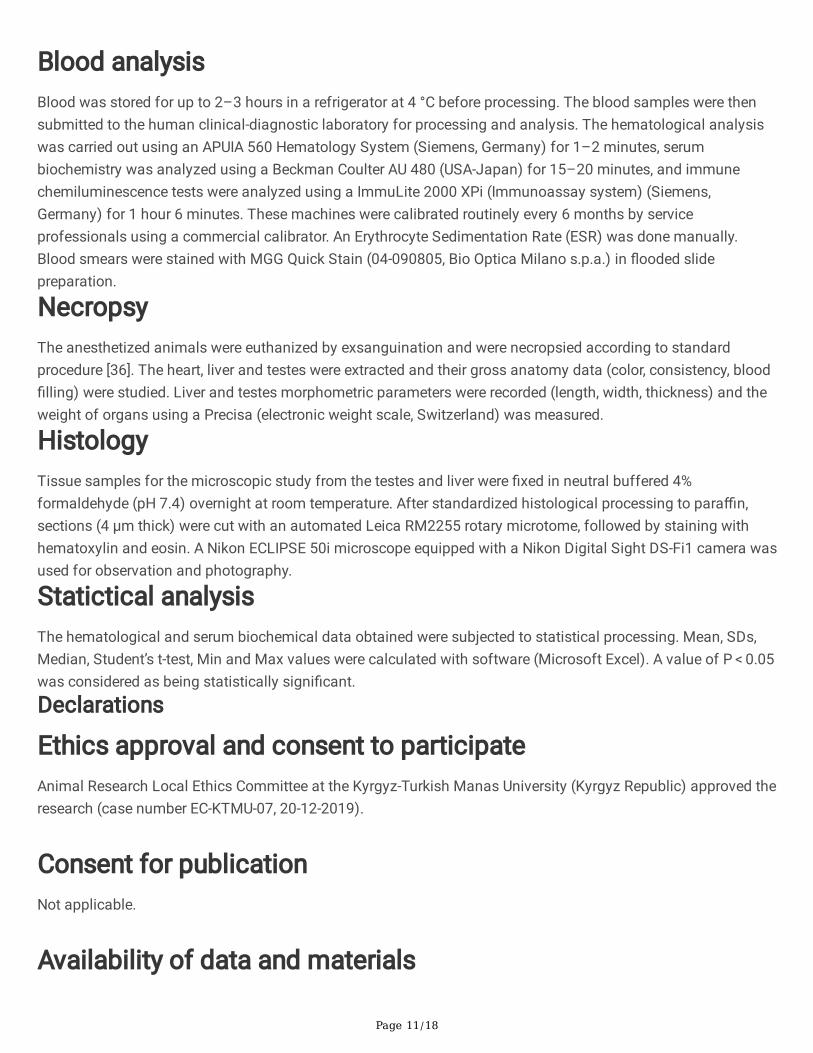
Blood analysisBlood was stored for up to 2–3 hours in a refrigerator at 4 °C before processing. The blood samples were thensubmitted to the human clinical-diagnostic laboratory for processing and analysis. The hematological analysiswas carried out using an APUIA 560 Hematology System (Siemens, Germany) for 1–2 minutes, serumbiochemistry was analyzed using a Beckman Coulter AU 480 (USA-Japan) for 15–20 minutes, and immunechemiluminescence tests were analyzed using a ImmuLite 2000 XPi (Immunoassay system) (Siemens,Germany) for 1 hour 6 minutes. These machines were calibrated routinely every 6 months by serviceprofessionals using a commercial calibrator. An Erythrocyte Sedimentation Rate (ESR) was done manually.Blood smears were stained with MGG Quick Stain (04-090805, Bio Optica Milano s.p.a.) in �ooded slidepreparation.
NecropsyThe anesthetized animals were euthanized by exsanguination and were necropsied according to standardprocedure [36]. The heart, liver and testes were extracted and their gross anatomy data (color, consistency, blood�lling) were studied. Liver and testes morphometric parameters were recorded (length, width, thickness) and theweight of organs using a Precisa (electronic weight scale, Switzerland) was measured.
HistologyTissue samples for the microscopic study from the testes and liver were �xed in neutral buffered 4%formaldehyde (pH 7.4) overnight at room temperature. After standardized histological processing to para�n,sections (4 µm thick) were cut with an automated Leica RM2255 rotary microtome, followed by staining withhematoxylin and eosin. A Nikon ECLIPSE 50i microscope equipped with a Nikon Digital Sight DS-Fi1 camera wasused for observation and photography.
Statictical analysisThe hematological and serum biochemical data obtained were subjected to statistical processing. Mean, SDs,Median, Student’s t-test, Min and Max values were calculated with software (Microsoft Excel). A value of P < 0.05was considered as being statistically signi�cant.
Declarations
Ethics approval and consent to participateAnimal Research Local Ethics Committee at the Kyrgyz-Turkish Manas University (Kyrgyz Republic) approved theresearch (case number EC-KTMU-07, 20-12-2019).
Consent for publicationNot applicable.
Availability of data and materials

Page 12/18
The datasets used and/or analysed during the current study are available from the corresponding author onreasonable request.
Competing interestsThe authors declare that they have no competing interests.
FundingThis study was not funded.
Authors’ contributionsAN and JJ conceived, designed and realized the study. JJ, ON, KB and SK participated in the experimental work.AN, TA and SR took blood and necropsied the animals. ChK, KG and TA provided academic instruction. AN, TA,KG, BB and ShG conducted data collection and analysis. ChK undertook the statistical processing of the data.AN interpreted and wrote the draft manuscript. All authors read and approved the �nal manuscript.
AcknowledgementsWe thank Dr. Zarima Jumakanova, Peil Esengul kyzy and Ella Abylaeva for support technical editing andproofreading.
Author details1Department of Biology, Faculty of Sciences, Kyrgyz-Turkish Manas University, Bishkek 720042, Kyrgyz Republic.2Department of Chemical Engineering, Faculty of Engineering, Kyrgyz-Turkish Manas University, Bishkek 720042,Kyrgyz Republic. 3 Department of Basic Sciences, Faculty of Veterinary medicine, Kyrgyz-Turkish ManasUniversity, Bishkek 720042, Kyrgyz Republic.
References1. Suleman S. et al. Treatment of malaria and related symptoms using traditional herbal medicine in Ethiopia.
Ethnopharmacol. 2018. https:/doi.org/10.1016/j.jep.2017.10.034.
2. Sõukand R. and Pieroni A. The importance of a border: Medical, veterinary, and wild food ethnobotany of theHutsuls living on the Romanian and Ukrainian sides of Bukovina. Ethnopharmacol. 2016.https://doi.org/10.1016/j.jep.2016.03.009.
3. Tulobaev A.Z. Range of Medicinal Plants Used in Folk Veterinary Medicine in Kyrgyzstan. Manas J. Agric.Vet. Life Sci. 2019; 9(2):91–98.
4. Wang Guo-Qiang, Huang Lu-Qi, Xie Dong-Mei. [Introduction of traditional medicinal plants in Kyrgyzstan].Zhongguo Zhong Yao Za Zhi 2014; 39(3):391–396.

Page 13/18
5. Eisenman S.W., Zaurov D.E. and Struwe L. Medicinal plants of Central Asia: Uzbekistan and Kyrgyzstan.Springer; 2013. https:/doi.org/10.1007/978-1-4614-3912-7.
�. Haigh A. et al. Arum korolkowii Regel. Plants of the World online. 2011.http://powo.science.kew.org/taxon/urn:lsid:ipni.org:names:86049-1.
7. Guo R. et al. Botany, Phytochemistry, Pharmacology and Toxicity of Strychnos nux-vomica L.: A Review. J.Chin. Med. 2018. https:/doi.org/10.1142/S0192415X18500015.
�. Patel K., Laloo D., Singh G.K., Gadewar M., Patel D.K. A review on medicinal uses, analytical techniques andpharmacological activities of Strychnos nux-vomica Linn.: A concise report. J. Integr. Med. 2017.https:/doi.org/10.1007/s11655-016-2514-1.
9. Orozbakov S. Manas II. Bishkek: Kyrgyzstan; 1995. https://new.bizdin.kg/kniga/manas-eposu-4-kitep.
10. Mamai J. Zhusup Mamais variant of Semetey. 2017. https://new.bizdin.kg/media/books/Эпос_Семетей._Вариант_Жусупа_Мамая.pdf.
11. Williams W.R., Jonston M.S., Higgins S., Izzo A.A., Kendall L.V. Blood pro�les in unanesthetized andanesthetized Guinea pigs (Cavia porcellus),” Lab Anim. (NY). https:/doi.org/ 10.1038/laban.911.
12. Padilla-Carlin D.J., McMurray D.N. and Hickey A.J. The guinea pig as a model of infectious diseases. Med.2008; 58(4):324–340.
13. Suzuki O., Koura M., Noguchi Y., Takano K., Yamamoto Y., Matsuda J. Optimization of superovulationinduction by human menopausal gonadotropin in guinea pigs based on follicular waves and FSH-receptorhomologies. Reprod. Dev. 2003. https:/doi.org/ 10.1002/mrd.10242.
14. Gradela A. et al. Morphological and morphometric study of the prostate of guinea pigs (Cavia porcellus,Linnaeus, 1758) during postnatal development. Biotemas https:/doi.org/10.5007/2175-7925.2013v26n4p221.
15. Gradela A. et al. Morphologic and morphometric description of the guinea pigs vesicular gland duringpostnatal development. Vet. Bras. 2013. https:/doi.org/10.1590/S0100-736X2013000700017.
1�. Rodríguez-Casuriaga R., Geisinger A., Santiñaque F.F., López-Carro B., Folle G.A. High-purity �ow sorting ofearly meiocytes based on DNA analysis of guinea pig spermatogenic cells. Part A 2011.https:/doi.org/10.1002/cyto.a.21067.
17. Rodríguez R.E., Wettstein R.M. Quantitative Study on Guinea Pig Spermatogenesis Shows a Relative HighPercentage of Early Meiotic Prophase Stages. Rec. - Part A Discov. Mol. Cell. Evol. Biol. 2004.https:/doi.org/10.1002/ar.a.20037.
1�. Nunes A.K.R. et al. Morphological development of the testicles and spermatogenesis in Guinea pigs (Caviaporcellus Linnaeus, 1758). Morphol. Sci. 2017. https:/doi.org/10.4322/jms.107816.
19. Acosta S., Dizeyi N., Feinstein R., Pierzynowski S., Abrahamsson P-A. Long-term testosterone stimulationinduces hyperplasia in the guinea-pig prostate. Prostate Cancer Prostatic Dis.https:/doi.org/10.1038/sj.pcan.4500744.
20. Noonan D.E. The Guinea Pig (Cavia porcellus). Genet. 1975; no. September:275–307.
21. Card S.E., Brien J.F. No effect of chronic ethanol administration of the activity of alcohol dehydrogenase andaldehyde dehydrogenases in the near-term pregnant guinea pig. J. Physiol. Pharmacol. 1989.https:/doi.org/10.1139/y89-096.

Page 14/18
22. Nunes AKR et al. Morphological and functional analysis of spermatogenesis in guinea pigs (Caviaporcellus) from pre-puberty to post-puberty. Vet. Bras. 2013; 33:1–7.
23. Pelardy G. and Delost P. Secretion of the androgens in the male guinea-pig during the perinatal period,” ActaEndocrinol. (Copenh). https:/doi.org/ 10.1530/acta.0.0890770.
24. Rigaudiere N., Pelardy G., Robert A., Delost P. Changes in the concentrations of testosterone andandrostenedione in the plasma and testis of the guinea pig from birth to death. Reprod. Fertil. 1976.https:/doi.org/10.1530/jrf.0.0480291.
25. Simões L.S.et al. The quanti�cation of testicular cells during the postnatal development in two Caviomorphrodents: The guinea pig (Cavia porcellus) and the cutia (Dasyprocta agouti). 2017.https:/doi.org/10.1590/0001-3765201720170038.
2�. Uppal V., Bansal N., Pathak D., Kumar A. Histomorphochemical studies on the epididymis of guinea pig.Indian J. Anim. Sci. 2009; 79(8):809–812.
27. Simões L.S. et al. Ultrastructural analysis of the spermatogenesis in the guinea pig (Cavia porcellus). Vet.Bras. 2016. https:/doi.org/10.1590/S0100-736X2016001300013.
2�. Genzer S.C., Huynh T., Coleman-McCray J.A.D., Harmon J.R., Welch S.R., Spengler J.R. Hematology andclinical chemistry reference intervals for inbred strain 13/N Guinea pigs (Cavia porcellus). Am. Assoc. Lab.Anim. Sci. 2019. https:/doi.org/10.30802/AALAS-JAALAS-18-000118.
29. Fan Y., Liu S., Chen X., Feng M., Song F., Gao X. Toxicological effects of Nux Vomica in rats urine and serumby means of clinical chemistry, histopathology and 1H NMR-based metabonomics approach.Ethnopharmacol. 2018. https:/doi.org/ 10.1016/j.jep.2017.06.027.
30. Uche F., Obianime A., Gogo-Abite M. Effects of Vanadium Pentoxide on the Histological and SpermParameters of Male Guinea Pigs. Appl. Sci. Environ. Manag. 2010.https:/doi.org/10.4314/jasem.v12i3.55512.
31. STAN F.G. Comparative Study of the Liver Anatomy in the Rat, Rabbit, Guinea Pig and Chinchilla. Univ. Agric.Sci. Vet. Med. Cluj-Napoca. Vet. Med. 2018. https:/doi.org/10.15835/buasvmcn-vm:002717.
32. Rosas C.C., Vásquez B.P., del Sol M. Histological and Histochemical description of the liver of the guinea pig(Cavia porcellus). J. Morphol. 2010. https:/doi.org/10.4067/S0717-95022010000100021.
33. Garber J.C. et al. GUIDE FOR THE CARE AND USE OF LABORATORY ANIMALS. 8th ed. 21(3). Washington;2011.
34. Percie du Sert N. et al. The ARRIVE guidelines 2.0: Updated guidelines for reporting animal research. PLoSBiol. https:/doi.org/10.1371/journal.pbio.3000410.
35. Williams W.R. and Kendall L.V. Blood collection in the guinea pig (Cavia porcellus). Lab Anim. (NY).https:/doi.org/10.1038/laban.787.
3�. Kopteva K.E. et al. The technique of autopsy and removal of organs in laboratory animals. Anim. Sci. Res.2019. https:/doi.org/10.29296/2618723X-2019-02-05.
Figures

Page 15/18
Figure 1
View of the outside (a) and on transvers sections (b) dried kuchala (Arum korolkowii Regel) tubers and prepared10% tincture (c).
Figure 2
Hematological parameters (mean ± s.e.m.) which were signi�cantly different between the control and theexperimental groups of guinea pigs. Notable differences were observed in percentage of neutrophils (dР < 0.001),lymphocytes (dР < 0.001), RBC (dР < 0.001), Hgb (dР < 0.001), Hct (cР < 0.01), MCH (dР < 0.001), MCHC (dР <0.001), Hct (bР < 0.05) and mean platelet volume (dР < 0.001).

Page 16/18
Figure 3
Serum biochemistry parameters (mean ± s.e.m.) of guinea pigs in control and experimental groups. Besidespercentage of glucose in serum, notable differences were observed in percentage of ALT (dР < 0.001), AST (dР <0.001) and testosterone (dР < 0.001).
Figure 4
WBC are commonly found in guinea pig blood smears. Lymphocyte with biggest nucleus and small cytoplasmicrim (a), bean-shaped nuclear monocyte (b), segmented neutrophil (c), and eosinophil with purple-coloredgranules in the cytoplasm (d), basophil with characteristic blue-purple granules (e) and Foa-Kurloff cell with pinkintracytoplasmic inclusion body (f). MGG Quick Stain staining, x100 (oil immersion).

Page 17/18
Figure 5
Photomicrographs of para�n sections of control (a) and experimental (a*) liver, and control (b) and experimental(b*) testes of guinea pigs. Intensive stained normal hepatic lobule with hepatocytes, sinusoids, somemacrophages (a) and pale stained experimental hepatic lobules with clearly visible sinusoids, some intensivelystained Kupffer cells, apoptotic �gures (a*). The control testes section showed the round-oval seminiferoustubule with multilayered spermatogenic cells and sustentacular cells (b). The experimental testes sectionshowed the same picture (b*), but with an increase in spermatogenic cells, hematoxylin and eosin staining, x20(a, a*) and x40 (b, b*).

Page 18/18
Figure 6
The main phases of the experimental study.
Supplementary Files
This is a list of supplementary �les associated with this preprint. Click to download.
Table1and2data.xlsx