CTLA4Ig-mediated blockade of T-cell costimulation in ...
10
Introduction Psoriasis is a multifactorial disease of uncertain etiology that affects approximately 2% of the population (1). Pso- riatic lesions are characterized by a clinical triad consist- ing of skin induration, scaling, and erythema. The histo- logic correlates of these clinical findings include inflammation, abnormal keratinocyte proliferation/ter- minal differentiation, and dermal angiogenesis. The inflammatory infiltrate, particularly pronounced at the dermal-epidermal junction, consists largely of activated T cells and antigen-presenting cells (APCs) and precedes the development of epidermal hyperproliferation (2). Increased levels of inflammatory cytokines have been detected in lesional psoriatic epidermis, which may result in the potentiation of T-cell activation (3) as well as hyper- proliferation and accelerated differentiation of ker- atinocytes (4, 5). These and other data derived from T cell–based therapeutics (6–8) suggest that activated T cells play an important role in triggering and perpetuat- ing the disease. The Journal of Clinical Investigation | May 1999 | Volume 103 | Number 9 1243 CTLA4Ig-mediated blockade of T-cell costimulation in patients with psoriasis vulgaris Judith R. Abrams, 1 Mark G. Lebwohl, 2 Cynthia A. Guzzo, 3 Brian V. Jegasothy, 4 Michael T. Goldfarb, 5 Bernard S. Goffe, 6 Alan Menter, 7 Nicholas J. Lowe, 8 Gerald Krueger, 9 Michael J. Brown, 1 Russell S. Weiner, 1 Martin J. Birkhofer, 1 Garvin L. Warner, 10 Karen K. Berry, 1 Peter S. Linsley, 11 James G. Krueger, 12 Hans D. Ochs, 13 Susan L. Kelley, 1 and Sewon Kang 5 1 Bristol-Myers Squibb Pharmaceutical Research Institute, Wallingford, Connecticut 06492, USA 2 Department of Dermatology, Mount Sinai Medical Center, New York, New York 10029, USA 3 Department of Dermatology, University of Pennsylvania, Philadelphia, Pennsylvania 19104, USA 4 Department of Dermatology, University of Pittsburgh Medical Center, Montefiore University Hospital, Pittsburgh, Pennsylvania 15213, USA 5 Department of Dermatology, University of Michigan, Ann Arbor, Michigan 48109, USA 6 Minor and James Medical Clinic, Seattle, Washington 98104-2138, USA 7 Baylor Psoriasis Research Center, Dallas, Texas 75246, USA 8 Clinical Research Specialists, Santa Monica, California 90404-2102, USA 9 Department of Dermatology, The University of Utah Health Science Center, Salt Lake City, Utah 84132-0001, USA 10 Genetics Institute, Andover, Massachusetts 01810, USA 11 Rosetta Inpharmatics, Kirkland, Washington 98034, USA 12 Laboratory for Investigative Dermatology, The Rockefeller University, New York, New York 10021-6399, USA 13 Division of Immunology, Infectious Diseases and Rheumatology, Department of Pediatrics, University of Washington School of Medicine, Seattle, Washington 98195-6320, USA Address correspondence to: Judith R. Abrams, Bristol-Myers Squibb Pharmaceutical Research Institute, 5 Research Parkway, Wallingford, Connecticut 06492, USA. Phone: (203) 677-7448; Fax: (203) 677-7690; E-mail: [email protected] Portions of this work have appeared in abstract form (1997. J. Invest. Dermatol. 108:570a). Received for publication November 20, 1998, and accepted in revised form March 15, 1999. Engagement of the B7 family of molecules on antigen-presenting cells with their T cell–associated lig- ands, CD28 and CD152 (cytotoxic T lymphocyte–associated antigen-4 [CTLA-4]), provides a pivotal costimulatory signal in T-cell activation. We investigated the role of the CD28/CD152 pathway in pso- riasis in a 26-week, phase I, open-label dose-escalation study. The importance of this pathway in the gen- eration of humoral immune responses to T cell–dependent neoantigens, bacteriophage φX174 and key- hole limpet hemocyanin, was also evaluated. Forty-three patients with stable psoriasis vulgaris received 4 infusions of the soluble chimeric protein CTLA4Ig (BMS-188667). Forty-six percent of all study patients achieved a 50% or greater sustained improvement in clinical disease activity, with progressive- ly greater effects observed in the highest-dosing cohorts. Improvement in these patients was associated with quantitative reduction in epidermal hyperplasia, which correlated with quantitative reduction in skin-infiltrating T cells. No markedly increased rate of intralesional T-cell apoptosis was identified, sug- gesting that the decreased number of lesional T cells was probably likely attributable to an inhibition of T-cell proliferation, T-cell recruitment, and/or apoptosis of antigen-specific T cells at extralesional sites. Altered antibody responses to T cell–dependent neoantigens were observed, but immunologic tol- erance to these antigens was not demonstrated. This study illustrates the importance of the CD28/CD152 pathway in the pathogenesis of psoriasis and suggests a potential therapeutic use for this novel immunomodulatory approach in an array of T cell–mediated diseases. J. Clin. Invest . 103:1243–1252 (1999).
Transcript of CTLA4Ig-mediated blockade of T-cell costimulation in ...

IntroductionPsoriasis is a multifactorial disease of uncertain etiologythat affects approximately 2% of the population (1). Pso-riatic lesions are characterized by a clinical triad consist-ing of skin induration, scaling, and erythema. The histo-logic correlates of these clinical findings includeinflammation, abnormal keratinocyte proliferation/ter-minal differentiation, and dermal angiogenesis. Theinflammatory infiltrate, particularly pronounced at thedermal-epidermal junction, consists largely of activated
T cells and antigen-presenting cells (APCs) and precedesthe development of epidermal hyperproliferation (2).Increased levels of inflammatory cytokines have beendetected in lesional psoriatic epidermis, which may resultin the potentiation of T-cell activation (3) as well as hyper-proliferation and accelerated differentiation of ker-atinocytes (4, 5). These and other data derived from Tcell–based therapeutics (6–8) suggest that activated Tcells play an important role in triggering and perpetuat-ing the disease.
The Journal of Clinical Investigation | May 1999 | Volume 103 | Number 9 1243
CTLA4Ig-mediated blockade of T-cell costimulationin patients with psoriasis vulgaris
Judith R. Abrams,1 Mark G. Lebwohl,2 Cynthia A. Guzzo,3 Brian V. Jegasothy,4
Michael T. Goldfarb,5 Bernard S. Goffe,6 Alan Menter,7 Nicholas J. Lowe,8
Gerald Krueger,9 Michael J. Brown,1 Russell S. Weiner,1 Martin J. Birkhofer,1
Garvin L. Warner,10 Karen K. Berry,1 Peter S. Linsley,11 James G. Krueger,12
Hans D. Ochs,13 Susan L. Kelley,1 and Sewon Kang5
1Bristol-Myers Squibb Pharmaceutical Research Institute, Wallingford, Connecticut 06492, USA2Department of Dermatology, Mount Sinai Medical Center, New York, New York 10029, USA3Department of Dermatology, University of Pennsylvania, Philadelphia, Pennsylvania 19104, USA4Department of Dermatology, University of Pittsburgh Medical Center, Montefiore University Hospital, Pittsburgh, Pennsylvania 15213, USA
5Department of Dermatology, University of Michigan, Ann Arbor, Michigan 48109, USA6Minor and James Medical Clinic, Seattle, Washington 98104-2138, USA7Baylor Psoriasis Research Center, Dallas, Texas 75246, USA8Clinical Research Specialists, Santa Monica, California 90404-2102, USA9Department of Dermatology, The University of Utah Health Science Center, Salt Lake City, Utah 84132-0001, USA10Genetics Institute, Andover, Massachusetts 01810, USA11Rosetta Inpharmatics, Kirkland, Washington 98034, USA12Laboratory for Investigative Dermatology, The Rockefeller University, New York, New York 10021-6399, USA13Division of Immunology, Infectious Diseases and Rheumatology, Department of Pediatrics, University of Washington School of
Medicine, Seattle, Washington 98195-6320, USA
Address correspondence to: Judith R. Abrams, Bristol-Myers Squibb Pharmaceutical Research Institute, 5 Research Parkway,Wallingford, Connecticut 06492, USA. Phone: (203) 677-7448; Fax: (203) 677-7690; E-mail: [email protected]
Portions of this work have appeared in abstract form (1997. J. Invest. Dermatol. 108:570a).
Received for publication November 20, 1998, and accepted in revised form March 15, 1999.
Engagement of the B7 family of molecules on antigen-presenting cells with their T cell–associated lig-ands, CD28 and CD152 (cytotoxic T lymphocyte–associated antigen-4 [CTLA-4]), provides a pivotal costimulatory signal in T-cell activation. We investigated the role of the CD28/CD152 pathway in pso-riasis in a 26-week, phase I, open-label dose-escalation study. The importance of this pathway in the gen-eration of humoral immune responses to T cell–dependent neoantigens, bacteriophage φX174 and key-hole limpet hemocyanin, was also evaluated. Forty-three patients with stable psoriasis vulgaris received4 infusions of the soluble chimeric protein CTLA4Ig (BMS-188667). Forty-six percent of all studypatients achieved a 50% or greater sustained improvement in clinical disease activity, with progressive-ly greater effects observed in the highest-dosing cohorts. Improvement in these patients was associatedwith quantitative reduction in epidermal hyperplasia, which correlated with quantitative reduction inskin-infiltrating T cells. No markedly increased rate of intralesional T-cell apoptosis was identified, sug-gesting that the decreased number of lesional T cells was probably likely attributable to an inhibitionof T-cell proliferation, T-cell recruitment, and/or apoptosis of antigen-specific T cells at extralesionalsites. Altered antibody responses to T cell–dependent neoantigens were observed, but immunologic tol-erance to these antigens was not demonstrated. This study illustrates the importance of theCD28/CD152 pathway in the pathogenesis of psoriasis and suggests a potential therapeutic use for thisnovel immunomodulatory approach in an array of T cell–mediated diseases.
J. Clin. Invest . 103:1243–1252 (1999).

The B7 family of molecules on APCs regulate T-cell acti-vation by delivering antigen-independent stimulatory sig-nals through CD28 and inhibitory signals through CD152(cytotoxic T lymphocyte–associated antigen-4 [CTLA-4])(9, 10). CTLA4Ig (BMS-188667) is a soluble chimeric pro-tein consisting of the extracellular domain of humanCD152 and a fragment (hinge, CH2, and CH3 domains) ofthe Fc portion of human IgG1 (11). CTLA4Ig binds to B7-1 (CD80) and B7-2 (CD86) molecules on APCs and there-by blocks the CD28-mediated costimulatory signal for T-cell activation. Biologic activity of CTLA4Ig has beendemonstrated in a variety of animal models of transplan-tation (12–16) and autoimmunity (17–20). The biologiceffects of CTLA4Ig in some transplantation models havebeen reported to persist well after the clearance of alldetectable drug from the circulation. Occasionally, donor-specific tolerance has been observed (13–15). In some ani-mal models of autoimmunity, CTLA4Ig not only preventsthe induction of an autoimmune process but also sup-presses disease activity late in the course of an establishedautoimmune response (18–20).
We evaluated the role of ongoing T-cell costimulationin the development and perpetuation of psoriaticplaques. Prior in vitro experiments have shown thatCTLA4Ig inhibits, in a dose-dependent fashion, thecapacity of B7 molecules present on epidermal Langer-hans’ cells and dermal dendritic cells to serve as costim-ulatory molecules for the proliferation of T cells in a pri-mary immune response (21–23). The importance of theCD28/CD152 pathway in a chronic cutaneous Tcell–mediated disease such as psoriasis was previouslyunknown. We also assessed the ability of CTLA4Ig toalter a humoral immune response to 2 T-dependent
neoantigens, bacteriophage φX174 and keyhole limpethemocyanin (KLH). The findings in this phase I clinicalstudy suggest that the blockade of T-cell costimulatorysignals mediated by the B7 family of molecules may be apotent strategy of immune modulation in psoriasis andother T cell–mediated diseases.
MethodsStudy design and patient characteristics. This phase I, multicenter,open-label dose-escalation study was approved by the ethicscommittee at each participating center. Patients providinginformed consent for use of the investigational agents wereenrolled in this study if they had a history of stable psoriasisvulgaris of at least 6 months’ duration (involving 10–49% oftotal body surface area) and had failed at least 1 prior anti-pso-riatic therapy. No evidence of active bacterial or viral infectionswas present at the time of enrollment. Prior to enrollment,retinoids were discontinued for at least 2 years; investigationaldrugs, methotrexate, cyclosporine, and systemic corticosteroidswere discontinued for at least 16 weeks; phototherapy and pho-tochemotherapy were not administered for at least 4 weeks;topical treatments other than emollients were not administeredfor 2 or more weeks. Of the 43 patients who were treated in thestudy, 35 (81%) were men and 8 (19%) were women. Patients’ages ranged from 23 to 57 years (median age: 39 years). Themedian duration of psoriasis was 16.4 years, with an overallrange of 0.7–43.0 years. The median body surface area involve-ment at day 1 was 25% (range: 11–55%). Previously failed ther-apies included topical corticosteroids (n = 37), phototherapy (n= 31), calcipotriol (n = 19), systemic corticosteroids (n = 15), coaltar (n = 12), methotrexate (n = 8), etretinate (n = 6), andcyclosporine (n = 6). Twenty-three psoriatic patients matchedto this study population for age and overall disease severityserved as a parallel control group. Baseline characteristics weresimilar in all groups.
CTLA4Ig administration and evaluation of patients. CTLA4Ig(BMS-188667) was administered as a 1-hour intravenous infu-sion on day 1 (week 1), day 3 (week 1), day 16 (week 3), and day29 (week 5). The starting dose of CTLA4Ig in this open-label
1244 The Journal of Clinical Investigation | May 1999 | Volume 103 | Number 9
Figure 1Clinical findings representative of a 50% or greater improvement frombaseline Physician’s Global Assessment observed in 19 of 41 studypatients. Serial photographs are obtained at baseline (left panels), day36 (middle panels), and day 71 (right panels) in a patient accrued to theCTLA4Ig 50 mg/kg dose level. A maximal improvement of 90%, com-pared with baseline, was reported for this patient.
Figure 2Median Physician’s Global Assessment of disease activity in theCTLA4Ig 0.5 mg/kg, 4 mg/kg, and 50 mg/kg dose groups during thestudy. A significant dose response across the 8 CTLA4Ig treatmentgroups (P < 0.05) was detected at study weeks 3, 4, 10, 12, and 15;patient numbers were too limited in some dose groups at study weeks18–26 to allow statistical comparison.

dose-escalation study was 0.5 mg/kg. Four to 6 patients wereaccrued to each of 8 dose levels: 0.5 mg/kg, 1 mg/kg, 2 mg/kg, 4mg/kg, 8 mg/kg, 16 mg/kg, 25 mg/kg, and 50 mg/kg. There wasno intrapatient dose escalation. Patients were monitored for a4-hour period following each infusion, at weekly intervals dur-ing the first 8 weeks of study, and then at biweekly-to-monthlyintervals through a follow-up of 176 days’ median duration(week 26). Safety (hematology and biochemistry, includingserum protein electrophoresis, immunophenotyping, and urinestudies), immunogenicity, pharmacokinetic, and biologic activ-ity assessments (antibody titers; Physician’s Global Assessment)were performed at each of these scheduled visits. The Physician’sGlobal Assessment, an evaluation of the extent/worsening of thepatient’s condition relative to pretreatment, was performed inthe traditional manner employing a 7-point scale, with thedegree of improvement compared with baseline evaluationdenoted in the following manner: 0 = 100%, completely clear; 1= 90%; 2 = 75%; 3 = 50%; 4 = 25%; 5 = 0%; 6 = deterioration (24).
Bacteriophage φX174 and KLH-ImmuneActivator immunizationprotocol. Bacteriophage φX174 (provided by H.D. Ochs, Univer-sity of Washington, Seattle, Washington, USA) was adminis-tered as an intravenous bolus at a dose of 0.02 mL/kg bodyweight (2 × 10⁹ plaque-forming units/kg) on days 1 (week 1), 29(week 5), 71 (week 11), and 148 (week 22). KLH-ImmuneActi-vator (INTRACEL Corp., Rockville, Maryland, USA) wasadministered at a dose of 1.0 mg intradermally on day –14(week –2) and day 29 (week 4) to patients accrued to theCTLA4Ig dose levels above 0.5 mg/kg and to 19 of 23 patientsaccrued to the control cohort; all other study patients received
a dose of 0.1 mg intradermally at the appointed times. Patientsera were collected pre and 1, 2, and 4 weeks after each admin-istration of bacteriophage φX174 or KLH-ImmuneActivator foranalysis of antibody titers.
CTLA4Ig serum level determination and pharmacokinetics.CTLA4Ig serum concentrations were determined using a vali-dated ELISA with a lower level of CTLA4Ig quantitation of 1ng/mL (25). Values were determined by a standard curveemploying known quantities of CTLA4Ig in 10% human serum.Within the dose range of 0.5–50 mg/kg, CTLA4Ig exhibited lin-ear pharmacokinetics, with little apparent variability betweenpatients. The CTLA4Ig mean peak serum concentrations(Cmax ± SD) following administration of the fourth dose were17.0 ± 4.6 µg/mL and 2,201 ± 578 µg/mL at the 0.5 and 50mg/kg dose levels, respectively. The mean serum eliminationhalf-life (T-HALF) of CTLA4Ig was 14.7 days; there was noapparent change in the T-HALF with increasing doses ofCTLA4Ig. The rate of elimination remained constant followingdosing, reflecting the lack of anti-CTLA4Ig antibody genera-tion following infusion of CTLA4Ig.
Anti-CTLA4Ig antibody determination. Sera were collected fromstudy patients to detect anti-CTLA4Ig antibodies at baseline andthen at weekly-to-monthly intervals through the final study day(day 176). An ELISA was employed in the analyses that utilizedCTLA4Ig as a capture reagent (plates coated with 2 µg/mLCTLA4Ig). Specific antibody binding was detected using a mix-ture of commercially available, alkaline phosphatase–conjugat-ed, goat anti-human κ and λ light chain antibodies (SouthernBiotechnology Associates Inc., Birmingham, Alabama, USA).
The Journal of Clinical Investigation | May 1999 | Volume 103 | Number 9 1245
Figure 3Reversal of molecular markers of epidermal and vascular pathology following administration of CTLA4Ig. Representative immunohistologic findingsin the 19 patients demonstrating a 50% or greater improvement in global clinical parameters following administration of CTLA4Ig. Serial biopsies atday 1 (upper row), day 36 (middle row), and day 78 (lower row) obtained from the perimeter of a single representative lesion in a patient accruedto the CTLA4Ig 25 mg/kg dose level. Hematoxylin and eosin–stained sections (labeled H & E in a–c) demonstrate progressive epidermal thinning,diminution in the inflammatory cellular infiltrate, and normalization of keratinocyte maturation on or prior to day 36. Scale bar in a: 100 µm. T cellsand proliferating cells present in the psoriatic lesions were detected by immunostaining with mAb’s to CD3 (d–f) and Ki67 nuclear protein (g–i),respectively. A progressive decrease in the number of positively staining cells was evident in the serial biopsies. Expression of keratin 16 (j–l) and α-3integrin (m–o) in lesional biopsies was reduced following administration of CTLA4Ig. Immunostaining with mAb’s to laminin (p–r), present in thebasement membrane of blood vessels, illustrates the serial decrease in the ectasia of the lesional vessels.

Serum samples were serially diluted 2-fold; the end-point titerwas the reciprocal of the greatest dilution of sera that resultedin an A450 at least 5 times greater than the plate background.Seroconversion was defined as a minimum 4-fold increase inend-point titer at any study day relative to baseline examination.
Anti-bacteriophage φX174 antibody and anti-KLH antibody meas-urements. Anti-bacteriophage antibody was assayed as neutral-izing activity and expressed as the K value (Kv), the rate ofphage inactivation over time (26). The titer of anti-KLH anti-bodies was measured by ELISA (27). Values were determined bya standard curve employing sera containing a known quantityof anti-KLH antibody (a gift from C.O. Elson, University ofAlabama–Birmingham, Birmingham, Alabama, USA).
Peripheral blood leukocyte immunophenotyping. Blood samples forimmunophenotyping were obtained prior to administration ofCTLA4Ig (days –14 and 1) and at study days 8, 22, 36, 64, 99,and 148. Single cell suspensions for flow cytometry were incu-bated with combinations of the following mouse anti-human,fluorochrome-conjugated mAb’s at saturation for 15 minutesat room temperature and fixed in PBS containing 1%paraformaldehyde: (a) FITC-conjugated anti-CD8 [SK1]; anti-CD25 [2A3]; anti-CD28 [L293]; anti-CD45 [2D1]; anti-CD45RA [L48]; anti-TCR-α/β [WT31] (Becton DickinsonImmunocytometry Systems, San Jose, California, USA); anti-CD40 [EA5]; anti-CD86 [BU63]; anti-CD154 [24.31] (AncellCorp., Bayport, Minnesota, USA); anti-CD54 [15.2] (BIODE-SIGN International, Kennebunk, Maine, USA); anti-CD80[BB1] (PharMingen, San Diego, California, USA); (b) phyco-erythrin-conjugated (PE-conjugated) anti-CD4 [SK3]; anti-CD14 [MOP9]; anti-CD19 [4G7]; CD45RO [UCHL-1]; anti-CD56 [MY31]; anti-CD80 [L307.4]; TCR-γ/δ [11F2]; HLA-DR[L243] (Becton Dickinson Immunocytometry Systems); anti-CD86 [FUN-1] (PharMingen); (c) peridinin chlorophyll protein–conjugated (PerCP-conjugated) anti-CD3 [SK7]; anti-CD4 [SK3]; anti-CD8 [SK1] (Becton Dickinson Immuno-
cytometry Systems); (d) Tri-Color–conjugated (TC-conjugated)anti-CD14 [TUK4]; anti-CD19 [SJ25.C1] (Caltag LaboratoriesInc., Burlingame, California, USA). The controls for nonspecif-ic Ig binding were mouse IgG1 directly conjugated to FITC, PE,or PerCP (Becton Dickinson Immunocytometry Systems);mouse IgG2a directly conjugated to PE or TC (Caltag Labora-tories Inc.); and mouse IgM-FITC (PharMingen). Ten thousandlymphocyte-gated or 1,000 monocyte-gated events were col-lected for triple staining on a Cytoron Absolute flow cytometerand data were analyzed using Immunocount software (bothfrom Ortho Diagnostic Systems Inc., Raritan, New Jersey, USA).
Pathologic analysis. Five serial samples for histologic analysiswere obtained with a 6-mm punch biopsy from a single repre-sentative lesion before administration of CTLA4Ig and atstudy days 8, 16, 36, and 78. Each specimen was split for rou-tine histopathology and for histochemical analysis on cryo-stat-cut 6-µm sections. Immunohistochemical staining proce-dures were performed as previously described (7, 28). Serialsections from selected patients were also processed using a ter-minal deoxynucleotidyl transferase–mediated dUTP nick end-labeling (TUNEL) assay (29). Terminal deoxynucleotidyl trans-ferase (TdT; Pharmacia Biotech AB, Uppsala, Sweden) andbiotin-16-dUTP (Boehringer Mannheim Biochemicals Inc.,Indianapolis, Indiana, USA) were used in these reactions. Aspositive controls, sections treated with DNase (Sigma Chemi-cal Co., St. Louis, Missouri, USA) at 1 U/mL and biopsy speci-mens obtained from patients treated with ultraviolet B lightwere included with each experiment.
Statistical analysis. Data are summarized as frequencies forcategorical data or means and confidence intervals for con-tinuous data. Confidence intervals were calculated based onthe t distribution. For transformed variables, the summarieswere computed on the transformed data and then reportedon the original scale. Comparisons of on-study values withbaseline were made with a paired t test. Longitudinal com-
1246 The Journal of Clinical Investigation | May 1999 | Volume 103 | Number 9
Table 1Biologic end points by dose level on study
CTLA4Ig ≥50% improvement in Mean % reduction in Mean % reduction Antibody titersdose Physician’s Global AssessmentA epidermal T cellsB in epidermal thicknessB
Day 78 Day 36 Day 78 φX174 1°C φX174 2°D KLH 2°D
mg/kg No. of subjects % % % No. of subjects >2 SDs below mean of control group
0 1/23 – – – – – –0.5 0/4 51 2 -18 2/4 2/4 –1 1/5 75 15 53 2/5 2/5 1/52 1/5 82 30 52 3/5 2/5 3/54 3/5 71 40 57 3/5 2/4 1/48 3/6 56 44 16 6/6 6/6 3/6
16 2/5 67E 32 57E 5/6 6/6 4/625 5/6 82F 45F 51F 6/6 6/6 3/650 4/5 93F 47F 61F 4/6 3/5 1/5
T cell–mediated and T-dependent humoral biologic end points were studied following administration of CTLA4Ig at unit doses ranging between 0.5 and 50 mg/kg. A par-allel control group (0 mg/kg) received all clinical assessments and immunizations but were not administered CTLA4Ig infusions. APhysician’s Global Assessment is an eval-uation of the extent of improvement or worsening of the patient’s condition, relative to pretreatment. A response was defined as a 50% or greater improvement observedon 2 or more consecutive visits over the study period. Two patients who received CTLA4Ig were not evaluable for clinical response: one patient accrued to the 16 mg/kgdose level received concomitant prednisone (5 mg/day) and an incorrect dose (2 mg/kg) of CTLA4Ig on day 3; a patient enrolled in the 50 mg/kg dose level was admin-istered intravenous steroids on study day 32 for an exacerbation of asthma. BMean percent change was calculated by computing the mean log10 ratio of the day 36 or day78 value to the baseline (day 1) value. The mean log ratio was then transformed to the percent change scale according to the following: mean percent change = 100 ×(10MLR – 1), where MLR = mean log ratio. CA patient was defined as a responder if the titers at 2 weeks and/or 4 weeks after primary bacteriophage φX174 immunizationwere suppressed to values 2 SDs or greater below the geometric mean of a parallel control population (denoted as CTLA4Ig dose of 0 mg/kg). DA patient was defined asa responder if at least 2 antibody titers obtained following secondary immunization and prior to the tertiary immunization were suppressed to values 2 SDs or greaterbelow the geometric mean of a parallel control population. Patients accrued to the 0.5 mg/kg dose level received KLH at a dose of 0.1 mg intradermally at day –14; a nom-inal humoral antibody response to KLH was observed, and summary statistics were not tabulated. Patients accrued to all subsequent dose levels were administered KLHat a set dose of 1.0 mg intradermally at days –14 and 29. EP < 0.05, and FP < 0.01 based on two-sided t test for no change from baseline.

parisons of Physician’s Global Assessment among the dosegroups were done at each time point using an exact test for alinear-by-linear association, with dose level and percentimprovement used as category weights (30). No adjustmentswere made for multiple comparisons.
ResultsClinical response to CTLA4Ig. Nineteen (46%) of the 41patients evaluable for clinical response achieved a 50% orgreater improvement in their Physician’s Global Assess-ment of disease activity, compared with baseline psoriasisevaluation (Table 1 and Figure 1). In contrast, only 1 (4%)of 23 patients in a parallel control group of psoriaticpatients spontaneously exhibited a 50% or greaterimprovement in the Physician’s Global Assessment. Whencomparisons are made across the 8 CTLA4Ig treatmentgroups, a significant dose response was demonstrated (P < 0.05) at study weeks 3, 4, 10, 12, and 15; patient num-bers were too limited in some dose groups at study weeks18–26 to allow statistical comparison. A 50% or greaterimprovement in global clinical parameters of psoriasis wasobserved in 9 of 11 patients accrued to the top 2 CTLA4Igdosing cohorts and in 1 of 9 patients enrolled in the 2 low-est dosing levels. Sustained clinical improvement wasobserved in some cases for at least 147 days followingadministration of the final dose of CTLA4Ig (Figure 2). Ingeneral, concordant responses were observed in dosingcohorts across the various biologic response endpoints.
CTLA4Ig was well tolerated in this study. There wereno reports of clinically significant adverse sequelae ofimmunosuppression, such as opportunistic infectionsor malignancy. Serial immunophenotyping studiesrevealed no detectable cellular depletion of B7-bearingtarget cell populations or alterations in lymphocyte subset distribution. Suppression of total serumimmunoglobulins and increases in anti-CTLA4Ig anti-body titers were not observed. The most commonadverse events reported throughout the period of obser-
vation (median duration: 176 days) were uncomplicatedupper respiratory tract infection (16%) and transientheadache (16%). Five of the 7 reported cases of upper res-piratory tract infection occurred at CTLA4Ig unit dosesof 2 mg/kg or less. No patients discontinued treatmentbecause of adverse events. One patient accrued to the 50mg/kg dose level required hospitalization for exacerba-tion of preexisting asthma 3 days following the fourth(day 29) administration of CTLA4Ig and the second doseof KLH and bacteriophage φX174.
Reversal of keratinocyte maturational abnormalities and vas-cular pathology. Histologic changes in psoriatic lesionswere studied using immunohistochemistry on cryostat-prepared skin specimens obtained serially from a singlerepresentative lesion prior to CTLA4Ig administration(day 1) and at days 8, 16, 36, and 78 following the initialinfusion. Clinical improvement was associated withreversal of disease-defining pathology in serial biopsies.All study patients who attained a 50% or greaterimprovement in global clinical parameters of psoriasisdisplayed a normalization of keratinocyte matura-tion/proliferation analogous to those changes illustrat-ed in Figure 3. Reformation of the granular cell layer,absence of retained keratinocyte nuclei in the stratumcorneum (parakeratosis), and a reversion of the thick-ened stratum corneum (hyperkeratosis) to a “basket-weave” configuration are all characteristic of the normalization of the keratinocyte terminal differen-tiation process (Figure 3, a–c). Cellular infiltration with CD3+ (T) cells, most marked at the dermal-epidermaljunction, was serially decreased following day 1 dosing(Figure 3, d–f). The expanded pool of cycling ker-atinocytes became restricted to the basal layer, as seen innormal skin (Figure 3, g–i). Keratins (Figure 3, j–l) andintegrins (Figure 3, m–o) expressed in the basal andsuprabasal layers of the hyperplastic psoriatic epidermis(31) were confined to the basal layer of keratinocytes inthe subset of patients demonstrating a 50% or greater
The Journal of Clinical Investigation | May 1999 | Volume 103 | Number 9 1247
Table 2Bacteriophage φX174 antibody response at 2 weeks after primary and secondary immunization with bacteriophage φX174
CTLA4Ig dose Two weeks post primary immunization Two weeks post secondary immunization
n Mean titerA 95% confidence n Mean titerA 95% confidence Mean % IgGB 95% confidenceinterval interval interval
mg/kg Kv Kv %
0 23 125 (69, 227) 21 410 (259, 569) 27 (20, 34)0.5 4 11 (2, 82) 3 33 (0, 3,717) 7 (0, 31)1 5 16 (6, 46) 5 168 (48, 591) 4 (1, 10)2 5 13 (2, 82) 5 95 (14, 647) 2 (0, 7)4 5 11 (3, 40) 4 69 (10, 495) 5 (0, 16)8 6 6 (3, 11) 6 15 (3, 79) 1 (0, 2)
16 6 3 (1, 13) 6 8 (3, 24) 0 (0, 2)25 6 4 (2, 9) 6 18 (4, 87) 1 (0, 2)50 6 9 (2, 49) 5 28 (1, 629) 2 (0, 6)
The ability of CTLA4Ig to alter a T-dependent humoral immune response to bacteriophage φX174 was assessed in all study patients. A parallel control group (0 mg/kg) wasadministered bacteriophage φX174 under the identical immunization schedule but did not receive CTLA4Ig infusions. The primary immunization with bacteriophage φX174was concurrent with the day 1 infusion with CTLA4Ig; the secondary immunization was concurrent with the fourth CTLA4Ig infusion (day 29). Results at 2 weeks followingprimary and secondary immunization are shown. Antibody titers are expressed as the rate of phage inactivation, or K value (Kv), as described in Methods. AThe geometricmean titer of anti-bacteriophage antibody was derived from the arithmetic mean of the log10 titer, with subsequent transformation back to the original titer scale. BIndivid-ual isotypes of anti-phage antibody in sera were measured by the ability of in vitro treatment with 2-mercaptoethanol to abolish activity of the IgM, but not the IgG isotypeantibody. The mean and confidence interval were calculated using the arcsin transformation [y = sin–1 (√ X)] and then transformed back to the percent scale for reporting.
clinical improvement following CTLA4Ig administra-tion. A decreased dermal angiogenic tissue reaction wasalso noted in this subset of patients (Figure 3, p–r).
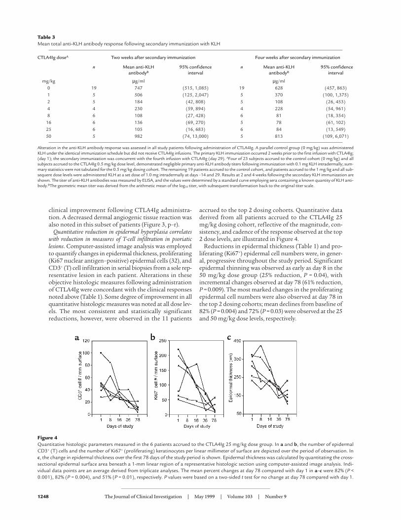
Quantitative reduction in epidermal hyperplasia correlateswith reduction in measures of T-cell infiltration in psoriaticlesions. Computer-assisted image analysis was employedto quantify changes in epidermal thickness, proliferating(Ki67 nuclear antigen–positive) epidermal cells (32), andCD3+ (T) cell infiltration in serial biopsies from a sole rep-resentative lesion in each patient. Alterations in theseobjective histologic measures following administrationof CTLA4Ig were concordant with the clinical responsesnoted above (Table 1). Some degree of improvement in allquantitative histologic measures was noted at all dose lev-els. The most consistent and statistically significantreductions, however, were observed in the 11 patients
accrued to the top 2 dosing cohorts. Quantitative dataderived from all patients accrued to the CTLA4Ig 25mg/kg dosing cohort, reflective of the magnitude, con-sistency, and cadence of the response observed at the top2 dose levels, are illustrated in Figure 4.
Reductions in epidermal thickness (Table 1) and pro-liferating (Ki67+) epidermal cell numbers were, in gener-al, progressive throughout the study period. Significantepidermal thinning was observed as early as day 8 in the50 mg/kg dose group (25% reduction, P = 0.04), withincremental changes observed at day 78 (61% reduction,P = 0.009). The most marked changes in the proliferatingepidermal cell numbers were also observed at day 78 inthe top 2 dosing cohorts; mean declines from baseline of82% (P = 0.004) and 72% (P = 0.03) were observed at the 25and 50 mg/kg dose levels, respectively.
1248 The Journal of Clinical Investigation | May 1999 | Volume 103 | Number 9
Table 3Mean total anti-KLH antibody response following secondary immunization with KLH
CTLA4Ig doseA Two weeks after secondary immunization Four weeks after secondary immunization
n Mean anti-KLH 95% confidence n Mean anti-KLH 95% confidenceantibodyB interval antibodyB interval
mg/kg µg/ml µg/ml0 19 747 (515, 1,085) 19 628 (457, 863)1 5 506 (125, 2,047) 5 370 (100, 1,375)2 5 184 (42, 808) 5 108 (26, 453)4 4 230 (59, 894) 4 228 (54, 961)8 6 108 (27, 428) 6 81 (18, 354)
16 6 136 (69, 270) 5 78 (61, 102)25 6 105 (16, 683) 6 84 (13, 549)50 5 982 (74, 13,000) 5 813 (109, 6,071)
Alteration in the anti-KLH antibody response was assessed in all study patients following administration of CTLA4Ig. A parallel control group (0 mg/kg) was administeredKLH under the identical immunization schedule but did not receive CTLA4Ig infusions. The primary KLH immunization occurred 2 weeks prior to the first infusion with CTLA4Ig;(day 1); the secondary immunization was concurrent with the fourth infusion with CTLA4Ig (day 29). AFour of 23 subjects accrued to the control cohort (0 mg/kg) and allsubjects accrued to the CTLA4Ig 0.5 mg/kg dose level, demonstrated negligible primary anti-KLH antibody titers following immunization with 0.1 mg KLH intradermally; sum-mary statistics were not tabulated for the 0.5 mg/kg dosing cohort. The remaining 19 patients accrued to the control cohort, and patients accrued to the 1 mg/kg and all sub-sequent dose levels were administered KLH at a set dose of 1.0 mg intradermally at days –14 and 29. Results at 2 and 4 weeks following the secondary KLH immunization areshown. The titer of anti-KLH antibodies was measured by ELISA, and the values were determined by a standard curve employing sera containing a known quantity of KLH anti-body.BThe geometric mean titer was derived from the arithmetic mean of the log10 titer, with subsequent transformation back to the original titer scale.
Figure 4Quantitative histologic parameters measured in the 6 patients accrued to the CTLA4Ig 25 mg/kg dose group. In a and b, the number of epidermalCD3+ (T) cells and the number of Ki67+ (proliferating) keratinocytes per linear millimeter of surface are depicted over the period of observation. Inc, the change in epidermal thickness over the first 78 days of the study period is shown. Epidermal thickness was calculated by quantitating the cross-sectional epidermal surface area beneath a 1-mm linear region of a representative histologic section using computer-assisted image analysis. Indi-vidual data points are an average derived from triplicate analyses. The mean percent changes at day 78 compared with day 1 in a–c were 82% (P <0.001), 82% (P = 0.004), and 51% (P = 0.01), respectively. P values were based on a two-sided t test for no change at day 78 compared with day 1.

Reductions in epidermal thickness correlated withreductions in infiltrating epidermal T cells (r = 0.60). Ateach dose level, the mean number of epidermal and der-mal T cells decreased serially following day 1 dosingthrough the final histologic sampling (day 78). Acrossthe 8 dose levels, the most significant decreases in thenumber of infiltrating epidermal and dermal T cellscompared with baseline were observed at day 78 in the 2top dosing cohorts. At these 2 highest dose levels, theabsolute number of epidermal and dermal intralesionallymphocytes fell to near normal range at day 78.
One possible mechanism for the diminution of intrale-sional T-cell numbers is enhanced intralesional T-cellapoptosis. To test this possibility, a TUNEL assay was usedto measure apoptosis in skin biopsy material selectedacross all dose levels and biopsy time points. No increasedrate of DNA fragmentation in skin histologic sections wasobserved following administration of CTLA4Ig (Figure 5).In contrast, apoptotic T cells were found in lesional biop-sies from psoriatic patients receiving ultraviolet B light(UVB), a therapy believed to be associated with in situ T-cell apoptosis (33). These data suggest that the largedecrease in intralesional T-cell numbers observed follow-ing administration of CTLA4Ig was not attributable to amarkedly increased rate of intralesional T-cell apoptosis.
Humoral immune response to bacteriophage φX174 and KLH.CTLA4Ig has been shown to abrogate a humoral immuneresponse to T cell–dependent antigens in preclinical mod-els (27, 34, 35). To test the importance of theCD28/CD152 (CTLA-4) costimulatory pathway in thegeneration of a T cell–dependent humoral immuneresponse in humans, study patients were immunizedwith 2 neoantigens, bacteriophage φX174 and KLH-ImmuneActivator. Suppression of antibody titers to lev-els 2 or more SDs below the mean of a parallel controlgroup were observed in 1 or more patients accrued to alldose levels following primary immunization with bacte-riophage φX174 (φX174 1°), secondary immunization
with bacteriophage φX174 (φX174 2°), and secondaryimmunization with keyhole limpet hemocyanin (KLH2°). Suppression of each of these antibody titers wasobserved most consistently in the 18 patients enrolled inthe 8, 16, and 25 mg/kg dose levels (Table 1). An appar-ent dose effect was evident in the mean antibody titersmeasured following φX174 1°, φX174 2°, and KLH 2°immunizations in patients receiving CTLA4Ig unit dosesof 0.5–25 mg/kg (Tables 2 and 3). Paradoxically, normalpeak antibody titers (predominantly IgM) were observedfollowing bacteriophage φX174 and KLH immunizationsin several patients in the CTLA4Ig 50 mg/kg dosingcohort. At unit doses of 1 mg/kg CTLA4Ig or greater, thepercent of anti-bacteriophage antibody of IgG isotypewas significantly reduced compared with control values(Table 2). This is consistent with a role for CD28/CD152in antibody class switching (35), a T cell–dependent event.
Across all doses of CTLA4Ig evaluated, the humoralimmune response was documented to be reconstituted instudy patients administered tertiary (day 71) and quater-nary (day 148) immunizations with bacteriophage φX174(Figure 6). There was no evidence of permanent nonre-sponsiveness (tolerance induction) to this soluble proteinantigen. One study patient at the 50 mg/kg dose level,though demonstrating sequential amplification of thehumoral immune response, did not display full reconsti-tution to within 2 SDs of the primary and secondary anti-bacteriophage antibody control values following tertiaryand quaternary immunizations, respectively. The CTLA4Igmean (± SD) through serum concentrations at days 78 and148 were 104 ± 19.6 µg/mL and 12.4 ± 12.6 µg/mL, respec-tively, in patients accrued to the 50 mg/kg dose level.
DiscussionTo our knowledge, this report constitutes the firsthuman experience with an agent designed to simultane-ously block T-cell costimulatory signals transducedthrough the CD28 and CD152 (CTLA-4) receptors. The
The Journal of Clinical Investigation | May 1999 | Volume 103 | Number 9 1249
Figure 5Absence of increased intralesional apoptosis followingadministration of CTLA4Ig. Illustrated in a and b are rep-resentative negative TUNEL reactions obtained followinghistochemical examination across all sampling timepoints and all dose levels of the study. In representativesections from a patient accrued to the 50 mg/kg doselevel, no increased rate of in situ cell death was evidentwhen comparisons were made between pretreatment (a)and day 36 (b) lesional biopsies. Scale bar in a: 100 µm.Positive controls included in these experiments are illus-trated in c and d, which are paired biopsy specimensfrom patients receiving ultraviolet B light (UVB) therapy.An increased rate of in situ apoptosis at day 15 (d) com-pared with baseline examination (c) was seen followingdaily administration of UVB treatment. Double stainingof these sections with mAb’s reactive with CD3 identifiedthe apoptotic cells as T cells.

data from this multidose, phase I clinical trial in patientswith psoriasis suggest that CTLA4Ig (BMS-188667) maybe an effective strategy for immunomodulation of T-cellfunction and provide insights into the pathogenesis ofpsoriasis. CTLA4Ig, administered to study patients withstable plaque psoriasis, reduced the number of intrale-sional T cells in a dose-dependent manner. The changesin lesional T-cell numbers correlated with reductions inepidermal proliferation, epidermal thickness, reversionof keratinocyte maturational abnormalities, and clinicalimprovement of psoriasis. The highly selective nature ofthis therapeutic intervention supports prior findingsand suggests that activated T cells, and the cytokinesthat they elaborate, are the prime effectors in the patho-genesis of psoriasis (2–8).
This study also illustrates the importance of the con-tinued activation of T cells interacting with B7 molecules
on APCs in the maintenance of the psoriatic phenotype.Despite the evidence that signals transduced throughCD28 are important in T-cell survival (36, 37), noincreased rate of T-cell apoptosis was discernible in pso-riatic lesions following administration of CTLA4Ig. Theobserved diminution in lesional T-cell numbers may,therefore, be attributed to factors altering T-cell recruit-ment into psoriatic lesions (38, 39), failure of localexpansion of antigen-specific T cells (40, 41), and/orapoptosis of antigen-specific T cells at extralesional sites.
The durability of the clinical responses observed fol-lowing administration of CTLA4Ig was notable in thispreliminary clinical experience. At the lower dose levelsevaluated, clinical quiescence was maintained wellbeyond the presence of detectable levels of CTLA4Ig inthe serum. The sustained responses in clinical measuresof psoriasis activity may be attributed to the degree ofnormalization of the pathologic epidermal and ker-atinocyte activation in the psoriatic lesions. Applyingsimilar quantitative methodology, the degree of nor-malization observed following administration ofCTLA4Ig was comparable to psoralen and ultraviolet Alight (PUVA) therapy and greater than that observed fol-lowing cyclosporine treatment (28, 42).
In addition to modulation of the cutaneous cell-medi-ated immune response in patients with psoriasis,CTLA4Ig also demonstrated activity in the humorallimb of the immune system. When comparisons aremade between the 2 immunogens employed in thisstudy, peak secondary antibody titers were more sup-pressed following bacteriophage φX174 challenge thanafter KLH secondary immunization. The immunizationschedule of these 2 immunogens varied; both the pri-mary and secondary bacteriophage φX174 immuniza-tions occurred following initiation of dosing withCTLA4Ig. Conversely, the KLH immunization schemainvolved a primary immunization with this immunogen2 weeks prior to the first dose of CTLA4Ig. The second-ary immunization with both immunogens was concur-rent with the fourth infusion of CTLA4Ig (day 29). Thesedata, in accordance with murine studies (32), suggest
1250 The Journal of Clinical Investigation | May 1999 | Volume 103 | Number 9
Figure 6Reconstitution of the humoral immune response to bacteriophage φX174in the 1 mg/kg, 4 mg/kg, 16 mg/kg, and 50 mg/kg dosing cohorts fol-lowing tertiary and quaternary immunization. Bacteriophage φX174 wasadministered at week 1 (1°), week 5 (2°), week 11 (3°), and week 22(4°). Patient sera were collected before and 1, 2, and 4 weeks after eachadministration of bacteriophage. Anti-bacteriophage antibody titers weredetermined by a neutralizing assay and expressed as a rate of phage inac-tivation, or K value (Kv). Antibody titers for patients accrued to the 1mg/kg (a), 4 mg/kg (b), 16 mg/kg (c), and 50 mg/kg (d) dose groups aredepicted. The geometric mean Kv’s of the 23 psoriatic control patientsare illustrated by the dark black line in each part. Vertical bars indicate±2 SD of the control group mean Kv. Following tertiary and/or quater-nary immunization, all patients’ titers were within 2 SD of the controlgroup primary and secondary responses, respectively, with the exceptionof 1 patient enrolled in the 50 mg/kg dose group (diamonds in d).Despite progressive titer amplification and a immunoglobulin classswitch following the 4° immunization, this patient did not achieve thestudy definition of full reconstitution of the humoral immune responseto bacteriophage φX174.

that fully primed secondary T cell–dependent humoralimmune responses, such as those elicited to KLH in thisstudy, are less dependent upon T-cell costimulationthrough the CD28/CD152 pathway (43, 44). Therefore,although meaningful clinical responses were noted inthis chronic autoimmune disease population character-ized by ongoing T-cell activation, lower unit doses ofCTLA4Ig may yield equivalent or superior blockade of T-cell activation in clinical settings where primary immuneresponses dominate.
Paradoxically, normal peak antibody titers followingKLH and bacteriophage φX174 immunization wereobserved in some patients receiving CTLA4Ig unit dosesof 50 mg/kg. Though the dose response was fairly con-sistent across the cell-mediated and humoral biologicend points incorporated in this study, there was cleardivergence at this top dose level. A possible explanationcould be that at very high unit doses of CTLA4Ig, univa-lent, lower-affinity binding with B7 molecules (45)would predominate and thereby reduce the effectivenessof CTLA4Ig as a blocking agent. Alternatively, at this topdose level, more efficient blockade of the interaction ofB7 with CTLA-4 may ensue, disrupting an inhibitory sig-nal transduced through CTLA-4 (46–49). Further stud-ies are needed to more fully elucidate the mechanismsresponsible for these observations.
Tolerance induction has been noted following CTLA4Igadministration in certain preclinical settings (13–15).This immunologic property, however, generally has notbeen observed following exposure to soluble protein anti-gens and, similarly, was not evident in this clinical study.Additional studies will be required to explore the optimalschedule of CTLA4Ig administration (50) and combina-tions with other immunosuppressive agents (12, 16) thatmay lead to antigen-specific nonresponsiveness in clini-cal settings where tolerance is a desired outcome.
A favorable safety profile was observed in this phase Isetting; however, full characterization of the clinical safe-ty of CTLA4Ig awaits larger clinical trials providingincreased duration of exposure to this novel therapeuticagent. It is postulated that CD152 (CTLA-4) may be anegative regulator of T-cell activation, and CTLA-4–/–
mice are known to succumb to a fatal lymphoprolifera-tive disorder at 3 weeks of age (46–48). No such alter-ations in lymphocyte homeostasis were evident in thisstudy. The combined effect of the simultaneous block-ade of CD28 and CD152 receptor-ligand interactionsmay offer a neutral effect on peripheral T-cell home-ostasis. Indeed, the lethal lymphoproliferative disorderassociated with CTLA-4–/– mice can be prevented by theadministration of CTLA4Ig, presumably because the B7receptors are blocked from interacting with the intactCD28 ligand (48, 49). Furthermore, mice lacking CTLA-4, B7-1, and B7-2 do not display a lymphoproliferativedisorder (58), additionally supporting the importance ofunopposed CD28 signaling for disease progression inCTLA-4–/– mice.
Our results indicate that the maintenance of the pso-riatic phenotype is dependent, to a large degree, uponthe activation of T cells utilizing the CD28/CD152 cos-timulatory pathway. This pathway is effectively blockedin a dose-dependent fashion following the administra-
tion of CTLA4Ig (BMS-188667). This therapeutic strat-egy may have great utility in the treatment of other T cell–mediated diseases and warrants further explo-ration in additional clinical studies.
AcknowledgementsThe authors wish to thank Robert Bruschini (Bristol-MyersSquibb, Princeton, New Jersey, USA) for help in preparing thefigures.
1. Greaves, M.W., and Weinstein, G.D. 1995. Treatment of psoriasis. N. Engl.J. Med. 332:581–588.
2. Demidem, A., Taylor, R., Grammer, S.F., and Streilein, J.W. 1991. T-lym-phocyte-activating properties of epidermal antigen-presenting cells fromnormal and psoriatic skin: evidence that psoriatic epidermal antigen-presenting cells resemble cultured normal Langerhans cells. J. Invest. Der-matol. 97:454–460.
3. Chang, E.Y., et al. 1992. T-cell activation is potentiated by cytokinesreleased by lesional psoriatic, but not normal epidermis. Arch. Dermatol.128:1479–1485.
4. Strange, P., et al. 1993. T-lymphocyte clones initiated from lesional pso-riatic skin release growth factors that induce keratinocyte proliferation.J. Invest. Dermatol. 101:695–700.
5. Bata-Csorgo, Z., Hammerberg, C., Voorhees, J.J., and Cooper, K.D. 1995.Kinetics and regulation of human keratinocyte stem cell growth inshort-term primary ex vivo culture. J. Clin. Invest. 95:317–327.
6. Bachelez, H., et al. 1998. Treatment of recalcitrant plaque psoriasis witha humanized non-depleting antibody to CD4. J. Autoimmun. 11:53–62.
7. Gottlieb, S.L., et al. 1995. Response of psoriasis to a lymphocyte-selec-tive toxin (DAB389IL-2) suggests a primary immune, but not ker-atinocyte, pathogenic basis. Nat. Med. 1:442–447.
8. Mueller, W., and Herrman, B. 1979. Cyclosporin A for psoriasis. N. Engl.J. Med. 301:555.
9. Mueller, D.L., Jenkins, M.K., and Schwartz, R.H. 1989. Clonal expansionversus functional inactivation: a costimulatory signaling pathway deter-mines the outcome of T cell antigen receptor occupancy. Annu. Rev.Immunol. 7:445–480.
10. Linsley, P.S., et al. 1991. Binding of the B cell activation antigen B7 toCD28 costimulates T cell proliferation and interleukin 2 mRNA accu-mulation. J. Exp. Med. 173:721–730.
11. Linsley, P.S., et al. 1991. CTLA-4 is a second receptor for the B cell acti-vation antigen B7. J. Exp. Med. 174:561–569.
12. Sayegh, M.H., and Turka, L.A. 1998. The role of T-cell costimulatory acti-vation pathways in transplant rejection. N. Engl. J. Med. 338:1813–1821.
13. Lenschow, D.J., et al. 1992. Long-term survival of xenogeneic pancreaticislet grafts induced by CTLA4Ig. Science. 257:789–792.
14. Lin, H., et al. 1993. Long-term acceptance of major histocompatabilitycomplex mismatched cardiac allografts induced by CTLA4Ig plus donor-specific transfusion. J. Exp. Med. 178:1801–1806.
15. Sayegh, M.H., et al. 1995. CD28-B7 blockade after alloantigenic challengein vivo inhibits Th1 cytokines but spares Th2. J. Exp. Med. 181:1869–1874.
16. Kirk, A.D., et al. 1997. CTLA4-Ig and anti-CD40 ligand prevent renalallograft rejection in primates. Proc. Natl. Acad. Sci. USA. 94:8789–8794.
17. Reiser, H., and Stadecker, M.D. 1996. Costimulatory B7 molecules in thepathogenesis of infectious and autoimmune diseases. N. Engl. J. Med.335:1369–1377.
18. Finck, B.K., Linsley, P.S., and Wofsy, D. 1994. Treatment of murine lupuswith CTLA4Ig. Science. 265:1225–1227.
19. Cross, A.H., et al. 1995. Long-term inhibition of murine experimentalautoimmune encephalomyelitis using CTLA-4-Fc supports a key role forCD28 costimulation. J. Clin. Invest. 95:2783–2789.
20. Webb, L.M.C., Walmsley, M.J., and Feldmann, M. 1996. Prevention andamelioration of collagen-induced arthritis by blockade of the CD28 co-stimulatory pathway: requirement for both B7-1 and B7-2. Eur. J.Immunol. 26:2320–2328.
21. Larsen, C.P., et al. 1994. Regulation of immunostimulatory function andcostimulatory molecule (B7-1 and B7-2) expression on murine dendrit-ic cells. J. Immunol. 152:5208–5219.
22. Symington, F.W., Brady, W., and Linsley, P.S. 1993. Expression and functionof B7 on human epidermal Langerhans cells. J. Immunol. 150:1286–1295.
23. Nestle, F.O., Turka, L.A., and Nickoloff, B.J. 1994. Characterization ofdermal dendritic cells in psoriasis. J. Clin. Invest. 94:202–209.
24. Jones, E.L., Epinette, W.W., Hackney, V.C., Menendez, L., and Frost, P.1975. Treatment of psoriasis with oral mycophenolic acid. J. Invest. Der-matol. 65:537–542.
25. Srinivas, N.R., et al. 1996. Pharmacokinetics and pharmacodynamics ofCTLA4Ig (BMS-188667), a novel immunosuppressive agent, in monkeysfollowing multiple doses. J. Pharm. Sci. 85:1–4.
The Journal of Clinical Investigation | May 1999 | Volume 103 | Number 9 1251
26. Ochs, H.D., Davis, S.D., and Wedgwood, R.J. 1971. Immunologicresponses to bacteriophage φX174 in immunodeficiency diseases. J. Clin.Invest. 50:2559–2568.
27. Linsley, P.S., et al. 1992. Immunosuppression in vivo by a soluble formof the CTLA-4 T cell activation molecule. Science. 257:792–795.
28. Vallat, V.P., et al. 1994. PUVA bath therapy strongly suppresses immuno-logical and epidermal activation in psoriasis: a possible cellular basis forremittive therapy. J. Exp. Med. 180:283–296.
29. Gavrieli, Y., Sherman, Y., and Ben-Sasson, S.A. 1992. Identification ofprogrammed cell death in situ via specific labeling of nuclear DNA frag-mentation. J. Cell. Biol. 119:493–501.
30. Agresti, A., Mehta, C.R., and Patel, N.R. 1990. Exact inference for con-tingency tables with ordered categories. J. Am. Stat. Assoc. 85:453–458.
31. Hotchin, N.A., Gandarillas, A., and Watt, F.M. 1995. Regulation of cellsurface β1 integrin levels during keratinocyte terminal differentiation.J. Cell Biol. 128:1209–1219.
32. Gerdes, J., et al. 1984. Cell cycle analysis of a cell proliferation-associat-ed human nuclear antigen defined by the monoclonal antibody Ki-67. J.Immunol. 133:1710–1715.
33. Ozawa, M., et al. 1999. 312-nanometer ultraviolet B light (narrow-bandUVB) induces apoptosis of T cells within psoriatic lesions. J. Exp. Med.189:711–718.
34. Cabrian, K.M., et al. 1996. Suppression of T-cell-dependent immuneresponses in monkeys by CTLA4Ig. Transplant. Proc. 28:3261–3262.
35. Ronchese, F., Hausmann, B., Hubele, S., and Lane, P. 1994. Mice trans-genic for a soluble form of murine CTLA-4 show enhanced expansion ofantigen-specific CD4+ T cells and defective antibody production in vivo.J. Exp. Med. 179:809–817.
36. Boise, L.H, et al. 1995. CD28 costimulation can promote T cell survivalby enhancing the expression of Bcl-xL. Immunity. 3:87–98.
37. Van Parijs, L., Ibraghimov, A., and Abbas, A.K. 1996. The roles of cos-timulation and FAS in T cell apoptosis and peripheral tolerance. Immu-nity. 4:321–328.
38. Austrup, F., et al. 1997. P- and E-selectin mediate recruitment of T-helper-
1 but not T-helper-2 cells into inflamed tissues. Nature. 385:81–83.39. Chin, Y.-H., Falanga, V., Taylor, J.R., Cai, J.-P., and Bax, J. 1990. Adherence
of human helper/memory T-cell subsets to psoriatic dermal endotheli-um. J. Invest. Dermatol. 94:413–417.
40. Dunn, D., et al. 1993. T cell receptor Vβ expression in normal humanskin. Proc. Natl. Acad. Sci. USA. 90:1267–1271.
41. Chang, J.C.C., et al. 1994. CD8+ T cells in psoriatic lesions preferentiallyuse T-cell receptor Vβ3 and/or Vβ13.1 genes. Proc. Natl. Acad. Sci. USA.91:9282–9286.
42. Gottlieb, A.B., et al. 1992. Studies of the effect of cyclosporine in psori-asis in vivo: combined effects on activated T lymphocytes and epidermalregenerative maturation. J. Invest. Dermatol. 98:302–309.
43. Viola, A., and Lanzavecchia, A. 1996. T cell activation determined by Tcell receptor number and tunable thresholds. Science. 273:104–106.
44. Iezzi, G., Karjalainen, K., and Lanzavecchia, A. 1998. The duration ofantigenic stimulation determines the fate of naive and effector T cells.Immunity. 8:89–95.
45. Greene, J.L., et al. 1996. Covalent dimerization of CD28/CTLA-4 andoligomerization of CD80/CD86 regulate T cell costimulatory interac-tions. J. Biol. Chem. 271:26762–26771.
46. Krummel, M.F., and Allison, J.P. 1996. CTLA-4 engagement inhibits IL-2 accumulation and cell cycle progression upon activation of resting Tcells. J. Exp. Med. 183:2533–2540.
47. Tivol, E.A., et al. 1995. Loss of CTLA-4 leads to massive lymphoprolifer-ation and fatal multiorgan tissue destruction, revealing a critical nega-tive regulatory role of CTLA-4. Immunity. 3:541–547.
48. Tivol, E.A., et al. 1997. CTLA4Ig prevents lymphoproliferation and fatalmultiorgan tissue destruction in CTLA-4-deficient mice. J. Immunol.158:5091–5094.
49. Mandelbrot, D.A., McAdam, A.J., and Sharpe, A.H. 1999. B7-1 or B7-2 isrequired to produce the lymphoproliferative phenotype in mice lacking cyto-toxic T lymphocyte-associated antigen 4 (CTLA-4). J. Exp. Med. 189:435–440.
50. Perez, V.L., et al. 1997. Induction of peripheral T cell tolerance in vivorequires CTLA-4 engagement. Immunity. 6:411–417.
1252 The Journal of Clinical Investigation | May 1999 | Volume 103 | Number 9