![Relationship between Urate Crystal Deposits Detected by Dual … · 2019-03-27 · Relationship between MSU Depositions and Bone Erosions in Non-tophaceous Gout 125 ment of gout [3,6,10].](https://static.fdocuments.in/doc/165x107/5f105d197e708231d448be03/relationship-between-urate-crystal-deposits-detected-by-dual-2019-03-27-relationship.jpg)
Crystal Arthropathy - learnorthopaedics.com · Crystal Arthropathy Tim Coughlin Gout Gout is a true...
8
Crystal Arthropathy Tim Coughlin Gout Gout is a true crystal deposition disease. It can be defined as the pathological reaction of the joint or periarticular tissues to the presence of monosodium urate monohydrate (MSUM) crystals. MSUM crystals preferentially deposit in the peripheral connective tissues in and around synovial joints, initially favouring the lower limbs and especially targeting the first metatarsophalangeal and small joints of the feet and hands. As the crystal deposits slowly increase and enlarge there is progressive involvement of more proximal sites and the potential for cartilage and bone damage, and development of ‘secondary’ OA. MSUM crystals take months or years to grow to a detectable size, implying a long asymptomatic phase. Prolonged hyperuricaemia is necessary, but not sufficient, for development of gout. Hyperuricaemia is most logically defined as a serum uric acid level above the theoretical solubility of MSUM in physiological conditions (0.42 mmol/L or 7.1 mg/dL). In practical terms, however, it is usually defined as a serum uric acid level greater than 2 standard deviations above the mean of the population (~ 0.40mmol/L or 6.7 mg/dL for men, 0.35 mmol/L or 5.9 mg/dL for women). Probably 95% of hyperuricaemic subjects never develop gout. Prevalence of gout is around 1% with a strong male predominance (10:1). Prevalence increases with age and increasing serum uric acid concentration. Primary Gout Is almost exclusively a male disease and the most common cause of inflammatory arthritis in men over the age of 40. About one-third of the body uric acid pool is derived from dietary sources and two-thirds from endogenous purine metabolism. The concentration of uric acid in body fluids depends on the balance between its synthesis and its elimination via the kidneys (two-thirds) and gut (one-third). Purine nucleotide synthesis and degradation are regulated by a network of enzyme pathways; xanthine oxidase catalyses the end conversion of hypoxanthine to xanthine and then xanthine to uric acid. Factors that predispose to chronic hyperuricaemia and gout: a) Diminished renal excretion (Renal Impairment, Long Term Diuretics, Low Dose Aspirin) b) Increased production of uric acid-uncommon c) Increased purine turnover (Chronic myeloproliferative or lymphoproliferative disorders) d) Increased de novo synthesis (Unidentified abnormality most common, Enzyme defect) In over 90% of patients with primary gout, hyperuricaemia results from an inherited isolated defect in fractional uric acid excretion which impairs their ability to increase renal excretion in response to a purine load (‘under-excretors’) Some primary gout patients are intrinsic ‘over-producers’ of uric acid through no identifiable cause. Rare individuals (< 1% primary gout patients) have a specific inherited enzyme defect of purine synthesis which should be suspected if gout develops: a) Under the age of 25 years b) In patients presenting with uric acid stones in the urinary tract Copyright © 2011 Tim Coughlin. All rights reserved. www.learnorthopaedics.com
-
Upload
vuongthuan -
Category
Documents
-
view
218 -
download
1
Transcript of Crystal Arthropathy - learnorthopaedics.com · Crystal Arthropathy Tim Coughlin Gout Gout is a true...

Crystal ArthropathyTim Coughlin
Gout
Gout is a true crystal deposition disease. It can be defined as the pathological reaction of the joint or periarticular tissues to the presence of monosodium urate monohydrate (MSUM) crystals. MSUM crystals preferentially deposit in the peripheral connective tissues in and around synovial joints, initially favouring the lower limbs and especially targeting the first metatarsophalangeal and small joints of the feet and hands. As the crystal deposits slowly increase and enlarge there is progressive involvement of more proximal sites and the potential for cartilage and bone damage, and development of ‘secondary’ OA. MSUM crystals take months or years to grow to a detectable size, implying a long asymptomatic phase. Prolonged hyperuricaemia is necessary, but not sufficient, for development of gout. Hyperuricaemia is most logically defined as a serum uric acid level above the theoretical solubility of MSUM in physiological conditions (0.42 mmol/L or 7.1 mg/dL). In practical terms, however, it is usually defined as a serum uric acid level greater than 2 standard deviations above the mean of the population (~ 0.40mmol/L or 6.7 mg/dL for men, 0.35 mmol/L or 5.9 mg/dL for women). Probably 95% of hyperuricaemic subjects never develop gout.Prevalence of gout is around 1% with a strong male predominance (10:1). Prevalence increases with age and increasing serum uric acid concentration.
Primary GoutIs almost exclusively a male disease and the most common cause of inflammatory arthritis in men over the age of 40. About one-third of the body uric acid pool is derived from dietary sources and two-thirds from endogenous purine metabolism. The concentration of uric acid in body fluids depends on the balance between its synthesis and its elimination via the kidneys (two-thirds) and gut (one-third).Purine nucleotide synthesis and degradation are regulated by a network of enzyme pathways; xanthine oxidase catalyses the end conversion of hypoxanthine to xanthine and then xanthine to uric acid.
Factors that predispose to chronic hyperuricaemia and gout:a) Diminished renal excretion (Renal Impairment, Long Term Diuretics, Low Dose Aspirin)b) Increased production of uric acid-uncommonc) Increased purine turnover (Chronic myeloproliferative or lymphoproliferative disorders)d) Increased de novo synthesis (Unidentified abnormality most common, Enzyme defect)
In over 90% of patients with primary gout, hyperuricaemia results from an inherited isolated defect in fractional uric acid excretion which impairs their ability to increase renal excretion in response to a purine load (‘under-excretors’)
Some primary gout patients are intrinsic ‘over-producers’ of uric acid through no identifiable cause.
Rare individuals (< 1% primary gout patients) have a specific inherited enzyme defect of purine synthesis which should be suspected if gout develops:a) Under the age of 25 yearsb) In patients presenting with uric acid stones in the urinary tract
Copyright © 2011 Tim Coughlin. All rights reserved. www.learnorthopaedics.com
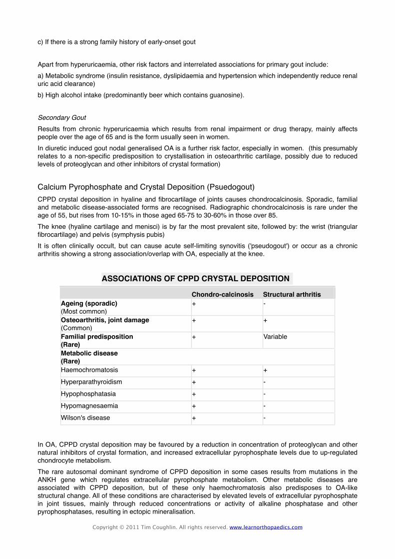
c) If there is a strong family history of early-onset gout
Apart from hyperuricaemia, other risk factors and interrelated associations for primary gout include:a) Metabolic syndrome (insulin resistance, dyslipidaemia and hypertension which independently reduce renal uric acid clearance) b) High alcohol intake (predominantly beer which contains guanosine).
Secondary GoutResults from chronic hyperuricaemia which results from renal impairment or drug therapy, mainly affects people over the age of 65 and is the form usually seen in women. In diuretic induced gout nodal generalised OA is a further risk factor, especially in women. (this presumably relates to a non-specific predisposition to crystallisation in osteoarthritic cartilage, possibly due to reduced levels of proteoglycan and other inhibitors of crystal formation)
Calcium Pyrophosphate and Crystal Deposition (Psuedogout)CPPD crystal deposition in hyaline and fibrocartilage of joints causes chondrocalcinosis. Sporadic, familial and metabolic disease-associated forms are recognised. Radiographic chondrocalcinosis is rare under the age of 55, but rises from 10-15% in those aged 65-75 to 30-60% in those over 85.The knee (hyaline cartilage and menisci) is by far the most prevalent site, followed by: the wrist (triangular fibrocartilage) and pelvis (symphysis pubis)It is often clinically occult, but can cause acute self-limiting synovitis ('pseudogout') or occur as a chronic arthritis showing a strong association/overlap with OA, especially at the knee.
ASSOCIATIONS OF CPPD CRYSTAL DEPOSITION
Chondro-calcinosis Structural arthritisAgeing (sporadic)(Most common)
+ -
Osteoarthritis, joint damage(Common)
+ +
Familial predisposition(Rare)
+ Variable
Metabolic disease(Rare)Metabolic disease(Rare)Metabolic disease(Rare)Haemochromatosis + +
Hyperparathyroidism + -
Hypophosphatasia + -
Hypomagnesaemia + -
Wilson's disease + -
In OA, CPPD crystal deposition may be favoured by a reduction in concentration of proteoglycan and other natural inhibitors of crystal formation, and increased extracellular pyrophosphate levels due to up-regulated chondrocyte metabolism.The rare autosomal dominant syndrome of CPPD deposition in some cases results from mutations in the ANKH gene which regulates extracellular pyrophosphate metabolism. Other metabolic diseases are associated with CPPD deposition, but of these only haemochromatosis also predisposes to OA-like structural change. All of these conditions are characterised by elevated levels of extracellular pyrophosphate in joint tissues, mainly through reduced concentrations or activity of alkaline phosphatase and other pyrophosphatases, resulting in ectopic mineralisation.
Copyright © 2011 Tim Coughlin. All rights reserved. www.learnorthopaedics.com

Calcific Periarthritis
Basic Calcium Phosphate (BCP) Deposition: Hydroxyapatite (apatite) is the principal mineral in bone and teeth. Apatite and other basic, as opposed to acidic, calcium phosphates (octacalcium phosphate, tricalcium phosphate) are also the usual minerals to deposit in extraskeletal tissues. In MSK tissues abnormal deposition may occur in:
• periarticular tissues, particularly tendon • hyaline cartilage in association with OA • subcutaneous tissue and muscle, principally in connective tissue diseases.
Under normal circumstances mineralisation of soft tissues is prevented by inhibitors such as pyrophosphate and proteoglycans. When these protective mechanisms break down, abnormal calcification due to BCP occurs.Calcific periarthritis is deposition of apatite in the supraspinatus tendon is an incidental radiographic finding in around 7% of adults. It occasionally results in severe acute inflammation of the subacromial bursa and periarticular tissues through crystal shedding from the tendon into and around the bursa. Periarticular sites around the greater trochanter of the hip, foot or hand are less commonly affected.Calcific periarthritis may result from metabolic abnormality (renal failure, hyperparathyroidism, hypophosphatasia) but measurements of serum creatinine, calcium and alkaline phosphatase are usually normal. The CRP is elevated during the episode.
Signs and Symptoms
Acute crystal-associated synovitis (gout, pseudogout)In almost all first attacks a single distal joint is affected. The first metatarsophalangeal joint is affected in over 50% of cases-'podagra'. Other common sites (in order of decreasing frequency) are the ankle, midfoot, knee, small joints of hands, wrist and elbow. The axial skeleton and large proximal joints are rarely involved and never as the first site.
Typical attacks have the following characteristics:• extremely rapid onset, reaching maximum severity in just 2-6 hours, often waking the patient in the
early morning • severe pain, often described as the 'worst pain ever' • extreme tenderness-the patient is unable to wear a sock or to let bedding rest on the joint • marked swelling with overlying red, shiny skin • self-limiting over 5-14 days, with complete return to normality.
During the attack the joint shows signs of marked synovitis but also periarticular swelling and erythema.There may be accompanying fever, malaise and even confusion, especially if a large joint such as the knee is involved.As the attack subsides, pruritus and desquamation of overlying skin are common.The main differential diagnosis is septic arthritis, infective cellulitis or another crystal disease. Sepsis, however, is usually more subacute in onset and progresses in severity until treated.Acute attacks may also manifest as bursitis, tenosynovitis or cellulitis. These attacks have the same characteristics-rapid onset, severe pain, florid inflammation and erythema.Many patients describe milder episodes lasting just a few days ('petite attacks').
Copyright © 2011 Tim Coughlin. All rights reserved. www.learnorthopaedics.com
Some have attacks in more than one joint; sometimes one attack, by triggering the acute phase response, triggers attacks in other joints a few days later ('cluster attacks'). Polyarticular attacks are rare.Pseudogout: The typical attack resembles acute gout and develops rapidly, with severe pain, stiffness and swelling, maximal within 6-24 hours of onset.Overlying erythema is common and examination reveals a very tender joint held in the flexed 'loose-pack' position with signs of marked synovitis (large/tense effusion, warmth, restricted movement with stress pain). Fever is common and the patient may appear confused and ill. The attack is self-limiting but may take 1-3 weeks to resolve.
Chronic tophaceous goutLarge MSUM crystal deposits produce irregular firm nodules ('tophi') at the usual sites for nodules:
• around extensor surfaces of fingers• hands• forearm,• elbows• Achilles tendons • sometimes the helix of the ear.
The white colour of MSUM crystals may be evident and permits distinction from rheumatoid nodules. Large nodules may ulcerate, discharging white gritty material and associating with local inflammation (erythema, pus), even in the absence of secondary infection. Although tophi are usually a very late feature, they may appear surprisingly rapidly, in under 1 year, in patients with chronic renal failure.Secondary gout may present with painful, sometimes discharging tophi without preceding acute attacks. This is particularly seen in older, mainly female patients with nodal OA who develop tophi in and around their osteoarthritic finger joints as a consequence of chronic (> 1-2 years) diuretic therapy.
Acute calcific periarthritisThe acute episode may occur spontaneously or follow local trauma. Within just a few hours shoulder pain and tenderness are extreme and the area appears swollen, hot and sometimes red. Modest systemic upset and fever are common. X-rays confirm the diagnosis by showing tendon calcification. If the subacromial bursa is aspirated, thick white fluid containing many calcium-staining (alizarin red S) aggregates may be obtained.The condition usually resolves spontaneously over 1-3 weeks, often accompanied by radiographic dispersal and disappearance of small to modest-sized deposits (i.e. complete crystal shedding).
Pathogenesis and Radiological Findings of Crystal Arthropathy
A variety of crystals can deposit in and around joints and associate with both acute inflammatory and chronic syndromes. In some instances crystals are the primary pathogenic agents-true 'crystal deposition disease' (e.g. gout). In other situations MSK disease predisposes to secondary crystal formation (e.g. predisposition to calcium pyrophosphate and apatite crystal formation in OA).Crystals may subsequently amplify symptoms and damage, or be an incidental epiphenomenon of no clinical consequence.Several factors influence crystal formation:
Copyright © 2011 Tim Coughlin. All rights reserved. www.learnorthopaedics.com
There must be sufficient concentration of the chemical components (ionic product). Whether a crystal then forms, however, depends on the balance of tissue factors that promote or inhibit crystal nucleation and growth. Many tissues are supersaturated for various products but depend on natural inhibitors to prevent crystallisation. Alteration in the balance of inhibitors and promoters may allow crystallisation. Crystals can also dissolve and the yield of crystals at any one time will depend on the relative rates of crystallisation, growth and dissolution.
Factors relating to crystal formation and tissue concentration at any one timeThe inflammatory potential of crystals resides in the physical irregularity and high negative charge of their surface which can induce inflammation and damage cell membranes. Crystals can also cause mechanical damage to the tissues in which they lie and act as wear particles at the joint surface. Crystals forming deep within cartilage or tendon are prevented from interaction with proteins and cells and can paradoxically reside in MSK tissues for years without causing inflammation or symptoms. It is only when they are released from their protected sites of origin ('crystal shedding') that they trigger acute attacks of inflammation. Such attacks may occur spontaneously, result from mechanical loosening (local trauma), partial dissolution and reduced crystal size (e.g. initiation of hypouricaemic treatment), or occur in association with an acute phase response due to intercurrent illness or surgery (mechanism unknown).
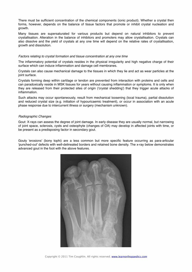
Radiographic ChangesGout: X-rays can assess the degree of joint damage. In early disease they are usually normal, but narrowing of joint space, sclerosis, cysts and osteophyte (changes of OA) may develop in affected joints with time, or be present as a predisposing factor in secondary gout. Gouty 'erosions' (bony tophi) are a less common but more specific feature occurring as para-articular 'punched-out' defects with well-delineated borders and retained bone density. The x-ray below demonstrates advanced gout in the foot with the above features.
Copyright © 2011 Tim Coughlin. All rights reserved. www.learnorthopaedics.com

Tophi may also be visible as eccentric soft tissue swellings. In late disease changes may be hard to distinguish from other forms of inflammatory polyarthritis.Pseudogout: X-rays may show chondrocalcinosis in hyaline cartilage and/or fibrocartilage (occasionally capsule or ligament) with or without associated structural changes of OA.Chondrocalcinosis is not always evident, especially in joints showing some degree of cartilage loss, and its absence does not exclude the diagnosis of pseudogout.CPPD crystal deposition in hyaline and fibrocartilage of joints causes chondrocalcinosis.Radiographic chondrocalcinosis is rare under the age of 55, but rises from 10-15% in those aged 65-75 to 30-60% in those over 85. The x-ray of a knee below shows calcification in the menisci of the knee joint.
Calcific periarthritis – shows calcification of tissues i.e. the supraspinatus tendon. The x-ray below shows calcification of the common extensor tendon at the elbow.
Copyright © 2011 Tim Coughlin. All rights reserved. www.learnorthopaedics.com
Diagnosis and Management of Crystal Arthritis
InvestigationsDefinitive diagnosis of gout requires identification of MSUM crystals in the aspirate from a joint, bursa or tophus. In acute gout synovial fluid shows increased turbidity due to the greatly elevated cell count (> 90% neutrophils) Chronic gouty fluid is more variable but occasionally appears white due to the high crystal load. During remission aspiration of an asymptomatic first metatarsophalangeal joint or knee may still permit crystal identification.Although hyperuricaemia is usually consistently present, it does not confirm gout. Equally, a normal uric acid level, especially during an attack, does not exclude gout (uric acid falls as part of the acute phase reaction). Measurement of 24-hour urinary uric acid excretion on a low purine diet will identify an over-producer. Assessment of the following are required:
• renal function (serum creatinine, urine testing)• hypertension (B.P.)• blood glucose• serum lipid profile
An FBC and ESR should detect myeloproliferative disorders during remission of acute gout. During an attack a marked acute phase response (elevated CRP, neutrophilia) is usual; the ESR is often modestly raised in tophaceous gout.In acute pseudogout examination of synovial fluid using compensated polarised microscopy will demonstrate CPPD crystals and permit distinction from urate gout. The aspirated fluid is often turbid and may be uniformly blood-stained, reflecting the severity of inflammation. Gram stain and culture of the fluid will exclude sepsis. CPPD crystals may also be identified in the less inflamed fluids aspirated from chronic pyrophosphate arthritis.X-ray’s to look for the changes as above.Screening for metabolic or familial predisposition should be undertaken in patients who show CPPD deposition aged < 55; florid polyarticular, as opposed to pauciarticular, chondrocalcinosis; recurrent acute attacks without chronic arthropathy; or additional clinical or radiographic features of predisposing disease.
ManagementThe acute attack of gout: A fast-acting oral NSAID (e.g. naproxen, diclofenac, indometacin) can give effective pain relief and is the standard treatment. Patients can keep a supply of an NSAID with which they are familiar and take it as soon as the first symptoms are noticed, continuing for the duration of the attack. Oral colchicine (a potent inhibitor of neutrophil microtubular assembly) can be very effective, but unfortunately often causes vomiting and severe diarrhoea at the doses needed for rapid relief (1 mg loading dose, then 0.5 mg 6-hourly until symptoms abate). The compromise is to try lower doses (0.5 mg 8-12-hourly) for a slower onset of benefit.Aspiration of the joint will give instant relief and, when combined with an intra-articular corticosteroid injection to prevent fluid reaccumulation, often effectively aborts the attack.For acute pseudogout: aspiration quickly reduces pain and may alone be sufficient. Fluid reaccumulation, however, is common, particularly early in an attack, and additional intra-articular injection of corticosteroid is usually required. Oral NSAIDs and colchicine are also effective, as in gout, but should be avoided if possible in older people.
Copyright © 2011 Tim Coughlin. All rights reserved. www.learnorthopaedics.com
Early active mobilisation is also important in this age group. For chronic arthropathy management is the same as for OA
Hypouricaemic Drug TreatmentThe aim of treatment is to bring the serum uric acid level into the lower half of the normal range to ensure dissolution of crystals and to prevent new ones forming. The serum uric acid should therefore be measured every 3-4 weeks and the dose of allopurinol increased in 100 mg increments until this is achieved (maximum 900 mg daily). Infrequent (e.g. yearly) monitoring is advised to ensure maintenance of effective treatment. In most cases allopurinol will need to be continued indefinitely.Indications for hypouricaemic drugs are:
• Recurrent attacks of acute gout • Tophi • Evidence of bone or joint damage • Associated renal disease• Gout with greatly elevated serum uric acid
Allopurinol is the usual drug of choice because of its once-daily convenience and low incidence of side-effects. It inhibits xanthine oxidase and reduces conversion of hypoxanthine and xanthine to uric acid.The usual starting dose is 100-300 mg daily but lower doses (100 mg or less) should be used in older patients or if renal function is impaired. The sharp reduction in tissue uric acid levels that follows initiation of treatment can partially dissolve MSUM crystals and trigger acute attacks. The patient should be warned of this and told to continue treatment even if an attack occurs. This risk can be minimised by using a lower starting dose (100 mg) or by concurrent administration of oral colchicine (0.5 mg 12-hourly) or an NSAID for the first few weeks.Initiation of treatment during an attack can exacerbate and prolong the episode so it is prudent to wait until the attack settles.Uricosuric drugs such as probenecid or sulfinpyrazone can achieve equivalent reductions in serum uric acid to allopurinol but require several doses each day and maintenance of a high urine flow (to avoid uric acid crystallisation in renal tubules). Salicylates antagonise the uricosuric action of these drugs and should be avoided. Uricosurics are contraindicated in:
• over-producers (they already have gross uricosuria)• those with renal impairment (ineffective)• patients with urolithiasis (increased stone formation).
The uricosuric benzbromarone is effective in patients with mild to moderate renal impairment but can cause hepatotoxicity and has limited availability in most countries.
Copyright © 2011 Tim Coughlin. All rights reserved. www.learnorthopaedics.com