Covalent Conjugation to Cytodiagnostics Carboxylated Gold ... · PDF fileCovalent Conjugation...
5
919 Fraser Drive Unit 11, Burlington, ON, L7L 4X8, Canada Tel: 866-344-3954 Fax: 289-288-0122 www.cytodiagnostics.com 1 Covalent Conjugation to Cytodiagnostics Carboxylated Gold Nanoparticles – Tech Note #105 Background Gold nanoparticle conjugates have been widely used in biological research and biosensing applications. For example, gold nanoparticles can function as probes in light or electron microscopy, and in lateral flow immunoassays. There are two ways of preparing gold conjugates, i.e. passive absorption and covalent coupling via a crosslinker. Although a relatively simple preparation process, passive absorption of proteins to gold nanoparticles does not provide a permanent attachment of the coating because molecules may desorb from the surface over time. In addition, in some cases, the proteins lose their properties after being absorbed to the surface, which can be caused by changes in tertiary structure or binding of the active/antigen to the gold surface rendering it inaccessible. Preparation of gold nanoparticle protein conjugates through passive adsorption can be optimized with our convenient Gold Conjugation Optimization Kits. In comparison to passive adsorption, covalent coupling permanently immobilizes molecules of interest to the functionalized particles (e.g. with carboxyl or amine groups). Thus this approach provides much improved stability of the protein coating over the passive absorption method. Covalent coupling uses chemical linkers (e.g. EDC/NHS) that react with certain groups on the molecules. Therefore, the reaction is more specific and controllable than the passive absorption method and the number of covalently conjugated ligands can be optimized to the particular application. The covalent coupling process is also less harsh, maintaining the tertiary structure of proteins with the advantage of minimizing possible negative effects on the properties and activities after conjugation. Our Carboxyl Gold Nanoparticles rely on EDC/NHS chemistry for conjugation. EDC and NHS “activate” the carboxyl groups on the particle surface to form an intermediate that can subsequently react with primary amine groups on the specific protein or other ligand to be conjugated. The efficiency of EDC conjugation is typically low and sensitive to pH and covalent coupling protocols requires some degree of optimization to achieve desired performance or stability. The following protocol provides general guidelines for coupling biomolecules to our Carboxyl Gold Nanoparticles, with conjugation of a standard IgG to our 20nm Carboxyl Gold Nanoparticles given as an example. For conjugation of other biomolecules, the optimal conjugation conditions may vary. To obtain maximum conjugation to the particle surface, the amount of protein for conjugation is about 1 to 10X excess that of its theoretical quantity needed for full coverage. Materials and Equipment Required • Carboxyl Gold Nanoparticles • Negative control: Methyl Gold Nanoparticles • 1-Ethyl-3-[3-dimethylaminopropyl]carbodiimide hydrochloride (EDC) (Sigma, Cat# E1769) • N-hydroxysulfosuccinimide (Sulfo-NHS) (Sigma, Cat# 56485) • Positive control: Horse Radish Peroxidase (HRP) or IgG from human serum (Sigma, Cat# I4506) • Blocker: Bovine Serum Albumin (BSA) (Sigma, Cat# A3059) • Activation buffer: 2-(N-morpholino)ethanesulfonic acid (MES) buffer (10 mM, pH 5.5) • Coupling buffer: 1X Phosphate Buffered Saline (PBS) • Washing buffer: 1X Phosphate Buffered Saline + 0.05% Tween 20 (PBST) • UV-VIS Spectrophotometer
Transcript of Covalent Conjugation to Cytodiagnostics Carboxylated Gold ... · PDF fileCovalent Conjugation...

919 Fraser Drive Unit 11, Burlington, ON, L7L 4X8, Canada Tel: 866-344-3954 Fax: 289-288-0122
www.cytodiagnostics.com
1
Covalent Conjugation to Cytodiagnostics Carboxylated Gold Nanoparticles – Tech Note #105 Background Gold nanoparticle conjugates have been widely used in biological research and biosensing applications. For example, gold nanoparticles can function as probes in light or electron microscopy, and in lateral flow immunoassays. There are two ways of preparing gold conjugates, i.e. passive absorption and covalent coupling via a crosslinker. Although a relatively simple preparation process, passive absorption of proteins to gold nanoparticles does not provide a permanent attachment of the coating because molecules may desorb from the surface over time. In addition, in some cases, the proteins lose their properties after being absorbed to the surface, which can be caused by changes in tertiary structure or binding of the active/antigen to the gold surface rendering it inaccessible. Preparation of gold nanoparticle protein conjugates through passive adsorption can be optimized with our convenient Gold Conjugation Optimization Kits. In comparison to passive adsorption, covalent coupling permanently immobilizes molecules of interest to the functionalized particles (e.g. with carboxyl or amine groups). Thus this approach provides much improved stability of the protein coating over the passive absorption method. Covalent coupling uses chemical linkers (e.g. EDC/NHS) that react with certain groups on the molecules. Therefore, the reaction is more specific and controllable than the passive absorption method and the number of covalently conjugated ligands can be optimized to the particular application. The covalent coupling process is also less harsh, maintaining the tertiary structure of proteins with the advantage of minimizing possible negative effects on the properties and activities after conjugation. Our Carboxyl Gold Nanoparticles rely on EDC/NHS chemistry for conjugation. EDC and NHS “activate” the carboxyl groups on the particle surface to form an intermediate that can subsequently react with primary amine groups on the specific protein or other ligand to be conjugated. The efficiency of EDC conjugation is typically low and sensitive to pH and covalent coupling protocols requires some degree of optimization to achieve desired performance or stability. The following protocol provides general guidelines for coupling biomolecules to our Carboxyl Gold Nanoparticles, with conjugation of a standard IgG to our 20nm Carboxyl Gold Nanoparticles given as an example. For conjugation of other biomolecules, the optimal conjugation conditions may vary. To obtain maximum conjugation to the particle surface, the amount of protein for conjugation is about 1 to 10X excess that of its theoretical quantity needed for full coverage. Materials and Equipment Required
• Carboxyl Gold Nanoparticles • Negative control: Methyl Gold Nanoparticles • 1-Ethyl-3-[3-dimethylaminopropyl]carbodiimide hydrochloride (EDC) (Sigma, Cat# E1769) • N-hydroxysulfosuccinimide (Sulfo-NHS) (Sigma, Cat# 56485) • Positive control: Horse Radish Peroxidase (HRP) or IgG from human serum (Sigma, Cat# I4506) • Blocker: Bovine Serum Albumin (BSA) (Sigma, Cat# A3059) • Activation buffer: 2-(N-morpholino)ethanesulfonic acid (MES) buffer (10 mM, pH 5.5) • Coupling buffer: 1X Phosphate Buffered Saline (PBS) • Washing buffer: 1X Phosphate Buffered Saline + 0.05% Tween 20 (PBST) • UV-VIS Spectrophotometer

919 Fraser Drive Unit 11, Burlington, ON, L7L 4X8, Canada Tel: 866-344-3954 Fax: 289-288-0122
www.cytodiagnostics.com
2
• Protein of interest to be conjugated
Note: for effective conjugation, the purity of the protein should be concerned. Any other molecules containing primary amines may compete for EDC with the protein of conjugation and reduce the conjugation efficiency. The protein should also have enough accessible primary amine groups for conjugation. Lysine residues are the primary target sites for EDC conjugation. A higher number of lysine groups on the outer surface of the protein will probably lead to higher conjugation efficiency. For example, bovine serum albumin (BSA) has 30 to 35 lysine groups available on its surface for EDC conjugation. An IgG antibody molecule typically has about 90 lysine residues, and 30 are potentially useful for conjugation.
Procedure
1. If the concentration of gold nanoparticles is less than OD 50, concentrate them by centrifugation. If particles are in buffers, wash them in pure water by centrifugation. Refer to Tech Note #101 for handling instructions.
2. Prepare fresh EDC/NHS mix solution in MES buffer at a concentration of 30 and 30 mg/mL, respectively. * Note: EDC/NHS rapidly hydrolyzes in aqueous solutions and should be prepared fresh just prior to conjugation.
3. Take 10 µL of 20 nm particles (OD 50 in water) and mix with 10 µL of EDC/NHS mix solution as prepared in step 2
4. Incubate for 30 min at room temperature 5. Add 1 mL of PBST and vortex thoroughly ** 6. Spin down by centrifugation at 6,500 g for 30 min 7. Remove most of the supernatant 8. Add 10 µL of IgG (1 mg/mL in 1X PBS)*** 9. Sonicate in a water bath sonicator for 10 sec 10. Incubate for 2 to 4 hours at room temperature with mixing 11. Add 1 mL of PBST and vortex thoroughly 12. Spin down by centrifugation at 3,500 g for 30 min 13. Remove most of the supernatant 14. Add 50 µL PBS with 1% BSA 15. Store at 4 degrees and ready to use
* Activation rate or efficiency is pH dependent, the more acidic, the more efficient, but acidity may affect particle stability. ** Alternatively, 1-step conjugation procedure may be employed for smaller proteins, peptides, or other molecules like oligo DNAs. *** The concentration of protein may vary depending on the particle size and protein to be conjugated. In general, the amount of protein should be 1 to 10X excess of the amount of full surface coverage. The total surface area of particles and the docking area should be estimated to calculate the optimal amount of protein. The following table is a general reference for conjugating IgG to our Carboxyl Gold Particles of different sizes. The calculation for the other proteins are similar.
919 Fraser Drive Unit 11, Burlington, ON, L7L 4X8, Canada Tel: 866-344-3954 Fax: 289-288-0122
www.cytodiagnostics.com
3
Table 1. Suggested quantities of IgG for Carboxyl Gold Nanoparticles of different sizes during EDC conjugation. The surface areas of particles are calculated according to our Gold Nanoparticle Properties. The docking area of IgG is estimated to be 45 nm2, with a molecular weight of 150 kDa. “N X full coverage amount” means the excess ratio between the incubation amount and the amount needed for full coverage of particle surface.
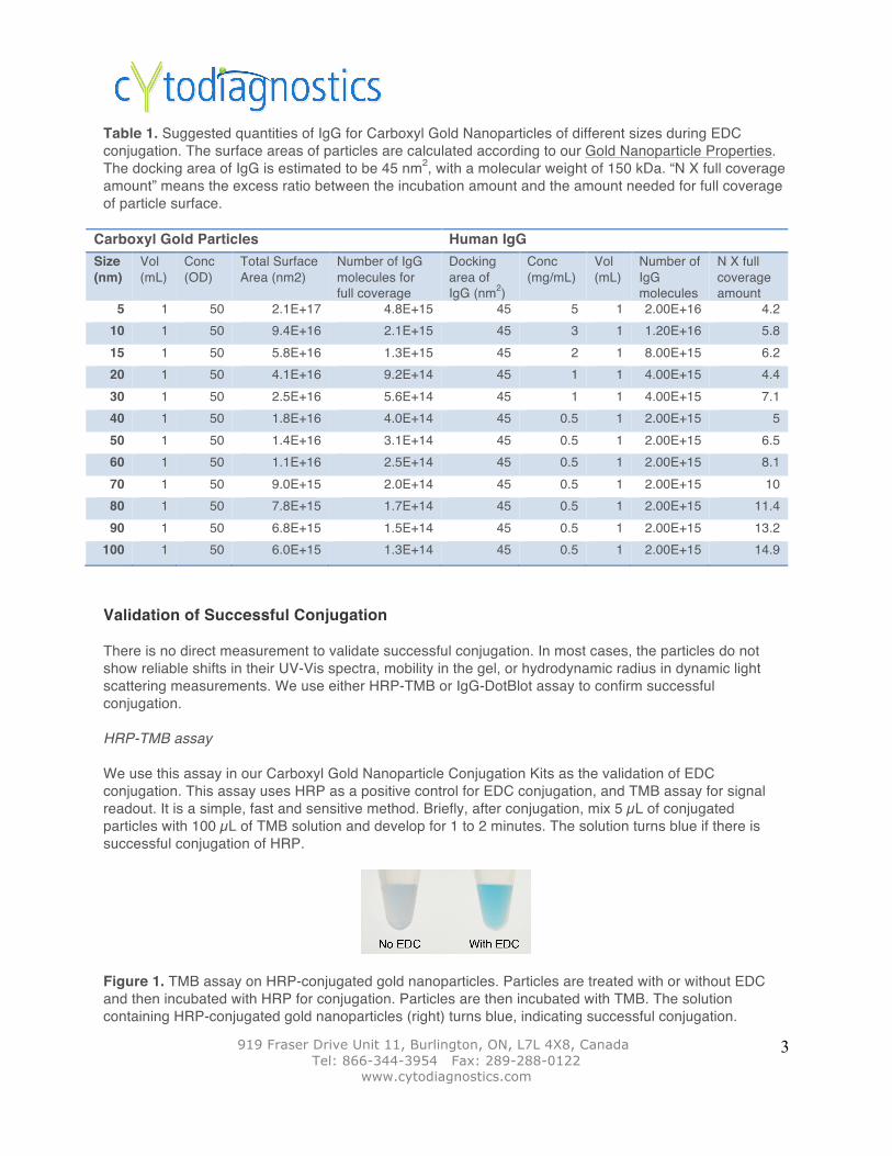
Validation of Successful Conjugation There is no direct measurement to validate successful conjugation. In most cases, the particles do not show reliable shifts in their UV-Vis spectra, mobility in the gel, or hydrodynamic radius in dynamic light scattering measurements. We use either HRP-TMB or IgG-DotBlot assay to confirm successful conjugation. HRP-TMB assay We use this assay in our Carboxyl Gold Nanoparticle Conjugation Kits as the validation of EDC conjugation. This assay uses HRP as a positive control for EDC conjugation, and TMB assay for signal readout. It is a simple, fast and sensitive method. Briefly, after conjugation, mix 5 µL of conjugated particles with 100 µL of TMB solution and develop for 1 to 2 minutes. The solution turns blue if there is successful conjugation of HRP.
Figure 1. TMB assay on HRP-conjugated gold nanoparticles. Particles are treated with or without EDC and then incubated with HRP for conjugation. Particles are then incubated with TMB. The solution containing HRP-conjugated gold nanoparticles (right) turns blue, indicating successful conjugation.
Carboxyl Gold Particles Human IgG Size (nm)
Vol (mL)
Conc (OD)
Total Surface Area (nm2)
Number of IgG molecules for full coverage
Docking area of IgG (nm2)
Conc (mg/mL)
Vol (mL)
Number of IgG molecules
N X full coverage amount
5 1 50 2.1E+17 4.8E+15 45 5 1 2.00E+16 4.2 10 1 50 9.4E+16 2.1E+15 45 3 1 1.20E+16 5.8 15 1 50 5.8E+16 1.3E+15 45 2 1 8.00E+15 6.2 20 1 50 4.1E+16 9.2E+14 45 1 1 4.00E+15 4.4 30 1 50 2.5E+16 5.6E+14 45 1 1 4.00E+15 7.1 40 1 50 1.8E+16 4.0E+14 45 0.5 1 2.00E+15 5 50 1 50 1.4E+16 3.1E+14 45 0.5 1 2.00E+15 6.5 60 1 50 1.1E+16 2.5E+14 45 0.5 1 2.00E+15 8.1 70 1 50 9.0E+15 2.0E+14 45 0.5 1 2.00E+15 10 80 1 50 7.8E+15 1.7E+14 45 0.5 1 2.00E+15 11.4 90 1 50 6.8E+15 1.5E+14 45 0.5 1 2.00E+15 13.2
100 1 50 6.0E+15 1.3E+14 45 0.5 1 2.00E+15 14.9

919 Fraser Drive Unit 11, Burlington, ON, L7L 4X8, Canada Tel: 866-344-3954 Fax: 289-288-0122
www.cytodiagnostics.com
4
IgG Immuno-Dot Blot assay This assay uses IgG as a positive control for EDC conjugation, and dot-blot assay for signal read out. This assay may be a better control for confirmation of IgG functionality after conjugation onto particles. Materials Required
• Goat anti-human IgG(H+L) antibody (Cytodiagnostics Cat# AB-01-08 ) • Membrane Blocking Solution (Cytodiagnostics Cat# SR-05-01) • Silver Enhancer Kit for Membranes (Cytodiagnostics Cat# SR-01-02) • Nitrocellulose Membrane (Whatmen, Cat# 10 402 594C) • Optional: Mini Incubation Trays (Bio-Rad, Cat#170-3902)
Procedure
1. Prepare a serial dilution of Goat anti-human IgG(H+L) antibody in 1X PBS: 0.01, 0.05, and 0.1 µg/µL
2. Spot 1 µL of the above solutions onto a nitrocellulose membrane strip and air-dry for 15 minutes 3. Transfer the membrane strips to a Mini Incubation Tray or a regular glass/plastic 2-mL vial 4. Add 1 mL of the Membrane Blocking Solution (make sure the solution covers the whole
membrane) 5. Put the tray or vials on a rocking plate and incubate for 30 minutes at room temperature 6. Add calculated amount of human IgG-conjugated particles to reach the final concentration of OD
0.2 7. Continue incubation for 2 hours on a rocking platform at room temperature 8. Remove the solution 9. Add 1 mL of water containing 0.05% Tween 20 to wash the membrane 10. Remove the water and repeat washing step twice 11. Add 1 mL of silver enhancing reagents (prepare freshly before use according to instructions in kit) 12. Develop for 15 minutes and observe color change
Figure 2. Dot-blot assay of Goat anti-human IgG antibody using human IgG-conjugated gold particles. Particles are treated with or without EDC and then incubated with human IgG for conjugation. There is concentration-dependent positive signal only when the particles are treated with EDC (lower row), suggesting successful conjugation.

919 Fraser Drive Unit 11, Burlington, ON, L7L 4X8, Canada Tel: 866-344-3954 Fax: 289-288-0122
www.cytodiagnostics.com
5
Frequently Asked Questions Q: what is the optimal conjugation pH for conjugation? A: The EDC/NHS prefers an acidic environment for higher conjugation efficiency. However, conjugation can occur at pH between 4.5 to 7.4. In our protocol, we activate the carboxyl groups at pH 5.5 first to maximize the carboxyl activation. The excess EDC/NHS is then washed away to prevent protein crosslinking. At this step, the protein to be conjugated can be in buffers of pH from 4.5 to 7.4, depending on the protein. Q: what is the optional conjugation time? A: In our standard protocol, we conjugate 2 to 4 hours at room temperature for proteins. Based on the stability of proteins, a shorter or longer conjugation time should be tested. The conjugation efficiency of EDC is usually low, so a 2 hour incubation are often seen in many protocols. A shorter than 2-hour conjugation period may be enough, depending on the applications and protein concentrations. It is necessary to carry out a kinetic study on conjugation time versus final result output for different proteins or molecules to be conjugated. For example, we conjugated human IgG to our Carboxyl Gold Nanoparticles for 1 hour, 2 hours, 3 hours and overnight. 2 hours of conjugation time demonstrate the best dot-blot assay performance:
Figure 3. Effect of conjugation time on the performance of dot-blot assay. Particles are incubated with human IgG for 1 hour, 2 hours, 3 hours and overnight. 2 hours of incubation shows the best dot-bot signal among all conjugation times. Q: what are the pros and cons of 1-step conjugation? A: The advantage of 1-step conjugation is its simple procedure. However, 1-step conjugation comes with a risk of crosslinking proteins among themselves, instead of conjugating them onto the particles. This risk is dependent on the structure of the protein to be conjugated, which determines the ratio of the accessible carboxyl groups between the proteins and particles. Also, excess amounts of EDC/NHS will increase the chance of protein crosslinking. It is recommended to carry out titration experiments of protein and EDC/NHS concentrations to find the conditions of maximal conjugation/performance with minimal protein crosslinking. Q: what other factors can influence conjugation results? A: if the conjugation pH and conjugation time are suitable, but there is no positive result, it is necessary to make sure EDC/NHS is freshly prepared just before conjugation. EDC should always be stored at -20 degrees. Effective removal of excess EDC/NHS after activation is important to prevent them from crosslinking proteins. Related Products
• Conjugation Services – let us solve your problem! We provide a variety of conjugation services that make your work more efficient and pleasant.