Course: SPA 4030 - FIU College of Nursing and Health …cnhs.fiu.edu/csd/CAA/syllabi/Syllabi/Summer...
284
Introduction to Audiology SPA 4030, Spring, 2014 COURSE NUMBER SPA 4030 COURSE TITLE Introduction to Audiology SECTION [U01 (17084)] PLACEMENT COURSE CREDITS 3 CLOCK HOURS 3 FACULTY Cindy Ann Simon, Au.D. 305 663-9301 (private line in office) 954 270-0856 (cell phone) 954 583-1395 (home phone) [email protected] – email, [email protected] – phone email Meetings may be set outside of the classroom at student’s request Office address: 7000 SW 62 nd Ave., Ph-S South Miami, Florida 33143 Individual appointments possible between 4 – 5 pm in classroom COURSE DESCRIPTION (NOT to exceed 200 spaces, including blanks) This course will introduce the profession of audiology and its myriad components. It will review the psychoacoustic properties of sound and hearing, anatomy and physiology of the auditory system. You will learn common theories, tests, and (re)habilitation. You will learn types of hearing loss, the disorders that go with them, and the test results associated with them. COURSE OBJECTIVES Upon completion of this course, the student will be able to: Explain the scope of practice of the profession of audiology Identify types of hearing loss and where they are in the ear Understand common disorders, their symptoms, tests, and results Understand assistive devices, whether hearing aids, implants, or other devices Be aware of concomitant issues including but not limited to tinnitus, vertigo, psychosocial issues, educational issues, and quality of life issues. Be aware of the laws that exist to help the client. Be aware of infection control. 1
Transcript of Course: SPA 4030 - FIU College of Nursing and Health …cnhs.fiu.edu/csd/CAA/syllabi/Syllabi/Summer...

Introduction to AudiologySPA 4030, Spring, 2014
COURSE NUMBER SPA 4030 COURSE TITLE Introduction to Audiology SECTION [U01 (17084)] PLACEMENT COURSE CREDITS 3 CLOCK HOURS 3 FACULTY Cindy Ann Simon, Au.D.
305 663-9301 (private line in office)954 270-0856 (cell phone)954 583-1395 (home phone)[email protected] – email,[email protected] – phone emailMeetings may be set outside of the classroom at student’s requestOffice address: 7000 SW 62nd Ave., Ph-SSouth Miami, Florida 33143Individual appointments possible between 4 – 5 pm in classroom
COURSE DESCRIPTION (NOT to exceed 200 spaces, including blanks)This course will introduce the profession of audiology and its myriad components. It will review the psychoacoustic properties of sound and hearing, anatomy and physiology of the auditory system. You will learn common theories, tests, and (re)habilitation. You will learn types of hearing loss, the disorders that go with them, and the test results associated with them. COURSE OBJECTIVES Upon completion of this course, the student will be able to:
Explain the scope of practice of the profession of audiology Identify types of hearing loss and where they are in the ear Understand common disorders, their symptoms, tests, and results Understand assistive devices, whether hearing aids, implants, or other devices Be aware of concomitant issues including but not limited to tinnitus, vertigo, psychosocial issues,
educational issues, and quality of life issues. Be aware of the laws that exist to help the client. Be aware of infection control.
TOPICAL OUTLINE
SYLLABUS FOR SPA 4030
May 12: Introductions, Course Overview, Basic acoustics, Anatomy and Physiology, Chapt 1 - 2
May 19: Continuation of the class above1

Assignments due on comparison of ASHA and AAA scope of practice
May 26: No Class, Memorial Day
June 02: Exam on anatomy and physiologyChapter 3 & 4, begin to look at Hepfner
bookBegin types of hearing loss: conductive, sensori-neural, mixed, pseudohypacusis
June 09: Continue with what is left from May 23
June 16: (1) Continue with HL and disorders(2) Chapter 5 & 6 – types of disorders, where they occur, and type of hearing loss causedBegin the Alphabet Soup of terminology
June 23: Chapter 6The Alphabet Soup of Terminology: SPL, HL, SL, SRT, MCL, UCL, SDT, masking, crossover, types of masking, tuning fork tests continued
June 30: (1) symbols of the audiogram, how to plot the audiogram, how to do the test, speech audiometry In Hepfner book, begin cases 1 - 15
July 07: Chapter 8 & 9
Immittance/Impedance testing.Otoacoustic emissions testing, ABR
July 14: (1) Look at the big picture of the audiogram, speech results, immitance results, OAEs,
2

and other special tests and what they mean together.(2) Begin to tie in disorders of the various parts of the ear and their auditory tests results.(3) Cases 1 through 15 from Hepfner book due(4) Review cases assignment with class
July 21: (1) Exam 2(2) Begin chapters 10, 12, 13
July 28: Continue with Chapters 10, 12, 13(1) Variations of test procedures for children
and their management (Rehab) of their hearing loss.
(2) Some infection control. Tie up loose ends.
(1) Hearing aids, their parts, what they do, types
of circuitry.(2) Aural (re)Habilitation and Counseling in
Adults
August 4: Final exam
Please note that the readings are approximate. The class is fluid and things may be taken out of order, depending on the needs of the class.We may meet on Sundays as desired to review audios.
Please note that the readings are approximate. The class is fluid and things may be taken out of order, depending on the needs of the class.
3

Additionally, we may meet on Sundays as desired to review audios. TEACHING STRATEGIES The class is mostly lecture by the instructor. There will be some presentations by students. There may be guest speakers as well. Additional time to practice audiogram will be offered outside of class with the instructor. Class will receive all notes and audiograms examples for the course in advance of the beginning of the course.
EVALUATION METHODS There will be 3 examinations and 2 assignments. All points will be totaled together and divided by 3 to obtain your grade. Please see the University’s grading system to determine the points needed for each grade range. REQUIRED TEXTS
1. Clinical Audiology – An Introduction 2nd edition by Brad Stach2. The Audiogram Workbook by Sharon T. Hepfner.
SUPPLEMENTAL TEXTS OR MATERIALS
Materials will be sent to students prior to the first day of class.
UNIVERSITY DROP DATE FOR THE COURSE – see your University schedule
FIU POLICIES
Attendance Policy
(As per the department)
Instruction Policy regarding Make-up for Exams, Assignments, or Performance Measures
This instructor needs to be notified in advance of any issues with examinations or assignments and arrangements made prior to the schedules date.
Students with DisabilitiesIf you have a disability and need assistance, please contact the Disability Resource Center (University Park: GC190; 305-348-3532) (North Campus: WUC139, 305-919-5345). Upon contact, the Disability Resource Center will review your request and contact your professors or other personnel to make arrangements for appropriate modification and/or assistance.
Sexual Harassment
For information on sexual harassment, please visit:
http://regulations.fiu.edu/regulation
Religious Holy DaysThe University's policy on religious holy days as stated in the University Catalog and Student Handbook will be followed in this class. Any student may request to be excused from class to observe a religious holy day of his or her faith.
Academic Integrity
To view our Code of Academic Integrity, please visit:
http://academic.fiu.edu/AcademicBudget/misconductweb/Code_of_Academic_Integrity.pdf
4

Academic MisconductFlorida International University is a community dedicated to generating and imparting knowledge through excellent teaching and research, the rigorous and respectful exchange of ideas, and community service. All students should respect the right of others to have an equitable opportunity to learn and honestly to demonstrate the quality of their learning. Therefore, all students are expected to adhere to a standard of academic conduct, which demonstrates respect for themselves, their fellow students, and the educational mission of Florida International
University. All students are deemed by the University to understand that if they are found responsible for academic misconduct, they will be subject to the Academic Misconduct procedures and sanctions, as outlined in the Student Handbook. Students who plagiarize or cheat can be charged with academic misconduct. Penalties for academic misconduct can include up to dismissal from the University.
Misconduct includes:
Cheating: The unauthorized use of books, notes, aids, electronic sources; or assistance from another person with respect to examinations, course assignments, field service reports, class recitations; or the unauthorized possession of examination papers or course materials, whether originally authorized or not.
Plagiarism: The use and appropriation of another's work without any indication of the source and the representation of such work as the student's own. Any student, who fails to give credit for ideas, expressions or materials taken from another source, including internet sources, is guilty of plagiarism. As a student taking this class:
I will not represent someone else’s work as my own.
I will not cheat, nor will I aid in another’s cheating.
I will be honest in my academic endeavors.
I understand that if I am found responsible for academic misconduct, I will be subject to the academic misconduct procedures and sanctions as outlined in the Student Handbook.
I promise to adhere to FIU’s Student Code of Academic Integrity.
Failure to adhere to the guidelines stated above may result in one of the following:
Expulsion: Permanent separation of the student from the University, preventing readmission to the institution. This sanction shall be recorded on the student's transcript.
Suspension: Temporary separation of the student from the University for a specific period of time.
5

What is audiology and what is an audiologist? (the AAA website with some additions from your
instructor:)What is Audiology and what is an Audiologist?
• Au-di-ol-o-gists: Audiologists are the primary health-care professionals who evaluate, diagnose, treat, and manage hearing loss and balance disorders in adults and children.
• Audiologists a. prescribe and fit hearing aids b. evaluate and diagnose HL and balancec. assist in cochlear implant programs d. perform ear- or hearing-related surgical monitoring e. design and implement hearing conservation programs (incl measuring sound levels)f. design and implement newborn hearing screening programs g. provide rehabilitation training such as
i. auditory training ii. speech reading iii. listening skills improvement (incl.
APD)iv. vestibular rehab for BPPVv. tinnitus therapy
h. may consult or work for industry in the
design, development and training of new
technology
6

i. audiologists may be professors in universities
j. may deal with forensics as in expert witness within their specialityk. work in auditory, vestibular, tinnitus and related research
l. let’s not forget the basics – perform diagnostic evaluations for auditory and vestibular problems (must be performed before any of the above can exist)
m. cerumen management as needed and trained n. design and implement newborn screening o. assess and treat debilitating tinnitus or misophonia p. treat all ages from newborn throughout the lifespan q. Audiologists may work as consultants when designing and building classrooms, meeting
rooms in retirement homes, etc. in order to reduce reverberation and increase a listening-friendly environment.
• While most audiologists earn a doctor of audiology (AuD)
degree, there are other doctoral degrees that audiologists can obtain, i.e., PhD, ScD, etc., from accredited universities with special training in the
7

prevention, identification, assessment, and treatment of hearing disorders.
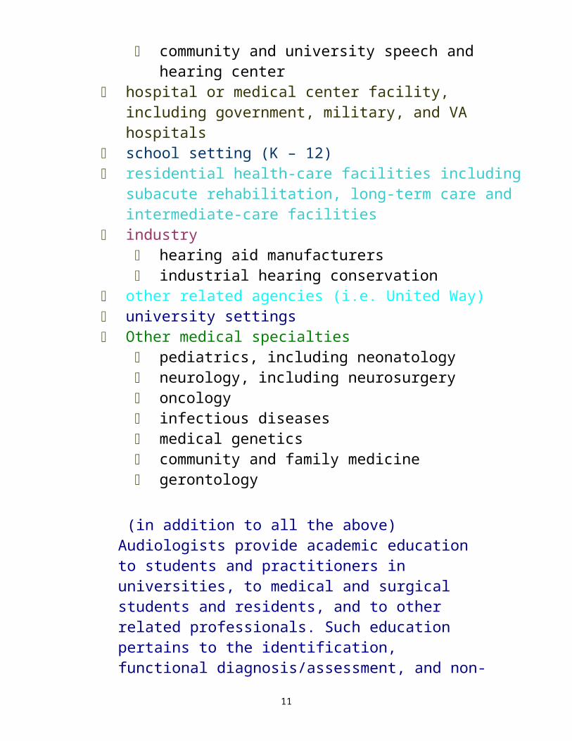
• Audiologists must be licensed in most states. • Audiologists treat all ages and types of hearing loss: adults, teens, children, and infants. • Audiologists work in a variety of settings, such as:
private practice & other nonresidential health-care facilities private practice private or public clinics ENT offices community and university speech and hearing
center hospital or medical center facility, including
government, military, and VA hospitals school setting (K – 12) residential health-care facilities including subacute
rehabilitation, long-term care and intermediate-care facilities
industry hearing aid manufacturers industrial hearing conservation
other related agencies (i.e. United Way) university settings Other medical specialties
pediatrics, including neonatology neurology, including neurosurgery oncology infectious diseases medical genetics community and family medicine gerontology
(in addition to all the above) Audiologists provide academic education to students and practitioners in universities, to medical and
8

surgical students and residents, and to other related professionals. Such education pertains to the identification, functional diagnosis/assessment, and non-medical treatment/management of auditory, vestibular, balance, and related impairments.
• Almost all types of hearing loss are treatable by an audiologist. • Most hearing loss that is caused by nerve damage can be
treated by an audiologist with hearing aids, assistive listening devices, and hearing rehabilitation.
9

Please note that the scope of practice and definitions from AAA and ASHA can be found in the back of your text at appendix A and B in their entirety.
The AAAScope of Practice document updated January 2004 says:The Scope of Practice document describes the range of interests, capabilities and professional activities of audiologists. It defines audiologists as independent practitioners and provides examples of settings in which they are engaged. It is not intended to exclude the participation in activities outside of those delineated in the document. As a dynamic and growing profession, the field of audiology will change over time as new information is acquired. This Scope of Practice document will receive regular review for consistency with current knowledge and practice. services, and the general public.
Definition of an AudiologistAn audiologist is a person who, by virtue of academic degree, clinical training, and license to practice and/or professional credential, is uniquely qualified to provide a comprehensive array of professional services related to the prevention of hearing loss and the audiologic identification, assessment, diagnosis, and treatment of persons with impairment of auditory and vestibular function, and to the prevention of impairments associated with them. Audiologists serve in a number of roles including clinician, therapist, teacher, consultant, researcher and administrator. The supervising audiologist maintains legal and ethical responsibility for all assigned audiology activities provided by audiology assistants and audiology students.
The central focus of the profession of audiology is concerned with all auditory impairments and their relationship to disorders of communication. Audiologists identify, assess, diagnose, and treat individuals with impairment of either peripheral or central auditory and/or vestibular function, and strive to prevent such impairments.
10
Audiologists provide clinical and academic training to students in audiology. Audiologists teach physicians, medical students, residents, and fellows about the auditory and vestibular system. Specifically, they provide instruction about identification, assessment, diagnosis, prevention, and treatment of persons with hearing and/or vestibular impairment. They provide information and training on all aspects of hearing and balance to other professions including psychology, counseling, rehabilitation, and education. Audiologists provide information on hearing and balance, hearing loss and disability, prevention of hearing loss, and treatment to business and industry. They develop and oversee hearing conservation programs in industry. Further, audiologists serve as expert witnesses within the boundaries of forensic audiology.
The audiologist is an independent practitioner who provides services in hospitals, clinics, schools, private practices and other settings in which audiologic services are relevant such as industry (measuring noise and designing protection) and with hearing aid manufacturers for design development, and/or training of new technology and products).
11

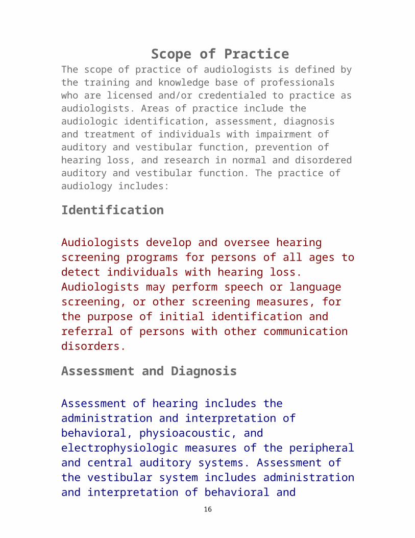
Scope of PracticeThe scope of practice of audiologists is defined by the training and knowledge base of professionals who are licensed and/or credentialed to practice as audiologists. Areas of practice include the audiologic identification, assessment, diagnosis and treatment of individuals with impairment of auditory and vestibular function, prevention of hearing loss, and research in normal and disordered auditory and vestibular function. The practice of audiology includes:
Identification
Audiologists develop and oversee hearing screening programs for persons of all ages to detect individuals with hearing loss. Audiologists may perform speech or language screening, or other screening measures, for the purpose of initial identification and referral of persons with other communication disorders.
Assessment and Diagnosis
Assessment of hearing includes the administration and interpretation of behavioral, physioacoustic, and electrophysiologic measures of the peripheral and central auditory systems. Assessment of the vestibular system includes administration and interpretation of behavioral and electrophysiologic tests of equilibrium. Assessment is accomplished using standardized testing procedures and appropriately calibrated instrumentation and leads to the diagnosis of hearing and/or vestibular abnormality.
12

Treatment
The audiologist is the professional who provides the full range of audiologic treatment services for persons with impairment of hearing and vestibular function. The audiologist is responsible for the evaluation, fitting, and verification of amplification devices, including assistive listening devices. The audiologist determines the appropriateness of amplification systems for persons with hearing impairment, evaluates benefit, and provides counseling and training regarding their use. Audiologists conduct otoscopic examinations, clean ear canals and remove cerumen, take ear canal impressions, select, fit, evaluate, and dispense hearing aids and other amplification systems. Audiologists assess and provide audiologic treatment for persons with tinnitus using techniques that include, but are not limited to, biofeedback, masking, hearing aids, education, and counseling.
Audiologists also are involved in the treatment of persons with vestibular disorders. They participate as full members of balance treatment teams to recommend and carry out treatment and rehabilitation of impairments of vestibular function.
Audiologists provide audiologic treatment services for infants and children with hearing impairment and their families. These services may include clinical treatment, home intervention, family support, and case management.
13

The audiologist is the member of the implant team (e.g., cochlear implants, middle ear implantable hearing aids, fully implantable hearing aids, bone anchored hearing aids, and all other amplification/signal processing devices) who determines audiologic candidacy based on hearing and communication information. The audiologist provides pre and post surgical assessment, counseling, and all aspects of audiologic treatment including auditory training, rehabilitation, implant programming, and maintenance of implant hardware and software.
The audiologist provides audiologic treatment to persons with hearing impairment, and is a source of information for family members, other professionals and the general public. Counseling regarding hearing loss, the use of amplification systems and strategies for improving speech recognition is within the expertise of the audiologist. Additionally, the audiologist provides counseling regarding the effects of hearing loss on communication and psycho-social status in personal, social, and vocational arenas.
The audiologist administers audiologic identification, assessment, diagnosis, & treatment programs to children of all ages with hearing impairment from birth & preschool through school age. The audiologist is an integral part of the team within the school system that manages students with hearing impairments & students with central auditory processing disorders. The audiologist participates in the development of Individual Family Service Plans (IFSPs) and Individualized Educational Programs (IEPs), serves as a consultant in matters pertaining to classroom acoustics, assistive listening systems, hearing aids, communication, & psycho-social effects of hearing loss, and maintains both classroom assistive systems as well as students' personal hearing aids. The audiologist administers hearing screening programs in schools, & trains & supervises non audiologists performing hearing screening in the ed setting.
14

Hearing Conservation
The audiologist designs, implements and coordinates industrial and community hearing conservation programs. This includes identification and amelioration of noise-hazardous conditions, identification of hearing loss, recommendation and counseling on use of hearing protection, employee education, and the training and supervision of non audiologists performing hearing screening in the industrial setting.
Intraoperative Neurophysiologic Monitoring
Audiologists administer and interpret electrophysiologic measurements of neural function including, but not limited to, sensory and motor evoked potentials, tests of nerve conduction velocity, and electromyography. These measurements are used in differential diagnosis, pre- and postoperative evaluation of neural function, and neurophysiologic intraoperative monitoring of central nervous system, spinal cord, and cranial nerve function.
Research
Audiologists design, implement, analyze and interpret the results of research related to auditory and balance systems.
Additional Expertise
Some audiologists, by virtue of education, experience and personal choice choose to specialize in an area of practice not otherwise defined in this document. Nothing in this document shall be construed to limit individual freedom of choice in this regard provided that the activity is consistent with the American Academy of Audiology Code of Ethics. For example, as consultants to buildings such as schools and nursing homes desiring acoustically friendly construction to eliminate problems such as reverberation. Now let’s look at what ASHA has to say:
15
Definition of an Audiologist
Audiologists are professionals engaged in autonomous practice to promote healthy hearing, communication competency, and quality of life for persons of all ages through the prevention, identification, assessment, and rehabilitation of hearing, auditory function, balance, and other related systems. (also in your book on page 4) They facilitate prevention through the fitting of hearing protective devices, education programs for industry and the public, hearing screening/conservation programs, and research. The audiologist is the professional responsible for the identification of impairments and dysfunction of the auditory, balance, and other related systems. Their unique education and training provides them with the skills to assess and diagnose dysfunction in hearing, auditory function, balance, and related disorders. The delivery of audiologic (re)habilitation services includes not only the selecting, fitting, and dispensing of hearing aids and other hearing assistive devices, but also the assessment and follow-up services for persons with cochlear implants. The audiologist providing audiologic (re)habilitation does so through a comprehensive program of therapeutic services, devices, counseling, and other management strategies. Functional diagnosis of vestibular disorders and management of balance rehabilitation is another aspect of the professional responsibilities of the audiologist. Audiologists engage in research pertinent to all of these domains.
now to what they consider the scope of practice
16

Professional Roles and Activities
Audiologists serve a diverse population and may function in one or more of a variety of activities. The practice of audiology includes:
1.Prevention1. Promotion of hearing wellness, as well as the
prevention of hearing loss and protection of hearing function by designing, implementing, and coordinating occupational, school, and community hearing conservation and identification programs;
2. Participation in noise measurements of the acoustic environment to improve accessibility and to promote hearing wellness.
2.Identification1. Activities that identify dysfunction in hearing,
balance, and other auditory-related systems;2. Supervision, implementation, and follow-up of
newborn and school hearing screening programs;
3. Screening for speech, orofacial myofunctional disorders, language, cognitive communication disorders, and/or preferred communication modalities that may affect education, health, development or communication and may result in recommendations for rescreening or comprehensive speech-language pathology assessment or in referral for other examinations or services;
4. Identification of populations and individuals with or at risk for hearing loss and other auditory dysfunction, balance impairments, tinnitus, and
17

associated communication impairments as well as of those with normal hearing;
5. In collaboration with speech-language pathologists, identification of populations and individuals at risk for developing speech-language impairments.
3.Assessment1. The conduct and interpretation of behavioral,
electroacoustic, and/or electrophysiologic methods to assess hearing, auditory function, balance, and related systems;
2. Measurement and interpretation of sensory and motor evoked potentials, electromyography, and other electrodiagnostic tests for purposes of neurophysiologic intraoperative monitoring and cranial nerve assessment;
3. Evaluation and management of children and adults with auditory-related processing disorders;
4. Performance of otoscopy for appropriate audiological management or to provide a basis for medical referral;
5. Cerumen management to prevent obstruction of the external ear canal and of amplification devices;
6. Preparation of a report including interpreting data, summarizing findings, generating recommendations and developing an audiologic treatment/management plan;
7. Referrals to other professions, agencies, and/ or consumer organizations.
4.Rehabilitation18
1. As part of the comprehensive audiologic (re)habilitation program, evaluates, selects, fits and dispenses hearing assistive technology devices to include hearing aids;
2. Assessment of candidacy of persons with hearing loss for cochlear implants and provision of fitting, mapping, and audiologic rehabilitation to optimize device use;
3. Development of a culturally appropriate, audiologic rehabilitative management plan including, when appropriate:
1. Recommendations for fitting and dispensing, and educating the consumer and family/caregivers in the use of and adjustment to sensory aids, hearing assistive devices, alerting systems, and captioning devices;
2. Availability of counseling relating to psycho social aspects of hearing loss, and other auditory dysfunction, and processes to enhance communication competence;
3. Skills training and consultation concerning environmental modifications to facilitate development of receptive and expressive communication;
4. Evaluation and modification of the audiologic management plan.
4. Provision of comprehensive audiologic rehabilitation services, including management procedures for speech and language habilitation and/or rehabilitation for persons with hearing loss or other auditory dysfunction, including but not exclusive to speechreading, auditory
19
training, communication strategies, manual communication and counseling for psychosocial adjustment for persons with hearing loss or other auditory dysfunction and their families/caregivers;
5. Consultation and provision of vestibular and balance rehabilitation therapy to persons with vestibular and balance impairments;
6. Assessment and non-medical management of tinnitus using biofeedback, behavioral management, masking, hearing aids, education, and counseling;
7. Provision of training for professionals of related and/or allied services when needed;
8. Participation in the development of an Individual Education Program (IEP) for school-age children or an Individual Family Service Plan (IFSP) for children from birth to 36 months old;
9. Provision of in-service programs for school personnel, and advising school districts in planning educational programs and accessibility for students with hearing loss and other auditory dysfunction;
10. Measurement of noise levels and provision of recommendations for environmental modifications in order to reduce the noise level;
11. Management of the selection, purchase, installation, and evaluation of large-area amplification systems.
5.Advocacy/ Consultation1. Advocacy for communication needs of all
individuals that may include advocating for the 20

rights/funding of services for those with hearing loss, auditory, or vestibular disorders;
2. Advocacy for issues (i.e., acoustic accessibility) that affect the rights of individuals with normal hearing;
3. Consultation with professionals of related and/or allied services when needed;
4. Consultation in development of an Individual Education Program (IEP) for school-age children or an Individual Family Service Plan (IFSP) for children from birth to 36 months old;
5. Consultation to educators as members of interdisciplinary teams about communication management, educational implications of hearing loss and other auditory dysfunction, educational programming, classroom acoustics, and large-area amplification systems for children with hearing loss and other auditory dysfunction;
6. Consultation about accessibility for persons with hearing loss and other auditory dysfunction in public and private buildings, programs, and services;
7. Consultation to individuals, public and private agencies, and governmental bodies, or as an expert witness regarding legal interpretations of audiology findings, effects of hearing loss and other auditory dysfunction, balance system impairments, and relevant noise-related considerations;
8. Case management and service as a liaison for the consumer, family, and agencies in order to monitor audiologic status and management and
21

to make recommendations about educational and vocational programming;
9. Consultation to industry on the development of products and instrumentation related to the measurement and management of auditory or balance function.
6.Education/ Research/Administration1. Education, supervision, and administration for
audiology graduate and other professional education programs;
2. Measurement of functional outcomes, consumer satisfaction, efficacy, effectiveness, and efficiency of practices and programs to maintain and improve the quality of audiologic services;
3. Design and conduct of basic and applied audiologic research to increase the knowledge base, to develop new methods and programs, and to determine the efficacy, effectiveness, and efficiency of assessment and treatment paradigms; disseminate research findings to other professionals and to the public;
4. Participation in the development of professional and technical standards;
5. Participation in quality improvement programs;6. Program administration and supervision of
professionals as well as support personnel.
22

All the above is included in the ASHA scope of practice.
23

The following were taken from the 2nd edition of the Comprehensive Dictionary of Audiology Illustrated
AUDIOLOGYThe branch of healthcare devoted to the study, diagnosis, treatment, and prevention of hearing disorders.
This is further broken into subspecialties:a. educational: subspecialty devoted to the
hearing needs of school-age children in an academic setting
b. forensic: subspecialty devoted to legal proceedings related to hearing loss and noise matters
c. pediatric: subspecialty devoted to the study, diagnosis, and treatment of hearing impairment in children
d. recreational: subspecialty devoted to the conservation of hearing during recreational activities, such as shooting, listening to music, etc.
AUDIOLOGIST
A healthcare professional who is credentialed in the practice of audiology to provide a comprehensive array of services related to prevention, diagnosis, and treatment of hearing impairment and its associated communication disorder
24

This was broken down into subspecialties as well including: dispensing, educational, and pediatric
25

This from ASHA, 1996ASHA further states in the Scope of Practice that audiologists provide their services “across the entire age span from birth through adulthood; to individuals from diverse language, ethnic, cultural, and socioeconomic backgrounds; and to individuals who have multiple disabilities.” It further notes that audiologists are engaged in “counseling for psycho-social adjustments to hearing loss and to persons with hearing loss, their caregivers/families.”From page 3 – 4 of your text – 3 definitions
Audiologist(according to the author of this text)
A professional who, by virtue of academic degree, clinical education, and appropriate (certification and/or) licensure or other credential, is uniquely qualified to provide a comprehensive array of professional services related to the prevention of hearing loss and the audiologic identification, diagnosis, and treatment of patients with impairments in hearing and balance function.According to AAA
An audiologist is a person who, by virtue of academic degree, clinical training, and license to practice and/or professional credential is uniquely qualified to provide a comprehensive array of professional services related to the prevention of hearing loss and the audiologic identification,
26

assessment, diagnosis, and treatment of persons with impairment of auditory and vestibular function, and to the prevention of impairments associated with them. According to ASHA
Audiologists are professionals engaged in the autonomous practice to promote healthy hearing, communication competency, and quality of life for persons of all ages through the prevention, identification, assessment, and rehabilitation of hearing, auditory function, balance, and other related systems.
The unique mission:The evaluation of the auditory and vestibular system and the amelioration of the impairments that result from auditory (hearing) and vestibular (balance) disorders.
Audiologists may diagnose and treat and provide remediation for disorders from any part of the auditory system from the outer ear to the middle ear to the inner ear (including the vestibular system) to the central auditory nervous system. Remediation or rehabilitation may include hearing aids for hearing loss, tinnitus maskers and counseling for tinnitus, ALDs for APD, limited vestibular rehab such as repositioning maneuvers for vestibular problems,
Audiologists may be found in many roles; they may be found as:
clinicians therapists teachers, educators, supervisors research investigators
27

administrators consultants – education, prevention, forensics
Various studies showed that 80 – 90% of hearing loss is not medically treatable. Thus, the audiologist serves as the primary expert in the assessment and nonmedical diagnosis of auditory impairment.
Some situations in which SLPs and audiologists work closely together:
a. the hearing impaired child will likely need the assistance for speech delay
b. those children determined to have auditory perceptual problems due to an impaired central auditory nervous system (those with Auditory Processing Disorder or APD)
c. with older individuals after stroke or other neurologic insult (to determine the extent the hearing loss is impacting on receptive language ability)
28

Chapter 2 of text
What is Sound?
Sound: a type of energy occurring as a result of pressure waves
that come from a force being applied to a sound source. It results from the compression and rarefaction of the molecules in the medium though which it is traveling. As the medium is compressible or elastic, the molecules themselves return to the point of origin, however, they have bumped into other molecules during the compression(aka condensation due to increased density of molecules) and rarefaction (the elasticity allowing reduced density of molecules) phase and those other molecules keep the pressure waves moving and so the energy is passed along.
So we have a (1) source of vibratory energy which (2) causes a disturbance in a medium which (3) propagates that disturbance as sound waves to (4) carry energy away from the source.
Vibration: to and fro movement in a massa. free – the mass is displaced from rest and allowed
to oscillate without outside influenceb. forced – the mass is moved back and forth by
applying an external force
There are 2 ways to define sound:2. in a psychological sense which is the act of hearing3. in a physical sense which is a series of disturbances of
the molecules within, and propagated through, an elastic medium
29
Waves: molecules are shoved close together and pulled apart
a. condensation – molecules are close together; increased density of molecules during sound; a compression of molecules
b. rarefaction – when there’s space between the areas of compression; decreased density of molecules during sound; an expansion of molecules.
Compression and expansion results in pressure changes that travel though a medium such as air or water. These pressure waves have mass, are elastic and pass energy along.
Properties of Sound
Simple harmonic motion (or sinusoidal motion): periodic back and forth movement (and may be plotted as a function of time). Sinusoidal waveform: a graphic display of simple harmonic motion in magnitude versus time. Aka sine waveWaveform: the shape of a wave seen as amplitude of displacement versus time and this is used to describe various properties of sound. Magnitude of the waveform or the amplitude dictates the intensity of the sound and is what we are more familiar with known as loudness.Frequency of the waveform is how often a cycle is completed and is what we are more familiar with known as pitch.
Cycle: one complete sound wave (or an oscillation); a condensation and rarefaction as a function of time. This
30

determines the frequency of a sound. Any stage of a cycle is known as the phase. (so this is the location at any point in time in the displacement of an air molecule during a cycle)One cycle may also be known as the period.Period =_____1___ FrequencySo if the time is for 1 second, the answer used to be cps or cycles/second. We no longer use that term, we use the term Hertz (Hz). So cps and Hz may be used interchangeably.Frequency – the number of complete cycles of a vibrating body per unit of time.Example: If the time period you are measuring is one second and there are 3 full cycles in that period, then the measurement is 3 cycles/sec
Pure tone: 1 sine wave with no tones superimposedSimple Sounds – a sound that has all its energy at one frequency, a pure tone.
31

32

There is a large range of intensity. Barely audible sounds occur at 20 μPa and painful intensity is at 200,000,000 μPa. As these units become rather large, intensities are described as decibels (dB).
Today, we describe intensity in decibels sound pressure level or dBSPL. SPL is the magnitude of sound energy relative to a reference pressure of .0002dynes/cm2 or 20μPa.Decibels are expressed as a ratio of a measured pressure to a reference pressure. Thus, 0 dB does not mean that there is no sound. It just means that the measured pressure equals the reference pressure.Velocity – the speed with which the sound wave travels from the source to another point. The denser the medium, the faster sound travels as molecules are closer together and bounce off one another faster.
Intensity: quantity or magnitude of a sound. It is the perception of sound loudness. The distance mass moves from the point of
rest; the amplitude a body vibrates, this is measured in dB or decibels
Pressure: this is generated whenever force is distributed over a surface area. Damaging sound waves have a high pressure.
So once we understand this, and we now know frequency and intensity, another way to think of it is:
Physical Measurement Psychological Correlate Measurement Frequency Pitch Hz or kHz
33

Intensity Loudness dB (decibels)
Decibels (dB): the units in which we measure intensitySound Pressure Level (SPL): the magnitude of sound energy relative to a reference pressure. Most commonly used reference is .0002dynes/cm2 also known as 20uPa (micropascals)
Remember: intensity changes are not a 1:1 relationship but a logarithmic relationship. We change the sound pressure into numbers we can use.
Since decibels are a ratio of a measured pressure to a reference pressure, that does not mean that 0dB is no sound but rather the measured pressure is equal to the reference pressure.
But when performing hearing testing we measure decibels in hearing level.
Hearing level (HL): this is the decibels according to average normal hearing or audiometric 0.
Normals were measured in dBSPL and at each frequency, the average minimal level of hearing was turned into audiometric 0.
The human ear can hear from 20Hz to 20,000Hz.
We test in octaves. An octave is twice the frequency of a given frequency. We normally test at 250Hz, 500Hz, 1000Hz, 2000Hz, 4000Hz, and 8000Hz. Not that each is double the one before. So the next frequency tested is always an octave interval of the one tested prior.
34

Waves with more than one sinusoid are complex. The content of the complex sounds, the interaction of intensity (or magnitude) and frequency, is known as the sound’s spectrum.
35
Terms to Know
Bel – a logarithm expressing a ratio between 2 lengthy numbersDecibel (dB) – 1/10 of a bel – used because the bel can be a
very long number, the unit of measurement of intensity or loudness used in audiometrics
As these represent logarithms, we must realize it is not a 1:1 relationship. When the intensity of the wave is not doubled but raised by three. This 3dB refers to intensity.
Sound Pressure Level (SPL) – commonly measured in dynes/cm2, and most commonly, the reference is .0002 dynes/cm2, this is also known at 20 micropascals (Pa). This is an absolute measurement, as we would think of centigrade when using absolute temperatures.
When dealing with 2 sound pressure measurements to be added, we are again dealing with logarithms and it is not a 1:1 relationship of direct addition. So, for sound pressure level, we increase the level by 6dB when it is doubled.
Again:When doubling intensities, we add 3dB.When doubling sound pressure, we add 6dB.
The formula to convert to dBSPL, or sound pressure is: ( output pressure )
dBSPL = 20 X log ( referent pressure) and this is .002 dynes/cm2
The formula to convert power to decibels intensity level dBIL is:
(Wo ) Wo = watts per cm2 (power) output36
dB = 10 X log ( Wr ) Wr = watts per cm2 (power) reference and the agreed-upon intensity level is 10-16W/cm2
Hearing Level (HL) – this is a relative term, much as farenheit
is a relative term for temperatures. To obtain this, a number of normal hearing adults were tested and their hearing in SPL was converted into 0HL for each frequency. Example: if the average was 7dBSPL for 1Hz, then the HL level for 7dB at 1kHz now became 0dB on the audiometer.
Sensation Level (SL) – this is another relative term, relative to
HL. It is the number of decibels above the minimum level of hearing. Example, if someone hears speech at 30dBHL, and you speech at 60dBHL, it is their 30dBSL at it is 30dB in sensation level above the hearing level.
Threshold () – this is the minimum level where an individual
can hear a tone 50% of the time.
Fundamental Frequency – the lowest rate of a sound’s vibration. This is determined by the physical properties of the vibrating body.
Harmonics – whole-number multiples of the fundamental frequency. So a 1000Hz tone would have a first harmonic of 2000Hz and a second harmonic of 4000Hz, etc. (aka overtones)
Sounds can be measured by their waveforms, consisting of peaks and valleys and the peaks are called formants. These formants are important for vowel perception.
37

Moving sound encounters resistance that impedes its progress. The more dense the medium, the more impedance there is. For example, when the ossicles move correctly, there is little impedance. When the stapes becomes stiff there is more mass and thus more impedance to movement and transmission of sound.
Audiometric zero: the sound pressure level at which the threshold of audibility occurs for normal listeners. Aka audiometric zeroFrequency dBSPL (absolute) dBHL (relative) 250Hz 26.5 0500Hz 13.3 01000Hz (1kHz) 7.5 02000Hz (2kHz) 11.0 03000Hz (3kHz) 9.5 04000Hz (4kHz) 10.5 06000Hz (6kHz) 13.5 08000Hz (8kHz) 13.0 0
38
THE EAR
The ear can be divided into three portions:1.The outer ear2.The middle ear3.The inner ear
Sound is primarily mechanical transmission till the inner ear.Physical processing of acoustic information occurs in the outer, middle, and inner ears.Physiological processing begins in the inner ear and goes along the 8th cranial nerve to the central auditory nervous system (CANS).Psychological processing begins in the brainstem and continues to the auditory cortex and onward.
A problem in the first 2 portions of the ear is easily taken care of viaa. medical treatmentb. surgeryc. hearing aids
A problem in the inner ear and beyond causes significant difficulties that are not easily managed and requires additional counseling and usually audiologic habilitation/rehabilitation.
Let’s look at each portion of the ear.
39
The Outer Ear
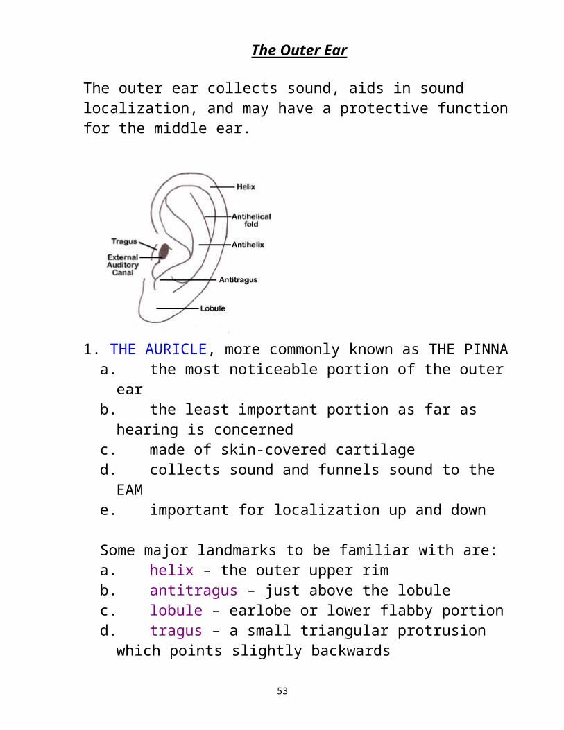
The outer ear collects sound, aids in sound localization, and may have a protective function for the middle ear.
1. THE AURICLE, more commonly known as THE PINNAa. the most noticeable portion of the outer earb. the least important portion as far as hearing is
concernedc. made of skin-covered cartilaged. collects sound and funnels sound to the EAMe. important for localization up and down
Some major landmarks to be familiar with are:a. helix – the outer upper rimb. antitragus – just above the lobulec. lobule – earlobe or lower flabby portiond. tragus – a small triangular protrusion which points
slightly backwardse. concha – bowl-like shape in the middle; may increase
sound by as much as 10 – 15dB at around 4500Hz; funnels sound into
f. External Auditory Canal or external auditory meatus (EAC or EAM) aka the ear canal and serves as a resonator to enhance sound at 2700Hz
40

2. THE EXTERNAL AUDITORY CANALa. a tunnel beginning at the concha ; 23 – 29 mm in
lengthb. it is elliptical, slants down at TM, and lined with skinc. the outer 2/3 is skin-covered cartilage (continuous
with the cartilage of the pinna)d. the inner 1/3 is skin-covered bone (bony portion of
the ear canal) and goes through the temporal bone.e. Osseocartilaginous junction – where the 2 portions of
the EAC meetf. Protective function to protect TM due to narrow
opening and the cerumen protects from foreign objects and creatures.
g. there are several sets of glands in the skin of the cartilaginous portion, including those which cause cerumen or earwax. Note: cerumen is normal and when combined with the little hairs in the ear canal (cilia), serves as a protective mechanism to keep foreign objects from going in further and to minimize bacteria and fungus from infecting the ear canal
h. In infants and very young children, the angle of the EAC is very different from that of the adult. It slopes downward at a sharper angle
i. The canal serves as a tube resonator for frequencies around 2700Hz for most adults by 10 – 20dB.
41
1. THE TYMPANIC MEMBRANE (EARDRUM)a. occurs at the termination of the EACb. constructed in 3 layers of skin embedded in the
bony portion of the canali. outer layer is skinii. middle layer is tough, fibrous, connective
tissue which contributes most to the ability to vibrate
iii. mucous membrane which also lines the middle ear space
c. very thind. concave, curving slightly inward, and taut like a
drume. extremely efficient vibrating surfacef. rich in blood supplyg. embedded in fibrous portion is the malleus or
largest bone of the middle earh. umbo – tip of the malleus is set to cause the center
of the eardrum to be pulled inward and this is the point of greatest retraction
i. annulus – a ring of tissue that holds the ear drum in position
42

j. vibrates when sound waves hit it and vibrates with a magnitude proportional to the intensity of sound at a speed proportional to its frequency
k. semitransparent
When looking into the ear with an otoscope, it is common to observe a reflection of the otoscope’s light in the anterior and inferior quadrant and this is referred to as the cone of light.
The ear drum can be divided into 4 quadrants:1. anterior-superior2. posterior-superior3. anterior-inferior4. posterior-inferior
Has 2 main sections:a. pars tensa - most of the surface of the eardrum is
taut, it is the larger, inferior portion with 4 membranous layers and much stiffer than the pars flaccida
b. pars flaccida – smaller and more compliant, located superiorly and has 2 layers of tissue; mostly epidermal and mucus membrane layers aka Shrapnell’s membrane.
The external ear, comprised of the pinna and external auditory canal up to the eardrum, provides a resonant tube through which sound waves pass. The TM vibrates with a magnitude proportional to the intensity (loudness) of the sound wave at a speed proportional to its frequency (cycles/second or pitch). As sound waves hit the eardrum, the eardrum vibrates, and then causes vibration of the malleus, which is attached to it.
43

THE MIDDLE EAR
The average middle ear is an almost oval, air-filled space within the temporal bone of the skull. This is an air cavity with suspended structures within. It begins with the inner layers of the TM. It serves as an impedance matching device between the sound waves hitting the TM and the fluid waves of the cochlea. Thus, they provide a bridge between the pressure waves coming in to the TM and the fluid traveling waves of the cochlea; it connects the tympanic membrane to the oval window of the cochlea via the ossicular chain.
44

a. The roof is a thin layer of bone separating the middle ear cavity from the brain.
b. Tegmen tympani – thin layer of bone separating ME cavity from the brain
c. Fundus tympani – thin plate of bone separating ME from jugular bulb
d. Jugular bulb – below the floor of the middle eare. Membranous wall – lateral part of the middle ear and
contains the TMf. Epitympanic recess – space within the ME and above
the TMg. Eustachian tube – connects the middle ear to the
nasopharynx (area where the nose and back of the throat come together) This keeps the middle ear at atmospheric pressure
h. Lined with a mucous membrane and is ciliated (cilia are tiny hairs). These hair cells create a motion that cleanses the middle ear by moving particles down and out through the Eustachian tube.
45

1. THE EUSTACHIAN TUBEa. enters the ear at a 30 degree angle and passes into
the nasopharynx; it is the passage way from the nasopharynx to the anterior wall of the ME
b. opened by the action of 3 sets of muscles and this occurs duringi. yawningii. sneezing, or iii. swallowing.
c. With sudden pressure changes, if not equalized, the TM is pushed in and pressure is felt. May need to swallow or valsalva
d. Keeps the air in the middle ear at atmospheric pressure
2. THE MASTOIDThe skull area just behind the ear is bone that is honeycombed with hundreds of air cells. This is called the pneumatic mastoid of the temporal bone. The mastoid borders the middle ear space posteriorly. The bony protuberance behind the pinna is called the mastoid process.
3. WINDOWSa. Oval window – filled by the base of the stapes (the
smallest bone in the body) and beyond is the inner ear.
b. Round window – covered by a thin, tough, elastic membrane
c. Promontory – a section of the inner ear that protrudes into the middle ear. It separates the oval and round window (oval is above and round below)
46

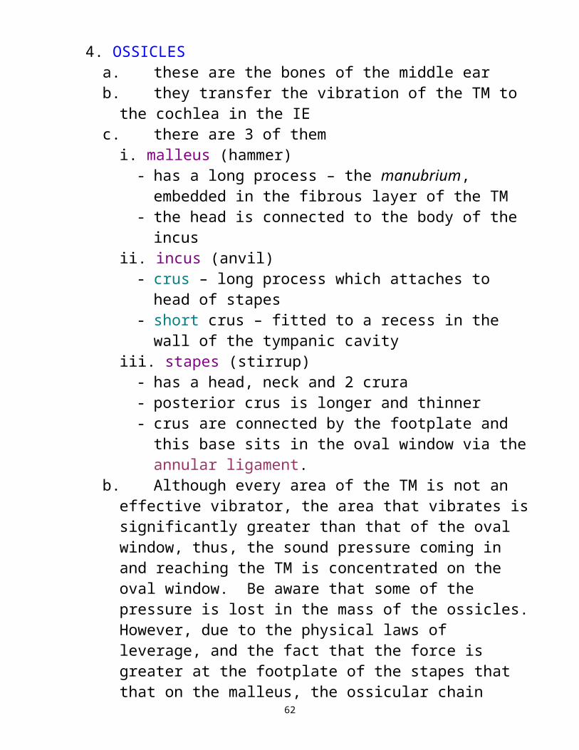
4. OSSICLESa. these are the bones of the middle earb. they transfer the vibration of the TM to the cochlea in
the IEc. there are 3 of them
i. malleus (hammer)- has a long process – the manubrium, embedded
in the fibrous layer of the TM- the head is connected to the body of the incus
ii. incus (anvil)- crus – long process which attaches to head of
stapes- short crus – fitted to a recess in the wall of the
tympanic cavityiii. stapes (stirrup)
- has a head, neck and 2 crura- posterior crus is longer and thinner- crus are connected by the footplate and this
base sits in the oval window via the annular ligament.
b. Although every area of the TM is not an effective vibrator, the area that vibrates is significantly greater than that of the oval window, thus, the sound pressure coming in and reaching the TM is concentrated on the oval window. Be aware that some of the pressure is lost in the mass of the ossicles. However, due to the physical laws of leverage, and the fact that the force is greater at the footplate of the stapes that that on the malleus, the ossicular chain rocks and acts as a pivot rather than a piston.Thus, the increased pressure + lever action give a significantly increased pressure at the oval window.
47

So the middle ear has impedance matching characteristics. This is why there is no loss of sound energy due to the impedance mismatch. The ease of energy flow in air is very different than energy flow in a fluid filled space. So the ossicular chain plays the role of mediator between what comes in the OE and what affects the IE. Without it, the air pressure alone would have to set the fluid into motion and a lot of energy could be lost. With the middle ear, the air waves are changed into mechanical energy and this mechanical energy in the ME helps convert the energy from air to hydraulic. It does this in 3 ways:
A. sound is collected over a large surface area (TM) and directed
to a smaller surface area (footplate of the stapes), increasing the sound pressure by as much as 23dB.
B. TM is curved with more movement on the curved areas and
less near the manubrium. This results in an increase in the force transmitted through the middle ear.
C. The difference in length between ossicles, from the malleus to
the incus, creates a lever-type action that result in a small increase in sound pressure. This lever action provides a boost of about 2.4dB to the signal.
Without these impedance matching characteristics, there could possibly be a loss of energy of 26 to 30dB of sound during the transmission of sound from air to fluid in the cochlea.
48

5. There are 2 main middle ear muscles:a. stapedius muscle: this muscle is in a canal next to
the facial canal. It is innervated by a branch of the VIIth nervei. stapedius tendon: attached the muscle to the stapes.
The stapedius tendon is attached to the neck of the stapes and when the muscle is contracted, the stapes moves and tenses the oval window. It is theorized that we understand better than we should in noise because this reduced the low pitch parts of noise by reducing the amplitude of vibration. This tendon also supplies blood to the lenticular process of the incus.
b. tensor tympani muscle: the tendon from this muscle goes into the manubrium of the malleus and when contracted causes the malleus to move in a way that tenses the TM. The innervation of this nerve is from the trigeminal (Vth) nerve.
Later, we will learn of acoustic reflexes and the reflex arc. The stapedial muscle will play a major role in the acoustic reflexes, both ipsilateral and contralateral.
49

THE INNER EAR
The inner ear has also been called a labyrinth. There are 2 primary portions with numerous parts.
I. THE VESTIBULAR APPARATUS – balance portion 1. vestibule: the immediate entryway after just past the
oval window; through here the various partitions of the inner ear can be reached.
2. perilymph: the fluid which fills the vestibule 3. here is where the organs of balance or the vestibular apparatus exists 4. this system relies on the forces of gravity and inertia 5. receives input from visual and somatosensory system or the skeletal muscular system. 6. In the vestibule are the utricle and the saccule. Both are membranous sacs surrounded by perilymph and
containing endolymph. The saccule is slightly smaller than the utricle. Coming off the utricle are the 3 semicircular canals, the:
a.superior semicircular canalb.lateral semicircular canalc.posterior semicircular canal
These are filled with endolymph and surrounded in the cavity by perilymph. They are arranged perpendicular to each other to cover all dimensions in space.
50

7. Ampulla (ampullae pl.): enlarged areas at the end of each canal which goes back into the utricle. Each contains an end organ called 8. crista: an end organ in the ampulla which is used for the sense of balance 9. macula of otoliths: otoconia are calcium carbonate crystals and contains sensory receptors called macular making hair cells sensitive to gravity(note: the above crista of ampullae and maula of otoliths are in the uticle) 10. When your head moves, the fluids inside this system moves and this stimulates the entire vestibular mechanism.
So the 5 sensory receptors in this portion are the utricle, saccule, and the 3 semicircular canals. This all acts as a motion detector:a. the utricle orients to gravity and horizontal movement, while the saccule orients in the vertical plain (these 2 structures are responsive to linear acceleration)b. the semicircular canals and the ampullae are responsive to angular acceleration (movement like tilting your head to one side)
11. When a problem occurs, whether by illness or damage, the result is vertigo. 12. Nystagmus: a rapid movement of the eyes that
51

occurs with vertigo
52
II. THE AUDITORY MECHANISM1.Cochlea: a snail-like structure, 2.5 turns in the temporal bone composed of three canals or scala where the outermost scala are filled with perilymph
fluid and the center scala is filled with endolymph.a.scala vestibuli: close to vestibule, topmost
portion closest to vestibular apparatus, perilymph filled, terminates basally at the oval window
b.scala tympani: the bottom-most portion; terminates basally at the round window, perilymph filled
c.scala media: the middle canal, AKA as the cochlear duct or cochlear partition and separates the scala vestibule from the scala tympani; filled with endolymph- Reissner’s membrane covers the partition and separates it from the scala vestibuli- Basilar membrane separates it from the scala tympani and runs the length of the cochlea from the base to the apex, on it is the organ of Corti which has the sensory cells of hearing
2. Helicotrema: a small passage at the tip/apex of the
cochlea which allows perilymph to go from the s. vestibuli to the s. tympani. As the oval window is pushed in, perilymph from the s. vestibuli flows through the
53

helicotrema to the s. tympani & pushes the round window out.
3. The endolymph in the scala media, via a duct, is
continuous with the endolymph in the semicircular canals, saccule, and vestibule.
4. Reisner’s membrane: separates scala media from scala vestibuli 5. Organ of Corti: lies along the full length of the scala
media on the basilar membrane. It contains the sensory cells of hearing of which there are 2 types – outer hair cells (about 13,000) and inner hair cells (about 3,500)
6. Spiral Ligament: supports the scala media 7. Stria vascularis: produces endolymph, supplies oxygen, & other nutrients to the cochlea 8. Modiolus: the place where the blood and nerve
supply enter the cochlea; the central bony pillar of the cochlear through which blood vessels and nerve fibers of the labyrinth course
9. BASILAR MEMBRANE:
54

a.narrow at the basal end and wider at the apical turn (the opposite of the cochlear duct)
b.has 3 – 5 parallel rows of 12,000 – 15,000 outer hair cells (OHC) and one row of 3,500 inner hair cells (IHC)
c.Corti’s arch: separates inner and outer hair cells
d.Stereocilia: located on top of each HC; depending on the direction they bend, nerve impulses are either stimulatory or inhibitory
e.Tectorial membrane – the membrane where the stereocilia on the OHCs are embedded; gelatinous
f. When the hair cells shear, a chemical is released at the base of the hair cell
g.Each IHC is supplied by about 20 nerve fibers. Each nerve fiber serves only 1 hair cell
h.With OHCs, each nerve fiber may go to several OHCs
i. OHCs have cilia embedded in a gelatinous layer covering the organ of Corti know as the tectorial membrane
j. Spiral ganglion: all nerve fibers leave the cochlea and extend to the modiolus where the cell bodies group together to form the spiral ganglion; a collection of cell bodies of the auditory nerve fibers clustered in the modiolus
k.IHCs connect to the brain and OHCs are innate biological amplifiers
55

l. OHCs are susceptible to noise, disease, head trauma, vascular problems, etc for about 40 – 60dBHL.
m. OHCs are innervated by efferent or motor fibers of the nervous system – they take information from the brain back to the cochlea
n.IHCs are innervated by the afferent or sensory fibers of the nervous system – they send information to the brain
o.IHCs do not make direct contact with the tectorial membrane
56

57

10. THE AUDITORY NEURONa.There are 2 types of neurons:
i. afferent (sensory) neurons (30,000): carries impulses from the cochlea to the central auditory systemii. efferent neurons (1800): projects to the brainstem and contacts other hair cells
b.The neurons are specialized cells designed as a conductor of nerve impulses and consist of:i. cell bodyii. axon: transmits impulses along the neuronsiii. dendrites: receives impulses from other nerve cellsThe axons and dendrites are a branching system. Electrical impulses travel along the axon to be received by the dendrites. This is accomplished through:i. synapses: connections between neuronsii. neurotransmission: the act of sending information between neuronsiii. neurotransmitters: at the connecting junction, these are released and cause activation or inhibition of adjacent neurons.
58

As mentioned numerous times in connection with the various structures, there are 2 types of fluid, endolymph and perilymph. Endolymph is high in potassium ions and low in sodium ions and perilymph is low in potassium ions and high in sodium ions.
59

IN the past, there have been many theories of hearing. It is no longer fashionable to go into them but be aware only of the TRAVELING WAVE THEORY as explained by von Bekesy: as the stapes footplate moves in and out, the basilar membrane moves down and up due to a disturbance of the endolymph; the wave moving down the cochlear duct from base to apex with max amplitude for high frequency tones at the basal end and low frequencies at the apical end. So the input frequency determines the distance the traveling wave moves before peak as well as the rate of basilar membrane vibration. Therefore, you can see that the basilar membrane is arranged tonotopically.
However, the traveling wave theory does not explain the sensitivity and frequency selectivity of the cochlea. It may be that the sensitivity of the IHCs is controlled by the OHC and at maximum displacement of the traveling wave, the IHCs become stimulated, thus releasing neurotransmitters that stimulate nerve endings.
So let’s recap what we know to the point about the energy transmission of sound from when it enters the ear to the brain.
When sound enters the ear it is acoustical energy. In the middle era it is changed to mechanical energy. In the inner ear, when the fluids begin to move, we have hydraulic energy. As we get higher into the auditory nerve and above, the energy becomes electrical via the synapses.
60

Acoustical (outer ear) Mechanical (middle ear) hydraulic (inner ear) electrical (auditory nerve and
above)
61
62

AUDITORY NERVE AND CENTRAL AUDITORY PATHWAYS
Almost all structures on one side of the brain have a corresponding partner on the other side. The auditory nervous system is filled with nuclei that serve as a relay station for neural information from the cochlea and VIIIth nerve to other nuclei in the ANS and to nuclei of other sensory and motor systems. The ones involved in the primary auditory pathway of the CANS are
- cochlear nucleus- superior olivary complex- lateral lemniscus- inferior colliculus- medial geniculate
1. THE AUDITORY NERVE: this is carried in the internal auditory canal (IAC). The IAC runs from the base of the modiolus and ends at the base of the brain. The canal carries the vestibular portion of the 8th nerve (the fibers going into the utricle, saccule, and semicircular canals). But the auditory portion spirals through like a cable, creating the nerve trunk.
a. the fibers from the basal turn (high frequencies) form the outer portion of the cable and
b. the apical areas (low frequencies) form the center
This nerve continues past the IAC to attach to the brainstem at the cerebellopontine angle (CPA). This is where the cerebellum, medulla oblongata, and pons join. At this point, the auditory and vestibular portions of the nerve separate.
The 8th nerve codes intensity as the rate of neural discharge and it codes frequency as the place of neural discharge (tonotopically).
63

2. THE COCHLEAR NUCLEUS: The cochlear nucleus probably
preserves but does not enhance the information it receives. This is tonotopic as well as the cochlea. The auditory nerve fiber termination separates into areas, one for apical turn fibers and one for basal turn fibers, etc.
As fibers travel up the tract, some travel straight up (ipsilateral) and some (75%) crossover to the same structure on the opposite side of the brain (contralateral). The crossover is also called decussation & the bundles that crossover to the other side are called commissures. The 1st crossover point is at the trapezoid body.
3. RETICULAR FORMATION: this “diffusely organized area” is in the center of the brainstem and communicates everywhere. It plays a role in auditory alertness, reflexes, and habituation. It is responsible for (a) cardio-respiratory reflex function and (b) fight/flight reflex (auditory based threats invoke the reticular formation). It may have an important role in selective attention.
4. SUPERIOR OLIVARY COMPLEX (SOC): gets most of the fibers
from the cochlear nucleus, both ipsilateral and contralateral. Senses direction. It also functions as a relay station on the way to the cortex. Note that this
64

mediates the reflex activity of the 2 middle ear muscles, the crossovers where could explain the stapedial reflex in both ears when sound is presented to one ear. The SOC processes time & intensity cues. It (a) measures the time of arrival of sound at each ear & (b) measures how loud the same sound is in both ears. This is localization & how we lock on a target sound, separate it from the noise. A –12 to –20 S:N ratio still allows us to hear in the presence of noise. If there is a problem in one ear, you lose localization & the ability to listen in noise when the target sound is softer than the noise.
5. LATERAL LEMNISCUS: it is a major pathway for transmission of impulses from the ipsilateral lower brainstem. It is considered to be the primary brainstem auditory pathway.
6. INFERIOR COLLICULUS (IC): receives afferent stimulation from both SOCs. This is in the midbrain and sensitive to binaural stimulation. Exhibits a great degree of tonotopicity.
7. MEDIAL GENICULATE BODY (MGB): this is located in the
thalamus. At this point the nerve fibers become auditory radiations and go up to the auditory cortex.
8. AUDITORY CORTEX: Up here is Heschl’s gyrus which is also
tonotopic. Some discrimination goes on here. Its 2 main functions are (a) to be involved in localization and (b) to determine what is speech and what is not; what is language and what is not.
65

The ANS blood supply comes mainly from 2 sources:a. the basilar artery supplies the auditory brainstem
and its branches supplies the brainstem structures and other subcortical structures
b. the auditory subcortex and cortex receive the blood supply from the middle cerebral artery (a branch of the carotid artery).
Descending Auditory Pathways or the Efferent TractThe efferent pathway starts down the same path it followed up to terminate in the olivocochlear bundle (OCB) in the pons. When this bundle is activated, there may be better detection of signal in the presence of background noise.
So the afferent system provides stimulation from one ear to both sides of the brain and the efferent system provides inhibition to both cochleas.
The processing of speech information occurs throughout the central auditory system with the primary location for processing occurring in the left temporal lobe, thus the right ear is dominant for the processing of speech.
66

Characteristic of Outer and Inner Hair CellsOuter Hair Cells Inner Hair Cells
About 12,000 in each cochlea About 3,500 in each cochleacylindrical in shape rounded or flask-like in shape
Makes contact with the bottom does not make contact with of the tectorial membrane the tectorial membrane
Communicates mostly through Communicates mostly with thethe olivocochlear bundle and the 8th nerve fibers and ends ends at the OHC at the lower brainstem
Mostly efferent (take information Are mostly afferent; that is from brain back to the cochlea) they send information to the Messages from brain tells them brainto stretch or shrink
Are stimulated by soft sounds Stimulated by sounds of 40 – 60 dBSPL
Helps IHC sense soft sounds by When they are closer to the tectorial amplifying them, the OHC shrinks membrane, the IHC can be bent and & pulls the tectorial membrane can send sound to the brain closer to the tips of the IHC
Are usually damaged before IHC
Damage results in losses in the Losses greater than 60dB, most40 – 60 dBHL range likely involves OHC and IHC damage
Presbycusis and NIHL thought Impact noise may cause both to cause damage to these HC OHC and IHC damage
Damage may result in difficulty Damage often results in difficulty understanding speech difficulty understanding speech in quiet and
67

in noise in noise due to reduction of sound from the ear to the brainFrom Survey of Audiology: Fundamentals for Audiologists and Health Professional, 2nd Edition, by DeBonis and DonohueSome additional characteristics
Characteristic of Outer and Inner Hair CellsOuter Hair Cells Inner Hair Cells
Has 3 rows Has 1 row
Cilia embedded in tectorial cilia in proximity to but not touchingmembrane tectorial membrane
motor (efferent) fibers sensory (afferent) fibers
68

So what can go wrong?1. there can be hearing loss2. there can be interruption of the development and
maturation processes of the central auditory system
Almost all causes of hearing loss destroy sensory cells, but, usually not the nerve.
The fetus is able to hear by 20 – 22 weeks gestation.
Due to neuromaturation, the auditory system is adult-like at 9 – 11 years of age. Puberty is the marker for maturation in the brain. So we need to keep everything stimulated as early as possible for the best development.
There are 3 dimensions of sound that allows us to communicate:
a. frequencyb. intensityc. how they change as a function of time.
The inner ear encodes this with exquisite precision. From the brainstem and up this is decoded. So receptive language is the processing of these basic sound characteristics essential for the understanding of speech.
So let’s go back to neuromaturation of the auditory system and look at synapses and how they change over time.
a. At birth there are 50 trillion synapsesb. At 1 year there are 1000 trillion synapses (it required
external stimulation to increase from 50 to 1000)c. At age 20, there are 500 trillion.
It is not really new neurons that develop but rather dendritic branches. At maturity, every part of the brain is connected to all others.
69

The auditory system is so important that within 4 days after conception, we can identify the auditory system and the auditory structures that support hearing occurs from about the 3rd week of gestation through the 37th week. Some developmental landmarks for the inner ear are:Week 4 vestibular/cochlear developmentWeek 7 1st coil of the cochlea, sensory cells in utricle and sacculeWeek 112 ½ coils of the cochlea; VIII attaches to cochlear ductWeek 12sensory cells in the cochlea (the cochlea is connected
to the brainstem)Week 20the cochlea is adult sizeWeek 22the cochlea is functional
Note that by week 11, 2 ½ coils of the cochlea are developed. The full adult cochlea has 2 ¾ coils. So you can see that most of the development occurs in the first trimester (the first 12 weeks).
FIRST TEST MATERIAL ENDS HERE
70

Types of Hearing Loss
First, let’s review the pathway of sound.
1. The sound comes in the outer ear; collected by the concha, funneled down the ear canal and hits the eardrum. This is acoustic energy.
2. The middle ear now plays its role. The eardrum vibrates and sets the 3 middle ear bones to vibrating which pushes against the oval window. This is mechanical energy.
3. The piston action of the stapes pushing against the round window sets fluid moving in the inner ear which moves the sensory cells (cilia or hairs) in the cochlea, causing sound to continue to the nerve of hearing. So the mechanical energy has been transformed to hydraulic energy to electrical energy.
Depending on where the problem is will determine the type of hearing loss the individual will have.
We characterize hearing impairment by type (site of the disorder) and degree of loss (extent it affects normal function).
71

Chapter 3
One may look at hearing loss as being:1)hearing sensitivity loss
i) conductive hearing lossii) sensori-neural hearing lossiii) mixed hearing loss
2)suprathreshold hearing disorders – may or may not include sensitivity loss
i) APDii) Other central issues
3) functional hearing loss – fabrication of a hearing loss
There are 3 main types of hearing loss:
1. Conductive: When we hear through the entire ear system, the sound is conducted by air. If there is a problem within the outer ear or middle ear system, then the conduction of sound is partially blocked and we cannot hear as well, the sound is attenuated. When there is such a problem, we call it a conductive hearing loss.
2. Sensori-neural: When there is no problem in conducting the sound through the outer ear or middle ear, however, there is a problem in the inner ear, then the nerve is affected. This is called a sensori-neural hearing loss. That is because we did not know if it was the sensory (cochlear) or neural (retrocochlear) system affected without further testing.
3. Mixed: When there is both a problem in the conductive mechanism (outer and middle ear) as well as a problem in the nerve, we have both types of hearing loss and this is called a mixed hearing loss.
72

The above three are losses of hearing sensitivity.
4. Pseudohypacusis: This is a whole other type of hearing loss. There really is not hearing loss, even though the individual says there is. Other names for this are functional hearing loss, malingering, non-organic hearing loss. We have to tease out the truth using our diagnostic tests.
There are also auditory nervous system disorders. These may or may not have physical hearing loss. They result in a reduced ability to hear suprathreshold sounds properly.
5. Auditory Nervous System Impairments: This occurs with disease or damage to the auditory nervous system and may or may not be accompanied by physical hearing loss.
A. retrocochlear (due to a change in neural structure and function such as what is caused by a space-occupying lesion (like an acoustic neuroma) or by stroke. The more peripheral the lesion, the more impact on auditory function, the more central the lesion, the less impact on auditory function.
B. APD: When the problem is from a developmental disorder or diffuse changes, such as auditory processing disorder in children or central changes from the aging process, then we have Auditory Procession Disorder. There may be ADD, LD, and language problems. These may exhibit as receptive language processing disorders or may be neuropsychological disorders (auditory attention and auditory memory – deficits in cognitive ability). In the elderly, this is due to
73
neural degeneration. In children, it is generally idiopathic.
TYPES OF HEARING LOSSConductive Hearing Loss:
1. The loss of sound produced by abnormalities of the outer ear and/or middle ear. ( Martin text)
2. This occurs if sound waves are disrupted before reaching the inner ear (from Hearing Aids: A Guide to Selection, Wear, and Care)
3. Type of hearing impairment resulting from an interruption of sound transmission through an abnormal outer and/or middle ear (Singular’s 1999 Illustrated Dictionary of Audiology)
4.A reduction in hearing sensitivity due to a disorder of the outer or middle ear (text) p. 102
5. reduction in hearing sensitivity despite normal cochlear function, due to impaired sound transmission through the external auditory meatus, tympanic membrane, and ossicular chain (2nd edition Comprehensive Dictionary of Audiology Illustrated)
Sensori-Neural Hearing Loss1. The loss of sound sensitivity produced by
abnormalities of the inner ear or nerve pathways beyond the inner ear to the brain. (Martin text)
2. This occurs when sound energy reaching the cochlea is not properly processed or if the nerve signals are disrupted on the way to the brain (from Hearing Aids: A Guide to Selection, Wear, and Care)
3. Type of hearing loss stemming from a lesion in the cochlea and in adjacent parts of the auditory nerve (Singular’s 1999 Illustrated Dictionary of Audiology)
74

4.A reduction in hearing sensitivity due to a disorder of the inner ear (text) p. 103
5. cochlear or retrocochlear loss in hearing sensitivity due to disorders involving the cochlea and/or the auditory nerve fibers of Cranial Nerve VIII (2nd edition Comprehensive Dictionary of Audiology Illustrated)
Mixed Hearing Loss1. The sum of the hearing losses produced by
abnormalities in both the conductive and sensori-neural mechanisms of hearing. (Martin text)
2. Both conductive and sensori-neural hearing loss. (from Hearing Aids: A Guide to Selection, Wear, and Care)
3. Hearing loss with both conductive (outer and/or middle ear pathology) and sensory (cochlear or auditory nerve pathology) components. (Singular’s 1999 Illustrated Dictionary of Audiology)
4.A reduction in hearing sensitivity due to a combination of a disordered outer or middle ear and inner ear (text)p. 103
5. hearing loss with both a conductive and a sensorineural component (2nd edition Comprehensive Dictionary of Audiology Illustrated)
Pseudohypacusis – Nonorganic hearing loss: (aka malingering, functional hearing loss)
1. The exaggerated elevation of auditory thresholds (Martin text)
2. Faked or exaggerated hearing loss with no known physiologic cause (Singular’s 1999 Illustrated Dictionary of Audiology)
3. The exaggeration or feigning of hearing impairment (text)
4. Hearing sensitivity loss that is exaggerated or feigned (2nd edition Comprehensive Dictionary of Audiology Illustrated)
75

Another type of nonorganic loss or pseudohypacusis might be Hysterical Deafness, defined as a rare psychogenic disorder of hearing caused by conversion of emotional trauma to a physical manifestation (2nd edition Comprehensive Dictionary of Audiology Illustrated).
76
Hearing loss may also be identified by:1. time of onset 2. time course 3. # ears involved congenital acute unilateral acquired chronic bilateral adventitious sudden
gradualtemporarypermanentprogressivefluctuating
Hearing loss may also be identified by configuration:Flat, sloping, reverse curve, cookie-bite, reverse cookie-bite, precipitous, noise-induced notch, corner audiogram, etc.
77

AUDIOMETRIC SYMBOLS right left
air unmasked O X
air masked
bone unmasked
bone masked [ ]
sound field S or SF
aided sound field A A
Red is always the right ear.Blue is always the left ear.
Headphones are color-coded the same way.
78
p. 266 – 268 in text
The earliest tests we had for the testing of hearing were the tuning fork tests. This helped us to differentiate where the hearing problem might be, if anywhere.
When held in front of the ear, we are testing the conduction system.When held against the mastoid bone, we are testing the sensori-neural system.
Some old tests used without tuning forks by ENTs were: the whisper test the watch-tick test
So tuning fork tests are preferable to the aforementioned.
Tuning forks come in different sizes. The larger the tines, the lower the frequency or the more the bass. The smaller the tines, the higher the frequency.
This way, if using multiple tuning forks, the physician could obtain an idea of low frequency, mid frequency, and high frequency hearing. But guesses about hearing could only be made for the frequency tuning forks used. There are still physicians today who use the above methods.
79

The following are some of the more commonly used tuning fork tests. They were all named for German otologists.
1. The Weber Test: a. place the fork stem midline on the forehead b. ask where they hear the sound c. Depending on where they hear it (lateralization), we can
make certain assumptions:i. midline = normal or symmetrical hearing
loss (sensori-neural or conductive)ii. lateralize = conductive hearing loss,
unilateral sensori-neural hearing loss; if the loss is conductive, the tone is louder in the poorer ear, if the loss is sensori-neural, the sound is louder in the better ear.
2. The Bing Test a. place the fork stem on the mastoid bone b. as you close off the ear by pressing on the tragus, ask them if it is louder, softer, or the same when you close the ear c. if they say:
i. louder – then hearing is normal or sensori-neural hearing loss
ii. the same – then the problem is conductive
80

3. The Rinne Test: compares length of time tone is perceived by air conduction versus bone conduction a. alternate holding a vibrating tuning fork against the mastoid versus next to the entrance to the ear b. ask them where the sound is heard longer: c. if they say:
i. same amount of time for both – hearing is normal or there is a sensori-neural hearing loss (positive Rinne)
ii. heard longer on the mastoid – there is a conductive hearing loss (negative Rinne)
4. The Schwabach Test: a. compares examiner’s hearing and patient’s hearing b. place the fork stem on the mastoid and alternate between examiner and patient c. when the patient no longer hears the tone, the examiner places it on his/her mastoid d. If:
i. both stop hearing at the same time, the hearing is normal
ii. patient stops hearing before the examiner, then it is a diminished Schwabach and there is a sensori-neural hearing loss
iii. if they not only hear as long as the examiner but beyond the examiner (prolonged Schwabach), there is probably a conductive hearing loss.
81
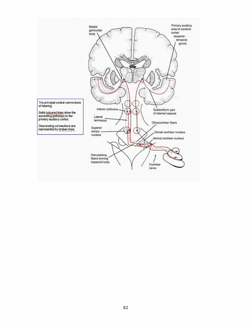
Problems With Tuning Fork Tests
Problems with the Schwabach:1. examiner must have normal hearing2. sometimes it is difficult to distinguish between
normal and conductive loss as they hear the tone the same amount of time
3. difficult to interpret in the case of mixed hearing loss4. difficult to interpret in the case of asymmetrical
hearing as the better ear will hear the sound
Problems with the Rinne and Bing:1. When the inner ear not being tested responds to the
tone, this is due to the immediate crossover of sound.
Problems with the Weber:1. it is difficult to determine with mixed hearing loss2. may report the tone for the wrong (better) ear
because what they hear makes no sense to them (even though they hear it in the worse ear, they think they shouldn’t)
Note: when testing bone conduction, the entire skull vibrates. This is a 0dB crossover. If one ear is better than another, the better ear always responds. That is why we need the audiometer, so we can “mask” the better ear with a noise and separate out the test ear.
With bone conduction testing, the crossover threshold is 0dB.With air conduction testing, the crossover threshold is about 60dB.
82

Now lets backtrack to the pseudohypacusis.There are many reasons for this to occur.
There have been cases of hysterical hearing loss which might also be called psychogenic hearing loss or hysterical deafness. This might happen after a trauma, and it is possible to regain this as it was never really lost.
Other terms for this are nonorganic hearing loss, functional hearing loss, malingering. We must be careful using these terms.
Various reasons for this problem:1. hysteria2. attention-getter (mostly children)3. accident case for:
a. rewardb. workman’s compensation
Things to look for on your tests:1. air conduction is greater than bone conduction2. crossover does not occur3. they answer you when you whisper behind their head4. Stenger test5. tests we will learn about being tympanometry,
acoustic reflexes, OAEs, ABRs, partial spondee response, inconsistent speech and tone results.
83

Chapter 4 – Causes of Hearing Impairment
There are several categories of pathology and noxious influence that adversely affect the auditory system. These include:1. Developmental defects
a. hereditary – dominant and recessiveb. congenital or progressive
2. InfectionsI. maternal
a. rubellab. CMVc. Herpesd. Ingestion of teratogenic drugs (ie thalidomide or
accutane)II. acquired
a. meningitisb. bacterialc. Herpes
3. Toxinsa. ingestion of teratogenic drugs like thalidomideb. exposure to toluenec. aminoglycosidesd. chemotherapeutics
4. Traumaa. noise traumab. head trauma
5. Vascular disordersa. embolismb. strokec. diabetes mellitus
6. Neural disordersa. neuritis (inflammation of the nerve)b. MS
7. Immune-system disordersa. autoimmune diseaseb. rheumatoid arthritis
84

8. Bone disordersa. otosclerosisb. otospongeosis
9. Aginga. presbycusis
10. Tumors and other growths a. acoustic neuroma
We can look at these disorders anatomically. Disorders of the outer and middle ear are usually structural due to congenital malformations or secondary to infection or trauma. They may be hereditary or acquired.
OUTER EARMICROTIAATRESIAMACROTIASTENOSISIMPACTED CERUMENEXTERNAL OTITISPREAURICULAR TAGSCARCINOMA OF THE AURICLE (Basal/Squamous Cell)EXOSTOSIS/OSTEOMA
MIDDLE EARTYMPANIC MEMBRANE PERFORATIONOTITIS MEDIAEUSTACHIAN TUBE DYSFUNCTIONOTOSCLEROSISGLOMUS TUMORDISARTICULATION OF OSSICULAR CHAINTYMPANOSCLEROSISCHOLESTEATOMABAROTRAUMATINNITUS (may be involved with middle ear, inner ear, or mixed losses)
85

COCHLEAR DISORDERS1. SYNDROMIC
a. ALPORT SYNDROME b. BOR (BRACHIO-OTO=RENAL SYNDROME)
c. CHARGE ASSOCIATIONd. PENDRED SYNDROMEe. USHER SYNDROMEf. WAARDENBURG SYNDROMEg. CERVICO-OCULO-ACOUSTIC SYNDROMEh. CHARGE ASSOCIATIONi. JERVELL AND LANGE-NIELSON SYNDROME
2. NONSYNDROMICa. PRESBYCUSISb. NOISE INDUCED HEARING LOSS/ACOUSTIC TRAUMAc. CYTOMEGALOVIRUS (CMV)d. RUBELLAe. SYPHILISf. OTOTOXICITY
i. AMINOGLYCOSIDESii. ANTINEOPLASTIC DRUGSiii. INDUSTRIAL SOLVENTS iv. LOOP DIURETICS
g. MENINGITISh.LABYRINTHITISi. HERPES ZOSTER OTICUSj. MENIERE’S DISEASEk. COCHLEAR NEURITISl. AUTOIMMUNE DISEASEm. BENIGN POSITIONAL PAROXYSMAL VERTIGO (BPPV)n. PERILYMPHATIC FISTULAo. ENLARGED VESTIBULAR AQUEDUCT SYNDROME
CENTRAL AUDITORY NERVOUS SYSTEM DISORDERSBRAIN INFARCTSMULTIPLE SCLEROSISACOUSTIC NEUROMA (AKA COCLEOVESTIUBLAR SCHWANNOMA)CPA TUMOR (CEREBELLOPONTINE ANGLE TUMOR)CEREBROVASCULAR ACCIDENT (STROKE)DIABETIC CRANIAL NEUROPATHYAUDITORY PROCESSING DISORDERS
86

OUTER EAR
MICROTIA – congenital malformation of the auricleThis may not affect hearing in any significant way, however it may affect sound localization and affect the acoustics of how we are accustomed to listening to sound. Also remember, when there is a craniofacial anomaly, there may be other abnormalities linked to it.
MACROTIA – Similar to microtia but the opposite, it is a congenital condition of excessive enlargement of the auricle. Again, this may not affect hearing in any significant way. They may be made smaller surgically.
ATRESIA – another congenital malformation, primarily of the ear canal. At
some point, the ear canal is closed off. There may be no problems past the point of closure or there may be other accompanying abnormalities such as congenital ossicular malformations. This will cause a significant conductive hearing loss if the sensory mechanism is intact which may be corrected through the surgical opening of an ear canal, assuming all structures of the middle ear are present and normal.
STENOSIS – another condition that is usually congenital, it is a narrowing
of the ear canal. Again this may or may not affect hearing. It does often cause wax to build up in the individual. Sometimes the ear canal is widened surgically. It may also occur later in life as the body changes.
IMPACTED CERUMEN – an accumulation of wax in the ear canal. It causes
a conductive hearing loss of varying degrees from 15dB to as much as 60dB. A good cleaning should take care of the problem.
EXTERNAL OTITIS – this is an infection of the ear canal or auricle. There
may be inflammation, bacteria, or even fungal growth. The most common type known is “swimmer’s ear”. This can be treated by the physician. Hearing loss, if any, is conductive and when the inflammation is gone, hearing returns to normal.
CARCINOMA OF THE AURICLE (Basal/Squamous Cell) – There are various
87
types of cell carcinoma that may occur such as basal call, epidermoid, and squamous cell. This must be treated or it can spread. Sometimes the external portion with this problem is surgically removed. As with the above disorders, this may or may not cause hearing loss, however, important acoustic information dependent on the shape of the auricle may be lost.
88

EXOSTOSIS – rounded hard bony nodule growing from the osseous portion of the EAM and caused by extended exposure to cold water Usually wide base, usually bilateral, history of exposure to cold, can occlude the ear canal, not roundOSTEOMA – skin covered by bone growth, usually round, narrow base,
Usually unilateral, skin covered, may obstruct normal migration of squamous epithelium outward
89

MIDDLE EAR
TYMPANIC MEMBRANE PERFORATION – this is a hole of the eardrum. It
may occur due to spontaneous bursting due to otitis media, a foreign object being pushed though the canal too far, or even due to a trauma such as extreme changes in pressure or jumping into a pool on the side. Hearing loss may or may not occur, depending on the cause. If there is hearing loss, it will be conductive. There may be pain and even dizziness. Sometimes it will heal on its own. Sometimes a paper patch is used. If this doesn’t work, an graft may be placed to replace the damaged membrane.
OTITIS MEDIA – This is one of the most common childhood problems. It
will cause a conductive hearing loss. The most common type is OM with effusion. Without effusion, there is just inflammation. Purulent effusion has pus and mucoid effusion thick and mucuslike. It can be acute or chronic or recurrent. Depending on type and duration, this can cause educational problems. While the child has OM, there is usually some conductive hearing loss. If left untreated, it can become permanent and become sensori-neural. Usually, antibiotics are used and if this does not help, then a myringotomy and placement of ventilation tube may be needed.
EUSTACHIAN TUBE DYSFUNCTION – This tube allows for pressure equalization between the ME and the outside. If it becomes restricted due to swelling, or even because of the small, torturous curvature of young children, then oxygen or fluid can be trapped. First, with oxygen, the pressure is negative, much like being on a plane and not equalizing. If left uncheck and to persist, then this can lead into a bout of OM. ET dysfunction may or may not cause a conductive hearing loss. If the negative pressure persists, then skin cells can become trapped in a pocket and cause a cholesteatoma.
BAROTRAUMA – This occurs when there is a sudden, extreme change in
atmospheric pressure, whether due to diving and coming up too quickly or as occurs when an airplane is descending. Even if the change is not extreme, if the ET is not functioning correctly, this can still occur. Extreme negative middle ear pressure may be observed. If any hearing loss occurs, it will
90

be conductive. If not corrected, it can become an otitis media. Treatment may include nasal sprays and decongestants.
GLOMUS TUMOR – This is the most common tumor of the middle ear. It may occur on the jugular bulb, in the middle ear, or along the vagus nerve. It is a heavy vascular supply of cells that arises in the middle ear near the jugular bulb and may cause pulsatile tinnitus. It is bluish in color. It may cause a conductive hearing loss and if untreated, can press on the cochlea causing a reduction in oxygen getting to the cochlea and thus resulting in a mixed hearing loss. Depending on the location, there may also be facial nerve paralysis. Hearing loss maybe anything and the most common treatment is surgery. OTOSCLEROSIS – This is a stiffness of the ossicles in the middle ear. It
usually affects the stapes and it is usually hereditary. If a female does not exhibit this before childbirth, it may often rear its head when she is pregnant. It is usually hereditary and more often in women. It is characterized by resorption of bone and then new, spongy formations occur about the footplate of the stapes and where it attaches to the oval window. The cure is surgical. They may chip away and free the stapes or place a prosthesis so the ossicular chain moves and transmits sound again. Hearing loss often begins as a low frequency conductive hearing loss and may become a flat conductive hearing loss across all frequencies.
DISARTICULATION OF OSSICULAR CHAIN – also known as a discontinuity.
There is a separation somewhere in the ossicular chain causing a separation within the chain. This may occur in the incus, in the incudostapedial joint, in the crura of the stapes, or even of the malleus. The result would be a conductive
91

hearing loss. Again, surgical treatment would be necessary. Hearing aids might be selected instead.
TYMPANOSCLEROSIS – This can occur due to aging or as a change in the
structure of the tympanic membrane due to chronic otitis media. White plaques are formed on the TM and cause an increased stiffening of the TM. Depending on the severity, conductive hearing loss can occur. This is usually a surgical treatment.
CHOLESTEATOMA – This can occur following chronic OM or due to a
chronic retraction pocket of the TM. A pocket of cells form and results in a growth. If unchecked, it can erode through the bones of the skull. Adhesions can grow on the ossicles. It may or may not cause a conductive hearing loss, depending on the side and extent of the growth. It must be removed surgically but should be watched as it is not unusual for it to recur. There is also congenital cholesteatomas which must also be removed.
92

COCHLEAR DISORDERS
PRESBYCUSIS – gradually decreasing hearing due to the aging process.
Mostly affects those over age 65 with sensori-neural hearing loss. Other issues during the normal lifetime may exacerbate this such as noise exposure, reduced oxygen due to vascular disease, medications, etc. Usually begins as a sloping hearing loss with most hearing loss in the higher frequencies. Hearing aids are the only solution.
NOISE INDUCED HEARING LOSS/ACOUSTIC TRAUMA – continual exposure
to loud noises over long periods of time without ear protection will cause a sensori-neural hearing loss. This may also occur from a one-time blast of extremely loud noise near the ear. Shorter exposures may cause something called TTS (temporary threshold shift) such as after a concert, when the hearing is muffled but eventually returns to normal. When it no longer returns to normal it becomes PTS (permanent threshold shift). Use of ear protectors to avoid loud noise and hearing aids if desired are the only solutions.
CYTOMEGALOVIRUS (CMV) – this is a congenital infection and it induces a
sensori-neural hearing loss. This is the leading cause of congenital hearing loss that is not genetic and it usually transmitted in utero. It may be of delayed onset, it may be progressive, it may be asymmetrical. The mother may not even know she has this as she may think she only has a cold. Hearing aids or cochlear implant and appropriate educational placement are the treatments.
RUBELLA – this is due to a viral infection and usually affects the child in
utero during the first trimester. In the 60’s, this was the leading cause of nongenetic congenital sensori-neural hearing loss in infants and the one of the prime factors for the development of all the educational laws, including inclusion and mainstreaming that have occurred since. As with CMV, hearing aids, CI, and appropriate educational placement are the treatments.
SYPHILIS – A venereal disease which can affect hearing during the more
93

advanced stages if left untreated. It can be transmitted in utero from an infected mother and may result in progressive sensori-neural hearing loss. In adults, during the later stages of syphilis, the hearing will deteriorate both sensory-wise and neurally. Hearing aids and antibiotics such as penicillin if it is not too late are the treatments.
94

OTOTOXICITY – sensori-neural hearing loss caused by drugs and chemicals that are toxic to the ear. It may also affect the vestibular system. It is usually permanent but in the case of aspirin and quinine, it may be reversible. For the majority, hearing aids or CI will be the only solutions. The following are the main causes of ototoxicity.
AMINOGLYCOSIDES – bacteriocidal antibiotics which include among them amikacin, gentamicin, kanamycin, neomycin, netilmicin, streptomycin, tobramycin, viomycin, garamycin.
ANTINEOPLASTIC DRUGS – generally drugs for cancer – hearing should be periodically monitored and keep the patient well hydrated. (cisplatin/carboplatin)
INDUSTRIAL SOLVENTS – most commonly inhaled on the job are styrene, toluene, and trichlorethylene
LOOP DIURETICS – most commonly seen with lasix, furosemide, and ethacrynic acid. Lasix is very popular and causes degeneration of the stria vascularis.MENINGITIS – bacterial infection may cause inflammagion of the cochlea
or labyrinth. The meninges become inflamed. It may cause total deafness, it may be asymmetrical. If early treatment with corticosteroids is initiated, it may arrest the hearing loss before it becomes severe. Hearing aids, CI, and educational placement are the final outcomes.
LABYRINTHITIS – Also caused by infection, the vestibular labyrinth becomes inflames and may produce vertigo, with or without hearing loss. It may be transient or may become toxic and become permanent along with hearing loss. In the more severe form, the membranous labyrinth of the cochlea is usually affected and hearing loss is permanent. Hearing aids would be suggested.
HERPES ZOSTER OTICUS – aka Ransay Hunt syndrome. Caused by the
95

chicken pox virus and often occurs in older people or when there is extreme stress. Must have had the chicken pox when younger. Causes little pimple-like eruptions that are very painful, may cause facial nerve paralysis, dizziness, and sensori-neural hearing loss. Medical treatment and hearing aids if necessary are the treatments.
96

MENIERE’S DISEASE – often used synonymously with endolymphatic
hydrops. There is excessive fluid in the cochlea and the vestibular labyrinth and symptoms include fullness in the ear, reduced hearing, roaring in the ear and vertigo. Cochlear Meniere’s may only have fluctuating hearing loss without the vertigo and vestibular Meniere’s may have the extreme vertigo without the hearing loss. Over time, any hearing loss may become permanent and be progressive. Often, patients are told to watch their diet, avoid salt and caffeine, and are given diuretics. When hearing loss is permanent, hearing aids may be suggested.
AUDITORY NEUROPATHY/DYS-SYNCHRONY – this is a condition in which the patient displays auditory characteristics that support normal outer hair cell function and abnormal or sys-synchronous responses from the 8th nerve and brainstem. So the site of dysfunction is between the OHC and brainstem. Auditory test will be normal, the OAE will be normal, and the ABR will be abnormal or absent. Speech recognition will be poor in noise but in quiet it is variable, may be slightly or greatly reduced.AUTOIMMUNE DISEASE – a disorder in which the body produced antibodies that attacks itself, one common example being rheumatoid arthritis. May affect the auditory system and may be characterized by bilateral, progressive, sensori-neural hearing loss. Treatment would be hearing aids or CI. BENIGN POSITIONAL PAROXYSMAL VERTIGO (BPPV) – a form of dizziness
characterized by latency of onset when in a certain position, severe vertigo for seconds, transiency, and fatigability. It is the most common cause of dizziness in the elderly but may occur due to head trauma, cold, whiplash, etc. Otoliths made of calcium carbonate crystals are not reabsorbed into the body and float in the semicircular canals, causing temporary vertigo
97

when the head is turned toward the side with the excess. This may be cured with a canalith repositioning maneuvers.
WAARDENBURG SYNDROME – a genetic syndrome with the following as
the most common characteristics – white forelock, multi-colored iris, with displacement of medial canthi, hearing loss. The hearing loss may be unilateral or bilaterally, may be symmetrical or asymmetrical, may be of any degree. Hearing aids would be helpful.
BOR (BRACHIO-OTO-RENAL SYNDROME) – these may have brachial
clefts, fistulas, cysts, renal malformation. There may be preauricular tags. It is also possible to have hearing loss whether conductive, sensori-neural, or mixed. Hearing aids would be the treatment.
CHARGE ASSOCIATION – CHARGE stands for Colomboma, Heart disease,
Atresia Choanae (nasal cavity), Retarded growth, Genital Hypoplasia, and Ear anomalies. The ear anomalies may cause any type of hearing loss, conductive, sensori-nerual, or mixed. Depending on the child is how the hearing loss will be handled but usually with hearing aids.
PENDRED SYNDROME – This is a recessive genetic disorder of the endocrine metabolism. There may be goiter and congenital hearing loss. The loss is generally symmetrical, moderate to profound, sensori-neural. Treated with hearing aids.
USHER SYNDROME – This is also a recessive genetic disorder. There is usually hearing loss that is sensori-neural as well as retinitis pigmentosa causing a progressive loss of vision. Again, the hearing loss may be treated with hearing aids.TREACHER-COLLINS SYNDROME – rare genetic disorder characterized by craniofacial abnormalities, underdevelopment of certain bones of the head. Many involve the ear, mostly malformation of the external ear and middle ear structures, including atresia. 40% are autosomal
98
dominant inheritance but 60% are likely new mutations. Most hearing loss will be conductive.
TINNITUS – A noise in the ears or head, usually described as crickets, whistle, steam, sometimes a roaring, but may also be voices. May accompany middle ear, cochlear, or even CANS disorders. If there is hearing loss, a hearing aid often helps. Sometimes maskers, TRT, or Neuromonics device, or other devices help retrain the brain so this becomes a nonissue. Within this family of disorders may also be found HYPERACUSIS and a new one – MISOPHONIA. In hyperacusis, they are unusually sensitive to sounds at levels that would normally not be bothersome. In misophonia, they react to certain sounds, and can almost become violent in their dislike.
99

CENTRAL AUDITORY NERVOUS SYSTEM DISORDERS
BRAIN INFARCTS – some localized area is denied its normal blood supply
because of occlusion of an artery or drainage from a vein. When blood supply is denied to an area servicing the ear, an auditory disorder can occur, whether sensory, neural, or both. Hearing aids if possible would be recommended.
MULTIPLE SCLEROSIS – in this disorder, the myelin sheaths of the nerves
are disrupted causing scattered areas of demyelination of white matter within the nervous system. If the auditory nervous system is affected, then there an be hearing disorder of any degree with speech perception problems. The problem is retrocochlear. Hearing aids may or may not be helpful. Medical management through medication may sometimes arrest and even reverse the plaques.
ACOUSTIC NEUROMA (AKA COCLEOVESTIUBLAR SCHWANNOMA) – the
most common growth affecting the auditory nerve. It is always benign and may or may not cause hearing loss, depending on the size. They are most often unilateral and often develop from the vestibular branch of the VIIIth nerve. There may be tinnitus, hearing loss, unsteadiness, difficulty understanding words, etc. They are bilateral for those suffering from NFII. If very small they may be watched, if surgically removed, the hearing is generally lost, or the newer treatment using the Gamma Knife may be used and then the area is monitored for necrosis of the tumor over time. This is one way the hearing can be preserved if it had not been affected.
CPA TUMOR (CEREBELLOPONTINE ANGLE TUMOR) – This is a tumor
arising in the angle between the cerebellum and pons where the VIIIth nerve enters the brainstem. If large enough and presses on the nerve, then again hearing loss may occur, and may be sensory, neural or both. Hearing aids could be used.
CEREBROVASCULAR ACCIDENT (STROKE) – This is caused by an interruption of the blood supply to the brain resulting in sudden loss of function from that portion that was damaged. Hearing may or may not be affected but usually not. Sometimes there may be problems understanding words.
100

DIABETIC CRANIAL NEUROPATHY – Diabetes mellitus is an insufficiency of
insulin with complications that include neuropathy and degenerative alterations of blood vessels. This may attach all systems and may re with vestibular and auditory disorders. Generally, the auditory disorder is retrocochlear (neural). Hearing aids may or may not help.
AUDITORY PROCESSING DISORDERS – This may exhibit in multiple symptoms. It may be merely a mild problem understanding the teacher in a noisy class to having problems even listening at home. It may be accompanied by distractibility and behavioral issues. There may be problems following directions and there may be reading and spelling problems. The elderly also tend to experience this problem manifesting as they individual having greater difficulty understanding words than would be indicated from the audiogram (phonemic regression). While there may be some benefit with hearing aid use, other programs and therapy that train to listen in noise or react more quickly may be helpful.
101

Name of Disorder What is it? Symptoms
Microtia/Mactritia
Microtia - congenital deformity, can be unilateral or bilateral from birth. Ear can be normally shaped incomplete or absent, small… Macrotia - LARGE EARS
Microtia, conductive hearing loss in the affected ear
Artesia/Stenosis
happens at birth, presence of bone formation obstructs sound from reaching the inner ear.
common to have malformed pinna but not necessary
External Otitis
inflammation or bacterial infection of the external auditory canal and/or outer ear (auricle) swimmers ear or ear ache ear ache
Carcinoma of the auricleMost common cancer involving the outer ear
Raised, ulcerated lesion on the helix
Exostosis
Surfer Ears; development of benign bony growths caused by the repeated exposure to cold water and wind.
narrows the pathway trapping water and debris, and ear wax in the canal, causing an infection.
Tympanic Membrane Perforation
most common is ear infection, one bad ear infection or many mild ones can cause eardrum perforation
conductive hearing loss, degree vares
Otitis Media
inflammation of the middle ear; the space behind the ear drum. Common ear infection in childhood. puss, inflammation middle ear
Eustachian Tube Dysfunctionambien air pressure different than that of middle ear fullness and pain in the ears
Otosclerosis
abnormal bone in the ossicles of the middle ear, which leads to the fixation of the stapes. No root cause known yet, but have found that hereditary can play factor hearing loss
Glomus Tumor
benign tumors which arise from glomus jugular. Most common tumor in the middle ear
patient tested -looks like a red ball behind the TM
Disarticulation of the ossicular chain
Leverage of ossicles increases the intensity of the sound to TM 25db, therefore disarticulation results in impairment of the passage of sound vibrations in the inner ear.
this normally comes from head trauma, violent head injury
Cholesteatoma
expanding, tumorlike mass of epithelium and cholesterol, usually occing in the middle ear and mastoid region
can eat into ossicles and even the bony labarynth. Type of loss is conductive
Barotrauma
condition where pressure difference b/w the inside and outside of the eardrum may cause discomfort mild to severe hearing loss
102

Persbycusisgradual loss of hearing that occurs as people age
typically bilateral, and characterized by difficulty in hearing high frequencies
Noise induced hearing loss/acoustic trauma
trauma that occurs when exposed to loud noises including music
TTS-temporary comes back PTS-permenant
Ototoxicity
"ear poisoning" exposure to drugs that damage the inner ear, specifically the cochlea & sometimes the vestibulum
Temporary or permenant disturbance of hearing, balance, or both.
Meningitis
inflamation of the meinges (membranes that protect brain and spinal cord) may be aquired through kissing. There are Viral/Bacterial<-most severe.
fever, nausea, and vomiting, headaches. Joint pain, stiff neck, and a low tolerance to bright light.
Labyrinthititis
inflammatory process of the vestibular labyrinth membrane located at the inner ear, in the middlemost part of the cochelar duct.
"rapid eye movement" causes chronic anxiety, hearing loss may or not be affected, and tinnitus. Vertigo and Nausea problems…
Meniere's Diseasechange of fluid volume within a portion of the inner ear known as the labyrinth
think of vertigo attack, roaring of ear especially in the low frequencies combined with stuffiness in the ear that may proceed vertigo.
Benign positional paroxymal vertigo (BBPV)
dizziness and imbalance due to the debris which is collected within the ear… "ear rocks"
dizziness or vertigo, lightheadness, imbalance, and nausea
Autoimmune DiseaseBody's own immune system attacks the inner ear degree of loss varies
Multiple Sclerosisinflamatory disease in the central auditory system
WHOLE LIST… muscle spasm, there are speech perception problems
Perilymphatic Fistula
tear in the round or oval window which causes a leak from the perilymphatic spaces of the bony layrinth into the middle ear space; can happen from head trauma, barotraumas, bony erosion..
vertigo, dizziness, imbalance, vomiting, and nausea
Herpes Zoster Oticus
Viral infection of the inner, middle, and external ear common complication of shingles
burning pain close to the ear. Shortly thereafter, eruption of vesicles occurs in the ear canal and sometime on the face, trunk. Facial paralysis
Acoustic Neuromaa noncancerous tumor of the cranial 8th nerve
most common brain tumor located at the base of the brain
103

Rubella
"German Measles" viral infection charac. By fever and a skin rash resembling measles red bumps, fever, etc.
Auditory Processing Disordersdifficulty in processing; child hears well but can't understand speech
cause is unknown, but some cases prove hereditary, no technical prefiferal hearing loss at all, you add noise though and the discrim goes down
Usher Syndrome
inherited condition that causes serious hearing loss present from birth and thereafter and progressive vision loss
hearing loss is sensori neural and ranges from moderate to profound some folks have balance problems
Waardenburg SyndromeGenetic disorder. Very common to have a hearing disorder with this syndrome.
Changes in both hair and skin color, 2 color eyes; premature patch of grey hair; synophrys-unibrow
Syphillis STD, it is treatable can cause tinnitus
104

chapter 5
So why are we evaluating these patients?
CASE HISTORY!!!!
Get Information from referral source, patient, or family – begins to give you an idea of where you are going.Is it self-referral? Medical referral? Family decision? Child? Adult? Obtain before testing patient.
This will give us an idea of what we are looking for. Maybe they are merely looking for an augmentative device, perhaps they are dizzy or have tinnitus. Perhaps a parent suspects a hearing loss in a child or the child failed a screening before entering school or at birth. Perhaps it is a workman’s comp or other litigious case. Perhaps they have a learning disability or may have a tumor. We begin to decide what we are going to do and the order in which we go about it based on the information we obtain.
So a case history is a must. It can give us much valuable information. From this, we can being to determine the onset of the problem, possible causes (ototoxicity, NIHL, sudden onset, hereditary disorders like otosclerosis, etc) and begin to determine the next step in the process. It may be that we are the ones who determine that the disorder is treatable and an ENT consultation is needed. Or it may be that we are the ones determining that it is the services of the SLP that is truly what is needed. Sometimes we may be the ones to tract a dizzy or tinnitus complaint to medications being taken/recently changed/recently added and this may be the true cause of the problem.
105

In order to truly understand the effects of hearing loss on any individual, we must look at:
a. degree of hearing loss (mild, moderate, severe, etc.)
b. configuration of hearing loss (flat, sloping, reverse slope, cookie-bite, precipitously sloping, low frequency, high frequency, etc.)
c. type of hearing loss (sensori-neural, conductive, mixed, APD)
d. speech perception deficit (if any)e. age at onset (congenital or acquired – pre or
postlinguistic)f. sudden or gradual (has or does not have
compensatory strategies)g. patient’s normal communication demands
(sitting home versus being in school and learning language)
106

A case history or interview, as suggested by DeBonis and Donohue, suggest including the following questions:
1. What is the reason for your visit here today? (identify communication difficulties, rule out medical pathology, monitor medication effects, etc)
2. Do you believe that you have a hearing loss? (if yes, discuss time of onset).
3. Do you believe that one ear is better than the other? (normally, hearing is the same, asymmetry is a warning)
4. Do you believe that your hearing loss is gradual, or have you noted any sudden changes in it? (gradual is the norm, sudden is a warning)
5. How would you describe your ability to understand conversational speech when on the phone? Watching TV? In noisy environments? At distances? (Ideas for client-specific rehab)
6. Do you experience a sensation of ringing, buzzing, or other noises in your head or ears? If so, do these sounds keep you awake at night? (determine disturbance of tinnitus, a common sign of hearing loss or medical conditions)
7. Do you experience any pain or drainage from your ears? Do you have a plugged or fullness sensation in one or both of your ears? Have you ever been seen by and ENT physician? (check for ME pathology with drainage, ME or IE pathology with stuffiness)
8. Have you experienced dizziness or a sensation that the room is spinning (vertigo)? (Associated with numerous problems)
9. Do you have a history of or are you currently exposed to loud sounds, whether through your work or at home or through hobbies/leisure activities? (to determine likelihood of NIHL)
107

10. Do you currently wear hearing aids and are the aids helping you to your satisfaction? (May need adjustment)
11. Is there a family history of hearing loss? (genetic etiology?)
12. What do you think may have caused your hearing loss? (a client-centered question)
I like to add a question regarding medications they are currently taking and medications they have taken in the past. Many of our patients are on medications with side effects such as tinnitus, vertigo, ototoxic hearing loss, confustion, etc.
If your patient is a child, additional issues to look at may include:
1. If there were any difficulties during the pregnancy2. If there were any difficulties during the birth3. Again, familial history of hearing loss4. Did the child reach the developmental milestones at
appropriate ages?5. Illnesses, high fevers, ear infections, etc.6. Are they doing well in school or any teacher comments
about difficulties observed7.Do you have any concerns about hearing,
speech/language development, or general development?
Do we need to refer for medical consultation?There are many common problems that are medically treatable. Some are stenosis, impacted cerumen, perforation, tympanosclerosis, and ossicular discontinuity.Such reduction in function may be due to structural changes and are amenable to medical management.
108

The most efficient way of answering these questions, aside from superficial otoscopy, is through immittance audiometry – an electroacoustic assessment technique measuring middle ear function.
What is the hearing sensitivity?Measurement of hearing sensitivity – thresholdType of hearing loss? CHL, SNHL, mixed,
retrocochlearAEP or ABR for those not measurable by conventional methods. May make guesses from acoustic reflexes as well.
How well does the patient understand speech?This allows for a realistic prognosis for the expectations with amplification.
How well does the patient process the auditory information?
This evaluates auditory processing. This is the process by which the central auditory nervous system transfers information from the 8th nerve to the auditory cortex. This plays a role in localization of sound and in detection of desired signal in the presence of extraneous noise.
Does the hearing impairment cause a hearing handicap?This is a current favorite to evaluate. It is difficult to evaluate objectively as it is such a subjective matter. But to evaluate this, we need to understand the difference between hearing impairment, hearing disability, and hearing handicap.
Hearing Impairment: refers to abnormal or reduced function; the actual dysfunction described by the measures of hearing status.
109

Hearing Disability: refers to a functional limitation imposed by a hearing impairment
Hearing Handicap: refers to the obstacles to psychosocial function resulting from or imposed by a disability
Much evaluation is done via patient questionnaires. These scales assess the extent of hearing disability as well as the social and emotional consequences of hearing impairment. These questionnaires are now known by the term clinometrics. These self-assessment scales are likely the most efficacious way of measuring the activity limitations, the extent to which the auditory disorder is causing ah erring problems, the extent to which the hearing problems is affecting the quality of life. Scales have also been similarly developed for vertigo and tinnitus. In some cases, the effects of vertigo and tinnitus on quality of life may be worse than the hearing loss.
110

Histories
Depend on this a lot. Always listen to your patients or the parents. Listen to how they answer the questions.
- Statement of the problem- Why services are sought- Patient’s own attitude- Duration and degree of loss- Family history- Other pertinent history like noise exposure or trauma
Referrals
Do so when you feel additional help is needed. Forward reports and results of audiogram. Suggested organization of a report is:
A.First paragraph: identification of the patient – name, age, sex, history, reason for referral.
B.Second paragraph: statement of type and degree of loss, what needs special attention, interpretation, implications for communication difficulties.
C.Third paragraph: specific recommendations like aids, speech, follow-up, use of ear protectors, use of tinnitus maskers, etc.
Referral we might make: Physicians (usually otorhinolaryngologists) (when referring to the doctor, state the area of concern and reason for referral.) Clinical psychologist (State concern and reason for referral) Speech-language pathologists Teachers of the hearing impaired Regular school classroom teachers
111

So once referrals have been determined we need to decide:Best fit of hearing aid for the individual (as determined by hearing loss, life style, physical limitations if any, etc.)Best formula for fitNeeds as determined by questionnaires
We need to give adequate orientation to the hearing aid and to coping with limitations.
We need to ensure that educational recommendations are observed.
We need to EMPOWER for INDEPENDENCE and QUALITY OF LIFE for all concerned, the hearing impaired and their family/significant others/caregivers.
112

Chapter 6
THE AUDIOMETER
Pure tone audiometer: the instrument by which we measure
hearing. It measures frequency, intensity, and speech. It runs from –10dBHL to 120dBHL for intensity and measures from 125Hz – 12000Hz in frequency. Some special audiometers, known as high frequency audiometers, may measure up to 20000Hz. It also produces broad-band noise, speech noise, and narrow-band noise. May also direct signals from an external CD player or tape recorder for speech testing or use a microphone for monitored live voice.
Oscillator/ Frequency dial – generates pure tones at discrete
frequencies and controlled by frequency dial.
Interruptor – the switch that may keep a sound on or keeps the
sound off until pressed; controls duration of sound.
Attenuator dial – changes the intensity of the signal – usually in
5dB steps, some may be even smaller
Frequency dial – changes the frequency of the signal
Selector switch – changes from air conduction to bone conduction
to speech, etc.
VU meter – monitors output of the oscillator, microphone, or
auxiliary items such as tape or CD player113

Output Transducer – the device that converts one form of energy
to another such as acoustical or vibratory. These may include earphones (insert or supraaural), loudspeakers, or bone conduction vibrators.
ANSI – American National Standards Institute – the governing
body that sets the standard for audiometers, hearing aid specifications, etc. These may change. The current standard is ANSI 1996, prior to this was ANSI, 1969. An association of specialists, manufacturers, & consumers that determines standards for measuring instruments, including audiometers. They are the specs by which our equipment is calibrated.
ISO – International Standards Organization – another standard
used but not usually in the US, usually in Europe.
Threshold – The level at which the patient can just barely detect
the signal correctly 50% of the time. This is the same for pure tones and speech; air conduction and bone conduction; the “just-audible” concept
Air conduction – the hearing test as measured through supra-
aural or insert earphones, tests the entire auditory system.
Bone conduction – the hearing test via a bone vibrator, tests the
nerve of hearing directly – may be felt due to bone oscillator
114

vibration in those with severe hearing losses and usually
occurs at 250Hz & 500Hz. This is called a vibrotactile response.
Masking – the introduction of a noise into the nontest ear as needed, usually when there is great asymmetry.
a. typically used for air conduction when the difference between ears is equal to or greater than 40 - 50dB
b. typically used for bone conduction when there is any difference between ears
c. typically used for speech when there is a difference between ears of 40 – 50dB or greater
Use 40dB with supra-aural headphones, 50dB with insert earphones, and 0dB for bone
115

Insert Headphones – this is a newer way of testing, whereby a
receiver, covered by a foam cushion is inserted deeply into the ear canal and reduces external noise (ambient noise) as well as the need for masking. Often used with collapsing ear canals.
Ways to respond to pure tone testing:a. raising a hand or fingerb. pushing a buttonc. verballyd. dropping a block in a pail or box in the case of a child
false positive response: patient hears something when there is nothingfalse negative response: patient does not respond when they do hear a tone – may happen in the case of malingerers
The pure tone audiogram: establishes threshold sensitivity across the frequency range important for human communication and is placed on a graph.
Hughson-Westlake “Ascending Method” or Hughson-Westlake technique – the method by which we obtain thresholdVerbal instructions to a patientNote that infants and children use modified methods we will review later in the course.Collapsing ear canal:
a. can happen with a narrow ear from the pressure of the headphones
b. may give a false conductive hearing lossc. may be overcome by the use of insert headphones or
holding the headphone off the ear
116

A review of testing technique – down 10dB and up 5dBPure tone average – this is an average by which to estimate the severity of the hearing loss:
a. the more common average is 500Hz, 1000Hz, and 2000Hz
b. now we often use 500Hz, 1000Hz, 2000Hz, and 3000Hz for a more accurate representation in the presence of high frequency hearing loss.
c. Fletcher Average – when there is a precipitous drop at 1kHz, a Fletcher average may be more accurate, this is the best 2 frequency average so in this case you might average 250Hz in.
Refer to symbols of the audiogram.
Occlusion effect – when the intensity of the tone during bone
conduction is increased due to the one of the ears being covered. Usually for 1000Hz and below. Does not occur in conductive hearing loss.
Vibrotactile response (VT) – usually seen at 250Hz and 500Hz and the stimulus is felt as a vibration rather than hears; usually occurs primarily for bone conduction testing.
Air-Bone Gap – the difference between the air conduction and bone conduction
air conduction – bone conduction = air-bone gap
In the low frequencies, with bone conduction, they may experience a vibrotactile response where they feel a vibration rather than hear a response.
117

Interaural attenuation – the amount of reduction in intensity that occurs as a signal crosses over the head (is transmitted by bone conduction) from one ear to the other ear; the point at which the better ear begins to respond:
a. about 40 - 50dB for air conduction (depending on phones)
b. at 0dB for bone conductionc. also the loss of intensity of a sound introduced to one
ear and heard by the other.
Masking – a way to remove the nontest ear from the test procedure when cross-hearing is suspected. The difference for air conduction is based on the BC of the better ear to the AC of the worse ear.Noises used for pure tones are:
a. white noiseb. narrow band noise
For speech testing we use:a. speech noiseb. pink noise
Rule for AC masking:If the thresholds from the test ear exceed the bone conduction threshold of the nontest ear by the amount of minimum interaural attenuation, then masking MUST be used.Rule for BC masking:Always use masking in the non-test ear during bone conduction testing if there is any difference at all between ears.
Overmasking – the noise presented is so loud that it crosses
118

back over to the other (test) ear and masks the test ear. Central masking – a small shift in threshold with the
introduction of masking and the shift continues to increase with increased noise.
Effective Masking – the level where increased masking noise
will no longer result in a shift of thresholdMasking dilemma – occurs when the difference between the BC
threshold in the test ear and the AC threshold in the non-test ear approaches the amount of interaural attenuation. Most often seen in bilateral conductive hearing loss.
Review plateau method for masking to determine effective masking
So what does the audiogram tell us?1. gives the degree of hearing loss (mild, moderate,
severe, profound)2. describes shape of loss (flat, sloping, low frequency,
high frequency, precipitous, cookie-bite, reverse slope or rising)
3. measures interaural symmetry (the difference between ears)
4. differentiates hearing loss (conductive, sensori-neural, mixed)
0 – 25dB normal hearing25 – 40dB mild hearing loss40 – 55dB moderate hearing loss55 – 70dB moderately severe hearing loss70 – 90dB severe hearing loss90dB+ profound hearing loss
119

Personally, I go by a slightly different scale. Once you say moderately severe, then where is mildly severe or severely severe? So I use the following:
0 – 25dB normal hearing25 – 40dB mild hearing loss40 – 60dB moderate hearing loss60 – 90dB severe hearing loss90dB+ profound hearing loss
As you see, there is not much of a difference.chapter 7
Speech tests:The goal is to quantify a patient’s ability to understand everyday communication at suprathreshold levels.
Why?- Gives us a threshold to crosscheck against our pure tone average- Measurement of threshold for speech- Assists in differential diagnosis- Assesses central auditory processing- Gives estimates of communicative function, aided as well as unaided
Speech reception or recognition threshold (SRT) – The
lowest level at which speech can be detected or recognized. The minimum level where spondee words that they have been familiarized with can be correctly detected 50% of the time. AKA Speech detection threshold or speech recognition threshold
120

Speech recognition or discrimination – the percentage
correct of 50 or 25 monosyllabic word lists presented at some level above the SRT, usually 30 – 40SL (SL meaning above the minimum hearing threshold of the spondee threshold).
The ability to perceive and recognize speech
Lets us know the prognosis with amplification.
Speech detection threshold (SDT) = Speech awareness threshold (SAT) – lowest level at which a speech signal is
audible or detected 50% of the time; this is used when the individual cannot speak, has a profound hearing loss, or is a child who cannot communicate, this may be used in place of SRT if they cannot repeat spondees
The SDT requires that the client merely detect the present of speech, the SRT requires that the client recognize the words. So the STD may often be at a somewhat better threshold than the SRT as it often reflects the best threshold.
There are some special speech tests, outside of the usual monosyllabic words, to evaluate auditory processing, or the transmission of information from the nerve to the auditory cortex via the various levels of the brain that make up part of the central auditory nervous system. Rather than just words in quiet, we tax the system by assessing speech recognition in the presence of competing speech signals, processing 2 different signals presented simultaneously to both ears (dichotic listening), etc. We may change the speech signal by using low-pass filtering, time compression, high level
121

suprathreshold testing (as in PB rollover), speech in competition (as with SPIN), or dichotic measures (as in SSW). We reduce the extrinsic redundancy of speech to tax the system.
Speech testing can be performed via:a.MLV – monitored live voice: the tester gives the
words while monitoring their voice level through a VU meter
b.Taped: a cassette or CD is hooked up to the audiometer and the speech stimuli are delivered this way.
Ways to deliver speech stimuli:a. through the headphonesb. via bone conductorc. via speakers for sound field testing
Sound field testing (SF): when the stimuli, whether speech or
pure tone or noise, are delivered through speakers in the room. This maybe used to check the efficacy of hearing aids or to test children who won’t tolerate headphones. May also be used for localization for infants and toddlers.
With children, pictures of the spondees may be used and the child will point for the response.
Ensure you do not allow them to read your lips.
SRT – speech reception threshold: the point where they are able to repeat 50% (2 out of 4) of the spondee words (compound words of equal emphasis) correctly after they have been familiarized with the words. ASHA recently recommended this be changed to speech recognition threshold.
122

SDT – speech detection threshold: for a child who cannot repeat or an individual who cannot discriminate words, the point at which they are able to detect the speech and respond by raising their hand or pushing a button.
The ability to repeat what is heard is considered open-set material. If we use a picture-pointing test, that is a closed-set approach, which limits possible choices.
Test procedure for SRT. Similar to Hughson-Westlake procedure.Note that your SRT level will always be at a greater intensity than the SDT. This is because it is easier to detect a sound than to identify the sound. These results should be in good agreement with the audiogram’s best pure-tone threshold.
Times when the SRT/SDT will not agree with the PTA
a. when there is a precipitous drop of hearing in the high frequencies (e.g., hearing is normal at 500Hz and then drops to 70 at 1000Hz, etc) – SRT better than PTA, closer to the best 2 frequency average.
b. When there is a central auditory problem, the elderly may have a problem in recognizing words and thus cannot get 50% correct – the SRT is poorer than PTA
c. In pseudohypacusis – anything goes here: NOTE: often, in the case of spondees, they will give half the word correct and not the other half.
d. SAT or SDT usually matches the single best frequency.
123
The same crossover problems for speech occur that occur for pure tones.
If we test recognition at a number of increasing intensities, we develop a PI function or performance versus intensity function. When rollover occurs (the recognition scores are reduced), we can assume there is a retrocochlear pathology.
MCL – most comfortable listening level or most comfortable loudness level: just talk to them and tell them to tell you when your voice is comfortable or “just right”. I like to tell them to tell me when my voice is at a level where they would like to listen to television for 2 hours.
UCL or ULL– uncomfortable listening level or uncomfortable loudness level aka TD (threshold of discomfort) and LDL (loudness discomfort level): the level at which speech is uncomfortably loud. May also be done with pure tones. A normal ear should tolerate 90 – 100dB. If there are tolerance problems, it suggests a cochlear problem and it sets the limits of the hearing aid output.Dynamic Range – the difference between the SRT and the UCL is the range of useful hearing.
There are many tests to measure speech discrimination.a. Phonetically balanced words (PB): These are word
lists and tested using 50 or 25 words. There are a number of sets.
i. CID W-22: a popular set of wordsii. NU6 words: another popular setiii. PBK words: similar to CID W-22 but for
childrenOther tests are:
b. CNC (consonant-nucleus-consonant)124

c. High frequency emphasisd. Nonsense-syllable lists – CVCVe. Closed set response lists
i. PIT – picture identification testii. WIPI select picture that matches wordiii. NUCHIPS – similar to WIPIiv. California consonant test for those with high
frequency hearing losses.Newer tests are using sentences:Some of these tests might include:
a. SSI (synthetic sentence identification)b. SPIN (speech perception in noise test)c. CST (connected speech test)d. SIN (speech in noise test)
We use some of the speech tests in noise as the main complaint of the hearing impaired is they can’t hear in noise and the quiet testing is very artificial.
Sensitized tests for retrocochlear problems may be:a. PB rolloverb. SPIN test (Speech Perception in Noise)c. SSI (synthetic sentence index): SSI-C or SSI-Id. SSW (Staggered Spondaic Words)e. DSI (Dichotic Sentence Identification)
125

Appendix A
Alphabetical List of Spondaic Words
airplanearmchairbackbonebaseballbirthdayblackboardcookbookcowboydoormat
drawbridgeduck pondeardrumearthquakeeyebrowgreyhoundhardwareheadlighthorseshoe
hotdogice creaminkwellmousetrapmushroomnorthwestnutmegoatmealoutside
padlockpancakeplaygroundrailroadstairwaysunsettoothbrushwhitewashwoodwork
126
CID W-22
LIST 1A LIST 2A LIST 3A LIST 4A
1. AN 1. YORE 1. BILL 1. ALL 2. YARD 2. BIN 2. ADD 2. WOOD 3. CARVE 3. WAY 3. WEST 3. AT 4. US 4. CHEST 4. CUTE 4. WHERE 5. DAY 5. THEN 5. START 5. CHIN 6. TOE 6. EASE 6. EARS 6. THEY 7. FELT 7. SMART 7. TAN 7. DOLLS 8. STOVE 8. GAVE 8. NEST 8. SO 9. HUNT 9. PEW 9. SAY 9. NUTS 10. RAN 10. ICE 10. IS 10. OUGHT11. KNEES 11. ODD 11. OUT 11. IN 12. NOT 12. KNEE 12. LIE 12. NET13. MEW 13. MOVE 13. THREE 13. MY 14. LOW 14. NOW 14. OIL 14. LEAVE15. OWL 15. JAW 15. KING 15. OF 16. IT 16. ONE 16. PIE 16. HANG 17. SHE 17. HIT 17. HE 17. SAVE 18. HIGH 18. SEND 18. SMOOTH 18. EAR19. THERE 19. ELSE 19. FARM 19. TEA20. EARN 20. TARE 20. THIS 20. COOK 21. TWINS 21. DOES 21. DONE 21. TIN22. COULD 22. TOO 22. USE 22. BREAD23. WHAT 23. CAP 23. CAMP 23. WHY24. BATHE 24. WITH 24. WOOL 24. ARM25. ACE 25. AIR 25. ARE 25. YET
LIST 1A LIST 2A LIST 3A LIST 4A
26. YOU 26. AND 26. AIM 26. DARN (dawn)27. AS 27. YOUNG 27. WHEN 27. ART28. WET 28. CARS 28. BOOK 28. WILL 29. CHEW 29. TREE 29. TIE 29. DUST 30. SEE 30. DUMB 30. DO 30. TOY31. DEAF 31. THAT 31. HAND 31. AID32. THEM 32. DIE 32. END 32. THAN 33. GIVE 33. SHOW 33. SHOVE 33. EYES 34. TRUE 34. HURT 34. HAVE 34. SHOE 35. ISLE 35. OWN 35. OWES 35. HIS36. OR 36. KEY 36. JAR 36. OUR37. LAW 37. OAK 37. NO 37. MEN38. ME 38. NEW 38. MAY 38. NEAR 39. NONE 39. LIVE 39. KNIT 39. FEW40. JAM 40. OFF 40. ON 40. JUMP 41. POOR 41. ILL 41. IF 41. PALE 42. HIM 42. ROOMS 42. RAW 42. GO 43. SKIN 43. HAM 43. GLOVE 43. STIFF44. EAST 44. STAR 44. TEN 44. CAN45. THING 45. EAT 45. DULL 45. THROUGH46. DAD 46. THIN 46. THOUGH 46. CLOTHES47. UP 47. FLAT 47. CHAIR 47. WHO48. BELLS 48. WELL 48. WE 48. BEE49. WIRE 49. BY 49. ATE 49. YES50. ACHE 50. AIL 50. YEAR 50. AM
127

128

Other Behavioral Measures
Remember that we talked about sensori-neural hearing loss being so called because we cannot decide it is sensory (only a problem in the cochlea) or neural (8th nerve and above through the brain, aka as retrocochlear). The following are a series of tests that were used extensively in the past as additional behavioral measure to determine site of lesion. They have fallen into disuse nowadays with all the other tests available. The ABR and OAE will be discussed and are widely used. MRI and CT scans are now extensively used as well.
To understand these tests, we must first understand the term RECRUITMENT and ADAPTATION.
Adaptation: in an ear with a neural involvement, audibility of suprathreshold sounds diminish rapidly due to excessive auditory adaptation. The normal ear adapts at low levels and the audible signal becomes inaudible. However, at loud suprathreshold intensity levels, the sound remains audible. In the retrocochlear disorder, the sound disappears rapidly.
Recruitment: an unusually rapid growth of loudness of an impaired ear. Loudness grows more rapidly than normal at intensity levels just above threshold in an ear with a cochlear problem. It may also be defined as a disproportionate increase in loudness as a function of intensity of the impaired ear. This is symptomatic of the majority of hearing losses that are sensory. There are other variations of recruitment:
1. partial recruitment – some recruitment is noted, however, the growth never complete it to the level of the better ear.
129
2. hyperrecruitment – the impaired ear not only catcher up to the better ear but appears to be louder in the impaired ear than the normal ear with the same high intensity.
3. decruitment – the loudness grows more slowly in the impaired ear than a normal ear. Even intense sounds may not produce much loudness. This is often associated with problems in the 8th nerve.
ABLB – Alternate Binaural Loudness Balance TestAn old popular test of recruitment. This is a standard measurement of recruitment. We perform this test at 2 frequencies, a low frequency (usually 500Hz) and a high frequency (usually 2kHz). The bad ear is compared to the good ear as intensity is increased until they are equal. Usually, the intensity required for equal loudness is less for the impaired ear than the normal ear. Slightly different tests with the same idea are:
a. AMLB (Alternate Monaural Loudness Balance Test) This test compares a normal frequency to a hearing impaired frequency in the same ear
b. SBLB (Simultaneous Binaural Loudness Balance Test) This test presents the tone simultaneously to both ears. It was found that this was not a good test for recruitment.
When recruitment is found, it is suggestive of a cochlear disorder.
SISI – Short Increment Sensitivity IndexThis was another popular test of recruitment.This procedure tests the patient’s ability to detect the presence of a 1dB increment superimposed on a tone that is presented at 20SL. It is a suprathreshold test so beware of crossover. Again, this is presented at a low freq. and a high freq. The high frequency scores are greater, suggestive of a cochlear disorder.
130

Example: Threshold of the poorer ear is 20dB at 500 and 40dB at 2kHz. A tone is presented at 500Hz with an intensity of 40dB and every so often, it is increased by 1dB as a blip. They are to raise their finger every time they hear it. We score for 20 blips. The same is done at 2kHz with an intensity of 60dB. They will hear the blip much more often at 2kHz than at 500Hz. A score over 50% indicative of recruitment and so a cochlear disorder.TDT or Tone Decay TestThis is a test of adaptation.This test represents a sustained tone to the ear. Even normal ears will change threshold at soft levels to the sustained tone. As with the other tests, this is performed with a low frequency (500Hz) tone and a high frequency (2kHz) tone. A major change may be indicative of a CNS disorder. This is a test of adaptation. In adaptation, we know that a normal ear will adapt to ongoing sound, especially at near threshold levels, and eventually the sound is inaudible but higher intensity sounds will remain audible. We also know that with a retrocochlear disorder, audibility will be lost much faster, even at loud levels, due to excessive auditory adaptation.
A. Olsen-Noffsinger tone decay test: Present the tone at 20dBSL. As soon as they hear it the finger goes up and down when it disappears. Record how long they hear it at 20dL and increase level by 5dB without interrupting the tone. Continue in this way until they hear it for a full minute or until 30dB above the starting level has been reached.
B. Rosenberg tone decay test: Present and raise tone in the same way as above, however, this goes on only for 1 minute and the number of decibels raised at the end of a minute is the decay.
C. STAT – suprathreshold adaptation test: Same as the others, however, present the tone at a very loud level (usually 70dBSL not to exceed 100 or 105dB) and this
131

is only for 1 minute. If complete adaptation occurs, it is positive for a neural problem (central).
Generalizations:Most decay is noted for high frequencies. Thus, usually a low frequency and high frequency is tested.Decay was very significant when there is 30dB or more in one minute. Only moderate with 20 – 25dB of decay.
Bekesy AudiometryThis was another way to measure adaptation.This was once a popular test and had its own type of audiometer. It also measures auditory adaptation. It is a patient controlled test. They press a button and let go, depending on whether or not they hear the tone. A comparison is made between continuous and pulsed tones. The patterns produced were categorized into one of several types. Depending on the type noted was the assumed site-of-lesion. The types and their accompanying pathologies are as follows:
1. Type I: normal and conductive hearing loss. The tracings overlap almost completely.
2. Type II: above 1kHz, the tracing for continuous tone drops below the interrupted tone and this is indicative of cochlear pathology.
3. Type III: the continuous trace shows a pronounced separation for all frequencies and is associated with problems beyond the cochlea (neural or central). This is the most dramatic tracing. That is because the continuous tone shows adaptation and so it is fading as would be seen with a tone decay test.
4. Type IV: Similar to type II but the breakaway is at 500Hz and the separation between the 2 tracings is 20+dB. This may be cochlear or neural.
5. Type V: this is most often seen with nonorganic disorders. The continuous tracing is better than the pulsed tracing (they tend to run parallel).
132

133

Masking Level Differences – MLDs
This is another test still used. It measures lower brainstem function. It measures binaural release from masking due to interaural phase relationships. First, an identical, in-phase, low frequency tone is present to both ears. Then noise is added to each ear until the tone is masked. If the phase of the tone to one ear is reversed, the tone should become audible again. We look at the difference in threshold between the in-phase and out-of-phase conditions. MLDs should be greater than 7dB.
134
Chapter 7
Electrophysiological tests:a. serve as an indication of peripheral sensitivityb. serve as an indicator for site-of-lesion within the
systemc. is objective and requires no subjective response from
the patient.
IMMITTANCE AUDIOMETRY
A test battery that has become as common as pure tones and speech is acoustic immittance. This is a term that can be used to refer to acoustic impedance or acoustic admittance. Either terminology is correct, but acoustic impedance is the inverse of acoustic immittance. It is now accepted that the BEST measure of middle ear disorder is this battery of tests.
It has 4 main functions:1. it is very sensitive to middle ear disorder2. it can separate out cochlear from retrocochlear
involvement3. it may help estimate hearing sensitivity 4. it may help confirm your audiometric results
Thus we can differentiate between a medically treatable condition versus a non-treatable condition.Additionally, it may also give information regarding the integrity of the facial nerve and Eustachian tube function. The former may be important with Bell’s palsy and monitoring return of nerve function.
135

Immittance , in general, it is a measure of how readily a system can be set into vibration by a force.
Admittance is the ease with which energy will flow through the vibrating system; the total energy flow through a system
Impedance is the reciprocal of admittance and is the extent to which the system resists the flow of energy; the total opposition to energy flow of resistance to the absorption of energy
So immittance is a term encompassing both the concepts of impedance and admittance.
Compliance, on the measurements, measures the ability of the eardrum and ossicular chain to move.
Immittance audiometry is now an all-encompassing term to describe eardrum membrane impedance, compliance, or admittance. It is also known as middle-ear measurements. All the measures of ME function are indirect as they are determined by measurements made in the plane of the eardrum membrane. It assesses how energy flows through the outer and middle ears to the cochlea. It indirectly assess the appropriateness of the flow of energy throughout the system.
136
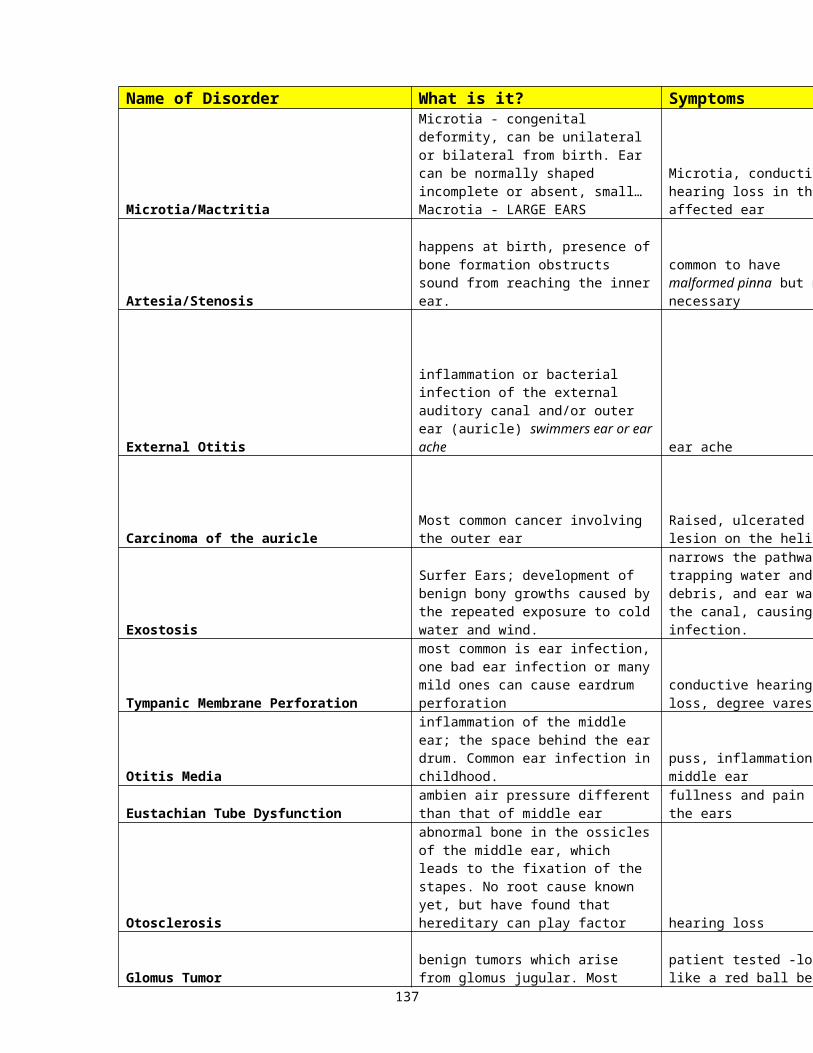
There are 3 (4) measurements made in the plane of the TM:
1. static acoustic compliance or immitance: mobility of the TM at a given volume of pressure
2. tympanometry: the graph of how the ear drum is moving as we vary the pressure from positive to negative; how the immittance of the middle ear changes as air pressure is varied in the external canal
3. acoustic reflexes: contraction of the middle ear muscle to very loud sounds; a reflex of the stapedius muscle, however, in animals it may be the tensor tympani muscle
4. acoustic reflex decay: the ability to hold the contraction for 10 seconds)
Resistance, mass, and stiffness make up the impedance of the system: Mass affects the high frequencies. Stiffness affects the low frequencies.
Resistance – determined by the ligaments supporting the ossiclesMass – determined by the weight of the ossicles and TMStiffness – load of fluid pressure from the inner ear on the
stapes
By combining the mass and stiffness, we can obtain the measurements needed for acoustic immittance.
One ear has a headphone or insert phone and the other ear has a probe creating an airtight seal. A constant tone comes through the probe side. (The probe tone is either 220Hz or 660Hz; the 660Hz usually used with infants.) Pressure on that side is then varied and the eardrum movement to different pressures recorded. ( The pressure range is typically +200daPa to -200daPa although it may go as far as -400daPa, depending on the peak pressure.)
137

This is based on how far back the probe tone comes as the pressure is varied. Reflex-eliciting stimuli may be delivered ipsilaterally or contralaterally, depending on what you are measuring.
138

Static Acoustic Compliance or Static Immittance
C2 (eardrum at maximum compliance)- C1 (eardrum stiffened with +200 pressure
C3 (total compliance of the TM)
.3 is the minimum compliance for normal ears1.6 to 1.75 is the maximum compliance for normal ears
This refers to the isolated contribution of the middle ear to the overall acoustic immittance of the system. It’s a measure of the height of the peak relative to the minimum or start height. It may be thought of as the absolute height of the tympanogram at its peak. (Stress point versus maximum mobility) Thus, a normal system and a negative system may both have the same peak or static immittance.
If the system is below the norm, there is less than normal mobility and the system is stiff or hypocompliant. Some examples that could cause this problem are otitis media or otosclerosis where the stapes is fixed.
If the system exceeds the norm, then there is greater than normal mobility and the system is hypercompliant. Some examples that could cause this problem are a separation of the ossicles (disarticulation) or a very thin eardrum characterized by abnormal elasticity (monomeric membrane). This may occur due to a healed perforation.
When testing, one of the measurements obtained is the ear canal volume is ECV. If the overall volume is greater than 5.0, or if one ear’s volume is twice that of the other ear, then the ear probably has a perforation or a patent ventilation tube.
139

TympanometryTympanogram: this is a chart that shows your curve of compliance.
To measure this, we start at +200daPa (aka mmH2O), measure the compliance, and gradually change the pressure until –200daPa. This highest peak shows the point of greatest compliance. A low-frequency probe tone of 220 or 226Hz is used to bounce off the TM and measure the distance back to determine compliance at each point of the tympanogram.
Types of tympanograms:a. type A: this indicates normal middle ear function. It
is almost an inverted V. The limits of the peak to still remain normal are +100daPa. The pressure in the outer ear and the pressure of the middle ear are equal
b. type As: the peak of the curve is within normal limits but the compliance is below .3. The “S” stands for stiff.
c. type AD: the peak of the curve is within normal limits but the compliance is above the upper end of normal. The “D” stands for discontinuous (or disarticulation).
d. type B: this is basically flat with little compliance and no peak. It usually indicates fluid is present. If the probe is packed with wax, this can occur as well as with cerumen impaction of the ear canal or a perforation of the eardrum.
e. type C: in this, the compliance is normal but the pressure peak exceeds the bounds of +100daPa. This might be seen on someone after a flight or with Eustachian tube dysfunction due to a cold.
140

Note:Also, probe tone frequency should be taken into account. When testing newborns and young infants, it is common to use a 1kHz probe tone versus the 226Hz tone used for adults.
Gradient (slope): the ratio of the height and width of the tymp.
WidthHeight
All of this also helps us to evaluate for perforations, even pinhole perforations that are invisible to the naked eye. We always get a volume measurement of the ear canal. When there is a perforation, it becomes very large as we are measuring the outer ear and the middle ear together. Also, if one is volume is twice that of the other, it is also suggestive of perforation.
Another way you might see middle ear measurements written is with the measures for ECV, COMP, and MEP. These stand for the following:
ECV: This stands for the ear canal volume and is given in ml. Again, when one ear ECV is twice the other or more, or, if the ECV is great than 5ml, there is a perforation or open tube.
COMP: This stands for the compliance measurement and should be between .3ml and 1.75m. If below .3ml, the system is hypocompliant and you can expect a conductive hearing loss. There may be fluid, otosclerosis, a perf, or other ME problem. If above 1.75, the system is hypercompliant and may or may not have a conductive problem. There may be a disarticulation.
141
MEP: This stands for middle ear pressure. This is where the peak of the most compliant point is. If it is between -100daPa and +100daPa, then the pressure peak is normal. If it is more negative than -100daPa, then you may have Eustachian tube function problems.
142

Tympanometry: A tympanogram is a graphic representation of the relationship of external auditory canal air pressure to impedance; the latter is usually reported in terms of tone of its derivatives, compliance in arbitrary units. Pressure in the external auditory canal is varied from -200 daPa* through +200daPa while monitoring impedance. Impedance is lowest (maximal compliance) when pressure in the canal equals pressure in the middle ear. Ears can be classified into three basic groups on the basis of the configuration of the tympanogram.
o Type A. The peak compliance occurs at or near atmospheric pressure indicating normal pressure in the middle ear. There are three subgroups.
A - normal shape reflects a normal mechanism
AD - A deep curve with a tall peak indicates an abnormally compliant middle ear, as seen in ossicular dislocation or erosion, or loss of elastic fibers in the tympanic membrane.
AS - A shallow curve indicates a stiff system, as in otosclerosis.
o Type B - No sharp peak, with little or no variation in impedance over a wide range, usually secondary to non-compressible fluid in the middle ear (otitis media), tympanic membrane perforation or obstructing cerumen.
o Type C - Peak compliance is significantly below zero, indicating negative pressure (sub-atmospheric) in the middle ear space. This finding is often indicative eustachian tube dysfunction.
*daPa = decaPascal = mm H20 One system of tympanogram classification.
A = normal AS = stiffened tympano-ossicular system; AD = disarticulation
B = Middle ear effusion, tympanic membrane perforation or impacted cerumenC = negative middle ear pressure.
143

Acoustic Reflex measures (AR): Contraction of the stapedius muscle occurs with loud sounds, producing a measurable change in compliance. Abnormalities of hearing may be suspected by the following results:
o Elevated threshold - indicates cochlear sensitivity loss or VIII nerve disorder
o Absent reflex
Abnormal middle ear system
Severe sensitivity loss
VIII nerve lesion
Ipsilateral VII nerve lesion
Some otherwise "normal" ears
o Threshold low in proportion to sensitivity level ("recruitment")- seen in cochlear loss
o Abnormal "shape" of reflex.
144

Acoustic Reflex
The acoustic reflex is activated by the RAS or reflex activating signal. The meter reads the decrease in compliance from the ipsilateral (same) ear or contralateral (opposite) ear. Acoustic reflexes may also assist in differentiating cochlear and retrocochlear disorders based on the reflex pattern. We look at crossed and uncrossed reflexes. There can be no reflex with a conductive involvement of as little at 5 – 10dB air-bone gap.
Generally, pure tones are used to elicit these responses. The response is a contraction of the stapedius muscle in response to an acoustic stimulus that is usually at least 60dB greater than the hearing threshold. If there is significant hearing loss (greater than 60dB) or a conductive problem, there will be no reflex. The ipsilateral (probe ear) is measured at 1kHz and 2kHz. The contralateral (opposite ear) is measures at 500Hz, 1kHz, 2kHz, and 4kHz. White noise may be used as well. The intensity of the tone is raised until a reflex is obtained or until you hit the maximum allowable level, whichever is first. For example, ipsilateral reflexes have a limit of 105dB and contralateral reflexes go to 110dB. Due to recent lawsuits, there is controversy whether to go up to 110dB or stop at 105dB as is done with ipsilateral reflexes. Above 110dB you will see a message to warn that this level can cause damage to the ear.
The pattern for measuring the acoustic reflex is the same as with all other measurement, down 10 and up 5.
So these thresholds may:1. detect a loss of hearing above a specific degree2. detect an auditory disorder; conductive, sensori-
neural, or central145

146

ART (Acoustic reflex threshold): the lowest level at which the reflex is obtained twice.
Ipsilateral reflex pathway: a sound in that ear evokes a response from the same ear.Sound OE ME cochlea (IE) along 8th nerve and to the brainstem. In the brainstem, it is received by CN SOC facial nerve (VII) ipsilaterally, and descends to innervate the stapedius muscle.
Contralateral reflex pathway: the sound goes in the ear opposite the probe and the reflex is picked up by the ear with the probe.Sound OE ME cochlea (IE) along 8th nerve and to the brainstem. In the brainstem, it is received by CN SOC and crosses over to the opposite SOC facial nerve (VII) contralaterally, and descends to innervate the stapedius muscle, thus evoking a contralateral reflex.
The reflex will generally occur at about 85dBSL or at about 85dB above the threshold of hearing, up to about 50 or 60dB hearing loss. Till 70dB of hearing loss, the reflex may be elevated by that amount above 50dB of loss. Above 70dB, the reflex will be absent as the sound level presented is not loud enough above threshold to elicit the reflex.
If the is a problem in any part of the chain (or reflex arc as it is known), the reflex may be absent. By analyzing the reflex response pattern, we begin to made educated analysis of possible pathologies.
147

Acoustic Reflex DecayThe tone is given at 10dBSL or 10dB above the reflex threshold and maintained for 10 seconds. It the reflex does not decay to half its original amplitude, it is negative. If it is positive, there is a more central problem to worry about.
Abnormal decay may be defined as a 50% reduction of the reflex. Your handout says within 5 seconds, however, all previous literature uses 10 seconds. The Comprehensive Dictionary of Audiology Illustrated (2nd Edition) defines acoustic reflex decay as “perstimulatory reduction in the magnitude of the acoustic reflex reduced by over 50% of the initial amplitude within 10 seconds of stimulus onset”.
So…..Static compliance and tympanometry measure mobility.Acoustic reflexes and acoustic reflex decay give information about probable disorders.
148
WHAT WILL THE TYMP LOOK LIKE?
TYMP
A
LOSS
Normal/Sensori-neural
DISORDER EXAMPLE
Presbycusis
PRESENT OR ABSET REFLEXES
P/A
AS Conductive/Mixed
Otosclerosis A
AD Conductive/Mixed Ossicular Discontinuity
A
B Conductive/Mixed
Otitis Media A
149

C Normal/Conductive
Eustachian Tube Dysfunction
P/A
Normal hearing->better than 25db with air conduction and bone conduction equal
Sensori-neural hearing loss->hearing worse than 25db with air conduction and bone conduction equal
Conductive hearing loss-> there is a gap of greater than 10 db b/w the air conduction and bone conduction of the same ear at the same frequency
Mixed hearing loss->bone conduction thresholds are worse than 25 db and there is an air-bone gap greater than 10db in the same ear at the same frequency
SPEECH100% - 90% - excellent90% - 80% - good70% - 79% - fairBelow 70% - poorBelow 40% - very poor
150
Sensitivity Prediction by the acoustic Reflex (SPAR):This is a popular test for predicting hearing sensitivity. It is based on the difference between acoustic reflex thresholds to the pure tones and the reflex threshold to broad band noise (BBN).
SPAR = Reflex PTA (500, 1, &2kHz) – BBN +5dB correction factor
If a SPAR value is less than 15, there is a high probability of a SNHL. In most normal hearing individuals, the reflex threshold for BBN is much lower than for tones. In a SNHL, the reflex threshold for BBN is significantly higher while the pure tones do not change. Thus, the closer the BBN reflex threshold to the PT reflex thresholds, the greater the likelihood of SNHL. This test may be used for children who cannot respond. It may also be used for malingerers.
Other tests which do not involve voluntary responses are:
1. Auditory evoked potentials (AEPs)2. Otoacoustic emissions (OAEs)
151

152

Chapter 9AEP – Auditory Evoked Potentials
There are neuroelectric events; they are measured from the scalp via electrodes. The electroencephalograph (EEG) is used and looks for a change in activity. All waves, except for the change associated with the introduction of the click stimulus, are then filtered out. They are subdivided based on where and when they occur:
1. ECoG or Electrocochleography: this is the electrical response generated within the cochlea. This test is used primarily for determining Meniere’s disease. We look at the action potential (AP), cochlear microphonic (CM), and the summating potential (SP). Reflects activity of the cochlea and VIIIth nerve and is the earliest of the Evoked Potentials, occurring in the first 5 ms.
2. ABR (Auditory Brainstem Response) aka BAER (brainstem Auditory “Evoked Response aka BERA (brainstem evoked Response Audiometry): This occurs in the first 10 – 15 ms and the response comes from the 8th nerve and brainstem to the midbrain. It is the most commonly used test of the the evoked potentials. There are 5 waves we measure. We can approximate hearing sensitivity for high frequency click stimuli from this. It is also a good test for neonates, and until recently, the gold standard for newborn screening in the screening version called is the AABR (Automated Auditory Brainstem Response). If there is a delay of waveforms, when using this test for diagnostics, we can guess where the probable site of pathology lies: Wave I from the distal portion of the VIIIth nerve where the fibers leave the cochlea (also the equivalent of the ECoG), Wave II from the proximal portion of the nerve
153
near the brainstem, Wave III from the proximal portion of the nerve and from the cochlear nucleus, and waves IV and V have contributions from the cochlear nucleus, superior olivary complex, and lateral lemniscus. These waves are referred to as Jewett Waves. Interpeak latencies are also evaluated, I-V, I – III, and III-V. Finally, the interaural latency difference, or ILD is looked at. This is the difference for the same measurement between ears and should not exceed .3msec. This test may be used to find acoustic neuromas (vestibular schwannomas). One may assess the integrity of the central auditory pathway using a fast rate to find demyelinating disease (MS). Multiple components to this may be:
a. The standard ABR – just look at absolute peak latencies, interpeak latencies, and differences between ears
b. The Latency-Intensity Function – may obtain wave V down to within 10 – 20dB of threshold, good for babies and malingerers
c. Rate series: same sound and click but at different speeds, sensitive to demylinating disease (MS).
154

155

3. AMLR (Auditory Middle Latency Response) – This originates in the midbrain and occurs in the fist 50 ms after the introduction of the stimuli. It reflects the activity at or near the auditory cortex. It uses tone pips and may provide information for frequencies below 2KHz as ABR measures 2 – 4kHz. It is characterized by 2 positive peaks, Pa at 25 – 35msec and Pb at 40-60msec after stimulus presentation. It is of questionable use with infants and uncooperative patients. May also be used with central auditory processing disorder. At times it may be used for threshold determination as well.
4. LLR (late latency response) or (ALR - Auditory Late Response) or LER (Late Evoked Response) – Occurs beyond 60ms, within the first 250msec and comes from the cortex, specifically the activity of the primary-audiotry and association areas of the cerebral cortex. It has a negative peak, N1, at about 90msec and a positive peak, P2, at about 180msec after sound presentation. At 300ms it is known as the P300. It may assess neurological function. The patient must be alert. Be aware that there is a developmental effect for the ALR during the first 8 – 10 years and then it becomes robust.
The above tests may be used in intraoperative monitoring to assess the integrity of the 8th nerve as surgery progresses. It is a way to preserve hearing during surgical procedures. The standard ABR or ECoG are the most commonly used tests for this.
The 4 main applications of the above are:a. to predict hearingb. infant hearing screeningc. diagnostic assessment of CANS functiond. intraoperative monitoring as mentioned above
156

5. STACKED ABR – this also uses clicks and high pass pink noise masking. It is the composite of activity from all frequency regions of the cochlea. – may screen and detect for small acoustic tumors and may be a good estimation of threshold for hearing for those who are difficult to test.
6. AUDITORY STEADY STATE RESPONSE (ASSR): This test used modulated tones that can be used to predict hearing sensitivity. It is evoked by a periodic modulation of a tone (usually 500Hz, 1kHz, 2kHz, or 4kHz) and the potential follows the time course of the modulation. Good for those who cannot response normally.
157
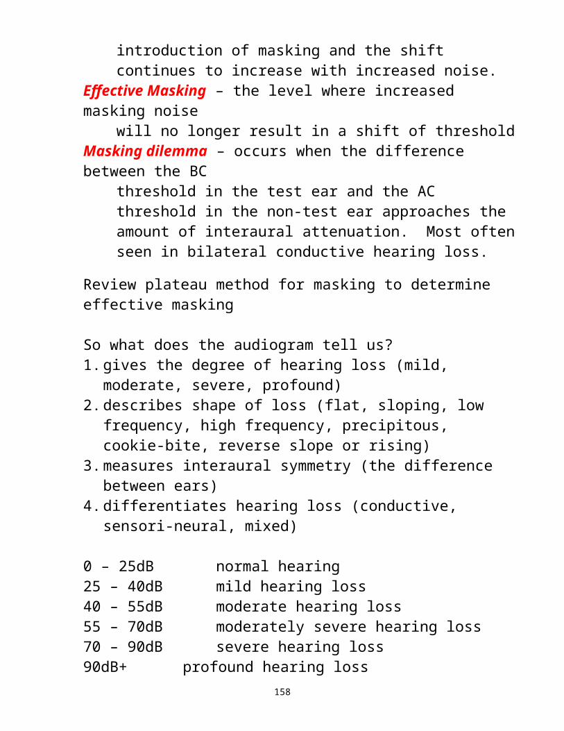
Otoacoustic Emissions Audiometry
This test is a preneural response giving information up to but not including the auditory nerve. They are an active byproduct of the outer hair cell system. If there is a compromise in the outer or middle ear, the response will be affected and most likely be absent. Remember, this is not a direct measure of hearing.
158

1. OAE – Otoacoustic Emissions aka Evoked Otoacoustic Emissions – Most normal cochleas react to acoustic stimulation with a very tiny sound of its own. So these are low intensity sounds generated by the cochlea and transmitted into the middle ear and ear canal. That is why if there is a middle ear problem, the OAE will be absent except in the case of ventilation tubes. This equipment is used to detect and amplify this miniscule response. This has become a popular test for newborn screenings. There are different types of evoked otoacoustic emissions:
159
2. TOAE (transient otoacoustic emissions) aka as TEOAE (transient evoked otoacoustic emissions) – These are produced by brief acoustic stimuli such as tone pips or clicks. The response is usually all or none. Anyone with normal hearing and normal ear structures should have emissions. A middle ear problem or hearing loss greater than 35 - 40dB will eliminate the response.
3. DPOAE (distortion product otoacoustic emissions) – For this test, 2 primary tones are presented and the normal ear produces energy at additional frequencies known as the distortion product. As the primary tones vary, so does the distortion product. A response may be obtained as long as the loss does not exceed 40 – 50dB.
4. SOAE (spontaneous otoacoustic emissions) – This is not an evoked response. Many normal ears (50 – 70%) have natural emissions and these may be measured. It was initially thought that this would explain tinnitus, however, this has not proved to be the case.
Both the TOAE and DPOAE give a measure of outer hair cell function. That is why they are sensitive to cochlear vs retrocochlear pathology for hearing loss below 65dBHL.
Some advantages to using OAEs over the ABR in newborn testing:
2. the test time is much faster3. noninvasive tests that does not involve the use of
electrodes4. less sensitive to environmental noise
160

So, OAEs are applicable for the following:a. infant screening (all or none phenomenon, sensitive
to the most minimal of hearing losses)b. pediatric assessment – if we can only obtain sound
field results, we cannot determine if one ear is better than another, but this would give us ear specific results easily and without needing a response from the child
c. cochlear function monitoring – are potentially ototoxic meds having an effect on the cochlea
d. some diagnostic cases – is the problem in the cochlea?, outer hair cells vs inner hair cells, auditory neuropathy?
e. Also good for verifying hearing in pseudohypacusisf. To diagnose cochlear dys-synchrony (aka
neuropathy). Is the inability to hear or discriminate words due to disruption of synchronous neuronal firing in the auditory pathways? This is especially helpful for early infant detection when combined with ABR testing. If OAE only without ABR, this disorder would have been missed.
MATERIAL FOR SECOND TEST ENDS HERE
161

Chapter 10, p. 437 – 461Young children often cannot be tested in the same manner as adults. The procedures used will vary, based on the age and maturity of the child. We must be flexible and alert, able to modify a procedure to obtain what we need. Sometimes the response may be barely detectable, very subtle, and you must be looking for it to see it. With universal infant screening, the challenges we face now will often be transferred to the SLP. Our current challenges:
Identify the children at risk and needing further evaluation
Determine if there is an actual hearing loss and differential diagnosis of the loss and quantification of the loss
Assess preschoolers with possible CAPD, identify, quantify and refer
The challenge for audiologists and the SLP: Once these children are identified and found,
what are the referrals to be made Are there professionals ready to immediately
begin the necessary habilitation Are we prepared for the influx due to universal
infant screening. If not, all our screenings and early detection is meaningless.
Some additional case history information, in addition to those questions discussed earlier, might be:1. Were there any problems during the pregnancy or
birth? If so, what?2. Is there any history of ear infections?3. Were the child’s developmental milestones to date
normal?4.Do you have any concerns about hearing,
speech/language development, or general development?
162

So let’s have a brief, cursory review of language development.
3 months: There is no difference in response to stimuli between the hearing and deaf infant.6 months: They go from reflexive babbling to intentional babbling and vocal play. A deaf baby will revert to reflexive babbling if they cannot hear themselves. If they don’t make this transition out of reflexive babbling, they will never rise to higher levels.
Cannonical babbling: raspberries, mama, dada, change voiceThis stimulates the auditory system and encourages further development of the auditory system between the ears.
At 12 months of age they start linking words to meanings.At 18 months of age there is one new word every 2 hours.At 24 months of age, there are 2-word phrases.At 36 months of age, they make short phrases.At 30 – 36 months, they apply the rules to language and overgeneralize rules.
Overall, the temporal lobe dominates. In central system analysis, they look for frequency shifts to identify speech and send it to the left hemisphere. Only speech produces frequency transitions with the exception of computer-generated synthesized speech. Sounds determined not to be language are sent to the right hemisphere.
So the left hemisphere is for concrete language and the right hemisphere is for suprasegmentals and the abstract. The 2 halves together give the full reception and expression of language.
163

We know there is a difference between females and males.
Thus, in babies, the low and mid frequency regions develop first. The high frequency region develops after birth.
At 4 days after birth, an infant can distinguish the mother’s language from others.At 6 – 10 months of age, babies can differentiate phonemes.At 6 months of age, syllable pattern recognition begins and then comes the baby’s first word – NO!
The children learn language by shared experience. They learn syllable patterns. This is important to brain development and the development of listening skills.
The auditory brain map is formed by 12 months of age. An absence of stimuli between 6 – 12 months produces a measurable speech delay.
The most critical period for speech learning is birth – 12 months.2nd most important period for this is 12 – 36 months.
Below 3 months
Speech and language are imitative processes, acquired mostly through audition. A problem early on can interfere with the development of speech and language acquisition, thus culminating in a deficit of normal communication. Most of these infants were referred because of high risk factors or they failed and OAE/AABR screening.
164

Babbling is not indicative of anything. If anything, it will decrease later on in the hearing impaired child. Also, a difference in vocal quality may be noted between infants with normal hearing and infants with hearing losses.
Until recently, our detection of children with hearing loss could often take till 3 years of age. Yet numerous studies have shown that the earlier intervention is initiated, the better the chance of a well adjusted, good communicator.
Hearing loss has been ranked 6th in prevalence of chronic conditions in the US. Thus, it occurs more often than many other disorders that we currently screen for. The savings to the government in special education and other needed services later on more than makes up for the cost of newborn screening.Three states, Rhode Island, Colorado, and Hawaii voluntarily began their own programs without mandate. The biggest problem noted is with the number of false positives. Some, such as Luterman question:a. Whether proper bonding with the mother can occur if
hearing is at question early on b. if the hearing loss is genuine, every parental activity
becomes a learning activity rather than just enjoying the child
c. is the audiologist legally liable for the mental stress, especially if it was a false positive
It has been suggested that the newborn screening methods, in conjunction with the high risk registry, can reduce the false positives. Our state has mandated newborn screening, however, they are always trying to cut the budget.
10 High Risk Factors (as determined by a joint committee)
165
1. Family history of congenital or delayed-onset childhood SNHL
2. In utero infection associated with SNHL (i.e. CMV, rubella, etc.)
3. Craniofacial anomalies, including those of pinna and ear canal
4. Birth weight less than 1500 grams (about 3.3 lbs) 5. Hyperbilirubenemia at levels needing exchange
transfusion6. Ototoxic meds, & diuretics used in combination with
the drugs7. Bacterial meningitis8. Severe depression at birth, including low Apgar scores9. Prolonged mechanical ventilation of at least 5 days10. Stigmata or other findings associated with a
syndrome known to include SN or conductive hearing loss
166
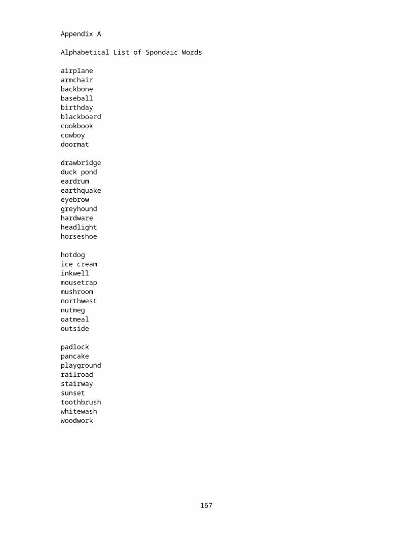
Risk factors that would suggest the necessity of a screening during the first 2 years:1. concern regarding hearing, speech, language, &/or
developmental delay2. bacterial meningitis or other infections associated
with SNHL3. head trauma associated with concussion or skull
fracture4. stigmata or other findings associated with syndromes
known to include SNHL5. ototoxic medications and diuretics6. recurrent or persistent OM with effusion for at least 3
months
It is possible that there may be some disorders with delayed onset of HL, therefore, the following are risk factors for periodic monitoring of hearing till 3 years old:
1. Indicators associated with delayed onset of SNHL are:
a. family history of hereditary childhood hearing lossb. in utero infection like CMV, rubella, syphilis,
herpes, etc.c. neurofibromatosis & certain neurodegenerative
disorders
2. Indicators associated with conductive hearing loss are:
a. recurrent or persistent otitis media with effusionb. anatomic deformities or other disorders affecting
Eustachian tube function (i.e. Treacher-Collins)c. neurodegenerative disorders
With the high risk registry, only about 50% of newborns with hearing loss are caught because the other 50% did not have the high risk factors.
167

There have been different techniques for screening newborns over the years:
a. cardiac responseb. respiration audiometry, alternation of sucking, and
startle responsec. crib-o-gramd. High Risk Registrye. ABRf. AABRg. ABR with High Risk Registry h. OAE
ABR was the first real way of testing that gave better sensitivity. When combined with acoustic reflex testing, it produced a more sensitive and specific approach to new born testing.
Now we have OAEs. These are even more cost effective. There is no need to use electrodes and clean the skin. It is a simple tip in the ear and very quick. It is less sensitive to environmental noise, making it excellent for the NICU.
Testing birth to 1 year
1. casually observe the child.2. Personally, I like to listen to the mother. Parents know
their child and are more sensitive than most pediatricians to the presence of hearing loss.
3. Listen to the child vocalizations. Severe hearing loss can be detectable in their vocalizations.
4. Behavioral Observation Audiometry (BOA) in sound field
168

Behavioral Observation Audiometry (BOA) 0 – 1yrThis is conducted in a sound field environment through speakers. Speech, various noises, warble tones, etc. may be presented and we look for:
a. cessation of activity whether it be sucking, crying, or breathing or even eyeblinks.
b. Localization to the speaker where the sound comes from by 8 months old. Prior to that, a searching behavior should be noted.
Some children will respond to the sound off rather than when the sound is turned on.
Visual Reinforcement Audiometry (VRA)This is the same as BOA, however, at this time the child is trying to localize to the source of the sound. Localization or sound acknowledgement is rewarded by a clapping toy or lighted toy, etc.
Testing 1 – 5 years of ageYoung children fatigue quickly, therefore you jump in and get what you can. Before 3 years of age, the VRA is most likely to be used. At 2, they may even repeat some words. If nothing else works, you can ask them to point to body parts or ask questions like “where is mommy?”. Listen to their vocalizations, if they are almost silent, there may be hearing loss suspected.
Conditioned Play Audiometry (CPA)By 3 – 5, you can try conditioned play audiometry. They respond to the stimulus most often by placing a block in a
169
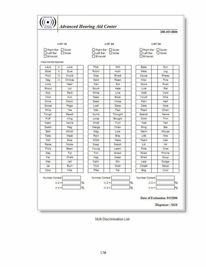
box or a ring on a post, or a peg in a hole. They may also respond to the command “put it in”. For most, at this point, you can probably perform this task under headphones.
Often, I can even get them to say “birdie” to warble tones.
For the immature or very young child or children with multiple disabilities, when you can only obtain response through the speakers, it is impossible to know whether it is indicative of the better ear or both ears. That is where the OAE come in as it can tell you if the outer hair cell activity is normal in both ears or not.
Always include your impedance measures to allow you to guess normal, SNHL, or conductive problem.
THE CROSS CHECK PRINCIPLEAccording to your book, “no single test result obtained during pediatric assessment should be considered valid until you have obtained an independent cross-check of its validity”.
A recent article in Advance magazine (Jan, 2013) gave the following article to check for possible speech delay due to hearing loss:
170

NEWS WATCH
Several Development Milestones Can Signal Possible Hearing Loss
Posted on: January 8, 2013
Infants and young children have developmental milestones that parents can watch for to identify a possible hearing loss.
By 3 months, your baby recognizes your voice and makes cooing noises. Sudden, loud noises should startle your baby.
By 6 months, your baby recognizes speech sounds and familiar noises. Interesting noises make your baby turn their head, and your baby plays with their own voice and laughs. Your baby also uses their voice to indicate pleasure and discomfort.
By 9 months, your baby understands simple words like 'mommy' and 'daddy,' 'no', 'bye-bye' and their own name.
By 12 months, your toddler can speak one or more real, recognizable word.
By 18 months, your toddler understands simple phrases and retrieves familiar objects on command and speaks between 20 to 50 words and short phrases and your toddler learns new words each week.
By 24 months, your toddler's spoken vocabulary should be 200 to 300 words and simple sentences can be spoken. Adults who are not around the child on a daily basis can understand your child's speech. A toddler at this age should also be able to sit and listen while being read books.
171

Central Auditory Processing Disorder (CAPD) aka APD
Central Auditory Processing: May be involved in the following: Sound localization Separation of signal from noise Detection of signal in noise Preprocessing for pattern identification Providing the foundation for concrete language Processing the abstract portions of language
CAPD: children whose recognition or use of language is not age-appropriate, and/or is inconsistent with their level of intelligence. Many of these kids also have learning disabilities which prevent them from being able to progress normally in their education. It should be noted that CAPD measures are very sensitive to the effects of attention.
Language delays may occur due to: Hearing Drugs/alcohol Environmental And high frequency hearing loss can be responsible
for language delay
Most of these children have normal peripheral hearing. While this has become a catch-all category, many symptoms of CAPD include: poor listening skills easily distracted slow responsiveness articulation disorders language delay
172

short attention span poor memories and reading comprehension difficulty in linguistic sequencing problems in learning to read and spell difficulty recognizing speech in the presence of background noise
Some auditory processing symptoms in social behavior might be:
- Hyperactive - Fatigues - Lethargic- Disruptive - Withdrawn - Poor
memory- Passive - Slow Starter - Fail to
complete task
There is no particular behavior for a child that classifies them as APD.As can be seen, many of these symptoms overlap with:
- ADD- LD- Speech/language disorders- Behavior disorders associated with
Fine motor coordination Visual Disorders
A more structured learning environment can be very helpful for these children.The speech tests administered in CAPD will be more challenging than standard tests. These usually are words or sentences presented with competition, both ipsilaterally as well as contralaterally.
Hearing loss and/or CAPD can have an effect on social, intellectual, and emotional development.Some of the effects may include egocentricity, difficulty in empathizing with others, rigidity, impulsivity, coercive dependency, and a tendency to act up.
173

Auditory Processing Therapy: There may be environmental manipulations
One to one contact Personal FM system Preferential seating Multi-modality input
Manipulations of temporal/spectral characteristics of sound
Decrease presentation velocity for improved processing then gradually increase presentation velocity
“Velocity” Memory exercises
There have been articles on the importance of Music in assistance of development of the right side.
Development of spacio-temporal reasoning ? development of math skills ? development of pattern analysis ? perception of meaning
Music, math, and logic may be connected.
The neurons are exercised by music. This strengthens the same circuits used for mathematical reasoning. Therefore, preschoolers taking piano or singing lesions improve spatial reasoning. The melody must be identifiable however.
Children with developmental disability are tested by whatever means we can.
In the public schools, children are typically screened every other year. It is not perfect and there is an inverse relationship between efficiency and accuracy in any screening procedure. It is also true that the environment in which the screening is performed is less than perfect
174

and often we raise the pass values at 500Hz to allow for the excessive noise.
Now, with so many more infants and children being detected, we really need the personnel with training and programs in place to adequately service these infants and children in order to:1. facilitate speech and language development2. properly counsel parents and give unbiased information
of all possible educational routes that can be taken3. provide support programs4. mitigate some of the psychological consequences of
hearing loss on the child and the family.The current climate has moved from a client-centered medical model to a family-centered model. In the latter, the family must be educated and empowered to work as a co-partner in planning strategies for their child.
175
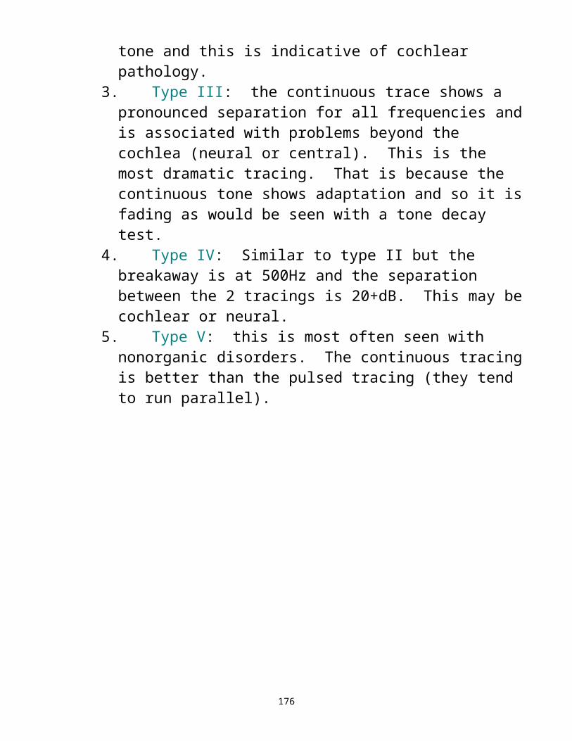
Chapters 12, 13, 14 Management of Hearing Loss
Some type of aid to hearing has been utilized throughout history, ranging from animal horns and seashells to ear horns, even using a hand cupped behind the ear. Whether these primitive methods, or more modern hearing aids, they all collect and direct sound energy.
19th century – first electronic hearing aids – they were huge!!!!Reading the history of hearing aids is like a delayed history of electronics, there were:Carbon hearing aids that were body wornVacuum tube aids allowing for more power and smaller aidsTransistors, further miniaturizing hearing aids and completely replacing vacuum tube aids.Now we have integrated circuits.Even better, we have computer technology!!!!This is also known as digital signal processing.Now, even more, we have WIRELESS CONNECTIVITY!!!!
Rule of thumb for fitting one or two hearing aids:1. if all is symmetrical, both hearing and speech
discrimination, fit both ears2. if asymmetric and both ears have good speech
discrimination, try fitting both ears3. if asymmetric and speech is much better in one ear
than the other, fit the ear with better speech discrimination
4. if symmetrical but speech is much better in one ear than the other and other issues have been ruled out, fit the ear with better speech discrimination
5. if asymmetric and there is a severe to profound loss on the bad side, fit the good side only
176

6. if profound loss bilaterally with poor to no speech discrimination, consider referral for cochlear implant if used to be part of hearing world, otherwise leave in Deaf Comm.
Hearing aids consist of a microphone, amplifier, and receiver and are used to amplify sound and deliver it to the user’s ear. Sounds strike the microphone (input transducer), are amplified, and transmitted to the output transducer (receiver or loudspeaker) and into the external ear canal. For those for whom, for some reason, a standard hearing aid cannot be used, a bone conduction hearing aid may be feasible. Reasons for not being able to use standard hearing aids may include atresia, stenosis, chronic middle ear drainage, perforation, dermatologic problems, etc.
Hearing Aid CircuitsSo hearing aids are signal processors, they alter the signal input to improve it for the wearer. Types may include:
Analog Hearing Aids: similar to an LP, the waves coming out are like those going in. They modify a continuous electric signal. Fine tuning is very limited to the type of circuit.Hybrid Hearing Aids (Programmables): These use both analog and digital technology. They shape the sound digitally but use analog processing. While more precise in fine tuning than the analog aids, they are not as fine tunable as the digital aids.Digital Hearing Aids: These are the newest. Sound waves are converted to binary digits (computer talk using 0’s and 1’s) much like data storage. It changes the output via analog-to-digital converter. Advanced processing operations can then be carried out and the altered signal changed back to analog form by the digital-to-analog (D/A) converter. They provide improved clarity
177

of signal and better signal to noise ratio. They may separate wanted sound from unwanted sound but are still not perfect. They also allow for more fine tuning than ever before.
Electroacoustic Characteristics of Hearing Aids:
1.Output Sound Pressure Level: the maximum sound pressure the hearing aid can produce at high input levels. Also known as saturation sound pressure level. This is especially important in the case of babies. Failing to be careful may cause a noise-induced hearing loss if there are dangerously high sound levels.
2.Frequency Gain: this is the difference between the input signal and the output signal. For example, if a signal of 60dB goes into the hearing aid and 110dB comes out of the hearing aid into the ear, then 110 – 60 = 50dB of gain. That is the amount amplified to get the output.
3.Frequency Response: The range over which the hearing aid provides amplification. With advances in technology, we can cover a wider range of frequencies. The microphone and receiver of the aid are the prime limiters of the frequency range. With today’s hearing aids, we can set the response for each hearing loss specifically.
4.Distortion: This occurs when the sound leaving the hearing aid is very different from what went in. It reduces clarity and intelligibility. Most common is harmonic distortion. The greater the distortion, the poorer the quality of amplified speech.
5.Input-Output Characteristics: describes how the hearing aid functions at different loudness levels. Tells
178

how much the amplification is increased with increase in the input signal. You can see if this is linear or nonlinear. Unless the loss is sever to profound, almost all amplification is nonlinear.
6.Compression, Peak Clipping, Output Limiting, Wide Dynamic Range Compression (WDRC), Automatic Gain Control (AGC): There are additional parameters that limit the output of the hearing aid as sounds gets louder to avoid peaking at an individual’s uncomfortable listening level.
179

All the above are measured in a test box with a 2cc coupler. We can also measure some of these characteristics in the individual’s ears using a probe microphone system. This may also be used as a verification of your fittings.
A more subjective method of verification could be obtaining function gain:Functional Gain: This is similar to acoustic gain. It is measured by obtaining hearing levels in sound field without the hearing aid and with the hearing aid. May be done with warble tones or speech.This allows to show how much gain the individual actually perceives when using the hearing aid and their improvement as compared to how they do at the same level without the hearing aid.
The validation, which may be defined as a determination of whether the disability has been reduced and goals addressed, may be established through clinometrics, or questionnaires/scales.
Another method of establishing you fit the individual properly is Probe-Microphone Measurements and Speech Mapping.Real ear Gain, measured by probe mic equipment, shows us the amount of gain delivered to the ear directly. A loudspeaker is placed close to the patient’s ears, a thin tube inserted into the ear canal close to the TM and attached to a mic. Sounds are presented out the speaker and the hearing aid response in the patient’s ear is recorded.With Speech Mapping, ongoing speech is presented via the probe mic at a fixed level and the output displayed. The hearing aid parameters can then be adjusted as needed.
180

Parts of a Hearing Aid
1. The microphone – in addition to what was stated earlier, we now consider whether we will be omnidirectional (sensitive to sounds from everywhere), or directional (sensitive to sounds in front of a person), directional fixed or automatic directional adaptive (comes and goes as needed for noise and sensitive to sounds from where the speech comes)
2. The amplifier – controls how much amplification occurs at different frequencies, may boost sounds differently, and may be able to limit sound from becoming too loud. (The last used to be known by many names over the years including output limiting, peak clipping, compression limiting, etc).
3. The receiver – also considered the loudspeaker and it is a transducer that changes electrical energy back to acoustical energy. With RIC or RITE hearing aids, the receiver is placed in the ear canal itself rather than being part of the body of the hearing aid.
4. Potentiometers – these are screwset controls, present on old analog hearing aids and some of the older programmable aids, to set high and low tones, limit loudness, limit gain. We no longer need this as things are adjusted by computer.
5. Remote Controls – not available for all hearing aids but many manufacturers have optional remotes for adjusting volume, program, or wireless connectivity.
7.Manual Controls – Older hearing aids have an MTO (mic/telephone/off) switch. Even some digitals have a pushbutton control or toggle to change volume or program.
8.Battery door – found on all aids except disposables, some current manufacturers offer to charge batteries while in the hearing aid
9.Earhook (on standard BTEs) – where earmold connects
181

10. DAI – Direct Audio-Input, usually at the battery door of a BTE so other systems such as FM can be connected
11. Earmold – a part that attaches to the BTE earhook to send the sound into the ear. Sometimes also used with RIC/RITE hearing aids.
12. Auto T – much more common now, and option which allows a hearing aid to go into telephone mode without having to move any switches on the hearing aid or hit anything on the remote control.
13. Ear Domes – the disposable tips that fit the end of the RIC/RITE/thin tubes in the ear canal
14. Color options 15. Connectivity – special remotes and other parts to
change televisions, phones, iPods, etc into Bluetooth devices for the hearing aids (i.e. Oticon Streamer, Siemens Tek, Phonak iCom, etc) Now there is even a mic for others to talk into and sent via Bluetooth without getting a FM system.
16. noise/feedback reduction – most use noise cancellation technology today so feedback is not the issue it used to be.
182

EARMOLDSAn earmold can change the acoustics or frequency response of the hearing aid. This is known as earmold acoustics. There are different types of earmold and various degrees of venting.
Styles of earmoldsThe earmolds block the ear canal to various degrees. High frequency hearing losses use open earmolds that do not fully block the canal. The more severe hearing losses use earmolds that fully seal the ear canal to prevent sound leakage that cause feedback from high power hearing aids. See figure 13-18 for examples of 3 types of molds.
VentingA vent is nothing more than an opening in the mold that allows air and sound to escape. Depending on the loss, it will vary in diameter. Most commonly is the SAV or select-a-vent that allows the fitter to vary the diameter by using plugs with varying diameters. The vent usually runs parallel to the channel containing the tubing. It would be minimal for severe losses and large for minimal losses.
Horn EffectThe horn effect can increase the high frequencies. One way is to use a Libby horn which is a tubing that increases from 2mm to 3mm or 4mm going into the mold. Another way to obtain this is to widen the bore at the end of the mold or “bell” the bore.
DampingThis may be a filter or piece of damping material that is placed into the earhook of the hearing aid or into the earmold to decrease and smooth out peaks in the amplitude of the hearing aid response.
183

Venting, horn effect, and damping all affect different portions of the frequency response. They are affected in the following manner:Venting - low frequenciesDamping - mid frequenciesHorn Effect - high frequencies
There are now special molds made by most labs to work with the RITE/RIC and special high power canal molds for the power RITE/RIC hearing aids.
184

Types of Hearing Aids Body Aids (used with earmolds) Eyeglass hearing aids (used with earmolds) Behind-the-ear (BTE) hearing aids (used with
earmolds) OTE open ear fit RITE/RIC OTE open ear thin tube fit In-the-ear hearing aids (ITE) Half shell hearing aids In-the-canal hearing aid (ITC) Completely-in-the-canal hearing aids (CIC) CROS and BiCROS hearing aids Bone-conduction hearing aids Implantable Bone Conduction devices/BAHA Middle ear implants Cochlear implants Auditory brainstem implants – for NF2 patients –
can’t use CI so implant electrode in brainstem and an external sound processor sends electrical stimulation to the auditory neurons of the CN, hoping to improve the client’s ability to detect or understand incoming speech information
Vibrotactile devices (ie tactaid) For the future: completely implantable hearing aids Hair cell regeneration
185
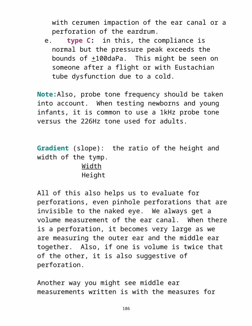
186
187

Discuss why 2 are better than one – Directionality – The left and right location is determined by perception of the difference of arrival time or difference in phase of sounds at each ear. If there are more than two arrivals, as in a reverberant environment, we choose the direction of the first sound to arrive, even if later ones are louder. Height information is provided by the shape of our ears. If a HF sound arrives from the front, a small amount of energy is reflected from the back edge of the ear lobe. This reflection is out of phase for one specific frequency, so a notch is produced in the spectrum. The elongated shape of the lobe causes the notch frequency to vary with the vertical angle of incidence, and we can interpret that effect as height.
When both ears are amplified, the sound from each ear fuses together at the level of the brainstem giving about 3dB increase in gain as compared to one (binaural summation) and so there is more usable gain.When both ears are amplified, the brainstem can separate speech information from background noise better (background squelch)Localization is improved with a binaural fit due to 2 ears working with time and intensity cues from the arrival of sound to each ear as explained above. Finally, if a hearing aid is worn in one ear, then sound from the other side is not amplified in the unaided ear and the head shadow effect causes the gain to be reduced in the aided ear.
Exercise both sides of the brain
No need to turn the headWhat else?
Discuss reasonable expectations.
188

There are other devices to aid in listening besides hearing aids. These are mostly termed Assistive Listening Devices or ALDs. These include:
- FM systems- Personal amplifiers (Pocket Talker)- Closed captioning- Infra-red systems- Amplifying headsets- Personal sound field amplification systems- Large room sound field amplification systems- Amplified telephones
o built in amplifierso portable amplifiers
- TTY/TTD and relay services- Cap-Tel- Caption Call- Video Remote Access- Sign language interpreters- Hearing dogs- Voice recognition technology- Alerting devices: Vibrating, blinking, flashing,
beeping devices for alarm clocks, doorbells, smoke detectors, baby-crying detectors, etc.
- Bluetooth device connection is here now- CART- Notetaker
These devices may be used in addition to or in place of hearing aids. They may make an individual’s life easier, improve the quality of life, improve relations with the family, and give more of a feeling of independence.
For example, the individual who can use a phone amplifier, no longer needs to depend on someone else to answer the phone.
189

Another example is the mother who can use a vibrator alert to let her know her baby is crying.
The deaf person who can wake up on their own by using a bed vibrator connected to the alarm.
They allow for the security of knowing you will wake up in the event of a fire alarm, even if you are sleeping by using blinking lights, vibrators, etc.
This independence also allows for empowerment!!
This independence may also allow protective parents to let go!!! Let the kid be a kid!!!!
And we can all expect this to improve quality of life!!
In the future, a way to ensure compliance of treatment and verification/validation of treatment results may be via the idea of TELEMEDICINE. It is gaining in use across the country. With telemedicine, treatment or therapy is conducted via computer and monitors and cameras and even testing can be performed in this manner. The individual performing the testing/therapy MUST be licensed both in the state where they are conducting therapy from, as well as in the state where the patient resides. In this state, they are still trying to decide on what to write into the licensure law and/or rule and treatment is only going to be within the state.
190

ADULTS
The US Census Bureaus has shown that those individuals 65 years of age or older is the fasted growing segment of the US population and so the need for audiological services will be large as such a high percentage of the elderly suffer from some degree of hearing loss. Some of the natural changes in the body include:
a. outer ear: loss of cartilage elasticity, perhaps resulting in collapsing ear canals
b. middle ear: arthritic changes causing stiffness of ossicles and/or tympanic membrane
c. inner ear: sensori-neural hearing loss, especially of high freq
d. 8th nerve and neural processing: these changes may include degeneration of the auditory nerve, brainstem, and auditory association areas of the brain, thus compromising the ability to understand, especially in noise, and compounding the peripheral changes discussed above. These changes are changes due to the aging process, aka senescent changes, and counsel appropriately. Be aware of the hassle factor. Depending on lifestyle, for some of the elderly, a hearing aid may be more trouble than it is worth and an assistive listening device would be more appropriate. In some cases, these elderly develop problems similar to very young children, such as CAPD. The loss of vision further compounds these issues as they do not have the assistance, whether consciously or unconsciously, of lipreading or reading facial expressions.
Although the goal is to reduce hearing handicap by maximizing the use of residual hearing, approaches to reach that goal may vary from patient to patient. We must look at age, type of disorder and patient need. We must ease the burden on listening in noise.
191

We must consider other needs; emotional, educational, psychosocial, and the effects on interactions. Much of this can be found via our assessment scales/questionnaires.
192

Audiological Rehabilitation: management of these patient; aka Aural Rehabilitation. If you get someone at birth, rather than rehabilitation, you use habilitation since there was nothing to rehabilitate.
The goal of adult AR is to make maximum use of residual hearing and they may need more help than would be thought based on the test results. The broader their dynamic range (the range of sounds they can tolerate), the better they will probably do.
Combine audiometric data and questionnaires/scales to determine an individual’s impairment, handicap, and needs.
So aural rehabilitation is more than speechreading.
Counseling = LISTENING = uh huh
Know when the problem has become pathological and when the counseling needed is beyond your scope of practice and don’t be afraid to REFER.Be there with the individual, watch your body language, give them what they need, not what you think they need.
Do not use the medical model – you are the boss and tell the patient what they are to do, authoritativeUse the client-centered model – you discuss and conclude together, everything is collaborative.It is best to include the entire family, including significant others and caregivers.
Give orientation individually or in groups or some combination thereof. The environment may be in the traditional setting of a professional office or a home, senior center, house of worship, ALF, etc. may be used as well.
193

Teach them to maximize communicative skills by managing environments. Train the patient with hearing loss in maximum use of residual hearing.
One aural rehab consideration is a new, interactive, computer-based tool called LACE (Listening and Communication Enhancement) It provides both interactive and adaptive tasks. By adaptive, we mean that the user’s responses determine the level of difficulty of the task and as they improve, there are more difficulty stimulus items. The 3 main areas addressed are
a. degraded speech – in this category, tasks are designed to help develop better skills when listening to speech in noise and when listening to fast speech
b. cognitive skills – These activities give short and long-term memory training and improve processing speed
c. communication strategies – these tasks are designed to help clients develop skills to improve performance in daily communication situations
Look for support groups for those you work with.FamilyParentsTeensSiblings of the HIGrandparentsLate-deafened adults, etc.
EMPOWER
With children, the management will differ, depending if the loss is prelinguistic or postlinguistic.
194

Parents reactions: Refer to Kubler-Ross – major grief and denial, then on to anger, guilt, bargaining, and finally, acceptance.Don’t forget to recommend genetic counseling.
Prelinguistic:You need to train them as to what sounds are.Begin speechreading early in life. Encourage them to watch faces. Some educational beliefs do not encourage this.
Some believe that humans are lazy and will do whatever is easiest. Manual communication and visual cues are easiest and there will never be a total dependence on audition if you start with anything else. Others feel that it is more beneficial to just get that communication going, keep the frustration down, and let them feel the ability to communicate with others.
195

Before beginning educational options, we first need to look at the basic laws:
THE AMERICANS WITH DISABILITIES ACT (ADA) OF 1990This law applies to all ages. It provides comprehensive civil rights protection and prohibits discrimination to individuals with disabilities in the areas of Title 1 employment, Title II public services (state and local government services including education) Title III public accommodationsTitle IV telecommunicationsTitle V miscellaneous
INDIVIDUALS WITH DISABILITIES EDUCATION ACT (3-21 - IDEA PL 94-132)This ensures for identification and habilitation services for children 3 – 21, including speech, hearing evaluation, counseling and guidance, and educational adjustments/specialized services. Within this portion of IDEA, each student gets an IEP or individualized education program.
IDEA Part B (3 – 21 years)Ensures early services for the children 3 – 21 years and address identification, evaluation, and rehabilitation of hearing loss. Utilized the IFSP or individualized family service plan.Part C (birth – 3 years)Ensures services birth through 3 and may provide the hearing aids for the child if no other insurance coverage. Also works with parents.
SECTION 504 OF THE REHABILITATION ACT OF 1973
196

This section applies to all students who do not quality for services under IDEA. If a child with hearing loss does not meet the eligibility requirements for special education services under IDEA, provision of adaptations to or modification of regular education services to support the student’s academic performance under this section can be explored.
IDEA and Section 504 of the Rehabilitation Act address numerous issues, including but not limited to: FAPE, LRE, compliance and enforcement, protection for parent’s rights, evaluation, reevaluation, composition of multidisciplinary team, documentation, due process, etc. While an assistive device such as FM may not be sent home under IDEA, it may be sent home under Section 504 if needed to complete homework assignments (i.e. FM system so they can listen to the television documentary for an assignment).
Educational OptionsOralists: emphasis is placed on speech and amplification is designed to take advantage of residual hearing.
Some methods are:a. Auditory-Verbal approach (uses only audition and
amplification)b. Aural/Oral Method (auditory and speech reading;
child’s output is oralManualists: communicate by sign and finger spelling
Some methods are:a. ASL – American Sign Language – different grammar
than oral English. Communicates concepts rather than individual words, uses facial and body movement and is processed spatially
b. Cued Speech – enhances those speech sounds that are difficult to differentiate
c. Finger Spelling – this preserves rules of grammar and syntax
197

d. Manually Coded English (MCE) – Signs correspond to English words and syntax is the same, includes Seeing Essential English and Signing Exact English)
198

Mainstreaming: Children spend their entire day in a regular class except for times to received support services related to their disability. They are entitled to the “least restrictive environment”, however, this is not the right choice for all children.Inclusion: Children are in a regular classroom but may have an individual to assist them to keep on target.Self-contained Classroom or Day Classes: children are in a class for the hearing impaired and attend a regular school equipped for this class.Total Communication: This method uses all modalities and may occur in any educational setting.
Depending on the educational method used and school attended, some ALDs might be appropriate. Some ALD examples are FM systems, FM mini speaker systems, infra-red systems, vibrating watches, a sign language interpreter, etc.
Educational terms to be familiar withIEP: individualized education planIFSP: individualized family service plan – educate parent to make the decisionIDEA: Individuals with Disabilities Education ActSection 504 – Part of ADA and applies to those who do have an IEPPart C – for those found to be significantly hearing impaired under 3Part B – for those over 3
Review Deaf controversy
199
Issues of vertigo and tinnitus have not been reviewed in detail. We touched more on the vertigo portion during the disorders. However, be aware that most individuals with hearing loss also experience some degree of tinnitus. Tinnitus may be defined as a noise that is perceived in the ear or head that does not come from any external sources. It can be very debilitating. Some causes for this condition may include:- ear surgery - ototoxic drugs- aging - noise exposure- head trauma - reduced blood flow- Meniere’s disease - tumor- otosclerosis - cerumen
Some of the devices and treatments available for tinnitus are:
tinnitus maskers – devices that look like hearing aids and emit a noise to cover over their tinnitus
noise generators – similar to tinnitus maskers but more broad-band noise and go to mixing point where they hear both, their own sound and the noise
Tinnitus Retraining Therapy – This mixes using noise generators and directive counseling and may take up to 18 months to give full relief
Neuromonics Oasis – a new device from Australia using music with underlying noise & takes about 6 months for treatment
Hearing Aids – Often, just the use of hearing aids for those with hearing loss will help the tinnitus.
Sound Oasis – A homeopathic device with different sound cards including environmental sounds for sleep/relaxation and a card with sounds for tinnitus; often helpful during the evening for those suffering from tinnitus or to help get to sleep.
Progressive Tinnitus Management (PTM) – created by Jim Henry of the VA. It looks at sounds an individuals
200

likes for distraction, interest, and background and tries this before going toward expensive devices.
The Inhibitor – a handheld device which emits an ultra high frequency sound for one minute to temporarily inhibit tinnitus
Combination HA/Masker Devices – This is a hearing aid which permits a concomitant noise. As of the writing of these notes, two manufacturers had devices for amplification and tinnitus and two other manufacturers are about to get into it.
PAXX100 – customized audio device to be used daily using customized acoustic enrichment sounds. As with all other devices, the objective is to beat the link between the tinnitus and sympathetic nervous system so the user’s consciousness and attention is not to the perception of their tinnitus.
SoundCure Sernade Solution – This device looks like an iPod as does the Neuromonics and uses “S” tones, their proprietary sound along with options for narrow band noise and broad band noise as a noise therapy.
There are many other treatments that have been and are being investigated which include drugs, cold laser, biofeedback, and even some brain surgery, however, the above are the most common and noninvasive.
201

First, let’s discuss the basics of microbiology that leads us to:
a. how germs are spread from one person to anotherb. how infections are spread from one person to
anotherINFECTIONMicrobes
1.Pathogenic versus opportunistic: we usually think about pathogenic, HIV, Hepatitis B. They are inherently pathogenic organisms that make humans sick when they come in contact. The other part of infection control deals with opportunistic organisms. That is, those organisms that are supposed to be there, they live with us, they live on us, and they could make us sick by their absence. These opportunistic organisms are capable of causing disease if we give them the opportunity and generally we give them the opportunity by some deficiency in our immune system. Audiologists usually deal with people at 2 opposite ends of the age continuum:
a. We deal with young children with underdeveloped immune systems
b. We deal with older adults whose immune system begins to wear out as they continue to age.
Because of this, our daily patient population is much more vulnerable to opportunistic infection than the general population. This is significant if the first AM patient is a 50 year old health male and the 3rd is an 80 year old diabetic female with a compromised immune system.
202

2.Mode of Transmission (Vehicle): How germs are spread from one person to another. There are 4 common modes of transmission.
a. Contact: the most prevalent form of transmission of germs. There are 3 types:i. Direct: spread of germs between people by
touch. May occur from handshakes to pulling the ear back.
ii. Indirect: germs are spread via inanimate objects or surface, i.e. countertops, armchairs, etc.iii. Droplet: occurs as we talk, cough, sneeze, etc. Droplets of moisture are sent from our respiratory system into the air and the droplets of moisture contain little microorganisms that live in our bodies and our head is usually close to the patient and you may exchange respiratory droplets with those patients.
b. Vehicle: this may be from food, water, blood, or other body substances. Pathogenic and opportunistic organisms may be transferred through blood but ear drainage (mucous) and cerumen can be potentially infectious bodily substances. The dangerous thing about cerumen is that because of its color and viscosity and the overall nature of that substance, it is difficult to ascertain whether or not that particular substance is contaminated.
c. Airborne: This is the sum total of all the things we put into the air. The respiratory droplets, the heating and cooling system, walking on carpet, all join together. Remember, what goes up into
203

the air eventually settles down on surfaces and objects. So we need to regularly clean surfaces, whether or not they need it.
d. Vectorborne: Not very pertinent to this discussion but this is where organisms are spread from one person to another via a vector. A vector is an insect or small animal (e.g. lyme disease and ticks, malaria and mosquitoes).
3.Route (pathway): This is how organisms enter the body. The most common routes are the normal body openings, eyes, ears, nose, and mouth. They are normal body openings. But the ears are connected via the Eustachian tubes to the rest of the respiratory tract, the possibility for an organism to come from outside the body, find its way into the middle ear through the Eustachian tubes and into the respiratory tract is a very real possibility. This is possible with small, microscopic teas and fissures on the TM that you cannot even see. You can create a body opening through nicking, cutting, or scratching the patient through cerumen management or impression taking. So you can crate a route or pathway allowing the organisms in to cause infection.
INFECTION PROCESSThere are 3 criteria which determine whether or not the invading organism will make us sick:
1.Virulence: This relates to how quickly our body can identify the organism, create the troops to kill the organism, and get rid of it. That virulence is also related to how quickly that organism can reproduce in us. If we don’t identify it quickly and it reproduces
204

very quickly, then it can make us sick quickly and it is a virile organism.
2.Titer (number): This is the number of organisms you’ve been in contact with. The titer being the overall number you are spreading from 1 person to another and is directly related to
a. when was the last time you cleaned your handsb. when was the last time you disinfected the
speculac. when was the last time you cleaned the hearing
aidAll of these will have a dramatic impact on the titer or the number of germs that are available for infection.
3.Resistance of the Host: An intact immune system is very good at fending off organisms but our immune systems aren’t always intact. As we age, the immune system goes down and when we are very young, it is underdeveloped. Then there are various states of health based on how you take care of yourself.
Looking at these 3 points:a. you have no control over the virulenceb. you have some control over resistance of the
host (take care of yourself and vaccinate)c. BUT YOU HAVE THE MOST CONTROL OVER
THE TITER!!! And this becomes the basis for an infection control program.
The basic premise of an infection control program is to:
205

Reduce the number of pathogens in the environment to a level where the normal resistance mechanism of the body may take over and prevent infection.
206
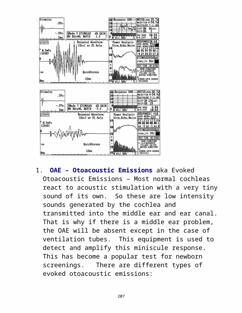
There are a couple of important points from this definition. We talk about reducing the number of pathogens in the environment, not eliminating them. Reduce them to a point where the normal resistance mechanism of the body can take over and resist disease. The resistance mechanism fluctuates. Protect the individual who is the most vulnerable person in your practice.
Based on this premise, the definition for an infection control program is:
An organized effort to manage one’s environment in order to minimize exposure to pathogenic microbes that can make you or your patients sick.
The key point here it that it is an organized effort. An infection control program is an effort where you actually commit it to paper, not just because OSHA requires a written infection control plan on the premises, but also because of the fact that if you write it down, you have the document that you can teach new people from, you can monitor it, and you can train the staff every year.
Why do we need to do this? There are 4 main points:You deal with patient with compromised resistance because of the nature of the practiceProcedures you do require patient contact. They require face to face contact and you actually have to touch the patient during the procedureThere is a potential for exposure to bodily fluids, whether ear drainage, blood, cerumen, incontinence, droplets from the mouth, etc.There may be changes attributable to AIDS. Now life is prolonged but the drugs may be ototoxic. With
207

greater interest in quality of life and hearing aids, there is a higher chance of seeing such a patient.What are the risks?
1. A risk to your personal health2. a risk to your patient’s health3. a risk to your practice’s health if illness always
occurs after leaving 4. legal implications (malpractice)5. regulatory (OSHA): OSHA’s bloodborn pathogens
applies to audiologists and speech pathologists and failure to comply puts you at regulatory risk from OSHA.
Specific Health Concerns:1. HIV, HBV, CMV
a. HIV – difficult to contractb. Hepatitis B: Transferred by blood to blood
contact through saliva and the human bite. It is scary because many with this problem never show symptoms and so it doesn’t show up on a history form
c. CMV: there is concern with this due to the fact it generally leads to central nervous defects. The most common is hearing loss in unborn children. We get exposed when we see these children. The most dire consequences occur during pregnancy.
2. TB, Flu, coldsa. TB: WHO thought this would be gone by Y2K and it isn’t. It
is now at epidemic proportions around the world.b. Flu: an airborne disease that is transmitted via respiratory
particles. For many we see, this can be life-threatening so it behooves us to look into flu shots.
c. Colds: you can reduce this by simple things like washing your hands.
208

3. Fungal/Bacterial TransmissionThis is probably the least severe but most common form of infectious situation. You can transmit this through the handling of hearing aids and earmolds. As the ear is the “greenhouse of the body”, it is the perfect place for growing organisms. They are usually kept in check by our immune system. But as you change the PH of the ear canal, it becomes vulnerable and the organism prolific. They can spread and cause otitis externa. The organisms can colonize on the hearing aid and earmold and then when you handle it, you can transmit them as well. So disinfect hearing aids and earmolds carefully before handling them. Always wash hands before and after patient contact and handling of hearing aids.
209

Basic Tenets of Infection Control1. Environmental (do not use the following 3 words interchangeably) Clean: you remove the gross contamination from an
object or surface. You may or may not be killing germs. You do this to everything. Remove the gross, visible contamination.Disinfect: You kill a specific number of germs. The number killed is related to the level of disinfectant you are using (household or hospital). EPA is the regulatory body that oversees the marketing and distribution of disinfectants. Data must be submitted to the EPA of germs and amounts needed to kill germs. The disinfectants are graded from household to hospital grade disinfectants. Hospital disinfectants kill more germs in less time. Use a minimum of a hospital grade disinfectant. You can soak things in an ultrasonic machine, use wipes to clean and disinfect, or use brushes to clean and disinfect with spray, wipes, or soaking.
210

Sterilize: You kill 100% of the germs 100% of the time. You not only kill the vegetative form of the germ (form capable of reproducing) but also the spore form of the germ. When organisms are challenged, you throw chemical on them, you put heat on them, and they have the ability to revert to a spore form and this is more resistant than the vegetative form. However, that organism is constantly trying to get back to the vegetative form of life. So when you remove the challenge, it reverts to the vegetative from where it can reproduce. Sterilize means killing all of them, including spores, 100% of them 100% of the time. You sterilize everything contaminated with bodily substances or any object capable of breaking the skin.i. autoclave: this is the preferred method of sterilization, it is heat or pressure. This is not a good one for us.ii. chemclave: a large chamber that is capable of handling things vulnerable to being melted because it uses a chemical bath and a gas to kill the organisms. They are expensive to acquire and maintain and thus impractical for us.iii. gluteraldehyde: this is cold sterilization. It is a sterilant in the 2% concentration or greater but not ideal. It is toxic and carcinogenic. Now you may use SPOROX as an alternative.
211
2 . HumanA. Hands: The single most important thing you can do to control the spread of infection and disease in your practice is to WASH OUR HANDS and do it before and after every patient. You can used soap an water or “No-Rinse”. If you use soap, use liquid soap as soap bars attract germs and if creamier, it will moisturize. The germ-killing action is the rubbing.B. Gloves: this is an important of any setting and you will want these in a number of instances. If you cannot use latex or are allergic, use vinyl.C. Eyes, Mouth, Nose: these are common routes for pathways into the body so be careful. Don’t eat where there is patient contact and wash your hands first. Don’t touch contact lenses. Cover your nose and mouth when you sneeze and cough.D. Medical history: do this from an infection control standpoint. Write not only what they are being treated for but what meds they are taking and look them up.
212

SURFACE DISINFECTION
This is a 2-step process. You must first:a. clean to remove the gross contamination and thenb. disinfect to kill germs.Areas to be disinfected are:1. Counter tops, chair arm rests in all rooms, all tables,
etc.2. The lab area after working on devices.3. The reception counter in the morning, at noon, and at
closing.
Note that some products that both clean & disinfect. You should:1. Choose a hospital-grade, EPA registered,
disinfectant/cleaner. 2. Spray or wipe the surface with the disinfectant/cleaner.
Wipe away all gross contamination with a paper tower or coarse brush if needed.
3. Spray or wipe the surface again and leave it wet to the time as specified on the label. It is during this dwell time that germs are killed.
4. When there are multiple items, they can be disinfected in the ultrasonic machine.
213

WAITING ROOM AND MOTIVATIONAL TOYS
Most toys in the waiting room as well as those used for conditioning audiometry in the booth end up in children’s mouths or are touched with hands wet from saliva. This is a common vector for passing disease along. To control for this & provide a safe environment for children:1. Use nonporous, easily cleaned toys, preferably those
that can get wet. This allows for the use of disinfectant sprays or wipes.
2. Disinfect daily & routinely, & immediately after known handling by a child.
3. Wash your hands thoroughly with an antibacterial soap after touching these toys. It is preferable to wear gloves to pick up the toys.
4. Replace old, broken, or worn-out toys. Avoid stuffed animals, small toys, and non-washable items in your office.
214

STERILIZATION
Any instruments that contact, blood, ear drainage, mucous, or cerumen or saliva containing blood or drainage become CRITICAL and MUST be sterilized prior to reuse. 1. Gross contamination must be cleaned away first.2. If using heat sterilization in an autoclave, follow
instructions.3. If using cold sterilization with 2% gluteraldehyde or
sporox, you must:a. prepare the solution in a covered, plastic tray that
was approved for use with them. Wear gloves when handling the solution (do not use in ultrasonic cleaners).
b. Submerge them at least 10 minutes for high level disinfection or for the hours as stated on the solution (usually 10) for sterilization.
c. Remove instruments and rinse with water or wipe with disinfectant to remove residual solution and allow to air dry.
d. Change solution at least every 28 days or as instructed on the label. Change sooner if solution is visibly soiled or viscous.
Warning: If using gluteraldehyde, the fumes may irritate the eyes and nose and even cause respiratory problems. Try to use rubber gloves and safety goggles and handle in an area with good ventilation. Persons who pour or mix the solution should also consider wearing eye protection. The chemical should always be stored in a covered tray. Keep the tray (or container with the unused solution) tightly shut at all times.
215

IMMERSION DISINFECTION
An ultrasonic cleaning machine can be utilized to clean and disinfect noncritical objects and instruments. The items to be disinfected in this manner include specula, probe tips and earmolds that appear free of blood, mucous, or significant cerumen. They will remain in the disinfectant bath as long as directed on the label. Remember to clean before disinfecting.
216

HANDWASHING
Handwashing is the single most important activity you can do to limit the spread of infectious disease. 1. Always wash hands or at least clean with a degermer or
towelette before & after each patient or after handling any item that may contain blood, drainage, or spittle (such as toys that young children have handled) if you were not wearing gloves when such items were touched.
2. Always wash hands before and after eating.3. Always wash hands before and after adjusting contact
lenses4. Always wash hands after handling waiting room toys.5. Always wash hands after performing sterilization
procedures.6. Always wash hands after applying cosmetics, lip balm,
or smoking.7. Always wash hands after removing gloves.8. Always wash hands after using the washroom.9. Always wash hands after completing the day’s work.10. Always wash hands after touching surfaces that
patients might have touch that have not been disinfected.
217

The following guidelines apply to washing hands:1. Remove all rings and put them in a safe place away
from drains. This is because microorganisms cannot be washed away from the skin beneath the rings and as it is warm and moist and dark under the rings, growth of microorganisms is assured. This growth could cause a potential risk to the patient as well as to yourself.
2. Wash hands before & after each patient. If water is not available, use a no-rinse antibacterial hand degermer. If soap & water, use medical-grade antibacterial soap with emollients to protect hands from drying.
3. First start the water, then apply the liquid antibacterial soap. Lather the soap, scrubbing your palms, the backs of your hand & up over the wrists onto the forearms for a minimum of 15 seconds. Clean all surfaces, especially under fingernails and between fingers.
4. Thoroughly rinse off the soap under running water.5. Use a paper towel to blot hands dry.6. Turn the tap off using the paper towel (it may be
contaminated with colonies that rubbed off when you turned the water on with your hand).
7. Use lotion as needed to keep hands from chapping. Avoid petroleum-based lotions, as these negatively affect latex gloves.
218

PROPER USE OF GLOVES
1. Select latex (or vinyl if you or your patient shows sensitivity to latex) examination gloves & make sure that they fit properly (they should fit like a second skin).
2. Always change gloves between examinations. If a glove becomes torn or perforated in any way, replace it.
3. Explain that gloves are worn to protect patients & provide the best in modern care.
4. Place band-aids on open sores or cuts prior to putting on gloves.
5. Double-glove when treating patients known to be infected with HIV or hepatitis B.
6. To safely remove the gloves & make sure that the hands do not make contact with potentially infectious material:a. Peel off a glove from wrist to finger tip, then grasp in
the gloved hand.b. Next, using the bare hand, peel off the second glove
from the inside, tucking the first glove inside the second glove as it is removed.
c. Wash hands thoroughly when completed.
219
WASTE MANAGEMENT
If you should find yourself with disposable materials that contain bodily fluids such as blood, mucous, spittle, or cerumen that may contain such bodily substances, etc., discard in a manner that reduces the risk to employees, patients, & the outside environment. Waste, such as paper towels & gloves, that are contaminated by significant amounts of blood will be disposed of in an impermeable plastic bag marked with the international symbol for biohazard. This waste will be handled by a waste hauler licensed for carrying medical waste. Most waste can be placed in the regular garbage. All garbage containers will contain disposable plastic bags serving as a liner. When placing less contaminated waste in the regular garbage, attempt to separate it from the rest of the garbage by sealing it to minimize the chance of maintenance or cleaning personnel making casual contact with it. Waste contaminated with cerumen, drainage, saliva, vomit, etc. will be placed in a sealable plastic bag, then placed in the regular garbage. Diapers, or material used to clean up vomit or any bodily substance other than blood, will be handled likewise. Used disinfectant or sterilizing solution will be poured down a drain in accordance with label directions.
220