Coronavirus Species Specificity: Murine Coronavirus Binds to...
9
JOURNAL OF VIROLOGY, Dec. 1992, p. 7420-7428 Vol. 66, No. 12 0022-538X/92/127420-09$02.00/0 Copyright X) 1992, American Society for Microbiology Coronavirus Species Specificity: Murine Coronavirus Binds to a Mouse-Specific Epitope on Its Carcinoembryonic Antigen-Related Receptor Glycoprotein SUSAN R. COMPTON,t CHARLES B. STEPHENSEN,t STUART W. SNYDER,§ DAVID G. WEISMILLER,II AND KATHRYN V. HOLMES* Department of Pathology, Uniformed Services University of the Health Sciences, Bethesda, Maryland 20814-4799 Received 28 January 1992/Accepted 21 September 1992 Like most coronaviruses, the coronavirus mouse hepatitis virus (MHV) exhibits strong species specificity, causing natural infection only in mice. MHV-A59 virions use as a receptor a 110- to 120-kDa glycoprotein (MHVR) in the carcinoembryonic antigen (CEA) family of glycoproteins (G. S. Dveksler, M. N. Pensiero, C. B. Cardellichio, R. K. Williams, G. S. Jiang, K. V. Holmes, and C. W. Dieffenbach, J. Virol. 65:6881-6891, 1991; and R. K. Williams, G. S. Jiang, and K. V. Holmes, Proc. Natl. Acad. Sci. USA 88:5533-5536, 1991). The role of virus-receptor interactions in determining the species specificity of MHV-A59 was examined by comparing the binding of virus and antireceptor antibodies to cell lines and intestinal brush border membranes (BBM) from many species. Polyclonal antireceptor antiserum (anti-MHVR) raised by immunization of SJL/J mice with BALB/c BBM recognized MHVR specifically in immunoblots of BALB/c BBM but not in BBM from adult SJIJJ mice that are resistant to infection with MHV-A59, indicating a major difference in epitopes between MHVR and its SJUJ homolog which does not bind MHV (7). Anti-MHVR bound to plasma membranes of MHV-susceptible murine cell lines but not to membranes of human, cat, dog, monkey, or hamster cell lines. Cell lines from these species were resistant to MHV-A59 infection, and only the murine cell lines tested were susceptible. Pretreatment of murine fibroblasts with anti-MHVR prevented binding of radiolabeled virions to murine cells and prevented virus infection. Solid-phase virus-binding assays and virus overlay protein blot assays showed that MHV-A59 virions bound to MHVR on intestinal BBM from MEIV-susceptible mouse strains but not to proteins on intestinal BBM from humans, cats, dogs, pigs, cows, rabbits, rats, cotton rats, or chickens. In immunoblots of BBM from these species, both polyclonal and monoclonal antireceptor antibodies that block MHV-A59 infection of murine cells recognized only the murine CEA-related glycoprotein and not homologous CEA-related glycoproteins of other species. These results suggest that MEIV-A59 binds to a mouse-specific epitope of MIIVR, and they support the hypothesis that the species specificity of MHV-A59 infection may be due to the specificity of the virus-receptor interaction. To determine the mechanism of the strong species speci- ficity for infection exhibited by most coronaviruses (49), we have studied the species specificity of binding of the coro- navirus mouse hepatitis virus (MHV) and anti-MHVR anti- bodies to cell lines and membranes from tissues of many different species. MHV causes frequent and widespread infections of feral and laboratory mice (3). Depending in part on the virus strain, MHV infection causes a variety of syndromes, including inapparent enteric and respiratory infection, neonatal enteritis, hepatitis, and acute and chronic demyelinating diseases (9, 49). Susceptibility to MHV differs markedly among inbred strains of mice (1, 17, 40). BALB/c mice are susceptible to MHV, whereas adult SJL/J mice are highly resistant to MHV infection (1, 17, 36). Using virus * Corresponding author. t Present address: Section of Comparative Medicine, Yale Uni- versity School of Medicine, New Haven, CT 06510. t Present address: Department of Public Health Sciences, School of Public Health, University of Alabama-Birmingham, University Station, Birmingham, AL 35294. § Present address: PRI, Dynco, National Cancer Institute-Fred- erick Cancer Research Center, Frederick, MD 21702. 11 Present address: Department of Family Practice, University of Virginia, Charlottesville, VA 22901. overlay protein blots, a 110-kDa glycoprotein receptor (MHVR) for MHV-A59 was identified on intestinal brush border membranes (BBM) and hepatocyte plasma mem- branes from adult BALB/c, but homologous glycoproteins from BBM of adult SJL/J mice failed to bind virus (7, 53). We suggested that the failure of MHV to bind effectively to receptors on intestinal and liver membranes from SJL/J mice may account for the marked resistance of SJL/J mice to MHV infection (7). N-terminal amino acid sequencing of immunoaffinity-pu- rified MHV receptor glycoprotein (MHVR) from Swiss- Webster mouse liver suggested that the receptor was a member of the carcinoembryonic antigen (CEA) family of glycoproteins in the immunoglobulin superfamily (52, 53) and antibodies to human CEA cross-reacted with affinity- purified MHVR (52). Cloning and sequencing of the MHVR cDNA from BALB/c mouse liver (11) showed that the open reading frame of MHVR was almost identical to that of mmCGM1, a partial cDNA clone of a mouse CEA-related glycoprotein (4), which is a homolog of human biliary glycoprotein (14) and rat ecto-ATPase (19). Although glyco- proteins homologous to MHVR were recognized in SJL/J mouse liver and intestine by antibody directed against the amino-terminal peptide of MHVR, these SJL/J glycoproteins did not bind MHV or monoclonal antireceptor antibody (53). 7420 on November 9, 2018 by guest http://jvi.asm.org/ Downloaded from
Transcript of Coronavirus Species Specificity: Murine Coronavirus Binds to...
JOURNAL OF VIROLOGY, Dec. 1992, p. 7420-7428 Vol. 66, No. 120022-538X/92/127420-09$02.00/0Copyright X) 1992, American Society for Microbiology
Coronavirus Species Specificity: Murine Coronavirus Binds toa Mouse-Specific Epitope on Its Carcinoembryonic
Antigen-Related Receptor GlycoproteinSUSAN R. COMPTON,t CHARLES B. STEPHENSEN,t STUART W. SNYDER,§
DAVID G. WEISMILLER,II AND KATHRYN V. HOLMES*Department ofPathology, Uniformed Services University of the
Health Sciences, Bethesda, Maryland 20814-4799
Received 28 January 1992/Accepted 21 September 1992
Like most coronaviruses, the coronavirus mouse hepatitis virus (MHV) exhibits strong species specificity,causing natural infection only in mice. MHV-A59 virions use as a receptor a 110- to 120-kDa glycoprotein(MHVR) in the carcinoembryonic antigen (CEA) family of glycoproteins (G. S. Dveksler, M. N. Pensiero,C. B. Cardellichio, R. K. Williams, G. S. Jiang, K. V. Holmes, and C. W. Dieffenbach, J. Virol. 65:6881-6891,1991; and R. K. Williams, G. S. Jiang, and K. V. Holmes, Proc. Natl. Acad. Sci. USA 88:5533-5536, 1991).The role of virus-receptor interactions in determining the species specificity of MHV-A59 was examined bycomparing the binding of virus and antireceptor antibodies to cell lines and intestinal brush border membranes(BBM) from many species. Polyclonal antireceptor antiserum (anti-MHVR) raised by immunization of SJL/Jmice with BALB/c BBM recognized MHVR specifically in immunoblots of BALB/c BBM but not in BBM fromadult SJIJJ mice that are resistant to infection with MHV-A59, indicating a major difference in epitopesbetween MHVR and its SJUJ homolog which does not bind MHV (7). Anti-MHVR bound to plasmamembranes of MHV-susceptible murine cell lines but not to membranes of human, cat, dog, monkey, orhamster cell lines. Cell lines from these species were resistant to MHV-A59 infection, and only the murine celllines tested were susceptible. Pretreatment of murine fibroblasts with anti-MHVR prevented binding ofradiolabeled virions to murine cells and prevented virus infection. Solid-phase virus-binding assays and virusoverlay protein blot assays showed that MHV-A59 virions bound to MHVR on intestinal BBM fromMEIV-susceptible mouse strains but not to proteins on intestinal BBM from humans, cats, dogs, pigs, cows,rabbits, rats, cotton rats, or chickens. In immunoblots of BBM from these species, both polyclonal andmonoclonal antireceptor antibodies that block MHV-A59 infection of murine cells recognized only the murineCEA-related glycoprotein and not homologous CEA-related glycoproteins of other species. These resultssuggest that MEIV-A59 binds to a mouse-specific epitope of MIIVR, and they support the hypothesis that thespecies specificity of MHV-A59 infection may be due to the specificity of the virus-receptor interaction.
To determine the mechanism of the strong species speci-ficity for infection exhibited by most coronaviruses (49), wehave studied the species specificity of binding of the coro-navirus mouse hepatitis virus (MHV) and anti-MHVR anti-bodies to cell lines and membranes from tissues of manydifferent species. MHV causes frequent and widespreadinfections of feral and laboratory mice (3). Depending in parton the virus strain, MHV infection causes a variety ofsyndromes, including inapparent enteric and respiratoryinfection, neonatal enteritis, hepatitis, and acute and chronicdemyelinating diseases (9, 49). Susceptibility to MHV differsmarkedly among inbred strains of mice (1, 17, 40). BALB/cmice are susceptible to MHV, whereas adult SJL/J mice arehighly resistant to MHV infection (1, 17, 36). Using virus
* Corresponding author.t Present address: Section of Comparative Medicine, Yale Uni-
versity School of Medicine, New Haven, CT 06510.t Present address: Department of Public Health Sciences, School
of Public Health, University of Alabama-Birmingham, UniversityStation, Birmingham, AL 35294.
§ Present address: PRI, Dynco, National Cancer Institute-Fred-erick Cancer Research Center, Frederick, MD 21702.
11 Present address: Department of Family Practice, University ofVirginia, Charlottesville, VA 22901.
overlay protein blots, a 110-kDa glycoprotein receptor(MHVR) for MHV-A59 was identified on intestinal brushborder membranes (BBM) and hepatocyte plasma mem-branes from adult BALB/c, but homologous glycoproteinsfrom BBM of adult SJL/J mice failed to bind virus (7, 53).We suggested that the failure of MHV to bind effectively toreceptors on intestinal and liver membranes from SJL/J micemay account for the marked resistance of SJL/J mice toMHV infection (7).
N-terminal amino acid sequencing of immunoaffinity-pu-rified MHV receptor glycoprotein (MHVR) from Swiss-Webster mouse liver suggested that the receptor was amember of the carcinoembryonic antigen (CEA) family ofglycoproteins in the immunoglobulin superfamily (52, 53)and antibodies to human CEA cross-reacted with affinity-purified MHVR (52). Cloning and sequencing of the MHVRcDNA from BALB/c mouse liver (11) showed that the openreading frame of MHVR was almost identical to that ofmmCGM1, a partial cDNA clone of a mouse CEA-relatedglycoprotein (4), which is a homolog of human biliaryglycoprotein (14) and rat ecto-ATPase (19). Although glyco-proteins homologous to MHVR were recognized in SJL/Jmouse liver and intestine by antibody directed against theamino-terminal peptide of MHVR, these SJL/J glycoproteinsdid not bind MHV or monoclonal antireceptor antibody (53).
7420
on Novem
ber 9, 2018 by guesthttp://jvi.asm
.org/D
ownloaded from
CORONAVIRUS SPECIES SPECIFICITY 7421
Thus, the resistance of SJL/J mice to MHV infection appearsto be due, at least in part, to absence of a virus-bindingepitope on the SJL/J homolog of MHVR (53).
Natural MHV infection appears to occur only in mice.Although antibodies which cross-react with MHV are foundin rats and humans, these may be elicited by natural infec-tion of rats with the rat coronavirus RCV or sialodacryoad-enitis virus (5, 25) and humans with the human coronavirusHCV-OC43, which are antigenically related to MHV (21).Although rats are much less susceptible to MHV than mice,intracerebral inoculation of rats with MHV-JHM, MHV-3,or MHV-A59 can cause neurological infection. Susceptibil-ity of rats to MHV is age dependent; only young rats aresusceptible, and adult rats are highly resistant to MHVinfection (37, 50). Thus, like many other coronaviruses,MHV is highly species specific, causing natural infectiononly in susceptible strains of mice and experimentally infect-ing only young animals of a closely related species.To determine whether the species specificity of MHV is
determined by the specificity of virus interactions withCEA-related glycoproteins homologous to MHVR, we stud-ied the interactions of MHV-A59 or antireceptor antibodieswith membranes isolated from the intestinal BBM of avariety of vertebrate species and with cell cultures frommany of these species. Cell lines and BBM from susceptiblestrains of mice expressed virus-binding and antireceptorantibody-binding activities, whereas cultured cells and BBMfrom MHV-resistant SJL/J mice or from nonmurine speciesfailed to bind virus or antireceptor antibodies. Thus, suscep-tibility of cells or animals to MHV appears to depend onexpression of a mouse-specific virus-binding epitope onMHVR, a CEA-related glycoprotein that serves as a recep-tor for MHV.
MATERIALS AND METHODS
Virus and cell propagation. The 17 Cl 1 line of spontane-ously transformed BALB/c 3T3 mouse fibroblasts was usedfor propagation of the A59 strain of MHV (MHV-A59) aspreviously described (42). For studies on binding of radiola-beled virus, [3H]uridine (20 ,uCi/ml) was present in themedium from 8 to 24 h after virus inoculation, and virionsreleased into the supernatant medium were purified andconcentrated by sucrose density ultracentrifugation as pre-viously described (41) and stored at -70°C in mediumcontaining 10% fetal bovine serum.Sac- cells were obtained from W. Spaan (University of
Utrecht, Utrecht, The Netherlands); L2 cells were obtainedfrom L. Sturman (New York State Department of Health,Albany, N.Y.); RD cells were obtained from Ortwin Schmitt(University of Oklahoma, Stillwater, Okla.); fwcf-4 cellswere obtained from N. Pedersen (University of California,Davis, Calif.); IMR-90 cells were obtained from R. Silver-man (Uniformed Services University of the Health Sciences,Bethesda, Md.); A72 cells were obtained from L. Binn(Walter Reed Army Institute for Research, Washington,D.C.); BHK-21, CV-1, and HFF cells were obtained from J.Hay (Uniformed Services University of the Health Sciences,Bethesda, Md.); and J774A-1 and HL-60 cells were obtainedfrom the American Type Culture Collection, Rockville, Md.Peritoneal macrophages were harvested from adult BALB/cand SJL/J mice 5 days after intraperitoneal injection of 3.0ml of sterile 3% thioglycolate solution. Macrophages weremaintained in RPMI medium (GIBCO Laboratories, GrandIsland, N.Y.) as previously described (48).Animals. BALB/c and SJL/J mice were obtained from the
National Cancer Institute or Jackson Laboratories (BarHarbor, Maine). Wistar Furth rats were obtained fromCharles River Breeding Laboratories (Wilmington, Mass.).Intestinal tissues from cats, chickens, cotton rats, dogs,pigs, rabbits, and Sprague-Dawley rats were obtained fromexperimental animals sacrificed by other investigators forother purposes. Small samples of normal human small intes-tine were obtained from surgical specimens from T. Scott(Uniformed Services University of the Health Sciences,Bethesda, Md.).
Antisera. Goat antibody directed against the S glycopro-tein of MHV-A59 was raised by immunization of a goat withMHV-A59 peplomers isolated from detergent-disrupted vir-ions by sucrose density gradient ultracentrifugation (41).
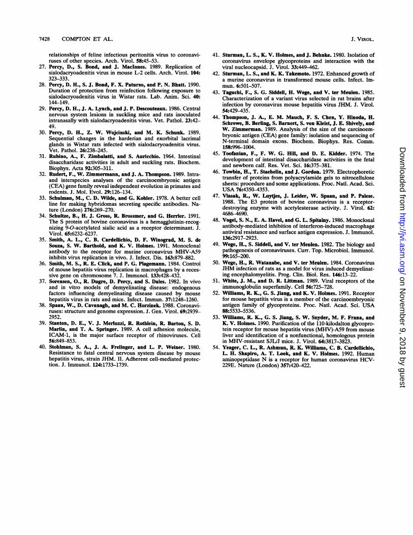
Polyclonal antibody directed against the MHV receptor(anti-MHVR) was obtained by immunization of receptor-negative SJL/J mice with deoxycholate extracts from puri-fied intestinal BBM from receptor-positive BALB/c mice.Mice which produced antireceptor antibodies, as shown inFig. 1, were given Sarcoma 180 cells by intraperitonealinoculation to induce the ascites fluid called polyclonalanti-MHVR. Control ascites fluid was obtained in a similarmanner from nonimmunized mice.Monoclonal anti-MHV receptor antibodies were gener-
ated by fusion of Sp2/0 cells with splenocytes from SJL/Jmice immunized with deoxycholate-extracted BALB/c BBMto produce receptor-specific hybridomas by establishedmethods (33). Monoclonal antibodies (MAbs) were screenedby using an enzyme-linked immunoassay on wells coatedwith MHVR eluted from a 110-kDa band of a preparativepolyacrylamide slab gel of BALB/c intestinal BBM. Inimmunoblots of BALB/c BBM, MAb CC1 (immunoglobulinGl [IgGl] isotype) bound to the 110- to 120-kDa receptorglycoprotein and to a related 58-kDa glycoprotein (52, 53).Pretreatment of L2 and 17 Cl 1 cells with MAb CC1protected the cells from MHV-A59 infection (53). Treatmentof infant mice with MAb CC1 markedly reduced the yield ofvirus from the liver, nose, and brain (35). A MAb of the IgGlisotype directed against an irrelevant antibody (choleratoxin) was used as a control for MAb CCl.
Blocking of virus attachment. L2 cells from spinner cul-tures in Eagles minimum essential medium without Ca2" orMg2+ and with 5% dialyzed fetal bovine serum were incu-bated for 2 h at 37°C in polypropylene tubes (5 x 10⁶ cells in1 ml) on a roller wheel with control medium or serialdilutions of anti-MHVR or control ascites fluid and thenwere incubated for 90 min at 37°C with 104 cpm of [3H]uri-dine-labeled, gradient-purified MHV-A59 virions (specificactivity 4.3 x 10⁴ PFU/cpm). The cells were harvested byfiltration onto 0.45-p,m-pore-size Millipore filters and washedthree times with medium. Radioactivity bound to the cellswas determined by counting dried filters in Liquifluor scin-tillation fluid in a Beckman (Palo Alto, Calif.) liquid scintil-lation counter. Values represent the averages of triplicatesamples. Percent inhibition of virus binding was calculatedas 100 x [1 - (EXPT - BKGD)/(MAX - BKGD)], whereBKGD was the counts per minute on filters with virus alone(typically 500 to 1,000 cpm), MAX was the counts perminute for cells plus virus (typically 3,500 cpm), and EXPTwas the counts per minute of samples containing virus andcells with antibody dilutions.
Immunofluorescence. Monolayer cultures of cells grownon glass coverslips were incubated for 1 h at 4°C withanti-MHVR or control antibody, washed three times inphosphate-buffered saline (PBS), then fixed in acetone or 2%paraformaldehyde in calcium- and magnesium-free PBS, and
VOL. 66, 1992
on Novem
ber 9, 2018 by guesthttp://jvi.asm
.org/D
ownloaded from
7422 COMPTON ET AL.
washed three times in PBS with 2% normal goat serum. Forsome experiments, cells were fixed for 1 h with 2% para-formaldehyde and washed three times with PBS beforeincubation with anti-MHVR. Bound antibody was detectedwith affinity-purified, fluorescein- or rhodamine-labeled rab-bit or goat anti-mouse IgG (Organon-Teknika Co., WestChester, Pa.). Cells were mounted in 40% glycerol andexamined with a Zeiss photomicroscope III.
Intestinal BBM preparation. Mouse, rat, cotton rat, andrabbit intestines were flushed with ice-cold PBS, snap frozenin liquid nitrogen, and stored at -70°C. Mucosa werescraped from cat, dog, pig, cow, chicken, and human intes-tines prior to snap freezing. Intestinal BBM were preparedfrom frozen intestines or intestinal mucosa by the method ofKessler et al. (15). Briefly, intestinal tissue was thawed,homogenized with a Tissumizer (Tekmar Co., Cincinnati,Ohio), and clarified by precipitation with 10 mM CaCl2; andBBM were prepared by differential centrifugation and storedin TE buffer (10 mM Tris HCl [pH 7.4], 1 mM EDTA).Protein concentrations of the BBM preparations were deter-mined by the Bradford method, using bovine serum albuminas a standard (8). To determine the comparability of BBMpreparations from different species, we measured the activ-ity of sucrase, an enzyme associated with BBM, by themethod of Messer and Dahlqvist (23). All BBM preparationswere comparable, releasing 3 to 20 p,mol of glucose/ml/min/mg of protein, except cow and neonatal rat BBM whichare known to lack sucrase activity (31, 45).
Chorioallantoic membrane preparation. Chorioallantoicmembranes were removed from 14-day-old chicken em-bryos, washed in PBS, swollen in reticulocyte standardbuffer (10mM Tris HCl [pH 7.4], 10 mM NaCl, 5 mM MgCl2)with 1 ,ug of phenylmethylsulfonyl fluoride per ml, andhomogenized. The homogenate was centrifuged at 1,000 x gfor 5 min to remove large debris, and the resulting superna-tant was centrifuged for 2 h at 125,000 x g. Membranes inthe pellet were resuspended in TE buffer and stored at-700C.
Solid-phase virus-binding assay. The solid-phase MHVRassay was performed as previously described (7), except thatBBM were treated with 5% j-mercaptoethanol to reduce anybound immunoglobulins before being applied to nitrocellu-lose sheets in a 96-well minifold apparatus (Schleicher &Schuell, Inc., Keene, N.H.). This step was needed becausewithout it, intestinal BBM of several nonmurine speciesbound staphylococcal protein A (SPA) in the absence ofadded virus or antibody. The nitrocellulose was blockedwith 2% bovine serum albumin, incubated with MHV-A59in minimum essential medium with 10% fetal bovine serum-10 mM HEPES (N-2-hydroxyethylpiperazine-N'-2-ethane-sulfonic acid; Sigma Chemical Co., St. Louis, Mo.), washed,incubated with goat antibody directed against the S glyco-protein of MHV-A59 (41), and washed again. Bound anti-body was detected by incubation with radioiodinated SPA(125I-SPA; 2 to 10 mCi/mg; New England Nuclear Co.,Boston, Mass.) and then by autoradiography. Controls forthe specificity of virus binding used conditioned mediumfrom uninfected cells in place of virus or normal goat serumor buffer in place of anti-S antibody.VOPBA and immunoblotting. The virus overlay protein
blot assay (VOPBA) was performed as previously described(7). Briefly, 100 ,ug of protein from BBM was separated bysodium dodecyl sulfate-polyacrylamide gel electrophoresis(SDS-PAGE) (18) and electroblotted onto nitrocellulosesheets which were then blocked with 2% bovine serumalbumin. Subsequent steps of the VOPBA were performed
abc d ef g h ij
200-
9 7- 5 *
68-
43-
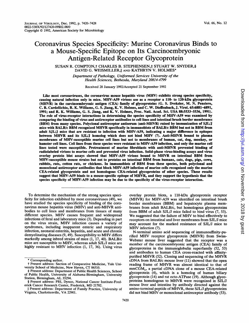
FIG. 1. Immunoreactivity with BALB/c mouse intestinal BBMof polyclonal SJL/J mouse antibody to BALB/c BBM. Strips from apreparative SDS-PAGE gel blotted to nitrocellulose were reactedwith virus or antireceptor antibodies. Lane a shows a virus overlayprotein blot of BALB/c BBM in which the 100- to 120-kDa CEA-related MHVR glycoprotein was detected by binding of MHV-A59virions followed by goat anti-MHV and 1"I-SPA. Strips in lanes bthrough i were incubated with a 1:20 dilution of mouse serum; laneb was incubated with preimmunization serum of an SJLVJ mouse,and lanes c through i were incubated with sera from different SJVJmice immunized with a deoxycholate extract of adult BALB/cintestinal BBM. Lane j is an immunoblot with a 1:50 dilution ofascites fluid raised in a mouse immunized with BALB/c BBM asdescribed in the legend to lanes c through i. This was the source ofthe polyclonal mouse anti-MHVR used in this report.
as described above for the solid-phase virus-binding assay.For immunoblots, BBM proteins were separated by SDS-PAGE, electroblotted onto nitrocellulose sheets, and incu-bated with a 1:100 dilution of polyclonal anti-MHVR orapproximately 20 ng of MAb CCl per ml, followed byaffinity-purified rabbit anti-mouse IgG and then 1`I-SPA(46).
RESULTS
Antibody directed against the MHV receptor blocks bindingof virus to mouse fibroblasts. In order to develop antibody toa putative MHV receptor from BALB/c BBM, we immu-nized SJL/J mice with extracts of these membranes. Theresulting polyclonal antibody, anti-MHVR, recognized the110- to 120-kDa CEA-related MHVR glycoprotein in immu-noprecipitates (data not shown) and immunoblots ofBALB/c BBM but not those of SJL/J BBM (Fig. 1). Immu-nofluorescence experiments showed that anti-MHVR alsoreacted with paraformaldehyde-fixed BHK-21 cells stablytransfected with MHVR1 cDNA and expressing the MHVRglycoprotein (11) but not with control BHK-21 cells (data notshown). Thus, although SJL/J and BALB/c mice are notclosely related and differ in major histocompatibility com-plex antigens, when SJL/J mice were immunized with ex-tracts of BALB/c BBM, an immunodominant antigen wasthe MHVR glycoprotein. This suggests that the epitope(s) onMHVR recognized by anti-MHVR may not be shared by thehomologous SJL/J glycoproteins.The mouse polyclonal anti-MHVR was directed at least in
part against the virus-binding determinant of MHVR, be-cause mouse L2 cells pretreated with serial dilutions up to1:200 of anti-MHVR were resistant to challenge with infec-tious MHV-A59. To determine whether this inhibition ofvirus infection by polyclonal anti-MHVR was due to block-ing of virus attachment to the receptor, we pretreated cellswith serial dilutions of polyclonal anti-MHVR, normalmouse ascites, or medium and then incubated the cells with
J. VIROL.
on Novem
ber 9, 2018 by guesthttp://jvi.asm
.org/D
ownloaded from

CORONAVIRUS SPECIES SPECIFICITY 7423
80
60 -
% Inhibitionof MHV Binding
40
20
0 1 2 3 4 5
- Log Antibody Dilution
FIG. 2. Inhibition of binding of radiolabeled MHV-A59 virionsto mouse fibroblasts by polyclonal antireceptor antibody (anti-MHVR). Spinner cultures of L2 cells were incubated with serialdilutions of mouse polyclonal antireceptor antibody (solid circles) orof ascites from nonimmunized mice (open circles) and then wereincubated with [3H]uridine-labeled, gradient-purified MHV-A59 vir-ions. Radioactivity binding to the cells was decreased in a concen-tration-dependent manner by anti-MHVR.
gradient-purified, radiolabeled MHV-A59 virions. Figure 2shows that mouse polyclonal anti-MHVR prevented bindingof MHV-A59 virions to mouse fibroblasts. Thus, anti-MHVR blocks infection of mouse fibroblasts by blocking the
epitope of the MHV receptor glycoprotein which is recog-nized by the viral S glycoprotein.The epitope(s) of MHVR recognized by polyclonal anti-
MHVR is not expressed on nonmurine cell lines. Becauseanti-MHVR recognized one or more epitopes on MHVR,including one at or near enough to the virus-binding site toblock virus attachment, we used this antibody to test celllines from different species for expression of homologs ofMHVR that might have MHV-binding activity. Indirectimmunofluorescence showed that only cell lines of murineorigin bound mouse polyclonal anti-MHVR (Fig. 3 and Table1). Like the BHK line of baby hamster kidney cells (Fig. 3B),all other nonmurine cell lines tested (Table 1) showed nospecific immunofluorescence with the mouse polyclonal anti-MHVR. These data indicate that the polyclonal anti-MHVRraised in SJL/J mice was specific for antigens on mouse cellsand did not recognize proteins homologous to MHVR on celllines from other species.
Cells of nonmurine species are resistant to infection withMHV-A59. Mouse, hamster, cat, dog, monkey, and humancell lines were inoculated with MHV-A59 at a multiplicity ofinoculation of 1 to 10 PFU per cell and incubated for 1 to 3days at 37°C. Cultures which showed cytopathic effects suchas cell fusion, rounding, and/or cell death were scored assusceptible to MHV (Table 1). All of the murine cell culturestested except SJL/J macrophages developed cytopathic ef-fects from MHV infection. Cultures which did not exhibitvirus-induced cytopathic effects were fixed and examined fordevelopment of MHV-specific antigens by indirect immuno-
FIG. 3. Expression of the 110-kDa glycoprotein receptor for MHV on the cell surface. Polyclonal mouse anti-MHVR was incubated withan MHV-susceptible murine cell line (L2; panel A), an MHV-resistant hamster cell line (BHK-21; panel B), and primary macrophage culturesfrom MHV-susceptible BALB/c mice (C) or MHV-resistant SJL/J mice (D). The cell lines (A and B) were fixed with acetone after incubationwith antibody, and the macrophages (C and D) were fixed with 2% paraformaldehyde prior to incubation with antibody. Anti-MHVR wasvisualized with rhodamine-conjugated goat anti-mouse IgG.
VOL. 66, 1992
on Novem
ber 9, 2018 by guesthttp://jvi.asm
.org/D
ownloaded from
7424 COMPTON ET AL.
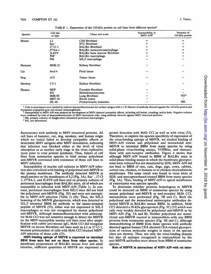
TABLE 1. Expression of the 110-kDa protein on cell lines from different speciesa
Cell line . . Susceptibility to Presence ofSpecies or type Tissue and strai MHV-A59' 110-kDa proteinMouse L2 C3H fibroblast + +
Sac- STU fibroblast + +17 Cl 1 BALB/c fibroblast + +J774A-1 BALB/c monocyte/macrophage + +JLSV9 BALB/c bone marrow fibroblast + +PMC BALB/c macrophage + +PM SJL/J macrophage - -
Hamster BHK-21 Kidney fibroblast
Cat fwcf-4 Fetal tissue
Dog A72 Tumor tissue
Monkey CV-1 Kidney fibroblast
Human HFF Foreskin fibroblastRD RhabdomyosarcomaIMR-90 Lung fibroblast - NDdHRT-18 Rectal tumorHL-60 Promyelocytic leukemia - ND
a Cells in monolayers were labeled by indirect immunofluorescence for surface antigen with a 1:25 dilution of antibody directed against the 11O-kDa protein andrhodamine-conjugated goat anti-mouse immunoglobulin.
b Susceptibility to MHV-A59 was assayed by development of MHV-induced cytopathic effects, including cell fusion, rounding, and/or lysis. Negative cultureswere confirmed by lack of immunofluorescence of MHV-inoculated cells, using antibody directed against MHV structural proteins.
c PM, primary cultures of thioglycolate-stimulated peritoneal macrophages.d ND, not determined.
fluorescence with antibody to MHV structural proteins. Allcell lines of hamster, cat, dog, monkey, and human originwhich we tested failed to develop cytopathic effects ordetectable MHV antigens after MHV inoculation, indicatingthat infection was blocked either at the level of virusadsorption or at another early stage in the virus replicativecycle prior to viral protein synthesis. Thus, failure of celllines from nonmurine species to bind mouse polyclonalanti-MHVR correlated with resistance of these cell lines toMHV infection.
Susceptibility of murine cell cultures to MHV-A59 infec-tion also correlated with binding of polyclonal anti-MHVR tothe plasma membrane. The antibody detected MHVR insmall patches on the membranes of L2 (Fig. 3A), Sac-, 17 Cl1, J774A-1, and JLSV9 cell lines and on primary cultures ofperitoneal macrophages from BALB/c mice, all of which aresusceptible to infection with MHV-A59 (Table 1). In con-trast, peritoneal macrophages from SJL/J mice did not bindthe polyclonal anti-MHVR (Fig. 3D) and were not suscepti-ble to MHV-A59 infection (Table 1) (36). Thus, the SJL/Jhomolog of the MHVR glycoprotein, which was detected inSJL/J intestinal BBM by antibody to the amino-terminalpeptide of MHVR (53), either was not expressed in SJL/Jmacrophages or was not recognized by mouse polyclonalanti-MHVR. Although immunofluorescence with antirecep-tor MAb CCl was not sensitive enough to detect the MHVRon the MHV-susceptible murine cell lines even after ampli-fication with rabbit anti-mouse IgG, MAb CCl did recognizeMHVR on mouse fibroblast cell lines such as L2 or 17 Cl 1,because pretreatment of cells with MAb CCl blocked MHV-A59 infection of these cells (53).MHV-binding epitope of MHVR is expressed on intestinal
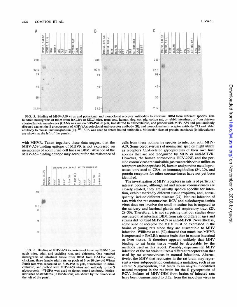
BBM from mice but not on those from other species. Inmembrane preparations of BALB/c mouse liver and smallintestine, sufficient quantities of MHVR are expressed to
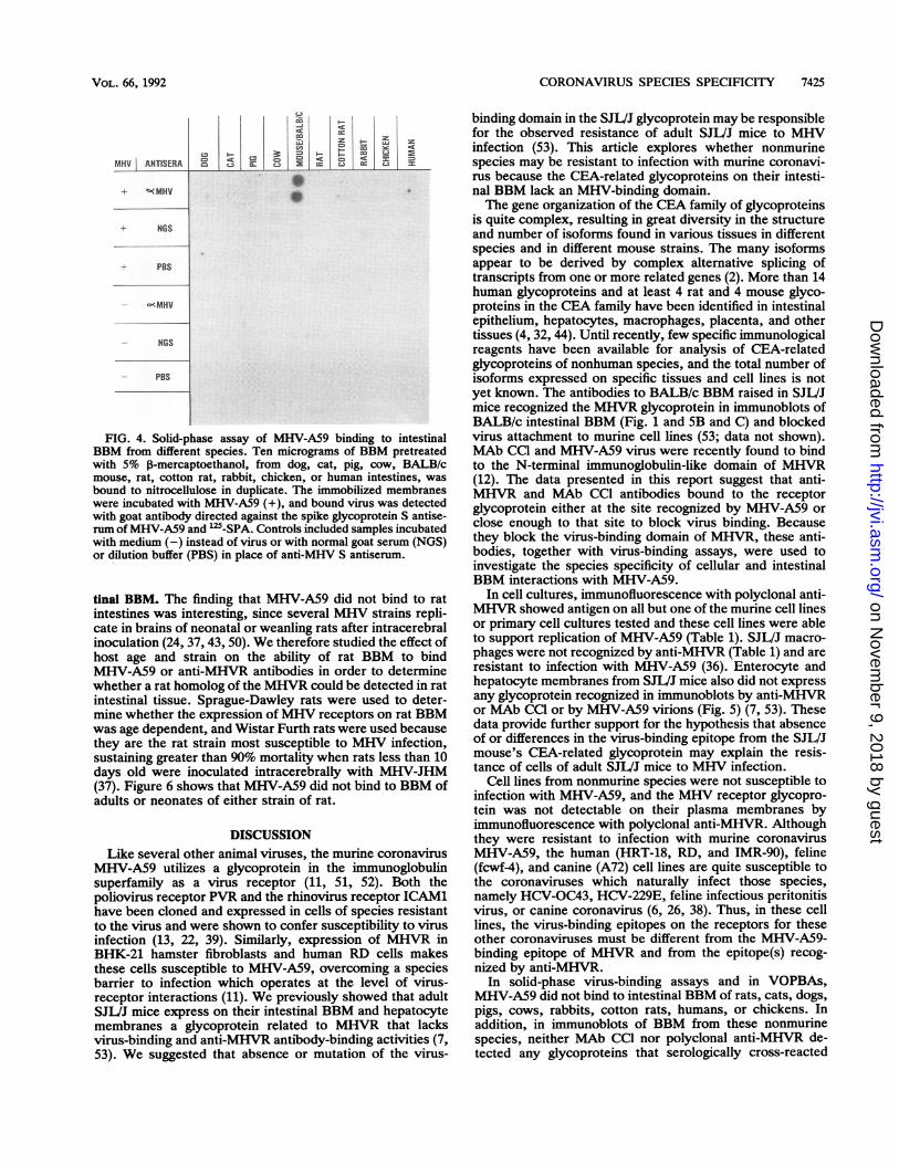
permit detection with MAb CC1 as well as with virus (52).Therefore, to explore the species specificity of expression ofthe virus-binding epitope of MHVR, we studied binding ofMHV-A59 virions and polyclonal and monoclonal anti-MHVR to intestinal BBM from many species by usingsolid-phase virus-binding assays, VOPBAs, and immuno-blots with anti-receptor antibodies. Figure 4 shows thatalthough MHV-A59 bound to BBM of BALB/c mice insolid-phase binding assays in which the membrane glycopro-teins were reduced but not denatured by SDS, MHV-A59 didnot bind to BBM of rats, cats, dogs, pigs, cows, rabbits,cotton rats, chicken, or humans or to chicken chorioallantoicmembranes. The same result was found in virus blots ofSDS- and mercaptoethanol-treated BBM from many species(Fig. 5A). Thus, binding of MHV-A59 to apical membranesof enterocytes was species specific.To determine whether proteins homologous to MHVR
could be detected on BBM of nonmurine species by usingmouse polyclonal anti-MHVR or anti-receptor MAb CCI,immunoblots were performed (Fig. SB and C). Both thepolyclonal and the monoclonal antireceptor antibodies de-tected MHVR in BALB/c mouse BBM. In addition, MAbCC1 detected a 58-kDa glycoprotein (Fig. 5C) (53) which wasonly very weakly detected by polyclonal anti-MHVR or byMHV-A59 (Fig. SA and B). Neither polyclonal nor mono-clonal anti-MHVR reacted in immunoblots with any BBMproteins from nonmurine species (Fig. 5B and C), althoughimmunoblotting of BBM from these species with antibodydirected against human CEA showed CEA-related glycopro-teins of various molecular weights in many of the species(data not shown). Thus, not only the virus-binding domainbut also all epitopes recognized in immunoblots by theanti-MHVR antibodies were absent from BBM of nonmurinespecies.
Role of MHVR in interactions of MHV-A59 with rat intes-
J. VIROL.
on Novem
ber 9, 2018 by guesthttp://jvi.asm
.org/D
ownloaded from
CORONAVIRUS SPECIES SPECIFICITY 7425
MHV ANTISERA
LU CDLu
C-t: . = I- CD Q
0m C.11 E C.3 2 C3 cc C-
+ Y(MHV
+ NGS
+- PBS
- c'MHV
- NGS
- PBS
FIG. 4. Solid-phase assay of MHV-A59 binding to intestinalBBM from different species. Ten micrograms of BBM pretreatedwith 5% P-mercaptoethanol, from dog, cat, pig, cow, BALB/cmouse, rat, cotton rat, rabbit, chicken, or human intestines, was
bound to nitrocellulose in duplicate. The immobilized membraneswere incubated with MHV-A59 (+), and bound virus was detectedwith goat antibody directed against the spike glycoprotein S antise-rum ofMHV-A59 and `2'-SPA. Controls included samples incubatedwith medium (-) instead of virus or with normal goat serum (NGS)or dilution buffer (PBS) in place of anti-MHV S antiserum.
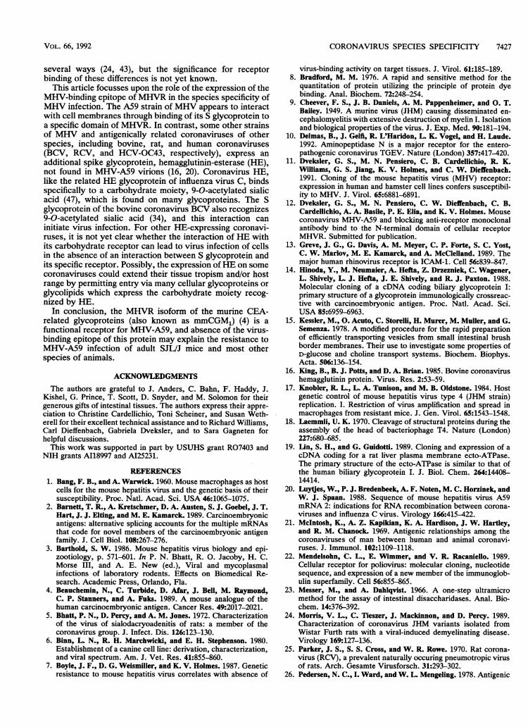
tinal BBM. The finding that MHV-A59 did not bind to ratintestines was interesting, since several MHV strains repli-cate in brains of neonatal or weanling rats after intracerebralinoculation (24, 37, 43, 50). We therefore studied the effect ofhost age and strain on the ability of rat BBM to bindMHV-A59 or anti-MHVR antibodies in order to determinewhether a rat homolog of the MHVR could be detected in ratintestinal tissue. Sprague-Dawley rats were used to deter-mine whether the expression of MHV receptors on rat BBMwas age dependent, and Wistar Furth rats were used becausethey are the rat strain most susceptible to MHV infection,sustaining greater than 90% mortality when rats less than 10days old were inoculated intracerebrally with MHV-JHM(37). Figure 6 shows that MHV-A59 did not bind to BBM ofadults or neonates of either strain of rat.
DISCUSSIONLike several other animal viruses, the murine coronavirus
MHV-A59 utilizes a glycoprotein in the immunoglobulinsuperfamily as a virus receptor (11, 51, 52). Both thepoliovirus receptor PVR and the rhinovirus receptor ICAM1have been cloned and expressed in cells of species resistantto the virus and were shown to confer susceptibility to virusinfection (13, 22, 39). Similarly, expression of MHVR inBHK-21 hamster fibroblasts and human RD cells makesthese cells susceptible to MHV-A59, overcoming a species
barrier to infection which operates at the level of virus-receptor interactions (11). We previously showed that adultSJL/J mice express on their intestinal BBM and hepatocytemembranes a glycoprotein related to MHVR that lacksvirus-binding and anti-MHVR antibody-binding activities (7,53). We suggested that absence or mutation of the virus-
binding domain in the SJL/J glycoprotein may be responsiblefor the observed resistance of adult SJLVJ mice to MHVinfection (53). This article explores whether nonmurinespecies may be resistant to infection with murine coronavi-rus because the CEA-related glycoproteins on their intesti-nal BBM lack an MHV-binding domain.The gene organization of the CEA family of glycoproteins
is quite complex, resulting in great diversity in the structureand number of isoforms found in various tissues in differentspecies and in different mouse strains. The many isoformsappear to be derived by complex alternative splicing oftranscripts from one or more related genes (2). More than 14human glycoproteins and at least 4 rat and 4 mouse glyco-proteins in the CEA family have been identified in intestinalepithelium, hepatocytes, macrophages, placenta, and othertissues (4, 32, 44). Until recently, few specific immunologicalreagents have been available for analysis of CEA-relatedglycoproteins of nonhuman species, and the total number ofisoforms expressed on specific tissues and cell lines is notyet known. The antibodies to BALB/c BBM raised in SJLV/Jmice recognized the MHVR glycoprotein in immunoblots ofBALB/c intestinal BBM (Fig. 1 and 5B and C) and blockedvirus attachment to murine cell lines (53; data not shown).MAb CCl and MHV-A59 virus were recently found to bindto the N-terminal immunoglobulin-like domain of MHVR(12). The data presented in this report suggest that anti-MHVR and MAb CC1 antibodies bound to the receptorglycoprotein either at the site recognized by MHV-A59 orclose enough to that site to block virus binding. Becausethey block the virus-binding domain of MHVR, these anti-bodies, together with virus-binding assays, were used toinvestigate the species specificity of cellular and intestinalBBM interactions with MHV-A59.
In cell cultures, immunofluorescence with polyclonal anti-MHVR showed antigen on all but one of the murine cell linesor primary cell cultures tested and these cell lines were ableto support replication of MHV-A59 (Table 1). SJL/J macro-phages were not recognized by anti-MHVR (Table 1) and areresistant to infection with MHV-A59 (36). Enterocyte andhepatocyte membranes from SJL/J mice also did not expressany glycoprotein recognized in immunoblots by anti-MHVRor MAb CCl or by MHV-A59 virions (Fig. 5) (7, 53). Thesedata provide further support for the hypothesis that absenceof or differences in the virus-binding epitope from the SJL/Jmouse's CEA-related glycoprotein may explain the resis-tance of cells of adult SJIVJ mice to MHV infection.
Cell lines from nonmurine species were not susceptible toinfection with MHV-A59, and the MHV receptor glycopro-tein was not detectable on their plasma membranes byimmunofluorescence with polyclonal anti-MHVR. Althoughthey were resistant to infection with murine coronavirusMHV-A59, the human (HRT-18, RD, and IMR-90), feline(fcwf-4), and canine (A72) cell lines are quite susceptible tothe coronaviruses which naturally infect those species,namely HCV-OC43, HCV-229E, feline infectious peritonitisvirus, or canine coronavirus (6, 26, 38). Thus, in these celllines, the virus-binding epitopes on the receptors for theseother coronaviruses must be different from the MHV-A59-binding epitope of MHVR and from the epitope(s) recog-nized by anti-MHVR.
In solid-phase virus-binding assays and in VOPBAs,MHV-A59 did not bind to intestinal BBM of rats, cats, dogs,pigs, cows, rabbits, cotton rats, humans, or chickens. Inaddition, in immunoblots of BBM from these nonmurinespecies, neither MAb CCl nor polyclonal anti-MHVR de-tected any glycoproteins that serologically cross-reacted
VOL. 66, 1992
on Novem
ber 9, 2018 by guesthttp://jvi.asm
.org/D
ownloaded from
7426 COMPTON ET AL.
B
Ki '
cnc9L~=~ La - L-)
92.
69
30
L,1 5-
im, KnCD ICD cLl<51 zlcNlCc
200 -
92.5-b9 -
46 --
30 -
21 5-
mCZ (IIm C C L. C-i
FIG. 5. Binding of MHV-A59 virus and polyclonal and monoclonal receptor antibodies to intestinal BBM from different species. Onehundred micrograms of BBM from BALB/c or SJL/J mice, from cow, human, dog, cat, pig, cotton rat, or rabbit intestines, or from chickenchorioallantoic membranes (CAM) was run on SDS-PAGE gels, transferred to nitrocellulose, and probed with MHV-A59 and goat antibodydirected against the S glycoprotein of MHV (A); polyclonal anti-receptor antibody (B); and monoclonal anti-receptor antibody CCl and rabbitantibody to mouse immunoglobulin (C). '25I-SPA was used to detect bound antibodies. Molecular sizes of protein standards (in kilodaltons)are shown at the left of the panels.
with MHVR. Taken together, these data suggest that the
MHV-A59-binding epitope of MHVR is not expressed on
membranes of nonmurine cell lines or BBM. Absence of theMHV-A59-binding epitope may account for the resistance of
200
92.bg.
46
30-
SPRAiUE DAWLEY P A'. WIL' rAR ;Jf Tri h
t_
FIG. 6. Binding of MHV-A59 to proteins of intestinal BBM fromadult mice, adult and suckling rats, and chickens. One hundredmicrograms of intestinal tissue from BBM from BALB/c mice,chickens, three female adult rats, or pools of 5- or 10-day-old WistarFurth rats was separated on SDS-PAGE gels, transferred to nitro-cellulose, and probed with MHV-A59 virus and antibody to the Sglycoprotein. '25 -SPA was used to detect bound antibody. Molec-ular sizes of standards (in kilodaltons) are shown by the numbers atthe left of the panel.
cells from these nonmurine species to infection with MHV-A59. Some coronaviruses of nonmurine species might utilizeas receptors CEA-related glycoproteins of their own hostspecies that are not recognized by MHV or anti-MHVR.However, the human coronavirus HCV-229E and the por-cine coronavirus transmissible gastroenteritis virus utilize as
receptors aminopeptidase N, human and porcine metallopro-teases unrelated to CEA, or immunoglobulins (54, 10), andprotein receptors for other coronaviruses have not yet beenidentified.The investigation of MHV receptors in rats is of particular
interest because, although rat and mouse coronaviruses are
closely related, they are usually species specific for infec-tion, exhibit markedly different tissue tropisms, and, conse-
quently, induce different diseases (27). Natural infection ofrats with the rat coronavirus RCV and sialodacryoadenitisvirus does not involve the small intestine but is targeted tothe salivary and lacrimal glands and respiratory tract (25,28-30). Therefore, it is not surprising that our studies dem-onstrated that intestinal BBM from rats of different ages andstrains did not bind MHV-A59 or anti-MHVR. Nevertheless,some kind of receptor for MHV must be expressed in thebrains of young rats since they are susceptible to MHVinfection. Williams et al. (52) showed that much less MHVRis expressed in BALB/c mouse brain than in mouse intestinalor liver tissue. It therefore appears unlikely that MHVbinding to rat brain tissue would be detectable by themethods used in this report. Possibly, experimental MHVinfection of the rat brain utilizes a different receptor than thatused by rat coronaviruses in natural infections. Alterna-tively, the MHV that replicates in the rat brain may repre-sent a virus subpopulation containing a mutation, such as an
altered S glycoprotein, that binds to an as-yet-unidentifiednatural receptor in the rat brain for the S glycoprotein ofRCV. Isolates of MHV-JHM from brains of infected ratshave been demonstrated to differ from the inoculum virus in
A
200
92.5 -
69
46
30 --
21.5
J. VIROL.
on Novem
ber 9, 2018 by guesthttp://jvi.asm
.org/D
ownloaded from
CORONAVIRUS SPECIES SPECIFICITY 7427
several ways (24, 43), but the significance for receptorbinding of these differences is not yet known.
This article focusses upon the role of the expression of theMHV-binding epitope of MHVR in the species specificity ofMHV infection. The A59 strain of MHV appears to interactwith cell membranes through binding of its S glycoprotein toa specific domain of MHVR. In contrast, some other strainsof MHV and antigenically related coronaviruses of otherspecies, including bovine, rat, and human coronaviruses(BCV, RCV, and HCV-OC43, respectively), express anadditional spike glycoprotein, hemagglutinin-esterase (HE),not found in MHV-A59 virions (16, 20). Coronavirus HE,like the related HE glycoprotein of influenza virus C, bindsspecifically to a carbohydrate moiety, 9-O-acetylated sialicacid (47), which is found on many glycoproteins. The Sglycoprotein of the bovine coronavirus BCV also recognizes9-O-acetylated sialic acid (34), and this interaction caninitiate virus infection. For other HE-expressing coronavi-ruses, it is not yet clear whether the interaction of HE withits carbohydrate receptor can lead to virus infection of cellsin the absence of an interaction between S glycoprotein andits specific receptor. Possibly, the expression ofHE on somecoronaviruses could extend their tissue tropism and/or hostrange by permitting entry via many cellular glycoproteins orglycolipids which express the carbohydrate moiety recog-nized by HE.
In conclusion, the MHVR isoform of the murine CEA-related glycoproteins (also known as mmCGM1) (4) is afunctional receptor for MHV-A59, and absence of the virus-binding epitope of this protein may explain the resistance toMHV-A59 infection of adult SJL/J mice and most otherspecies of animals.
ACKNOWLEDGMENTSThe authors are grateful to J. Anders, C. Bahn, F. Haddy, J.
Kishel, G. Prince, T. Scott, D. Snyder, and M. Solomon for theirgenerous gifts of intestinal tissues. The authors express their appre-ciation to Christine Cardellichio, Toni Scheiner, and Susan Weth-erell for their excellent technical assistance and to Richard Williams,Carl Dieffenbach, Gabriela Dveksler, and to Sara Gagneten forhelpful discussions.
This work was supported in part by USUHS grant R07403 andNIH grants A118997 and AI25231.
REFERENCES1. Bang, F. B., and A. Warwick. 1960. Mouse macrophages as host
cells for the mouse hepatitis virus and the genetic basis of theirsusceptibility. Proc. Natl. Acad. Sci. USA 46:1065-1075.
2. Barnett, T. R., A. Kretschmer, D. A. Austen, S. J. Goebel, J. T.Hart, J. J. Elting, and M. E. Kamarck. 1989. Carcinoembryonicantigens: alternative splicing accounts for the multiple mRNAsthat code for novel members of the carcinoembryonic antigenfamily. J. Cell Biol. 108:267-276.
3. Barthold, S. W. 1986. Mouse hepatitis virus biology and epi-zootiology, p. 571-601. In P. N. Bhatt, R. 0. Jacoby, H. C.Morse III, and A. E. New (ed.), Viral and mycoplasmalinfections of laboratory rodents. Effects on Biomedical Re-search. Academic Press, Orlando, Fla.
4. Beauchemin, N., C. Turbide, D. Afar, J. Bell, M. Raymond,C. P. Stanners, and A. Fuks. 1989. A mouse analogue of thehuman carcinoembryonic antigen. Cancer Res. 49:2017-2021.
5. Bhatt, P. N., D. Percy, and A. M. Jones. 1972. Characterizationof the virus of sialodacryoadenitis of rats: a member of thecoronavirus group. J. Infect. Dis. 126:123-130.
6. Binn, L. N., R. H. Marchwicki, and E. H. Stephenson. 1980.Establishment of a canine cell line: derivation, characterization,and viral spectrum. Am. J. Vet. Res. 41:855-860.
7. Boyle, J. F., D. G. Weismiller, and K. V. Holmes. 1987. Geneticresistance to mouse hepatitis virus correlates with absence of
virus-binding activity on target tissues. J. Virol. 61:185-189.8. Bradford, M. M. 1976. A rapid and sensitive method for the
quantitation of protein utilizing the principle of protein dyebinding. Anal. Biochem. 72:248-254.
9. Cheever, F. S., J. B. Daniels, A. M. Pappenheimer, and 0. T.Bailey. 1949. A murine virus (JHM) causing disseminated en-cephalomyelitis with extensive destruction of myelin I. Isolationand biological properties of the virus. J. Exp. Med. 90:181-194.
10. Delmas, B., J. Gelfi, R. L'Haridon, L. K. Vogel, and H. Laude.1992. Aminopeptidase N is a major receptor for the entero-pathogenic coronavirus TGEV. Nature (London) 357:417-420.
11. Dveksler, G. S., M. N. Pensiero, C. B. Cardellichio, R. K.Williams, G. S. Jiang, K. V. Holmes, and C. W. Dieffenbach.1991. Cloning of the mouse hepatitis virus (MHV) receptor:expression in human and hamster cell lines confers susceptibil-ity to MHV. J. Virol. 65:6881-6891.
12. Dveksler, G. S., M. N. Pensiero, C. W. Dieffenbach, C. B.Cardellichio, A. A. Basile, P. E. Elia, and K. V. Holmes. Mousecoronavirus MHV-A59 and blocking anti-receptor monoclonalantibody bind to the N-terminal domain of cellular receptorMHVR. Submitted for publication.
13. Greve, J. G., G. Davis, A. M. Meyer, C. P. Forte, S. C. Yost,C. W. Marlov, M. E. Kamarck, and A. McClelland. 1989. Themajor human rhinovirus receptor is ICAM-1. Cell 56:839-847.
14. Hinoda, Y., M. Neumaier, A. Hefta, Z. Drzezniek, C. Wagener,L. Shively, L. J. Hefta, J. E. Shively, and R. J. Paxton. 1988.Molecular cloning of a cDNA coding biliary glycoprotein I:primary structure of a glycoprotein immunologically crossreac-tive with carcinoembryonic antigen. Proc. Natl. Acad. Sci.USA 85:6959-6963.
15. Kessler, M., 0. Acuto, C. Storelli, H. Murer, M. Muller, and G.Semenza. 1978. A modified procedure for the rapid preparationof efficiently transporting vesicles from small intestinal brushborder membranes. Their use to investigate some properties ofD-glucose and choline transport systems. Biochem. Biophys.Acta. 506:136-154.
16. King, B., B. J. Potts, and D. A. Brian. 1985. Bovine coronavirushemagglutinin protein. Virus. Res. 2:53-59.
17. Knobler, R. L., L. A. Tunison, and M. B. Oldstone. 1984. Hostgenetic control of mouse hepatitis virus type 4 (JHM strain)replication. I. Restriction of virus amplification and spread inmacrophages from resistant mice. J. Gen. Virol. 65:1543-1548.
18. Laemmli, U. K. 1970. Cleavage of structural proteins during theassembly of the head of bacteriophage T4. Nature (London)227:680-685.
19. Lin, S. H., and G. Guidotti. 1989. Cloning and expression of acDNA coding for a rat liver plasma membrane ecto-ATPase.The primary structure of the ecto-ATPase is similar to that ofthe human biliary glycoprotein I. J. Biol. Chem. 264:14408-14414.
20. Luytjes, W., P. J. Bredenbeek, A. F. Noten, M. C. Horzinek, andW. J. Spaan. 1988. Sequence of mouse hepatitis virus A59mRNA 2: indications for RNA recombination between corona-viruses and influenza C virus. Virology 166:415-422.
21. McIntosh, K., A. Z. Kapikian, K. A. Hardison, J. W. Hartley,and R. M. Chanock. 1969. Antigenic relationships among thecoronaviruses of man between human and animal coronavi-ruses. J. Immunol. 102:1109-1118.
22. Mendelsohn, C. L., E. Wimmer, and V. R. Racaniello. 1989.Cellular receptor for poliovirus: molecular cloning, nucleotidesequence, and expression of a new member of the immunoglob-ulin superfamily. Cell 56:855-865.
23. Messer, M., and A. Dahlqvist. 1966. A one-step ultramicromethod for the assay of intestinal disaccharidases. Anal. Bio-chem. 14:376-392.
24. Morris, V. L., C. Tieszer, J. Mackinnon, and D. Percy. 1989.Characterization of coronavirus JHM variants isolated fromWistar Furth rats with a viral-induced demyelinating disease.Virology 169:127-136.
25. Parker, J. S., S. S. Cross, and W. R. Rowe. 1970. Rat corona-virus (RCV), a prevalent naturally occuring pneumotropic virusof rats. Arch. Gesamte Virusforsch. 31:293-302.
26. Pedersen, N. C., I. Ward, and W. L. Mengeling. 1978. Antigenic
VOL. 66, 1992
on Novem
ber 9, 2018 by guesthttp://jvi.asm
.org/D
ownloaded from
7428 COMPTON ET AL.
relationships of feline infectious peritonitis virus to coronavi-ruses of other species. Arch. Virol. 58:45-53.
27. Percy, D., S. Bond, and J. MacInnes. 1989. Replication ofsialodacryoadenitis virus in mouse L-2 cells. Arch. Virol. 104:323-333.
28. Percy, D. H., S. J. Bond, F. X. Paturzo, and P. N. Bhatt. 1990.Duration of protection from reinfection following exposure tosialodacryoadenitis virus in Wistar rats. Lab. Anim. Sci. 40:144-149.
29. Percy, D. H., J. A. Lynch, and J. P. Descouteaux. 1986. Centralnervous system lesions in suckling mice and rats inoculatedintranasally with sialodacryoadenitis virus. Vet. Pathol. 23:42-49.
30. Percy, D. H., Z. W. Wojcinski, and M. K. Schunk 1989.Sequential changes in the harderian and exorbital lacrimalglands in Wistar rats infected with sialodacryoadenitis virus.Vet. Pathol. 26:238-245.
31. Rubino, A., F. Zimbalatti, and S. Auricchio. 1964. Intestinaldisaccharidase activities in adult and suckling rats. Biochem.Biophys. Acta 92:305-311.
32. Rudert, F., W. Zimmermann, and J. A. Thompson. 1989. Intra-and interspecies analyses of the carcinoembryonic antigen(CEA) gene family reveal independent evolution in primates androdents. J. Mol. Evol. 29:126-134.
33. Schulman, M., C. D. Wilde, and G. Kohler. 1978. A better cellline for making hybridomas secreting specific antibodies. Na-ture (London) 276:269-270.
34. Schultze, B., H. J. Gross, R. Brossmer, and G. Herrler. 1991.The S protein of bovine coronavirus is a hemagglutinin-recog-nizing 9-O-acetylated sialic acid as a receptor determinant. J.Virol. 65:6232-6237.
35. Smith, A. L., C. B. Cardellichio, D. F. Winograd, M. S. deSouza, S. W. Barthold, and K. V. Holmes. 1991. Monoclonalantibody to the receptor for murine coronavirus MHV-A59inhibits virus replication in vivo. J. Infect. Dis. 163:879-882.
36. Smith, M. S., R. E. Click, and P. G. Plagemann. 1984. Controlof mouse hepatitis virus replication in macrophages by a reces-sive gene on chromosome 7. J. Immunol. 133:428-432.
37. Sorensen, O., R. Dugre, D. Percy, and S. Dales. 1982. In vivoand in vitro models of demyelinating disease: endogenousfactors influencing demyelinating disease caused by mousehepatitis virus in rats and mice. Infect. Immun. 37:1248-1260.
38. Spaan, W., D. Cavanagh, and M. C. Horzinek 1988. Coronavi-ruses: structure and genome expression. J. Gen. Virol. 69:2939-2952.
39. Stanton, D. E., V. J. Merluzzi, R. Rothlein, R. Barton, S. D.Marlin, and T. A. Springer. 1989. A cell adhesion molecule,ICAM-1, is the major surface receptor of rhinoviruses. Cell56:849-853.
40. Stohlman, S. A., J. A. Frelinger, and L. P. Weiner. 1980.Resistance to fatal central nervous system disease by mouse
hepatitis virus, strain JHM. II. Adherent cell-mediated protec-tion. J. Immunol. 124:1733-1739.
41. Sturman, L. S., K. V. Holmes, and J. Behnke. 1980. Isolation ofcoronavirus envelope glycoproteins and interaction with theviral nucleocapsid. J. Virol. 33:449-462.
42. Sturman, L. S., and K. K. Takemoto. 1972. Enhanced growth ofa murine coronavirus in transformed mouse cells. Infect. Im-mun. 6:501-507.
43. Taguchi, F., S. G. Siddell, H. Wege, and V. ter Meulen. 1985.Characterization of a variant virus selected in rat brains afterinfection by coronavirus mouse hepatitis virus JHM. J. Virol.54:429-435.
44. Thompson, J. A., E. M. Mauch, F. S. Chen, Y. Hinoda, H.Schrewe, B. Berling, S. Barnert, S. von Kleist, J. E. Shively, andW. Zimmerman. 1989. Analysis of the size of the carcinoem-bryonic antigen (CEA) gene family: isolation and sequencing ofN-terminal domain exons. Biochem. Biophys. Res. Comm.158:996-1004.
45. Toofanian, F., F. W. G. Hill, and D. E. Kidder. 1974. Thedevelopment of intestinal disaccharidase activities in the fetaland newborn calf. Res. Vet. Sci. 16:375-381.
46. Towbin, H., T. Staehelin, and J. Gordon. 1979. Electrophoretictransfer of proteins from polyacrylamide gels to nitrocellulosesheets: procedure and some applications. Proc. Natl. Acad. Sci.USA 76:4350-4353.
47. Vlasak, R., W. Luytjes, J. Leider, W. Spaan, and P. Palese.1988. The E3 protein of bovine coronavirus is a receptor-destroying enzyme with acetylesterase activity. J. Virol. 62:4686-4690.
48. Vogel, S. N., E. A. Havel, and G. L. Spitalny. 1986. Monoclonalantibody-mediated inhibition of interferon-induced macrophageantiviral resistance and surface antigen expression. J. Immunol.136:2917-2923.
49. Wege, H., S. Siddell, and V. ter Meulen. 1982. The biology andpathogenesis of coronaviruses. Curr. Top. Microbiol. Immunol."9:165-200.
50. Wege, H., R. Watanabe, and V. ter Meulen. 1984. CoronavirusJHM infection of rats as a model for virus induced demyelinat-ing encephalomyelitis. Prog. Clin. Biol. Res. 146:13-22.
51. White, J. M., and D. R. Littman. 1989. Viral receptors of theimmunoglobulin superfamily. Cell 56:725-728.
52. Williams, R. K., G. S. Jiang, and K. V. Holmes. 1991. Receptorfor mouse hepatitis virus is a member of the carcinoembryonicantigen family of glycoproteins. Proc. Natl. Acad. Sci. USA88:5533-5536.
53. Williams, R. K., G. S. Jiang, S. W. Snyder, M. F. Frana, andK. V. Holmes. 1990. Purification of the 110-kilodalton glycopro-tein receptor for mouse hepatitis virus (MHV)-A59 from mouseliver and identification of a nonfunctional, homologous proteinin MHV-resistant SJUJ mice. J. Virol. 64:3817-3823.
54. Yeager, C. L., R. Ashmun, R. K. Williams, C. B. Cardellichio,L. H. Shapiro, A. T. Look, and K. V. Holmes. 1992. Humanaminopeptidase N is a receptor for human coronavirus HCV-229E. Nature (London) 357:420-422.
J. VIROL.
on Novem
ber 9, 2018 by guesthttp://jvi.asm
.org/D
ownloaded from