
Controllable Medical Image Generation via Generative ...
5
Controllable Medical Image Generation via Generative Adversarial Networks Zhihang Ren, Stella X. Yu, David Whitney; UC Berkeley / ICSI; Berkeley, California, USA Abstract Radiologists and pathologists frequently make highly conse- quential perceptual decisions. For example, visually searching for a tumor and recognizing whether it is malignant can have a life-changing impact on a patient. Unfortunately, all human per- ceivers—even radiologists—have perceptual biases. Because hu- man perceivers (medical doctors) will, for the foreseeable future, be the final judges of whether a tumor is malignant, understand- ing and mitigating human perceptual biases is important. While there has been research on perceptual biases in medical image perception tasks, the stimuli used for these studies were highly ar- tificial and often critiqued. Realistic stimuli have not been used because it has not been possible to generate or control them for psychophysical experiments. Here, we propose to use Generative Adversarial Networks (GAN) to create vivid and realistic medical image stimuli that can be used in psychophysical and computer vision studies of medical image perception. Our model can gener- ate tumor-like stimuli with specified shapes and realistic textures in a controlled manner. Various experiments showed the authen- ticity of our GAN-generated stimuli and the controllability of our model. Introduction Because of its significant impact on health and well-being, medical image perception has been the focus of a great deal of research [1, 2, 3], from studies on its limits to studies on how to improve it. However, researchers often encounter a relative paucity of data and resources needed to pursue further investiga- tion. While there are many publicly available medical imaging datasets, these are often limited, inadequately annotated, or out- dated, e.g., The Digital Database for Screening Mammography (DDSM[19]). Moreover, the public datasets (e.g.,[5]) are not suf- ficiently large to support certain research questions. Therefore, many researchers resort to using their own data from hospitals. Although this approach can ensure sufficient data is collected, it is often extracted from small geographic areas that are not representative of the broader population. Another issue with this method is the tedious and time-consuming process it re- quires to sort, categorize, and de-identify the data, and make it public. Moreover, it requires experts to perform meticulous an- notations that are costly and time intensive [4]. Finally, and most importantly for the present purposes, these types of medical im- ages are specific to each individual patient, which allows almost no room for researchers to manipulate them in order to meet de- sired experimental configurations. To tackle this problem in psychophysical experiments, arti- ficial medical stimuli have been employed [6, 7]. On one hand, artificial stimuli can be easily generated and manipulated, such as shape morphing and background replacement. On the other hand, those stimuli are obviously fake and completely unlike those that doctors routinely examine. Consequently, expert ra- diologists rightly worry that these psychophysical experiments do not accurately represent their daily diagnostic tasks. Thus, generating authentic and easily controllable medical image stimuli is critical for medical image perception studies. Current research in computer vision provides us with a promising approach, using Generative Adversarial Networks (GAN). Gen- erative Adversarial Networks have been utilized for generating authentic materials [14, 13, 8] for years, such as faces, cars, land- scapes, and so on. Trained on real image datasets, GAN can gen- erate various authentic samples that have similar semantic con- text to that of real images. Besides this, the generation is easily conditioned [8], which means manipulating generated samples is possible through the design and the input of the GAN. Inspired by Generative Adversarial Networks, we utilized this computational model to generate medical image stimuli. We then tested our model on mammogram stimuli generation. Fur- thermore, we generated tumor-like stimuli with specified shapes and realistic textures using our GAN model, which effectively controls the similarity of the generated stimuli. Various experi- ments showed the authenticity of our GAN-generated stimuli and the controllability of our model. Generative Adversarial Network A Generative Adversarial Network (GAN) is a powerful deep learning model with two networks, i.e., a generator network and a discriminator network [9]. The two networks learn from each other in an adversarial way. In summary, the generator pro- duces authentic images from random noise vectors and tries to fool the discriminator, while the discriminator tries to distinguish the fake samples (generated from generator) from the real sam- ples. The whole process can be conceptually described as a min- max game shown in Equation 1, where G represents the genera- tor, D represents the discriminator, p data (x) indicates the real data distribution, and p z (z) indicates the noise vector distribution. min G max D E x∼p data (x) [log D(x)]+ E z∼p z (z) [log(1 - D(G(z)))] (1) Ideally, through the adversarial training, GAN can approx- imate the real data distribution (manifold) parameterized by the generator network. Similar samples between two specific samples can be picked along the path between the corresponding points on the manifold. This can be done by interpolating the corresponding latent vectors and forwarding them through the generator. Originally, the training of the Generative Adversarial Net- work (GAN) was highly unstable and many strategies have been proposed to tackle this problem [10, 11, 12, 13]. In this paper, we adopt the structure from StyleGAN [14], where the latent space
Transcript of Controllable Medical Image Generation via Generative ...

Controllable Medical Image Generation via Generative AdversarialNetworksZhihang Ren, Stella X. Yu, David Whitney; UC Berkeley / ICSI; Berkeley, California, USA
AbstractRadiologists and pathologists frequently make highly conse-
quential perceptual decisions. For example, visually searchingfor a tumor and recognizing whether it is malignant can have alife-changing impact on a patient. Unfortunately, all human per-ceivers—even radiologists—have perceptual biases. Because hu-man perceivers (medical doctors) will, for the foreseeable future,be the final judges of whether a tumor is malignant, understand-ing and mitigating human perceptual biases is important. Whilethere has been research on perceptual biases in medical imageperception tasks, the stimuli used for these studies were highly ar-tificial and often critiqued. Realistic stimuli have not been usedbecause it has not been possible to generate or control them forpsychophysical experiments. Here, we propose to use GenerativeAdversarial Networks (GAN) to create vivid and realistic medicalimage stimuli that can be used in psychophysical and computervision studies of medical image perception. Our model can gener-ate tumor-like stimuli with specified shapes and realistic texturesin a controlled manner. Various experiments showed the authen-ticity of our GAN-generated stimuli and the controllability of ourmodel.
IntroductionBecause of its significant impact on health and well-being,
medical image perception has been the focus of a great deal ofresearch [1, 2, 3], from studies on its limits to studies on howto improve it. However, researchers often encounter a relativepaucity of data and resources needed to pursue further investiga-tion. While there are many publicly available medical imagingdatasets, these are often limited, inadequately annotated, or out-dated, e.g., The Digital Database for Screening Mammography(DDSM[19]). Moreover, the public datasets (e.g.,[5]) are not suf-ficiently large to support certain research questions.
Therefore, many researchers resort to using their own datafrom hospitals. Although this approach can ensure sufficient datais collected, it is often extracted from small geographic areas thatare not representative of the broader population. Another issuewith this method is the tedious and time-consuming process it re-quires to sort, categorize, and de-identify the data, and make itpublic. Moreover, it requires experts to perform meticulous an-notations that are costly and time intensive [4]. Finally, and mostimportantly for the present purposes, these types of medical im-ages are specific to each individual patient, which allows almostno room for researchers to manipulate them in order to meet de-sired experimental configurations.
To tackle this problem in psychophysical experiments, arti-ficial medical stimuli have been employed [6, 7]. On one hand,artificial stimuli can be easily generated and manipulated, suchas shape morphing and background replacement. On the otherhand, those stimuli are obviously fake and completely unlike
those that doctors routinely examine. Consequently, expert ra-diologists rightly worry that these psychophysical experiments donot accurately represent their daily diagnostic tasks.
Thus, generating authentic and easily controllable medicalimage stimuli is critical for medical image perception studies.Current research in computer vision provides us with a promisingapproach, using Generative Adversarial Networks (GAN). Gen-erative Adversarial Networks have been utilized for generatingauthentic materials [14, 13, 8] for years, such as faces, cars, land-scapes, and so on. Trained on real image datasets, GAN can gen-erate various authentic samples that have similar semantic con-text to that of real images. Besides this, the generation is easilyconditioned [8], which means manipulating generated samples ispossible through the design and the input of the GAN.
Inspired by Generative Adversarial Networks, we utilizedthis computational model to generate medical image stimuli. Wethen tested our model on mammogram stimuli generation. Fur-thermore, we generated tumor-like stimuli with specified shapesand realistic textures using our GAN model, which effectivelycontrols the similarity of the generated stimuli. Various experi-ments showed the authenticity of our GAN-generated stimuli andthe controllability of our model.
Generative Adversarial NetworkA Generative Adversarial Network (GAN) is a powerful
deep learning model with two networks, i.e., a generator networkand a discriminator network [9]. The two networks learn fromeach other in an adversarial way. In summary, the generator pro-duces authentic images from random noise vectors and tries tofool the discriminator, while the discriminator tries to distinguishthe fake samples (generated from generator) from the real sam-ples. The whole process can be conceptually described as a min-max game shown in Equation 1, where G represents the genera-tor, D represents the discriminator, pdata(x) indicates the real datadistribution, and pz(z) indicates the noise vector distribution.
minG
maxD
Ex∼pdata(x)[logD(x)]+Ez∼pz(z)[log(1−D(G(z)))] (1)
Ideally, through the adversarial training, GAN can approx-imate the real data distribution (manifold) parameterized by thegenerator network. Similar samples between two specific samplescan be picked along the path between the corresponding points onthe manifold. This can be done by interpolating the correspondinglatent vectors and forwarding them through the generator.
Originally, the training of the Generative Adversarial Net-work (GAN) was highly unstable and many strategies have beenproposed to tackle this problem [10, 11, 12, 13]. In this paper, weadopt the structure from StyleGAN [14], where the latent space

(a) Real Mammograms (b) Previous Stimuli [6] (C) GAN Generated Mammograms
Figure 1. Comparison of (a) real mammograms, (b)stimuli used in previous studies [6], and (c)our GAN generated mammograms. A particular example
from previous study consists of naive morphed shapes (tumors) and healthy mammogram backgrounds, which are obviously fake and do not represent realistic
stimuli for radiologists. It is clear that our mammogram generation can mimic the texture for both tumor and non-tumor regions and they have reasonable shapes
compared to real mammograms.
Z is first mapped into the W space through a non-linear map-ping network (an 8-layer MLP) and then merged into the synthe-sis network via adaptive instance normalization (AdaIN) at eachconvolutional layer [15, 16, 17]. Gaussian noise is added aftereach convolutional layer, before the AdaIN layer.
StyleGAN has the ability to generate high-resolution real-istic images of faces, cats, bedrooms, and cars. It can controlthe image details from coarse to fine by changing the AdaIN pa-rameters and the input noises at different network levels. UsingStyleGAN, we can easily generate various authentic medical im-ages by changing the input noise vectors. However, the genera-tion is unconditional; this means, in order to get the desire input(e.g., the mammogram having a specific shape or texture.), weneed to pick samples from a large number of generated imagesbecause we have no control over the attributes of the images (e.g.,the shape and the texture of the mammogram). It is tedious andtime-consuming. Moreover, we do not have the latent codes forreal data if we want to generate similar medical images betweentwo real ones. An intuitive idea is that we can encode the latentvector z from the desired medical image. Then the similar med-ical images can be generated by interpolating the correspondingencoded vectors.
MethodTo train then utilize the encoder, first, the discriminator and
generator of StyleGAN [14] are pretrained. Then the generatorof styleGAN is fixed. While training the encoder network, tradi-tional methods [20, 21] regularize the encoder on the latent space,encouraging the encoder to encode the same latent codes for thecorresponding generated images regardless of the reconstructedimages. This method can deteriorate the reconstruction quality.Instead, we adopt the idea from In-domain GAN inversion [18],where the regularization of the encoder is on the image space.In detail, the encoded vector is fed into the generator again andthe L2 reconstruction loss regularizes the encoder on the imagespace. The training is conducted on the real data and the adversar-ial loss helps the reconstructed image to be more realistic. More-
Figure 2. Overview of proposed method. The generator (G) and discrim-
inator (D) are from StyleGAN[14]. The training has two phases. In the first
phase, the generator and discriminator will be trained first without the en-
coder (E) via adversarial loss Ladversarial . In the second phase, the generator
(G) will be fixed. The encoder (E) and discriminator (D) will be trained adver-
sarially via the reconstruction loss Lreconstruction, the perceptual loss Lperceptual ,
and the adversarial loss Ladversarial . The dashed lines indicate how to compute
the corresponding loss metrics.
over, perceptual loss [22] is utilized. The whole process can besummarized as follows
minE||x−G(E(x))||2 +λ1||F(x)−F(G(E(x)))||2
−λ2Ex∼pdata(x)[logD(G(E(x)))](2)
minD
Ex∼pdata(x)[logD(G(E(x)))]−Ex∼pdata(x)[logD(x)]
+γ
2Ex∼pdata(x)[||∇xD(x)||22]
(3)
where pdata(x) indicates the real data distribution, x is thereal image, E represents the encoder, F indicates the VGG featureextraction [23], and λ1, λ2 and γ are weights for the perceptualloss, the adversarial loss, and the gradient penalty. An overviewof the training process can be find in Figure 2.
Since inverse mapping will never be perfect, additional op-timization is required for a better reconstruction for each image.Starting with the output code from the encoder, the optimizationupdates the encoded vectors based on the reconstruction loss and

the perceptual loss but is still regularized by the encoder. Theoptimization process can be described as below.
zinv = minz||x−G(E(x))||2 +λ3||F(x)−F(G(z))||2
+λ4||z−E(G(z))||2(4)
where zinv is the optimized inverse code, λ3 and λ4 areweights for the perceptual loss, and the code reconstruction loss(i.e., the encoder regularization).
We test our proposed method on the mammogram genera-tion task. Similar experiments can be conducted with differentmedical imaging modalities.
Perceptual lossPerceptual loss has been utilized as a similarity metric in
many computer vision tasks, such as style transfer [24, 25], andimage super-resolution [22], both of which are ill-posed prob-lems. For style transfer, there is no ground truth to act as a ref-erence. For image super-resolution, many high-resolution imagescan be sampled to generate the same low-resolution image. In or-der to achieve the tasks, semantic information of the input imagesshould be maintained. Thus, per-pixel loss is no longer suitablesince it cannot capture the semantic difference between the outputand ground truth. For example, in style transfer, there are usuallydrastic changes in color and texture compared to the input images.
Perceptual loss is computed as the difference between high-level features from a pretrained loss network which is usually afeature extractor of an image classification network. Compared tothe per-pixel loss, which depends only on low-level pixel infor-mation, perceptual loss is more robust in image similarity mea-surement during training.
Implementation detailsWe conduct our experiment on Digital Database for Screen-
ing Mammography (DDSM) [19] dataset which consists of 2,620cases of normal, benign, and malignant cases. During training,we only use the mammograms which have tumors inside, i.e., thebenign and malignant cases. The GAN part is pretrained basedon StyleGAN. The encoder consists of one initial convolution, 8residual blocks, and one dense layer. And the batch normaliza-tion is utilized for all modules in the encoder. While training theencoder, the generator is fixed. Only the encoder and discrimina-tor are updated according to the loss function shown in Equation2 and Equation 3 respectively. For the perceptual loss, we useconv4 3 feature layer in VGG [23] as illustrated in [18]. Hyper-parameters are set as λ1 = 5e−5, λ2 = 0.1, λ3 = 5e−5, λ4 = 2, andγ = 10. And we use the Adam optimizer [27] with β1 = 0.9 andβ2 = 0.99. The learning rate is set to 0.0001
Mammogram generationThe pretrained StyleGAN[14] approximates the real mam-
mogram distribution (manifold) which is parameterized by thegenerator. Then, the authentic mammograms can be sampledfrom the learned manifold. To do so, we sample latent codes froma normal distribution and use the generator to map the latent codesonto the learned manifold.
Mammogram interpolationMammogram interpolation is utilized to generate similar
stimuli between two desired mammograms. Since the GAN gen-
erator already approximates the real data manifold, similar mam-mograms can be found between any two mammograms on thepath that links them on the manifold. Given two latent codesfrom either unconditional generation or encoded from real mam-mograms, we interpolate the latent codes and the generator canhelp to find one of the linking paths by mapping the interpolatedlatent codes onto the manifold.
Controllable mammogram generationControllable mammogram generation is utilized to generate
similar desired mammograms (i.e. the end points for interpola-tion). With the generator and the encoder network, we can achievethe controllable mammogram generation, where we can combinethe desired tumor texture with the given shape template.
First, we crop the tumor texture region and paste it onto theshape template. Then, we use the encoder to obtain a latent codefor this combined raw image. Because the regularization of theencoder is on the image space, the latent code can already carrycertain semantic information from both the shape template mam-mogram and the tumor texture mammogram. Finally, we applythe masked optimization, only using the tumor texture region tocompute the reconstruction loss.
Human evaluationTo verify the authenticity of our GAN generated mammo-
grams, we designed a judgement test where participants were ran-domly presented 100 mammograms with the same amount of realand generated (fake) samples. In this task, they were asked toclassify each mammogram as real or fake as well as rate theirlevel of confidence with their selection. In total, 6 participantswere involved.
To make sure participants were paying attention and notguessing randomly, we asked a subset of participants to do thejudgment test a second time. Participants were not told about thesecond judgment task prior to the first judgment, eliminating anychance that they purposely remember their first responses. At last,we compute the test-retest similarity in term of the Sokal-Michenemetric[26].
ResultsIn this section, we will show the mammogram stimuli gener-
ation quality, the corresponding human evaluation result, the in-terpolation result, and the results when generating mammogramsgiven specific shapes and textures.
Mammogram generationExamples of the GAN generated samples are shown in Fig-
ure 1 (c). A particular example from previous study [6] consistsof naive morphed shapes (tumors) and healthy mammogram back-grounds, which are obviously fake and do not represent realisticstimuli for radiologists. It is clear that our mammogram genera-tion can mimic the texture for both tumor and non-tumor regionsand has reasonable shapes compared to real mammograms.
Mammogram interpolationThe interpolation results are shown in Figure 3. Through the
interpolation, the mammograms change gradually from one to theother and they are similar to the neighboring images. Moreover,through the interpolation, we can generate a similar stimuli loopwhere the stimuli gradually changing from image A to image B,then to image C, and finally back to image A, as shown in Figure
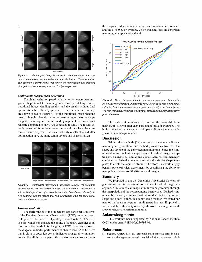
3.
Imag
e A
Imag
e B
Interpolations
Imag
e B
Imag
e C
Imag
e C
Imag
e A
Figure 3. Mammogram interpolation result. Here we evenly pick three
mammograms along the interpolation just for illustration. We show that we
can generate a similar stimuli loop where the mammogram can gradually
change into other mammograms, and finally change back.
Controllable mammogram generationThe final results compared with the tumor texture mammo-
gram, shape template mammograms, directly stitching results,traditional image blending results, and the results without finaloptimization (i.e., directly generated from the encoder output),are shown shown in Figure 4. For the traditional image blendingresults, though it blends the tumor texture region into the shapetemplate mammogram, the surrounding region of the tumor is notrealistic compared to our GAN generated results. The results di-rectly generated from the encoder outputs do not have the sametumor texture as given. It is clear that only results obtained afteroptimization have the same tumor texture and shape as given.
Tumor Texture
Shape Template Directly Stitching Image Blending W/O Optimization W/ Optimization
Figure 4. Controllable mammogram generation results. We compared
our final results with the traditional image blending method and the results
without final optimization (i.e., directly generated from the encoder output).
It is clear that only the results after final optimization have the same tumor
texture and shapes as given.
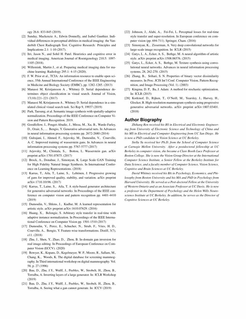
Human evaluationThe performance of the judgement test participants in terms
of the Receiver Operating Characteristic (ROC) curve is shownin Figure 5. The Receiver Operating Characteristic (ROC) curveis a plot which can indicate the ability of a binary classifier as itsdiscrimination threshold is changing. A ROC curve that is close tothe diagonal indicates performance at chance level. A ROC curvethat is close to upper left corner indicates stronger discriminationpower. For all the participants, their performance curves are near
the diagonal, which is near chance discrimination performance,and the d’ is 0.02 on average, which indicates that the generatedmammograms appeared authentic.
0 0.2 0.4 0.6 0.8 1False positive rate
0
0.2
0.4
0.6
0.8
1
True
pos
itive
rate
ZY 0.64LY 0.70WJ 0.59TR 0.67YM 0.66TCRandom 0
Test-Retest Similarity
ROC Curves for the Judgement Test
Figure 5. Human judgement test for our mammogram generation quality.
All the Receiver Operating Characteristic (ROC) curves lie near the diagonal,
indicating that our generated mammogram successfully fooled participants.
The high test-retest similarities indicate that participants did not just randomly
guess the result.
The test-retest similarity in term of the Sokal-Michenemetric[26] is shown after each participant initial in Figure 5. Thehigh similarities indicate that participants did not just randomlyguess the mammogram label.
DiscussionWhile other methods [28] can only achieve unconditional
mammogram generation, our method provides control over theshape and texture of the generated mammograms. Since the stim-uli used in psychophysical experiments of medical image percep-tion often need to be similar and controllable, we can manuallycombine the desired tumor texture with the similar shape tem-plates to create the required stimuli. Therefore, this work largelybenefits psychophysical experiments by establishing the ability tomanipulate and control life-like medical images.
SummaryWe proposed to use the Generative Adversarial Network to
generate medical image stimuli for studies of medical image per-ception. Similar medical image stimuli can be generated throughthe interpolation of the corresponding latent codes. Desired stim-uli can be manually combined with desired attributes, e.g. objectshape and tumor texture, in a controllable manner. We tested ourmethod on the mammogram stimuli generation task. Empirically,we proved the authenticity of our synthesized mammograms witha psychophysical discrimination task.
AcknowledgmentsThis work has been supported by National Cancer Institute
(NCI) under grant # 1R01CA236793-01.
References[1] Degnan, Andrew J., et al. Perceptual and interpretive error in diag-
nostic radiology—causes and potential solutions. Academic radiol-
ogy 26.6: 833-845 (2019).[2] Sunday, Mackenzie A., Edwin Donnelly, and Isabel Gauthier. Indi-
vidual differences in perceptual abilities in medical imaging: the Van-derbilt Chest Radiograph Test. Cognitive Research: Principles andImplications 2.1: 1-10 (2017).
[3] Itri, Jason N., and Sohil H. Patel. Heuristics and cognitive error inmedical imaging. American Journal of Roentgenology 210.5: 1097-1105 (2018).
[4] Willemink, Martin J., et al. Preparing medical imaging data for ma-chine learning. Radiology 295.1: 4-15 (2020).
[5] F. W. Prior et al., TCIA: An information resource to enable open sci-ence, 35th Annual International Conference of the IEEE Engineeringin Medicine and Biology Society (EMBC), pp. 1282-1285. (2013)
[6] Manassi M, Kristjansson A , Whitney D. Serial dependence de-termines object classification in visual search. Journal of Vision,17(10):221–221 (2017)
[7] Manassi M, Kristjansson A , Whitney D. Serial dependence in a sim-ulated clinical visual search task. Sci Rep 9, 19937 (2019)
[8] Park, Taesung, et al. Semantic image synthesis with spatially-adaptivenormalization. Proceedings of the IEEE Conference on Computer Vi-sion and Pattern Recognition. 2019.
[9] Goodfellow, I., Pouget-Abadie, J., Mirza, M., Xu, B., Warde-Farley,D., Ozair, S., ... Bengio, Y. Generative adversarial nets. In Advancesin neural information processing systems pp. 2672-2680 (2014)
[10] Gulrajani, I., Ahmed, F., Arjovsky, M., Dumoulin, V., Courville,A. C. Improved training of wasserstein gans. In Advances in neuralinformation processing systems pp. 5767-5777 (2017)
[11] Arjovsky, M., Chintala, S., Bottou, L. Wasserstein gan. arXivpreprint arXiv:1701.07875. (2017)
[12] Brock, A., Donahue, J., Simonyan, K. Large Scale GAN Trainingfor High Fidelity Natural Image Synthesis. In International Confer-ence on Learning Representations. (2018)
[13] Karras, T., Aila, T., Laine, S., Lehtinen, J. Progressive growingof gans for improved quality, stability, and variation. arXiv preprintarXiv:1710.10196. (2017)
[14] Karras, T., Laine, S., Aila, T. A style-based generator architecturefor generative adversarial networks. In Proceedings of the IEEE con-ference on computer vision and pattern recognition pp. 4401-4410(2019)
[15] Dumoulin, V., Shlens, J., Kudlur, M. A learned representation forartistic style. arXiv preprint arXiv:1610.07629. (2016)
[16] Huang, X., Belongie, S. Arbitrary style transfer in real-time withadaptive instance normalization. In Proceedings of the IEEE Interna-tional Conference on Computer Vision pp. 1501-1510 (2017)
[17] Dumoulin, V., Perez, E., Schucher, N., Strub, F., Vries, H. D.,Courville, A., Bengio, Y. Feature-wise transformations. Distill, 3(7),e11. (2018)
[18] Zhu, J., Shen, Y., Zhao, D., Zhou, B. In-domain gan inversion forreal image editing. In Proceedings of European Conference on Com-puter Vision (ECCV). (2020)
[19] Bowyer, K., Kopans, D., Kegelmeyer, W. P., Moore, R., Sallam, M.,Chang, K., Woods, K. The digital database for screening mammog-raphy. In Third international workshop on digital mammography. Vol.58, p. 27 (1996)
[20] Bau, D., Zhu, J.Y., Wulff, J., Peebles, W., Strobelt, H., Zhou, B.,Torralba, A. Inverting layers of a large generator. In: ICLR Workshop(2019)
[21] Bau, D., Zhu, J.Y., Wulff, J., Peebles, W., Strobelt, H., Zhou, B.,Torralba, A. Seeing what a gan cannot generate. In: ICCV (2019)
[22] Johnson, J., Alahi, A., Fei-Fei, L. Perceptual losses for real-timestyle transfer and super-resolution. In European conference on com-puter vision (pp. 694-711). Springer, Cham. (2016)
[23] Simonyan, K., Zisserman, A. Very deep convolutional networks forlarge-scale image recognition. In: ICLR (2015)
[24] Gatys, L. A., Ecker, A. S., Bethge, M. A neural algorithm of artisticstyle. arXiv preprint arXiv:1508.06576. (2015)
[25] Gatys, L., Ecker, A. S., Bethge, M. Texture synthesis using convo-lutional neural networks. Advances in neural information processingsystems, 28, 262-270. (2015)
[26] Zhang, B., Srihari, S. N. Properties of binary vector dissimilaritymeasures. In Proc. JCIS Int’l Conf. Computer Vision, Pattern Recog-nition, and Image Processing (Vol. 1). (2003)
[27] Kingma, D. P., Ba, J. Adam: A method for stochastic optimization.In: ICLR (2015)
[28] Korkinof, D., Rijken, T., O’Neill, M., Yearsley, J., Harvey, H.,Glocker, B. High-resolution mammogram synthesis using progressivegenerative adversarial networks. arXiv preprint arXiv:1807.03401.(2018)
Author BiographyZhihang Ren received his BS in Electrical and Electronic Engineer-
ing from University of Electronic Science and Technology of China andhis MS in Electrical and Computer Engineering from UC San Diego. Heis now a PhD candidate in Vision Science at UC Berkeley.
Stella Yu received her Ph.D. from the School of Computer Scienceat Carnegie Mellon University. After a postdoctoral fellowship at UCBerkeley in computer vision, she became a Clare Booth Luce Professor atBoston College. She is now the Vision Group Director at the InternationalComputer Science Institute, a Senior Fellow at the Berkeley Institute forData Science, and a faculty member of Computer Science, Vision Science,Cognitive and Brain Sciences at UC Berkeley.
David Whitney received his BA in Psychology, Economics, and Phi-losophy from Boston University and his MA and PhD in Psychology fromHarvard University. He served as a Post-doctoral Fellow at the Universityof Western Ontario and as an Associate Professor at UC Davis. He is nowa professor in the Department of Psychology and the Helen Wills Neuro-science Institute at UC Berkeley. In addition, he serves as the Director ofCognitive Sciences at UC Berkeley.


![Invisible steganography via generative adversarial networks · 2019-04-29 · Steganography Since its introduction, generative adversarial networks [10] have received more and more](https://static.fdocuments.in/doc/165x107/5fb139f476033e4a47717d30/invisible-steganography-via-generative-adversarial-networks-2019-04-29-steganography.jpg)