Control # 848 Title: “Polkadots and Moonbeams” Neurocutaneous Syndromes Made Easy eEdE# eEdE-197.
Upload
bethanie-maloneCategory
view
222download
1
Control #:1805Title: Imaging of Medial Canthus of the Orbit: An Unexplored TerritoryeEdE#: eEdE-110

Disclosure
The authors have nothing to disclose
Imaging of Medial Canthus of the Orbit: An Unexplored Territory
J Nair, C Torres, J Chankowsky, R del carpio

Learning Objectives1) To revisit detailed anatomy of the medial canthus of the orbit.
2) To list the common and uncommon pathologies.
3) Describe the CT and MRI findings of each of these pathologies.
4) To discuss the relevance of imaging as regards patient management.

Normal Anatomy
Medial Canthal Ligament
Medial Orbital Septum
Lacrimal Sac Medial Rectus Muscle
Anterior Lacrimal crest
Posterior Lacrimal crest
S
M
S
M

Lacrimal drainage apparatus
Orbit
Paranasal Sinuses and Nasal Cavity
Miscellaneous
Local Disease
Systemic
Medial Canthus lesions
Our Approach
Lymphoma
Metastasis
Langerhans Cell Histiocytosis
Sarcoidosis
Others
Lacrimal Drainage Apparatus

Anatomy of Lacrimal Drainage System
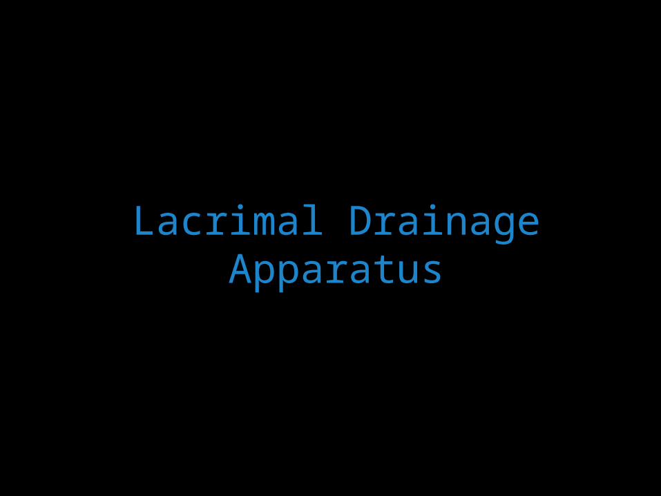
Formation of Congenital Mucocele of Nasolacrimal Duct/ Dacryocystocele
Obstruction at the valve of Hasner by imperforate Hasner membraneCo-existing obstruction at entrance to lacrimal sac Formation of closed cystic space with amniotic fluid- Amniotocele
Filling of cyst with mucous and epithelial debris from nasolacrimal duct – Mucocele

Post-Contrast CT scan in 18-day-old infant : Rounded, well-defined, rim-enhancing lesion abutting the medial preseptal right orbit extending down the lacrimal canal into the inferior meatus, causing obstruction of the nasal cavity.
Dacryocystocele
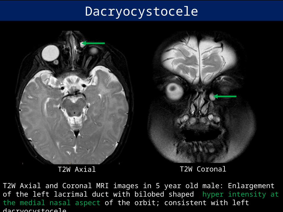
T2W Axial and Coronal MRI images in 5 year old male: Enlargement of the left lacrimal duct with bilobed shaped hyper intensity at the medial nasal aspect of the orbit; consistent with left dacryocystocele.
T2W Axial T2W Coronal
Dacryocystocele
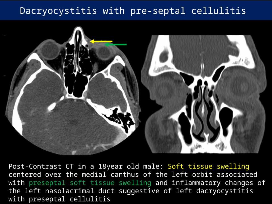
Post-Contrast CT in a 18year old male: Soft tissue swelling centered over the medial canthus of the left orbit associated with preseptal soft tissue swelling and inflammatory changes of the left nasolacrimal duct suggestive of left dacryocystitis with preseptal cellulitis
.
Dacryocystitis with pre-septal cellulitis

Post- contrast CT scan: Axial and Coronal images of the orbit in 47 year old female with left preseptal and post septal extra-conal cellulitis and dacryocystitis.
Dacryocystitis with pre and post-septal cellulitis
Teaching points:
1) Dacryocysitis is diagnosed clinically unless associated with periorbital cellulitus.
2) Imaging allows to distinguish between post-septal orbital inflammation (treated surgically) from dacryocystitis ( treated non-surgically)

Post- contrast CT scan of the orbit in 45 year old male : Left dacryocystitis and abscess formation
Dacryocystitis with abscess

Post-contrast CT in 75 year old male: Heterogeneously enhancing soft tissue along the medial wall of the left orbit with destruction of the lamina papyracea, widening of nasolacrimal duct and infiltration of the left globe along the insertion of superior oblique and medial recti muscles.
Lacrimal sac squamous cell ca
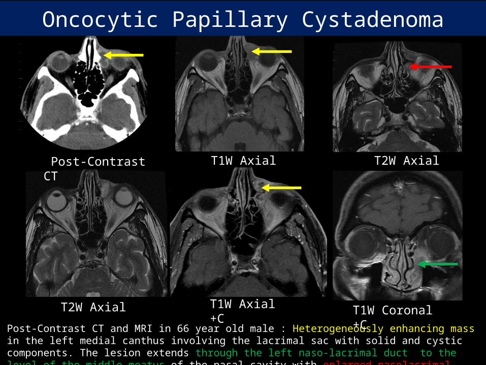
Post-Contrast CT and MRI in 66 year old male : Heterogeneously enhancing mass in the left medial canthus involving the lacrimal sac with solid and cystic components. The lesion extends through the left naso-lacrimal duct to the level of the middle meatus of the nasal cavity with enlarged nasolacrimal duct.
T2W Axial T1W Axial Post-Contrast CT
T2W Axial T1W Axial +C
T1W Coronal +C
Oncocytic Papillary Cystadenoma

CT and MRI of orbit in 52 year old female: Heterogeneously enhancing mass along the length of the left nasolacrimal duct with enlarged nasolacrimal duct.
Post-Contrast Coronal CT Post-Contrast Axial CT T2W Axial
T1W Axial T2W Coronal T1W Axial +C
Lacrimal duct invasive squamous cell carcinoma

Orbit

Post- contrast CT scan of the orbit in 28 year old female: Oval-shaped fat density lesion in the medial canthus of the left orbit with no enhancement or bony erosion consistent with dermoid tumor
Dermoid
Teaching Points:
1) Dermoids are the most common congenital orbital masses.2) Presence of fat, rim calcification , scalloping of bone are the common
imaging findings.

T1W Axial T2W Axial
T1W Fat sat Axial DWI
T2W Fat sat Coronal CT and MRI of the orbit in 14 year old male: Well circumscribed lesion in the region of the right medial canthus with no underlying bony changes. It shows high signal on T1 and T2 images and suppressed on fat sat images with no restricted diffusion or post-contrast enhancement
Dermoid

Sclerosing orbital pseudo tumor
T1W Axial T2W Axial T2W Coronal
T1W Axial +C
T1W Coronal + C
MRI of the orbit in 37 year old female : Homogenously enhancing soft tissue mass lesion in the medial canthus of the right orbit with expansion of the nasolacrimal duct, extending into the nasal cavity with obliterating of the right osteomeatal complex. Extension also noted into the intra conal compartment of the orbit with thickening of the medial and inferior rectus muscle.
Teaching Points:
1) Orbital Pseudo tumor is an autoimmune disorder usually restricted to one or more extraocular muscles.
2) Restricted eye movements due to pain, diplopia and proptosis are the common clinical manifestation.
3) Pseudotumors are iso to hypointense on T2 weighted images with loss of fat between involved muscle and periosteum of involved orbital wall.
4) Differential Diagnosis: Grave’s disease, Lymphoma, Rhabdomyosarcoma and Metastasis

Paranasal Sinuses and Nasal Cavity

Ethmoid mucocele
Contrast enhanced CT scan of the paranasal sinuses: Mucocele of the right anterior ethmoid air cells, protruding through the right lamina papyracea into the medial canthus in a 48 year old male with known sinonasal polyposis

Post-Contrast Axial CT T1W Axial
T2W Axial Post-Contrast Coronal CT
T1W Coronal+C
Invasive Aspergillus of right ethmoid sinus
CT and MRI of the orbit in 43 yearold male: Left sided naso-ethmoidal tumoral mass with irregular contours and low signal on the T1 and T2W images with post contrast enhancement extending in to the region of the right medial canthus with signs of invasion of nasolacrimal duct.

T1W Coronal
T1W Axial T2W Axial
T1W Axial +C
Malignant transformation of inverted papilloma
MRI of the orbits with contrast in 67 year old female: Heterogeneously enhancing soft tissue mass lesion representing malignant transformation of a long standing inverted papilloma, centered in the right osteomeatal complex, invading the right orbit with infiltration of the globe and the soft tissues extending to the medial canthus

T1W Axial T1W Axial +C
DWI
T1W Axial +C
T2W Axial
Nasal lymphoma to medial canthus
MRI of the orbit in 56 year old male : Soft tissue mass lesion appearing predominantly isointense on T1, hypo intense on T2 weighted images with significant restricted diffusion and homogeneous post-contrast enhancement centered in the nasal cavity extending to the medial canthus of right orbit with widened nasolacrimal duct
T1W Axial T1W Axial T2W Axial
T1W Axial +C
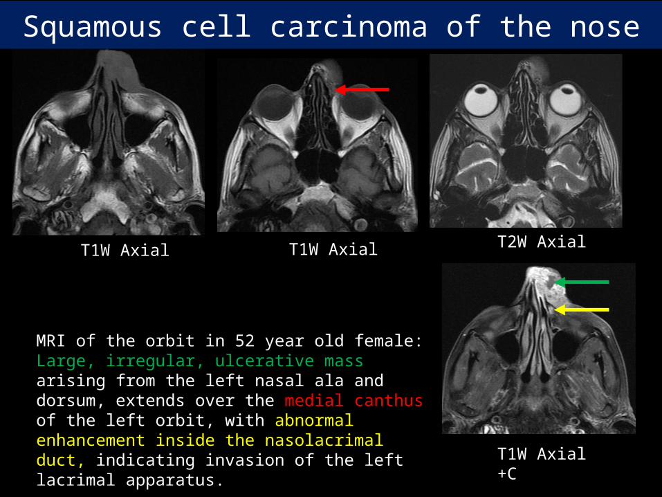
Squamous cell carcinoma of the nose
MRI of the orbit in 52 year old female: Large, irregular, ulcerative mass arising from the left nasal ala and dorsum, extends over the medial canthus of the left orbit, with abnormal enhancement inside the nasolacrimal duct, indicating invasion of the left lacrimal apparatus.

T2W Axial
T2W Axial
T1W Axial
Wegner’s Granulomatosis
MRI of the orbit in 26 year old female: Soft tissue thickening at the left medial canthus with extensive changes in the nasal cavity and paranasal sinuses resulting from combination of post surgical changes and inflammatory disease

Miscellaneous

Basal Cell Carcinoma
T1W Axial
T1W Axial +C
Post-Contrast CT
CT and MRI of the orbit in 58 year old male : Well-defined homogenously enhancing soft tissue mass limited to the medial canthus of the left orbit.
T1W Axial T1W Axial
T2W Axial T2W Axial T1W Axial +C
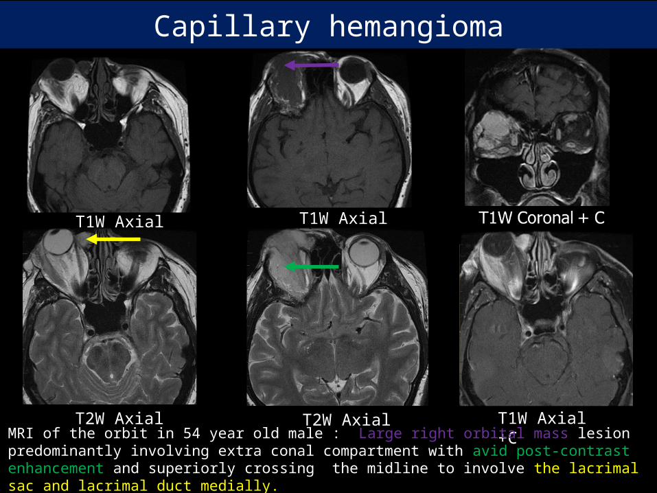
Capillary hemangioma
MRI of the orbit in 54 year old male : Large right orbital mass lesion predominantly involving extra conal compartment with avid post-contrast enhancement and superiorly crossing the midline to involve the lacrimal sac and lacrimal duct medially.

Systemic Disease

Post-Contrast Axial CT T2W Axial T2W Coronal
T1W Axial +C
DW1
CT and MRI of the orbit in 68 year old male : Well-defined homogenously enhancing soft tissue mass with low signal on T2W image and restricted diffusion on DWI images in the medial canthus of the left orbit.
Lymphoma

T2W Axial
T2W Axial
T1W Axial +C
T1W Axial +C
T2W Coronal
T1W Axial
T1W Axial
MRI of the orbit in 65 year old female : Hypo intense lobulated enhancing soft tissue in the medial canthus and paranasal sinuses with diffusion restriction. Diffuse leptomeningeal enhancement also noted.
Leukemic deposits

Ultrasound with Doppler of the right orbit in 12 year old child: Heterogeneous lesion in the medial canthus at the inferomedial aspect of the orbit. Color Doppler evaluation shows flow within.the lesion
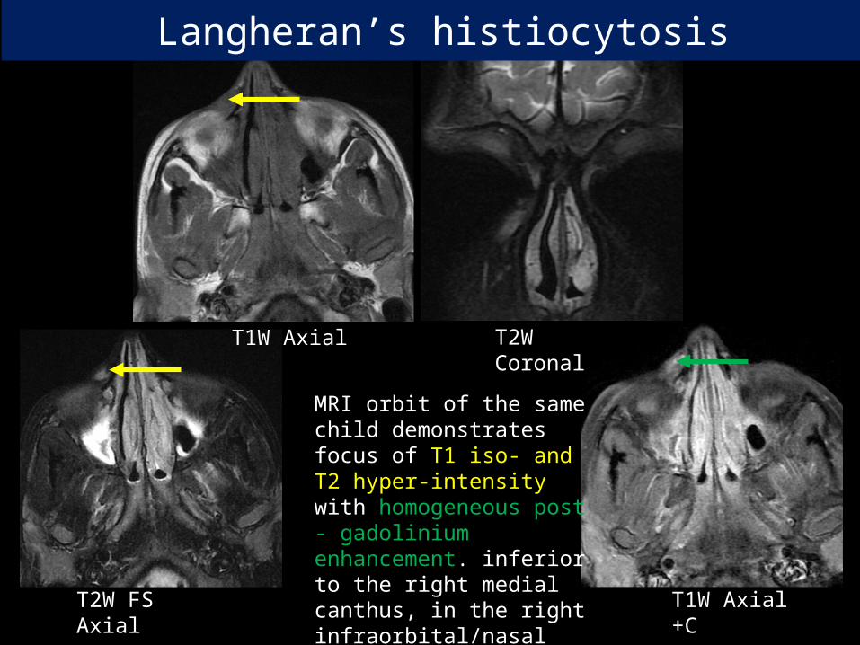
Langheran’s histiocytosis
T1W Axial T2W Coronal
T1W Axial +C
T2W FS Axial
MRI orbit of the same child demonstrates focus of T1 iso- and T2 hyper-intensity with homogeneous post - gadolinium enhancement. inferior to the right medial canthus, in the right infraorbital/nasal subcutaneous tissues.
Langheran’s histiocytosis

Meta
stasis fro
m sq
uam
ou
s cell ca
of
ton
silM
eta
stasi
s f
rom
poorl
y d
iffere
nti
ate
d
ca o
f su
bm
and
ibula
r g
land
Metastasis

ConclusionMedial Canthus of the orbit can be involved by a wide range of pathologies.
Careful examination and systematic imaging approach with knowledge of the pathologies is the key to successful patient management.
This pictorial review from our institution will familiarize the radiologists with imaging features of common and uncommon lesions involving the medial canthus.

References Russell EJ, Czervionke L, Huckman M, Daniels D, McLachlan D. CT of the infenomedial orbit and the lacrimal drainage apparatus: normal and pathologic anatomy. AJNR 1985;6:759-766
Rand P, Ball WS, Kulwin DR. Congenital nasolacrimal mucoceles: CT evaluation. Radiology 1989; 173:691-694
Cibis GW, Spurney RO, Waeltermann J . Radiographic visualization of congenital lacrimal sac mucoceles. Ann Ophthalmol I 986; 18:68-69
Escott EJ. A variety of appearances of malignant melanoma in the head: a review. RadioGraphics 2001;21(3):625–639.
Isiklar I, Leeds NE, Fuller GN, Kumar AJ. Intracranial metastatic melanoma: correlation between MR imaging characteristics and melanin content. AJR Am J Roentgenol 1995;165(6):1503–1512.

Imaging of Medial Canthus of the Orbit: An Unexplored Territory
J Nair, C Torres, J Chankowsky, R del carpio