Contrast-sensitive perceptual grouping and object-based ...
20
Vision Research 40 (2000) 1413 – 1432 Contrast-sensitive perceptual grouping and object-based attention in the laminar circuits of primary visual cortex Stephen Grossberg *, Rajeev D.S. Raizada Department of Cogniti6e and Neural Systems, Center for Adapti6e Systems, Boston Uni6ersity, 677 Beacon Street, Boston, MA 02215, USA Received 3 May 1999; received in revised form 11 October 1999 Abstract Recent neurophysiological studies have shown that primary visual cortex, or V1, does more than passively process image features using the feedforward filters suggested by Hubel and Wiesel. It also uses horizontal interactions to group features preattentively into object representations, and feedback interactions to selectively attend to these groupings. All neocortical areas, including V1, are organized into layered circuits. We present a neural model showing how the layered circuits in areas V1 and V2 enable feedforward, horizontal, and feedback interactions to complete perceptual groupings over positions that do not receive contrastive visual inputs, even while attention can only modulate or prime positions that do not receive such inputs. Recent neurophysiological data about how grouping and attention occur and interact in V1 are simulated and explained, and testable predictions are made. These simulations show how attention can selectively propagate along an object grouping and protect it from competitive masking, and how contextual stimuli can enhance or suppress groupings in a contrast-sensitive manner. © 2000 Elsevier Science Ltd. All rights reserved. Keywords: Attention; Grouping; Layers; Striate cortex; Feedback www.elsevier.com/locate/visres 1. Introduction: attention and perceptual grouping in visual cortex Perceptual grouping is the process whereby spatially distributed visual features become linked into object representations. There is evidence that it is a preatten- tive process that requires no top-down influences (e.g. Moore & Egeth, 1997). Attention enables observers to selectively process some object representations at the expense of others. It is clearly a top-down process. The past few years have seen an explosion of interest in the neurophysiological substrates of attention and percep- tual grouping in visual cortex, at first in extrastriate areas but more recently also in striate cortex, or V1 (Grosof, Shapley & Hawken, 1993; Motter, 1993; Ka- padia, Ito, Gilbert & Westheimer, 1995; Sheth, Sharma, Rao & Sur, 1996; Polat, Mizobe, Pettet, Kasamatsu & Norcia, 1998; Roelfsema, Lamme & Spekreijse, 1998; Watanabe, Sasaki, Nielsen, Takino & Miyakawa, 1998; Somers, Dale, Seiffert & Tootell, 1999). Anatomical studies have also revealed much of the intricate cortico- cortical and intracortical laminar circuitry of visual cortex which supports these processes (Callaway, 1998). However, explicit computational theories of how the cortical layers join these ‘higher-order’ perceptual func- tions to the better understood visual filtering processes have been lacking. In order to interpret the seemingly bewildering tangle of known laminar circuitry, an anal- ysis is needed of the functional roles which these cir- cuits subserve. Given the evidence cited above that those roles include attention and perceptual grouping, several sets of tight constraints emerge. Three lines of neurophysiological and psychophysical evidence suggest that attention and grouping share some common mechanisms. Firstly, both processes act to enhance weak stimuli, but may have a neutral or even suppressive effect on stimuli that are already strong. In particular, the threshold level of contrast required to detect a target stimulus can be reduced, either by directing attention to the target location (Reynolds, Pasternak & Desimone, 1996) or by adding * Corresponding author: Tel.: +1-617-3537858; fax: +1-617- 3537755. E-mail addresses: [email protected] (S. Grossberg), ra- [email protected] (R.D.S. Raizada) 0042-6989/00/$ - see front matter © 2000 Elsevier Science Ltd. All rights reserved. PII:S0042-6989(99)00229-1
Transcript of Contrast-sensitive perceptual grouping and object-based ...
Vision Research 40 (2000) 1413–1432
Contrast-sensitive perceptual grouping and object-based attentionin the laminar circuits of primary visual cortex
Stephen Grossberg *, Rajeev D.S. RaizadaDepartment of Cogniti6e and Neural Systems, Center for Adapti6e Systems, Boston Uni6ersity, 677 Beacon Street, Boston, MA 02215, USA
Received 3 May 1999; received in revised form 11 October 1999
Abstract
Recent neurophysiological studies have shown that primary visual cortex, or V1, does more than passively process imagefeatures using the feedforward filters suggested by Hubel and Wiesel. It also uses horizontal interactions to group featurespreattentively into object representations, and feedback interactions to selectively attend to these groupings. All neocortical areas,including V1, are organized into layered circuits. We present a neural model showing how the layered circuits in areas V1 and V2enable feedforward, horizontal, and feedback interactions to complete perceptual groupings over positions that do not receivecontrastive visual inputs, even while attention can only modulate or prime positions that do not receive such inputs. Recentneurophysiological data about how grouping and attention occur and interact in V1 are simulated and explained, and testablepredictions are made. These simulations show how attention can selectively propagate along an object grouping and protect itfrom competitive masking, and how contextual stimuli can enhance or suppress groupings in a contrast-sensitive manner. © 2000Elsevier Science Ltd. All rights reserved.
Keywords: Attention; Grouping; Layers; Striate cortex; Feedback
www.elsevier.com/locate/visres
1. Introduction: attention and perceptual grouping invisual cortex
Perceptual grouping is the process whereby spatiallydistributed visual features become linked into objectrepresentations. There is evidence that it is a preatten-tive process that requires no top-down influences (e.g.Moore & Egeth, 1997). Attention enables observers toselectively process some object representations at theexpense of others. It is clearly a top-down process. Thepast few years have seen an explosion of interest in theneurophysiological substrates of attention and percep-tual grouping in visual cortex, at first in extrastriateareas but more recently also in striate cortex, or V1(Grosof, Shapley & Hawken, 1993; Motter, 1993; Ka-padia, Ito, Gilbert & Westheimer, 1995; Sheth, Sharma,Rao & Sur, 1996; Polat, Mizobe, Pettet, Kasamatsu &Norcia, 1998; Roelfsema, Lamme & Spekreijse, 1998;
Watanabe, Sasaki, Nielsen, Takino & Miyakawa, 1998;Somers, Dale, Seiffert & Tootell, 1999). Anatomicalstudies have also revealed much of the intricate cortico-cortical and intracortical laminar circuitry of visualcortex which supports these processes (Callaway, 1998).However, explicit computational theories of how thecortical layers join these ‘higher-order’ perceptual func-tions to the better understood visual filtering processeshave been lacking. In order to interpret the seeminglybewildering tangle of known laminar circuitry, an anal-ysis is needed of the functional roles which these cir-cuits subserve. Given the evidence cited above thatthose roles include attention and perceptual grouping,several sets of tight constraints emerge.
Three lines of neurophysiological and psychophysicalevidence suggest that attention and grouping sharesome common mechanisms. Firstly, both processes actto enhance weak stimuli, but may have a neutral oreven suppressive effect on stimuli that are alreadystrong. In particular, the threshold level of contrastrequired to detect a target stimulus can be reduced,either by directing attention to the target location(Reynolds, Pasternak & Desimone, 1996) or by adding
* Corresponding author: Tel.: +1-617-3537858; fax: +1-617-3537755.
E-mail addresses: [email protected] (S. Grossberg), [email protected] (R.D.S. Raizada)
0042-6989/00/$ - see front matter © 2000 Elsevier Science Ltd. All rights reserved.PII: S 0 0 4 2 -6989 (99 )00229 -1

S. Grossberg, R.D.S. Raizada / Vision Research 40 (2000) 1413–14321414
flanking stimuli with which the target can collinearlygroup (Kapadia et al., 1995; Polat et al., 1998). Suchenhancement tends not to occur for higher-contraststimuli (Reynolds et al., 1996; see also Hupe, James,Payne, Lomber, Girard & Bullier, 1998). This sort ofcontrast-dependent effect was found for perceptualgrouping in the recent V1 study by Polat et al. (1998)(data shown in Fig. 1b), where neuronal responses tolow-contrast Gabor patches were enhanced by the pres-ence of collinear flankers outside the classical receptivefield, but responses to Gabors which were well abovecontrast threshold were suppressed.
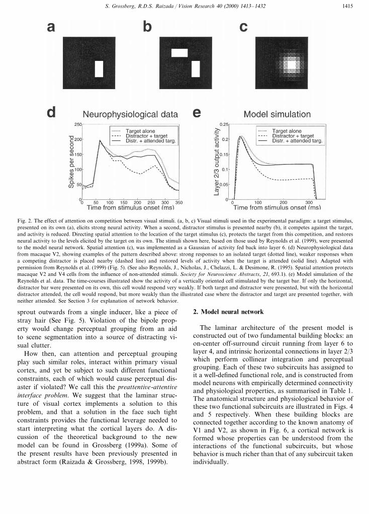
Secondly, attention and grouping both act to sup-press rival stimuli. Distinct ways of perceptually group-ing a scene compete against each other, for example thealternative horizontal and vertical groupings which mayform when viewing a grid of dots (Kubovy, Holcombe& Wagemans, 1998). Spatially directed attention has aninhibitory off-surround which suppresses unattendedstimuli (Somers et al., 1999), and which declines instrength with distance from the attended location (Ca-puto & Guerra, 1998). Both the suppressive and theenhancing effects of attention have been shown in workby Reynolds, Chelazzi and Desimone (1999) (datashown in Fig. 2d), in which attention directed to atarget stimulus protects it from the competitive effectsof a nearby distractor, and also suppresses neural re-sponses to the distractor itself.
The third reason for suggesting partial sharing oftheir underlying mechanisms is that attention and per-ceptual grouping reciprocally interact. Although group-ings may arise preattentively (Moore & Egeth, 1997),
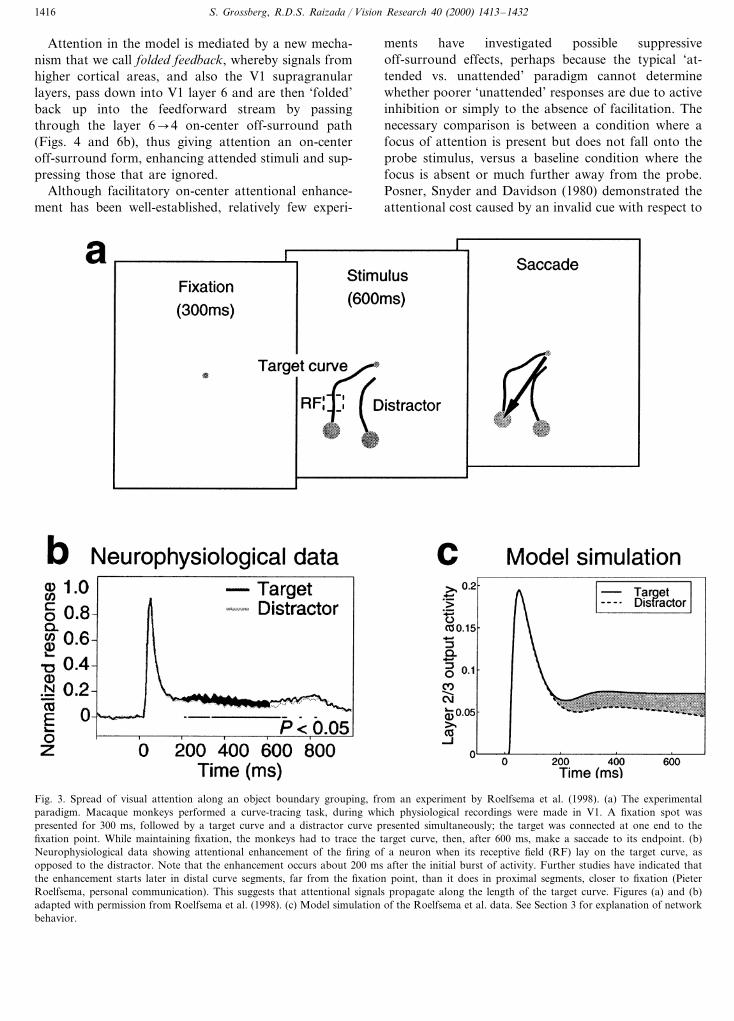
attentional task demands can influence which of vari-ous alternative groupings actually form, and thesegroupings in turn affect attentional phenomena such asthe occurrence of illusory conjunctions (Prinzmetal &Keysar, 1989) or reaction times in a visual search task(Carrasco & Chang, 1995). Physiological recordings instriate cortex of macaques performing a visual curve-tracing task (Roelfsema et al., 1998, data illustrated inFig. 3b) have shown that attention enhancementspreads along line segments which are grouped togetherto form a smooth curve, but not into contiguous seg-ments which are grouped as a different object.
Despite these shared properties, attention and group-ing must satisfy markedly different functional con-straints if they are to avoid giving spurious output.Although top-down attention can slightly elevate thebaseline firing rate of a neuron whose receptive fieldcontains no visual stimulus (Luck, Chelazzi, Hillyard &Desimone, 1997), it does not produce suprathresholdneural activity of the sort that would be produced bybottom-up input. Violation of this constraint couldcause continual hallucinations. However, illusory con-tours, prime examples of perceptual grouping, do, bydefinition, form over parts of visual space where thereis no visual stimulus (von der Heydt, Peterhans &Baumgartner, 1984), clearly breaking the rule that at-tention must obey. Groupings must satisfy a quitedifferent constraint referred to here as the ‘bipole prop-erty’ (Grossberg & Mingolla, 1985): an illusory contourmay form between two or more inducers, such as the‘pacman’ corners of a Kanizsa square, but cannot
Fig. 1. Contrast-dependent perceptual grouping in primary visual cortex. (a) Illustrative visual stimuli. A variable-contrast oriented Gabor patchstimulates the classical receptive field (CRF), with collinear flanking Gabors of fixed high-contrast outside of the CRF. The stimulus shown here,based on those used Polat et al. (1998), was presented to the model neural network. (b) Neural responses recorded from cat V1. The collinearflankers have a net facilitatory effect on weak targets which are close to the cell’s contrast-threshold, but they act to suppress responses to stronger,above-threshold targets. When the flankers are presented on their own, with no target present, the neural response stays at baseline levels.Reproduced with permission from Polat et al. (1998). (c) Model simulation of the Polat et al. data. See Section 3 for explanation of networkbehavior.
S. Grossberg, R.D.S. Raizada / Vision Research 40 (2000) 1413–1432 1415
Fig. 2. The effect of attention on competition between visual stimuli. (a, b, c) Visual stimuli used in the experimental paradigm: a target stimulus,presented on its own (a), elicits strong neural activity. When a second, distractor stimulus is presented nearby (b), it competes against the target,and activity is reduced. Directing spatial attention to the location of the target stimulus (c), protects the target from this competition, and restoresneural activity to the levels elicited by the target on its own. The stimuli shown here, based on those used by Reynolds et al. (1999), were presentedto the model neural network. Spatial attention (c), was implemented as a Gaussian of activity fed back into layer 6. (d) Neurophysiological datafrom macaque V2, showing examples of the pattern described above: strong responses to an isolated target (dotted line), weaker responses whena competing distractor is placed nearby (dashed line) and restored levels of activity when the target is attended (solid line). Adapted withpermission from Reynolds et al. (1999) (Fig. 5). (See also Reynolds, J., Nicholas, J., Chelazzi, L. & Desimone, R. (1995). Spatial attention protectsmacaque V2 and V4 cells from the influence of non-attended stimuli. Society for Neuroscience Abstracts, 21, 693.1). (e) Model simulation of theReynolds et al. data. The time-courses illustrated show the activity of a vertically oriented cell stimulated by the target bar. If only the horizontal,distractor bar were presented on its own, this cell would respond very weakly. If both target and distractor were presented, but with the horizontaldistractor attended, the cell would respond, but more weakly than the illustrated case where the distractor and target are presented together, withneither attended. See Section 3 for explanation of network behavior.
sprout outwards from a single inducer, like a piece ofstray hair (See Fig. 5). Violation of the bipole prop-erty would change perceptual grouping from an aidto scene segmentation into a source of distracting vi-sual clutter.
How then, can attention and perceptual groupingplay such similar roles, interact within primary visualcortex, and yet be subject to such different functionalconstraints, each of which would cause perceptual dis-aster if violated? We call this the preattenti6e-attenti6einterface problem. We suggest that the laminar struc-ture of visual cortex implements a solution to thisproblem, and that a solution in the face such tightconstraints provides the functional leverage needed tostart interpreting what the cortical layers do. A dis-cussion of the theoretical background to the newmodel can be found in Grossberg (1999a). Some ofthe present results have been previously presented inabstract form (Raizada & Grossberg, 1998, 1999b).
2. Model neural network
The laminar architecture of the present model isconstructed out of two fundamental building blocks: anon-center off-surround circuit running from layer 6 tolayer 4, and intrinsic horizontal connections in layer 2/3which perform collinear integration and perceptualgrouping. Each of these two subcircuits has assigned toit a well-defined functional role, and is constructed frommodel neurons with empirically determined connectivityand physiological properties, as summarised in Table 1.The anatomical structure and physiological behavior ofthese two functional subcircuits are illustrated in Figs. 4and 5 respectively. When these building blocks areconnected together according to the known anatomy ofV1 and V2, as shown in Fig. 6, a cortical network isformed whose properties can be understood from theinteractions of the functional subcircuits, but whosebehavior is much richer than that of any subcircuit takenindividually.
S. Grossberg, R.D.S. Raizada / Vision Research 40 (2000) 1413–14321416
Attention in the model is mediated by a new mecha-nism that we call folded feedback, whereby signals fromhigher cortical areas, and also the V1 supragranularlayers, pass down into V1 layer 6 and are then ‘folded’back up into the feedforward stream by passingthrough the layer 6�4 on-center off-surround path(Figs. 4 and 6b), thus giving attention an on-centeroff-surround form, enhancing attended stimuli and sup-pressing those that are ignored.
Although facilitatory on-center attentional enhance-ment has been well-established, relatively few experi-
ments have investigated possible suppressiveoff-surround effects, perhaps because the typical ‘at-tended vs. unattended’ paradigm cannot determinewhether poorer ‘unattended’ responses are due to activeinhibition or simply to the absence of facilitation. Thenecessary comparison is between a condition where afocus of attention is present but does not fall onto theprobe stimulus, versus a baseline condition where thefocus is absent or much further away from the probe.Posner, Snyder and Davidson (1980) demonstrated theattentional cost caused by an invalid cue with respect to
Fig. 3. Spread of visual attention along an object boundary grouping, from an experiment by Roelfsema et al. (1998). (a) The experimentalparadigm. Macaque monkeys performed a curve-tracing task, during which physiological recordings were made in V1. A fixation spot waspresented for 300 ms, followed by a target curve and a distractor curve presented simultaneously; the target was connected at one end to thefixation point. While maintaining fixation, the monkeys had to trace the target curve, then, after 600 ms, make a saccade to its endpoint. (b)Neurophysiological data showing attentional enhancement of the firing of a neuron when its receptive field (RF) lay on the target curve, asopposed to the distractor. Note that the enhancement occurs about 200 ms after the initial burst of activity. Further studies have indicated thatthe enhancement starts later in distal curve segments, far from the fixation point, than it does in proximal segments, closer to fixation (PieterRoelfsema, personal communication). This suggests that attentional signals propagate along the length of the target curve. Figures (a) and (b)adapted with permission from Roelfsema et al. (1998). (c) Model simulation of the Roelfsema et al. data. See Section 3 for explanation of networkbehavior.
S. Grossberg, R.D.S. Raizada / Vision Research 40 (2000) 1413–1432 1417
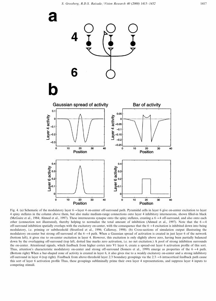
Fig. 4. (a) Schematic of the modulatory layer 6� layer 4 on-center off-surround path. Pyramidal cells in layer 6 give on-center excitation to layer4 spiny stellates in the column above them, but also make medium-range connections onto layer 4 inhibitory interneurons, shown filled-in black(McGuire et al., 1984; Ahmed et al., 1997). These interneurons synapse onto the spiny stellates, creating a 6�4 off-surround, and also onto eachother (connection not illustrated), thereby helping to normalize the total amount of inhibition (Ahmed et al., 1997). Note that the 6�4off-surround inhibition spatially overlaps with the excitatory on-center, with the consequence that the 6�4 excitation is inhibited down into beingmodulatory, i.e. priming or subthreshold (Stratford et al., 1996; Callaway, 1998). (b) Cross-sections of simulation output illustrating themodulatory on-center but strong off-surround of the 6�4 path. When a Gaussian spread of activation is created in just layer 6 of the network(bottom left), it gives rise to on-center excitation in layer 4. However, this excitation is only slightly above zero, having been partially balanceddown by the overlapping off-surround (top left, dotted line marks zero activation, i.e. no net excitation.) A pool of strong inhibition surroundsthe on-center. Attentional signals, which feedback from higher cortex into V1 layer 6, create a spread-out layer 6 activation profile of this sort.Thus, attention’s characteristic modulatory on-center and strong off-surround (Somers et al., 1999) emerge as properties of the 6�4 path.(Bottom right) When a bar-shaped zone of activity is created in layer 6, it also gives rise to a weakly excitatory on-center and a strong inhibitoryoff-surround in layer 4 (top right). Feedback from above-threshold layer 2/3 boundary groupings via the 2/3�6 intracortical feedback path causethis sort of layer 6 activation profile Thus, these groupings subliminally prime their own layer 4 representations, and suppress layer 4 inputs tocompeting stimuli.
S. Grossberg, R.D.S. Raizada / Vision Research 40 (2000) 1413–14321418
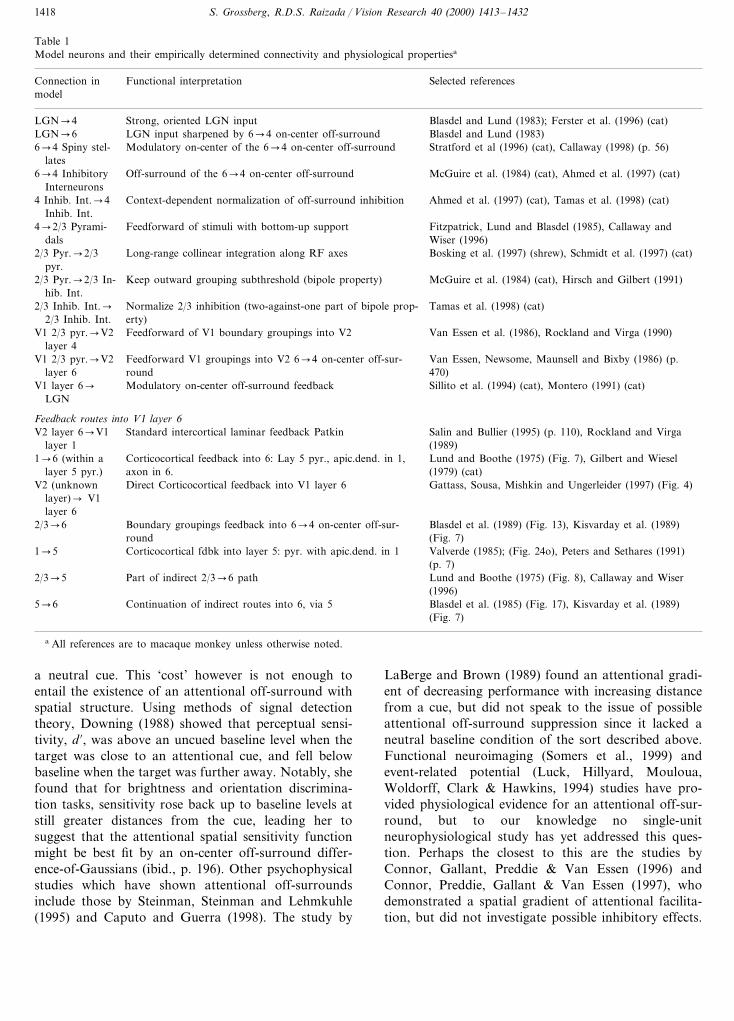
Table 1Model neurons and their empirically determined connectivity and physiological propertiesa
Functional interpretationConnection in Selected referencesmodel
Blasdel and Lund (1983); Ferster et al. (1996) (cat)LGN�4 Strong, oriented LGN inputBlasdel and Lund (1983)LGN input sharpened by 6�4 on-center off-surroundLGN�6
Modulatory on-center of the 6�4 on-center off-surround6�4 Spiny stel- Stratford et al (1996) (cat), Callaway (1998) (p. 56)lates
6�4 Inhibitory Off-surround of the 6�4 on-center off-surround McGuire et al. (1984) (cat), Ahmed et al. (1997) (cat)Interneurons
Context-dependent normalization of off-surround inhibition Ahmed et al. (1997) (cat), Tamas et al. (1998) (cat)4 Inhib. Int.�4Inhib. Int.
4�2/3 Pyrami- Feedforward of stimuli with bottom-up support Fitzpatrick, Lund and Blasdel (1985), Callaway andWiser (1996)dals
2/3 Pyr.�2/3 Long-range collinear integration along RF axes Bosking et al. (1997) (shrew), Schmidt et al. (1997) (cat)pyr.
2/3 Pyr.�2/3 In- Keep outward grouping subthreshold (bipole property) McGuire et al. (1984) (cat), Hirsch and Gilbert (1991)hib. Int.
2/3 Inhib. Int.� Normalize 2/3 inhibition (two-against-one part of bipole prop- Tamas et al. (1998) (cat)erty)2/3 Inhib. Int.
V1 2/3 pyr.�V2 Feedforward of V1 boundary groupings into V2 Van Essen et al. (1986), Rockland and Virga (1990)layer 4
Feedforward V1 groupings into V2 6�4 on-center off-sur-V1 2/3 pyr.�V2 Van Essen, Newsome, Maunsell and Bixby (1986) (p.roundlayer 6 470)
V1 layer 6� Modulatory on-center off-surround feedback Sillito et al. (1994) (cat), Montero (1991) (cat)LGN
Feedback routes into V1 layer 6Standard intercortical laminar feedback Patkin Salin and Bullier (1995) (p. 110), Rockland and VirgaV2 layer 6�V1
layer 1 (1989)Corticocortical feedback into 6: Lay 5 pyr., apic.dend. in 1,1�6 (within a Lund and Boothe (1975) (Fig. 7), Gilbert and Wieselaxon in 6.layer 5 pyr.) (1979) (cat)Direct Corticocortical feedback into V1 layer 6V2 (unknown Gattass, Sousa, Mishkin and Ungerleider (1997) (Fig. 4)
layer)� V1layer 6
2/3�6 Boundary groupings feedback into 6�4 on-center off-sur- Blasdel et al. (1989) (Fig. 13), Kisvarday et al. (1989)(Fig. 7)roundValverde (1985); (Fig. 24o), Peters and Sethares (1991)Corticocortical fdbk into layer 5: pyr. with apic.dend. in 11�5(p. 7)
Part of indirect 2/3�6 path Lund and Boothe (1975) (Fig. 8), Callaway and Wiser2/3�5(1996)Blasdel et al. (1985) (Fig. 17), Kisvarday et al. (1989)Continuation of indirect routes into 6, via 55�6(Fig. 7)
a All references are to macaque monkey unless otherwise noted.
a neutral cue. This ‘cost’ however is not enough toentail the existence of an attentional off-surround withspatial structure. Using methods of signal detectiontheory, Downing (1988) showed that perceptual sensi-tivity, d %, was above an uncued baseline level when thetarget was close to an attentional cue, and fell belowbaseline when the target was further away. Notably, shefound that for brightness and orientation discrimina-tion tasks, sensitivity rose back up to baseline levels atstill greater distances from the cue, leading her tosuggest that the attentional spatial sensitivity functionmight be best fit by an on-center off-surround differ-ence-of-Gaussians (ibid., p. 196). Other psychophysicalstudies which have shown attentional off-surroundsinclude those by Steinman, Steinman and Lehmkuhle(1995) and Caputo and Guerra (1998). The study by
LaBerge and Brown (1989) found an attentional gradi-ent of decreasing performance with increasing distancefrom a cue, but did not speak to the issue of possibleattentional off-surround suppression since it lacked aneutral baseline condition of the sort described above.Functional neuroimaging (Somers et al., 1999) andevent-related potential (Luck, Hillyard, Mouloua,Woldorff, Clark & Hawkins, 1994) studies have pro-vided physiological evidence for an attentional off-sur-round, but to our knowledge no single-unitneurophysiological study has yet addressed this ques-tion. Perhaps the closest to this are the studies byConnor, Gallant, Preddie & Van Essen (1996) andConnor, Preddie, Gallant & Van Essen (1997), whodemonstrated a spatial gradient of attentional facilita-tion, but did not investigate possible inhibitory effects.

S. Grossberg, R.D.S. Raizada / Vision Research 40 (2000) 1413–1432 1419
Fig. 5. Schematic of the boundary grouping circuit in layer 2/3.Pyramidal cells with collinear, coaxial receptive fields (shown asovals) excite each other via long-range horizontal axons (Bosking etal., 1997; Schmidt et al., 1997), which also give rise to short-range,disynaptic inhibition via pools of interneurons, shown filled-in black(McGuire et al., 1991). This balance of excitation and inhibition helpsto implement what we call the bipole property. (a) Illustration of howhorizontal input coming in from just one side is insufficient to causeabove-threshold excitation in a pyramidal cell (henceforth referred toas the target) whose receptive field does not itself receive any bottom-up input. The inducing stimulus (e.g. a Kanizsa ‘pacman’, shownhere) excites the oriented receptive fields of layer 2/3 cells, which sendout long-range horizontal excitation onto the target pyramidal. How-ever, this excitation brings with it a commensurate amount of disy-naptic inhibition. This creates a case of ‘one-against-one’, and thetarget pyramidal is not excited above-threshold. The boundary repre-sentation of the solitary pacman inducer produces only weak, sub-threshold collinear extensions (thin dashed lines). (b) When twocollinearly aligned induced stimuli are present, one on each side of thetarget pyramidal’s receptive field, a boundary grouping can form.Long-range excitatory inputs fall onto the cell from both sides, andsummate. However, these inputs fall onto a shared pool of inhibitoryinterneurons, which, as well as inhibiting the target pyramidal, alsoinhibit each other (Tamas et al., 1998), thus normalizing the totalamount of inhibition emanating from the interneuron pool, withoutany individual interneuron saturating. This summating excitation andnormalizing inhibition together create a case of ‘two-against-one’,and the target pyramidal is excited above-threshold. This processoccurs along the whole boundary grouping, which thereby becomesrepresented by a line of suprathreshold layer 2/3 cells (thick dottedline). Boundary strength scales in a graded analog manner with thestrength of the inducing signals.
cells’ monocularity (Callaway, 1998, p. 56). We suggestthat the on-center excitation is inhibited down intobeing modulatory by the overlapping and broader off-surround (Fig. 4). Thus, although the center excitationis weak, the suppressive effect of the off-surround inhi-bition can be strong. Because attentional excitationmust pass through the 6�4 path before it can effectvisual processing, it inherits this path’s properties: theattentional on-center is modulatory, able to enhanceexisting activity but only slightly to elevate neurons’baseline firing rates in the absence of visual input (Lucket al., 1997), but the off-surround can select stronglyagainst unattended stimuli.
Several routes exist through which feedback fromhigher cortex can reach V1 layer 6, as shown in Table1. Fig. 6(b) illustrates the route whereby feedback sig-nals pass into layer 1, where the majority of V2 feed-back axons terminate (Rockland & Virga, 1989), andthen stimulate the apical dendrites of layer 5 pyramidalcells whose axons send collaterals into layer 6 (Lund &Boothe, 1975; Gilbert & Wiesel, 1979), where the atten-tional signals are ‘folded’ back up into the 6�4 on-center off-surround. Reversible deactivation studies ofmonkey V2 have shown that feedback from V2 to V1does indeed have an on-center off-surround form (Bul-lier, Hupe, James & Girard, 1996), and moreover thatthe V1 layer whose activation is most reduced bycutting off V2 feedback is layer 6 (Sandell & Schiller,1982).
We suggest that the mechanism of folded feedback isalso used to help select the final layer 2/3 grouping.Like attentional signals from higher cortex, the group-ings which start to form in layer 2/3 also feedback intothe 6�4 path (Fig. 6c), to enhance their own positionsin layer 4 via the 6�4 on-center, and to suppress inputto other groupings via the 6�4 off-surround. Thereexist direct layer 2/3�6 connections in macaque V1, aswell as indirect routes via layer 5 (Table 1). Thiscompetition between layer 2/3 groupings, via layer 2/3�6�4�2/3 feedback, causes the strongest groupingto be selected, while suppresses weaker groupings, un-grouped distractors, and noise. The interlaminar feed-back also binds the cortical layers together intofunctional columns.
The fact that both attention and perceptual groupingshare the properties of enhancing weak stimuli, and ofsuppressing signals from nearby rival inputs, can thusbe parsimoniously explained by the hypothesis thatboth processes share the 6�4 folded feedback path.This laminar architecture also resolves the preatten-tive–attentive interface problem described above, sincedespite their shared properties and coexistence side-by-side within V1 and V2, attention and grouping behavequite differently in parts of visual space where there isno bottom-up visual stimulus. Above-thresholdboundary groupings can form over regions with no
A key prediction of the model is that the on-center ofthe 6�4 path is modulatory (or priming, or sub-threshold), consistent with the finding that layer 4EPSPs elicited by layer 6 stimulation are much weakerthan those caused by stimulation of LGN axons or ofneighbouring layer 4 sites (Stratford, Tarczy-Hornoch,Martin, Bannister & Jack, 1996), and also with the factthat binocular layer 6 neurons synapse onto monocularlayer 4 cells of both eye types without reducing these
S. Grossberg, R.D.S. Raizada / Vision Research 40 (2000) 1413–14321420
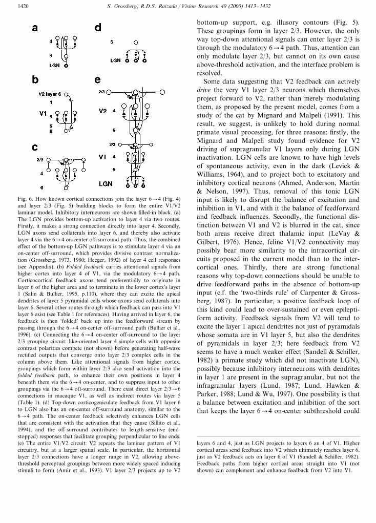
Fig. 6. How known cortical connections join the layer 6�4 (Fig. 4)and layer 2/3 (Fig. 5) building blocks to form the entire V1/V2laminar model. Inhibitory interneurons are shown filled-in black. (a)The LGN provides bottom-up activation to layer 4 via two routes.Firstly, it makes a strong connection directly into layer 4. Secondly,LGN axons send collaterals into layer 6, and thereby also activatelayer 4 via the 6�4 on-center off-surround path. Thus, the combinedeffect of the bottom-up LGN pathways is to stimulate layer 4 via anon-center off-surround, which provides divisive contrast normaliza-tion (Grossberg, 1973, 1980; Heeger, 1992) of layer 4 cell responses(see Appendix). (b) Folded feedback carries attentional signals fromhigher cortex into layer 4 of V1, via the modulatory 6�4 path.Corticocortical feedback axons tend preferentially to originate inlayer 6 of the higher area and to terminate in the lower cortex’s layer1 (Salin & Bullier, 1995, p.110), where they can excite the apicaldendrites of layer 5 pyramidal cells whose axons send collaterals intolayer 6. Several other routes through which feedback can pass into V1layer 6 exist (see Table 1 for references). Having arrived in layer 6, thefeedback is then ‘folded’ back up into the feedforward stream bypassing through the 6�4 on-center off-surround path (Bullier et al.,1996). (c) Connecting the 6�4 on-center off-surround to the layer2/3 grouping circuit: like-oriented layer 4 simple cells with oppositecontrast polarities compete (not shown) before generating half-waverectified outputs that converge onto layer 2/3 complex cells in thecolumn above them. Like attentional signals from higher cortex,groupings which form within layer 2/3 also send activation into thefolded feedback path, to enhance their own positions in layer 4beneath them via the 6�4 on-center, and to suppress input to othergroupings via the 6�4 off-surround. There exist direct layer 2/3�6connections in macaque V1, as well as indirect routes via layer 5(Table 1). (d) Top-down corticogeniculate feedback from V1 layer 6to LGN also has an on-center off-surround anatomy, similar to the6�4 path. The on-center feedback selectively enhances LGN cellsthat are consistent with the activation that they cause (Sillito et al.,1994), and the off-surround contributes to length-sensitive (end-stopped) responses that facilitate grouping perpendicular to line ends.(e) The entire V1/V2 circuit: V2 repeats the laminar pattern of V1circuitry, but at a larger spatial scale. In particular, the horizontallayer 2/3 connections have a longer range in V2, allowing above-threshold perceptual groupings between more widely spaced inducingstimuli to form (Amir et al., 1993). V1 layer 2/3 projects up to V2
bottom-up support, e.g. illusory contours (Fig. 5).These groupings form in layer 2/3. However, the onlyway top-down attentional signals can enter layer 2/3 isthrough the modulatory 6�4 path. Thus, attention canonly modulate layer 2/3, but cannot on its own causeabove-threshold activation, and the interface problem isresolved.
Some data suggesting that V2 feedback can activelydri6e the very V1 layer 2/3 neurons which themselvesproject forward to V2, rather than merely modulatingthem, as proposed by the present model, comes from astudy of the cat by Mignard and Malpeli (1991). Thisresult, we suggest, is unlikely to hold during normalprimate visual processing, for three reasons: firstly, theMignard and Malpeli study found evidence for V2driving of supragranular V1 layers only during LGNinactivation. LGN cells are known to have high levelsof spontaneous activity, even in the dark (Levick &Williams, 1964), and to project both to excitatory andinhibitory cortical neurons (Ahmed, Anderson, Martin& Nelson, 1997). Thus, removal of this tonic LGNinput is likely to disrupt the balance of excitation andinhibition in V1, and with it the balance of feedforwardand feedback influences. Secondly, the functional dis-tinction between V1 and V2 is blurred in the cat, sinceboth areas receive direct thalamic input (LeVay &Gilbert, 1976). Hence, feline V1/V2 connectivity maypossibly bear more similarity to the intracortical cir-cuits proposed in the current model than to the inter-cortical ones. Thirdly, there are strong functionalreasons why top-down connections should be unable todrive feedforward paths in the absence of bottom-upinput (c.f. the ‘two-thirds rule’ of Carpenter & Gross-berg, 1987). In particular, a positive feedback loop ofthis kind could lead to over-sustained or even epilepti-form activity. Feedback signals from V2 will tend toexcite the layer 1 apical dendrites not just of pyramidalswhose somata are in V1 layer 5, but also the dendritesof pyramidals in layer 2/3; here feedback from V2seems to have a much weaker effect (Sandell & Schiller,1982) a primate study which did not inactivate LGN),possibly because inhibitory interneurons with dendritesin layer 1 are present in the supragranular, but not theinfragranular layers (Lund, 1987; Lund, Hawken &Parker, 1988; Lund & Wu, 1997). One possibility is thata balance between excitation and inhibition of the sortthat keeps the layer 6�4 on-center subthreshold could
layers 6 and 4, just as LGN projects to layers 6 an 4 of V1. Highercortical areas send feedback into V2 which ultimately reaches layer 6,just as V2 feedback acts on layer 6 of V1 (Sandell & Schiller, 1982).Feedback paths from higher cortical areas straight into V1 (notshown) can complement and enhance feedback from V2 into V1.
S. Grossberg, R.D.S. Raizada / Vision Research 40 (2000) 1413–1432 1421
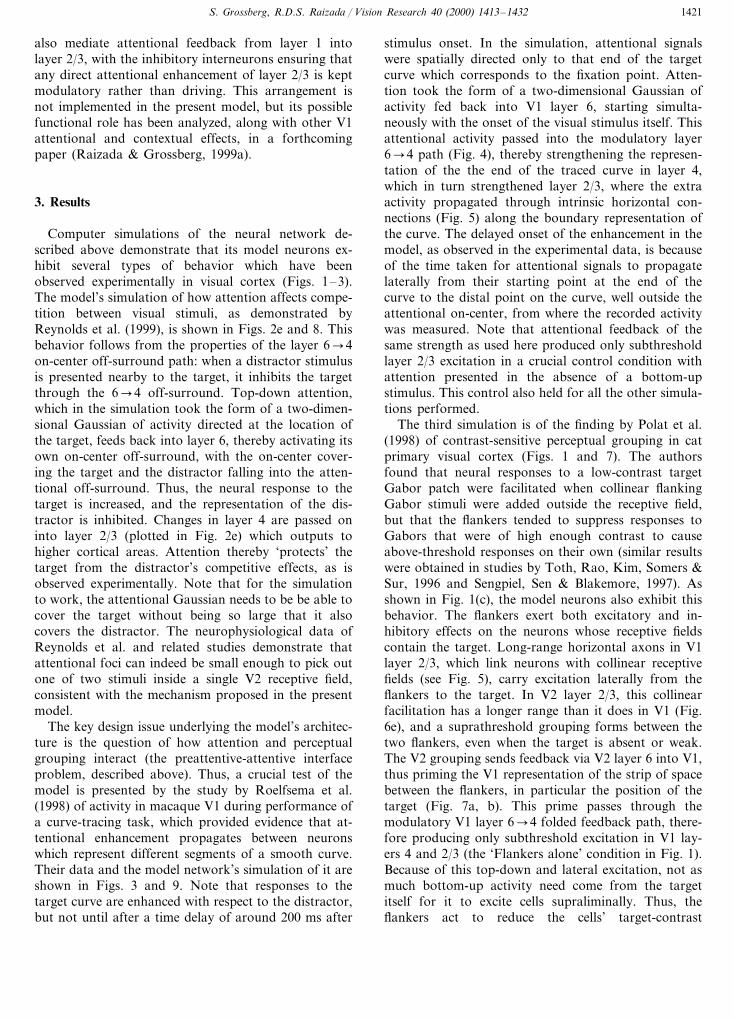
also mediate attentional feedback from layer 1 intolayer 2/3, with the inhibitory interneurons ensuring thatany direct attentional enhancement of layer 2/3 is keptmodulatory rather than driving. This arrangement isnot implemented in the present model, but its possiblefunctional role has been analyzed, along with other V1attentional and contextual effects, in a forthcomingpaper (Raizada & Grossberg, 1999a).
3. Results
Computer simulations of the neural network de-scribed above demonstrate that its model neurons ex-hibit several types of behavior which have beenobserved experimentally in visual cortex (Figs. 1–3).The model’s simulation of how attention affects compe-tition between visual stimuli, as demonstrated byReynolds et al. (1999), is shown in Figs. 2e and 8. Thisbehavior follows from the properties of the layer 6�4on-center off-surround path: when a distractor stimulusis presented nearby to the target, it inhibits the targetthrough the 6�4 off-surround. Top-down attention,which in the simulation took the form of a two-dimen-sional Gaussian of activity directed at the location ofthe target, feeds back into layer 6, thereby activating itsown on-center off-surround, with the on-center cover-ing the target and the distractor falling into the atten-tional off-surround. Thus, the neural response to thetarget is increased, and the representation of the dis-tractor is inhibited. Changes in layer 4 are passed oninto layer 2/3 (plotted in Fig. 2e) which outputs tohigher cortical areas. Attention thereby ‘protects’ thetarget from the distractor’s competitive effects, as isobserved experimentally. Note that for the simulationto work, the attentional Gaussian needs to be be able tocover the target without being so large that it alsocovers the distractor. The neurophysiological data ofReynolds et al. and related studies demonstrate thatattentional foci can indeed be small enough to pick outone of two stimuli inside a single V2 receptive field,consistent with the mechanism proposed in the presentmodel.
The key design issue underlying the model’s architec-ture is the question of how attention and perceptualgrouping interact (the preattentive-attentive interfaceproblem, described above). Thus, a crucial test of themodel is presented by the study by Roelfsema et al.(1998) of activity in macaque V1 during performance ofa curve-tracing task, which provided evidence that at-tentional enhancement propagates between neuronswhich represent different segments of a smooth curve.Their data and the model network’s simulation of it areshown in Figs. 3 and 9. Note that responses to thetarget curve are enhanced with respect to the distractor,but not until after a time delay of around 200 ms after
stimulus onset. In the simulation, attentional signalswere spatially directed only to that end of the targetcurve which corresponds to the fixation point. Atten-tion took the form of a two-dimensional Gaussian ofactivity fed back into V1 layer 6, starting simulta-neously with the onset of the visual stimulus itself. Thisattentional activity passed into the modulatory layer6�4 path (Fig. 4), thereby strengthening the represen-tation of the the end of the traced curve in layer 4,which in turn strengthened layer 2/3, where the extraactivity propagated through intrinsic horizontal con-nections (Fig. 5) along the boundary representation ofthe curve. The delayed onset of the enhancement in themodel, as observed in the experimental data, is becauseof the time taken for attentional signals to propagatelaterally from their starting point at the end of thecurve to the distal point on the curve, well outside theattentional on-center, from where the recorded activitywas measured. Note that attentional feedback of thesame strength as used here produced only subthresholdlayer 2/3 excitation in a crucial control condition withattention presented in the absence of a bottom-upstimulus. This control also held for all the other simula-tions performed.
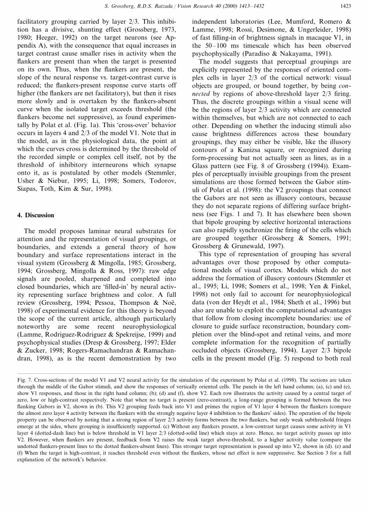
The third simulation is of the finding by Polat et al.(1998) of contrast-sensitive perceptual grouping in catprimary visual cortex (Figs. 1 and 7). The authorsfound that neural responses to a low-contrast targetGabor patch were facilitated when collinear flankingGabor stimuli were added outside the receptive field,but that the flankers tended to suppress responses toGabors that were of high enough contrast to causeabove-threshold responses on their own (similar resultswere obtained in studies by Toth, Rao, Kim, Somers &Sur, 1996 and Sengpiel, Sen & Blakemore, 1997). Asshown in Fig. 1(c), the model neurons also exhibit thisbehavior. The flankers exert both excitatory and in-hibitory effects on the neurons whose receptive fieldscontain the target. Long-range horizontal axons in V1layer 2/3, which link neurons with collinear receptivefields (see Fig. 5), carry excitation laterally from theflankers to the target. In V2 layer 2/3, this collinearfacilitation has a longer range than it does in V1 (Fig.6e), and a suprathreshold grouping forms between thetwo flankers, even when the target is absent or weak.The V2 grouping sends feedback via V2 layer 6 into V1,thus priming the V1 representation of the strip of spacebetween the flankers, in particular the position of thetarget (Fig. 7a, b). This prime passes through themodulatory V1 layer 6�4 folded feedback path, there-fore producing only subthreshold excitation in V1 lay-ers 4 and 2/3 (the ‘Flankers alone’ condition in Fig. 1).Because of this top-down and lateral excitation, not asmuch bottom-up activity need come from the targetitself for it to excite cells supraliminally. Thus, theflankers act to reduce the cells’ target-contrast

S. Grossberg, R.D.S. Raizada / Vision Research 40 (2000) 1413–14321422
threshold, raising the low-contrast section of the curveplotting neural response vs. target-contrast when theflankers are present.
However, the target also receives layer 6�4 off-sur-round inhibition from the flankers, which acts as a lessspecific ‘lateral masking’, as opposed to the collinear
Fig. 7. (Continued)
S. Grossberg, R.D.S. Raizada / Vision Research 40 (2000) 1413–1432 1423
facilitatory grouping carried by layer 2/3. This inhibi-tion has a divisive, shunting effect (Grossberg, 1973,1980; Heeger, 1992) on the target neurons (see Ap-pendix A), with the consequence that equal increases intarget contrast cause smaller rises in activity when theflankers are present than when the target is presentedon its own. Thus, when the flankers are present, theslope of the neural response vs. target-contrast curve isreduced; the flankers-present response curve starts offhigher (the flankers are net facilitatory), but then it risesmore slowly and is overtaken by the flankers-absentcurve when the isolated target exceeds threshold (theflankers become net suppressive), as found experimen-tally by Polat et al. (Fig. 1a). This ‘cross-over’ behavioroccurs in layers 4 and 2/3 of the model V1. Note that inthe model, as in the physiological data, the point atwhich the curves cross is determined by the threshold ofthe recorded simple or complex cell itself, not by thethreshold of inhibitory interneurons which synapseonto it, as is postulated by other models (Stemmler,Usher & Niebur, 1995; Li, 1998; Somers, Todorov,Siapas, Toth, Kim & Sur, 1998).
4. Discussion
The model proposes laminar neural substrates forattention and the representation of visual groupings, orboundaries, and extends a general theory of howboundary and surface representations interact in thevisual system (Grossberg & Mingolla, 1985; Grossberg,1994; Grossberg, Mingolla & Ross, 1997): raw edgesignals are pooled, sharpened and completed intoclosed boundaries, which are ‘filled-in’ by neural activ-ity representing surface brightness and color. A fullreview (Grossberg, 1994; Pessoa, Thompson & Noe,1998) of experimental evidence for this theory is beyondthe scope of the current article, although particularlynoteworthy are some recent neurophysiological(Lamme, Rodriguez-Rodriguez & Spekreijse, 1999) andpsychophysical studies (Dresp & Grossberg, 1997; Elder& Zucker, 1998; Rogers-Ramachandran & Ramachan-dran, 1998), as is the recent demonstration by two
independent laboratories (Lee, Mumford, Romero &Lamme, 1998; Rossi, Desimone, & Ungerleider, 1998)of fast filling-in of brightness signals in macaque V1, inthe 50–100 ms timescale which has been observedpsychophysically (Paradiso & Nakayama, 1991).
The model suggests that perceptual groupings areexplicitly represented by the responses of oriented com-plex cells in layer 2/3 of the cortical network: visualobjects are grouped, or bound together, by being con-nected by regions of above-threshold layer 2/3 firing.Thus, the discrete groupings within a visual scene willbe the regions of layer 2/3 activity which are connectedwithin themselves, but which are not connected to eachother. Depending on whether the inducing stimuli alsocause brightness differences across these boundarygroupings, they may either be visible, like the illusorycontours of a Kanizsa square, or recognized duringform-processing but not actually seen as lines, as in aGlass pattern (see Fig. 8 of Grossberg (1994)). Exam-ples of perceptually invisible groupings from the presentsimulations are those formed between the Gabor stim-uli of Polat et al. (1998): the V2 groupings that connectthe Gabors are not seen as illusory contours, becausethey do not separate regions of differing surface bright-ness (see Figs. 1 and 7). It has elsewhere been shownthat bipole grouping by selective horizontal interactionscan also rapidly synchronize the firing of the cells whichare grouped together (Grossberg & Somers, 1991;Grossberg & Grunewald, 1997).
This type of representation of grouping has severaladvantages over those proposed by other computa-tional models of visual cortex. Models which do notaddress the formation of illusory contours (Stemmler etal., 1995; Li, 1998; Somers et al., 1998; Yen & Finkel,1998) not only fail to account for neurophysiologicaldata (von der Heydt et al., 1984; Sheth et al., 1996) butalso are unable to exploit the computational advantagesthat follow from closing incomplete boundaries: use ofclosure to guide surface reconstruction, boundary com-pletion over the blind-spot and retinal veins, and morecomplete information for the recognition of partiallyoccluded objects (Grossberg, 1994). Layer 2/3 bipolecells in the present model (Fig. 5) respond to both real
Fig. 7. Cross-sections of the model V1 and V2 neural activity for the simulation of the experiment by Polat et al. (1998). The sections are takenthrough the middle of the Gabor stimuli, and show the responses of vertically oriented cells. The panels in the left hand column; (a), (c) and (e),show V1 responses, and those in the right hand column; (b); (d) and (f), show V2. Each row illustrates the activity caused by a central target ofzero, low or high-contrast respectively. Note that when no target is present (zero-contrast), a long-range grouping is formed between the twoflanking Gabors in V2, shown in (b). This V2 grouping feeds back into V1 and primes the region of V1 layer 4 between the flankers (comparethe almost zero layer 4 activity between the flankers with the strongly negative layer 4 inhibition to the flankers’ sides). The operation of the bipoleproperty can be observed by noting that a strong region of layer 2/3 activity forms between the two flankers, but only weak subthreshold fringesemerge at the sides, where grouping is insufficiently supported. (c) Without any flankers present, a low-contrast target causes some activity in V1layer 4 (dotted-dash line) but is below threshold in V1 layer 2/3 (dotted-solid line) which stays at zero. Hence, no target activity passes up intoV2. However, when flankers are present, feedback from V2 raises the weak target above-threshold, to a higher activity value (compare theundotted flankers-present lines to the dotted flankers-absent lines). This stronger target representation is passed up into V2, shown in (d). (e) and(f) When the target is high-contrast, it reaches threshold even without the flankers, whose net effect is now suppressive. See Section 3 for a fullexplanation of the network’s behavior.
S. Grossberg, R.D.S. Raizada / Vision Research 40 (2000) 1413–14321424
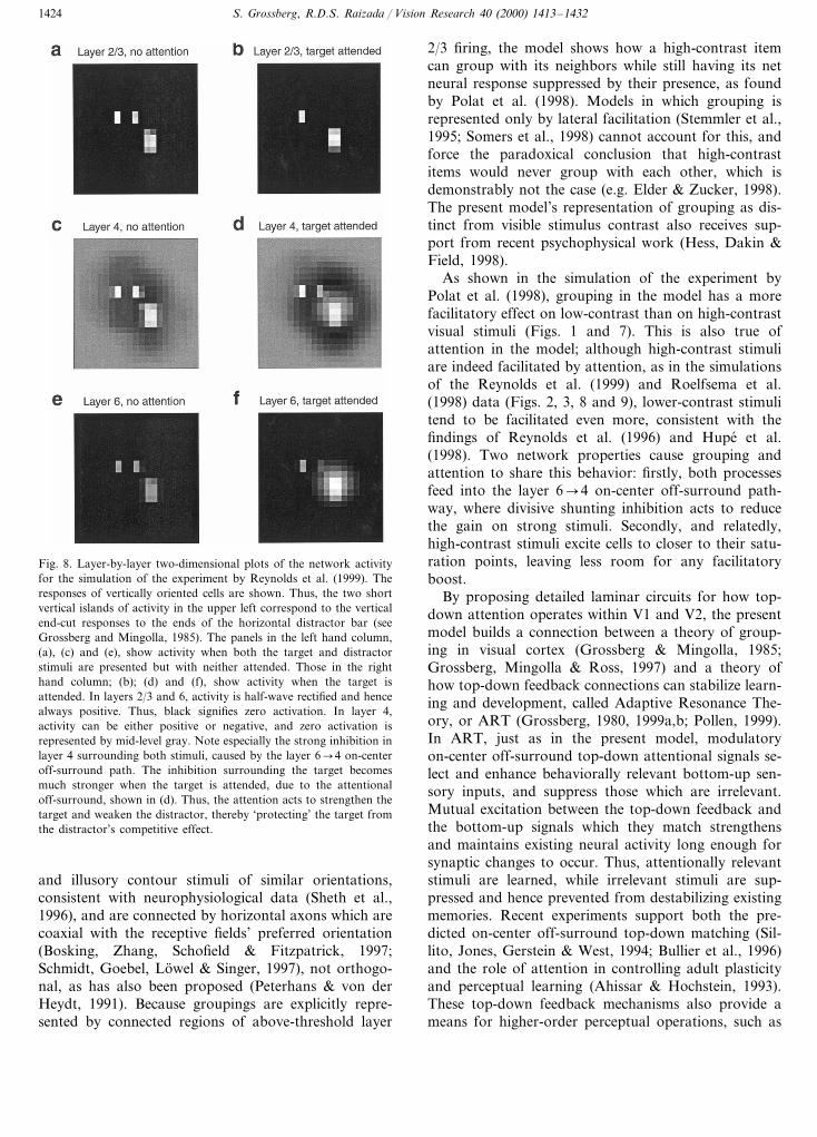
Fig. 8. Layer-by-layer two-dimensional plots of the network activityfor the simulation of the experiment by Reynolds et al. (1999). Theresponses of vertically oriented cells are shown. Thus, the two shortvertical islands of activity in the upper left correspond to the verticalend-cut responses to the ends of the horizontal distractor bar (seeGrossberg and Mingolla, 1985). The panels in the left hand column,(a), (c) and (e), show activity when both the target and distractorstimuli are presented but with neither attended. Those in the righthand column; (b); (d) and (f), show activity when the target isattended. In layers 2/3 and 6, activity is half-wave rectified and hencealways positive. Thus, black signifies zero activation. In layer 4,activity can be either positive or negative, and zero activation isrepresented by mid-level gray. Note especially the strong inhibition inlayer 4 surrounding both stimuli, caused by the layer 6�4 on-centeroff-surround path. The inhibition surrounding the target becomesmuch stronger when the target is attended, due to the attentionaloff-surround, shown in (d). Thus, the attention acts to strengthen thetarget and weaken the distractor, thereby ‘protecting’ the target fromthe distractor’s competitive effect.
2/3 firing, the model shows how a high-contrast itemcan group with its neighbors while still having its netneural response suppressed by their presence, as foundby Polat et al. (1998). Models in which grouping isrepresented only by lateral facilitation (Stemmler et al.,1995; Somers et al., 1998) cannot account for this, andforce the paradoxical conclusion that high-contrastitems would never group with each other, which isdemonstrably not the case (e.g. Elder & Zucker, 1998).The present model’s representation of grouping as dis-tinct from visible stimulus contrast also receives sup-port from recent psychophysical work (Hess, Dakin &Field, 1998).
As shown in the simulation of the experiment byPolat et al. (1998), grouping in the model has a morefacilitatory effect on low-contrast than on high-contrastvisual stimuli (Figs. 1 and 7). This is also true ofattention in the model; although high-contrast stimuliare indeed facilitated by attention, as in the simulationsof the Reynolds et al. (1999) and Roelfsema et al.(1998) data (Figs. 2, 3, 8 and 9), lower-contrast stimulitend to be facilitated even more, consistent with thefindings of Reynolds et al. (1996) and Hupe et al.(1998). Two network properties cause grouping andattention to share this behavior: firstly, both processesfeed into the layer 6�4 on-center off-surround path-way, where divisive shunting inhibition acts to reducethe gain on strong stimuli. Secondly, and relatedly,high-contrast stimuli excite cells to closer to their satu-ration points, leaving less room for any facilitatoryboost.
By proposing detailed laminar circuits for how top-down attention operates within V1 and V2, the presentmodel builds a connection between a theory of group-ing in visual cortex (Grossberg & Mingolla, 1985;Grossberg, Mingolla & Ross, 1997) and a theory ofhow top-down feedback connections can stabilize learn-ing and development, called Adaptive Resonance The-ory, or ART (Grossberg, 1980, 1999a,b; Pollen, 1999).In ART, just as in the present model, modulatoryon-center off-surround top-down attentional signals se-lect and enhance behaviorally relevant bottom-up sen-sory inputs, and suppress those which are irrelevant.Mutual excitation between the top-down feedback andthe bottom-up signals which they match strengthensand maintains existing neural activity long enough forsynaptic changes to occur. Thus, attentionally relevantstimuli are learned, while irrelevant stimuli are sup-pressed and hence prevented from destabilizing existingmemories. Recent experiments support both the pre-dicted on-center off-surround top-down matching (Sil-lito, Jones, Gerstein & West, 1994; Bullier et al., 1996)and the role of attention in controlling adult plasticityand perceptual learning (Ahissar & Hochstein, 1993).These top-down feedback mechanisms also provide ameans for higher-order perceptual operations, such as
and illusory contour stimuli of similar orientations,consistent with neurophysiological data (Sheth et al.,1996), and are connected by horizontal axons which arecoaxial with the receptive fields’ preferred orientation(Bosking, Zhang, Schofield & Fitzpatrick, 1997;Schmidt, Goebel, Lowel & Singer, 1997), not orthogo-nal, as has also been proposed (Peterhans & von derHeydt, 1991). Because groupings are explicitly repre-sented by connected regions of above-threshold layer

S. Grossberg, R.D.S. Raizada / Vision Research 40 (2000) 1413–1432 1425
sensitivity to figure vs. background (Grossberg, 1994),to modulate the activity of V1 cells (Lamme, Super &Spekreijse, 1998). The model’s proposal that bottom-up sensory activity is enhanced when matched by top-down signals is in accord with a hugeneurophysiological literature showing the facilitatoryeffect of attentional feedback (e.g. Luck et al., 1997;Roelfsema et al., 1998), but not with a recent modelof visual cortex (Rao & Ballard, 1999) in whichmatches with top-down feedback cause suppression.
Finally, many testable neurophysiological predic-tions follow from the model’s laminar functional ar-chitecture. A core prediction is that the layer 6�4on-center should be subthreshold: in particular, intra-cellularly evoked layer 6 activity should modulate, but
not drive, layer 4 spiny stellates and layer 2/3 pyrami-dals. The model proposes that attentional feedbackinto layer 6 passes into this modulatory 6�4 on-cen-ter to remain subthreshold in the absence of bottom-up visual input. Thus, it predicts that attentionalelevation of a neuron’s baseline firing rate when thereis no stimulus in its receptive field, as observed byLuck et al. (1997), should cause above-threshold acti-vation in layer 6, but below-threshold activation oflayer 4 spiny stellates. Note that Luck et al. foundthis baseline elevation in V2 but not in V1. Since wesuggest that the laminar mechanisms of attention aresimilar in both V1 and V2, differing only in spatialscale, we predict that this pattern of above-thresholdattentional activation of layer 6 but not 4 shouldhold in both areas. It is possible that only very atten-tionally demanding tasks, requiring discriminations atfine spatial resolution, will reveal such activity in V1.A similarity between attention and grouping whichthe model proposes is that V2 groupings should feedback into V1 through the same pathway as atten-tional signals. For example, widely spaced collinearinducers (like the flankers in the study by Polat et al.,1998), should cause illusory contour activation in V2layer 2/3, but not V1 layer 2/3, with feedback fromthis V2 grouping supraliminally activating V1 layer 6but not 4, just like attention to empty space. Suchdetailed structural and functional predictions by themodel will, we hope, help to stimulate further studyof the laminar organization of visual cortex, as wellas of other neocortical areas, whose horizontal inter-actions may be used to group different types of infor-mation, both as source and target of attentionalmodulation.
Fig. 9.
Fig. 9. Time-courses of activity across the layers of V1 at differentpositions along the traced curve in the simulation of the experimentby Roelfsema et al. (1998). Panels (a), (b) and (c) respectively showactivity at increasing distances away from the attended end of thecurve. For all panels, the solid lines represent activity when thereceptive field of the recorded neuron falls on the target curve, andhence is attended, and dashed lines represent activity when the curvehas become a distractor and is unattended. It can be seen that afteran initial onset transient, attention produces a sustained enhancementof neural activity across cortical layers 2/3, 4 and 6. The inter-laminarlayer 2/3�6�4 feedback is responsible for yoking the layers’ behav-ior together in this way. Note that at increasing distances from theattended zone, the strength of the attentional enhancement declines,and its latency increases. This can be seen most clearly by comparingthe time at which the target and distractor activities start to divergein panels (a) and (c). The weaker enhancement and longer latenciesare caused by the gradual attenuation of attentional signals as theyflow over time along layer 2/3. Note that although in these simula-tions layer 6 has stronger activity than layers 2/3 and 4, this is not astrong prediction of the model, since it is parameter-dependent. Seethe Discussion section for several testable predictions which derivefrom the model’s functional architecture, rather than its parametersettings.

S. Grossberg, R.D.S. Raizada / Vision Research 40 (2000) 1413–14321426
Acknowledgements
Stephen Grossberg was supported in part by theDefense Advanced Research Projects Agency and theOffice of Naval Research (ONR N00014-95-1-0409),the National Science Foundation (NSF IRI 97-20333)and the Office of Naval Research (ONR N00014-95-1-0657). Rajeev D.S. Raizida was supported in part bythe Defense Advanced Research Projects Agency andthe Office of Naval Research (ONR N00014-92-J-1309and ONR N00014-95-1-0657).
Appendix A. Model equations
The model LGN, V1 and V2 were simulated as anetwork of interacting neurons, each with a singlevoltage compartment whose membrane potential, V(t),was given by an equation of the form:
Cm
dV(t)dt
= − [V(t)−Eexcit]gexcit(t)
− [V(t)−Einhib]ginhib(t)− [V(t)−Eleak]gleak.
(1)In this equation, the time-varying conductances
gexcit(t) and ginhib(t) represent, respectively, the totalinputs from the excitatory and inhibitory neuronssynapsing onto the cell, as determined by the modelarchitecture shown in Fig. 6. The gleak term is a con-stant leakage conductance, and the E terms representreversal potentials. At equilibrium, the above equationcan be written as V= (Eexcit gexcit+Einhib ginhib+Eleak gleak)/(gexcit+ginhib+gleak). Thus, increases in theexcitatory and inhibitory conductances depolarise andhyperpolarise the membrane potential, respectively, asshown by the numerator of this term, and all theconductances contribute to divisive normalization ofthe membrane potential, as shown by the denominator.This divisive effect includes the special case of pure‘shunting’ inhibition when the reversal potential of theinhibitory channel is close to the cell’s resting potential(Borg-Graham, Monier & Fregnac, 1998). The follow-ing network equations are instances of this generalmembrane equation, where, for simplicity, the reversalpotentials are set to: Eexcit=1, Einhib= −1, Eleak=0,except where indicated. These continuous-time differen-tial equations were implemented in Matlab and numer-ically integrated using a fourth-order Runge-Kuttaalgorithm until equilibrium was reached. Computedintegrations were independently verified using Matlab’sbuilt-in adaptive-step-size second and third-order differ-ential equation solvers.
A.1. Retina
The model retina has at each position (i, j ) both anON cell, u+
ij , whose receptive field has the form of a
narrow on-center and a Gaussian off-surround, and anOFF cell, u−
ij , with a narrow off-center and a Gaussianon-surround (Schiller, 1992). As is observed in vivo,these ON and OFF cells feedforward into ON and OFFchannels of the LGN, and enable the network to re-spond both to light increments and to light decrements.The retinal cell activities caused by constant visualinputs I have the equilibrium values:
uij+=Iij−%
pq
Gpq(i, j, s1)Ipq (2)
uij−= −Iij+%
pq
Gpq(i, j, s1)Ipq (3)
where Gpq(i, j, s) is a two-dimensional Gaussian kernel,given by:
Gpq(i, j, s)=1
2 p s2 exp�
−1
2 s2((p− i)2+ (q−1)2)�
.
(4)
The Gaussian width parameter was set to: s1=1.
A.2. Lateral geniculate nucleus
The ON and OFF cells of the LGN, 6+ij and 6−ij , areexcited by the half-wave rectified ON and OFF cells ofthe retina, respectively. These retinal inputs are alsomultiplicatively gain-controlled by on-center off-sur-round feedback from V1 layer 6 (Sillito et al., 1994;Gove, Grossberg & Mingolla, 1995; Przybyszewski,Foote & Pollen, 1998). Layer 6 cells, xijk, at position (i,j ) and of all orientations, k, send on-center excitation,Aij, to LGN neurons at the same position, and send atwo-dimensional Gaussian spread of off-surround inhi-bition, Bij, to LGN neurons at the same and nearbypositions:
1d6
ddt6 ij+= −6 ij
++ (1−6 ij+)[uij
+]+(1+Aij)− (1+6 ij+)Bij
(5)
1d6
ddt6 ij−= −6 ij
−+ (1−6 il−)[uij
−]+(1+Aij)− (1+6 ij−)Bij
(6)
In Eqs. (5) and (6), the layer 6 on-center off-surroundfeedback terms, Aij and Bij, are given by:
Aij=C1 %k
xijk (7)
Bij=C2 %pqk
Gpq(i, j,s1)xijk, (8)
where the off-surround Gaussian, Gpq(i, j, s1) is definedby Eq. (4), and the notation [u+
ij ]+ signifies half-waverectification, [u+
ij ]+=max(u+ij , 0). The parameters for
the LGN were: d6=1.25, C1=1.5, C2=0.075.
S. Grossberg, R.D.S. Raizada / Vision Research 40 (2000) 1413–1432 1427
A.3. LGN inputs to cortical simple cells
Although the exact mechanisms through which sim-ple cells gain their orientation tuning are still controver-sial (Reid & Alonso, 1995; Ferster, Chung & Wheat,1996; Sompolinsky & Shapley, 1997; Adorjan, Levitt,Lund & Obermayer, 1999), recent intracellular studiesof simple cells in layers 4 and 6 have revealed muchabout the structure and interaction of their ON andOFF subfields (Hirsch, Alonso, Reid & Martinez,1998), supplementing and confirming earlier studies(Ferster, 1988). These results are incorporated, withsome simplifications, into the current model.
At each position (i, j ), and for each orientation, k,the model has a even-symmetric simple cell with twoparallel elongated parts: an ON subregion, Rijk, whichreceives excitation from LGN ON cells beneath it andis inhibited by LGN OFF cells at the same position;and an OFF subregion, Lijk, which has the converserelation to the LGN channels (Reid & Alonso, 1995;Hirsch et al., 1998). This physiology is embodied in theequation for the ON subregion by subtracting thehalf-wave rectified LGN OFF channel, [6−pq ]+, from therectified ON channel, [6+pq ]+, and convolving the resultwith the positive lobe of a Difference-of-Offset-Gaus-sians (DOOG) kernel, [D (k)
ijk ]+, which has the simple cellsubfield’s characteristic oriented elongated shape. TheOFF subregion, Lijk, is similarly constructed:
Rijk=%pq
([6pq+ ]+− [6pq
− ]+)[Dpqij(k) ]+ (9)
Lijk=%pq
([6pq− ]+− [6pq
+ ]+)[−Dpqij(k) ]+ (10)
where the oriented DOOG filter D (k)pqij is given by:
Dpqij(k) =Gpq(i−d cos u, j−d sin u, s2)
−Gpq(i+d cos u, j+d sin u, s2) (11)
with d=s2/2 and u=p(k−1)/K, where k ranges from1 to 2 K, K being the total number of orientations. Forsimplicity, the number of orientations was set to K=2(vertical and horizontal) in the present simulations. Thewidth parameter for the DOOG filter was s2=0.5.
At an oriented contrast edge, a suitably orientedsimple cell of the correct polarity will have its ONsubfield stimulated by a luminance increment and itsOFF subfield stimulated by an equal but oppositedecrement. The optimal nature of this stimulus is em-bodied in the following equation, in which simple cellactivity is the rectified sum of the activities of eachsubfield, minus their difference:
Sijk=g [Rijk+Lijk− �Rijk−Lijk �]+ (12)
Recent physiological studies have confirmed thatlayer 4 simple cells that are sensitive to opposite con-trast polarities pool their outputs at layer 2/3 complex
cells (Alonso & Martinez, 1998). In order to make thesimulations manageable, cells in layers 6 and 4 wereimplemented with their simple cell inputs alreadypooled, thus halving the number of cells. Since thepresent model is not used to simulate any polarity-spe-cific interactions in these layers, this simplificationleaves the output unaffected. Thus, the polarity-pooledinput from LGN to cortical layers 6 and 4 was calcu-lated as the term Cijk, which pools over opposite-polar-ity simple cells:
Cijk=Sijk+Sij(k+K) (13)
where k ranges from 1 to K. The parameter for thesimple cell responses, was set to g=10.
A.4. Layer 6 cells
V1 layer 6 cells, xijk, receive input from the LGN(Blasdel & Lund, 1983), which, as described above, isrepresented by the contrast-polarity pooled orientedinput, Cijk They also receive two types of folded feed-back excitation. The first type is intracortical feedbackfrom above-threshold pyramidal cells in V1 layer 2/3,zijk (Blasdel, Lund & Fitzpatrick, 1985; Kisvarday,Cowey, Smith & Somogyi, 1989). These are passedthrough a thresholding signal function, F, given by:
F(zijk, G)=max(zijk−G, 0), (14)
where G is the threshold value. The second type offolded feedback is intercortical attentional feedbackfrom V2, xV2
ijk (Sandell & Schiller, 1982), originating inV2 layer 6 (Rockland & Virga, 1989). The feedbackaxons from V2 terminate predominantly in V1 layer 1(Rockland, 1994). There exist several routes throughwhich these layer 1 signals can pass down into layer 6,notably via the layer 1 apical dendritic tufts of layer 5pyramidals with axon collaterals in 6 (Lund & Boothe,1975; Gilbert & Wiesel, 1979, see also Table 1). Thesepaths are not explicitly implemented in the presentmodel. In attentional simulations where only the V1part of the complete V1/V2 network is simulated, thecorticocortical feedback term, xV2
ijk , is replaced by atwo-dimensional Gaussian spread of attentional signals,centered on the attended location and exciting all orien-tations equally. Thus:
1dC
ddt
xijk= −xijk+ (1−xijk)
× (a Cijk+f F(zijk, G)+V21xijkV2), (15)
This equation was solved at equilibrium, giving:
xijk=a Cijk+f F(zijk, G)+V21 xijk
V2
1+a Cijk+f F(zijk, G)+V21 xijkV2. (16)
Parameters for the terms in the layer 6 equationwere: dC=0.25, a=0.5, f=2.0, G=0.2, V21=1.5.
S. Grossberg, R.D.S. Raizada / Vision Research 40 (2000) 1413–14321428
A.5. Layer 4 acti6ity
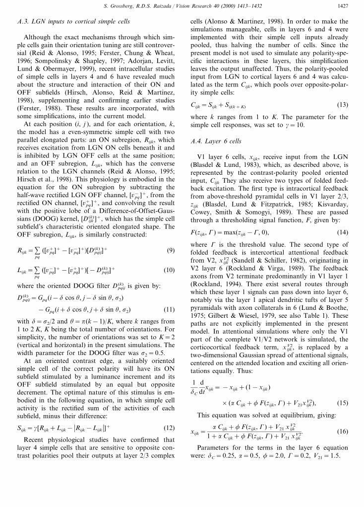
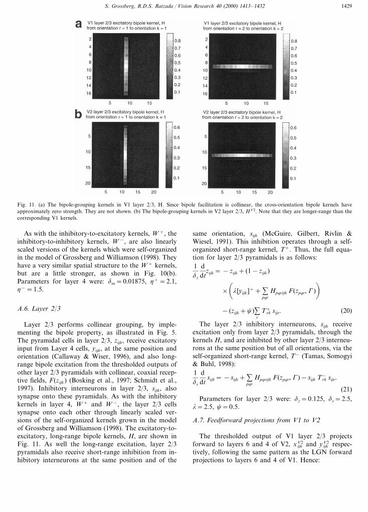
Model spiny stellate cells in layer 4, yijk, as well asreceiving the contrast-polarity pooled oriented input,Cijk, described above, also receive on-center off-sur-round input from layer 6. The on-center consists ofexcitatory connections from layer 6, xijk, to layer 4spiny stellates at the same position and of the sameorientation (Stratford et al., 1996; Wiser & Callaway,1997). The off-surround input is caused by medium-range projections from layer 6 onto layer 4 inhibitoryinterneurons (McGuire, Hornung, Gilbert & Wiesel,1984; Ahmed et al., 1997). The spatial distribution andstrength of these connections are determined by a two-dimensional kernel, W +
pqrijk, which is in the presentmodel a linearly scaled version of a self-organized 6–4inhibitory kernel grown in the developmental study byGrossberg and Williamson (1998) using the same net-work architecture, but without the corticocortical feed-back connections. The spatial distribution of thiskernel, which is approximately Gaussian, is shown inFig. 10(a). Therefore, the distribution of the off-sur-round inhibition in the present model is not hand-crafted by an algebraic equation, but is instead theproduct of a self-organized equilibrium reached by thesame network architecture in response to naturallystructured visual inputs. Thus, the equation for layer 4spiny stellates is:
1dC
ddt
yijk= −yijk+ (1−yijk)(Cijk+h+xijk)
− (yijk+1)%pqr
Wpqrijk+ mpqr. (17)
This was solved at equilibrium, giving:
yijk=Cijk+h+xijk− %
pqr
Wpqrijk+ mpqr
1+Cijk+h+xijk+%pqr Wpqrijk+ mpqr
(18)
Note that the 6–4 off-surround spatially overlapswith the on-center, with the consequence that the centerexcitation is inhibited down into being subthreshold, ormodulatory (Callaway, 1998, p. 56). This property isthe key to the attentional simulations performed, asdiscussed above (See Fig. 4).
Layer 4 inhibitory interneurons, mijk, also receiveon-center off-surround input, the on-center again com-ing from layer 6 cells with the same position andorientation, xijk, and the off-surround inhibition comingvia the spatial kernels, W −, of the other inhibitoryinterneurons in layer 4 (Ahmed et al., 1997). Theseinhibitory-to-inhibitory synapses help to normalize thetotal amount of inhibition present at a given position inlayer 4. Thus:
1dm
ddt
mijk= −mijk+h−xijk−mijk %pqr
Wpqrijk− mpqr. (19)
Fig. 10. (a) The inhibitory-to-excitatory off-surround kernels in Layer 4, W +. Only the kernels operating on vertically oriented cells are shown,since those operating on horizontally oriented cells are the same, but rotated by ninety degrees. (b) The inhibitory-to-inhibitory off-surroundkernels in layer 4, W −. Again, only the vertical kernels are shown.
S. Grossberg, R.D.S. Raizada / Vision Research 40 (2000) 1413–1432 1429
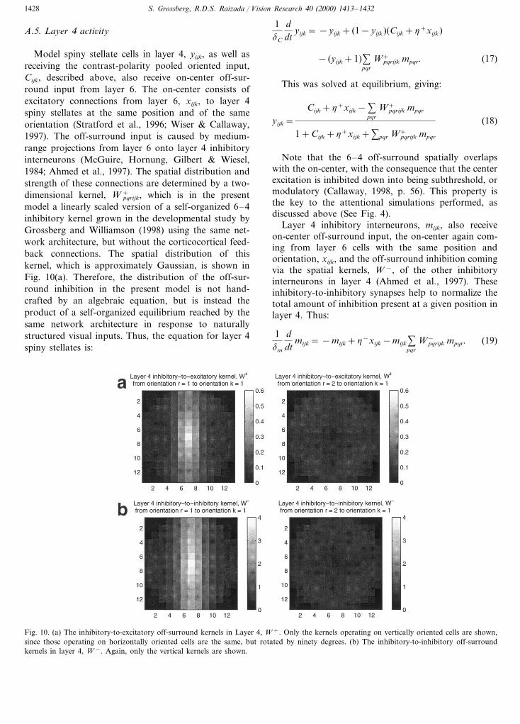
Fig. 11. (a) The bipole-grouping kernels in V1 layer 2/3, H. Since bipole facilitation is collinear, the cross-orientation bipole kernels haveapproximately zero strength. They are not shown. (b) The bipole-grouping kernels in V2 layer 2/3, HV2. Note that they are longer-range than thecorresponding V1 kernels.
As with the inhibitory-to-excitatory kernels, W +, theinhibitory-to-inhibitory kernels, W −, are also linearlyscaled versions of the kernels which were self-organizedin the model of Grossberg and Williamson (1998). Theyhave a very similar spatial structure to the W + kernels,but are a little stronger, as shown in Fig. 10(b).Parameters for layer 4 were: dm=0.01875, h+=2.1,h−=1.5.
A.6. Layer 2/3
Layer 2/3 performs collinear grouping, by imple-menting the bipole property, as illustrated in Fig. 5.The pyramidal cells in layer 2/3, zijk, receive excitatoryinput from Layer 4 cells, yijk, at the same position andorientation (Callaway & Wiser, 1996), and also long-range bipole excitation from the thresholded outputs ofother layer 2/3 pyramidals with collinear, coaxial recep-tive fields, F(zijk) (Bosking et al., 1997; Schmidt et al.,1997). Inhibitory interneurons in layer 2/3, sijk, alsosynapse onto these pyramidals. As with the inhibitorykernels in layer 4, W + and W −, the layer 2/3 cellssynapse onto each other through linearly scaled ver-sions of the self-organized kernels grown in the modelof Grossberg and Williamson (1998). The excitatory-to-excitatory, long-range bipole kernels, H, are shown inFig. 11. As well the long-range excitation, layer 2/3pyramidals also receive short-range inhibition from in-hibitory interneurons at the same position and of the
same orientation, sijk (McGuire, Gilbert, Rivlin &Wiesel, 1991). This inhibition operates through a self-organized short-range kernel, T+. Thus, the full equa-tion for layer 2/3 pyramidals is as follows:1dz
ddt
zijk= −zijk+ (1−zijk)
�
l [yijk ]++ %pqr
Hpqrijk F(zpqr, G)�
− (zijk+c)%r
Trk+ sijr. (20)
The layer 2/3 inhibitory interneurons, sijk receiveexcitation only from layer 2/3 pyramidals, through thekernels H, and are inhibited by other layer 2/3 interneu-rons at the same position but of all orientations, via theself-organized short-range kernel, T− (Tamas, Somogyi& Buhl, 1998):1ds
ddt
sijk= −sijk+ %pqr
Hpqrijk F(zpqr, G)−sijk Trk− sijr.
(21)Parameters for layer 2/3 were: dz=0.125, ds=2.5,
l=2.5, c=0.5.
A.7. Feedforward projections from V1 to V2
The thresholded output of V1 layer 2/3 projectsforward to layers 6 and 4 of V2, xV2
ijk and yV2ijk respec-
tively, following the same pattern as the LGN forwardprojections to layers 6 and 4 of V1. Hence:
S. Grossberg, R.D.S. Raizada / Vision Research 40 (2000) 1413–14321430
1dC
ddt
xijkV2= −xijk
V2+ (1−xijkV2)
× (V126 F(zijk, G)+f F(z ijk
V2, G)) (22)
1dC
ddt
y ijkV2= −yijk
V2+ (1−yijkV2)(V12
4 F(zijk, G)+h+xijkV2)
− (yijkV2+1)%
pqr
Wpqrijk+ mpqr
V2 . (23)
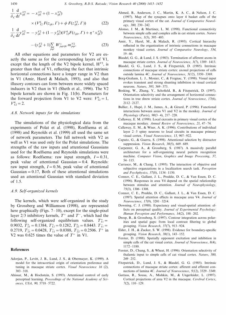
All other equations and parameters for V2 are ex-actly the same as for the corresponding layers of V1,except that the length of the V2 bipole kernel, HV2, isgreater than that of V1, reflecting the fact that intrinsichorizontal connections have a longer range in V2 thanin V1 (Amir, Harel & Malach, 1993), and also thatillusory contours can form between more widely spacedinducers in V2 than in V1 (Sheth et al., 1996). The V2bipole kernels are shown in Fig. 11(b). Parameters forthe forward projection from V1 to V2 were: V6
12=1,V4
12=2.
A.8. Network inputs for the simulations
The simulations of the physiological data from theexperiments of Polat et al. (1998), Roelfsema et al.(1998) and Reynolds et al. (1999) all used the same setof network parameters. The full network with V2 aswell as V1 was used only for the Polat simulations. Thestrengths of the raw inputs and attentional Gaussiansused for the Roelfsema and Reynolds simulations wereas follows: Roelfsema: raw input strength, I=0.31,peak value of attentional Gaussian=0.4. Reynolds:raw input strength, I=0.36, peak value of attentionalGaussian=0.17, Both of these attentional simulationsused an attentional Gaussian with standard deviationof 1.5.
A.9. Self-organized kernels
The kernels, which were self-organized in the studyby Grossberg and Williamson (1998), are representedhere graphically (Figs. 7–10), except for the single-pixellayer 2/3 inhibitory kernels, T+ and T−, which had thefollowing self-organized equilibrium values. T+
11=0.9032, T+
21=0.1384, T+12=0.1282, T+
22=0.8443. T−11=
0.2719, T−21=0.0428, T−
11=0.0388, T−22=0.2506. T+ in
V2 was 0.625 times the value of T+ in V1.
References
Adorjan, P., Levitt, J. B., Lund, J. S., & Obermayer, K. (1999). Amodel for the intracortical origin of orientation preference andtuning in macaque striate cortex. Visual Neuroscience 16 (2),303–318.
Ahissar, M., & Hochstein, S. (1993). Attentional control of earlyperceptual learning. Proceedings of the National Academy of Sci-ences, USA, 90, 5718–5722.
Ahmed, B., Anderson, J. C., Martin, K. A. C., & Nelson, J. C.(1997). Map of the synapses onto layer 4 basket cells of theprimary visual cortex of the cat. Journal of Comparati6e Neurol-ogy, 380, 230–242.
Alonso, J. M., & Martinez, L. M. (1998). Functional connectivitybetween simple cells and complex cells in cat striate cortex. NatureNeuroscience, 1(5), 395–403.
Amir, Y., Harel, M., & Malach, R. (1993). Cortical hierarchyreflected in the organization of intrinsic connections in macaquemonkey visual cortex. Journal of Comparati6e Neurology, 334,19–46.
Blasdel, G. G., & Lund, J. S. (1983). Termination of afferent axons inmacaque striate cortex. Journal of Neuroscience, 3(7), 1389–1413.
Blasdel, G. G., Lund, J. S., & Fitzpatrick, D. (1985). Intrinsicconnections of macaque striate cortex: axonal projections of cellsoutside lamina 4C. Journal of Neuroscience, 5(12), 3350–3369.
Borg-Graham, L. J., Monier, C., & Fregnac, Y. (1998). Visual inputevokes transient and strong shunting inhibition in visual corticalneurons. Nature, 393, 369–373.
Bosking, W., Zhang, Y., Schofield, B., & Fitzpatrick, D. (1997).Orientation selectivity and the arrangement of horizontal connec-tions in tree shrew striate cortex. Journal of Neuroscience, 17(6),2112–2127.
Bullier, J., Hupe, J. M., James, A., & Girard, P. (1996). Functionalinteractions between areas V1 and V2 in the monkey. Journal ofPhysiology (Paris), 90(3–4), 217–220.
Callaway, E. M. (1998). Local cicrcuits in primary visual cortex of themacaque monkey. Annual Re6iew of Neuroscience, 21, 47–74.
Callaway, E. M., & Wiser, A. K. (1996). Contributions of individuallayer 2–5 spiny neurons to local circuits in macaque primaryvisual cortex. Visual Neuroscience, 13, 907–922.
Caputo, G., & Guerra, S. (1998). Attentional selection by distractorsuppression. Vision Research, 38(5), 669–689.
Carpenter, G. A., & Grossberg, S. (1987). A massively parallelarchitecture for a self-organizing neural pattern recognitionmechine. Computer Vision, Graphics and Image Processing, 37,54–115.
Carrasco, M., & Chang, I. (1995). The interaction of objective andsubjective organzations in a localization search task. Perceptionand Psychphysics, 57(8), 1134–1150.
Connor, C. E., Gallant, J. L., Preddie, D. C., & Van Essen, D. C.(1996). Responses in area V4 depend on the spatial relationshipbetween stimulus and attention. Journal of Neurophysiology,75(3), 1306–1308.
Connor, C. E., Preddie, D. C., Gallant, J. L., & Van Essen, D. C.(1997). Spatial attention effects in macaque area V4. Journal ofNeuroscience, 17(9), 3201–3214.
Downing, C. J. (1988). Expectancy and visual-spatial attention: ef-fects on perceptual quality. Journal of Experimental Psychology :Human Perception and Performance, 14(2), 188–202.
Dresp, B., & Grossberg, S. (1997). Contour integration across polar-ities and spatial gaps: from local contrast filtering to globalgrouping. Vision Research, 37(7), 913–924.
Elder, J. H., & Zucker, S. W. (1998). Evidence for boundary-specificgrouping. Vision Research, 38(1), 143–152.
Ferster, D. (1988). Spatially opponent excitation and inhibition insimple cells of the cat visual cortex. Journal of Neuroscience, 8(4),1172–1180.
Ferster, D., Chung, S., & Wheat, H. (1996). Orientation selectivity ofthalamic input to simple cells of cat visual cortex. Nature, 380,249–252.
Fitzpatrick, D., Lund, J. S., & Blasdel, G. G. (1985). Intrinsicconnections of macaque striate cortex: afferent and efferent con-nections of lamina 4C. Journal of Neuroscience, 5(12), 3329–3349.
Gattass, R., Sousa, A., Mishkin, M., & Ungerleider, L. (1997).Cortical projections of area V2 in the macaque. Cerebral Cortex,7(2), 110–129.
S. Grossberg, R.D.S. Raizada / Vision Research 40 (2000) 1413–1432 1431
Gilbert, C. D., & Wiesel, T. N. (1979). Morphology and intracorticalprojections of functionally characterised neurones in the cat visualcortex. Nature, 280, 120–125.
Gove, A., Grossberg, S., & Mingolla, E. (1995). Brightness percep-tion, illusory contours, and corticogeniculate feedback. VisualNeuroscience, 12(6), 1027–1052.
Grosof, D. H., Shapley, R. M., & Hawken, M. J. (1993). MacaqueV1 neurons can signal ‘illusory’ contours. Nature, 365, 550–552.
Grossberg, S. (1973). Contour enhancement, short term memory, andconstancies in reverberating neural networks. Studies in AppliedMathematics, 52, 217–257. Reprinted in studies of mind andbrain, S. Grossberg (1982). D. Reidel Publishing Company: Dor-drecht, Holland.
Grossberg, S. (1980). How does a brain build a cognitive code?Psychological Re6iew, 87, 1–51.
Grossberg, S. (1994). 3-D vision and figure-ground separation byvisual cortex. Perception and Psychophysics, 55(1), 48–120.
Grossberg, S. (1999a). How does the cerebral cortex work? Learning,attention, and grouping by the laminar circuits of visual cortex.Spatial Vision, 12, 163–187.
Grossberg, S. (1999b). The link between brain learning, attention,and consciousness. Consciousness and Cognition, 8, 1–44.
Grossberg, S., & Grunewald, A. (1997). Cortical synchronization andperceptual framing. Journal of Cogniti6e Neuroscience, 9(1), 117–132.
Grossberg, S., & Mingolla, E. (1985). Neural dynamics of formperception: boundary completion, illusory figures, and neon colorspreading. Psychological Re6iew, 92(2), 173–211.
Grossberg, S., Mingolla, E., & Ross, W. D. (1997). Visual brain andvisual perception: how does the cortex do perceptual grouping?Trends in Neurosciences, 20(3), 106–111.
Grossberg, S., & Somers, D. (1991). Synchronized oscillations duringcooperative feature linking in a cortical model of visual percep-tion. Neural Networks, 4(4), 453–466.
Grossberg, S., & Williamson, J. R. (1998). A neural model of howvisual cortex develops a laminar architecture capable of adultperceptual grouping. Boston University Technical Report CAS/CNS-98-022.
Heeger, D. J. (1992). Normalization of cell responses in cat striatecortex. Visual Neuroscience, 9(2), 181–197.
Hess, R. F., Dakin, S. C., & Field, D. J. (1998). The role of ‘contrastenhancement’ in the detection and appearance of visual contours.Vision Research, 38, 783–787.
Hirsch, J. A., Alonso, J. M., Reid, R. C., & Martinez, L. M. (1998).Synaptic integration in striate cortical simple cells. Journal ofNeuroscience, 18(22), 9517–9528.
Hirsch, J. A., & Gilbert, C. D. (1991). Synaptic physiology ofhorizontal connections in the cat’s visual cortex. Journal of Neu-roscience, 11(6), 1800–1809.
Hupe, J. M., James, A. C., Payne, B. R., Lomber, S. G., Girard, P.,& Bullier, J. (1998). Cortical feedback improves discriminationbetween figure and background by V1, V2 and V3 neurons.Nature, 394, 784–787.
Kapadia, M. K., Ito, M., Gilbert, C. D., & Westheimer, G. (1995).Improvement in visual sensitivity by changes in local context:parallel studies in human observers and in V1 of alert monkeys.Neuron, 15, 843–856.
Kisvarday, Z. F., Cowey, A., Smith, A. D., & Somogyi, P. (1989).Interlaminar and lateral excitatory amino acid connections in thestriate cortex of monkey. Journal of Neuroscience, 9(2), 667–682.
Kubovy, M., Holcombe, A. O., & Wagemans, J. (1998). On thelawfulness of grouping by proximity. Cogniti6e Psychology, 35,71–98.
LaBerge, D., & Brown, V. (1989). Theory of attentional operations inshape identification. Psychological Re6iew, 96(1), 101–124.
Lamme, V. A. F., Rodriguez-Rodriguez, V., & Spekreijse, H. (1999).Separate processing dynamics for texture elements, boundaries
and surfaces in primary visual cortex of the macaque monkey.Cerebral Cortex, 9(4), 406–413.
Lamme, V. A. F., Super, H., & Spekreijse, H. (1998). Feedforward,horizontal and feedback processing in the visual cortex. CurrentOpinion in Neurobiology, 8(4), 529–535.
Lee, T. S., Mumford, D., Romero, R., & Lamme, V. A. F. (1998).The role of primary visual cortex in higher level vision. VisionResearch, 38(15/16), 2429–2454.
LeVay, S., & Gilbert, C. D. (1976). Laminar patterns of geniculocor-tical projection in the cat. Brain Research, 113, 1–19.
Levick, W. R., & Williams, W. O. (1964). Maintained activity oflateral geniculate neurones in darkness. Journal of Physiology(London), 170, 582–597.
Li, Z. (1998). A neural model of contour integration in the primaryvisual cortex. Neural Computation, 10, 903–940.
Luck, S. J., Chelazzi, L., Hillyard, S. A., & Desimone, R. (1997).Neural mechanisms of spatial selective attention in areas V1, V2,and V4 of macaque visual cortex. Journal of Neurophysiology, 77,24–42.
Luck, S., Hillyard, S. A., Mouloua, M., Woldorff, M. G., Clark, V.P., & Hawkins, H. L. (1994). Effects of spatial cuing on lumi-nance detectability: psychophysical and electrophysiological evi-dence for early selection. Journal of Experimental Psychology :Human Perception and Performance, 20(4), 887–904.
Lund, J. S. (1987). Local circuit neurons of macaque monkey striatecortex: I. Neurons of laminae 4C and 5A. Journal of Comparati6eNeurology, 257, 60–92.
Lund, J. S., & Boothe, R. G. (1975). Interlaminar connections andpyramidal neuron organisation in the visual cortex, area 17, of themacaque monkey. Journal of Comparati6e Neurology, 159, 305–334.
Lund, J. S., Hawken, M. J., & Parker, A. J. (1988). Local circuitneurons of macaque monkey striate cortex: II. Neurons of lami-nae 5B and 6. Journal of Comparati6e Neurology, 276, 1–29.
Lund, J. S., & Wu, C. Q. (1997). Local circuit neurons of macaquemonkey striate cortex: IV. Neurons of laminae 1-3A. Journal ofComparati6e Neurology, 384, 109–126.
McGuire, B. A., Gilbert, C. D., Rivlin, P. K., & Wiesel, T. N. (1991).Targets of horizontal connections in macaque primary visualcortex. Journal of Comparati6e Neurology, 305(3), 370–392.
McGuire, B. A., Hornung, J. P., Gilbert, C. D., & Wiesel, T. N.(1984). Patterns of synaptic input to layer 4 of cat striate cortex.Journal of Neuroscience, 4(12), 3021–3033.
Mignard, M., & Malpeli, J. G. (1991). Paths of information flowthrough visual cortex. Science, 251, 1249–1251.
Montero, V. M. (1991). A quantitative study of synaptic contacts oninterneurons and relay cells of the cat lateral geniculate nucleus.Experimental Brain Research, 86, 257–270.
Moore, C. M., & Egeth, H. (1997). Perception without attention:evidence of grouping under conditions of inattention. Journal ofExperimental Psychology : Human Perception and Performance,23(2), 339–352.
Motter, B. C. (1993). Focal attention produces spatially selectiveprocessing in visual cortical areas V1, V2 and V4 in the presenceof competing stimuli. Journal of Neurophysiology, 70, 909–919.
Paradiso, M. A., & Nakayama, K. (1991). Brightness perception andfilling-in. Vision Research, 31(7/8), 1221–1236.
Pessoa, L., Thompson, E., & Noe, A. (1998). Finding out aboutfilling-in: a guide to perceptual completion for visual science andthe philosophy of perception. Beha6ioral and Brain Sciences,21(6), 723–802.
Peterhans, E., & von der Heydt, R. (1991). Subjective contours—bridging the gap between psychophysics and physiology. Trends inNeurosciences, 14, 112–119.
Peters, A. G., & Sethares, C. (1991). Organization of pyramidalneurons in area 17 of monkey visual cortex. Journal of Compara-ti6e Neurology, 306, 1–23.
S. Grossberg, R.D.S. Raizada / Vision Research 40 (2000) 1413–14321432
Polat, U., Mizobe, K., Pettet, M. W., Kasamatsu, T., & Norcia, A.M. (1998). Collinear stimuli regulate visual responses dependingon cell’s contrast threshold. Nature, 391, 580–584.
Pollen, D. A. (1999). On the neural correlates of visual perception.Cerebral Cortex, 9(1), 4–19.
Posner, M. I., Snyder, C. R., & Davidson, B. J. (1980). Attention andthe detection of signals. Journal of Experimental Psychology,109(2), 160–174.
Prinzmetal, W., & Keysar, B. (1989). Functional theory of illusoryconjunctions and neon colours. Journal of Experimental Psychol-ogy : General, 118(2), 165–190.
Przybyszewski, A. W., Foote, W., & Pollen, D. A. (1998). Contrastgain control of the LGN neurons by V1. In6estigati6e Ophthal-mology and Visual Science (Suppl.), 38, S238 (Abstract).
Raizada, R. D. S., & Grossberg, S. (1998). What are the layers ofcortex for A neural model of attention, perceptual grouping andlearning in V1 and V2. Society for Neuroscience Abstracts, 28,105.10 (Abstract).
Raizada, R. D. S., & Grossberg, S. (1999a). Context-sensitive percep-tual processing by the laminar circuits of V1 and V2: Interactionsof attention, orientation-contrast, and grouping of real and illu-sory contours (submitted for publication).
Raizada, R. D. S., & Grossberg, S. (1999b). Laminar substrates ofattention, grouping and perceptual learning in V1 and V2. In6es-tigati6e Ophthalmology and Visual Science (Suppl.), 40(4), S645(Abstract).
Rao, R. P. N., & Ballard, D. H. (1999). Predictive coding in thevisual cortex: a functional interpretation of some extra-classicalreceptive field effects. Nature Neuroscience, 2, 79–87.
Reid, R. C., & Alonso, J. M. (1995). Specificity of monosynapticconnections from thalamus to visual cortex. Nature, 378, 281–284.
Reynolds, J., Chelazzi, L., & Desimone, R. (1999). Competitivemechanisms subserve attention in macaque areas V2 and V4.Journal of Neuroscience, 19, 1736–1753.
Reynolds, J., Pasternak, T., & Desimone, R. (1996). Attention in-creases contrast sensitivity of cells in macaque area V4. Society forNeuroscience Abstracts, 22, 475.3 (Abstract).
Rockland, K. S. (1994). The organization of feedback connectionsfron area V2 (18) to V1 (17). In A. Peters, & K. S. Rockland,Cerebral Cortex, vol. 10 (pp. 261–299). New York: Plenum Press.
Rockland, K. S., & Virga, A. (1989). Terminal arbors of individual‘feedback’ axons projecting from area V2 to V1 in the macaquemonkey: a study using immunohistochemistry of anterogradelytransported phaseolus vulgaris-leucoagglutinin. Journal of Com-parati6e Neurology, 285 (1), 54–72.
Rockland, K. S., & Virga, A. (1990). Organization of individualcortical axons projecting from area V1 (area 17) to V2 (area 18)in the macaque monkey. Visual Neuroscience, 4(1), 11–28.
Roelfsema, P. R., Lamme, V. A. F., & Spekreijse, H. (1998). Object-based attention in the primary visual cortex of the macaquemonkey. Nature, 395, 376–381.
Rogers-Ramachandran, D. C., & Ramachandran, V. S. (1998). Psy-chophysical evidence for boundary and surface systems in humanvision. Vision Research, 38(1), 71–77.
Rossi, A. F., Desimone, R., & Ungerleider, L. G. (1998). Late onsetresponses to extra-receptive field stimulation in V1. Society forNeuroscience Abstracts, 28, 789.2 (Abstract).
Salin, P., & Bullier, J. (1995). Corticocortical connections in thevisual system: structure and function. Physiological Re6iews,75(1), 107–154.
Sandell, J. H., & Schiller, P. H. (1982). Effect of cooling area 18 onstriate cortex cells in the squirrel monkey. Journal of Neurophysi-ology, 48(1), 38–48.
Schiller, P. H. (1992). The ON and OFF channels of the visualsystem. Trends in Neurosciences, 15(3), 86–92.
Schmidt, K. E., Goebel, R., Lowel, S., & Singer, W. (1997). Theperceptual grouping criterion of colinearity is reflected by an-isotropies of connections in the primary visual cortex. EuropeanJournal of Neuroscience, 9, 1083–1089.
Sengpiel, F., Sen, A., & Blakemore, C. (1997). Characteristics ofsurround inhibition in cat area 17. Experimental Brain Research,116, 216–228.
Sheth, B. R., Sharma, J., Rao, S. C., & Sur, M. (1996). Orientationmaps of subjective contours in visual cortex. Science, 274, 2110–2115.
Sillito, A. M., Jones, H. E., Gerstein, G. L., & West, D. C. (1994).Feature-linked synchronization of thalamic relay cell firing in-duced by feedback from the visual cortex. Nature, 369, 479–482.
Somers, D. C., Dale, A. M., Seiffert, A. E., & Tootell, R. B. (1999).Functional MRI reveals spatially specific attentional modulationin human primary visual cortex. Proceedings of the NationalAcademy of Sciences, USA, 96, 1663–1668.
Somers, D. C., Todorov, E. V., Siapas, A. G., Toth, L. J., Kim, D.,& Sur, M. (1998). A local circuit approach to understandingintegration of long-range inputs in primary visual cortex. CerebralCortex, 8, 204–217.
Sompolinsky, H., & Shapley, R. (1997). New perspectives on themechanisms for orientation selectivity. Current Opinion in Neuro-biology, 7(4), 514–522.
Steinman, B. A., Steinman, S. B., & Lehmkuhle, S. (1995). Visualattention mechanisms show a canter-surround organization. Vi-sion Research, 35(13), 1859–1869.
Stemmler, M., Usher, M., & Niebur, E. (1995). Lateral interactions inprimary visual cortex: A model bridging physiology and psycho-physics. Science, 269, 1877–1880.
Stratford, K. J., Tarczy-Hornoch, K., Martin, K. A. C., Bannister, N.J., & Jack, J. J. B. (1996). Excitatory synaptic inputs to spinystellate cells in cat visual cortex. Nature, 382, 258–261.
Tamas, G., Somogyi, P., & Buhl, E. H. (1998). Differentially inter-connected networks of GABAergic interneurons in the visualcortex of the cat. Journal of Neuroscience, 18(11), 4255–4270.
Toth, L. J., Rao, S. C., Kim, D. S., Somers, D., & Sur, M. (1996).Subthreshold facilitation and suppression in primary visual cortexrevealed by intrinsic signal imaging. Proceedings of the NationalAcademy of Sciences, 93, 9869–9874.
Valverde, F. (1985). The organizing principles of the primary visualcortex in the monkey. In A. Peters, & E. G. Jones, CerebralCortex, vol. 3 (pp. 207–257). New York: Plenum.
Van Essen, D. C., Newsome, W. T., Maunsell, J. H., & Bixby, J. L.(1986). The projections from striate cortex (V1) to areas V2 andV3 in the macaque monkey: asymmetries, areal boundaries, andpatchy connections. Journal of Comparati6e Neurology, 244(4),451–480.
von der Heydt, R., Peterhans, E., & Baumgartner, G. (1984). Illusorycontours and cortical neuron responses. Science, 224, 1260–1262.
Watanabe, T., Sasaki, Y., Nielsen, M., Takino, R., & Miyakawa, S.(1998). Attention-regulated activity in human primary visual cor-tex. Journal of Neurophysiology, 79, 2218–2221.
Wiser, A. K., & Callaway, E. M. (1997). Ocular dominance columnsand local projections of layer 6 pyramidal neurons in macaqueprimary visual cortex. Visual Neuroscience, 14, 241–251.
Yen, S. C., & Finkel, L. H. (1998). Extraction of perceptually salientcontours by striate cortical networks. Vision Research, 38(5),719–741.
.


![Extrinsic grouping factors in motion-induced blindness · In addition, we investigated whether the perception of an illusory Kanizsa triangle [27] is accompanied by a perceptual grouping](https://static.fdocuments.in/doc/165x107/5fccf16407295729ed5d4490/extrinsic-grouping-factors-in-motion-induced-blindness-in-addition-we-investigated.jpg)