Context Dependent Role of Type 2 Innate Lymphoid Cells in ...
14
REVIEW published: 06 November 2019 doi: 10.3389/fimmu.2019.02591 Frontiers in Immunology | www.frontiersin.org 1 November 2019 | Volume 10 | Article 2591 Edited by: Marina Cella, Washington University School of Medicine in St. Louis, United States Reviewed by: Stanley Ching-Cheng Huang, Case Western Reserve University, United States Christoph Wilhelm, University of Bonn, Germany *Correspondence: David A. Rafei-Shamsabadi david.rafei-shamsabadi@ uniklinik-freiburg.de Thilo Jakob [email protected] Specialty section: This article was submitted to NK and Innate Lymphoid Cell Biology, a section of the journal Frontiers in Immunology Received: 12 May 2019 Accepted: 18 October 2019 Published: 06 November 2019 Citation: Rafei-Shamsabadi DA, Klose CSN, Halim TYF, Tanriver Y and Jakob T (2019) Context Dependent Role of Type 2 Innate Lymphoid Cells in Allergic Skin Inflammation. Front. Immunol. 10:2591. doi: 10.3389/fimmu.2019.02591 Context Dependent Role of Type 2 Innate Lymphoid Cells in Allergic Skin Inflammation David A. Rafei-Shamsabadi 1 *, Christoph S. N. Klose 2 , Timotheus Y. F. Halim 3 , Yakup Tanriver 4,5 and Thilo Jakob 6 * 1 Allergy Research Group, Department of Dermatology, Medical Center-University of Freiburg, Faculty of Medicine, University of Freiburg, Freiburg, Germany, 2 Laboratory of Innate Immunity, Department of Microbiology, Infectious Diseases and Immunology, Charité-Universitätsmedizin Berlin, Berlin, Germany, 3 CRUK Cambridge Institute, University of Cambridge, Cambridge, United Kingdom, 4 Institute of Medical Microbiology and Hygiene, University Medical Center Freiburg, Freiburg, Germany, 5 Department of Internal Medicine IV, University Medical Center Freiburg, Freiburg, Germany, 6 Experimental Dermatology and Allergy Research Group, Department of Dermatology and Allergology, University Medical Center Giessen, Justus Liebig University Giessen, Giessen, Germany The discovery of innate lymphoid cells (ILC) has profoundly influenced the understanding of innate and adaptive immune crosstalk in health and disease. ILC and T cells share developmental and functional characteristics such as the lineage-specifying transcription factors and effector cytokines, but importantly ILC do not display rearranged antigen-specific receptors. Similar to T cells ILC are subdivided into 3 different helper- like subtypes, namely ILC1-3, and a killer-like subtype comprising natural killer (NK) cells. Increasing evidence supports the physiological relevance of ILC, e.g., in wound healing and defense against parasites, as well as their pathogenic role in allergy, inflammatory bowel diseases or psoriasis. Group 2 ILC have been attributed to the pathogenesis of allergic diseases like asthma and atopic dermatitis. Other inflammatory skin diseases such as allergic contact dermatitis are profoundly shaped by inflammatory NK cells. This article reviews the role of ILC in allergic skin diseases with a major focus on ILC2. While group 2 ILC are suggested to contribute to the pathogenesis of type 2 dominated inflammation as seen in atopic dermatitis, we have shown that lack of ILC2 in type 1 dominated contact hypersensitivity results in enhanced inflammation, suggesting a regulatory role of ILC2 in this context. We provide a concept of how ILC2 may influence context dependent the mutual counterbalance between type I and type II immune responses in allergic skin diseases. Keywords: innate lymphoid cells, allergic contact dermatitis, atopic dermatitis, counter regulation, immune crosstalk INTRODUCTION Innate lymphoid cells (ILC) are innate immune cells of the lymphoid lineage, which have a similar functional diversity as T cell subsets based on the developmental dependency on lineage-specifying transcription factors and effector functions. Like T and B lymphocytes, all ILC derive from a hematopoietic stem cell-derived common lymphoid precursor (CLP) cell in the bone marrow (Figure 1). The CLP gives rise to an early innate lymphoid precursor (EILP) that expresses the transcription factor (TF) T-cell factor 1 (Tcf-1). From this branching point natural killer (NK)
Transcript of Context Dependent Role of Type 2 Innate Lymphoid Cells in ...

REVIEWpublished: 06 November 2019
doi: 10.3389/fimmu.2019.02591
Frontiers in Immunology | www.frontiersin.org 1 November 2019 | Volume 10 | Article 2591
Edited by:
Marina Cella,
Washington University School of
Medicine in St. Louis, United States
Reviewed by:
Stanley Ching-Cheng Huang,
Case Western Reserve University,
United States
Christoph Wilhelm,
University of Bonn, Germany
*Correspondence:
David A. Rafei-Shamsabadi
david.rafei-shamsabadi@
uniklinik-freiburg.de
Thilo Jakob
Specialty section:
This article was submitted to
NK and Innate Lymphoid Cell Biology,
a section of the journal
Frontiers in Immunology
Received: 12 May 2019
Accepted: 18 October 2019
Published: 06 November 2019
Citation:
Rafei-Shamsabadi DA, Klose CSN,
Halim TYF, Tanriver Y and Jakob T
(2019) Context Dependent Role of
Type 2 Innate Lymphoid Cells in
Allergic Skin Inflammation.
Front. Immunol. 10:2591.
doi: 10.3389/fimmu.2019.02591
Context Dependent Role of Type 2Innate Lymphoid Cells in AllergicSkin InflammationDavid A. Rafei-Shamsabadi 1*, Christoph S. N. Klose 2, Timotheus Y. F. Halim 3,
Yakup Tanriver 4,5 and Thilo Jakob 6*
1 Allergy Research Group, Department of Dermatology, Medical Center-University of Freiburg, Faculty of Medicine, University
of Freiburg, Freiburg, Germany, 2 Laboratory of Innate Immunity, Department of Microbiology, Infectious Diseases and
Immunology, Charité-Universitätsmedizin Berlin, Berlin, Germany, 3CRUK Cambridge Institute, University of Cambridge,
Cambridge, United Kingdom, 4 Institute of Medical Microbiology and Hygiene, University Medical Center Freiburg, Freiburg,
Germany, 5Department of Internal Medicine IV, University Medical Center Freiburg, Freiburg, Germany, 6 Experimental
Dermatology and Allergy Research Group, Department of Dermatology and Allergology, University Medical Center Giessen,
Justus Liebig University Giessen, Giessen, Germany
The discovery of innate lymphoid cells (ILC) has profoundly influenced the understanding
of innate and adaptive immune crosstalk in health and disease. ILC and T cells
share developmental and functional characteristics such as the lineage-specifying
transcription factors and effector cytokines, but importantly ILC do not display rearranged
antigen-specific receptors. Similar to T cells ILC are subdivided into 3 different helper-
like subtypes, namely ILC1-3, and a killer-like subtype comprising natural killer (NK) cells.
Increasing evidence supports the physiological relevance of ILC, e.g., in wound healing
and defense against parasites, as well as their pathogenic role in allergy, inflammatory
bowel diseases or psoriasis. Group 2 ILC have been attributed to the pathogenesis of
allergic diseases like asthma and atopic dermatitis. Other inflammatory skin diseases
such as allergic contact dermatitis are profoundly shaped by inflammatory NK cells.
This article reviews the role of ILC in allergic skin diseases with a major focus on ILC2.
While group 2 ILC are suggested to contribute to the pathogenesis of type 2 dominated
inflammation as seen in atopic dermatitis, we have shown that lack of ILC2 in type
1 dominated contact hypersensitivity results in enhanced inflammation, suggesting a
regulatory role of ILC2 in this context. We provide a concept of how ILC2 may influence
context dependent the mutual counterbalance between type I and type II immune
responses in allergic skin diseases.
Keywords: innate lymphoid cells, allergic contact dermatitis, atopic dermatitis, counter regulation, immune
crosstalk
INTRODUCTION
Innate lymphoid cells (ILC) are innate immune cells of the lymphoid lineage, which have a similarfunctional diversity as T cell subsets based on the developmental dependency on lineage-specifyingtranscription factors and effector functions. Like T and B lymphocytes, all ILC derive from ahematopoietic stem cell-derived common lymphoid precursor (CLP) cell in the bone marrow(Figure 1). The CLP gives rise to an early innate lymphoid precursor (EILP) that expresses thetranscription factor (TF) T-cell factor 1 (Tcf-1). From this branching point natural killer (NK)

Rafei-Shamsabadi et al. ILC2 in Allergic Skin Inflammation
cells develop via a NK precursor (NKp) and by upregulatingthe TFs eomesodermin (EOMES) and T-box transcription factorTBX21 (T-bet). The other branch develops into an Id2 expressingcommon helper-like ILC progenitor (CHILP). C-C chemokinereceptor type 6 positive (CCR6+) ILC3 can directly evolve formthe CHILP depending on the expression of RAR-related orphanreceptor (ROR)γt. All the remaining helper-like ILC subtypes,namely ILC1, ILC2, and ILC3, evolve from an innate lymphoidcell precursor (ILCP) which expresses the TF promyelocyticleukemia zinc finger (PLZF). CCR6− ILC3 can adapt a moreILC1-like phenotype by downregulating RORγt and upregulatingT-bet. These cells are called ex ILC3. Production of their markercytokines attributes certain physiological and pathological rolesto the particular ILC subtype (Figure 1). Effector ILC can beclassified into three interleukin-7 receptor positive (IL-7R+)helper-like ILC groups (ILC1-3) and one IL-7R− cytotoxic ILCgroup (NK cells) (1–3). More recently, several groups have alsoidentified IL-10 secreting ILCwith proposed regulatory functions(4–6). Helper-like ILC and NK cells are mainly populated at
FIGURE 1 | Graphical summary of innate lymphoid cell (ILC) subtypes. ILC as well as T and B lymphocytes (T/B) derive from a common lymphoid precursor (CLP).
The CLP gives rise to an early innate lymphoid precursor (EILP) that expresses the transcription factor (TF) T-cell factor 1 (Tcf-1). From this point natural killer (NK) cells
develop via a NK precursor (NKp) and upregulate the TFs eomesodermin (EOMES) and T-box transcription factor TBX21 (T-bet). The helper-like ILC lineage derives
from an Id2 expressing common helper-like ILC progenitor (CHILP) from which C-C chemokine receptor type 6 positive (CCR6+) ILC3 can directly evolve depending
on the expression of RAR-related orphan receptor (ROR)γt. All the remaining helper-like ILC subtypes, namely ILC1, ILC2, and ILC3, evolve from an innate lymphoid
cell precursor (ILCP) which expresses the TF promyelocytic leukemia zinc finger (PLZF). CCR6− ILC3 can adapt a more ILC1-like phenotype by downregulating
RORγt and upregulating T-bet. These cells are called ex ILC3. Production of their marker cytokines attributes certain physiological and pathological roles to the
particular ILC subtype. IBD, inflammatory bowel disease; ACD, allergic contact dermatitis; AD, atopic dermatitis.
barrier surfaces like the skin, gut, and the respiratory tract,although significant numbers can be detected in secondaryand tertiary lymphoid organs in homeostasis and disease (7).Besides the bone-marrow, alternative sites of development exist,such as secondary lymphoid organs or even non-hematopoieticorgans such as the gut (8–10). While ILC development continuesthroughout life, it is known that some ILC lineages are long-lived, and seed their designated tissues early in embryogenesis asdemonstrated by parabiosis experiments in mice that show onlylittle replenishment of helper-like ILC from the bone marrow in
later life (11–13). Although some helper-like ILC express homingreceptors for certain tissues these cells are mainly thought toproliferate on site under proinflammatory conditions (7, 14).Given their localization at barrier surfaces ILC perfectly serve assensors for danger signals but also allergens and subsequentlymount early immune responses by rapid cytokine production.They can act as initiators of the adaptive immune response bycrosstalk with dendritic cells and T cells finally shaping full blowntype 1, 2, or 3 immune responses [reviewed in (15)]. This review
Frontiers in Immunology | www.frontiersin.org 2 November 2019 | Volume 10 | Article 2591

Rafei-Shamsabadi et al. ILC2 in Allergic Skin Inflammation
highlights the pathogenic role of ILC in the allergic skin diseaseswith a main focus on ILC2.
ILC CLASSIFICATION AND PLASTICITY
NK Cells and ILC1NK cells are considered the innate counterpart of memory
CD8+ T cells. They share similar functions such as cytotoxicity
and interferon-γ (IFN-γ) production and both express the
transcription factors Eomes and T-bet. ILC1 on the other hand
closely resemble TH1 cells. Both express and depend on T-
bet but lack EOMES and produce IFN-γ (16–19). NK cells
and ILC1 are involved in protecting the organism against
pathogens, viruses and tumors (16, 20, 21). Intraepithelial ILC1
can be found in Crohn’s disease patients and contribute as aproinflammatory IFN-γ-producing population in an anti-CD40-induced colitis model in mice (22). NK cells are suggested tobe important in enhancing inflammatory responses in a haptenbased contact hypersensitivity mouse model and human allergiccontact dermatitis (23, 24). Taken together these cell types aremainly involved in mounting a type 1 immune response.
ILC2ILC2, like TH2 cells, highly express the transcription factorGATA3 and produce type 2 cytokines including interleukin-5 (IL-5), IL-13 and the epidermal-growth-factor-like moleculeamphiregulin (7). ILC2 mediate pathology in a mouse modelof atopic dermatitis and promote wound healing in an IL-33-dependent manner (25, 26). ILC2 promote type 2 driven immuneresponses by promoting TH2 differentiation of naïve CD4+ Tcells through production of IL-13, and by expression of MHCclass II on their cell surface induce T cell priming (27–29). Inaddition, the inducible T-cell costimulatory (ICOS) molecule ishighly expressed on ILC2 regulating their activation status andproliferation (30, 31). Moreover, activated ILC2 can express theTNF receptor superfamily ligand OX40L, which promotes localTH2 cell proliferation and adaptive type 2 inflammation (32).Increased ILC2 numbers are linked to human allergic airway andskin diseases like allergic asthma atopic dermatitis (25, 33–36).Thus, type 2 immune responses are profoundly shaped by ILC2.
ILC3ILC3 share RORγt expression with TH17 cells and can produceIL-17 and IL-22 thereby helping the organism to fight againstbacteria and fungi and viruses, such as Citrobacter rodentium,Salmonella enterica, Candida albicans, and rotavirus (2, 7, 37–41). There are ILC3 expressing the chemokine receptor CCR6which comprise lymphoid-tissue-inducer (LTi) cells and canbe CD4+ or CD4−. These cells are crucially important in theembryonic development of many lymphoid organs, whereas inadult mice they reside mainly in cryptopatches of the intestinewith low proliferation (42–45). In mice, CCR6− ILC3 canexpress natural killer cell receptor such as NKp46 (NCR+ ILC3),loose RORγt expression and upregulate T-bet, finally leadingto IFN-γ production (46–50). These “ex-RORγt+ ILC3” closelyresemble ILC1. A large population of ILC3 can be found inthe intestine where they are essential for maintaining barrier
integrity and immunologic tolerance to commensal bacteria ofthe gut (51–53). IL-17 producing ILC3 are proposed to beinvolved in plaque formation in a psoriasis mouse model basedon the topical application of the Toll-like receptor 7 (TLR7)agonist imiquimod (54). Finally, elevated numbers of ILC3 arefound in blood and affected skin of psoriasis patients (55–57).Given this data ILC3 are part of type 3 immune responses andintestinal immunopathology.
ROLE OF ILC IN ATOPIC DERMATITIS
Impaired barrier function of the skin is a hallmark inthe pathogenesis of atopic dermatitis (AD). Loss-of-function-mutations in the gene coding for the epidermal structure proteinfilaggrin is strongly associated with an elevated risk to developatopic dermatitis by allowing elevated trans epidermal waterloss, higher prevalence of Staphylococcus aureus on the skinand facilitated penetration of allergens (58–61). The type 2inflammatory response in AD is known to involve innate andadaptive immune cells like mast cells, eosinophils, and CD4+
TH2 cells, the latter producing type 2 cytokines like IL-4, IL-5,and IL-13 (62). Since ILC2 are described in the skin (63) thisled to the hypothesis that innate lymphoid cells, especially ILC2,may contribute to the pathogenesis of this frequently occurringatopic disease (Figure 2).
ILC in Human Atopic DermatitisSignificantly more ILC2 can be found in lesional skin biopsiesfrom patients suffering from atopic dermatitis in relation toskin from healthy individuals (25, 36). These ILC2 produce highamounts of the type 2 cytokines IL-5 and IL-13 and express themembrane bound IL-33 receptor ST2 as well-receptors for IL-25 and thymic stromal lymphopoietin (TSLP) (25, 36). Thesechanges are even more profound when ILC2 are isolated fromskin of house dust mite (HDM) allergic individuals that havebeen challenged epicutaneously with HDM extract. IL-33 is ableto strongly enhance the expression of IL-13 and IL-5 and toincrease the migratory capacity of isolated skin-derived ILC2 invitro (36). Interestingly, ILC2 from atopic patients also expresshigher amounts of the killer cell lectin-like receptor G1 (KLRG1),which is even further elevated after stimulation with IL-33 orTSLP (36).
Human ILC2 express the prostaglandin D2 (PGD2) receptorchemoattractant receptor-homologous molecule expressed onTH2 cells (CRTH2) (64, 65). PGD2 which is mainly produced bymast cells induces ILC2migration, production of type 2 cytokinesand upregulation of the expression of IL-33 and IL-25 receptorsubunits (ST2 and IL-17RA) in vitro (66). The effects of PGD2 onILC2 can be mimicked by the supernatant from activated humanmast cells (through IgE-mediated degranulation) and inhibitedby a CRTH2 antagonist highlighting a cross-talk between mastcells and ILC2 (66).
ILC2 respond to further mast cell mediators like cysteinylleukotrienes, particularly LTE4 (67). Human ILC express thefunctional leukotriene receptors CysLT1 and its expressionis increased in patients with atopic dermatitis (67). LTE4not only induces migration, promotes cytokine productions
Frontiers in Immunology | www.frontiersin.org 3 November 2019 | Volume 10 | Article 2591
Rafei-Shamsabadi et al. ILC2 in Allergic Skin Inflammation
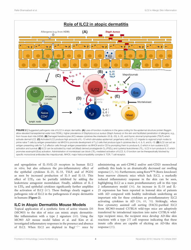
FIGURE 2 | Suggested pathogenic role of ILC2 in atopic dermatitis. (A) Loss-of-function-mutations in the gene coding for the epidermal structure protein filaggrin
allow elevated transepidermal water loss (TEWL), higher prevalence of Staphylococcus aureus (Staph Aureus) on the skin and facilitated penetration of allergens, e.g.,
from house dust mite (HDM). (B) Damaged keratinocytes (KC) release cytokines like interleukin-33 (IL-33), IL-25, and thymic stromal lymphopoietin (TSLP) which
activate dermal ILC2. (C) Activated ILC2 produce high amounts of IL-13 which stimulates epidermal Langerhans cells (LC). LC migrate to regional lymph nodes to
prime naïve T cells by antigen presentation via MHCII to promote development of TH2 cells that produce type II cytokines like IL-4, IL-5, and IL-13. (D) ILC2 can act as
antigen presenting cells for TH2 effector cells through antigen presentation via MHCII and/or CD1a prompting them to produce IL-2 which in turn sustains ILC2
activation and survival. (E) ILC2 can be activated by mast cell (Mast) derived prostaglandin D2 (PGD2) and cysteinyl leukotrienes LTE4. ILC2 in turn produce IL-5 which
promotes eosinophil (Eos) activation. Administration of montelukast can block LTE4-mediated activation of ILC2. IL-5 function can be therapeutically blocked by
specific monoclonal antibodies like mepolizumab. MHCII, major histocompatibility complex II; TCR, T cell receptor.
and upregulation of IL-33/IL-25 receptors in human ILC2in vitro, but also enhances the pro-inflammatory effect ofthe epithelial cytokines IL-25, IL-33, TSLP, and of PGD2as seen by increased production of IL-5 and IL-13. Thiseffect of LTE4 can be partially inhibited by adding theleukotriene antagonist montelukast. Finally, addition of IL-2to LTE4 and epithelial cytokines significantly further amplifiesthe activation of ILC2 (67). These findings clearly suggest apathogenic role of ILC2 in the pathogenesis if atopic dermatitisin humans (Figure 2).
ILC in Atopic Dermatitis Mouse ModelsTopical application of a synthetic form of active vitamin D3(MC903) to the skin of mice can mimic atopic dermatitis-like inflammation with a type 2 signature (68). Using theMC903 AD mouse model Salimi et al. and Kim et al.investigated inflammatory responses in the presence and absenceof ILC2. When ILC2 are depleted in Rag1−/− mice by
administering an anti-CD90.2 and/or anti-CD25 monoclonalantibody this leads to an dramatically decreased ear swellingresponse (25, 36). Furthermore, using Rorαsg/sg (Rorα-knockout)bone marrow chimeric mice which lack ILC2, a markedlyreduced inflammatory response in the skin can be seen,highlighting ILC2 as a main proinflammatory cell in this type2 inflammatory model (36). An increase in IL-33 and IL-25 expression has been reported in lesional skin of patientswith AD compared with healthy individuals underlining animportant role for these cytokines as proinflammatory ILC2activating cytokines in AD (36, 69, 70). Strikingly, whenflow cytometry assisted cell sorting (FACS)-purified ILC2from MC903-treated C57BL/6 wild-type mice are adoptivelytransferred by intradermal injection into naïve C57BL/6 wild-type recipient mice, the recipient mice develop AD-like skinreactions with a type 2 T cell response indicating that theseinnate cells alone are capable of eliciting an AD-like skinresponse (25).
Frontiers in Immunology | www.frontiersin.org 4 November 2019 | Volume 10 | Article 2591

Rafei-Shamsabadi et al. ILC2 in Allergic Skin Inflammation
FIGURE 3 | Context dependent role of ILC2 in type 1 and type 2 dominated contact hypersensitivity. Contact allergens irritate and penetrate the upper skin layers. (A)
Typical contact allergens like TNCB or oxazololone cause ROS and ATP release from damaged keratinocytes (KC) and uptake by epidermal Langerhans cells (LC).
(B,C) LC migrate to skin draining lymph nodes and promote a type 1 driven immune response mediated by TH1 and NK cells resulting in increased IFN-γ and IL-2
production. (D) Higher availability of IL-2 for NK cells results in their enhanced activation and effector cytokine production (IFN-γ, granzyme, perforin). (E) ILC2 are
likely to suppress LC migration, TH1 polarization and NK cells activation in this type 1 CHS response via mechanisms that are currently not well-understood. (F)
Protein allergens like papain or haptens like FITC cause IL-33 release from keratinocytes (KC) which in turn activates dermal ILC2 to produce large amounts of IL-13.
(G,H) ILC2 derived IL-13 promotes trafficking of Langerhans cells (LC) to regional lymph nodes where they prime naïve T cells by interaction of MHCII/peptide/hapten
complex and T cell receptor promoting the development of TH2 cells and a type 2 biased immune response. (I) ILC2 can act as antigen presenting cells for TH2
effector cells prompting them to produce IL-2 which in turn sustains ILC2 activation and survival. (J) ILC2 compete with other innate effector cells like NK cells for the
survival factor IL-2 leading to a reduced/moderate NK cell activation. FITC, Fluorescein isothiocyanate; MHCII, major histocompatibility complex II; ROS, reactive
oxygen species; TCR, T cell receptor; NK, natural killer cell; ATP, Adenosine triphosphate.
Another possible mouse model to study eczema likeskin reactions are the “flaky tail” mice. These mice beara frameshift mutation in the murine filaggrin gene (flg)resulting in expression of a truncated profilaggrin (∼215kDa) instead of the normal high-molecular-weight profilaggrin(>500 kDa) (71). Topical application of allergen to micehomozygous for this mutation results in cutaneous inflammatoryinfiltrates and enhanced cutaneous allergen priming withincreased development of allergen-specific antibody responses
(71). Saunders et al. characterized changes of ILC2 numbersand their cytokine production in flg-mutant mice (72). Thesemice show spontaneous atopic dermatitis-like inflammationand develop compromised pulmonary function. In the skinand skin draining lymph nodes of these mice, there is asignificant increase in the frequency of IL-5-producing ILCcompared to wild type animals. However, no differences incell numbers are seen for ILC1 and 3. Furthermore, flg-mutant mice show higher skin infiltrates of eosinophils, mast
Frontiers in Immunology | www.frontiersin.org 5 November 2019 | Volume 10 | Article 2591

Rafei-Shamsabadi et al. ILC2 in Allergic Skin Inflammation
cells and basophils (72). Even more astonishing, when flg-mutant mice are crossed with Rag1−/− mice (Flgft/ftRag1−/−)skin lesions but not lung inflammation occur as shown bycutaneous expansion of IL-5-producing ILC2, indicating thatskin inflammation can develop independently of the adaptiveimmune system in these mice (72). Regulation of ILC responsesby adaptive immune cells is also reported in other tissues (73).Finally, increased frequency of ILC2 can be found in skin blisterstaken from non-lesional skin of patients with filagrin mutationscompared with the skin of filagrin wildtype subjects (72). Takentogether, loss of filagrin function in humans and mice is clearlylinked to increased ILC2 activation and disease progression inatopic dermatitis.
This latter model, however, has been challenged recently bythe work of Schwartz et al. which provides evidence that atopicdermatitis like lesions can evolve independent of ILC2 and ILC2-derived cytokines in Filaggrin-mutant (Flgft/ft) mice bred onan ILC2-deficient background (74). Interestingly, inflammationin these mice following MC903 treatment requires IL-1β andIL-1R1-signaling but is independent of NOD-, LRR- and pyrindomain-containing protein 3 (NLRP3) inflammasome activationand results in elevated numbers of IL-1β-responsive connectivetissue mast cells (74). Finally, Flgft/ft mice do not develop skininflammation under germ-free compared to SPF conditionsindicting a crucial role for the microbiome in promotingproinflammatory immune responses in this mouse model (74).This issue will be discussed in more detail in a later section.
ILC2 as Possible Therapeutic Targets in ADDevelopment of ILC2 depends on the transcription factorreceptor-related orphan receptors alpha (RORα) and lack ofRORα results in impaired lung inflammation in response toprotease allergen in mice despite normal TH2 cell responses (75).Dai et al. provide evidence that a synthetic RORα/γ inverseagonist (SR1001) is able to suppress inflammation in the MC903-induced atopic dermatitis mouse models. Topical treatment withSR1001 reduces epidermal and dermal inflammation, suppressesthe production of type 2 cytokines and TSLP, and reversesimpaired keratinocyte differentiation (76). Since SR1001 alsoinhibits RORγ signaling it is quite possible that RORγt+ ILC3functions may also be impaired (42). If topical inverse agonistsfor RORα may have anti-inflammatory functions in humansremains to be elucidated.
A crucial role for the IL-33/ILC2 axis in the pathogenesisof AD has been proposed by Imai et al. The authorsgenerated a transgenic mouse line which overexpresses IL-33 in keratinocytes. These mice spontaneously develop anitchy dermatitis closely resembling AD at age 6–8 weekswith thickened epidermis, skin infiltration of eosinophils andmast cells, and high histamine and IgE levels in the blood(77). Moreover, IL-5 and IL-13 expressing ILC2 numbersare significantly increased in lesional skin, peripheral blood,and regional lymph nodes. Administering a neutralizingmonoclonal anti-IL-5 antibody results in a marked reductionof the inflammatory response as shown by a decreasedperipheral blood eosinophil count, milder thickened epidermisand lower inflammatory infiltrates including eosinophils (77).
Unfortunately, a randomized, placebo-controlled parallel groupdesign study in patients with AD could not detect a clinicalimprovement by administering a monoclonal antibody to humaninterleukin-5 (mepolizumab) in two single doses of 750mg, given1 week apart, despite a significant decrease in peripheral bloodeosinophils (78).
ROLE OF ILC IN ALLERGIC CONTACTDERMATITIS
Allergic contact dermatitis (ACD) is a prevalent inflammatoryskin disease triggered by low molecular weight organic chemicalsor metal ions which penetrate the skin and bind covalentlyor by complex formation to proteins thereby activating theinnate and adaptive immune response. ACD can be separatedinto two phases. The sensitization phase, were antigen uponfirst encounter with the skin is taken up by dendritic cellsand transferred to the regional draining lymph nodes tobe presented to antigen specific T-cells for priming. Andthe elicitation phase that is induced by subsequent antigencontact and leads to an infiltration of antigen-specific T-cellsinto the skin peaking 24–48 h after second antigen contact.In the mouse model of ACD, the contact hypersensitivity(CHS) model, hapten-specific CD8+ cytotoxic T-cells arethought to be the key effector cells in the elicitation phaserendering CHS a classical type 1 driven adaptive immuneresponse (Figure 3). Typical haptens used in these modelscomprise oxazolone, 2,4,6-Trinitrochlorobenzene (TNCB) or2,4-dinitrofluorobenzene (DNFB) (79–81). In addition, we andothers have previously demonstrated that sensing of dangersignals by cells of the innate immune system including dendriticcells, neutrophils, and mast cells represent a crucial element inthe initiation and elicitation of CHS responses (82–86).
NK Cells in Type 1 Dominated CHSResponsesGroup 1 ILCs consisting of NK cells and ILC1 are involved ininflammatory bowel and allergic skin diseases in mice (12, 24,87, 88). Regarding ACD Carbone et al. were able to characterizeCD56highCD16−CD62L− NK cells in an ex vivo human modelwhich accumulate in affected skin of hapten allergic humanindividuals and these NK cells release type 1 cytokines andinduce keratinocyte apoptosis in vitro (23). In mice NK cells canbe further subdivided into two distinct subsets: CD49a+DX5−
liver-resident (Trail+) and CD49a−DX5+ conventional NK cells(cNK) (12). Furthermore, cNK cells seem to express much higheramounts of the transcription factor EOMES (87). Liver-residentNK cells can mediate long-lived, antigen-specific adaptive recallresponses to haptens like DNFB and oxazolone independentof B cells and T cells (24). Preceding was the finding that aCHS response to several haptens can be elicited in Rag2−/−
mice lacking T- and B-cells but not in mice that either containdysfunctional NK cells (SCID × beige mice) or completely lackNK cells (Rag2−/− Il2rg−/− mice). A proper CHS responsecan be transferred by FACS-purified antigen-specific Thy-1+
Ly49C-I+ liver-resident NK cells from sensitized Rag2−/− mice
Frontiers in Immunology | www.frontiersin.org 6 November 2019 | Volume 10 | Article 2591
Rafei-Shamsabadi et al. ILC2 in Allergic Skin Inflammation
when transferred into naive Rag2−/− Il2rg−/− recipients (24).The same NK cell type seems to mount antigen specificimmunity against certain viral pathogens as well (88). Ourown investigations using the hapten TNCB support the role ofEOMES+ cNK cells as the dominant proinflammatory innatecell type in the early phase of contact hypersensitivity. NKcell numbers increase significantly 24 h in the ear skin of miceafter allergen challenge and produce type 1 marker cytokineslike IFN-γ and TNF (89). Taken together, NK cells seem torepresent a major driving force of the innate immune system inCHS pathogenesis pathogenesis (Figures 3A–D).
Helper-Like ILC in Type 1 Dominated CHSResponsesVery little is known about the involvement of helper-like ILC inthe pathogenesis of CHS, however there has been some indirectevidence for it in the past. ILC2 are known to be a major sourceof IL-13 production thus playing a crucial role in innate type2 immune responses to worms and inhaled allergens (90, 91).IL-13-deficient mice (Il13−/−) show impaired TH2 responsesinduced by epicutaneous ovalbumin (OVA) exposure whereasi.p. sensitization is normal and results in responses equivalentto wild type mice (92). Interestingly, Il13−/− mice display aneven enhanced ear swelling responses to the hapten DNFB,which is also known to elicit a type 1 T-cell driven immuneresponse (93), compared to wild type mice. At the time, thisfinding was interpreted as a lack of TH2-mediated suppressionbut it’s tempting to speculate that impaired ILC2 function inthis mouse model may also have contributed to a disinhibitedand thus exaggerated type 1 immune response. We recentlycharacterized cell numbers and cytokine production of all ILCsubgroups (ILC1-3 and NK cells) during the elicitation phaseof a CHS mouse model based on the hapten TNCB using anILC reporter system (89). Numbers of ILC are elevated in skindraining lymph nodes, show an activated phenotype and produceelevated amounts of their marker cytokines IL-13 and IL-5 atlate time points (48 and 72 h), i.e., during the resolution phaseof the inflammatory response in the skin. On the other hand,NK cell numbers and their production of IFN-γ and TNF arehighest 24 h after allergen challenge paralleling the strongest skininflammation period (89). The latter is expected since TNCBis known to elicit a type 1 driven immune response (93, 94).However, lack of ILC achieved by either antibody mediateddepletion using an anti CD90.2mAb in Rag1−/− mice or by usingmice that selectively lack ILC2 [Rorαsg/floxIl7rCre/+mice (29)]results in a significantly enhanced and long lasting inflammatoryresponse (89). The ear infiltrate of ILC depleted mice showa tendency toward a more type 1 biased immune responseindicated by increased numbers of T-bet+ CD4+ T-cells (89).This data supports the concept of a counter regulatory role forILC2 in CHS (Figures 3A–D).
Helper-Like ILC in Type 2 DominatedAllergic Skin ResponsesSome allergens like Fluorescein isothiocyanate (FITC) andpapain rather induce allergic type 2 immune responses withincreased IL-4 producing TH2 cell infiltrates in murine skin whenreapplied topically or intradermally (28, 95, 96), suggesting that
ILC2 might rather play a proinflammatory role in these models.Along this line we demonstrated in a papain skin challengemodel that lack of IL-13-producing ILC2 leads to a markedreduction of inflammation with less skin infiltrating TH2 cells[(28); (Figures 3F–J)]. A first therapeutic approach in type 2dominated allergic skin responses has been proposed by Baoet al. They demonstrate that ILC2 numbers are increased inthe skin of FITC-challenged mice. In addition, intraperitonealinjection of the cycloartane triterpene saponin AstragalosideIV during the sensitization phase leads to a reduction of theinflammatory response as seen by a decreased ear swellingresponse, less production of pro-allergic cytokines like IL-33 and TSLP, and significantly reduced numbers of ILC2in the skin of these mice (97). Thus, ILC2 seem to havecontrary roles in type 1 and type 2 dominated allergic skinreactions, respectively (Figure 3).
ROLE OF DERMAL ILC2 IN INNATE ANDADAPTIVE IMMUNE CROSS TALK
Antigen Presentation by MHCIIILC2 and ILC3 express MHCII molecules on their surface andcan act as antigen presenting cells for helper T cells (29, 51, 52).Our own analysis of MHCII expression on ILC2 revealed that inskin draining lymph nodes of mice ∼50% of the ILC2 expressMHCII, while in the skin only ∼3% express MHCII. Antibodymediated depletion of ILC leads to a significant reduction ofMHCII positive ILC2 both in skin and LN (89). Currently, wecan only speculate that ILC2 might regulate effector T cellsin a direct fashion via MHCII. In line with this, Oliphantet al. recently demonstrated that MHCII expression on ILC2and subsequent antigen presentation to CD4+ T cells is crucialfor successful helminth expulsion in mice (29). The crosstalkbetween ILC2 and CD4+ T cells seems to involve IL-2 sinceactivated CD4+ T cell-derived IL-2 has been shown to synergizewith IL-33 to stimulate ILC2 (29). Thus, lack of ILC2 maylead to a higher availability of IL-2 for proliferation of othereffector cells like NK cells leading to an augmented responsein CHS.
Antigen Presentation by CD1aAnother way how ILC2 might crosslink innate and adaptiveimmunity is by expressing the lipid-presenting molecule CD1a.Other than classical MHC proteins that present peptides, CD1molecules present endogenous and exogenous lipid antigensto T lymphocytes (98). In a CHS model using the poisonivy-derived lipid contact allergen urushiol, CD1a expressingLangerhans cells are important to promote CD1a-restrictedCD4+ T cells to produce IL-17 and IL-22. Furthermore,treatment with blocking antibodies against CD1a alleviates skininflammation dramatically (99). More recently Hardman et al.demonstrated in a human skin challenge model that skin-derivedILC2 not only express CD1a but are also capable of helpingCD1a-reactive T cells to sense S. aureus components in ancytosolic phospholipase A2 (PLA2G4A) and TLR-dependent–dependent manner, suggesting a new role for ILC2 in lipidsurveillance of the skin (100). Currently, it is unclear whetherthis also applies for the adaptive immune response against
Frontiers in Immunology | www.frontiersin.org 7 November 2019 | Volume 10 | Article 2591

Rafei-Shamsabadi et al. ILC2 in Allergic Skin Inflammation
urushiol. Taken together CD1a expression on ILC2 seems tobe clearly involved in shaping the phenotype of adaptive Tcell responses.
Crosstalk With Basophils andMacrophagesMashiko et al. reported significantly elevated frequenciesof basophils, ILC and TH2 cells in the lesional skin ofAD patients compared to patients suffering from psoriasis.Interestingly, basophils and ILC2 are positively correlated inskin, whereas skin basophils are inversely correlated with bloodILC2 suggesting that skin basophils may attract circulatingILC2 to skin of AD patients by IL-4 production (101). Kimet al. detected elevated numbers of basophils and ILC thatform clusters in inflamed human AD skin compared tocontrol skin. Using the MC903-based AD mouse model in IL-4/GFP reporter mice, they demonstrated that murine basophilresponses preceded ILC2 responses and those basophils arethe dominant IL-4-producing cell type in inflamed skin. Inaddition, ILC2 express the IL-4 receptor IL-4Rα and proliferatein an IL-4-dependent manner. Finally using Il4−/− mice Kimet al. provide evidence that especially basophil-derived IL-4 is necessary for proinflammatory ILC2 responses in theskin (102).
Most notably, Egawa et al. have shown that basophil-derivedIL-4 converts Ly6C+CCR2+ inflammatory monocytes into anti-inflammatory M2 macrophages in an IgE-mediated chronicallergic inflammation (IgE-CAI) mouse model, a model wherebasophils rather than mast cells and T cells play a criticalrole for the elicitation of allergic response (103, 104). In thismodel, skin infiltrating monocytes acquire anM2-like phenotypein an IL-4R- and basophil-dependent manner and adoptivetransfer of Ly6C+CCR2+ inflammatory monocytes dampensthe exacerbated IgE-CAI in CCR2−/− mice which also requiresIL-4R signaling (103). Thus, it is tempting to speculate, thatbasophil-derived IL-4 may promote pro-inflammatory responsesvia ILC2 and anti-inflammatory signals via M2 macrophages atthe same time, leading to a counterbalanced immune response.However, the role of ILC2 in the IgE-CAI model is not knownso far.
On the other side, ILC2 have been shown to promotepolarization of the anti-inflammatory M2 macrophages byproducing type-2 cytokines (IL-4, IL-5, and IL-13) in an renalischemia-reperfusion injury model and experimental cerebralmalaria (105, 106). Furthermore, in obese mice PD-1high ILC2are inhibited by PD-L1 expressing M1 macrophages which ispromoted by TNF. PD-1 blockade improves ILC2 function,reinforces type 2 innate responses and promotes adiposetissue homeostasis (107, 108). Interestingly, in an serum-induced arthritis mouse model ILC2 were indispensable fordampening proinflammatory IL-1β secretion by bone marrow-derived macrophages (109). Finally, basophil-derived IL-4 seemsto be essential for M2 macrophage mediated trapping ofNippostrongylus Brasiliensis larvae in the skin during secondinfection of mice thereby leading to reduced worm burden in thelung (110). However, basophils had no apparent contribution to
worm expulsion from the intestine highlighting their crucial rolein the skin (110).
Taken together, there seems to be an intense crosstalk betweenbasophils, ILC2 and macrophages involving cytokines like IL-4, IL-13, and IL1β and resulting in differential polarization ofmacrophages dependent on the disease model. How these threecell type interact in AD and CHS remains to be elucidated.
Crosstalk With Dendritic CellsUsing the protease-allergen papain which induces type 2 allergicairway and skin inflammation we showed that ILC2 arenecessary for mounting an appropriate antigen specific TH2memory response and that ILC2 activation clearly precedes TH2involvement in papain induced airway and skin inflammation(28). Furthermore, ILC2-derived IL-13 is needed for theactivation and expansion of an allergen-induced subset ofdendritic cells (CD11b+CD103−IRF4+) which produce the TH2cell chemoattractant CCL17. Using ILC2-deficient mice, wedemonstrated that dermal ILC2 are crucial to mediate expansionof CCL17+ dendritic cells after skin challenge with papainfinally leading to an effective TH2 memory response. Thus, ILC2licensing of dendritic cells is a critical component of the memoryTH2 cell response to certain allergens at barrier sites (28).
INFLUENCE OF SKIN MICROBIOTA ONILC2 IMMUNITY
As mentioned earlier, filaggrin mutant mice significantly differ intheir microbiome composition compared to wild type mice anddo not develop skin inflammation under germ-free conditionsprompting a crucial role for the microbiome in shaping thissetting (74). Several studies have investigated the role of skincommensal bacteria in shaping the host immune cell functions ofthis organ (111–113). This mostly involves skin derived dendriticcells as sensors of bacterial antigens which promote developmentof commensal-specific T cells. These T cells help to improvetissue repair and protection to pathogens rendering them asimportant players in the skin homeostasis (111, 112).
When analyzing different skin-derived bacterial strains in apediatric AD cohort over time, Byrd et al. were able to detectcertain clonal S. aureus strains which are associated with moresevere disease (113). Interestingly, heterogeneous Staphylococcusepidermidis strains were found in patients with less severe diseaseindicating that clonal expansion of certain bacterial strains cantrigger proinflammatory responses in human AD. Furthermore,S. aureus isolates from AD patients with more severe flares caninduce epidermal thickening and expansion of cutaneous TH2and TH17 cells in a murine AD model (113).
These findings are suggesting a role of the microbiome toshape ILC2 functions as well. Interestingly, ILC2 distributionand homeostatic function in bone marrow, fat, lung, gut,and skin seems to be independent of commensal microbiotawhen comparing SPF to germ free mice (114). However, inmouse model of chronic obstructive pulmonary disease (COPD),challenge with S. aureus or Haemophilis influenzae lead to lossof GATA-3 expression in ILC2 and a subsequent increase in the
Frontiers in Immunology | www.frontiersin.org 8 November 2019 | Volume 10 | Article 2591

Rafei-Shamsabadi et al. ILC2 in Allergic Skin Inflammation
expression of IL-12Rβ2, IL-18Rα, and T-bet giving them an ILC1-like phenotype (115). This ILC2 plasticity can also be influencedby viral stimuli especially influenza A virus (115).
Taken together, there is substantial evidence thatthe microbiome is involved in shaping ILC2 functionand plasticity, especially in inflammatory lung diseases.Whether this concept also applies to the pathogenesis ofinflammatory skin disease like AD and CHS remains tobe determined.
TYPE 1 AND TYPE 2 COUNTERREGULATION IN CHS
Type 1 and type 2 immune responses are known to tightlycounter-regulate each other (116). TH1 cytokines such as IFN-γ have been shown to antagonize the function of ILC2 andtype 2 innate immune responses in mouse models of allergiclung inflammation and viral respiratory tract infections (13,117). ILC2-mediated lung inflammation is enhanced in theabsence of the IFN-γ receptor on ILC2 cells in vivo and IFN-γeffectively suppresses the function of tissue-resident ILC2 cells,two observations that clearly suggest a suppressive function oftype 1 cytokines on ILC2 (13). Our own investigations revealthat TNCB based CHS in a mouse model is counter regulatedby activated ILC, since lack of all ILC or ILC2 alone leads toa dramatic increase in the inflammatory response with a type 1immune response bias (89). More recently, it has been reportedthat in the early stage of papain-induced lung inflammation inmice, depletion of NK cells results in increased numbers andcytokine production of ILC2, suggesting that NK cells negativelyregulate ILC2 (118). Hapten based CHS experiments in Il15−/−
mice, which lack NK cells, demonstrate dramatically reducedear swelling responses and at the same time increased numbersof ILC2 in skin and skin draining lymph nodes (89). Thus, amutual balance between type 1 and type 2 immunity may alsoexist in CHS, in whichNK cells negatively regulate ILC2 and ILC2counter regulate type 1 immune responses mainly driven by NKcells, TH1, and TC1 cells.
Recently, Kim et al. identified IL-10-producing lineagenegative lymphoid cells that show elevated numbers in theaxillary as well as inguinal lymph nodes and ear tissues ofOxazolone challenged mice suggesting a possible regulatory roleof ILC (119). These cells were designated “ILC10” and identifiedby expression of markers like CD45, CD127, and Sca-1, whiledetailed characterization of the exact ILC subpopulation was notprovided. Along the same line, an IL-10 producing ILC2 effectorcell population has recently been described in murine lung andsuggested to regulate immune responses in a papain inducedallergic lung inflammation model (4). These studies promptedus to address the presence of IL-10 producing ILC. Using highlysensitive IL-10 transcriptional reporter mice (120) we, however,could not identify relevant numbers of IL-10 transcribing lineagenegative cells in different tissues (skin, lymph nodes, blood, andspleen) in the TNCB induced CHS model (89). Thus, at least inour hands ILC derived IL-10 does not appear to be responsible forthe regulatory effects of ILC in type 1 dominated CHS of the skin.
Nevertheless, ILC2 are reported to promote regulatory T(Treg) cell expansion, thus framing the hypothesis that ILC2 canregulate inflammation indirectly. Molofski et al. demonstratedthat ICOSL expression by ILC2 can stimulate ICOS+ Treg cells,providing a potential indirect link between IL-33 and Tregcells (121). In line with this, Rauber et al. could demonstratethat IL-9 producing ILC2 are crucial in promoting Treg drivenanti-inflammatory effects in an antigen-induced arthritis mousemodel. This ILC2/Treg interaction was dependent on direct cellcontact involving ICOS–ICOSL interaction (122).
We recently showed that IL-33-induced OX40L expressionby ILC2 is critical for tissue-specific expansion of Tregcells (32). Moreover, our data indicates that OX40L/OX40-driven interactions between ILC2 and Treg cells preferentiallyexpands GATA3+ Treg cells, which are thought to betissue-resident and functionally primed (123). IL-33-inducedOX40L expression by ILC2 and the associated Treg cellexpansion seems to be restricted to specific anatomical locationssuch as the airway and adipose tissue but not LN or gut(32). Thus, it remains unknown if a similar mechanism oralternative ILC2-independent suppressive pathways are involvedin the skin.
Malhotra et al. recently found skin resident RORα-expressingTregs to dampen ILC2-driven inflammation in a mousemodel for atopic dermatitis (124). This effect is thoughtto be based on the enhanced expression of TNF ligand–related molecule 1 (TL1A) and death receptor 3 (DR3)on ILC2 as well as suppressed IL-4 expression. RORα-expressing Tregs are found in higher numbers in human skincompared to peripheral blood suggesting a possible counterregulatory role for these cells in ILC2-driven allergic skindiseases (124).
Taken together, these data show that ILC2 can act asmodulators of the adaptive immune response and that thefunctional outcome very much depends on the context of theinflammatory reaction that is analyzed. In type 2 dominatedskin inflammation ILC2 seem to be primarily proinflammatorywhile in the context of a type 1 dominated immune responseILC2 can act as regulators that help to counterbalance theinflammatory reaction (Figure 3).
CONCLUDING REMARKS AND OUTLOOK
Innate lymphoid cells are increasingly emerging as importanteffectors of the innate immune system finally shaping adistinctive adaptive immune response. This includes on theone side important physiological functions in promoting woundhealing, adipose tissue homeostasis, protection from pathogensand dampening of certain inflammatory disorders via Treginduction. On the other side, ILC2 have been shown to beimportant proinflammatory players in diseases like allergicasthma and atopic dermatitis. In the case of atopic dermatitisILC2 have been described to be the major proinflammatory ILCsubtype accountable for the production of marker cytokines likeIL-13 and IL-5, cross-talk with other innate cells like basophilsand dendritic cells, and finally promoting the development of
Frontiers in Immunology | www.frontiersin.org 9 November 2019 | Volume 10 | Article 2591
Rafei-Shamsabadi et al. ILC2 in Allergic Skin Inflammation
TH2 cells. ILC2 will continue to be of high interest as possibletargets in AD therapy, especially concerning their potential toproduce high amounts of cytokines.
Immunologic reaction in allergic contact dermatitis candiffer depending on the type of hapten used. Haptens likeTNCB or oxazolone inducing type 1 responses clearly favor NKcells and TH1 cells as the driving proinflammatory force. Inthese models ILC2 may have counter regulatory functions asour own investigations suggest. On the other side, in allergictype 2 responses of the skin, induced by distinct haptens likeFITC or protein allergens like papain, ILC2 seem to have aproinflammatory role. These observations clearly emphasize acontext dependent function of ILC2 which is determined by thetype of model analyzed (type 1 or type 2 dominated).
Additionally, ILC2 have recently been shown to be part ofa neuro-immune interface. ILC2 function can be influenced bythe neuropeptide neuromedin U (NMU) secreted by cholinergicneurons in the mucosal tissue of the gut and lungs. This goesin line with other studies showing that further neuroendocrinefactors like norepinephrine, vasoactive intestinal peptide (VIP),calcitonin gene-related peptide (CGRP), and acetylcholine canmodify ILC2 function as well (125–131). Furthermore, challengeof mouse skin with the poison ivy compound urushiol leads to anincrease in IL-33 expression which can act on small to medium-sized dorsal root ganglion neurons that innervate the skin andexpress the IL-33 receptor ST2 (132). Strikingly, targeting IL-33 by either neutralizing antibodies or intrathecal applicationof ST2 siRNA results in significantly reduced itching andsubsequently less scratching behavior in these mice, suggestinga new therapeutic approach in poison ivy ACD (132). Sincepruritus is a hallmark symptom of ACD in humans andmice which is mediated by certain sensory neurons (133) it
is tempting to speculate that this new identified “neuron-ILC2 unit” may also be important in the pathogenesis of ADand ACD. This hypothesis is further supported by studiesshowing that type 2 cytokines like TSLP and IL-4 can enhanceitching (134, 135).
Taken together, the picture of ILC function in allergic skindiseases is far from complete. Further investigations especiallyon the mode of action of how ILC modify immune responsesin a context dependent fashion are needed to fill this gapof knowledge.
AUTHOR CONTRIBUTIONS
DR-S did the main research and wrote the first draft of themanuscript. CK, TH, and YT provided substantial contributionsto acquisition, analysis, and interpretation of the scientificcontent of this work. TJ provided the main contribution to theconception and design of the work. All authors contributed tomanuscript revision, read, and approved the submitted version.
FUNDING
Research in the lab of YT was funded by the DFG (TA436/4-1 and P06/SFB1160) and the Else Kröner-FreseniusStiftung (2017_EKES.34). DR-S was supported by the clinicianscientist programme Excellent Clinician Scientists in Freiburg—Education for Leadership (EXCEL) of the Faculty of Medicine,University of Freiburg, funded by the Else Kröner-Fresenius-Stiftung. This work was supported by grants from the GermanResearch Foundation (DFG; KL 2963/2-1 and SPP1937 - KL2963/3-1 to CK) and the European Research Council (ERCEA;ERC Starting Grant #803087 to CK).
REFERENCES
1. Constantinides MG, McDonald BD, Verhoef PA, Bendelac A. A committed
hemopoietic precursor to innate lymphoid cells.Nature. (2014) 508:397–401.
doi: 10.1038/nature13047
2. Eberl G, Colonna M, Santo JPD, McKenzie ANJ. Innate lymphoid
cells: a new paradigm in immunology. Science. (2015) 348:aaa6566.
doi: 10.1126/science.aaa6566
3. Klose CS, Diefenbach A. Transcription factors controlling innate lymphoid
cell fate decisions. Curr Top Microbiol Immunol. (2014) 381:215–55.
doi: 10.1007/82_2014_381
4. Seehus CR, Kadavallore A, Torre B de la, Yeckes AR, Wang Y, Tang
J, et al. Alternative activation generates IL-10 producing type 2 innate
lymphoid cells. Nat Commun. (2017) 8:1900. doi: 10.1038/s41467-017-
02023-z
5. Crome SQ, Nguyen LT, Lopez-Verges S, Yang SYC, Martin B, Yam JY, et al. A
distinct innate lymphoid cell population regulates tumor-associated T cells.
Nat Med. (2017) 23:368–75. doi: 10.1038/nm.4278
6. Wang S, Xia P, Chen Y, QuY, Xiong Z, Ye B, et al. Regulatory innate lymphoid
cells control innate intestinal inflammation. Cell. (2017) 171:201–16.e18.
doi: 10.1016/j.cell.2017.07.027
7. Klose CSN, Artis D. Innate lymphoid cells as regulators of immunity,
inflammation and tissue homeostasis. Nat Immunol. (2016) 17:765–74.
doi: 10.1038/ni.3489
8. Vosshenrich CAJ, García-Ojeda ME, Samson-Villéger SI, Pasqualetto V,
Enault L, Goff OR-L, et al. A thymic pathway of mouse natural killer
cell development characterized by expression of GATA-3 and CD127. Nat
Immunol. (2006) 7:1217–24. doi: 10.1038/ni1395
9. Veinotte LL, Halim TYF, Takei F. Unique subset of natural killer cells
develops from progenitors in lymph node. Blood. (2008) 111:4201–8.
doi: 10.1182/blood-2007-04-087577
10. Huang Y, Mao K, Chen X, Sun M, Kawabe T, Li W, et al. S1P-dependent
interorgan trafficking of group 2 innate lymphoid cells supports host defense.
Science. (2018) 359:114–9. doi: 10.1126/science.aam5809
11. Gasteiger G, Fan X, Dikiy S, Lee SY, Rudensky AY. Tissue residency of
innate lymphoid cells in lymphoid and non-lymphoid organs. Science. (2015)
350:981–5. doi: 10.1126/science.aac9593
12. Peng H, Jiang X, Chen Y, Sojka DK, Wei H, Gao X, et al. Liver-resident NK
cells confer adaptive immunity in skin-contact inflammation. J Clin Invest.
(2013) 123:1444–56. doi: 10.1172/JCI66381
13. Moro K, Kabata H, Tanabe M, Koga S, Takeno N, Mochizuki M, et al.
Interferon and IL-27 antagonize the function of group 2 innate lymphoid
cells and type 2 innate immune responses. Nat Immunol. (2016) 17:76–86.
doi: 10.1038/ni.3309
14. Bando JK, Liang H-E, Locksley RM. Identification and distribution of
developing innate lymphoid cells in the fetal mouse intestine. Nat Immunol.
(2015) 16:153–60. doi: 10.1038/ni.3057
15. Schuijs MJ, Halim TYF. Group 2 innate lymphocytes at the interface between
innate and adaptive immunity. Ann N Y Acad Sci. (2018) 1417:87–103.
doi: 10.1111/nyas.13604
16. Klose CSN, Blatz K, d’Hargues Y, Hernandez PP, Kofoed-Nielsen M,
Ripka JF, et al. The transcription factor T-bet is induced by IL-15 and
Frontiers in Immunology | www.frontiersin.org 10 November 2019 | Volume 10 | Article 2591

Rafei-Shamsabadi et al. ILC2 in Allergic Skin Inflammation
thymic agonist selection and controls CD8αα+ intraepithelial lymphocyte
development. Immunity. (2014) 41:230–43. doi: 10.1016/j.immuni.2014.
06.018
17. Gordon SM, Chaix J, Rupp LJ, Wu J, Madera S, Sun JC, et al.
The transcription factors T-bet and Eomes control key checkpoints
of natural killer cell maturation. Immunity. (2012) 36:55–67.
doi: 10.1016/j.immuni.2011.11.016
18. Sojka DK, Plougastel-Douglas B, Yang L, Pak-Wittel MA, Artyomov MN,
Ivanova Y, et al. Tissue-resident natural killer (NK) cells are cell lineages
distinct from thymic and conventional splenic NK cells. eLife. (2014) 3:e1659.
doi: 10.7554/eLife.01659
19. Daussy C, Faure F, Mayol K, Viel S, Gasteiger G, Charrier E, et al. T-
bet and Eomes instruct the development of two distinct natural killer cell
lineages in the liver and in the bone marrow. J Exp Med. (2014) 211:563–77.
doi: 10.1084/jem.20131560
20. Abt MC, Lewis BB, Caballero S, Xiong H, Carter RA, Sušac B, et al. Innate
immune defenses mediated by two ILC subsets are critical for protection
against acute clostridium difficile infection. Cell Host Microbe. (2015) 18:27–
37. doi: 10.1016/j.chom.2015.06.011
21. Spits H, Bernink JH, Lanier L. NK cells and type 1 innate lymphoid
cells: partners in host defense. Nat Immunol. (2016) 17:758–64.
doi: 10.1038/ni.3482
22. Fuchs A, Vermi W, Lee JS, Lonardi S, Gilfillan S, Newberry RD, et al.
Intraepithelial type 1 innate lymphoid cells are a unique subset of IL-12- and
IL-15-responsive IFN-gamma-producing cells. Immunity. (2013) 38:769–81.
doi: 10.1016/j.immuni.2013.02.010
23. Carbone T, Nasorri F, Pennino D, Eyerich K, Foerster S, Cifaldi L, et al.
CD56highCD16−CD62L− NK cells accumulate in allergic contact dermatitis
and contribute to the expression of allergic responses. J Immunol. (2010)
184:1102–10. doi: 10.4049/jimmunol.0902518
24. O’Leary JG, Goodarzi M, Drayton DL, von Andrian UH. T cell- and B
cell-independent adaptive immunity mediated by natural killer cells. Nat
Immunol. (2006) 7:507–16. doi: 10.1038/ni1332
25. Kim BS, Siracusa MC, Saenz SA, Noti M, Monticelli LA, Sonnenberg
GF, et al. TSLP elicits IL-33–independent innate lymphoid cell responses
to promote skin inflammation. Sci Transl Med. (2013) 5:170ra16.
doi: 10.1126/scitranslmed.3005374
26. Rak GD, Osborne LC, Siracusa MC, Kim BS, Wang K, Bayat A, et al. IL-33-
dependent group 2 innate lymphoid cells promote cutaneous wound healing.
J Invest Dermatol. (2015) 136:487–96. doi: 10.1038/JID.2015.406
27. Christianson CA, Goplen NP, Zafar I, Irvin C, Good JT Jr, Rollins DR,
et al. Persistence of asthma requires multiple feedback circuits involving
type 2 innate lymphoid cells and IL-33. J Allergy Clin Immunol. (2015)
136:59–68.e14. doi: 10.1016/j.jaci.2014.11.037
28. Halim TY, Hwang YY, Scanlon ST, Zaghouani H, Garbi N, Fallon PG, et al.
Group 2 innate lymphoid cells license dendritic cells to potentiate memory T
helper 2 cell responses. Nat Immunol. (2016) 17:57–64. doi: 10.1038/ni.3294
29. Oliphant CJ, Hwang YY, Walker JA, Salimi M, Wong SH, Brewer JM, et al.
MHCII-mediated dialog between group 2 innate lymphoid cells and CD4+ T
cells potentiates type 2 immunity and promotes parasitic helminth expulsion.
Immunity. (2014) 41:283–95. doi: 10.1016/j.immuni.2014.06.016
30. Paclik D, Stehle C, Lahmann A, Hutloff A, Romagnani C. ICOS regulates
the pool of group 2 innate lymphoid cells under homeostatic and
inflammatory conditions in mice. Eur J Immunol. (2015) 45:2766–72.
doi: 10.1002/eji.201545635
31. Maazi H, Patel N, Sankaranarayanan I, Suzuki Y, Rigas D, Soroosh P, et al.
ICOS:ICOS-ligand interaction is required for type 2 innate lymphoid cell
function, homeostasis, and induction of airway hyperreactivity. Immunity.
(2015) 42:538–51. doi: 10.1016/j.immuni.2015.02.007
32. Halim TYF, Rana BMJ, Walker JA, Kerscher B, Knolle MD, Jolin HE, et al.
Tissue-restricted adaptive type 2 immunity is orchestrated by expression
of the costimulatory molecule OX40L on group 2 innate lymphoid
cells. Immunity. (2018) 48:1195–207.e6. doi: 10.1016/j.immuni.2018.
05.003
33. Smith SG, Chen R, Kjarsgaard M, Huang C, Oliveria J-P, O’Byrne PM, et al.
Increased numbers of activated group 2 innate lymphoid cells in the airways
of patients with severe asthma and persistent airway eosinophilia. J Allergy
Clin Immunol. (2016) 137:75–86.e8. doi: 10.1016/j.jaci.2015.05.037
34. Bartemes K, Kephart G, Fox SJ, Kita H. Enhanced innate type 2 immune
response in peripheral blood from patients with asthma. J Allergy Clin
Immunol. (2014) 134:671–8.e4. doi: 10.1016/j.jaci.2014.06.024
35. Liu T, Wu J, Zhao J, Wang J, Zhang Y, Liu L, et al. Type 2 innate
lymphoid cells: a novel biomarker of eosinophilic airway inflammation in
patients with mild to moderate asthma. Respir Med. (2015) 109:1391–6.
doi: 10.1016/j.rmed.2015.09.016
36. Salimi M, Barlow JL, Saunders SP, Xue L, Gutowska-Owsiak D, Wang X,
et al. A role for IL-25 and IL-33-driven type-2 innate lymphoid cells in atopic
dermatitis. J Exp Med. (2013) 210:2939–50. doi: 10.1084/jem.20130351
37. Gladiator A, Wangler N, Trautwein-Weidner K, LeibundGut-Landmann
S. Cutting edge: IL-17–secreting innate lymphoid cells are essential for
host defense against fungal infection. J Immunol. (2013) 190:521–5.
doi: 10.4049/jimmunol.1202924
38. Goto Y, Obata T, Kunisawa J, Sato S, Ivanov II, Lamichhane A, et al. Innate
lymphoid cells regulate intestinal epithelial cell glycosylation. Science. (2014)
345:1254009. doi: 10.1126/science.1254009
39. Hernández PP, Mahlakoiv T, Yang I, Schwierzeck V, Nguyen N, Guendel
F, et al. Interferon-λ and interleukin-22 cooperate for the induction of
interferon-stimulated genes and control of rotavirus infection.Nat Immunol.
(2015) 16:698–707. doi: 10.1038/ni.3180
40. Sonnenberg GF, Monticelli LA, Elloso MM, Fouser LA, Artis D. CD4+
lymphoid tissue inducer cells promote innate immunity in the gut.
Immunity. (2011) 34:122–34. doi: 10.1016/j.immuni.2010.12.009
41. Zheng Y, Valdez PA, Danilenko DM, Hu Y, Sa SM, Gong Q, et al. Interleukin-
22 mediates early host defense against attaching and effacing bacterial
pathogens. Nat Med. (2008) 14:282–9. doi: 10.1038/nm1720
42. Eberl G, Marmon S, Sunshine M-J, Rennert PD, Choi Y, Littman DR.
An essential function for the nuclear receptor RORγt in the generation
of fetal lymphoid tissue inducer cells. Nat Immunol. (2004) 5:64–73.
doi: 10.1038/ni1022
43. Yokota Y, Mansouri A, Mori S, Sugawara S, Adachi S, Nishikawa S-I,
et al. Development of peripheral lymphoid organs and natural killer cells
depends on the helix–loop–helix inhibitor Id2. Nature. (1999) 397:702–6.
doi: 10.1038/17812
44. Mebius RE, Rennert P, Weissman IL. Developing lymph nodes collect
CD4+CD3− LTβ+ cells that can differentiate to APC, NK cells, and
follicular cells but not T or B cells. Immunity. (1997) 7:493–504.
doi: 10.1016/S1074-7613(00)80371-4
45. Sun Z, Unutmaz D, Zou Y-R, Sunshine MJ, Pierani A, Brenner-
Morton S, et al. Requirement for RORγ in thymocyte survival
and lymphoid organ development. Science. (2000) 288:2369–73.
doi: 10.1126/science.288.5475.2369
46. Klose CSN, Kiss EA, Schwierzeck V, Ebert K, Hoyler T, d’Hargues Y, et al.
A T-bet gradient controls the fate and function of CCR6-RORγt+ innate
lymphoid cells. Nature. (2013) 494:261–5. doi: 10.1038/nature11813
47. Sciumé G, Hirahara K, Takahashi H, Laurence A, Villarino AV, Singleton KL,
et al. Distinct requirements for T-bet in gut innate lymphoid cells. J ExpMed.
(2012) 209:2331–8. doi: 10.1084/jem.20122097
48. Vonarbourg C, Mortha A, Bui VL, Hernandez PP, Kiss EA, Hoyler T, et al.
Regulated expression of nuclear receptor RORγt confers distinct functional
fates to NK cell receptor-expressing RORγt+ innate lymphocytes. Immunity.
(2010) 33:736–51. doi: 10.1016/j.immuni.2010.10.017
49. Rankin L, Groom JR, Chopin M, Herold M, Walker JA, Mielke LA, et al.
T-bet is essential for NKp46+ innate lymphocyte development through the
Notch pathway. Nat Immunol. (2013) 14:389–95. doi: 10.1038/ni.2545
50. Bernink JH, Peters CP, Munneke M, te Velde AA, Meijer SL, Weijer K, et al.
Human type 1 innate lymphoid cells accumulate in inflamedmucosal tissues.
Nat Immunol. (2013) 14:221–9. doi: 10.1038/ni.2534
51. Hepworth MR, Fung TC, Masur SH, Kelsen JR, McConnell FM, Dubrot
J, et al. Group 3 innate lymphoid cells mediate intestinal selection of
commensal bacteria-specific CD4+ T cells. Science. (2015) 348:1031–5.
doi: 10.1126/science.aaa4812
52. Hepworth MR, Monticelli LA, Fung TC, Ziegler CGK, Grunberg S, Sinha
R, et al. Innate lymphoid cells regulate CD4+ T-cell responses to intestinal
commensal bacteria. Nature. (2013) 498:113–7. doi: 10.1038/nature12240
53. Sonnenberg GF, Monticelli LA, Alenghat T, Fung TC, Hutnick NA,
Kunisawa J, et al. Innate lymphoid cells promote anatomical containment
Frontiers in Immunology | www.frontiersin.org 11 November 2019 | Volume 10 | Article 2591

Rafei-Shamsabadi et al. ILC2 in Allergic Skin Inflammation
of lymphoid-resident commensal bacteria. Science. (2012) 336:1321–5.
doi: 10.1126/science.1222551
54. Pantelyushin S, Haak S, Ingold B, Kulig P, Heppner FL, Navarini AA,
et al. Rorgammat+ innate lymphocytes and gammadelta T cells initiate
psoriasiform plaque formation in mice. J Clin Invest. (2012) 122:2252–6.
doi: 10.1172/JCI61862
55. Brüggen M-C, Bauer WM, Reininger B, Clim E, Captarencu C, Steiner GE,
et al. In situ mapping of innate lymphoid cells in human skin: evidence for
remarkable differences between normal and inflamed skin. J Invest Dermatol.
(2016) 136:2396–405. doi: 10.1016/j.jid.2016.07.017
56. Teunissen MBM, Munneke JM, Bernink JH, Spuls PI, Res PCM, te Velde
A, et al. Composition of innate lymphoid cell subsets in the human skin:
enrichment of NCR+ ILC3 in lesional skin and blood of psoriasis patients. J
Invest Dermatol. (2014) 134:2351–60. doi: 10.1038/jid.2014.146
57. Villanova F, Flutter B, Tosi I, Grys K, Sreeneebus H, Perera GK, et al.
Characterization of innate lymphoid cells in human skin and blood
demonstrates increase of NKp44+ ILC3 in psoriasis. J Invest Dermatol.
(2014) 134:984–91. doi: 10.1038/jid.2013.477
58. Irvine AD, McLean WHI, Leung DYM. Filaggrin mutations associated
with skin and allergic diseases. N Engl J Med. (2011) 365:1315–27.
doi: 10.1056/NEJMra1011040
59. Palmer CNA, Irvine AD, Terron-Kwiatkowski A, Zhao Y, Liao H, Lee SP,
et al. Common loss-of-function variants of the epidermal barrier protein
filaggrin are a major predisposing factor for atopic dermatitis. Nat Genet.
(2006) 38:441–6. doi: 10.1038/ng1767
60. Rodríguez E, Baurecht H, Herberich E, Wagenpfeil S, Brown SJ, Cordell HJ,
et al. Meta-analysis of filaggrin polymorphisms in eczema and asthma: robust
risk factors in atopic disease. J Allergy Clin Immunol. (2009) 123:1361–70.e7.
doi: 10.1016/j.jaci.2009.03.036
61. Sandilands A, Terron-Kwiatkowski A, Hull PR, O’Regan GM, Clayton TH,
Watson RM, et al. Comprehensive analysis of the gene encoding filaggrin
uncovers prevalent and rare mutations in ichthyosis vulgaris and atopic
eczema. Nat Genet. (2007) 39:650–4. doi: 10.1038/ng2020
62. Scheerer C, Eyerich K. Pathogenese des atopischen Ekzems.Hautarzt. (2018)
69:191–6. doi: 10.1007/s00105-018-4127-4
63. Roediger B, Kyle R, Yip KH, Sumaria N, Guy TV, Kim BS, et al. Cutaneous
immunosurveillance and regulation of inflammation by group 2 innate
lymphoid cells. Nat Immunol. (2013) 14:564–73. doi: 10.1038/ni.2584
64. Mjösberg JM, Trifari S, Crellin NK, Peters CP, van Drunen CM, Piet B, et al.
Human IL-25- and IL-33-responsive type 2 innate lymphoid cells are defined
by expression of CRTH2 and CD161. Nat Immunol. (2011) 12:1055–62.
doi: 10.1038/ni.2104
65. Tait Wojno E, Monticelli L, Tran S, Alenghat T, Osborne L, Thome J, et al.
The prostaglandin D2 receptor CRTH2 regulates accumulation of group
2 innate lymphoid cells in the inflamed lung. Mucosal Immunol. (2015)
8:1313–23. doi: 10.1038/mi.2015.21
66. Xue L, Salimi M, Panse I, Mjosberg JM, McKenzie ANJ, Spits H,
et al. Prostaglandin D2 activates group 2 innate lymphoid cells through
chemoattractant receptor-homologous molecule expressed on TH2 cells. J
Allergy Clin Immunol. (2014) 133:1184–94.e7. doi: 10.1016/j.jaci.2013.10.056
67. Salimi M, Stöger L, Liu W, Go S, Pavord I, Klenerman P, et al. Cysteinyl
leukotriene E4 activates human group 2 innate lymphoid cells and enhances
the effect of prostaglandin D2 and epithelial cytokines. J Allergy Clin
Immunol. (2017) 140:1090–100.e11. doi: 10.1016/j.jaci.2016.12.958
68. Li M, Hener P, Zhang Z, Kato S, Metzger D, Chambon P. Topical vitamin D3
and low-calcemic analogs induce thymic stromal lymphopoietin in mouse
keratinocytes and trigger an atopic dermatitis. Proc Natl Acad Sci USA.
(2006) 103:11736–41. doi: 10.1073/pnas.0604575103
69. Hvid M, Vestergaard C, Kemp K, Christensen GB, Deleuran B, Deleuran
M. IL-25 in Atopic Dermatitis: a Possible Link between Inflammation
and Skin Barrier Dysfunction? J Invest Dermatol. (2011) 131:150–7.
doi: 10.1038/jid.2010.277
70. Wang Y-H, Angkasekwinai P, Lu N, Voo KS, Arima K, Hanabuchi S, et al.
IL-25 augments type 2 immune responses by enhancing the expansion
and functions of TSLP-DC–activated Th2 memory cells. J Exp Med. (2007)
204:1837–47. doi: 10.1084/jem.20070406
71. Fallon PG, Sasaki T, Sandilands A, Campbell LE, Saunders SP, Mangan
NE, et al. A homozygous frameshift mutation in the murine filaggrin
gene facilitates enhanced percutaneous allergen priming. Nat Genet. (2009)
41:602–8. doi: 10.1038/ng.358
72. Saunders SP, Moran T, Floudas A, Wurlod F, Kaszlikowska A, Salimi M,
et al. Spontaneous atopic dermatitis is mediated by innate immunity,
with the secondary lung inflammation of the atopic march requiring
adaptive immunity. J Allergy Clin Immunol. (2016) 137:482–91.
doi: 10.1016/j.jaci.2015.06.045
73. Gasteiger G, Rudensky AY. Opinion: interactions of innate and
adaptive lymphocytes. Nat Rev Immunol. (2014) 14:631–9. doi: 10.1038/
nri3726
74. Schwartz C, Moran T, Saunders SP, Kaszlikowska A, Floudas A, Bom J,
et al. Spontaneous atopic dermatitis in mice with a defective skin barrier
is independent of ILC2 and mediated by IL-1β. Allergy. (2019) 74:1920–33.
doi: 10.1111/all.13801
75. Halim TY, MacLaren A, Romanish MT, Gold MJ, McNagny KM, Takei F.
Retinoic-acid-receptor-related orphan nuclear receptor alpha is required for
natural helper cell development and allergic inflammation. Immunity. (2012)
37:463–74. doi: 10.1016/j.immuni.2012.06.012
76. Dai J, Choo M-K, Park JM, Fisher DE. Topical ROR inverse agonists
suppress inflammation in mouse models of atopic dermatitis and
acute irritant dermatitis. J Invest Dermatol. (2017) 137:2523–31.
doi: 10.1016/j.jid.2017.07.819
77. Imai Y, Yasuda K, Sakaguchi Y, Haneda T, Mizutani H, Yoshimoto T, et al.
Skin-specific expression of IL-33 activates group 2 innate lymphoid cells and
elicits atopic dermatitis-like inflammation in mice. Proc Natl Acad Sci USA.
(2013) 110:13921–6. doi: 10.1073/pnas.1307321110
78. Oldhoff JM, Darsow U, Werfel T, Katzer K, Wulf A, Laifaoui J, et al. Anti-
IL-5 recombinant humanized monoclonal antibody (Mepolizumab)
for the treatment of atopic dermatitis. Allergy. (2005) 60:693–6.
doi: 10.1111/j.1398-9995.2005.00791.x
79. Asherson GL, Barnes RMR. Contact sensitivity in the mouse. Immunology.
(1973) 24:885–94.
80. Fyhrquist N, Wolff H, Lauerma A, Alenius H. CD8+ T cell migration to the
skin requires CD4+ help in a murine model of contact hypersensitivity. PLoS
ONE. (2012) 7:e41038. doi: 10.1371/journal.pone.0041038
81. Martin SF. Allergic contact dermatitis: xenoinflammation of the skin. Curr
Opin Immunol. (2012) 24:720–9. doi: 10.1016/j.coi.2012.08.003
82. Dudeck A, Dudeck J, Scholten J, Petzold A, Surianarayanan S,
Köhler A, et al. Mast cells are key promoters of contact allergy that
mediate the adjuvant effects of haptens. Immunity. (2011) 34:973–84.
doi: 10.1016/j.immuni.2011.03.028
83. Esser PR, Wölfle U, Dürr C, von Loewenich FD, Schempp CM, Freudenberg
MA, et al. Contact sensitizers induce skin inflammation via ROS
production and hyaluronic acid degradation. PLoS ONE. (2012) 7:e41340.
doi: 10.1371/journal.pone.0041340
84. Martin SF, Dudda JC, Bachtanian E, Lembo A, Liller S, Durr C, et al.
Toll-like receptor and IL-12 signaling control susceptibility to contact
hypersensitivity. J Exp Med. (2008) 205:2151–62. doi: 10.1084/jem.20070509
85. Weber FC, Németh T, Csepregi JZ, Dudeck A, Roers A, Ozsvári
B, et al. Neutrophils are required for both the sensitization and
elicitation phase of contact hypersensitivity. J Exp Med. (2015) 212:15–22.
doi: 10.1084/jem.20130062
86. Weber FC, Esser PR, Müller T, Ganesan J, Pellegatti P, Simon MM, et al.
Lack of the purinergic receptor P2X7 results in resistance to contact
hypersensitivity. J Exp Med. (2010) 207:2609–19. doi: 10.1084/jem.20092489
87. Tang L, Peng H, Zhou J, Chen Y, Wei H, Sun R, et al. Differential phenotypic
and functional properties of liver-resident NK cells and mucosal ILC1s. J
Autoimmun. (2016) 67:29–35. doi: 10.1016/j.jaut.2015.09.004
88. Paust S, Gill HS, Wang B-Z, Flynn MP, Moseman EA, Senman B, et al.
Critical role for the chemokine receptor CXCR6 in NK cell–mediated
antigen-specific memory of haptens and viruses. Nat Immunol. (2010)
11:1127–35. doi: 10.1038/ni.1953
89. Rafei-Shamsabadi DA, van de Poel S, Dorn B, Kunz S, Martin SF, Klose
CSN, et al. Lack of type 2 innate lymphoid cells promote a type I driven
enhanced immune response in contact hypersensitivity. J Invest Dermatol.
(2018) 138:1962–72. doi: 10.1016/j.jid.2018.03.001
90. Halim TYF, Steer CA, Mathä L, Gold MJ, Martinez-Gonzalez I, McNagny
KM, et al. Group 2 innate lymphoid cells are critical for the initiation of
Frontiers in Immunology | www.frontiersin.org 12 November 2019 | Volume 10 | Article 2591
Rafei-Shamsabadi et al. ILC2 in Allergic Skin Inflammation
adaptive T Helper 2 cell-mediated allergic lung inflammation. Immunity.
(2014) 40:425–35. doi: 10.1016/j.immuni.2014.01.011
91. Moro K, Yamada T, Tanabe M, Takeuchi T, Ikawa T, Kawamoto
H, et al. Innate production of T(H)2 cytokines by adipose tissue-
associated c-Kit(+)Sca-1(+) lymphoid cells. Nature. (2010) 463:540–4.
doi: 10.1038/nature08636
92. Herrick CA, Xu L, McKenzie ANJ, Tigelaar RE, Bottomly K. IL-
13 is necessary, not simply sufficient, for epicutaneously induced Th2
responses to soluble protein antigen. J Immunol. (2003) 170:2488–95.
doi: 10.4049/jimmunol.170.5.2488
93. Martin SF, Jakob T. From innate to adaptive immune responses in
contact hypersensitivity. Curr Opin Allergy Clin Immunol. (2008) 8:289–93.
doi: 10.1097/ACI.0b013e3283088cf9
94. Lass C, Merfort I, Martin SF. In vitro and in vivo analysis of pro- and anti-
inflammatory effects of weak and strong contact allergens. Exp Dermatol.
(2010) 19:1007–13. doi: 10.1111/j.1600-0625.2010.01136.x
95. Dearman RJ, Kimber I. Role of CD4+ T helper 2-type cells in cutaneous
inflammatory responses induced by fluorescein isothiocyanate. Immunology.
(2000) 101:442–51. doi: 10.1046/j.1365-2567.2000.01126.x
96. Ogawa A, Yoshizaki A, Yanaba K, Ogawa F, Hara T, Muroi E, et al.
The differential role of L-selectin and ICAM-1 in Th1-type and Th2-
type contact hypersensitivity. J Invest Dermatol. (2010) 130:1558–70.
doi: 10.1038/jid.2010.25
97. Bao K, Yu X,Wei X, Gui L, LiuH,Wang X, et al. Astragaloside IV ameliorates
allergic inflammation by inhibiting key initiating factors in the initial stage of
sensitization. Sci Rep. (2016) 6:38241. doi: 10.1038/srep38241
98. Porcelli S, Brenner MB, Greenstein JL, Terhorst C, Balk SP, Bleicher PA.
Recognition of cluster of differentiation 1 antigens by human CD4−CD8−
cytolytic T lymphocyte. Nature. (1989) 341:447–50. doi: 10.1038/341447a0
99. Kim JH, Hu Y, Yongqing T, Kim J, Hughes VA, Nours JL, et al. CD1a on
Langerhans cells controls inflammatory skin diseases. Nat Immunol. (2016)
17:1159–66. doi: 10.1038/ni.3523
100. Hardman CS, Chen Y-L, Salimi M, Jarrett R, Johnson D, Järvinen VJ, et al.
CD1a presentation of endogenous antigens by group 2 innate lymphoid cells.
Sci Immunol. (2017) 2:eaan5918. doi: 10.1126/sciimmunol.aan5918
101. Mashiko S, Mehta H, Bissonnette R, Sarfati M. Increased frequencies of
basophils, type 2 innate lymphoid cells and Th2 cells in skin of patients
with atopic dermatitis but not psoriasis. J Dermatol Sci. (2017) 88:167–74.
doi: 10.1016/j.jdermsci.2017.07.003
102. Kim BS, Wang K, Siracusa MC, Saenz SA, Brestoff JR, Monticelli LA,
et al. Basophils promote innate lymphoid cell responses in inflamed skin. J
Immunol. (2014) 193:3717–25. doi: 10.4049/jimmunol.1401307
103. Egawa M, Mukai K, Yoshikawa S, Iki M, Mukaida N, Kawano Y,
et al. Inflammatory monocytes recruited to allergic skin acquire an anti-
inflammatory M2 phenotype via basophil-derived interleukin-4. Immunity.
(2013) 38:570–80. doi: 10.1016/j.immuni.2012.11.014
104. Mukai K, Matsuoka K, Taya C, Suzuki H, Yokozeki H, Nishioka K, et al.
Basophils play a critical role in the development of IgE-mediated chronic
allergic inflammation independently of T cells and mast cells. Immunity.
(2005) 23:191–202. doi: 10.1016/j.immuni.2005.06.011
105. Cao Q, Wang Y, Niu Z, Wang C, Wang R, Zhang Z, et al. Potentiating
tissue-resident type 2 innate lymphoid cells by IL-33 to prevent
renal ischemia-reperfusion injury. J Am Soc Nephrol. (2018) 29:961–76.
doi: 10.1681/ASN.2017070774
106. Besnard A-G, Guabiraba R, Niedbala W, Palomo J, Reverchon F,
Shaw TN, et al. IL-33-mediated protection against experimental cerebral
malaria is linked to induction of type 2 innate lymphoid cells, M2
macrophages and regulatory T cells. PLoS Pathog. (2015) 11:e1004607.
doi: 10.1371/journal.ppat.1004607
107. Oldenhove G, Boucquey E, Taquin A, Acolty V, Bonetti L, Ryffel B,
et al. PD-1 is involved in the dysregulation of type 2 innate lymphoid
cells in a murine model of obesity. Cell Rep. (2018) 25:2053–60.e4.
doi: 10.1016/j.celrep.2018.10.091
108. Molofsky AB, Nussbaum JC, Liang H-E, Van Dyken SJ, Cheng LE,
Mohapatra A, et al. Innate lymphoid type 2 cells sustain visceral adipose
tissue eosinophils and alternatively activatedmacrophages. J ExpMed. (2013)
210:535–49. doi: 10.1084/jem.20121964
109. Omata Y, Frech M, Primbs T, Lucas S, Andreev D, Scholtysek C,
et al. Group 2 innate lymphoid cells attenuate inflammatory arthritis
and protect from bone destruction in mice. Cell Rep. (2018) 24:169–80.
doi: 10.1016/j.celrep.2018.06.005
110. Obata-Ninomiya K, Ishiwata K, Tsutsui H, Nei Y, Yoshikawa S,
Kawano Y, et al. The skin is an important bulwark of acquired
immunity against intestinal helminths. J Exp Med. (2013) 210:2583–95.
doi: 10.1084/jem.20130761
111. Naik S, Bouladoux N, Linehan JL, Han S-J, Harrison OJ, Wilhelm C, et al.
Commensal–dendritic-cell interaction specifies a unique protective skin
immune signature. Nature. (2015) 520:104–8. doi: 10.1038/nature14052
112. Linehan JL, Harrison OJ, Han S-J, Byrd AL, Vujkovic-Cvijin I, Villarino AV,
et al. Non-classical immunity controls microbiota impact on skin immunity
and tissue repair. Cell. (2018) 172:784–96.e18. doi: 10.1016/j.cell.2017.12.033
113. Byrd AL, Deming C, Cassidy SKB, Harrison OJ, Ng W-I, Conlan S,
et al. Staphylococcus aureus and Staphylococcus epidermidis strain diversity
underlying pediatric atopic dermatitis. Sci Transl Med. (2017) 9:eaal4651.
doi: 10.1126/scitranslmed.aal4651
114. Ricardo-Gonzalez RR, VanDyken SJ, Schneider C, Lee J, Nussbaum JC, Liang
H-E, et al. Tissue signals imprint ILC2 identity with anticipatory function.
Nat Immunol. (2018) 19:1093–9. doi: 10.1038/s41590-018-0201-4
115. Silver JS, Kearley J, Copenhaver AM, Sanden C, Mori M, Yu L, et al.
Inflammatory triggers associated with exacerbations of COPD orchestrate
plasticity of group 2 innate lymphoid cells in the lungs. Nat Immunol. (2016)
17:626–35. doi: 10.1038/ni.3443
116. Stehle C, Saikali P, Romagnani C. Putting the brakes on ILC2 cells. Nat
Immunol. (2016) 17:43–4. doi: 10.1038/ni.3353
117. Duerr CU, McCarthy CDA, Mindt BC, Rubio M, Meli AP, Pothlichet J,
et al. Type I interferon restricts type 2 immunopathology through the
regulation of group 2 innate lymphoid cells. Nat Immunol. (2015) 17:65–75.
doi: 10.1038/ni.3308
118. Bi J, Cui L, Yu G, Yang X, Chen Y, Wan X. NK cells alleviate lung
inflammation by negatively regulating group 2 innate lymphoid cells. J
Immunol. (2017) 198:3336–44. doi: 10.4049/jimmunol.1601830
119. Kim HS, Jang J-H, Lee MB, Jung ID, Kim YM, Park Y-M, et al.
A novel IL-10-producing innate lymphoid cells (ILC10) in a
Contact Hypersensitivity Mouse Model. BMB Rep. (2016) 49:293–6.
doi: 10.5483/BMBRep.2016.49.5.023
120. Madan R, Demircik F, Surianarayanan S, Allen JL, Divanovic S, Trompette
A, et al. Non-redundant roles for B cell-derived IL-10 in immune counter-
regulation. J Immunol. (2009) 183:2312–20. doi: 10.4049/jimmunol.0900185
121. Molofsky AB, Van Gool F, Liang H-E, Van Dyken SJ, Nussbaum JC, Lee
J, et al. Interleukin-33 and interferon-γ counter-regulate group 2 innate
lymphoid cell activation during immune perturbation. Immunity. (2015)
43:161–74. doi: 10.1016/j.immuni.2015.05.019
122. Rauber S, Luber M, Weber S, Maul L, Soare A, Wohlfahrt T, et al. Resolution
of inflammation by interleukin-9-producing type 2 innate lymphoid cells.
Nat Med. (2017) 23:938–44. doi: 10.1038/nm.4373
123. Wohlfert EA, Grainger JR, Bouladoux N, Konkel JE, Oldenhove G,
Ribeiro CH, et al. GATA3 controls Foxp3+ regulatory T cell fate
during inflammation in mice. J Clin Invest. (2011) 121:4503–15.
doi: 10.1172/JCI57456
124. Malhotra N, Leyva-Castillo JM, Jadhav U, Barreiro O, Kam C, O’Neill NK,
et al. RORα-expressing T regulatory cells restrain allergic skin inflammation.
Sci Immunol. (2018) 3:eaao6923. doi: 10.1126/sciimmunol.aao6923
125. Cardoso V, Chesné J, Ribeiro H, García-Cassani B, Carvalho T, Bouchery T,
et al. Neuronal regulation of type 2 innate lymphoid cells via neuromedin U.
Nature. (2017) 549:277–81. doi: 10.1038/nature23469
126. Galle-Treger L, Suzuki Y, Patel N, Sankaranarayanan I, Aron JL,
Maazi H, et al. Nicotinic acetylcholine receptor agonist attenuates
ILC2-dependent airway hyperreactivity. Nat Commun. (2016) 7:13202.
doi: 10.1038/ncomms13202
127. Klose CSN,Mahlakõiv T,Moeller JB, Rankin LC, Flamar A-L, Kabata H, et al.
The neuropeptide neuromedin U stimulates innate lymphoid cells and type
2 inflammation. Nature. (2017) 549:282–6. doi: 10.1038/nature23676
128. Moriyama S, Brestoff JR, Flamar A-L, Moeller JB, Klose CSN, Rankin
LC, et al. β2-adrenergic receptor–mediated negative regulation of
Frontiers in Immunology | www.frontiersin.org 13 November 2019 | Volume 10 | Article 2591

Rafei-Shamsabadi et al. ILC2 in Allergic Skin Inflammation
group 2 innate lymphoid cell responses. Science. (2018) 359:1056–61.
doi: 10.1126/science.aan4829
129. Sui P, Wiesner DL, Xu J, Zhang Y, Lee J, Dyken SV, et al. Pulmonary
neuroendocrine cells amplify allergic asthma responses. Science. (2018)
360:eaan8546. doi: 10.1126/science.aan8546
130. Talbot S, Abdulnour R-EE, Burkett PR, Lee S, Cronin SJF, Pascal MA, et al.
Silencing nociceptor neurons reduces allergic airway inflammation. Neuron.
(2015) 87:341–54. doi: 10.1016/j.neuron.2015.06.007
131. Wallrapp A, Riesenfeld SJ, Burkett PR, Abdulnour R-EE, Nyman
J, Dionne D, et al. The neuropeptide NMU amplifies ILC2-driven
allergic lung inflammation. Nature. (2017) 549:351–6. doi: 10.1038/nature
24029
132. Liu B, Tai Y, Achanta S, Kaelberer MM, Caceres AI, Shao X, et al. IL-
33/ST2 signaling excites sensory neurons and mediates itch response in a
mouse model of poison ivy contact allergy. Proc Natl Acad Sci USA. (2016)
113:E7572–9. doi: 10.1073/pnas.1606608113
133. Zhao Z-Q, Huo F-Q, Jeffry J, Hampton L, Demehri S, Kim S,
et al. Chronic itch development in sensory neurons requires BRAF
signaling pathways. J Clin Invest. (2013) 123:4769–80. doi: 10.1172/JCI
70528
134. Oetjen LK, Mack MR, Feng J, Whelan TM, Niu H, Guo CJ, et al.
Sensory neurons co-opt classical immune signaling pathways to mediate
chronic itch. Cell. (2017) 171:217–28.e13. doi: 10.1016/j.cell.2017.
08.006
135. Wilson SR, Thé L, Batia LM, Beattie K, Katibah GE, McClain SP,
et al. The epithelial cell-derived atopic dermatitis cytokine TSLP activates
neurons to induce itch. Cell. (2013) 155:285–95. doi: 10.1016/j.cell.2013.
08.057
Conflict of Interest: The authors declare that the research was conducted in the
absence of any commercial or financial relationships that could be construed as a
potential conflict of interest.
Copyright © 2019 Rafei-Shamsabadi, Klose, Halim, Tanriver and Jakob. This is an
open-access article distributed under the terms of the Creative Commons Attribution
License (CC BY). The use, distribution or reproduction in other forums is permitted,
provided the original author(s) and the copyright owner(s) are credited and that the
original publication in this journal is cited, in accordance with accepted academic
practice. No use, distribution or reproduction is permitted which does not comply
with these terms.
Frontiers in Immunology | www.frontiersin.org 14 November 2019 | Volume 10 | Article 2591