Crosstalk between Innate Lymphoid Cells and Other Immune Cells ...
15
Review Article Crosstalk between Innate Lymphoid Cells and Other Immune Cells in the Tumor Microenvironment Fabian Flores-Borja, 1 Sheeba Irshad, 1 Peter Gordon, 1 Felix Wong, 2 Ibrahim Sheriff, 2 Andrew Tutt, 1,3 and Tony Ng 1,2,4 1 Breast Cancer Now Research Unit, Division of Cancer Studies, Guy’s Hospital, King’s College London School of Medicine, London SE1 9RT, UK 2 Richard Dimbleby Department of Cancer Research, Randall Division & Division of Cancer Studies, King’s College London, Guy’s Medical School Campus, London SE1 1ULK, UK 3 Breast Cancer Now Toby Robins Research Centre, Institute of Cancer Research, London SW3 6JB, UK 4 UCL Cancer Institute, Paul O’Gorman Building, University College London, London WC1E 6DD, UK Correspondence should be addressed to Fabian Flores-Borja; fabian.fl[email protected] Received 16 June 2016; Revised 20 August 2016; Accepted 30 August 2016 Academic Editor: Menaka C. ounaojam Copyright © 2016 Fabian Flores-Borja et al. is is an open access article distributed under the Creative Commons Attribution License, which permits unrestricted use, distribution, and reproduction in any medium, provided the original work is properly cited. Our knowledge and understanding of the tumor microenvironment (TME) have been recently expanded with the recognition of the important role of innate lymphoid cells (ILC). ree different groups of ILC have been described based on their ability to produce cytokines that mediate the interactions between innate and adaptive immune cells in a variety of immune responses in infection, allergy, and autoimmunity. However, recent evidence from experimental models and clinical studies has demonstrated that ILC contribute to the mechanisms that generate suppressive or tolerant environments that allow tumor regression or progression. Defining the complex network of interactions and crosstalk of ILC with other immune cells and understanding the specific contributions of each type of ILC leading to tumor development will allow the manipulation of their function and will be important to develop new interventions and therapeutic strategies. 1. Introduction Developments in both basic immunology and tumor biology have increased our knowledge of the interactions between the tumor cells and the immune system. Collectively referred to as the tumor microenvironment (TME), cancers are complex tissues that are comprised of malignant cells and a multitude of stromal cells, such as fibroblasts, epithelial cells, and innate and adaptive immune cells. e TME also includes cells that form blood and lymphatic vasculature, as well as specialized mesenchymal cell types that are unique to each tissue microenvironment [1, 2]. Recently, innate lymphoid cells (ILC) have been added to the list of immune cells that may contribute to the TME [3]. Components within the TME have been shown in experimental models and clinical studies to provide either host protection leading to tumor regression or tumor promotion by providing an immunosuppressive milieu (Table 1). is review will focus primarily on current views of the role of ILC on the control or induction of tumor development and their crosstalk with other immune cells. We also comment on different experimental approaches to further investigate ILC function. 2. The Innate Lymphoid Cells (ILC) Family Lacking a B cell or T cell receptor, ILC are derived from a common lymphoid progenitor and possess a wide range of cell surface markers, many of which have only recently been elucidated [4, 5]. It has been suggested that these antigen receptor-lacking cells play a key role in facilitating and coor- dinating the innate and adaptive immune responses because they are evolutionary precursors of the adaptive immune Hindawi Publishing Corporation Journal of Immunology Research Volume 2016, Article ID 7803091, 14 pages http://dx.doi.org/10.1155/2016/7803091
Transcript of Crosstalk between Innate Lymphoid Cells and Other Immune Cells ...
Review ArticleCrosstalk between Innate Lymphoid Cells and Other ImmuneCells in the Tumor Microenvironment
Fabian Flores-Borja,1 Sheeba Irshad,1 Peter Gordon,1 Felix Wong,2
Ibrahim Sheriff,2 Andrew Tutt,1,3 and Tony Ng1,2,4
1Breast Cancer Now Research Unit, Division of Cancer Studies, Guy’s Hospital, King’s College London School of Medicine,London SE1 9RT, UK2Richard Dimbleby Department of Cancer Research, Randall Division & Division of Cancer Studies,King’s College London, Guy’s Medical School Campus, London SE1 1ULK, UK3Breast Cancer Now Toby Robins Research Centre, Institute of Cancer Research, London SW3 6JB, UK4UCL Cancer Institute, Paul O’Gorman Building, University College London, London WC1E 6DD, UK
Correspondence should be addressed to Fabian Flores-Borja; [email protected]
Received 16 June 2016; Revised 20 August 2016; Accepted 30 August 2016
Academic Editor: Menaka C. Thounaojam
Copyright © 2016 Fabian Flores-Borja et al. This is an open access article distributed under the Creative Commons AttributionLicense, which permits unrestricted use, distribution, and reproduction in any medium, provided the original work is properlycited.
Our knowledge and understanding of the tumormicroenvironment (TME) have been recently expandedwith the recognition of theimportant role of innate lymphoid cells (ILC). Three different groups of ILC have been described based on their ability to producecytokines that mediate the interactions between innate and adaptive immune cells in a variety of immune responses in infection,allergy, and autoimmunity. However, recent evidence from experimental models and clinical studies has demonstrated that ILCcontribute to the mechanisms that generate suppressive or tolerant environments that allow tumor regression or progression.Defining the complex network of interactions and crosstalk of ILC with other immune cells and understanding the specificcontributions of each type of ILC leading to tumor development will allow themanipulation of their function and will be importantto develop new interventions and therapeutic strategies.
1. Introduction
Developments in both basic immunology and tumor biologyhave increased our knowledge of the interactions between thetumor cells and the immune system. Collectively referred toas the tumor microenvironment (TME), cancers are complextissues that are comprised of malignant cells and a multitudeof stromal cells, such as fibroblasts, epithelial cells, andinnate and adaptive immune cells. The TME also includescells that form blood and lymphatic vasculature, as well asspecialized mesenchymal cell types that are unique to eachtissue microenvironment [1, 2]. Recently, innate lymphoidcells (ILC) have been added to the list of immune cells thatmay contribute to the TME [3]. Components within the TMEhave been shown in experimental models and clinical studiesto provide either host protection leading to tumor regression
or tumor promotion by providing an immunosuppressivemilieu (Table 1). This review will focus primarily on currentviews of the role of ILC on the control or induction of tumordevelopment and their crosstalk with other immune cells.We also comment on different experimental approaches tofurther investigate ILC function.
2. The Innate Lymphoid Cells (ILC) Family
Lacking a B cell or T cell receptor, ILC are derived from acommon lymphoid progenitor and possess a wide range ofcell surface markers, many of which have only recently beenelucidated [4, 5]. It has been suggested that these antigenreceptor-lacking cells play a key role in facilitating and coor-dinating the innate and adaptive immune responses becausethey are evolutionary precursors of the adaptive immune
Hindawi Publishing CorporationJournal of Immunology ResearchVolume 2016, Article ID 7803091, 14 pageshttp://dx.doi.org/10.1155/2016/7803091
2 Journal of Immunology Research
Table 1: Involvement of ILC in different types of tumors.The three different ILC groups have been linked andhave been shown to be associatedwith pro- or antitumor activities in diverse types of tumors.Themechanisms involved include secretion of cytokines and induction of changesin the tumor microenvironment that contribute to control of tumor growth or tumor progression and escape. For details, see main text.
ILC group Tumor type Effect Mechanism
ILC1 Intestinal tumors Antitumor Secretion of IFN-𝛾, activation of cytotoxic CD8+ T cells, inhibition ofmacrophage differentiation, and tumor angiogenesis
ILC2 Melanoma Antitumor Secretion of IL-5 and recruitment of eosinophilsILC2 Breast cancer Protumor TGF-𝛽-mediated induction of MDSC and TregILC2 Cholangiocarcinoma Protumor IL-13-mediated proliferationILC3 Colon cancer Protumor Induction of inflammation by secretion of IL-17 and IL-22ILC3 Colon cancer Protumor IL-22-induced proliferation of tumor cells
ILC3 Melanoma Antitumor Increased expression of ICAM and VCAM in tumor vasculature allows CD4+and CD8+ infiltration
system [6]. ILC comprise a small population of mononuclearhematopoietic cells that can be found in the circulation andtissues. Recent moves to propose a uniform nomenclaturedivide ILC into three subgroups based on the production ofTh1, Th2, and Th17 cell associated cytokines [6, 7]. This ledto an expert consortium recommending dividing ILC into3 distinct categories (group 1, group 2, and group 3 ILC)based on the expression of transcription factors, phenotypicmarkers, and effector cytokine production profiles [6].
2.1. Group 1 ILC. Group 1 ILC (ILC1) have a wide rangeof functions, including cytotoxicity, macrophage activation,immunity to viruses and cancer, and chronic inflammation[8]. ILC1 are dependent on the transcription factor T-bet(encoded by the Tbx21 gene). There are 2 main subgroups ofgroup 1 ILC in human and mouse—natural killer (NK) cellsand non-NK ILC1—and their phenotypic markers and effec-tor cytokines are well defined (Tables 2 and 3). NK cells andnon-NK ILC1 can be distinguished based on the expression ofthe transcription factor Eomesodermin (Eomes); while NKcells express it, non-NK ILC1 do not. [9]. Furthermore, NKcells do not express IL-1 receptor (IL-1R) and therefore donot require development of the transacting T cell-specifictranscription factor- (GATA-) 3, which is required by allother ILC including the non-NK ILC1 [10]. Further, onlyNK cells are distinguished by the expression of CD56 andnatural cytotoxicity receptors (NCRs), including NCR1 andNCR2 (also known as NKp46 and NKp44, resp.) [11]. ILC1produce a range of cytokines upon stimulation by IL-12 or IL-18. Amongst the characteristic cytokines of group 1 ILC areinterferon gamma (IFN𝛾) and tumor necrosis factor (TNF-𝛼), which are bothTh1-related cytokines [12].
2.2. Group 2 ILC. Group 2 ILC (ILC2) were first identifiedin experiments with RAG-deficient mice, in which IL-25 orIL-33 stimulation resulted in increased levels of IL-5 andIL-13 [13], the key characteristic markers and cytokines forthe ILC2 (Tables 2 and 3). ILC2 are dependent on epithelialcell-derived cytokines to coordinate responses during inflam-mation and infection [14] and have an important role inthe antihelminth response [15] and development of allergy-related inflammation [16, 17]. This group of ILC displays
little heterogeneity, with their development and maintenancedependent on the transcription factors GATA-3 and retinoicacid receptor-related orphan receptor-𝛼 (ROR𝛼), and thegrowth factor independent 1 transcriptional repressor (Gfi-1), respectively (reviewed in [18]). ILC2 are activated by IL-25, IL-33, and the thymic stromal lymphopoietin (TSLP) andplay an important role in type 2 inflammation in the lungand intestine due to their ability to affect T cell responsesto allergens through Th2-associated cytokines (IL-4, IL-5,and IL-13) [19, 20]. ILC2 have also been found residing inboth human and murine skin. These skin ILC2 are “criticallydependent” on activation by TSLP, which is key to promotingskin inflammation [21]. It is well known that the TNFsuperfamily cytokine TL1A (TNFSF15) promotes ILC2 toproduce IL-13 ex vivo. Furthermore, TL1A costimulates theexpansion of ILC2 via their highly expressed TNF-receptorsuperfamily member DR3 (TNFRSF25), independently ofthe IL-25 or IL-33 stimulation pathways [22]. Studies withDR3−/− mice demonstrated the importance of ILC2 stim-ulation by TL1A at the mucosal barriers. The lack of thiscostimulation leads to deregulated ILC2 functions, as thesemice developed gut helminth infections and were unable tomount ILC2 responses in the lungs upon induction of anallergic reaction by nasal papain challenge. The disruptionof ILC2 stimulation leads to reduced T cell accumulationand response in T cell dependent allergic models, whichwas suggested to be potentially beneficial for ILC2-relatedallergies such as allergic asthma [23].
2.3. Group 3 ILC. Group 3 ILC are unique in that theynot only are involved in immunity against extracellularbacteria and chronic inflammation but also play a key role inintestinal homeostasis [24, 25] and lymphoid tissue develop-ment. Indeed, one of the subgroups of ILC3-lymphoid tissueinducer cells (LTi) was first discovered in the developinglymph nodes, where they play a pivotal role in the formationof lymphoid tissue during organogenesis [26].
In addition to LTi, group 3 ILC include NCR+ILC3 andNCR−ILC3 (Table 2), which depend on GATA-3 and ROR𝛾texpression. Unlike ILC2, which require ROR𝛾t for theirdevelopment only, ILC3 require ROR𝛾t for both developmentand function, while GATA-3 regulates NCR+ILC3 as well as

Journal of Immunology Research 3
Table 2: Cell markers that define human and mouse ILC.
MarkerILC1 ILC2 ILC3
NK cells Noncytotoxic ILC1 LTi NCR−ILC3 NCR+ILC3H M H M H M H M H M H M
CD4∗1 − − − − − − − +/− − Low − −
CD11c − − − − − − − − − − − −
CD25 −/+ −/+ Low Low Low Low ND ND + + Low LowCD56 + + − − ND ND − − − − −/+ −/+CD117 − − − − + −/+ High High + + Low LowCD127 (IL-7R𝛼) −/+ −/+ − − + + + + + + + +NKp44 (NCR2)∗2 −/+ − − − − − − − − − + −
ICOS Low Low + + + + + + + + + +NKp46 (NCR1) + + − − − − − − − − + +CRTH2 (CD294) − − − − + + − − − − − −
IL-1R − − + + + + + + + + + +IL-23R − − − − ND ND + + + + + +IL-12R𝛽2 + + + + − − − − − − − −
ST2 − − − − + + − − − − − −
IL-17RB − − − − + + − − − − − −
NK1.1 (CD161) −/+ + −/+ − + − −/+ − ND − + −/+Sca1 (Ly6A)∗3 − + − ND − + − − − + − NDMHC class II − − − − − − + + + + − −
CCR6∗4 ND ND + − −/+ − + + + − + −
∗1There are differences between human and murine CD4 expression. Some murine LTi and a small number of NCR−ILC3 express CD4, whereas all humansubsets are negative. ∗2NKp44 is only expressed in human cells. ∗3Sca1 (also known as Ly6A) is a mouse cell surface protein of the Ly6 family and is not foundin human ILC. ∗4CCR6 expression in human and mouse ILC is different. In mice, CCR6 is not expressed in non-NK ILC1, ILC2, or NCR+ILC3. In humans,ILC1, ILC2, and NCR+ILC3 all express CCR6. CCR6, C-C chemokine receptor type 6; CRTH2, chemoattractant receptor-homologous molecule expressed onTh2 cells; ICOS, inducible T cell costimulator; IL, interleukin; ILC, innate lymphoid cell; LTi, lymphoid tissue inducer cells; MHC, major histocompatibilitycomplex; NCR, natural cytotoxicity triggering receptor; ND, not determined; NK, natural killer; Sca1, stem cell antigen 1; H, human; M, mouse.
Table 3: Effector cytokines produced by ILC.
Cytokines ILC1 ILC2 ILC3NK cells Noncytotoxic ILC1 LTi NCR−ILC3 NCR+ILC3
IFN𝛾 + + − − + −
TNF + + − − + +Perforin + − − − − −
Granzyme + − − − − −
IL-4 − − −/+ − − −
IL-5 − − + − − −
IL-9 − − + − − −
IL-13 − − + − − −
IL-17A − − − + + −
IL-22 − − − + + +Areg − − + − − −
LT-𝛼1𝛽2 − − − + + +GM-CSF + − − + + +Areg, amphiregulin; GM-CSF, granulocyte macrophage colony-stimulating factor; INF𝛾, interferon gamma; LT, lymphotoxin; and TNF, tumor necrosis factor.
NK cells and plays a critical role in the production of IL-22by these cells [6, 27, 28]. NCR+ILC3 express the activatingNKp46 orNKp44 receptors [11], and there is also a differentialexpression of chemokine receptors, whereby only LTi but notNCR+ILC3 express CCR6. Upon IL-23 or IL-1𝛽 stimulationall ILC3 produce IL-22 (Table 3). IL-22 is highly important for
ILC3 functions, and studies have shown that mice deficientin lymphotoxin- (LT-) 𝛼1𝛽2 were unable to produce IL-22 inresponse to colonic infection [29]. In addition to IL-22, LTiand NCR−ILC3 also produce the Th17 associated cytokineIL-17 and granulocyte macrophage colony-stimulating fac-tor (GM-CSF) which contribute to the proinflammatory
4 Journal of Immunology Research
response [30, 31]. Studies in models of intestinal infectionhave shown that NCR−ILC3 are able to produce IFN-𝛾 inaddition to IL-22 and IL-17 [25]. Interestingly, it was notedthat the ability of ILC3 to produce IFN-𝛾 is coupled with thedisappearance of ROR𝛾t expression and increased expressionof T-bet [32]. Other studies have shown that T-bet expressionhas the ability to induce a phenotype in ILC3 characterizedby high levels of IFN-𝛾 but not IL-17 [33]. These studiessuggest a degree of plasticity between ILC1 and ILC3, similarto that described between Th1 and Th17 cells (reviewed in[6]). This reported plasticity and ability to modify functionalphenotype might be important to explain the different effects(pro- or antitumor) of ILC in different models of cancer aswill be discussed next.
3. Migration and Tissue Distribution of ILC
ILC display a tissue specific distribution with ILC2 andNCR−ILC3 preferentially being distributed in skin, whileNCR+ILC3 are more prominent in the thymus, tonsils, bonemarrow, and gut (reviewed in [7]). The mechanism by whichthe different types of ILC migrate to different tissues isunder the control of a differential expression of integrinsand chemokine receptors gradients similar to that describedfor adaptive T cells [2]. Kim et al. have recently shown thatILC1 and ILC3 migrate from the bone marrow to mesentericlymph nodes in a process controlled by the expression oftheir homing receptor CCR7. Once in the lymph nodes,ILC1 and ILC3 undergo a homing receptor program switchand express CCR9 and 𝛼4𝛽7 receptors following stimulationby retinoic acid (RA) produced by dendritic cells (DC).This change in their receptor profile then allows migrationto intestinal tissue. ILC2 migration, on the other hand, isdevelopmentally controlled as the expression of gut homingreceptors occurs in the bone marrow [34]. Further evidencehas shown that expression of CXCR6 enables the definitionof subpopulations of ILC3 and dictates the distribution ofthese cells within gut microenvironments. In a model ofintestinal infection, CXCL16 released by DC induces themigration of ILC3 to the villus lamina propria where theyrespond to IL-23 and produce IL-22, which is essential for therelease of antimicrobial peptides and infection control [35].Recent studies using in vivo photoconversion to enable celltracking have also revealed how ILCmove frommucosal andperipheral tissues to local draining lymphoid tissues.Mackleyet al. have shown that mouse ROR𝛾t+ILC migrate fromthe intestine to draining mesenteric lymph nodes under theinfluence of chemokine receptor CCR7 [36]. In this way, ILCare enriched in locations such as the marginal sinus, bridgingchannels, and interfollicular areas where they can interactwith trafficking lymphocytes as they recirculate through theblood and lymphatic vessels [37, 38]. Given their anatomicallocation and ability to rapidly secrete immunoregulatorycytokines and crosstalk with other innate and adaptiveimmune cells, ILC are proving to be crucial in the regulationof immune responses. ILC respond to environmental stress inimmune disorders, infections, allergy, and autoimmunity byproducing cytokines that target stromal and epithelial cells,which then mediate the communication between ILC and
other immune cells [39]. Understanding themechanisms andmolecules that inhibit [40, 41] or enhance ILC function isvital for the development of immunotherapy for a number ofinflammatory diseases, including cancers [42, 43]. In the nextsections we will discuss evidence describing the role of ILC intumor biology.
4. Effector Mechanisms of ILC in Cancer
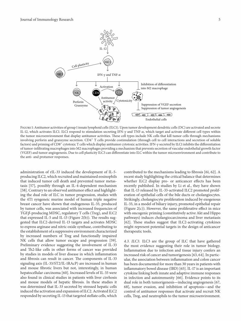
4.1. ILC1. The role of NK cells in cancer has been extensivelydiscussed in recent reviews [44] and here we will onlyfocus on non-NK ILC1. New insights from clinical and invivo studies have shown that non-NK ILC1, together withadaptive immune cells, might be involved in responses thateither mediate the elimination of tumors (natural cytotoxi-city, antibody-dependent cytotoxicity, and phagocytosis) orpromote tumor growth and metastasis (Figure 1) [15, 45–47].Recent reports have started to unravel the role of non-NKILC1 in tumorigenesis and although they lack expression ofgranzyme and perforin, they may have similar functions toNK cells in the antitumor response as they share a similarcytokine secretion signature. Recent studies in models ofinflammatory bowel disease [32, 48] and intestinal infectionwith Toxoplasma gondii [9] have demonstrated that cells withan ILC1 phenotype secrete IFN-𝛾 and TNF-𝛼 and contributeto the inflammatory response and pathology in responseto IL-12 and IL-15. The antiproliferative and proapoptoticproperties of IFN-𝛾 and TNF-𝛼, produced by activated Tcells and monocyte-macrophages, are well established inmany tumor models. However, whether these cytokines aresecreted by ILC and how thesemight inhibit proliferation andinduction of apoptosis need to be addressed. Interestingly,Djenidi et al. have shown that ILC1 had an integrin profile(expression of CD103, integrin alpha E) and a memory-activated phenotype similar to that observed in tumor-infiltrating CD8+ T cells, which are tumor-tissue specific andwhose presence correlates with improved early stage survivalin patients with non-small-cell lung cancer (NSCLC) [49].
4.2. ILC2. ILC2 were originally identified in mesentericlymph nodes and characterized by their ability to primeand stimulate Th2 responses. Following stimulation with IL-25 or IL-33, ILC2 produce IL-5, IL-13 [50, 51], IL-4, IL-6,IL-9, and amphiregulin which induce Th2 differentiation,production of antibodies, and class switching [52].The effectsof ILC2 can be mediated through the secretion of pro-Th2cytokines or by cell-cell interactions via presentation ofMHCclass II-associated antigens to T cells or OX40L stimulation[53, 54]. IL-5- and IL-13-producing ILC2 are associated withprotective immunity at mucosal surfaces. Clinical studiessuggested that increased numbers of ILC2 in peripheralblood, and the cytokines they secrete, could contribute tothe immunosuppressive environment maintained by Th2,myeloid-derived suppressor cells (MDSC), and macrophagesobserved in patients with gastric cancer [55, 56]. Ikutani et al.were amongst the first to show evidence of the role of ILC2 incancer. Using a mouse model of lung metastatic melanoma,ILC2 were specifically shown to produce IL-5 in response toIL-33 stimulation (Figure 2(a)). Following tumor induction,
Journal of Immunology Research 5
ILC3
Cell plasticity
DC
IL-12
ILC1
IFN- and TNF-
NK CD4 CD8
Activation
Perforin activity Cytotoxicity
Inhibition of differentiationinto M2 macrophage
Suppression of VGEF secretionSuppression of tumor angiogenesis
Endothelial cells
Granzyme
Antitumor
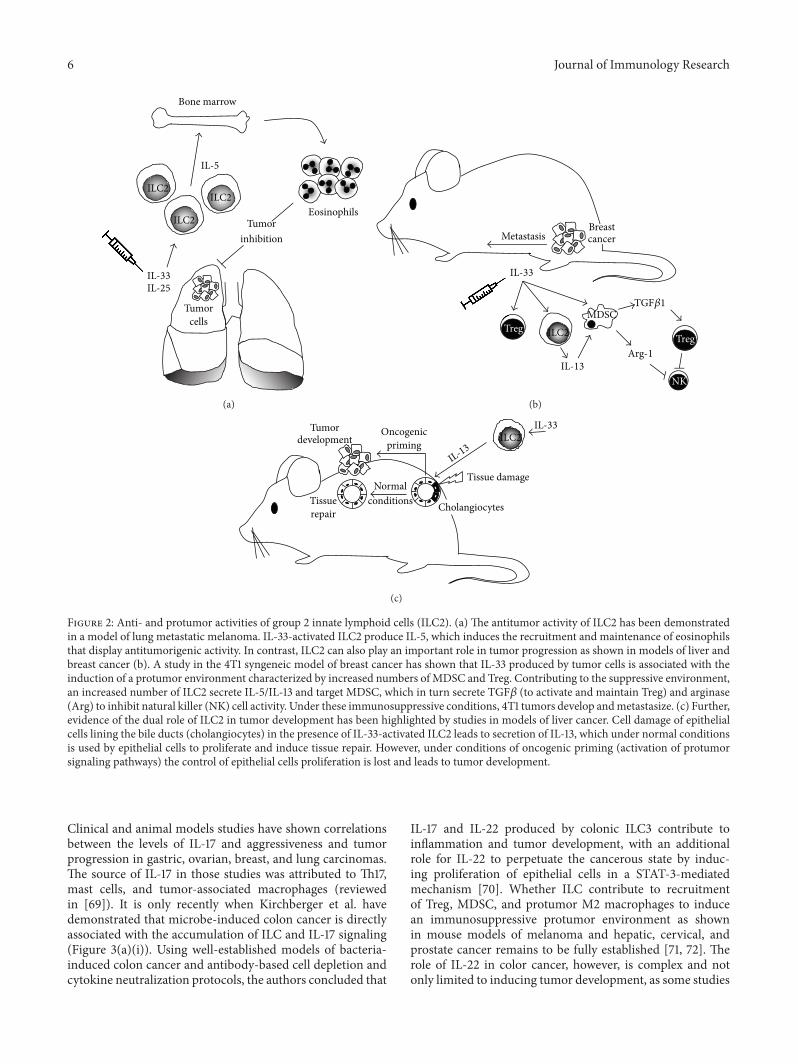
Figure 1: Antitumor activities of group 1 innate lymphoid cells (ILC1). Upon tumor development dendritic cells (DC) are activated and secreteIL-12, which activates ILC1. ILC1 respond to stimulation secreting IFN-𝛾 and TNF-𝛼, which target and activate different cell types withinthe tumor microenvironment that display antitumor activities. These cell types include NK cells that kill tumor cells through mechanismsinvolving perforin and granzyme secretion. CD4+ T cells provide costimulation (through cell to cell interactions and secretion of solublefactors) and priming of CD8+ cytotoxic T cells which display antitumor cytotoxic activities. IFN-𝛾 secreted by ILC1 inhibits the differentiationof tumor-infiltratingmacrophages intoM2macrophages providing amechanism that prevents secretion of vascular endothelial growth factor(VGEF) and tumor angiogenesis. Due to cell plasticity ILC3 can differentiate into ILC within the tumor microenvironment and contribute tothe anti- and protumor responses.
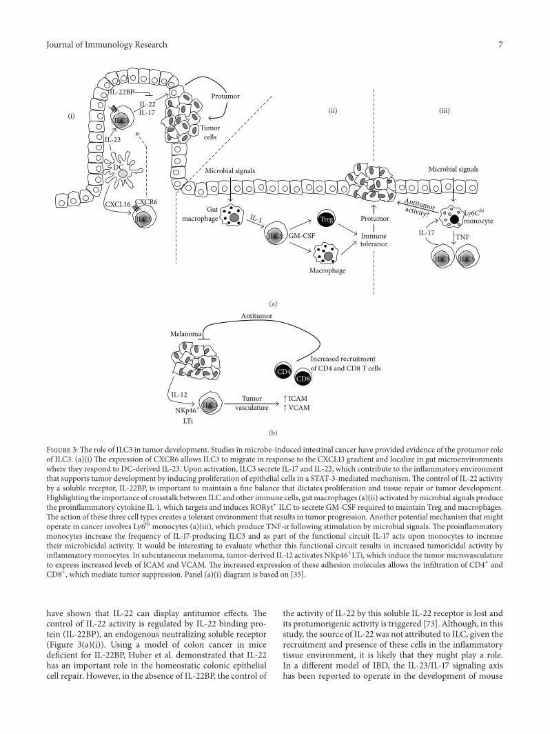
administration of rIL-33 induced the development of IL-5-producing ILC2, which recruited andmaintained eosinophilsthat induced tumor cell death and prevented tumor metas-tasis [57], possibly through an IL-4-dependent mechanism[58]. Contrary to an observed antitumor effect and highlight-ing the dual role of ILC in tumor progression, studies withthe 4T1 syngeneic murine model of human triple negativebreast cancer have shown that endogenous IL-33, producedby tumor cells, was associated with increased frequencies ofTGF𝛽-producing MDSC, regulatory T cells (Treg), and ILC2that expressed IL-5 and IL-13 (Figure 2(b)). The results sug-gested that ILC2-derived IL-13 targets and activates MDSCto express arginase and nitric oxide synthase, contributing tothe establishment of a suppressive environment characterizedby increased numbers of Treg and functionally impairedNK cells that allow tumor escape and progression [59].Preliminary evidence suggesting the involvement of IL-33and Th2-like cells in other forms of cancer was providedby studies in models of liver disease in which inflammationand fibrosis can result in cancer. The components of IL-33signaling axis (IL-33/ST2/IL-1RAcP) are increased in humanand mouse fibrotic livers but not, interestingly, in humanhepatocellular carcinoma [60]. Increased levels of IL-33 werealso found in clinical studies in patients with liver cirrhosisand mouse models of hepatic fibrosis. In these studies itwas determined that IL-33 secreted by stressed hepatic cellsinduced the activation and expansion of ILC2.Activated ILC2responded by secreting IL-13 that targeted stellate cells, which
contributed to the mechanisms leading to fibrosis [61, 62]. Arecent study highlighting the critical balance that determineswhether ILC2 display pro- or anticancer effects has beenrecently published. In studies by Li et al., they have shownthat IL-13 released by IL-33-activated ILC2 promoted prolif-eration of epithelial cells of the bile ducts or cholangiocytes.Strikingly, cholangiocyte proliferation induced by exogenousIL-33, in a model of biliary injury, promoted epithelial repair(Figure 2(c)). However, the same proliferative effect in micewith oncogenic priming (constitutively active Akt and Hippopathways) induces cholangiocarcinoma and liver metastasis[62]. These studies suggest that ILC2-activating cytokinesmight represent potential targets in the design of anticancertherapeutic tools.
4.3. ILC3. ILC3 are the group of ILC that have gatheredthe most evidence suggesting their role in tumor biology.Inflammation due to infection and tissue injury confers anincreased risk of cancer and tumorigenesis [63, 64]. In partic-ular, the association between inflammation and colon cancerhas been documented for more than 30 years in patients withinflammatory bowel disease (IBD) [65]. IL-17 is an importantcytokine linking both innate and adaptive immune responsesin infection and autoimmunity [66]. Evidence points to itsdual role in both tumorigenesis—inducing angiogenesis [67,68], tumor evasion, and inhibition of apoptosis—and thecontrol of antitumor responses that activate and recruit NKcells, Treg, and neutrophils to the tumor microenvironment.
6 Journal of Immunology Research
Bone marrow
IL-5
ILC2
ILC2
ILC2
IL-33IL-25
Tumorinhibition
Tumorcells
Eosinophils
(a)
Metastasis
IL-13
ILC2
IL-33
Breastcancer
TregTreg
MDSC
NK
Arg-1
TGF1
(b)
IL-13ILC2
IL-33Tumordevelopment
Oncogenicpriming
NormalconditionsTissue
repair Cholangiocytes
Tissue damage
(c)
Figure 2: Anti- and protumor activities of group 2 innate lymphoid cells (ILC2). (a) The antitumor activity of ILC2 has been demonstratedin a model of lung metastatic melanoma. IL-33-activated ILC2 produce IL-5, which induces the recruitment and maintenance of eosinophilsthat display antitumorigenic activity. In contrast, ILC2 can also play an important role in tumor progression as shown in models of liver andbreast cancer (b). A study in the 4T1 syngeneic model of breast cancer has shown that IL-33 produced by tumor cells is associated with theinduction of a protumor environment characterized by increased numbers of MDSC and Treg. Contributing to the suppressive environment,an increased number of ILC2 secrete IL-5/IL-13 and target MDSC, which in turn secrete TGF𝛽 (to activate and maintain Treg) and arginase(Arg) to inhibit natural killer (NK) cell activity. Under these immunosuppressive conditions, 4T1 tumors develop andmetastasize. (c) Further,evidence of the dual role of ILC2 in tumor development has been highlighted by studies in models of liver cancer. Cell damage of epithelialcells lining the bile ducts (cholangiocytes) in the presence of IL-33-activated ILC2 leads to secretion of IL-13, which under normal conditionsis used by epithelial cells to proliferate and induce tissue repair. However, under conditions of oncogenic priming (activation of protumorsignaling pathways) the control of epithelial cells proliferation is lost and leads to tumor development.
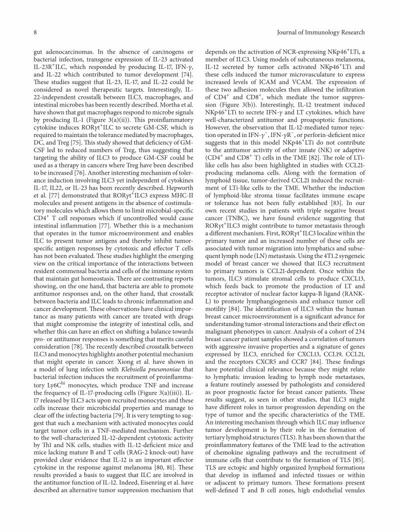
Clinical and animal models studies have shown correlationsbetween the levels of IL-17 and aggressiveness and tumorprogression in gastric, ovarian, breast, and lung carcinomas.The source of IL-17 in those studies was attributed to Th17,mast cells, and tumor-associated macrophages (reviewedin [69]). It is only recently when Kirchberger et al. havedemonstrated that microbe-induced colon cancer is directlyassociated with the accumulation of ILC and IL-17 signaling(Figure 3(a)(i)). Using well-established models of bacteria-induced colon cancer and antibody-based cell depletion andcytokine neutralization protocols, the authors concluded that
IL-17 and IL-22 produced by colonic ILC3 contribute toinflammation and tumor development, with an additionalrole for IL-22 to perpetuate the cancerous state by induc-ing proliferation of epithelial cells in a STAT-3-mediatedmechanism [70]. Whether ILC contribute to recruitmentof Treg, MDSC, and protumor M2 macrophages to inducean immunosuppressive protumor environment as shownin mouse models of melanoma and hepatic, cervical, andprostate cancer remains to be fully established [71, 72]. Therole of IL-22 in color cancer, however, is complex and notonly limited to inducing tumor development, as some studies
Journal of Immunology Research 7
ILC3
ILC3 ILC3
ILC3
ILC3
IL-22BPIL-22IL-17
IL-23
DC
CXCL16 CXCR6
Tumorcells
Microbial signals Microbial signals
Gutmacrophage IL-1
GM-CSF
Treg
Macrophage
Immunetolerance
activity?
IL-17 TNF
Ly6Chi
monocyte
(i)(ii) (iii)
Protumor
Protumor
Antitumor
(a)
ILC3
Melanoma
IL-12
LTi
Tumorvasculature
CD4CD8
Increased recruitmentof CD4 and CD8 T cells
↑ ICAM↑ VCAMNKp46+
Antitumor
(b)
Figure 3:The role of ILC3 in tumor development. Studies in microbe-induced intestinal cancer have provided evidence of the protumor roleof ILC3. (a)(i) The expression of CXCR6 allows ILC3 to migrate in response to the CXCL13 gradient and localize in gut microenvironmentswhere they respond to DC-derived IL-23. Upon activation, ILC3 secrete IL-17 and IL-22, which contribute to the inflammatory environmentthat supports tumor development by inducing proliferation of epithelial cells in a STAT-3-mediated mechanism.The control of IL-22 activityby a soluble receptor, IL-22BP, is important to maintain a fine balance that dictates proliferation and tissue repair or tumor development.Highlighting the importance of crosstalk between ILC and other immune cells, gutmacrophages (a)(ii) activated bymicrobial signals producethe proinflammatory cytokine IL-1, which targets and induces ROR𝛾t+ ILC to secrete GM-CSF required to maintain Treg and macrophages.The action of these three cell types creates a tolerant environment that results in tumor progression. Another potential mechanism that mightoperate in cancer involves Ly6hi monocytes (a)(iii), which produce TNF-𝛼 following stimulation by microbial signals. The proinflammatorymonocytes increase the frequency of IL-17-producing ILC3 and as part of the functional circuit IL-17 acts upon monocytes to increasetheir microbicidal activity. It would be interesting to evaluate whether this functional circuit results in increased tumoricidal activity byinflammatory monocytes. In subcutaneous melanoma, tumor-derived IL-12 activates NKp46+LTi, which induce the tumor microvasculatureto express increased levels of ICAM and VCAM. The increased expression of these adhesion molecules allows the infiltration of CD4+ andCD8+, which mediate tumor suppression. Panel (a)(i) diagram is based on [35].
have shown that IL-22 can display antitumor effects. Thecontrol of IL-22 activity is regulated by IL-22 binding pro-tein (IL-22BP), an endogenous neutralizing soluble receptor(Figure 3(a)(i)). Using a model of colon cancer in micedeficient for IL-22BP, Huber et al. demonstrated that IL-22has an important role in the homeostatic colonic epithelialcell repair. However, in the absence of IL-22BP, the control of
the activity of IL-22 by this soluble IL-22 receptor is lost andits protumorigenic activity is triggered [73]. Although, in thisstudy, the source of IL-22 was not attributed to ILC, given therecruitment and presence of these cells in the inflammatorytissue environment, it is likely that they might play a role.In a different model of IBD, the IL-23/IL-17 signaling axishas been reported to operate in the development of mouse
8 Journal of Immunology Research
gut adenocarcinomas. In the absence of carcinogens orbacterial infection, transgene expression of IL-23 activatedIL-23R+ILC, which responded by producing IL-17, IFN-𝛾,and IL-22 which contributed to tumor development [74].These studies suggest that IL-23, IL-17, and IL-22 could beconsidered as novel therapeutic targets. Interestingly, IL-22-independent crosstalk between ILC3, macrophages, andintestinal microbes has been recently described. Mortha et al.have shown that gutmacrophages respond tomicrobe signalsby producing IL-1 (Figure 3(a)(ii)). This proinflammatorycytokine induces ROR𝛾t+ILC to secrete GM-CSF, which isrequired tomaintain the tolerancemediated bymacrophages,DC, and Treg [75]. This study showed that deficiency of GM-CSF led to reduced numbers of Treg, thus suggesting thattargeting the ability of ILC3 to produce GM-CSF could beused as a therapy in cancers where Treg have been describedto be increased [76]. Another interestingmechanism of toler-ance induction involving ILC3 yet independent of cytokinesIL-17, IL22, or IL-23 has been recently described. Hepworthet al. [77] demonstrated that ROR𝛾t+ILC3 express MHC-IImolecules and present antigens in the absence of costimula-tory molecules which allows them to limit microbial-specificCD4+ T cell responses which if uncontrolled would causeintestinal inflammation [77]. Whether this is a mechanismthat operates in the tumor microenvironment and enablesILC to present tumor antigens and thereby inhibit tumor-specific antigen responses by cytotoxic and effector T cellshas not been evaluated. These studies highlight the emergingview on the critical importance of the interactions betweenresident commensal bacteria and cells of the immune systemthat maintain gut homeostasis. There are contrasting reportsshowing, on the one hand, that bacteria are able to promoteantitumor responses and, on the other hand, that crosstalkbetween bacteria and ILC leads to chronic inflammation andcancer development.These observations have clinical impor-tance as many patients with cancer are treated with drugsthat might compromise the integrity of intestinal cells, andwhether this can have an effect on shifting a balance towardspro- or antitumor responses is something that merits carefulconsideration [78]. The recently described crosstalk betweenILC3 andmonocytes highlights another potentialmechanismthat might operate in cancer. Xiong et al. have shown ina model of lung infection with Klebsiella pneumoniae thatbacterial infection induces the recruitment of proinflamma-tory Ly6Chi monocytes, which produce TNF and increasethe frequency of IL-17-producing cells (Figure 3(a)(iii)). IL-17 released by ILC3 acts upon recruited monocytes and thesecells increase their microbicidal properties and manage toclear off the infecting bacteria [79]. It is very tempting to sug-gest that such a mechanism with activated monocytes couldtarget tumor cells in a TNF-mediated mechanism. Furtherto the well-characterized IL-12-dependent cytotoxic activityby Th1 and NK cells, studies with IL-12-deficient mice andmice lacking mature B and T cells (RAG-2 knock-out) haveprovided clear evidence that IL-12 is an important effectorcytokine in the response against melanoma [80, 81]. Theseresults provided a basis to suggest that ILC are involved inthe antitumor function of IL-12. Indeed, Eisenring et al. havedescribed an alternative tumor suppression mechanism that
depends on the activation of NCR-expressing NKp46+LTi, amember of ILC3. Using models of subcutaneous melanoma,IL-12 secreted by tumor cells activated NKp46+LTi andthese cells induced the tumor microvasculature to expressincreased levels of ICAM and VCAM. The expression ofthese two adhesion molecules then allowed the infiltrationof CD4+ and CD8+, which mediate the tumor suppres-sion (Figure 3(b)). Interestingly, IL-12 treatment inducedNKp46+LTi to secrete IFN-𝛾 and LT cytokines, which havewell-characterized antitumor and proapoptotic functions.However, the observation that IL-12-mediated tumor rejec-tion operated in IFN-𝛾−, IFN-𝛾R−, or perforin-deficient micesuggests that in this model NKp46+LTi do not contributeto the antitumor activity of other innate (NK) or adaptive(CD4+ and CD8+ T) cells in the TME [82]. The role of LTi-like cells has also been highlighted in studies with CCL21-producing melanoma cells. Along with the formation oflymphoid tissue, tumor-derived CCL21 induced the recruit-ment of LTi-like cells to the TME. Whether the inductionof lymphoid-like stroma tissue facilitates immune escapeor tolerance has not been fully established [83]. In ourown recent studies in patients with triple negative breastcancer (TNBC), we have found evidence suggesting thatROR𝛾t+ILC3 might contribute to tumor metastasis througha differentmechanism. First, ROR𝛾t+ILC3 localize within theprimary tumor and an increased number of these cells areassociated with tumor migration into lymphatics and subse-quent lymphnode (LN)metastasis. Using the 4T1.2 syngeneicmodel of breast cancer we showed that ILC3 recruitmentto primary tumors is CCL21-dependent. Once within thetumors, ILC3 stimulate stromal cells to produce CXCL13,which feeds back to promote the production of LT andreceptor activator of nuclear factor kappa-B ligand (RANK-L) to promote lymphangiogenesis and enhance tumor cellmotility [84]. The identification of ILC3 within the humanbreast cancer microenvironment is a significant advance forunderstanding tumor-stromal interactions and their effect onmalignant phenotypes in cancer. Analysis of a cohort of 234breast cancer patient samples showed a correlation of tumorswith aggressive invasive properties and a signature of genesexpressed by ILC3, enriched for CXCL13, CCL19, CCL21,and the receptors CXCR5 and CCR7 [84]. These findingshave potential clinical relevance because they might relateto lymphatic invasion leading to lymph node metastases,a feature routinely assessed by pathologists and consideredas poor prognostic factor for breast cancer patients. Theseresults suggest, as seen in other studies, that ILC3 mighthave different roles in tumor progression depending on thetype of tumor and the specific characteristics of the TME.An interesting mechanism through which ILCmay influencetumor development is by their role in the formation oftertiary lymphoid structures (TLS). It has been shown that theproinflammatory features of the TME lead to the activationof chemokine signaling pathways and the recruitment ofimmune cells that contribute to the formation of TLS [85].TLS are ectopic and highly organized lymphoid formationsthat develop in inflamed and infected tissues or withinor adjacent to primary tumors. These formations presentwell-defined T and B cell zones, high endothelial venules
Journal of Immunology Research 9
(HEV), mature DC, germinal centre reactions, and B cellclass switch in the B cell follicles, suggesting the generation ofadaptive immune responses [86]. These lymphoid aggregatesare also characterized by the expression of chemokines(CCL19, CCL21, CXCL10, CXCL12, and CXCL13), adhesionmolecules (ICAM-2, ICAM-3, VCAM-1, and MAdCAM-1),and integrins (alphaL, alpha4, and alphaD) [87], which notonly attract effector immune cells such as Treg, DC, naıve Bcells, andT follicular helper (TFH) cells but also determine thearchitecture and cell segregation in specific compartments.The composition of TLS might be different depending on thetumor type and a growing number of clinical studies suggestthat TLS and associated biomarkers correlate with clinicaloutcome and prognostic value [88]. Studies of patients withmelanoma, breast [89], colorectal, lung, pancreas [90], orrenal cell carcinomas have shown that the presence of TLS isof positive prognostic value. However, two different studiesin patients with breast and renal cell carcinomas have alsoshown that the presence of TLS has a negative prognosticvalue (an extensive revision of the clinical studies and trialsare reviewed in [86, 91]). Detailed analyses have found thatparticular cell types within the TLS confer the prognosticvalue; for instance, CD8+ T cells and antibody-producing-plasma cells in ovarian cancer [92] and DC in primarylung tumors [93] confer a positive prognostic value. On theother hand, Joshi et al. have recently shown that tumor-infiltrating Treg in a mouse model of lung adenocarcinomaare increased and found at the tumor margins in tertiarylymphoid structures. At these sites, Treg actively restraineffector T cells [94]. These results, elevated numbers ofTreg, correlate with the adverse clinical outcome (poorsurvival) in patients with breast cancer [95]. In a strikingstudy, Finkin et al. have used a model of hepatocellularcarcinoma (HCC) with mice constitutively expressing theactive form of IKK-B in hepatocytes to activate the NF-𝜅Bpathway. They reported the presence of small clusters ofhepatocytes expressing markers of tumor progenitor cellswithin TLS formed in nontumor sites of the inflamed liver.These clusters progressively coalesced and egressed from theTLS to grow as HCC in all IKK-B-expressing livers. Theseresults suggest that TLS serve as niches supporting tumorcells growth and contribute to recurrence in HCC [96].Despite the strong correlation between clinical or prognosticvalue and the presence of TLS in different types of cancer,it is unclear which factors (intrinsic or extrinsic) contributeto their development. Studies from the rheumatology fieldhave suggested a role for ILC in the formation of TLS. Noortet al. have recently shown that TLS within the synovialtissue of some patients with rheumatoid arthritis contain lownumbers of CD3−RORC+ ILC3 which might play a role inTLS formation through a mechanism involving the release ofLT𝛽 and the expansion of follicular DC [97]. Interestingly,in the cancer setting, Carrega et al. have recently reportedthe presence of NCR+ILC3 at the edge of tumor-associatedTLS in NSCLC. Increased number of these cells in early stagetumors correlated with the density of intratumoral TLS andpredicted favorable clinical outcomes [98]. Further analysesare required to evaluate the role of ILC3 on TLS formationand function in other types of cancer. From the studies
mentioned above, it is clear that identifying the mechanismsand cellular components underlying TLS formation will behelpful to understand the pro- and antitumor responseswithin the tumor microenvironment. This knowledge willbe helpful to develop new therapies to promote or inhibitTLS formation. In fact, different drugs and antagonist ofthe LT𝛼/𝛽, RANK/RANK-L, and ICOS/ICOS-L signalingpathways are being developed and tested for their potentialto manipulate TLS formation (reviewed in [91]). Thus, theparadoxical role of ILC3 in both host protective and tumor-promoting immunosuppressive effects can be associated withthe reported functional plasticity that allows these cellsto respond to changes in the tumor microenvironmentaccordingly (reviewed in [6]). Whether ILC display pro- orantitumor activities seems to depend on the type of tumorand stage of development and on the complex network offine-tuned incoming signals that control cell-cell interactionsin themicroenvironment. It is likely that the ability to controlor influence those interactions will be important for thedevelopment of new therapeutic tools to fight cancer. Recentencouraging studies with chemical inhibitors of ROR𝛾t-mediated transcription (such as GSK805) have revealed atransient and specific targeting/inhibition tool for ROR𝛾t+cells. In a recent study of a model of intestinal infectionwith bacteria, administration of GSK805 reduced cytokineproduction by Th17, but not ILC3, thus preserving innateimmunity. This treatment resulted in a therapeutic responseas reduced activity of Th17, but not ILC3, contributed tothe control of inflammation in their infection model [99].Given the nonredundant functions of cytokines produced byT cells and ILC, this cell-specific and transient inhibition ofROR𝛾t+ cells might have applications in models of cancerwhere manipulating and enhancing the antitumor functionsof ILC over the proinflammatory functions of Th17 mightbe beneficial and important to control tumor development.Details on how these cytokines, chemokines, and growthfactors allow the cell-cell interactions that maintain thehomeostatic balance are currently being explored and will beessential for the development of new therapeutic tools againstcancer. In the next sectionwewill discuss imaging approachesto analyze the crosstalk between ILC and other innate andadaptive immune cells.
5. Imaging Strategies to Study theCrosstalk and Interactions betweenILC and Other Immune Cells
Immunohistochemistry (IHC), immunofluorescence, andflow cytometry methodologies are fundamental tools thathave contributed to the identification and our knowledgeof the different ILC subsets. In some studies and usingIHC techniques, ILC1 have been identified with markersNKp44+, CD103+, and CD3− [48] and ILC3 using Ror𝛾t+IL-7RB+CD11c− [100] or CD3−CD127+CD117+ROR𝛾t+ [101].With the use of many different markers and multicolorcytometric analyses, it has been possible to study anddetermine the presence and changes in ILC populationsin normal, disease, and inflammatory settings. However,understanding the roles of ILC in the control of immune
10 Journal of Immunology Research
responses requires a refined knowledge of the crosstalk andinteractions between ILC and other innate and adaptiveimmune cells. This has been approached through the useof novel imaging techniques using fluorescent probes. UsingILC3 as an example, here we describe some approaches thatcan be used to examine the role of ILC in the ex vivo and invivo settings.
A major complication arising when looking at a subset ofcells, such as the ILC, is the necessity to define that subset witha range of different markers. The current series of markersand phenotype accepted for identifying ILC3 require four(five including a nuclear stain) separate fluorophores whenimaging tissue to identify the one cell type. If ILC3 areto be imaged along with other cells of interest, the use ofserial sections is required, labeling a certain cell type persection and overlaying the images. Alternatively, a multiplexof 6 or 7 fluorescent probes can be used to identify twodifferent cell types on a single slide and imaging on astandard commercial confocal microscope. Using multiplefluorophores in a single slide is generally better acceptedas it avoids any slight alterations between sections and theinherent difficulty in obtaining good quality sequentiallycut sections. With using such a high number of fluorescentproteins on the same slide, it is common to find spectralbleed-through and cross-excitation between the detectedchannels. Fluorophore selection including fluorophores withlarge stokes shift, such as the Brilliant Violet and PacificBlue/Green/Orange, and the use of quantum dots [102] allowmultiplexing on a single excitation laser. This approach,combined with carefully customized microscope configura-tion, means it is possible to avoid spectral bleed-throughaltogether [103]. However, in the cases where it is unavoid-able, postacquisition correction algorithms can be utilized tocorrect for unwanted fluorescent emission crosstalk (spectraloverlap) by deconvolution/unmixing [104] to remove anyremaining overlaps between the fluorophores of the detectedchannels [105]. Multicolor confocal microscopy provideshigh-resolution tissue imaging, allowing the labeling ofdifferent cell types that require several markers to identify.This provides key localization information in fixed tissue ata particular time point and as such is extremely useful inindicating possible cellular interactions based on the relativecell-cell proximity, which can be further investigated usingtime-lapse microscopy or other means. A drawback of usingconfocal microscopy as an approach to imaging ILC3 isthe lack of temporal information. Relatively little is knownregarding the role these cells play in different scenarios;therefore, the ability to track where these cells migrate andthe cells they interact with is fundamental to decipheringtheir role and function.Multiphotonmicroscopy (also knownas 2-photon microscopy) allows the imaging of fluorescentlylabeled cells deep within tissue in an in vivo setting, visu-alizing cell behavior in the natural environment. Kineticinformation such as cell velocity, track length, meanderingindex, and displacement can reveal valuable informationregarding immune cells activity [106, 107] and the abilityto image and track the interactions between different cellsubsets is fundamental to further elucidate the roles ILC3mayplay in inhibiting particular signaling or receptor pathways.
Current commercial multiphoton systems typically have thecapacity for 2 to 4 fluorescent probes to be imaged at atime, and using the same multifluorescent labeling as usedin confocal microscope is impractical. In this situation, thepreferable method is to isolate ILC3 using flow cytometrybased cell sorting with the markers for ILC3 (CD3, CD127,CD117, and NKp46), followed by fluorescent labeling of thispopulation using cell tracker dyes [108]. These labeled cellscan then be injected intravenously into the target animal andimaged 18–72 hours later, allowing for the cells to migrateto their respective homing tissues [108, 109]. This methodallows for the imaging of ILC3 using a single fluorescentchannel, leaving the rest for additional labeled cells of interest.As multiphoton microscopy is primarily used for intravitalimaging, several approaches could be used to facilitate theimaging of ILC3. The activity of ILC3 within the lymph nodecould be imaged in a direct manner by surgically exposingthe inguinal lymph node [110]. While this approach allowsfor controllable intravital imaging, it is a terminal proce-dure and specific experimental time points must be chosenwhen imaging. This approach could be applied to modelsof breast cancer with direct exposure and imaging of thedraining inguinal lymph node and the primary tumor [111].Furthermore, in the same tumor model, the use of surgicallyimplanted windows can allow for longitudinal imaging ofthese organs, allowing repeated imaging of the same tissuethroughout tumor development, with or without experimen-tal intervention. A small abdominal window implanted overthe inguinal lymph node allows the inguinal lymph node tobe reexposed several times for imaging [112]. ILC3 can alsobe imaged deep within tumors via a mammary window thatis inserted in the skin over the lower mammary gland region.Breast cancer cell lines can then be injected/implanted andallowed to grow up into the recess created by the window,therefore allowing a surgery free method of imaging ILC3behavior over the development of the tumor [111, 113, 114].The use of this window method can be extended to imagingILC3 in the lung using a technique developed by Looney etal. [115] which, although terminal, can allow the studying ofILC3 and other target cells in a fully functional lung fromchronic obstructive pulmonary disease [116] and lung cancer[98]. Taking advantage of current photo-switchable mousemodels provides another tool to examine ILC3 function. Inthese mice, exposure to ultraviolet light induces changes influorescence on the reporter cells [117]. Using this, organs ortissues of interest can be activated using a UV light source,either through surgical exposure or using the aforementionedwindow systems, and the migration of cells from this tissuewas tracked, for instance, as has been shown between intes-tine and mesenteric lymph nodes [36]. This same approachcould be applied to a variety of tumor models to furtherunveil ILC3migration properties and the elements that affectthem.
6. Conclusions
Interest and attention on ILC has increased in recent yearsand a great deal of information has been gathered. Thediscovery of this family of innate immune cells, the definition
Journal of Immunology Research 11
of their phenotypes, and their classification into differentsubgroups based on their differentiation requirements andbiological functions represent important achievements inbiomedical research.The description of intricate interactionsin different tissues and organs, beyond the gut, with otherinnate and adaptive immune cells in homeostasis or duringdifferent types of infection, inflammatory diseases, and can-cer partially highlights the functional importance of thesecells. The challenge for future research is to fully understandand decipher the complex and specific contributions ofeach type of ILC to the control and regulation of immuneresponses. In particular, further studies on dissecting thedetailed nature and implications of the reported plasticitywill be required. In the specific case of cancer, observationsfrom the clinic and studies in a variety of animal modelshave shown opposing abilities of ILC to either promote orrepress tumor growth. A better understanding of the preciseroles of ILC cell types in tumorigenesis or control will allowunderstanding of the potential value of manipulation oftheir functions to develop new interventions and therapeuticstrategies.
Additional Points
Review Search Strategy.This is a narrative review.We searchedPUBMED for original articles focusing on innate lymphoidcells and cancer published in the last 10 years, from 2006to 2016. Search terms included “Group 1, 2, and 3 Innatelymphoid cells and tumor”, “ILC1, 2, 3 and cancer”, and“Innate lymphoid cells and tumor microenvironment”. Allpapers identified were English-language, full-text papers. Wealso searched the reference lists of identified articles forfurther papers.
Competing Interests
The authors declare that they have no competing interests.
Acknowledgments
Fabian Flores-Borja and Peter Gordon are supported by KCLBreast Cancer Now Unit funding (awarded to Andrew Tutt),including the Sarah Greene Fellowship to Sheeba Irshad.Tony Ng is supported by an endowment fund fromDimblebyCancer Care to King’s College London and by the KCL-UCLComprehensive Cancer Imaging Centre (CCIC) funding(CR-UK & EPSRC, in association with the MRC and DoHEngland).
References
[1] S. J. Turley, V. Cremasco, and J. L. Astarita, “Immunologicalhallmarks of stromal cells in the tumour microenvironment,”Nature Reviews Immunology, vol. 15, no. 11, pp. 669–682, 2015.
[2] W. W. Agace, “T-cell recruitment to the intestinal mucosa,”Trends in Immunology, vol. 29, no. 11, pp. 514–522, 2008.
[3] M. D. Vesely, M. H. Kershaw, R. D. Schreiber, and M. J. Smyth,“Natural innate and adaptive immunity to cancer,” AnnualReview of Immunology, vol. 29, pp. 235–271, 2011.
[4] L. L. Lanier, “Shades of grey-the blurring view of innate andadaptive immunity,” Nature Reviews Immunology, vol. 13, no. 2,pp. 73–74, 2013.
[5] G. Eberl, J. P. Di Santo, and E. Vivier, “The brave new world ofinnate lymphoid cells,” Nature Immunology, vol. 16, no. 1, pp. 1–5, 2015.
[6] H. Spits, D. Artis, M. Colonna et al., “Innate lymphoid cells—aproposal for uniform nomenclature,”Nature Reviews Immunol-ogy, vol. 13, no. 2, pp. 145–149, 2013.
[7] M. D. Hazenberg and H. Spits, “Human innate lymphoid cells,”Blood, vol. 124, no. 5, pp. 700–709, 2014.
[8] C. A. Biron, K. B. Nguyen, G. C. Pien, L. P. Cousens, andT. P. Salazar-Mather, “Natural killer cells in antiviral defense:function and regulation by innate cytokines,” Annual Review ofImmunology, vol. 17, pp. 189–220, 1999.
[9] C. S. N. Klose, M. Flach, L. Mohle et al., “Differentiation oftype 1 ILCs from a common progenitor to all helper-like innatelymphoid cell lineages,” Cell, vol. 157, no. 2, pp. 340–356, 2014.
[10] R. Yagi, C. Zhong, D. L. Northrup et al., “The transcriptionfactor GATA3 is critical for the development of all IL-7R𝛼-expressing innate lymphoid cells,” Immunity, vol. 40, no. 3, pp.378–388, 2014.
[11] A. Moretta, C. Bottino, M. Vitale et al., “Activating receptorsand coreceptors involved in human natural killer cell-mediatedcytolysis,” Annual Review of Immunology, vol. 19, pp. 197–223,2001.
[12] J. A. Walker, J. L. Barlow, and A. N. J. McKenzie, “Innatelymphoid cells—how did we miss them?” Nature ReviewsImmunology, vol. 13, no. 2, pp. 75–87, 2013.
[13] M. M. Fort, J. Cheung, D. Yen et al., “IL-25 Induces IL-4, IL-5, and IL-13 andTh2-associated pathologies in vivo,” Immunity,vol. 15, no. 6, pp. 985–995, 2001.
[14] Y. Huang, L. Guo, J. Qiu et al., “IL-25-responsive, lineage-negative KLRG1 hi cells are multipotential ‘inflammatory’ type2 innate lymphoid cells,” Nature Immunology, vol. 16, no. 2, pp.161–169, 2015.
[15] J. A.Walker and A. N. J. McKenzie, “Development and functionof group 2 innate lymphoid cells,” Current Opinion in Immunol-ogy, vol. 25, no. 2, pp. 148–155, 2013.
[16] P. Licona-Limon, L.K.Kim,N.W.Palm, andR.A. Flavell, “TH2,allergy and group 2 innate lymphoid cells,”Nature Immunology,vol. 14, no. 6, pp. 536–542, 2013.
[17] Y.-J. Chang, H. Y. Kim, L. A. Albacker et al., “Innate lymphoidcells mediate influenza-induced airway hyper-reactivity inde-pendently of adaptive immunity,” Nature Immunology, vol. 12,no. 7, pp. 631–638, 2011.
[18] J. Zhu, “T helper 2 (Th2) cell differentiation, type 2 innate lym-phoid cell (ILC2) development and regulation of interleukin-4(IL-4) and IL-13 production,” Cytokine, vol. 75, no. 1, pp. 14–24,2015.
[19] D. M. Zaiss, L. Yang, P. R. Shah, J. J. Kobie, J. F. Urban, andT. R. Mosmann, “Amphiregulin, a TH2 cytokine enhancingresistance to nematodes,” Science, vol. 314, no. 5806, p. 1746,2006.
[20] L. A. Monticelli, G. F. Sonnenberg, M. C. Abt et al., “Innatelymphoid cells promote lung-tissue homeostasis after infectionwith influenza virus,” Nature Immunology, vol. 12, no. 11, pp.1045–1054, 2011.
[21] B. S. Kim, M. C. Siracusa, S. A. Saenz et al., “TSLP elicits IL-33-independent innate lymphoid cell responses to promote skininflammation,” Science Translational Medicine, vol. 5, no. 170,Article ID 170ra16, 2013.
12 Journal of Immunology Research
[22] X. Yu, R. Pappu, V. Ramirez-Carrozzi et al., “TNF superfamilymember TL1A elicits type 2 innate lymphoid cells at mucosalbarriers,”Mucosal Immunology, vol. 7, no. 3, pp. 730–740, 2014.
[23] A. C. Richard, C. Tan, E. T. Hawley et al., “The TNF-family ligand TL1A and its receptor DR3 promote T cell-mediated allergic immunopathology by enhancing differentia-tion and pathogenicity of IL-9-producing T cells,” The Journalof Immunology, vol. 194, no. 8, pp. 3567–3582, 2015.
[24] N. Satoh-Takayama, C. A. J. Vosshenrich, S. Lesjean-Pottieret al., “Microbial flora drives interleukin 22 production inintestinal NKp46+ cells that provide innate mucosal immunedefense,” Immunity, vol. 29, no. 6, pp. 958–970, 2008.
[25] C. Vonarbourg, A. Mortha, V. L. Bui et al., “Regulated expres-sion of nuclear receptor ROR𝛾t confers distinct functional fatesto NK cell receptor-expressing ROR𝛾t+ innate lymphocytes,”Immunity, vol. 33, no. 5, pp. 736–751, 2010.
[26] R. E. Mebius, “Organogenesis of lymphoid tissues,” NatureReviews Immunology, vol. 3, no. 4, pp. 292–303, 2003.
[27] Z. Sun, D. Unutmaz, Y.-R. Zou et al., “Requirement for ROR𝛾 inthymocyte survival and lymphoid organ development,” Science,vol. 288, no. 5475, pp. 2369–2373, 2000.
[28] C. Zhong, K. Cui, C. Wilhelm et al., “Group 3 innate lymphoidcells continuously require the transcription factor GATA-3 aftercommitment,” Nature Immunology, vol. 17, no. 2, pp. 169–178,2016.
[29] N. Ota, K. Wong, P. A. Valdez et al., “IL-22 bridges thelymphotoxin pathway with the maintenance of colonic lym-phoid structures during infection with Citrobacter rodentium,”Nature Immunology, vol. 12, no. 10, pp. 941–948, 2011.
[30] H. Takatori, Y. Kanno, W. T. Watford et al., “Lymphoid tissueinducer-like cells are an innate source of IL-17 and IL-22,” TheJournal of Experimental Medicine, vol. 206, no. 1, pp. 35–41,2009.
[31] M. El-Behi, B. Ciric, H. Dai et al., “The encephalitogenicity ofTH 17 cells is dependent on IL-1- and IL-23-induced productionof the cytokine GM-CSF,”Nature Immunology, vol. 12, no. 6, pp.568–575, 2011.
[32] J. H. Bernink, C. P. Peters, M. Munneke et al., “Human type 1innate lymphoid cells accumulate in inflamed mucosal tissues,”Nature Immunology, vol. 14, no. 3, pp. 221–229, 2013.
[33] N. Powell, A. W. Walker, E. Stolarczyk et al., “The transcriptionfactor T-bet regulates intestinal inflammation mediated byinterleukin-7 receptor+ innate lymphoid cells,” Immunity, vol.37, no. 4, pp. 674–684, 2012.
[34] M. H. Kim, E. J. Taparowsky, and C. H. Kim, “Retinoic aciddifferentially regulates the migration of innate lymphoid cellsubsets to the gut,” Immunity, vol. 43, no. 1, pp. 107–119, 2015.
[35] N. Satoh-Takayama, N. Serafini, T. Verrier et al., “Thechemokine receptor CXCR6 controls the functional topographyof interleukin-22 producing intestinal innate lymphoid cells,”Immunity, vol. 41, no. 5, pp. 776–788, 2014.
[36] E. C. Mackley, S. Houston, C. L. Marriott et al., “CCR7-dependent trafficking of ROR𝛾+ ILCs creates a uniquemicroen-vironment within mucosal draining lymph nodes,” NatureCommunications, vol. 6, article 5862, 2015.
[37] M. Bajenoff, N. Glaichenhaus, and R. N. Germain, “Fibroblasticreticular cells guide T lymphocyte entry into and migrationwithin the splenic T cell zone,” Journal of Immunology, vol. 181,no. 6, pp. 3947–3954, 2008.
[38] P. J. L. Lane, F. M. Gaspal, F. M. McConnell, D. R. Withers,and G. Anderson, “Lymphoid tissue inducer cells: pivotal cells
in the evolution of CD4 immunity and tolerance?” Frontiers inImmunology, vol. 3, article 24, pp. 1–6, 2012.
[39] G. Magri, M. Miyajima, S. Bascones et al., “Innate lymphoidcells integrate stromal and immunological signals to enhanceantibody production by splenic marginal zone B cells,” NatureImmunology, vol. 15, no. 4, pp. 354–364, 2014.
[40] J. R. Huh, M. W. Leung, P. Huang et al., “Digoxin and itsderivatives suppress TH17 cell differentiation by antagonizingRORgammat activity,” Nature, vol. 472, pp. 486–490, 2011.
[41] L. A. Solt, N. Kumar, P. Nuhant et al., “Suppression of TH17differentiation and autoimmunity by a synthetic ROR ligand,”Nature, vol. 472, no. 7344, pp. 491–494, 2011.
[42] K. E. De Visser, A. Eichten, and L. M. Coussens, “Paradoxicalroles of the immune systemduring cancer development,”NatureReviews Cancer, vol. 6, no. 1, pp. 24–37, 2006.
[43] G. F. Sonnenberg and D. Artis, “Innate lymphoid cells in theinitiation, regulation and resolution of inflammation,” NatureMedicine, vol. 21, no. 7, pp. 698–708, 2015.
[44] S. Carotta, “Targeting NK cells for anticancer immunotherapy:clinical and preclinical approaches,” Frontiers in Immunology,vol. 7, article 152, 2016.
[45] S. I. Grivennikov, F. R. Greten, and M. Karin, “Immunity,inflammation, and cancer,” Cell, vol. 140, no. 6, pp. 883–899,2010.
[46] R. Nowarski, N. Gagliani, S. Huber, and R. A. Flavell, “Innateimmune cells in inflammation and cancer,” Cancer ImmunologyResearch, vol. 1, no. 2, pp. 77–84, 2013.
[47] B. Vallentin, V. Barlogis, C. Piperoglou et al., “Innate lymphoidcells in cancer,” Cancer Immunology Research, vol. 3, no. 10, pp.1109–1114, 2015.
[48] A. Fuchs, W. Vermi, J. S. Lee et al., “Intraepithelial type 1innate lymphoid cells are a unique subset of IL-12- and IL-15-responsive IFN-𝛾-producing cells,” Immunity, vol. 38, no. 4, pp.769–781, 2013.
[49] F. Djenidi, J. Adam, A. Goubar et al., “CD8+CD103+ tumor-infiltrating lymphocytes are tumor-specific tissue-residentmemory T cells and a prognostic factor for survival in lungcancer patients,”The Journal of Immunology, vol. 194, no. 7, pp.3475–3486, 2015.
[50] P. G. Fallon, S. J. Ballantyne, N. E. Mangan et al., “Identificationof an interleukin (IL)-25-dependent cell population that pro-vides IL-4, IL-5, and IL-13 at the onset of helminth expulsion,”Journal of Experimental Medicine, vol. 203, no. 4, pp. 1105–1116,2006.
[51] K. Moro, T. Yamada, M. Tanabe et al., “Innate production ofT(H)2 cytokines by adipose tissue-associated c-Kit(+)Sca-1(+)lymphoid cells,” Nature, vol. 463, no. 7280, pp. 540–544, 2010.
[52] M. Dancescu, M. Rubio-Trujillo, G. Biron, D. Bron, G. Dele-spesse, and M. Sarfati, “Interleukin 4 protects chronic lympho-cytic leukemic B cells from death by apoptosis and upregulatesBcl-2 expression,” The Journal of Experimental Medicine, vol.176, no. 5, pp. 1319–1326, 1992.
[53] C. J. Oliphant, Y. Y. Hwang, J. A. Walker et al., “MHCII-mediated dialog between group 2 innate lymphoid cells andCD4+ T cells potentiates type 2 immunity and promotesparasitic helminth expulsion,” Immunity, vol. 41, no. 2, pp. 283–295, 2014.
[54] L. Y. Drake, K. Iijima, and H. Kita, “Group 2 innate lymphoidcells and CD4+ T cells cooperate to mediate type 2 immuneresponse in mice,” Allergy, vol. 69, no. 10, pp. 1300–1307, 2014.
Journal of Immunology Research 13
[55] Z. Yang, B. Zhang, D. Li et al., “Mast cells mobilize myeloid-derived suppressor cells and Treg cells in tumor microenviron-ment via IL-17 pathway in murine hepatocarcinoma model,”PLoS ONE, vol. 5, no. 1, article e8922, 2010.
[56] Q. Bie, P. Zhang, Z. Su et al., “Polarization of ILC2s in peripheralblood might contribute to immunosuppressive microenviron-ment in patients with gastric cancer,” Journal of ImmunologyResearch, vol. 2014, Article ID 923135, 10 pages, 2014.
[57] M. Ikutani, T. Yanagibashi, M. Ogasawara et al., “Identificationof innate IL-5-producing cells and their role in lung eosinophilregulation and antitumor immunity,” The Journal of Immunol-ogy, vol. 188, no. 2, pp. 703–713, 2012.
[58] R. I. Tepper, R. L. Coffman, and P. Leder, “An eosinophil-dependent mechanism for the antitumor effect of interleukin-4,” Science, vol. 257, no. 5069, pp. 548–551, 1992.
[59] I. P. Jovanovic, N. N. Pejnovic, G. D. Radosavljevic etal., “Interleukin-33/ST2 axis promotes breast cancer growthand metastases by facilitating intratumoral accumulation ofimmunosuppressive and innate lymphoid cells,” InternationalJournal of Cancer, vol. 134, no. 7, pp. 1669–1682, 2014.
[60] P. Marvie, M. Lisbonne, A. L’Helgoualc’h et al., “Interleukin-33 overexpression is associated with liver fibrosis in mice andhumans,” Journal of Cellular andMolecularMedicine, vol. 14, no.6, pp. 1726–1739, 2010.
[61] T. Mchedlidze, M. Waldner, S. Zopf et al., “Interleukin-33-dependent innate lymphoid cells mediate hepatic fibrosis,”Immunity, vol. 39, no. 2, pp. 357–371, 2013.
[62] J. Li, N. Razumilava, G. J. Gores et al., “Biliary repair andcarcinogenesis are mediated by IL-33-dependent cholangiocyteproliferation,” The Journal of Clinical Investigation, vol. 124, no.7, pp. 3241–3251, 2014.
[63] E. Elinav, R. Nowarski, C. A. Thaiss, B. Hu, C. Jin, and R.A. Flavell, “Inflammation-induced cancer: crosstalk betweentumours, immune cells and microorganisms,” Nature ReviewsCancer, vol. 13, no. 11, pp. 759–771, 2013.
[64] L. Herszenyi, L. Barabas, P. Miheller, and Z. Tulassay, “Colorec-tal cancer in patients with inflammatory bowel disease: the trueimpact of the risk,” Digestive Diseases, vol. 33, no. 1, pp. 52–57,2015.
[65] B. C. Morson, “Precancer and cancer in inflammatory boweldisease,” Pathology, vol. 17, no. 2, pp. 173–180, 1985.
[66] S. L. Gaffen, R. Jain, A. V. Garg, and D. J. Cua, “The IL-23-IL-17immune axis: from mechanisms to therapeutic testing,” NatureReviews Immunology, vol. 14, no. 9, pp. 585–600, 2014.
[67] M. Numasaki, J.-I. Fukushi, M. Ono et al., “Interleukin-17promotes angiogenesis and tumor growth,” Blood, vol. 101, no.7, pp. 2620–2627, 2003.
[68] X. Chen, Q. Xie, X. Cheng et al., “Role of interleukin-17 inlymphangiogenesis in non-small-cell lung cancer: enhancedproduction of vascular endothelial growth factor C in non-small-cell lung carcinoma cells,” Cancer Science, vol. 101, no. 11,pp. 2384–2390, 2010.
[69] X. Qian, H. Chen, X. Wu, L. Hu, Q. Huang, and Y. Jin,“Interleukin-17 acts as double-edged sword in anti-tumorimmunity and tumorigenesis,” Cytokine, 2015.
[70] S. Kirchberger,D. J. Royston,O. Boulard et al., “Innate lymphoidcells sustain colon cancer through production of interleukin-22in a mouse model,” Journal of Experimental Medicine, vol. 210,no. 5, pp. 917–931, 2013.
[71] Q. Li, L. Liu, Q. Zhang, S. Liu, D. Ge, and Z. You, “Interleukin-17 indirectly promotes M2 macrophage differentiation through
stimulation of COX-2/PGE2 pathway in the cancer cells,”Cancer Research andTreatment, vol. 46, no. 3, pp. 297–306, 2014.
[72] D.He,H. Li,N. Yusuf et al., “IL-17 promotes tumor developmentthrough the induction of tumor promotingmicroenvironmentsat tumor sites and myeloid-derived suppressor cells,” TheJournal of Immunology, vol. 184, no. 5, pp. 2281–2288, 2010.
[73] S. Huber, N. Gagliani, L. A. Zenewicz et al., “IL-22BP isregulated by the inflammasome and modulates tumorigenesisin the intestine,” Nature, vol. 491, no. 7423, pp. 259–263, 2012.
[74] I. H. Chan, R. Jain, M. S. Tessmer et al., “Interleukin-23 is suffi-cient to induce rapid de novo gut tumorigenesis, independentof carcinogens, through activation of innate lymphoid cells,”Mucosal Immunology, vol. 7, no. 4, pp. 842–856, 2014.
[75] A. Mortha, A. Chudnovskiy, D. Hashimoto et al., “Microbiota-dependent crosstalk between macrophages and ILC3 promotesintestinal homeostasis,” Science, vol. 343, no. 6178, Article ID1249288, 2014.
[76] A. Banerjee, A. Vasanthakumar, and G. Grigoriadis, “Modulat-ing T regulatory cells in cancer: how close are we?” Immunology& Cell Biology, vol. 91, no. 5, pp. 340–349, 2013.
[77] M. R. Hepworth, L. A. Monticelli, T. C. Fung et al., “Innatelymphoid cells regulate CD4+ T-cell responses to intestinalcommensal bacteria,” Nature, vol. 498, no. 7452, pp. 113–117,2013.
[78] S. Viaud, R. Daillere, I. G. Boneca et al., “Gut microbiome andanticancer immune response: really hot Sh*t!,” Cell Death andDifferentiation, vol. 22, no. 2, pp. 199–214, 2015.
[79] H. Xiong, J. W. Keith, D. W. Samilo, R. A. Carter, I. M.Leiner, and E. G. Pamer, “Innate lymphocyte/Ly6Chi monocytecrosstalk promotes Klebsiella Pneumoniae clearance,” Cell, vol.165, no. 3, pp. 679–689, 2016.
[80] C. Carnaud, D. Lee, O. Donnars et al., “Cutting edge: cross-talkbetween cells of the innate immune system: NKT cells rapidlyactivate NK cells,”The Journal of Immunology, vol. 163, no. 9, pp.4647–4650, 1999.
[81] T. Kodama, K. Takeda, O. Shimozato et al., “Perforin-dependentNK cell cytotoxicity is sufficient for anti-metastatic effect of IL-12,” European Journal of Immunology, vol. 29, no. 4, pp. 1390–1396, 1999.
[82] M. Eisenring, J. vom Berg, G. Kristiansen, E. Saller, and B.Becher, “IL-12 initiates tumor rejection via lymphoid tissue-inducer cells bearing the natural cytotoxicity receptor NKp46,”Nature Immunology, vol. 11, no. 11, pp. 1030–1038, 2010.
[83] J. D. Shields, I. C. Kourtis, A. A. Tomei, J. M. Roberts, andM. A.Swartz, “Induction of lymphoidlike stroma and immune escapeby tumors that express the chemokine CCL21,” Science, vol. 328,no. 5979, pp. 749–752, 2010.
[84] S. Irshad, K. Lawler, R. Evans et al., “Effect of lymphoid tissueinducer cells on lymphatic tumor cell invasion via activation ofthe RANKL/RANK axis within triple-negative breast cancers,”Journal of Clinical Oncology, vol. 32, no. 15, Article ID 11082,2014.
[85] A. M. Weinstein and W. J. Storkus, “Therapeutic lymphoidorganogenesis in the tumor microenvironment,” Advances inCancer Research, vol. 128, pp. 197–233, 2015.
[86] M.-C. Dieu-Nosjean, J. Goc, N. A. Giraldo, C. Sautes-Fridman,and W. H. Fridman, “Tertiary lymphoid structures in cancerand beyond,” Trends in Immunology, vol. 35, no. 11, pp. 571–580,2014.
[87] L. de Chaisemartin, J. Goc, D. Damotte et al., “Characterizationof chemokines and adhesion molecules associated with T cell
14 Journal of Immunology Research
presence in tertiary lymphoid structures in human lung cancer,”Cancer Research, vol. 71, no. 20, pp. 6391–6399, 2011.
[88] W. H. Fridman, J. Galon, M.-C. Dieu-Nosjean et al., “Immuneinfiltration in human cancer: prognostic significance and dis-ease control,” Current Topics in Microbiology and Immunology,vol. 344, pp. 1–24, 2011.
[89] H. J. Lee, I. A. Park, I. H. Song et al., “Tertiary lymphoid struc-tures: prognostic significance and relationship with tumour-infiltrating lymphocytes in triple-negative breast cancer,” Jour-nal of Clinical Pathology, vol. 69, no. 5, pp. 422–430, 2016.
[90] N. Hiraoka, Y. Ino, R. Yamazaki-Itoh, Y. Kanai, T. Kosuge,and K. Shimada, “Intratumoral tertiary lymphoid organ is afavourable prognosticator in patients with pancreatic cancer,”British Journal of Cancer, vol. 112, no. 11, pp. 1782–1790, 2015.
[91] M. Dieu-Nosjean, N. A. Giraldo, H. Kaplon, C. Germain, W.H. Fridman, and C. Sautes-Fridman, “Tertiary lymphoid struc-tures, drivers of the anti-tumor responses in human cancers,”Immunological Reviews, vol. 271, no. 1, pp. 260–275, 2016.
[92] D. R. Kroeger, K. Milne, and B. H. Nelson, “Tumor-infiltratingplasma cells are associated with tertiary lymphoid structures,cytolytic T-cell responses, and superior prognosis in ovariancancer,” Clinical Cancer Research, vol. 22, no. 12, pp. 3005–3015,2016.
[93] J. Goc, C. Germain, T. K. D. Vo-Bourgais et al., “Dendriticcells in tumor-associated tertiary lymphoid structures signala th1 cytotoxic immune contexture and license the positiveprognostic value of infiltrating CD8+ t cells,” Cancer Research,vol. 74, no. 3, pp. 705–715, 2014.
[94] N. S. Joshi, E. H. Akama-Garren, Y. Lu et al., “Regulatory T cellsin tumor-associated tertiary lymphoid structures suppress anti-tumor T cell responses,” Immunity, vol. 43, no. 3, pp. 579–590,2015.
[95] M. Gobert, I. Treilleux, N. Bendriss-Vermare et al., “RegulatoryT cells recruited through CCL22/CCR4 are selectively activatedin lymphoid infiltrates surrounding primary breast tumors andlead to an adverse clinical outcome,” Cancer Research, vol. 69,no. 5, pp. 2000–2009, 2009.
[96] S. Finkin, D. Yuan, I. Stein et al., “Ectopic lymphoid structuresfunction asmicroniches for tumor progenitor cells in hepatocel-lular carcinoma,” Nature Immunology, vol. 16, no. 12, pp. 1235–1244, 2015.
[97] A. R. Noort, K. P. M. van Zoest, L. G. van Baarsen et al.,“Tertiary lymphoid structures in rheumatoid arthritis: NF-𝜅B-inducing kinase-positive endothelial cells as central players,”TheAmerican Journal of Pathology, vol. 185, no. 7, pp. 1935–1943,2015.
[98] P. Carrega, F. Loiacono, E. Di Carlo et al., “NCR + ILC3 con-centrate in human lung cancer and associate with intratumorallymphoid structures,” Nature Communications, vol. 6, article8280, 2015.
[99] D. R. Withers, M. R. Hepworth, X. Wang et al., “Transient inhi-bition of ROR-𝛾t therapeutically limits intestinal inflammationby reducing TH17 cells and preserving group 3 innate lymphoidcells,” Nature Medicine, vol. 22, pp. 319–323, 2016.
[100] S. Sawa, M. Lochner, N. Satoh-Takayama et al., “ROR𝛾t+ innatelymphoid cells regulate intestinal homeostasis by integrat-ing negative signals from the symbiotic microbiota,” NatureImmunology, vol. 12, no. 4, pp. 320–326, 2011.
[101] M. Killig, T. Glatzer, andC. Romagnani, “Recognition strategiesof group 3 innate lymphoid cells,” Frontiers in Immunology, vol.5, article 142, 2014.
[102] T. J. Deerinck, “The application of fluorescent quantum dotsto confocal, multiphoton, and electron microscopic imaging,”Toxicologic Pathology, vol. 36, no. 1, pp. 112–116, 2008.
[103] N. Eissing, L. Heger, A. Baranska et al., “Easy performance of6-color confocal immunofluorescence with 4-laser line micro-scopes,” Immunology Letters, vol. 161, no. 1, pp. 1–5, 2014.
[104] J. Wlodarczyk, A. Woehler, F. Kobe, E. Ponimaskin, A. Zeug,and E. Neher, “Analysis of FRET signals in the presence of freedonors and acceptors,” Biophysical Journal, vol. 94, no. 3, pp.986–1000, 2008.
[105] J.-A. Conchello and J. W. Lichtman, “Optical sectioningmicroscopy,” Nature Methods, vol. 2, no. 12, pp. 920–931, 2005.
[106] C. Sumen, T. R. Mempel, I. B. Mazo, and U. H. vonAndrian, “Intravital microscopy: visualizing immunity in con-text,” Immunity, vol. 21, no. 3, pp. 315–329, 2004.
[107] M. Bajenoff, J. G. Egen, L. Y. Koo et al., “Stromal cell networksregulate lymphocyte entry, migration, and territoriality inlymph nodes,” Immunity, vol. 25, no. 6, pp. 989–1001, 2006.
[108] V. B. Gibson, R. A. Benson, K. J. Bryson et al., “A novelmethod to allow noninvasive, longitudinal imaging of themurine immune system in vivo,” Blood, vol. 119, no. 11, pp. 2545–2551, 2012.
[109] M. B. Lappin, J. M. Weiss, V. Delattre et al., “Analysis ofmouse dendritic cell migration in vivo upon subcutaneous andintravenous injection,” Immunology, vol. 98, no. 2, pp. 181–188,1999.
[110] S. L. Sellers and G. W. Payne, “Intravital microscopy of theinguinal lymph node,” Journal of Visualized Experiments, vol. 4,no. 50, Article ID e2551, 2011.
[111] S. Irshad, F. Flores-Borja, R. Evans et al., “Use of live in-vivolymphatic imaging techniques to study the effects of immunecell interactions in a syngeneic mouse model of breast cancer,”Annals of Oncology, vol. 24, supplement 3, article iii15, 2013.
[112] K. Ito, B. R. Smith, N. Parashurama et al., “Unexpected dissem-ination patterns in lymphoma progression revealed by serialimaging within amurine lymph node,”Cancer Research, vol. 72,no. 23, pp. 6111–6118, 2012.
[113] R. Schafer, H. M. Leung, and A. F. Gmitro, “Multi-modalityimaging of a murine mammary window chamber for breastcancer research,” BioTechniques, vol. 57, no. 1, pp. 45–50, 2014.
[114] D. Kedrin, B. Gligorijevic, J. Wyckoff et al., “Intravital imagingof metastatic behavior through a mammary imaging window,”Nature Methods, vol. 5, no. 12, pp. 1019–1021, 2008.
[115] M. R. Looney, E. E. Thornton, D. Sen, W. J. Lamm, R. W.Glenny, and M. F. Krummel, “Stabilized imaging of immunesurveillance in the mouse lung,” Nature Methods, vol. 8, no. 1,pp. 91–96, 2011.
[116] K. C. De Grove, S. Provoost, F. M. Verhamme et al., “Charac-terization and quantification of innate lymphoid cell subsets inhuman lung,” PLoSONE, vol. 11, no. 1, Article ID e0145961, 2016.
[117] M. Tomura, N. Yoshida, J. Tanaka et al., “Monitoring cellularmovement in vivo with photoconvertible fluorescence protein‘Kaede’ transgenic mice,” Proceedings of the National Academyof Sciences of the United States of America, vol. 105, no. 31, pp.10871–10876, 2008.

Submit your manuscripts athttp://www.hindawi.com
Stem CellsInternational
Hindawi Publishing Corporationhttp://www.hindawi.com Volume 2014
Hindawi Publishing Corporationhttp://www.hindawi.com Volume 2014
MEDIATORSINFLAMMATION
of
Hindawi Publishing Corporationhttp://www.hindawi.com Volume 2014
Behavioural Neurology
EndocrinologyInternational Journal of
Hindawi Publishing Corporationhttp://www.hindawi.com Volume 2014
Hindawi Publishing Corporationhttp://www.hindawi.com Volume 2014
Disease Markers
Hindawi Publishing Corporationhttp://www.hindawi.com Volume 2014
BioMed Research International
OncologyJournal of
Hindawi Publishing Corporationhttp://www.hindawi.com Volume 2014
Hindawi Publishing Corporationhttp://www.hindawi.com Volume 2014
Oxidative Medicine and Cellular Longevity
Hindawi Publishing Corporationhttp://www.hindawi.com Volume 2014
PPAR Research
The Scientific World JournalHindawi Publishing Corporation http://www.hindawi.com Volume 2014
Immunology ResearchHindawi Publishing Corporationhttp://www.hindawi.com Volume 2014
Journal of
ObesityJournal of
Hindawi Publishing Corporationhttp://www.hindawi.com Volume 2014
Hindawi Publishing Corporationhttp://www.hindawi.com Volume 2014
Computational and Mathematical Methods in Medicine
OphthalmologyJournal of
Hindawi Publishing Corporationhttp://www.hindawi.com Volume 2014
Diabetes ResearchJournal of
Hindawi Publishing Corporationhttp://www.hindawi.com Volume 2014
Hindawi Publishing Corporationhttp://www.hindawi.com Volume 2014
Research and TreatmentAIDS
Hindawi Publishing Corporationhttp://www.hindawi.com Volume 2014
Gastroenterology Research and Practice
Hindawi Publishing Corporationhttp://www.hindawi.com Volume 2014
Parkinson’s Disease
Evidence-Based Complementary and Alternative Medicine
Volume 2014Hindawi Publishing Corporationhttp://www.hindawi.com
![Hematological malignancies - БГМУHematological malignancies Leukemia is a malignant proliferation of white blood cells (lymphoid cells [lymphocytes] or myeloid cells [granulocytes](https://static.fdocuments.in/doc/165x107/5f0624c37e708231d416825d/hematological-malignancies-oe-hematological-malignancies-leukemia-is-a-malignant.jpg)