ConstitutivelyActiveMutantgp130ReceptorProteinfrom ......
10
Constitutively Active Mutant gp130 Receptor Protein from Inflammatory Hepatocellular Adenoma Is Inhibited by an Anti-gp130 Antibody That Specifically Neutralizes Interleukin 11 Signaling * Received for publication, February 2, 2012 Published, JBC Papers in Press, March 6, 2012, DOI 10.1074/jbc.M111.349167 Jan Sommer ‡1 , Timo Effenberger §1 , Elena Volpi ‡ , Georg H. Waetzig ¶ , Marten Bernhardt § , Jan Suthaus § , Christoph Garbers ‡ , Stefan Rose-John § , Doreen M. Floss ‡ , and Jürgen Scheller ‡2 From the ‡ Institute of Biochemistry and Molecular Biology II, Medical Faculty, Heinrich-Heine-University, Düsseldorf, Germany, the § Institute of Biochemistry, Medical Faculty, Christian-Albrechts-University, Kiel, Germany, and the ¶ CONARIS Research Institute AG, Kiel, Germany Background: Constitutively active, mutant gp130 is responsible for the development of inflammatory hepatocellular ade- nomas (IHCA). Results: The anti-gp130 antibody B-P4 blocks constitutive activation of mutant gp130. Conclusion: B-P4 might be a drug candidate for IHCAs and rare cases of gp130-associated hepatocellular carcinoma. Significance: This is the first report on how to block oncogenic activation of gp130. Ligand-independent constitutively active gp130 mutants were described to be responsible for the development of inflam- matory hepatocellular adenomas (IHCAs). These variants had gain-of-function somatic mutations within the extracellular domain 2 (D2) of the gp130 receptor chain. Cytokine-dependent Ba/F3 cells were transduced with the constitutively active vari- ant of gp130 featuring a deletion in the domain 2 from Tyr-186 to Tyr-190 (gp130YY). These cells showed constitutive phos- phorylation of signal transducer and activator of transcription-3 (STAT3) and cytokine-independent proliferation. Deletion of the Ig-like domain 1 (D1) of gp130, but not anti-gp130 mAbs directed against D1, abolished constitutive activation of gp130YY, highlighting that this domain is involved in ligand- independent activation of gp130YY. Moreover, soluble vari- ants of gp130 were not able to inhibit the constitutive activation of gp130YY. However, the inhibition of constitutive activation of gp130YY was achieved by the anti-gp130 mAb B-P4, which specifically inhibits gp130 signaling by IL-11 but not by other IL-6 type cytokines. IL-11 but not IL-6 levels were found previ- ously to be up-regulated in IHCAs, suggesting that mutations in gp130 are leading to IL-11-like signaling. The mAb B-P4 might be a valuable tool to inhibit the constitutive activation of natu- rally occurring gp130 mutants in IHCAs and rare cases of gp130-associated hepatocellular carcinoma. Inflammatory hepatocellular adenoma (IHCA) 3 is a subtype of hepatocellular adenoma, which is a rare benign liver tumor mostly affecting younger females. IHCAs are characterized by polymorphic inflammatory cell infiltrates and activation of acute phase proteins, such as C-reactive protein and serum amyloid A (1). IHCAs show constitutive phosphorylation of signal transducer and activator of transcription 3 (STAT3), indicating a crucial role of gp130 signaling (2). The activation of gp130 receptor complexes leads to intracellular activation of Janus kinases (Jak/Tyk) as well as the STAT family of transcrip- tion factors such as STAT1 and STAT3. Furthermore, the acti- vation leads to stimulation of the Ras/Raf/MAP kinase path- ways (3). Importantly, the gp130 cytokine family member IL-6 is not overexpressed in IHCAs. However, about 60% of the investigated patient samples revealed small in-frame deletions within the binding site II of domain 2 (D2) of gp130 (2) and additional 12% carried activating STAT3 mutations (4). The marked activation of the gp130 signaling pathway in IHCAs was shown to be directly caused by these gain-of-function somatic mutations within the gp130 receptor chain, resulting in ligand- independent constitutively active mutant gp130 proteins (2). Sustained ligand-independent activation of gp130 homo- and heterotypic signaling pathways was demonstrated recently, showing that long-term activation was not suppressed by neg- ative feedback loops (5, 6). Mutant gp130 receptor chains were coexpressed along with wild-type gp130, suggesting a domi- nant effect of the mutations. -catenin mutations are fre- quently associated with IHCAs (2). The low transformation potential of IHCAs (below 5%) might be attributed to the coex- istence of gp130-mutations plus activated -catenin pathways (2). The presented experiments show that gp130YY also con- fers ligand-independent and sustained proliferation of Ba/F3- gp130YY cells, which adds gp130 to the list of oncogenes. Moreover, the immunoglobulin-like domain 1 (D1) of gp130 is crucial for the receptor autoactivation of a frequent in-frame gp130 deletion variant spanning a deletion in the domain 2 (D2) from Tyr-186 to Tyr-190 and designated as gp130YY. More- * This work was funded by Deutsche Forschungsgemeinschaft (DFG, Bonn, Germany) SCHE 907/2-1, SFB877 A2, SFB841 C1. 1 Both authors contributed equally to this work. 2 To whom correspondence should be addressed: Institute of Biochemistry and Molecular Biology II, Medical Faculty, Heinrich-Heine-University, Uni- versitätsstr. 1, 40225 Düsseldorf, Germany. Tel.: 49-211-8112724; Fax: 49-211-8112726; E-mail: [email protected]. 3 The abbreviations used are: IHCA, inflammatory hepatocellular adenoma; D1, domain 1; D2, domain 2; CBM, cytokine binding module. THE JOURNAL OF BIOLOGICAL CHEMISTRY VOL. 287, NO. 17, pp. 13743–13751, April 20, 2012 © 2012 by The American Society for Biochemistry and Molecular Biology, Inc. Published in the U.S.A. APRIL 20, 2012 • VOLUME 287 • NUMBER 17 JOURNAL OF BIOLOGICAL CHEMISTRY 13743 by guest on June 12, 2018 http://www.jbc.org/ Downloaded from
Transcript of ConstitutivelyActiveMutantgp130ReceptorProteinfrom ......

Constitutively Active Mutant gp130 Receptor Protein fromInflammatory Hepatocellular Adenoma Is Inhibited by anAnti-gp130 Antibody That Specifically NeutralizesInterleukin 11 Signaling*
Received for publication, February 2, 2012 Published, JBC Papers in Press, March 6, 2012, DOI 10.1074/jbc.M111.349167
Jan Sommer‡1, Timo Effenberger§1, Elena Volpi‡, Georg H. Waetzig¶, Marten Bernhardt§, Jan Suthaus§,Christoph Garbers‡, Stefan Rose-John§, Doreen M. Floss‡, and Jürgen Scheller‡2
From the ‡Institute of Biochemistry and Molecular Biology II, Medical Faculty, Heinrich-Heine-University, Düsseldorf, Germany, the§Institute of Biochemistry, Medical Faculty, Christian-Albrechts-University, Kiel, Germany, and the ¶CONARIS Research Institute AG,Kiel, Germany
Background: Constitutively active, mutant gp130 is responsible for the development of inflammatory hepatocellular ade-nomas (IHCA).Results: The anti-gp130 antibody B-P4 blocks constitutive activation of mutant gp130.Conclusion: B-P4 might be a drug candidate for IHCAs and rare cases of gp130-associated hepatocellular carcinoma.Significance: This is the first report on how to block oncogenic activation of gp130.
Ligand-independent constitutively active gp130 mutantswere described to be responsible for the development of inflam-matory hepatocellular adenomas (IHCAs). These variants hadgain-of-function somatic mutations within the extracellulardomain 2 (D2) of the gp130 receptor chain.Cytokine-dependentBa/F3 cells were transduced with the constitutively active vari-ant of gp130 featuring a deletion in the domain 2 from Tyr-186to Tyr-190 (gp130�YY). These cells showed constitutive phos-phorylationof signal transducer and activator of transcription-3(STAT3) and cytokine-independent proliferation. Deletion ofthe Ig-like domain 1 (D1) of gp130, but not anti-gp130 mAbsdirected against D1, abolished constitutive activation ofgp130�YY, highlighting that this domain is involved in ligand-independent activation of gp130�YY. Moreover, soluble vari-ants of gp130 were not able to inhibit the constitutive activationof gp130�YY.However, the inhibition of constitutive activationof gp130�YY was achieved by the anti-gp130 mAb B-P4, whichspecifically inhibits gp130 signaling by IL-11 but not by otherIL-6 type cytokines. IL-11 but not IL-6 levels were found previ-ously to be up-regulated in IHCAs, suggesting thatmutations ingp130 are leading to IL-11-like signaling. The mAb B-P4 mightbe a valuable tool to inhibit the constitutive activation of natu-rally occurring gp130 mutants in IHCAs and rare cases ofgp130-associated hepatocellular carcinoma.
Inflammatory hepatocellular adenoma (IHCA)3 is a subtypeof hepatocellular adenoma, which is a rare benign liver tumor
mostly affecting younger females. IHCAs are characterized bypolymorphic inflammatory cell infiltrates and activation ofacute phase proteins, such as C-reactive protein and serumamyloid A (1). IHCAs show constitutive phosphorylation ofsignal transducer and activator of transcription 3 (STAT3),indicating a crucial role of gp130 signaling (2). The activation ofgp130 receptor complexes leads to intracellular activation ofJanus kinases (Jak/Tyk) as well as the STAT family of transcrip-tion factors such as STAT1 and STAT3. Furthermore, the acti-vation leads to stimulation of the Ras/Raf/MAP kinase path-ways (3). Importantly, the gp130 cytokine family member IL-6is not overexpressed in IHCAs. However, about 60% of theinvestigated patient samples revealed small in-frame deletionswithin the binding site II of domain 2 (D2) of gp130 (2) andadditional 12% carried activating STAT3 mutations (4). Themarked activation of the gp130 signaling pathway in IHCAswasshown to be directly caused by these gain-of-function somaticmutations within the gp130 receptor chain, resulting in ligand-independent constitutively active mutant gp130 proteins (2).Sustained ligand-independent activation of gp130 homo- andheterotypic signaling pathways was demonstrated recently,showing that long-term activation was not suppressed by neg-ative feedback loops (5, 6). Mutant gp130 receptor chains werecoexpressed along with wild-type gp130, suggesting a domi-nant effect of the mutations. �-catenin mutations are fre-quently associated with IHCAs (2). The low transformationpotential of IHCAs (below 5%) might be attributed to the coex-istence of gp130-mutations plus activated �-catenin pathways(2).The presented experiments show that gp130�YY also con-
fers ligand-independent and sustained proliferation of Ba/F3-gp130�YY cells, which adds gp130 to the list of oncogenes.Moreover, the immunoglobulin-like domain 1 (D1) of gp130 iscrucial for the receptor autoactivation of a frequent in-framegp130 deletion variant spanning a deletion in the domain 2 (D2)from Tyr-186 to Tyr-190 and designated as gp130�YY. More-
* This work was funded by Deutsche Forschungsgemeinschaft (DFG, Bonn,Germany) SCHE 907/2-1, SFB877 A2, SFB841 C1.
1 Both authors contributed equally to this work.2 To whom correspondence should be addressed: Institute of Biochemistry
and Molecular Biology II, Medical Faculty, Heinrich-Heine-University, Uni-versitätsstr. 1, 40225 Düsseldorf, Germany. Tel.: 49-211-8112724; Fax:49-211-8112726; E-mail: [email protected].
3 The abbreviations used are: IHCA, inflammatory hepatocellular adenoma;D1, domain 1; D2, domain 2; CBM, cytokine binding module.
THE JOURNAL OF BIOLOGICAL CHEMISTRY VOL. 287, NO. 17, pp. 13743–13751, April 20, 2012© 2012 by The American Society for Biochemistry and Molecular Biology, Inc. Published in the U.S.A.
APRIL 20, 2012 • VOLUME 287 • NUMBER 17 JOURNAL OF BIOLOGICAL CHEMISTRY 13743
by guest on June 12, 2018http://w
ww
.jbc.org/D
ownloaded from

over, we demonstrate the specific and efficient inhibition ofautonomous gp130�YY receptor activation by the neutralizinganti-gp130 antibody B-P4, which specifically inhibits IL-11-mediated signaling.
EXPERIMENTAL PROCEDURES
Cells and Reagents—Ba/F3 and Ba/F3-gp130 cells wereobtained from Immunex (Seattle, WA) (7), COS-7 cells fromDeutsche Sammlung von Mikroorganismen und ZellkulturenGmbH (Braunschweig, Germany) and Phoenix-Eco cells fromU. Klingmüller (Deutsche Krebsforschungszentrum, Heidel-berg, Germany). All cells were grown in DMEM high-glucoseculture medium (PAA Laboratories, Cölbe, Germany) supple-mented with 10% FBS, penicillin (60 mg/liter), and streptomy-cin (100 mg/liter) at 37 °C with 5% CO2 in a water-saturatedatmosphere. For cultivation of Ba/F3-gp130 cells, standardDMEM was supplemented with 10 ng/ml Hyper-IL-6. Hyper-IL-6 is a fusion protein of IL-6 and the soluble IL-6 receptor thatmimics IL-6 trans-signaling (8, 9). Hyper-IL-6 was expressedand purified as described previously (8). Ba/F3 cells were cul-tured in the presence of IL-3 or conditioned medium fromWEHI-3B cells, which constitutively produce IL-3. Anti-Myc-tag (71D10), anti-STAT1/3, and anti-phospho STAT1/3 mAbswere purchased from Cell Signaling Technology (Frankfurt amMain, Germany). Anti-c-myc (9E10) mAbs were from SantaCruz Biotechnology (Heidelberg, Germany). The anti-gp130mAb B-T2 was obtained from Abcam (Cambridge, UK), anti-gp130 mAb B-R3 from Santa Cruz Biotechnology, and anti-gp130 mAb B-P4 from Hölzel (Hölzel Diagnostika GmbH,Köln,Germany). The recombinantproteins sgp130andsgp130Fcwere expressed and purified as described previously (10).Construction of Expression Plasmids—Standard cloning pro-
cedures were performed as described (11). The plasmid pBSK-gp130 was used as a template to amplify a fragment of gp130coding for the deletion from Tyr-186 to Tyr-190 with theprimer 5�gp130c2 (5�-GATATCGCCGCCATGTTGACGTT-GCAGACTTGGGT-3�) and 3�gp130delSY (5�-GACAAAAT-CAACAGTGCATGAGGTG-3�). The resulting PCR productwas subcloned into pBSK-gp130 via HincII (vector) and EcoRV(insert) to obtain the plasmid pBSK-gp130�YY.The deletion of the sequence coding for D1 of gp130 (from
proline 27 to glycine 123)was performed by splicing by overlap-extension PCR, which preserved the original signal peptidecoding sequence of gp130. The resulting plasmid was namedpBSK-gp130�D1. For producing the deletion from Tyr-186 toTyr-190 in the D2 domain of gp130, the plasmid pBSK-gp130�D1 was used as a template. Again, the resulting PCR-product was subcloned into pBSK-gp130�D1 via HincII (plas-mid) and EcoRV (insert) to obtain pBSK-gp130�D1�YY.The plasmid pEYFP-gp130 (12) was digested with NotI and
EcoRI for obtaining the enhanced YFP (EYFP)-tagged C termi-nus of gp130, which was subcloned into pBSK-gp130�YY andpBSK-gp130�D1�YY. The resulting plasmids were namedpBSK-gp130�YY-EYFP and pBSK-gp130�D1�YY-EYFP.These plasmids were used to create plasmids for the expressionof C-terminal myc-tagged gp130 expression vectors byexchange of the EYFP with the myc tag (5� primer 5�-GATCC-AGAACAAAAACTCATCTCAGAAGAGGATCTGTAGG-
C-3� and 3� primer 5�-GGCCGCCTACAGATCCTCTTCTG-AGATGAGTTTTTGTTCTG-3�) using NotI and BamHI. Theresulting plasmids were named pBSK-gp130�YY-myc andpBSK-gp130�D1�YY-myc.The plasmid pBSK-gp130�YY was digested with HincII and
ligated with an HincII fragment containing the gp130 signalpeptide, myc tag, and a part of the gp130 extracellular domain(synthesized by GENEART, Regensburg, Germany), leading topBSK-myc-gp130. To obtain the corresponding plasmid con-taining the deletion from Tyr-186 to Tyr-190, pBSK-myc-gp130 was used as a template to amplify a fragment of gp130coding for the deletion with the primer 5�gp130c2 and3�gp130delSY. The resulting plasmid was named pBSK-myc-gp130�YY. For deletion of D1, pBSK-myc-gp130 wasdigested with HincII, and a fragment coding for the signalpeptide of gp130, a myc-tag, and a part of gp130 containingthe deletion of D1 and Tyr-186 to Tyr-190 was inserted, re-sulting in pBSK-myc-gp130�D1�YY. cDNAs coding forgp130�YY, gp130�D1�YY, gp130�YY-myc, gp130�D1�YY-myc, myc-gp130, myc-gp130�YY, and myc-gp130�D1�YYwere subcloned into the retroviral expression vector pMOWS(13). The resulting plasmids were named pMOWS-gp130�YY,pMOWS-gp130�D1�YY, pMOWS-gp130�YY-myc, pMOWS-gp130�D1�YY-myc, pMOWS-myc-gp130, pMOWS-myc-gp130�YY, and pMOWS-myc-gp130�D1�YY. The vectorpMOWS-gp130-myc was obtained by digestion of pMOWS-gp130-EYFP and pMOWS-gp130�YY-myc with BlpI andHindIII. Subsequently, the resulting pMOWS vector backboneand the insert containing the C-terminal myc tag were ligated.cDNAs coding for myc-gp130, myc-gp130�YY, and myc-gp130�D1�YY were subcloned additionally into the expres-sion plasmid p409 (14).Transfection, Transduction, and Selection of Ba/F3-gp130
Cells—Themurine pre-B cell line Ba/F3 and Ba/F3-gp130 cells,stably transduced with human gp130, were used for retroviraltransduction with the plasmid derivatives of the retroviralexpression vector pMOWS. For this purpose, pMOWS plas-mids (1 �g each) were transiently transfected in 8 � 105 Phoe-nix-Eco cells using TurboFectTM according to manufacturer’sinstructions (Fermentas, St. Leon-Rot, Germany). The trans-fection efficiency was typically about 50%, which was estimatedby GFP expression 24 h after transfection (Axiovert 200 micro-scope, Zeiss). Retroviral supernatants were produced asdescribed (13). 250�l of the retroviral supernatantwere appliedto 1 � 105 Ba/F3 or Ba/F3-gp130 cells andmixed, and the solu-tionwas centrifuged at 1800 rpm for 2 h at 21 °C in the presenceof polybrene (8 �g/ml). Transduced cells were grown in stan-dard medium supplemented with either 10 ng/ml IL-3 (Ba/F3cells) or 10 ng/ml Hyper-IL-6 (Ba/F3-gp130 cells). 48 h aftertransduction, transduced cells were selected in 1.5�g/ml puro-mycin (PAA Laboratories) for at least 2 weeks. After 2 weeks ofantibiotic selection in the presence of IL-3 or Hyper-IL-6, thecells were screened for cytokine-independent proliferation.Proliferation Assays—Transduced Ba/F3-gp130 cells ex-
pressing the gp130 variants with or without the myc tag werewashed three times with sterile PBS and suspended in DMEMcontaining 10% FBS at 5 � 103 cells per well of a 96-well plate.The cells were cultured for 3 days in a final volume of 100 �l
Blocking Constitutively Active gp130 Signaling
13744 JOURNAL OF BIOLOGICAL CHEMISTRY VOLUME 287 • NUMBER 17 • APRIL 20, 2012
by guest on June 12, 2018http://w
ww
.jbc.org/D
ownloaded from

with or without additional cytokines or antibodies as indicated.The CellTiter-Blue� cell viability assay (Promega, Mannheim,Germany) was used to determine the cell number following themanufacturer’s instructions andmeasured on a Lambda Fluoro320 fluorometer (excitation filter 530/25, emission filter 590/35, sensitivity 75, software KC4). Relative light unit valueswere normalized by subtractions of negative control values(unstimulated Ba/F3-gp130 cells) from all other values. All val-ues were measured in triplicates.Western Blotting—For detection of phospho-STAT3, cells
were washed three times with sterile PBS and starved for 6 h inserum-free DMEM before adding additional cytokines orsgp130Fc as indicated. Subsequently, cells were centrifuged,and the pellet was directly frozen in liquid nitrogen. Cells werelysed in lysis buffer (50 mM Tris-HCl (pH 7.5), 150 mM NaCl, 2mM EDTA, 1 mM NaF, 1 mM Na3VO4, 1% IGEPAL (NonidetP-40) and 1%Trition-X-100, supplementedwith complete pro-tease inhibitor mixture tablets (Roche)).Proteins were separated by SDS-PAGE and transferred to a
polyvinylidene difluoride membrane (GE Healthcare). Themembranewas blockedwith 5% skimmedmilk in Tris-bufferedsaline with Tween 20 (TBS-T; 10 mM Tris-HCl (pH 7.6), 150mM NaCl, and 0.5% Tween 20) and probed with primary anti-bodies as indicated at 4 °C overnight. After washing withTBS-T, the membranes were incubated with the appropriatesecondary antibodies conjugated to HRP (Thermo Scientific/Pierce, Perbio), and protein bands were visualizedwith the ECLdetection system (GE Healthcare) according to the manufac-turer’s instructions.FlowCytometry Staining andAnalysis—Todetect the surface
expression of N-terminally myc-tagged gp130 variants, cellswere washed with FACS buffer (PBS, 1% BSA) and incubated at5 � 105 cells/100 �l of FACS buffer containing 1:100 dilutedanti-myc tag (71D10) mAb (Cell Signaling Technology) inFACS buffer for 60 min on ice. After a single washing step inFACS buffer, cells were incubated in 100 �l of FACS buffercontaining a 1:100 dilution of Alexa Fluor 488-conjugated anti-rabbit mAb (Life Technology, Darmstadt, Germany), respec-tively. Cells were washed once with FACS buffer, resuspended,and analyzed by flow cytometry (BD Biosciences, FACSCantoIIand FACS DIVA software). Detection of gp130 on the cell sur-facewas further performedwithmouse anti-gp130 (B-R3)mAb(sc-57189, Santa Cruz Biotechnology) followed by allophyco-cyanin-conjugated AffiniPure F(ab�)2 fragment goat anti-mouse IgG (Dianova, Hamburg, Germany).Coprecipitation Studies Using the Nanotrap System—For
coprecipitation, transiently transfected COS-7 cells were col-lected by scraping and subsequently lysed in 200 �l of lysisbuffer (20 mM Tris-HCl (pH 7.5), 150 mMNaCl, 0.5 mM EDTA,2 mM PMSF, 0.5% Nonidet P-40). The volume of the lysate wasadjusted to 500 �l with dilution buffer (20 mM Tris-HCl (pH7.5), 150 mM NaCl, 0.5 mM EDTA, 2 mM PMSF). 50 �l of eachlysate was boiled with 50 �l 4� Laemmli buffer. The remaining450�l of lysatewas incubatedwith anti-GFP-specific nanobod-ies (25 �g), coupled to N-hydroxysuccinimide-activated Sep-harose as described previously (15). Themixture was incubatedat room temperature in an overhead rotator for 2 h. Afterward,the Sepharose was washed four times with 250 �l dilution
buffer and subsequently boiled in 100 �l 2x Laemmli buffer.The lysate and the precipitated proteins were separated bySDS-PAGE and analyzed by Western blotting using mAbsagainst themyc-tag orGFP and the appropriate secondary anti-bodies conjugated to horseradish peroxidase (Thermo Scien-tific). Protein bands were visualized with the ECL detectionsystem (GE Healthcare) according to the manufacturer’sinstructions.
RESULTS
Cell-autonomous Proliferation of Ba/F3 Cells by the Ligand-independent, Constitutively Active gp130 Variant gp130�YY—We have generated the ligand-independent constitutivelyactive gp130 variant gp130�YY, featuring a deletion in thedomain 2 (D2) from Tyr-186 to Tyr-190 (Fig. 1A). This variantwas selected because it represents four of 26 identified muta-tions in IHCAs (2). Moreover, 20 of 26 patients carried dele-tions that include this region or a deletion from �Ser-187 toTyr-190 (gp130�SY, six of 26). Therefore, we conclude thatamino acids Tyr-186 to Tyr-190 of gp130 are representative formost of the ligand-independent gp130 receptor variants. Themurine pre-B cell line Ba/F3 was chosen as a model system toinvestigate the constitutive activation of gp130�YY. Ba/F3 cellsusually grow in dependence of the cytokine IL-3.However, aftertransduction with the gp130 receptor chain cDNA, Ba/F3-gp130 cells grow in the presence of IL-6 and the soluble IL-6Ror Hyper-IL-6, which is a fusion protein thereof (8, 9). ThecDNA encoding gp130�YY was stably transduced into Ba/F3-gp130 cells (Ba/F3-gp130-gp130�YY) because Ba/F-3 cellsexpressing the wild-type and the mutated gp130 receptorreflected the in vivo situation, with heterozygous cells having awild-type and a mutated gp130 allele. Ba/F3-gp130-gp130�YYcells showed ligand-independent STAT3 phosphorylation andlong-term proliferation, indicating that gp130�YY confers adominant ligand-independent, cell-autonomous gp130 recep-tor activation phenotype (Fig. 1, B and C). Even though STAT3phosphorylation has already been shown for transiently trans-fected Hep3B cells (2), it remained elusive whether gp130�YYalso mediates long-term receptor activation and cellularproliferation.Because Ba/F3-gp130-gp130�YY cells also expressed the
wild-type gp130 receptor, it was not possible to prove proteinexpression of the untagged gp130�YY protein in these cells.The size difference between wild-type gp130 and gp130�YYwas only five amino acids, and both gp130 receptor variantswere almost undetectable byWestern blotting using anti-gp130antibodies (data not shown). Therefore, stably transducedBa/F3-gp130 cells with C-terminally myc-tagged wild-typegp130 and gp130�YY proteins (referred to as Ba/F3-gp130-gp130-myc and Ba/F3-gp130-gp130�YY-myc) were generated.Expression of the corresponding cDNAs was demonstrated byWestern blotting with anti-myc mAbs (Fig. 1D). Again, onlygp130�YY-myc transduced Ba/F3-gp130-cells showed cyto-kine-independent proliferation and STAT3 phosphorylation(Fig. 1, E and F).Interestingly, the wild-type gp130 receptor present in Ba/F3-
gp130-gp130�YY cells did not interfere with cytokine-inde-pendent proliferation and STAT3 activation induced by
Blocking Constitutively Active gp130 Signaling
APRIL 20, 2012 • VOLUME 287 • NUMBER 17 JOURNAL OF BIOLOGICAL CHEMISTRY 13745
by guest on June 12, 2018http://w
ww
.jbc.org/D
ownloaded from

gp130�YY. Here, we cannot exclude that the expression ofwild-type gp130 receptor was too low to observe inhibition ofgp130�YY. Inhibition of gp130�YY was demonstrated forHep3B cells overexpressing both the wild-type and mutantgp130 receptor, albeit with an excess of the wild-type receptor(2). Of note, Ba/F3-gp130-gp130�YY-myc cells were selectedfor cytokine-independent growth, thus preferentially selecting
clones that express awild-type gp130/gp130�YY ratio that pro-motes gp130�YY activation.Cell-autonomous Proliferation of Ba/F3 Cells by the Ligand-
independent, Constitutively Active gp130 Variant gp130�YY IsDependent on the D1 Domain of gp130—The extracellular partof the gp130 receptor consists of one immunoglobulin-likedomain (Ig-like, D1), the cytokine binding module (CBM, D2
FIGURE 1. Biological activity of gp130�YY and gp130�D1�YY in stably transduced Ba/F3-gp130-gp130�YY and Ba/F3-gp130-gp130�D1�YY cells.A, schematic illustration of wild-type, gp130�YY, and gp130�D1�YY. B, equal numbers of Ba/F3-gp130 cells stably transduced with gp130�YY were culturedfor 3 days in the presence or absence of Hyper-IL-6. Proliferation was measured as indicated under “Experimental Procedures.” TM, transmembrane domain;ICD, intracellular domain. C, after 6 h of serum starvation, Ba/F3-gp130 cells stably transduced with gp130�YY were treated with Hyper-IL-6 for 5 min or leftuntreated. Phosphorylated STAT3 was detected by Western blot analysis. D, Western blot detection of C-terminal myc-tagged gp130�YY, gp130�D1�YY, andgp130. GFP served as a negative control. Arrows indicate the respective bands for gp130/gp130�YY and gp130�D1�YY. E, equal numbers of Ba/F3-gp130 cellsstably transduced with gp130�YY-myc, gp130�D1�YY-myc, or gp130-myc were cultured for 3 days in the presence or absence of Hyper-IL-6. Proliferation wasmeasured as indicated under “Experimental Procedures.” F, after 6 h of serum starvation, Ba/F3-gp130 cells stably transduced with gp130-myc, gp130�YY-myc,or gp130�D1�YY-myc were analyzed for STAT3 phosphorylation. The membrane was stripped and reprobed with anti-STAT3. As a control, Ba/F3-gp130-gp130-myc cells were stimulated for 5 min with Hyper-IL-6 or left untreated. G, coprecipitation of gp130�D1�YY-myc with gp130�D1�YY-EYFP using theNanotrap anti-GFP system as described under “Experimental Procedures. IP, immunoprecipitation; WB, Western blot.
Blocking Constitutively Active gp130 Signaling
13746 JOURNAL OF BIOLOGICAL CHEMISTRY VOLUME 287 • NUMBER 17 • APRIL 20, 2012
by guest on June 12, 2018http://w
ww
.jbc.org/D
ownloaded from

and D3), and three fibronectin-like type III domains (FNIII,D4-D6) (16). The binding of IL-6 and IL-11 to gp130 is medi-ated by theD1 domain of gp130 via site III of IL-6/IL-11 and theCBM of gp130 via site II of IL-6/IL-11 (17). A gp130 receptorlacking the Ig-like domain 1 (gp130�D1) is functionally inac-tive because gp130�D1 cannot form homodimers because ofthe missing binding site III for IL-6/IL-11 (18). To answer thequestion whether the D1 domain is crucial for the biologicalactivity of gp130�YY, we genetically deleted the D1 domainfrom gp130�YY-myc (named gp130�D1�YY-myc) and gener-ated Ba/F3-gp130-gp130�D1�YY-myc cells (Fig. 1A). Asdepicted in Fig. 1D, the gp130�D1�YY-myc protein was pro-duced by Ba/F3-gp130-gp130�D1�YY-myc cells. However,Ba/F3-gp130-gp130�D1�YY-myc cells were not able to prolif-erate cytokine-independently and showed no constitutiveSTAT3 phosphorylation (Fig. 1, E and F). Published coprecipi-tation studies showed that constitutively active gp130�SYinteracts with gp130�SY and wild-type gp130 in the absence ofa ligand (2). Surprisingly, the biologically inactive variantgp130�D1�YY was also precipitated with gp130�D1�YYin the absence of any ligand (Fig. 1G), indicating thatgp130�D1�YY can form inactive homodimers. These resultssuggested that the D1 domain of gp130�YY is crucial forligand-independent receptor activation.Using C-terminally tagged variants of gp130, we were not
able to show that the gp130�D1�YY-myc variant was trans-ported to the cell surface. Therefore, N-terminally taggedgp130 variants (myc-gp130, myc-gp130�YY, and myc-gp130�D1�YY) were generated and stably transduced intoBa/F3 and Ba/F3-gp130 cells. We were, however, not able todetect the N-terminal myc-tagged gp130 variants in lysates ofBa/F3 cells by Western blot analysis, but expression was veri-fied inCOS-7 cells (Fig. 2A). As expected, only theN-terminallytagged gp130�YY conferred ligand-independent proliferationof Ba/F3-gp130-myc-gp130�YY cells (Fig. 2B). Moreover,naive Ba/F3 cells stably transduced with myc-gp130, myc-gp130�YY, or myc-gp130�D1�YY were generated and testedfor STAT3 phosphorylation with and without Hyper-IL-6stimulation. Ba/F3-myc-gp130 showed Hyper-IL-6-inducedSTAT3 phosphorylation, Ba/F3-myc-gp130�YY showedSTAT3phosphorylation in the absence of cytokine stimulation,and Ba/F3-myc-gp130�D1�YY showed no STAT3 phosphor-ylation irrespective of cytokine stimulation (Fig. 2C). Eventhough Ba/F3-myc-gp130 cells showed STAT3 phosphoryla-tion after Hyper-IL-6 stimulation, these cells did not shift fromIL-3-dependent to Hyper-IL-6-dependent proliferation. How-ever, after long-term cultivation with IL-3, Ba/F3-myc-gp130�YY cells converted to ligand-independent proliferation.For unknown reasons, we were not able to adopt the prolifera-tion of Ba/F3-gp130 cells that were freshly transduced withwild-type gp130, to Hyper-IL-6-dependent growth (Fig. 2D).Finally, cell surface expression of all N-terminally myc-taggedgp130 variants of stably transduced Ba/F3 and Ba/F3-gp130cells was shown by FACS analysis using an anti-myc mAb,excluding the possibility that the biological inactivity ofgp130�D1�YY was due to inefficient transport to the plasmamembrane (Fig. 2E). Furthermore, cell surface expression of
gp130 variants of generated Ba/F3 cells was confirmed usingthe anti-gp130 mAb B-R3 (Fig. 2E).The Constitutively Active gp130 Variant gp130�YY Is Inhib-
ited by the Anti-gp130 Antibody B-P4—The D1 domain wasneeded for ligand-independent activation of gp130�YY.There-fore, we hypothesized that the mAb B-T2, which binds to theD1 domain and inhibits IL-6-induced activation of gp130,might also inhibit gp130�YY-induced proliferation (19). Asexpected, B-T2 inhibited Hyper-IL-6 induced proliferation ofBa/F3-gp130-myc-gp130 cells in a dose-dependent manner,but the proliferation of Ba/F3-gp130-myc-gp130�YY cells wasnot inhibited (Fig. 3A). From this experiment, we concludedthat even though the D1 domain of gp130 was critical for cyto-kine-independent proliferation mediated by gp130�YY, thebinding of IL-6 via site III to gp130 was not involved in theconstitutive activation of gp130�YY.Because we did not observe an inhibitory effect of wild-type
gp130 on gp130�YY-induced cell proliferation and STAT3phosphorylation in Ba/F3-gp130-gp130�YY cells, we testedwhether the soluble gp130 (sgp130) or the fusion proteinsgp130Fc, which both contain all extracellular domains ofgp130, inhibit the ligand-independent activation of gp130�YY.Sgp130 is thought to be the natural inhibitor of IL-6 trans-signaling via the soluble IL-6�IL-6 receptor complex (10).Hyper-IL-6-induced proliferation of Ba/F3-gp130-gp130-myccells was inhibited in a dose-dependent manner by sgp130 andsgp130Fc as described previously (Fig. 3, B and C) (10). How-ever, sgp130 and dimeric sgp130Fc did not inhibit proliferationof Ba/F3-gp130-gp130�YY-myc cells. Furthermore, activationof STAT3 was efficiently inhibited by sgp130Fc in Ba/F3-myc-gp130 cells but not in the corresponding myc-gp130�YY cells(Fig. 3D).Next, we tested two other neutralizing mAbs against gp130
for inhibition of the ligand-independent activation ofgp130�YY. The mAb B-R3 is directed against the CBM(domain 2 of gp130) (19, 20), whereas the mAb B-P4 binds tothe first of three fibronectin domains (domain 4 of gp130) (19,21). As shown in Fig. 4A, B-R3 inhibited the proliferation ofBa/F3-gp130 cells stimulatedwithHyper-IL-6 in a dose-depen-dent manner. However, the proliferation of Ba/F3-gp130-gp130�YY or Ba/F3-gp130-L-gp130 cells was not affected byB-R3. In L-gp130, the entire extracellular portion of gp130 wasreplaced with the c-jun leucine zipper region (5). As a control,B-R3 did not inhibit the proliferation of Ba/F3-gp130 cells stim-ulated with IL-3, indicating that B-R3 specifically blocked thereceptor activation of gp130 in Ba/F3-gp130 cells (Fig. 4B). Thebinding epitope of B-R3 is within the CBM (D2). The failure ofB-R3 to inhibit gp130�YY-induced cellular proliferation can-not be caused by the inability of B-R3 to bind to gp130�YYbecause this mAb was successfully used for detection of gp130,gp130�YY, and gp130�D1�YY in flow cytometry (Fig. 2E).Interestingly, B-P4 specifically inhibited the proliferation of
Ba/F3-gp130-gp130�YY in a concentration-dependent man-ner. Proliferation of Ba/F3-gp130-L-gp130 cells and Hyper-IL-6-induced proliferation of Ba/F3-gp130 cells was not inhibited(Fig. 4C). It was described previously that B-P4 specificallyinhibits gp130 receptor activation exclusively induced by IL-11but not by IL-6 (Hyper-IL-6), leukemia inhibitory factor,
Blocking Constitutively Active gp130 Signaling
APRIL 20, 2012 • VOLUME 287 • NUMBER 17 JOURNAL OF BIOLOGICAL CHEMISTRY 13747
by guest on June 12, 2018http://w
ww
.jbc.org/D
ownloaded from
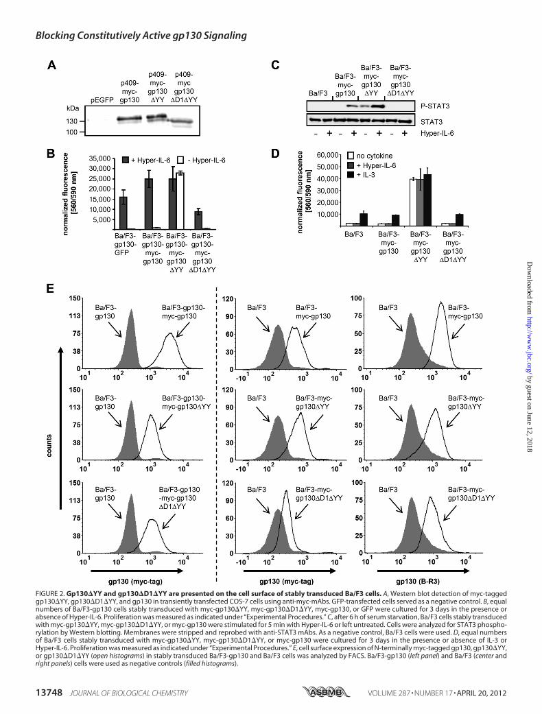
FIGURE 2. Gp130�YY and gp130�D1�YY are presented on the cell surface of stably transduced Ba/F3 cells. A, Western blot detection of myc-taggedgp130�YY, gp130�D1�YY, and gp130 in transiently transfected COS-7 cells using anti-myc-mAbs. GFP-transfected cells served as a negative control. B, equalnumbers of Ba/F3-gp130 cells stably transduced with myc-gp130�YY, myc-gp130�D1�YY, myc-gp130, or GFP were cultured for 3 days in the presence orabsence of Hyper-IL-6. Proliferation was measured as indicated under “Experimental Procedures.” C, after 6 h of serum starvation, Ba/F3 cells stably transducedwith myc-gp130�YY, myc-gp130�D1�YY, or myc-gp130 were stimulated for 5 min with Hyper-IL-6 or left untreated. Cells were analyzed for STAT3 phospho-rylation by Western blotting. Membranes were stripped and reprobed with anti-STAT3 mAbs. As a negative control, Ba/F3 cells were used. D, equal numbersof Ba/F3 cells stably transduced with myc-gp130�YY, myc-gp130�D1�YY, or myc-gp130 were cultured for 3 days in the presence or absence of IL-3 orHyper-IL-6. Proliferation was measured as indicated under “Experimental Procedures.” E, cell surface expression of N-terminally myc-tagged gp130, gp130�YY,or gp130�D1�YY (open histograms) in stably transduced Ba/F3-gp130 and Ba/F3 cells was analyzed by FACS. Ba/F3-gp130 (left panel) and Ba/F3 (center andright panels) cells were used as negative controls (filled histograms).
Blocking Constitutively Active gp130 Signaling
13748 JOURNAL OF BIOLOGICAL CHEMISTRY VOLUME 287 • NUMBER 17 • APRIL 20, 2012
by guest on June 12, 2018http://w
ww
.jbc.org/D
ownloaded from

oncostatin M, or ciliary neurtrophic factor (19). As a control,B-P4 did not inhibit the proliferation of Ba/F3-gp130-gp130�YY cells stimulated with IL-3, indicating that B-P4 spe-cifically blocked the activity of gp130�YY in Ba/F3-gp130-gp130�YY-cells (Fig. 4D).
DISCUSSION
Constitutive activation of the gp130-dependent transcrip-tion factor STAT3 has been implicated in many human neo-plastic malignancies, including multiple myeloma (4, 22, 23),prostate cancer, melanoma, ovarian cancer, renal carcinoma(24), as well as gastric cancer (25). Artificially dimerized STAT3has been shown to exhibit oncogenic potential, and STAT3wastherefore designated as an oncogene (26). The IL-6/gp130 sig-naling pathway is a candidate for constitutive STAT3 activationin tumors (27). Increased STAT3 phosphorylationwas found inIHCAs (2). Interestingly, gp130 gene mutations were found in60% of the analyzed IHCAs. It turned out that these mutationsresulted in ligand-independent dimerization of gp130 receptorchains and constitutive STAT3 phosphorylation. This was thefirst report on somatic mutation of gp130 in tumors (2), and incombination with the potential to induce cytokine-independ-ent cellular proliferation shown in this study, gp130 can bedefined as an oncogene involved in benign human tumors thatcontributes to the inflammatory phenotype (2).All mutations of gp130 found in IHCAs were deletions
within the cytokine binding interface of domain 2 (2). Here, weanalyzed a frequently occurring gp130mutation (gp130�Y186-Y190, gp130�YY) found in four of 26 IHCA patients. Six morepatients carried mutations from Ser-187-Y190 (gp130�SY)that were also covered in Tyr-186 to Tyr-190 (2).We show thatgp130�YY leads to ligand-independent, long-term prolifera-tion of Ba/F3 cells and constitutive STAT3 phosphorylation.Interestingly, deletion of domain 1 from gp130�YY resulted
in a signaling-incompetent receptor chain, indicating thatdomain 1 contributes to ligand-independent receptor activa-tion. However, dimerization of gp130�YY was independent ofthe presence of the D1 domain. The neutralizing anti-gp130mAb B-T2 directed against D1 did not inhibit receptor activa-tion of gp130�YY, indicating that the gp130 homodimerizationinduced by IL-6/IL-6R is fundamentally different from thehomodimerization of gp130�YY. Homodimerization of thewild-type gp130 receptor is facilitated by contacts of gp130CBM(domain 2 and 3) to the binding site II of IL-6 and of gp130D1 to the binding site III of IL-6, whereas the IL-6R contactsIL-6 via the binding site I (16). We speculate thathomodimerization of gp130�YY is facilitated by the interactionof the mutated D2 (CBM) of one receptor with the D2 of the
FIGURE 3. No inhibition of ligand-independent proliferation of Ba/F3-gp130-gp130�YY by sgp130 or sgp130Fc or the anti-gp130 mAb B-T2.A, equal numbers of Ba/F3-gp130 cells stably transduced with myc-gp130�YY were cultured for 3 days in the absence of Hyper-IL-6 and increas-ing amounts of B-T2 (0, 0.1, 0.5, 1, 5, and 10 �g/ml). Proliferation wasmeasured as indicated under “Experimental Procedures.” As a control, Ba/F3-gp130-myc-gp130 cells were treated with 1 ng/ml Hyper-IL-6 plus B-T2.B, equal numbers of Ba/F3-gp130 cells stably transduced with gp130�YY-myc were cultured for 3 days in the absence of Hyper-IL-6 and increasing
amounts of sgp130 (0, 0.1, 1, 5, and 10 �g/ml). Proliferation was measured asindicated under “Experimental Procedures.” As a control, Ba/F3-gp130-myc-gp130 cells were treated with 1 ng/ml Hyper-IL-6 plus sgp130. C, equal num-bers of Ba/F3-gp130 cells stably transduced with gp130�YY-myc were cul-tured for 3 days in the absence of Hyper-IL-6 and increasing amounts ofsgp130Fc (0, 0.1, 1, 5, and 10 �g/ml). Proliferation was measured as indicatedunder “Experimental Procedures.” As a control, Ba/F3-gp130 were treatedwith 1 ng/ml Hyper-IL-6 and sgp130Fc. D, after 6 h of serum starvation, Ba/F3cells stably transduced with myc-gp130�YY or myc-gp130 were stimulatedfor 5 min with Hyper-IL-6, Hyper-IL-6 � sgp130Fc, or sgp130Fc or leftuntreated. STAT3 phosphorylation was analyzed by Western blot analysis.
Blocking Constitutively Active gp130 Signaling
APRIL 20, 2012 • VOLUME 287 • NUMBER 17 JOURNAL OF BIOLOGICAL CHEMISTRY 13749
by guest on June 12, 2018http://w
ww
.jbc.org/D
ownloaded from

other receptor. However, future studies are needed to fullyexplore the mechanism of ligand-independent gp130�YYreceptor activation.Moreover, the wild-type gp130 receptor formed stable het-
erodimerswith gp130�YY, and overexpression of thewild-typegp130 receptor blocked constitutive activation of gp130�SY(2). A likely mechanism for this inhibition is that interaction ofthe extracellular parts of wild-type and mutated gp130 recep-tors resulted in inactive gp130/gp130�SY heterodimers. Sur-prisingly, soluble gp130 variants (sgp130 and sgp130Fc) did notinhibit gp130�YY-induced cellular proliferation. This para-doxical situation might be explained by a limited access ofsgp130 in the sterical correct orientation to the cell surfacebound gp130 protein, which might also explain why sgp130cannot inhibit gp130�YY-induced cellular proliferation.However, constitutive ligand-independent activation of
gp130�YY was blocked by the neutralizing anti-gp130 mAbB-P4. The epitope of B-P4 is located within the fibronectin typeIII domain 4 of gp130 (gp130-D4). Truncation of the fibronec-tin-like type III domains results in gp130 molecules devoid ofsignaling capacity (28), and it has been speculated that the func-tional role of the fibronectin type III domains is the assembly of
the transmembrane domains in close proximity to allow activa-tion of gp130-associated intracellular JAKs (29). Interestingly,B-P4 has been shown to block only gp130 signaling induced byIL-11 but not by IL-6 or the other members of the IL-6 family,leukemia inhibitory factor, oncostatin M, and ciliary neu-rotrophic factor (19). Cardiotrophin 1, cardiotrophin-like cyto-kine, and IL-27 were, however, not investigated so far (19). Thismight indicate that signaling of gp130�YYmimics IL-11 signal-ing. IL-11 was shown to promote gastric cancer via gp130 andSTAT3 phosphorylation (30). This view is supported by thefinding that IL-11 but not IL-6 was overexpressed in IHCAs (2).IL-11 was, however, only overexpressed in IHCAs that did notharbor gp130 mutations (2), suggesting that IHCAs are, tosome extent, driven by IL-11 via wild-type gp130. After somaticmutation of gp130 into an IL-11-like constitutively active gp130variant, the necessity of IL-11-driven gp130 signal transductionmight be abrogated, resulting in down-regulation of IL-11expression levels.No malignant transformation was found in IHCAs with
gp130 mutations, but two of 111 analyzed cases of malignanttransformation of IHCAs into hepatocellular carcinoma car-riedmutations in gp130 and in the �-catenin pathway, suggest-
FIGURE 4. Biological activity of gp130�YY can be suppressed by the neutralizing anti-gp130 mAb B-P4 but not by B-R3. A, equal numbers of Ba/F3-gp130-gp130�YY-myc cells were cultured for 3 days in the absence of Hyper-IL-6 and increasing amounts of B-R3 (0, 0.1, 0.5, 1, 5, and 10 �g/ml). Proliferationwas measured as indicated under “Experimental Procedures.” As a control, Ba/F3-gp130 cells were treated with 10 ng/ml Hyper-IL-6 and B-R3. B, equal numbersof Ba/F3-gp130-gp130�YY-myc cells were cultured for 3 days in the presence of IL-3 (1 ng/ml) and B-R3 (5 �g/ml). Proliferation was measured as indicatedunder “Experimental Procedures.” As a control, Ba/F3-gp130 cells were treated with 1 ng/ml IL-3 and B-R3 (5 �g/ml). C, equal numbers of Ba/F3-gp130-gp130�YY-myc cells were cultured for 3 days in the absence of Hyper-IL-6 and increasing amounts of B-P4 (0, 0.1, 0.5, 1, 5, and 10 �g/ml). Proliferation wasmeasured as indicated under “Experimental Procedures.” As a control, Ba/F3-gp130 cells were treated with 10 ng/ml Hyper-IL 6 and B-P4 (10 �g/ml). D, equalnumbers of Ba/F3-gp130-gp130�YY-myc cells were cultured for 3 days in the presence of IL-3 (1 ng/ml) and B-P4 (10 �g/ml). Proliferation was measured asindicated under “Experimental Procedures.”
Blocking Constitutively Active gp130 Signaling
13750 JOURNAL OF BIOLOGICAL CHEMISTRY VOLUME 287 • NUMBER 17 • APRIL 20, 2012
by guest on June 12, 2018http://w
ww
.jbc.org/D
ownloaded from

ing a rare interplay of these pathways in malignant transforma-tion (2). In conclusion, blockade of constitutive activation ofmutant gp130 by B-P4 might open a possibility to therapeuti-cally block gp130-induced STAT3 phosphorylation in hepaticadenomas and in a subclass of hepatocellular carcinomas.
REFERENCES1. Longerich, T., and Schirmacher, P. (2009) A new link between cancer and
inflammation? J. Hepatol. 51, 230–2322. Rebouissou, S., Amessou, M., Couchy, G., Poussin, K., Imbeaud, S., Pilati,
C., Izard, T., Balabaud, C., Bioulac-Sage, P., and Zucman-Rossi, J. (2009)Frequent in-frame somatic deletions activate gp130 in inflammatory hep-atocellular tumours. Nature 457, 200–204
3. Heinrich, P. C., Behrmann, I., Haan, S., Hermanns, H. M., Müller-Newen,G., and Schaper, F. (2003) Principles of interleukin (IL)-6-type cytokinesignalling and its regulation. Biochem. J. 374, 1–20
4. Pilati, C., Amessou, M., Bihl, M. P., Balabaud, C., Nhieu, J. T., Paradis, V.,Nault, J. C., Izard, T., Bioulac-Sage, P., Couchy, G., Poussin, K., and Zuc-man-Rossi, J. (2011) Somatic mutations activating STAT3 in human in-flammatory hepatocellular adenomas. J. Exp. Med. 208, 1359–1366
5. Stuhlmann-Laeisz, C., Lang, S., Chalaris, A., Krzysztof, P., Enge, S., Eichler,J., Klingmüller, U., Samuel, M., Ernst, M., Rose-John, S., and Scheller, J.(2006) Forced dimerization of gp130 leads to constitutive STAT3 activa-tion, cytokine-independent growth, and blockade of differentiation of em-bryonic stem cells.Mol. Biol. Cell 17, 2986–2995
6. Suthaus, J., Tillmann, A., Lorenzen, I., Bulanova, E., Rose-John, S., andScheller, J. (2010) Forced homo- and heterodimerization of all gp130-typereceptor complexes leads to constitutive ligand-independent signalingand cytokine-independent growth.Mol. Biol. Cell 21, 2797–2807
7. Gearing, D. P., Ziegler, S. F., Comeau, M. R., Friend, D., Thoma, B., Cos-man, D., Park, L., and Mosley, B. (1994) Proliferative responses and bind-ing properties of hematopoietic cells transfected with low-affinity recep-tors for leukemia inhibitory factor, oncostatinM, and ciliary neurotrophicfactor. Proc. Natl. Acad. Sci. U.S.A. 91, 1119–1123
8. Fischer, M., Goldschmitt, J., Peschel, C., Brakenhoff, J. P., Kallen, K. J.,Wollmer, A., Grötzinger, J., and Rose-John, S. (1997) I. A bioactive de-signer cytokine for human hematopoietic progenitor cell expansion. Nat.Biotechnol. 15, 142–145
9. Schroers, A., Hecht, O., Kallen, K. J., Pachta, M., Rose-John, S., and Grötz-inger, J. (2005) Dynamics of the gp130 cytokine complex: a model forassembly on the cellular membrane. Protein Sci. 14, 783–790
10. Jostock, T., Müllberg, J., Ozbek, S., Atreya, R., Blinn, G., Voltz, N., Fischer,M., Neurath, M. F., and Rose-John, S. (2001) Soluble gp130 is the naturalinhibitor of soluble interleukin-6 receptor transsignaling responses. Eur.J. Biochem. 268, 160–167
11. Sambrook, J., Fritsch, E. F., and Maniatis, T. (1989)Molecular Cloning: ALaboratory Manual, Cold Spring Harbor Laboratory Press, Cold SpringHarbor, NY
12. Tenhumberg, S., Schuster, B., Zhu, L., Kovaleva, M., Scheller, J., Kallen,K. J., and Rose-John, S. (2006) gp130 dimerization in the absence of ligand.Preformed cytokine receptor complexes.Biochem. Biophys. Res. Commun.346, 649–657
13. Ketteler, R., Glaser, S., Sandra, O., Martens, U. M., and Klingmüller, U.(2002) Enhanced transgene expression in primitive hematopoietic pro-genitor cells and embryonic stem cells efficiently transduced by optimizedretroviral hybrid vectors. Gene Ther. 9, 477–487
14. Schuster, B., Kovaleva, M., Sun, Y., Regenhard, P., Matthews, V., Grötz-inger, J., Rose-John, S., and Kallen, K. J. (2003) Signaling of human ciliaryneurotrophic factor (CNTF) revisited. The interleukin-6 receptor canserve as an � receptor for CTNF. J. Biol. Chem. 278, 9528–9535
15. Rothbauer, U., Zolghadr, K., Muyldermans, S., Schepers, A., Cardoso,
M. C., and Leonhardt, H. (2008) A versatile nanotrap for biochemical andfunctional studies with fluorescent fusion proteins.Mol. Cell. Proteomics7, 282–289
16. Scheller, J., Grötzinger, J., and Rose-John, S. (2006)Updating interleukin-6classic- and trans-signaling. Signal Transduction 6, 240–259
17. Boulanger, M. J., Chow, D. C., Brevnova, E. E., and Garcia, K. C. (2003)Hexameric structure and assembly of the interleukin-6/IL-6 �-receptor/gp130 complex. Science 300, 2101–2104
18. Pflanz, S., Kurth, I., Grötzinger, J., Heinrich, P. C., and Müller-Newen, G.(2000) Two different epitopes of the signal transducer gp130 sequentiallycooperate on IL-6-induced receptor activation. J. Immunol. 165,7042–7049
19. Gu, Z. J., Wijdenes, J., Zhang, X. G., Hallet, M.M., Clement, C., and Klein,B. (1996)Anti-gp130 transducermonoclonal antibodies specifically inhib-iting ciliary neurotrophic factor, interleukin-6, interleukin-11, leukemiainhibitory factor or oncostatin M. J. Immunol. Methods 190, 21–27
20. Wijdenes, J., Heinrich, P. C., Müller-Newen, G., Roche, C., Gu, Z. J., Clé-ment, C., and Klein, B. (1995) Interleukin-6 signal transducer gp130 hasspecific binding sites for different cytokines as determined by antagonisticand agonistic anti-gp130 monoclonal antibodies. Eur. J. Immunol. 25,3474–3481
21. Pflanz, S., Kernebeck, T., Giese, B., Herrmann, A., Pachta-Nick, M., Stahl,J., Wollmer, A., Heinrich, P. C., Müller-Newen, G., and Grötzinger, J.(2001) Signal transducer gp130: biochemical characterization of the threemembrane-proximal extracellular domains and evaluation of their oligo-merization potential. J. Immunol. 356, 605–612
22. Catlett-Falcone, R., Landowski, T. H., Oshiro,M.M., Turkson, J., Levitzki,A., Savino, R., Ciliberto, G., Moscinski, L., Fernández-Luna, J. L., Nuñez,G., Dalton, W. S., and Jove, R. (1999) Constitutive activation of Stat3signaling confers resistance to apoptosis in human U266 myeloma cells.Immunity 10, 105–115
23. Rawat, R., Rainey, G. J., Thompson, C. D., Frazier-Jessen, M. R., Brown,R. T., and Nordan, R. P. (2000) Constitutive activation of STAT3 is asso-ciated with the acquisition of an interleukin 6-independent phenotype bymurine plasmacytomas and hybridomas. Blood 96, 3514–3521
24. Bromberg, J. (2002) Stat proteins and oncogenesis. J. Clin. Invest. 109,1139–1142
25. Jenkins, B. J., Grail, D., Nheu, T., Najdovska, M., Wang, B., Waring, P.,Inglese, M., McLoughlin, R. M., Jones, S. A., Topley, N., Baumann, H.,Judd, L. M., Giraud, A. S., Boussioutas, A., Zhu, H. J., and Ernst, M. (2005)Hyperactivation of Stat3 in gp130 mutant mice promotes gastric hyper-proliferation and desensitizes TGF-� signaling. Nat. Med. 11, 845–852
26. Bromberg, J. F.,Wrzeszczynska,M. H., Devgan, G., Zhao, Y., Pestell, R. G.,Albanese, C., and Darnell, J. E. (1999) Stat3 as an oncogene. Cell 98,295–303
27. Grivennikov, S., and Karin, M. (2008) Autocrine IL-6 signaling. A keyevent in tumorigenesis? Cancer Cell 13, 7–9
28. Kurth, I., Horsten, U., Pflanz, S., Timmermann, A., Küster, A., Dahmen,H., Tacken, I., Heinrich, P. C., andMüller-Newen,G. (2000) Importance ofthe membrane-proximal extracellular domains for activation of the signaltransducer glycoprotein 130. J. Immunol. 164, 273–282
29. Skiniotis, G., Boulanger, M. J., Garcia, K. C., andWalz, T. (2005) Signalingconformations of the tall cytokine receptor gp130 when in complex withIL-6 and IL-6 receptor. Nat. Struct. Mol. Biol. 12, 545–551
30. Ernst, M., Najdovska, M., Grail, D., Lundgren-May, T., Buchert, M., Tye,H., Matthews, V. B., Armes, J., Bhathal, P. S., Hughes, N. R., Marcusson,E. G., Karras, J. G., Na, S., Sedgwick, J. D., Hertzog, P. J., and Jenkins, B. J.(2008)) STAT3 and STAT1 mediate IL-11-dependent and inflammation-associated gastric tumorigenesis in gp130 receptor mutant mice. J. Clin.Invest. 118, 1727–1738
Blocking Constitutively Active gp130 Signaling
APRIL 20, 2012 • VOLUME 287 • NUMBER 17 JOURNAL OF BIOLOGICAL CHEMISTRY 13751
by guest on June 12, 2018http://w
ww
.jbc.org/D
ownloaded from

Suthaus, Christoph Garbers, Stefan Rose-John, Doreen M. Floss and Jürgen SchellerJan Sommer, Timo Effenberger, Elena Volpi, Georg H. Waetzig, Marten Bernhardt, Jan
Neutralizes Interleukin 11 SignalingSpecificallyHepatocellular Adenoma Is Inhibited by an Anti-gp130 Antibody That
Constitutively Active Mutant gp130 Receptor Protein from Inflammatory
doi: 10.1074/jbc.M112.349167 originally published online March 6, 20122012, 287:13743-13751.J. Biol. Chem.
10.1074/jbc.M112.349167Access the most updated version of this article at doi:
Alerts:
When a correction for this article is posted•
When this article is cited•
to choose from all of JBC's e-mail alertsClick here
http://www.jbc.org/content/287/17/13743.full.html#ref-list-1
This article cites 29 references, 10 of which can be accessed free at
by guest on June 12, 2018http://w
ww
.jbc.org/D
ownloaded from