Conservative treatment for patients with subacromial...
20
Submitted 1 September 2017 Accepted 1 February 2018 Published 23 February 2018 Corresponding author Mikkel B. Clausen, [email protected] Academic editor Scotty Butcher Additional Information and Declarations can be found on page 16 DOI 10.7717/peerj.4400 Copyright 2018 Clausen et al. Distributed under Creative Commons CC-BY 4.0 OPEN ACCESS Conservative treatment for patients with subacromial impingement: Changes in clinical core outcomes and their relation to specific rehabilitation parameters Mikkel B. Clausen 1 ,2 ,3 , Mikas B. Merrild 1 , Adam Witten 2 , Karl B. Christensen 4 , Mette K. Zebis 1 , Per Hölmich 2 and Kristian Thorborg 2 ,3 1 Department of Physiotherapy and Occupational therapy, Metropolitan University College Copenhagen, Copenhagen, Denmark 2 Sports Orthopedic Research Center-Copenhagen (SORC-C), Department of Orthopedic Surgery, Copenhagen University Hospital, Amager-Hvidovre, Hvidovre, Denmark 3 Physical Medicine and Rehabilitation Research-Copenhagen (PMR-C), Copenhagen University Hospital, Amager-Hvidovre, Copenhagen, Denmark 4 Department of Biostatistics, University of Copenhagen, Copenhagen, Denmark ABSTRACT Background. Impaired patient-reported shoulder function and pain, external-rotation strength, abduction strength, and abduction range-of-motion (ROM) is reported in patients with subacromial impingement (SIS). However, it is unknown how much strength and ROM improves in real-life practice settings with current care. Furthermore, outcomes of treatment might depend on specific rehabilitation param- eters, such as the time spent on exercises (exercise-time), number of physiotherapy sessions (physio-sessions) and number of corticosteroid injections, respectively. However, this has not previously been investigated. The purpose of this study was to describe changes in shoulder strength, ROM, patient-reported function and pain, in real-life practice settings, and explore the association between changes in clinical core outcomes and specific rehabilitation parameters. Methods. Patients diagnosed with SIS at initial assessment at an outpatient hospital clinic using predefined criteria’s, who had not undergone surgery after 6 months, were included in this prospective cohort study. After initial assessment (baseline), all patients underwent treatment as usual, with no interference from the investigators. The outcomes Shoulder Pain and Disability Index (SPADI:0–100), average pain (NRS:0–10), external rotation strength, abduction strength and abduction ROM, pain during each test (NRS:0–10), were collected at baseline and at six month follow-up. Amount of exercise-time, physio-sessions and steroid-injections was recorded at follow-up. Changes in outcomes were analyzed using Wilcoxon Signed- Rank test, and the corresponding effect sizes (ES) were estimated. The associations between changes in outcomes and rehabilitation parameters were explored using multiple regression analyses. Results. Sixty-three patients completed both baseline and follow-up testing. Significant improvements were seen in SPADI (19 points, ES:0.53, p < 0.001) and all pain variables (median 1–1.5 points, ES:0.26–0.39, p < 0.01), but not in strength and ROM (ES:0.9–0.12, p > 0.2). A higher number of physio-sessions was significantly associated How to cite this article Clausen et al. (2018), Conservative treatment for patients with subacromial impingement: Changes in clinical core outcomes and their relation to specific rehabilitation parameters . PeerJ 6:e4400; DOI 10.7717/peerj.4400
Transcript of Conservative treatment for patients with subacromial...

Submitted 1 September 2017Accepted 1 February 2018Published 23 February 2018
Corresponding authorMikkel B. Clausen,[email protected]
Academic editorScotty Butcher
Additional Information andDeclarations can be found onpage 16
DOI 10.7717/peerj.4400
Copyright2018 Clausen et al.
Distributed underCreative Commons CC-BY 4.0
OPEN ACCESS
Conservative treatment for patients withsubacromial impingement: Changes inclinical core outcomes and their relationto specific rehabilitation parametersMikkel B. Clausen1,2,3, Mikas B. Merrild1, AdamWitten2, Karl B. Christensen4,Mette K. Zebis1, Per Hölmich2 and Kristian Thorborg2,3
1Department of Physiotherapy and Occupational therapy, Metropolitan University College Copenhagen,Copenhagen, Denmark
2 Sports Orthopedic Research Center-Copenhagen (SORC-C), Department of Orthopedic Surgery,Copenhagen University Hospital, Amager-Hvidovre, Hvidovre, Denmark
3Physical Medicine and Rehabilitation Research-Copenhagen (PMR-C), Copenhagen University Hospital,Amager-Hvidovre, Copenhagen, Denmark
4Department of Biostatistics, University of Copenhagen, Copenhagen, Denmark
ABSTRACTBackground. Impaired patient-reported shoulder function and pain, external-rotationstrength, abduction strength, and abduction range-of-motion (ROM) is reportedin patients with subacromial impingement (SIS). However, it is unknown howmuch strength and ROM improves in real-life practice settings with current care.Furthermore, outcomes of treatment might depend on specific rehabilitation param-eters, such as the time spent on exercises (exercise-time), number of physiotherapysessions (physio-sessions) and number of corticosteroid injections, respectively.However, this has not previously been investigated. The purpose of this study was todescribe changes in shoulder strength, ROM, patient-reported function and pain, inreal-life practice settings, and explore the association between changes in clinical coreoutcomes and specific rehabilitation parameters.Methods. Patients diagnosed with SIS at initial assessment at an outpatient hospitalclinic using predefined criteria’s, who had not undergone surgery after 6 months,were included in this prospective cohort study. After initial assessment (baseline), allpatients underwent treatment as usual, with no interference from the investigators.The outcomes Shoulder Pain and Disability Index (SPADI:0–100), average pain(NRS:0–10), external rotation strength, abduction strength and abduction ROM,pain during each test (NRS:0–10), were collected at baseline and at six monthfollow-up. Amount of exercise-time, physio-sessions and steroid-injections wasrecorded at follow-up. Changes in outcomes were analyzed using Wilcoxon Signed-Rank test, and the corresponding effect sizes (ES) were estimated. The associationsbetween changes in outcomes and rehabilitation parameters were explored usingmultiple regression analyses.Results. Sixty-three patients completed both baseline and follow-up testing. Significantimprovements were seen in SPADI (19 points, ES:0.53, p < 0.001) and all painvariables (median 1–1.5 points, ES:0.26–0.39, p< 0.01), but not in strength and ROM(ES:0.9–0.12, p> 0.2). A higher number of physio-sessions was significantly associated
How to cite this article Clausen et al. (2018), Conservative treatment for patients with subacromial impingement: Changes in clinicalcore outcomes and their relation to specific rehabilitation parameters . PeerJ 6:e4400; DOI 10.7717/peerj.4400

with larger improvements in external rotation strength (0.7Newton/session, p= 0.046),and higher exercise-time was significantly associated with decrease in average pain(−0.2 points/1,000 min, p= 0.048).Discussion. Patient-reported function and pain improved after six months of currentcare, but strength and ROM did not improve. This is interesting, as strengtheningexercises is part of most current interventions. While two significant associations wereidentified between self-reported rehabilitation parameters and outcomes, the smallgains per physio-session or 1,000 min of exercise-time reduces the clinical relevanceof these relationships. Collectively, the findings from this study indicate room forimprovement of the current rehabilitation of SIS, especially with regard to core clinicaloutcomes, such as strength and range of motion.
Subjects Evidence Based Medicine, Kinesiology, Orthopedics, Surgery and Surgical SpecialtiesKeywords Shoulder, Strength, Rehabilitation, Prospective, Cohort, Disability, Impingement
INTRODUCTIONSubacromial impingement syndrome (SIS) is the most common shoulder disorder (Vander Windt et al., 1995). The condition is characterized by shoulder pain and impairedpatient-reported function (Roach et al., 1991), but reduced strength and range of motion(ROM) has also been reported in patients with SIS (Clausen et al., 2017; MacDermid et al.,2004). More specifically, abduction and external rotation strength, as well as abductionROM, is only two-thirds of that in the unaffected shoulder (Clausen et al., 2017) or healthycontrols (MacDermid et al., 2004), which is why a∼50% increase is needed to reach normalstrength and ROM levels. Based on this, it is suggested that patient-reported function, pain,strength and ROM should be considered core outcomes related to SIS (Clausen et al., 2017).
Conservative treatment, including exercise therapy, is considered the first line oftreatment (Diercks et al., 2014; Sundhedsstyrelsen, 2013), and a range of randomizedcontrolled trials (RCTs) show promising improvements in patient-reported shoulderfunction, pain and ROM as a result of exercise therapy (Akyol et al., 2012; Dilek et al.,2016; Lombardi et al., 2008; Baskurt et al., 2011; Moezy, Sepehrifar & Solayman Dodaran,2014; Littlewood et al., 2015; Engebretsen et al., 2011; Yiasemides et al., 2011; Bal et al., 2009;McClure et al., 2004; Holmgren et al., 2012; Bennell et al., 2010). However, when maximumshoulder strength is measured, improvements vary, ranging from 4–36% in externalrotation strength (derived from Dilek et al., 2016; Lombardi et al., 2008; McClure et al.,2004; Bennell et al., 2010; Maenhout et al., 2013; Galace de Freitas et al., 2014; Ingwersen etal., 2017) and 4–42% in abduction strength (derived from Dilek et al., 2016; Lombardi etal., 2008; McClure et al., 2004; Bennell et al., 2010; Maenhout et al., 2013; Galace de Freitaset al., 2014; Ingwersen et al., 2017; Struyf et al., 2013). Importantly, however, strengthimprovements seem to be smallest in studies including patients whose duration ofsymptoms is greater than two months. In these studies, strength improvement rangesfrom 4% to 15% in external rotation strength and 4% to 20% in abduction strength(derived from Lombardi et al., 2008; Bennell et al., 2010; Maenhout et al., 2013; Ingwersen
Clausen et al. (2018), PeerJ, DOI 10.7717/peerj.4400 2/20

et al., 2017). This is far from the ∼50% increase needed to restore the above-mentionedshoulder strength deficits reported byMacDermid et al. (2004) and Clausen et al. (2017). Itshould also be noted that both the previously reported improvements in patient-reportedshoulder function, pain and ROM, as well as the less encouraging changes in strength,were all achieved in clinical trial settings. While such studies have high internal validity, itis possible that the observed improvements could partly be due to a trial effect, such as theHawthorne effect and/or placebo effect (Braunholtz, Edwards & Lilford, 2001), and henceit is possible that results obtained outside such controlled settings will be less encouraging.
Conservative treatment strategies often include home-based exercises (Holmgren et al.,2012; Bennell et al., 2010). The effectiveness of such exercise interventions likely dependson patients’ adherence to prescribed exercises, although this has not been demonstratedin patients with SIS. This is important because utilization of exercise therapy in therehabilitation of patients with shoulder disorders might be limited, as indicated in a recentregister-based study of 57,311 Danish shoulder patients in secondary care (70% with SIS),of whom only 43% had physiotherapy within 52 weeks after the first hospital visit, andonly 62% of physiotherapy sessions included exercise therapy or advice on self-training(Christiansen et al., 2016). This indicates a low utilization of some specific physiotherapyand rehabilitation parameters, such as exercise therapy, potentially leading to suboptimalrehabilitation for many patients. In addition to that, the actual time that patients with SISspend on exercise therapy has not been investigated and is therefore currently unknown.In relation to physiotherapy and exercise therapy, it is also unknown to what degree thenumber of completed physiotherapy sessions and the time spent on exercise therapy arerelated to improvements in the SIS-related core outcomes, such as shoulder strength,ROM, patient-reported function and pain (Sundhedsstyrelsen, 2013; Hopman et al., 2013).
Knowledge about improvements in SIS-related core outcomes following rehabilitationand their relationship to specific rehabilitation parameters will help researchers andclinicians understand to what degree these clinical core outcomes are addressed incurrent care.
PurposeThe purposes of this study were: first, to describe the changes in shoulder core outcomes,including strength, ROM, patient-reported shoulder function and pain, from baselineto six months follow-up, in patients diagnosed with SIS at the baseline examination at apublic hospital outpatient clinic; second, to describe specific rehabilitation parameters,such as the utilization of physiotherapy and the amount of home-based rehabilitationperformed during the same period; third, to investigate the association between shouldercore outcomes and specific rehabilitation parameters.
MATERIALS AND METHODSThis is a prospective cohort study based on the six-month follow-up of 129 SIS-patients(82%) from a consecutive cohort of 157 SIS-patients (Clausen et al., 2017). At baseline,patients underwent a clinical assessment of shoulder strength, ROM, patient-reportedshoulder function and pain impairments performed by one of six trained assessors (two
Clausen et al. (2018), PeerJ, DOI 10.7717/peerj.4400 3/20

physiotherapists, three physiotherapy undergraduates and onemedical student). After theseassessments, a clinical examination was performed by an orthopaedic surgeon shoulderspecialist blinded to the results of the baseline assessments. After baseline, all patientsunderwent usual care in settings not provided or controlled by the investigators of thisstudy. Accordingly, the content and progression of care was based solely on the choices ofthe doctors, therapists and patients, with no interference from any investigators or otherintruding parties.
Approximately six months after baseline (median 28 weeks [IQR: 27; 29]), patientsincluded in the consecutive cohort of SIS-patients were contacted by telephone, and aten-minute interview regarding treatment and rehabilitation since baseline was conducted.Information about sick leave due to the shoulder disorder was also obtained but notpresented in this study. Patients who had not undergone shoulder surgery were invited toa follow-up assessment of maximum isometric shoulder strength, ROM, patient-reportedfunction and pain, using the same procedures as in the baseline examination. Follow-upassessments were performed by one of five assessors (physiotherapy undergraduates) whodid not participate in the baseline assessment, but were trained by the primary investigator(MBC) and one of the assessors involved in the baseline assessments. Follow-up assessmentswere conducted at the same facility as the baseline assessments. Only the affected shoulderwas tested at the follow-up assessments. The study has been evaluated by the Capitol RegionCommittee on Health Research Ethics in Denmark, where it was evaluated as not requiringformal ethical approval (H-3-2013-FSP29). Written informed consent was obtained fromall participants.
ParticipantsInclusion and exclusion criteria for the consecutive cohort of 157 patients with SIS, whoform the basis of this follow-up study, have been described in more detail in a previouspaper (Clausen et al., 2017). Patients were considered eligible for inclusion based on thefollowing criteria: age 18 years or more, sufficient Danish language ability, referred toexamination of a shoulder problem and at least three of the five diagnostic tests forSIS were positive (Hawkins-Kennedy, Neer’s, painful arc, Resisted External Rotationand Jobe’s), as described by Michener et al. (2009). Patients were excluded based on thefollowing criteria: Presence of (1) a full thickness rotator cuff tear, luxation or sub-luxationof the glenohumeral or the acromioclavicular joint, frozen shoulder or osteoarthritis in theglenohumeral joint (based on clinical and/or paraclinical examination), (2) a labral lesionverified by paraclinical investigation, or (3) any competing disorder affecting the shoulderfunction or the ability to answer patient-reported questionnaires (e.g., neurological disease,cervical disorder, elbow disorder, mental disorder or blindness).
Core outcome measuresSPADIThe Shoulder Pain and Disability Index (SPADI) was used to measure patient-reportedshoulder function. SPADI consists of 13 items, each scored on an 11-point numeric ratingscale. The two domains pain (five questions) and disability (eight questions) are scored
Clausen et al. (2018), PeerJ, DOI 10.7717/peerj.4400 4/20

separately from 0 (worst) to 100 (best), and averaged into a total SPADI score. The Danishversion of SPADI is considered reliable and valid (Christiansen, Andersen & Haahr, 2013).
Active abduction ROMActive abduction ROM was measured in degrees using a digital inclinometer. First, withthe subject standing with the arm in the anatomical position and the elbow extended, theinclinometer was reset on a vertical surface. The subject raised the arm in the coronal planetowards the ceiling and a measurement was taken with the inclinometer aligned parallel tothe humerus, close to the insertion of the deltoid muscle. The inter-tester reliability of thetest is good (ICC 0.95) (Kolber et al., 2011).
StrengthMaximum isometric peak force in abduction and external rotation was assessed with theshoulder in neutral position and the elbow fully extended or flexed to 90 degrees. Thesubject was seated close to a wall, which was used as external resistance to the isometriccontraction. Measurements were obtained using a hand-held dynamometer and measuredin newtons (N). This method has been shown to have a high inter-tester reliability (ICC2,1
> 0.9) (Clausen et al., 2017). Detailed descriptions of assessment procedures are freelyavailable in the online supplement to a previous study (Clausen et al., 2017).
Patient characteristics and specific rehabilitation parametersDemographics and disease-specific characteristics, including severityThe following disease specific characteristics were collected at baseline: Duration (durationof current shoulder problem: 0–1months, 1–3months, 3–6months, >6months); Sick Leave(on sick leave or unemployed due to shoulder problem, yes/no); Affected Side (dominantside/non-dominant side diagnosed with SIS); Age (years); and gender (male/female).
Specific rehabilitation parametersInformation on shoulder surgery, corticosteroid injections, physiotherapy and time spenton exercises was based on the questions from the structured telephone interview (Table 1).First, patients who answered ‘yes’ to having had shoulder surgery (Q1) were categorisedas having had surgery since baseline, and were not asked further questions regardingtheir treatment since baseline. The number of corticosteroid injections and physiotherapysessions received since baseline was determined through the answer to question Q2 andQ3, respectively. Finally, total minutes of exercises was calculated as the number of weekswith exercises multiplied by the average time spent on exercises per week with exercises(Q5 * Q6).
Global impression of change since baseline and severity at follow-upInformation on patient-reported recovery was collected from answers to the question‘‘Is your shoulder disorder fully cured?’’ (yes/no). Improvement since baseline (Globalimpression of change, GIC) was collected through answers to the following questionusing a seven-point Likert scale: ‘‘How do you perceive your shoulder disorder now ascompared to when you underwent the first shoulder examination six months ago?’’ Muchworse, a very important aggravation; Worse, an important aggravation; Slightly worse,
Clausen et al. (2018), PeerJ, DOI 10.7717/peerj.4400 5/20

Table 1 Interview regarding treatment and rehabilitation since baseline.
Question
Shoulder surgery Q1 ‘‘Have you had shoulder surgery since your baselineassessment six months ago?’’
Corticosteroid injection Q2 ‘‘Have you had a corticosteroid injection for yourshoulder disorder since baseline, including the day of thebaseline examination? If yes, how many times?’’
Physiotherapy Q3 ‘‘Have you, since the baseline examination, seen aphysiotherapist for your shoulder disorder? If yes, howmany times?’’
Exercises Q4 ‘‘Have you been doing home-based (or non-supervised) exercises for your shoulder disorder sincebaseline?’’Q5 ‘‘For how many weeks in total since baseline have youactively been doing these exercises’’Q6 ‘‘During these weeks, how much time (in minutes) didyou in average use per week on the exercises?’’
but enough to be an important aggravation; The same; Slightly better, but enough tobe an important improvement; Better, an important improvement; Much better, a veryimportant improvement). The severity of the shoulder disorder was scored on a numericrating scale (range 1–5, 1 being very mild and 5 being very severe). All information onGIC and severity was collected through a self-developed, standardized, self-administeredquestionnaire.
Data reduction and statistics. Demographics, disease specific information, baseline valuesfor all outcomes and descriptive data on the treatment received since baseline are presentedas means ± SD, median [IQR] or numbers and proportions, for conservatively-treatedpatients who underwent follow-up assessment and those who did not, respectively.Age was compared between groups using independent samples t -test; proportions werecompared using Chi2-test or Fisher’s exact test as appropriate; baseline values, numberof physiotherapy sessions and total minutes of home exercises were compared using theMann–Whitney U test.
For patients who underwent follow-up assessment, SPADI-score, strength, ROM andpain values at baseline and follow-up, as well as changes from baseline to follow up, arepresented as both mean ± SD and median [IQR]. Change scores were calculated as thevalue at follow-up minus the value at baseline. A Wilcoxon signed-rank test was applied inorder to determine whether changes were significant, and corresponding effect sizes (ES)were calculated as the test statistic divided by the number of observations (ES= Z
√n).
Separately for each outcome (SPADI-score, external rotation strength, abductionstrength, abduction ROM, pain during each test and average pain level), the associationbetween the change in the outcome (dependent variable) and the number of physiotherapysessions, the time spent on home exercises and the number of steroid injections(independent variables) were investigated using multiple regression analysis. Theseexploratory multivariate regression analyses were performed separately for each pair
Clausen et al. (2018), PeerJ, DOI 10.7717/peerj.4400 6/20

Table 2 Baseline characteristics for conservatively treated patients, separately for those who participated in follow-up assessment and thosewho did not.
Conservativelytreated (n= 35)
Participated infollow-up assessment(n= 63)
P =
Age in years,mean± SD 51± 15 years 56± 13 years 0.14Gender, % females 51% (18 of 35) 54% (34 of 63) 0.84Affected side, % dominant side 53% (17 of 32) 59% (37 of 63) 0.66Sick Leave, % on sick leave at baseline 12% (4 of 33) 8% (5 of 62) 0.72Duration of symptoms at baseline in % 0.54
0–1 month 0% (0 of 34) 3% (2 of 62)1–3 months 21% (7 of 34) 16% (10 of 62)3–6 months 18% (6 of 34) 26% (16 of 62)>6 months 62% (21 of 34) 55% (34 of 62)
Baseline valuesSPADI Score, median [IQR] n= 34 61 [43; 73] n= 63 54 [38; 70] 0.17External rot. strength, Newton, median [IQR] n= 24 59 [36; 72] n= 56 52 [34; 86] 0.77Abduction strength, Newton, median [IQR] n= 25 55 [37; 92] n= 56 54 [36; 90] 0.96Abduction ROM, degrees, median [IQR] n= 27 101 [90; 140] n= 58 120 [87; 154] 0.30Average pain, median [IQR] n= 31 4 [2.5; 5] n= 60 3 [1.6; 4] 0.02Pain during external rot. strength, median [IQR] n= 24 4 [1; 6] n= 56 4 [2; 6] 0.63Pain during abduction strength, median [IQR] n= 24 4 [0.3; 7.8] n= 56 3 [1; 7] 0.95Pain during abduction ROM, median [IQR] n= 27 5 [5; 8] n= 58 5.5 [3; 8] 0.06
of dependent and independent variables, including the baseline value of the dependentvariable in question as covariate.
A significance level of 0.05 was applied for all statistical tests.
RESULTSFrom the original consecutive cohort of 157 patients with SIS, 129 (82%) completedthe standardized telephone interview. Thirty-one of these had undergone surgery for theirshoulder disorder since the baseline examination, leaving 98 conservatively-treated patientswho were all invited to follow-up assessment. Sixty-three of the 98 conservatively-treatedpatients participated in follow-up assessment approximately six months after baseline.Demographics, disease specific information and baseline values for all outcomes arepresented in Table 2, separating the conservatively-treated patients that participated infollow-up assessment (n= 63) from those who did not (n= 35). For further details onstudy flow, see Fig. 1.The group of patients participating in the follow-up assessment had significantly
improved from baseline to follow-up in SPADI-score (median 19 points, [3; 39], ES 0.53,p< 0.001) and all pain variables (median 1 to 1.5 points on NRS, ES 0.26 to 0.39, p< 0.01),with changes in average pain being the most pronounced. No changes were found inexternal rotation strength (median 2 N [−9; 17], ES 0.09, p= 0.337), abduction strength
Clausen et al. (2018), PeerJ, DOI 10.7717/peerj.4400 7/20

Original consecutive
cohort of patients with SIS
N=157
Patients completing
telephone interview
N=129
Conservatively treated
patients with SIS
N=98
Patients participating in
follow-up testing
N=63
Excluded: shoulder
surgery before follow-up
N=31
Figure 1 Flow-chart.Full-size DOI: 10.7717/peerj.4400/fig-1
(median 4 N [−10; 16], ES 0.12, p= 0.223) and Abduction ROM (−1◦ [−12; 30], ES 0.09,p= 0.324). See Table 3 for further details.
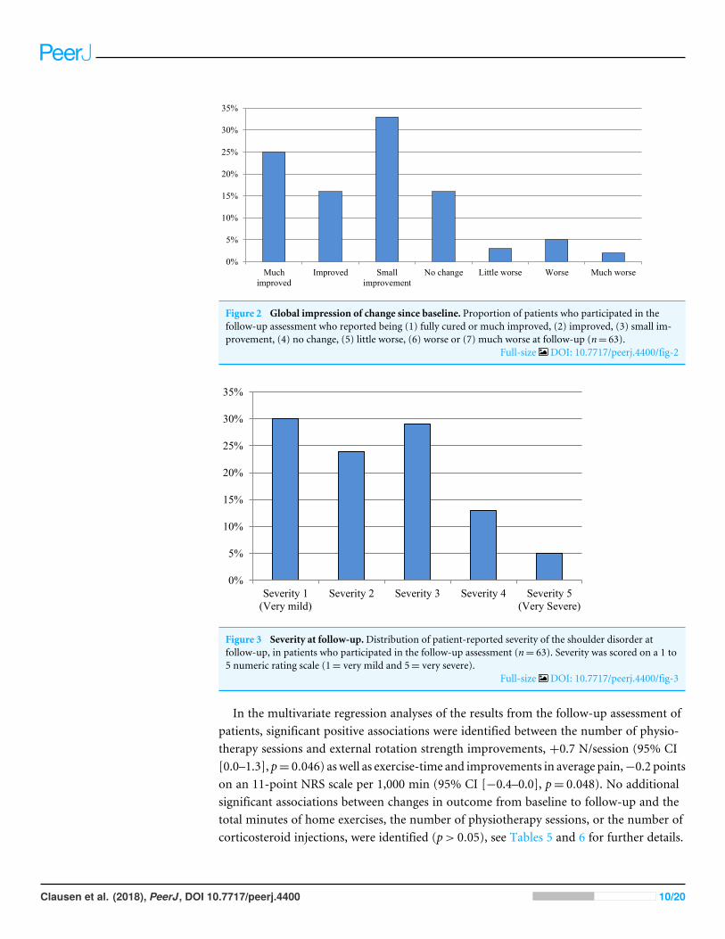
At follow-up, 25% (16 of 63) rated themselves as fully recovered or much improved. Thecurrent severity of the shoulder disorder was rated≥3 on a 1–5 scale (0 is best, 5 is worst) by46% (29 of 63) at follow-up. For further details on global impression of change and severityat follow-up, see Figs. 2 and 3. From the conservatively-treated patients that participatedin follow-up assessment 79% reported having had 0–1 steroid injections, 75% had receivedphysiotherapy (median 5 sessions [IQR 0; 11] for the entire group) and 87% had performedexercises for their shoulder disorder (median total exercise time of 1,040 min [IQR 220;2,700] for the entire group). The conservatively-treated patients who did not participatein follow-up assessment reported significantly lower total exercise time compared to theconservatively-treated patients who did participate in follow-up assessment (p= 0.045),while the other rehabilitation parameters listed in Table 4 did not differ significantlybetween groups. See Table 4 for further details on treatment since baseline.
Clausen et al. (2018), PeerJ, DOI 10.7717/peerj.4400 8/20

Table 3 Baseline, follow-up and change score for SPADI, strength, ROM and pain variables in conservatively treated patients who participatedin follow-up assessment.
Outcomes Normality test p-value change Effect size
Mean (SD) Median [IQR]
SPADI score,0–100 points (n= 63)
Baseline 54± 20 54 [38; 70] NormalFollow-up 31± 26 25 [8; 48] Non-norm.Change −23± 24 −19 [−39;−3] Normal <0.001 −0.53
External rot. strength,Newton (n= 56)
Baseline 62 N± 37 52 N [34; 84] Non-norm.Follow-up 65 N± 35 56 N [44; 79] Non-norm.Change 3 N± 25 2 N [−9; 17] Non-norm. 0.337 0.09
Abduction strength,Newton (n= 56)
Baseline 72 N± 51 54 N [36; 89] Non-norm.Follow-up 75 N± 46 63 N [44; 105] Non-norm.Change 4 N± 29 4 N [−10; 16] Non-norm. 0.223 0.12
Abduction ROM,degrees (n= 57)
Baseline 120◦ ± 40 121◦ [93; 154] NormalFollow-up 128◦ ± 35 137◦ [107; 153] Non-norm.Change 8◦ ± 41 −1◦ [−12; 30] Non-norm. .324 0.09
Average pain,0–10 points (n= 60)
Baseline 3.0± 1.5 3.0 [1.8; 4] NormalFollow-up 1.8± 2.1 1.0 [0; 3] Non-norm.Change −1.2± 1.9 −1.5 [−2.5; 0] Normal <0.001 −0.39
Pain duringexternal rot. strength,0–10 points (n= 56)
Baseline 4.1± 2.7 4.0 [2; 6] Non-norm.Follow-up 2.6± 3.1 1.0 [0; 6] Non-norm.Change −1.4± 2.6 −1.0 [−3; 0] Non-norm. <0.001 −0.34
Pain duringabduction strength,0–10 points (n= 56)
Baseline 3.8± 2.9 3.0 [1; 7] Non-norm.Follow-up 2.7± 3 2.0 [0; 5] Non-norm.Change −1.1± 2.5 −1.0 [−2; 0] Non-norm. 0.007 −0.26
Pain duringabduction ROM,0–10 points (n= 57)
Baseline 5.5± 2.5 6.0 [3; 8] Non-norm.Follow-up 3.0± 3 2.0 [1; 5] Non-norm.Change −2.3± 3.5 −1.0 [−5; 0] Normal <0.001 −0.38
Clausen et al. (2018), PeerJ, DOI 10.7717/peerj.4400 9/20
0%
5%
10%
15%
20%
25%
30%
35%
Much
improved
Improved Small
improvement
No change Little worse Worse Much worse
Figure 2 Global impression of change since baseline. Proportion of patients who participated in thefollow-up assessment who reported being (1) fully cured or much improved, (2) improved, (3) small im-provement, (4) no change, (5) little worse, (6) worse or (7) much worse at follow-up (n= 63).
Full-size DOI: 10.7717/peerj.4400/fig-2
0%
5%
10%
15%
20%
25%
30%
35%
Severity 1
(Very mild)
Severity 2 Severity 3 Severity 4 Severity 5
(Very Severe)
Figure 3 Severity at follow-up.Distribution of patient-reported severity of the shoulder disorder atfollow-up, in patients who participated in the follow-up assessment (n= 63). Severity was scored on a 1 to5 numeric rating scale (1= very mild and 5= very severe).
Full-size DOI: 10.7717/peerj.4400/fig-3
In the multivariate regression analyses of the results from the follow-up assessment ofpatients, significant positive associations were identified between the number of physio-therapy sessions and external rotation strength improvements, +0.7 N/session (95% CI[0.0–1.3], p= 0.046) aswell as exercise-time and improvements in average pain,−0.2 pointson an 11-point NRS scale per 1,000 min (95% CI [−0.4–0.0], p= 0.048). No additionalsignificant associations between changes in outcome from baseline to follow-up and thetotal minutes of home exercises, the number of physiotherapy sessions, or the number ofcorticosteroid injections, were identified (p> 0.05), see Tables 5 and 6 for further details.
Clausen et al. (2018), PeerJ, DOI 10.7717/peerj.4400 10/20

Table 4 Specific rehabilitation parameters in the conservatively treated patients, separately for those who participated in follow-up assessmentand those who did not.
Not participated infollow-up assessment(n= 35)
Participated infollow-up assessment(n= 63)
P =
Number of corticosteroid, by group0 injection 46% (16 of 35) 38% (24 of 63) 0.161 injection 31% (11 of 35) 41% (26 of 63)2 injections 9% (3 of 35) 18% (11 of 63)3 injections 6% (2 of 35) 3% (2 of 63)4 injections 6% (2 of 35)5 injections 3% (1 of 35)
Physiotherapy,%yes 57% (20 of 35) 75% (47 of 63) 0.11Number of physio-sessions,median [IQR] 2 [0; 12] 5 [0; 11] 0.26Grouped by number of physio-sessions
0 sessions 32% (15 of 35) 26% (16 of 62) 0.371 to 5 sessions 24% (7 of 35) 26% (16 of 62)6 to 10 sessions 18% (4 of 35) 21% (13 of 62)>10 sessions 27% (9 of 35) 27% (17 of 62)
Home Exercises, yes n(%) 76% (26 of 34) 87% (55 of 63) 0.25Total minutes of home exercises,median [IQR] 600 [0; 1560] 1,040 [220; 2,700] 0.045
Table 5 Regression analyses. The influence of specific rehabilitation parameters on the change in core clinical outcomes. Adjusted for baselinevalue of the relevant outcome.
SPADIa
(Points: 0–100)External rot.Strength(Newton)
Abductionstrength(Newton)
AbductionROM(Degrees)
Time spent on home exercisesB (1outcome per 1.000 min) −2.3 0.3 N −0.6 N 2◦
(95% CI) (−4.8 to 0.1) (−2.1 to 2.8) (−3.4 to 2.2) (−2 to 5)p-value 0.058 0.779 0.654 0.370
Number of physio-sessionsB (1outcome per session) 0.3 0.7 N 0.0 N 0◦
(95%CI) (−0.3 to 0.8) (0.0 to 1.3) (−0.8 to 0.8) (−1 to 1)p-value 0.385 0.046 0.984 0.962
Number of steroid injectionsB (1outcome per injection) 4.0 3.8 N 3.4 N 0◦
(95%CI) (−3.0 to 11.1) (−4.1 to 11.6) (−5.6 to 12.4) (−11 to 12)p-value 0.256 0.338 0.453 0.944
Notes.a0 is best, 100 is worst. Negative change score equals improvement in symptoms.
Clausen et al. (2018), PeerJ, DOI 10.7717/peerj.4400 11/20

Table 6 Regression analyses. Influence of specific rehabilitation parameters on the change in pain outcomes in patients participating in follow-upassessment. Adjusted for baseline value of relevant outcome.
Average painduring lastweeka (0–10 points)
Pain during tests (NPRS, 0–10 points)
External rot.strength testa
Abductionstrength testa
AbductionROM testa
Time spent on home exercisesB (1outcome per 1.000 min) −0.2 −0.2 +0.1 −0.2(95%CI) (−0.4 to 0.0) (−0.4 to 0.1) (−0.4 to 0.1) (−0.6 to 0.1)p-value 0.048 0.232 0.236 0.138
Number of physio-sessionsB (1outcome per session) 0.0 0.0 0.0 0.0(95%CI) (−0.0 to 0.1) (0.0 to 0.1) (−0.1 to 0.1) (−0.1 to 0.1)p-value 0.726 0.289 0.651 0.736
Number of steroid injectionsB (1outcome per injection) +0.5 +0.4 0.0 +0.2(95%CI) (−0.1 to 1.0) (−0.5 to 1.3) (−0.8 to 0.9) (−0.9 to 1.2)p-value 0.131 0.361 0.917 0.782
Notes.a0 is no pain, 10 is worst pain. Negative change score equals improvement in symptoms.
DISCUSSIONIn this prospective study of patients treated conservatively for SIS, medium to largeeffect sizes were seen for improvements in subjective outcomes of function and pain,approximately six months following initial assessment, in 63 conservatively treated SIS-patients. Interestingly, objective measures of strength and ROM did not improve.
Changes in strength and ROMTo the best of our knowledge, it has not previously been reported to what extent shoulderstrength and ROM have changed in patients with SIS who underwent conservativetreatment outside a controlled clinical trial setting. Therefore, the most important findingof this study is that usual care did not improve these core clinical outcomes assessingimportant impairments such as strength andmobility, with non-significantmedian changesin glenohumeral strength and shoulder abduction ROM close to zero (−1 degree and 2 to4 N, p> 0.2). This is despite the fact that shoulder mobility and strengthening exercisesaimed at the rotator cuff are a part of most treatment programs (Littlewood et al., 2015;Holmgren et al., 2012; Bennell et al., 2010; Struyf et al., 2013). Such lack of improvement isespecially relevant considering that the same group of patients have been shown to lack∼50% in abduction ROM as well as abduction and external rotation strength, to reach thesame level as the unaffected shoulder (Clausen et al., 2017).
Before our study, data regarding improvements in glenohumeral strength in patientswithSIS, who had received an intervention including rotator cuff strengthening exercises, havesolely been available from clinical trials, revealing somewhat varying results. Accordingly,external rotation strength (Dilek et al., 2016; McClure et al., 2004; Maenhout et al., 2013;
Clausen et al. (2018), PeerJ, DOI 10.7717/peerj.4400 12/20

Galace de Freitas et al., 2014; Ingwersen et al., 2017) and abduction strength (Dilek etal., 2016; McClure et al., 2004; Maenhout et al., 2013; Ingwersen et al., 2017) improvedsignificantly in some of the trials, with improvements ranging from 15% (derived fromIngwersen et al., 2017) to 43% (derived from Dilek et al., 2016). In five clinical trials,strength in abduction (Lombardi et al., 2008; Galace de Freitas et al., 2014; Struyf et al.,2013) and external rotation (Lombardi et al., 2008; Bennell et al., 2010) did not changesignificantly. Only in the two trials by Dilek et al. (2016) and Galace de Freitas et al. (2014)did average improvements in abduction and/or external rotation strength exceed 20%.The trial by Dilek et al. (2016) in particular stands out, not only with pronounced strengthimprovements in both abduction (43%) and external rotation (32%), but also near-perfectoutcomes in pain scores, with minimal-to-no pain at 12-weeks follow-up for patients whohad received an intervention including rotator cuff strengthening exercises. While thesetreatment outcomes are impressive, they indicate that the population included by Dilek etal. (2016) might not be comparable to that of other studies, including the current study,so the encouraging results regarding increases in shoulder strength may not represent thetypical response pattern. In support of Dilek et al. large improvements (36%) in externalrotation strength were also found in the trial by Galace de Freitas et al. (2014). In that trial,the population seems to mirror the population included in the present study and most ofthe aforementioned trials that included shoulder strength as an outcome (Lombardi et al.,2008;McClure et al., 2004; Bennell et al., 2010;Maenhout et al., 2013; Ingwersen et al., 2017;Struyf et al., 2013), indicating that pronounced strength improvement may be possible inthese populations, but is often not achieved. In summary, based on the findings from ourstudy and previous studies, it appears that current treatment strategies in the availablescientific literature often do not address shoulder strength impairments sufficiently inpatients with SIS, and future research should aim to improve the rehabilitation of theseimpairments.
The non-existent change in abduction ROM between baseline and follow-up (−1◦
[IQR −12; 30], p= 0.324) in this study, with the median at follow-up reaching only 137◦
[IQR 107; 153], is in contrast to the more encouraging results found in patients with SISwho underwent active treatment with shoulder exercises in clinical trial settings. In thesetrials, improvements of >20 degrees (Akyol et al., 2012; Lombardi et al., 2008) or follow-upresults close to 180 degrees (Dilek et al., 2016; Baskurt et al., 2011) were reported for groupswith varying starting levels of abduction ROM. Interestingly, the lack of improvementin abduction ROM found in our study is comparable to the treatment results in controlgroup patients on the waiting list for two months in the RCT study by Lombardi et al.(2008). Accordingly, it seems that the results of current care, regarding the core outcomeof shoulder abduction ROM, are a long way from matching the encouraging resultsfound in clinical trials. While this does question the effectiveness of current care in therehabilitation of patients with SIS, it also indicates an important difference in treatmentresponses obtained in clinical trials compared to real life settings. This apparent differencein treatment response underlines the importance of conducting RCTs using a morepragmatic approach to obtain valid data on the target group regarding the effectiveness
Clausen et al. (2018), PeerJ, DOI 10.7717/peerj.4400 13/20

of rehabilitation interventions. Such an approach would help inform both clinicians andpolicy makers when recommending or implementing specific rehabilitation strategies.
Changes in patient-reported function, pain, global impression ofchange and severityIn contrast to the objective outcomes of shoulder strength and ROM, both patient-reportedfunction and all pain outcomes improved significantly in the current study (mean SPADIimprovement of 23 points, median pain improvements of 1–1.5 points on 11-point NRS).This improvement in SPADI score is comparable to the mean improvement of 22.4 pointsafter 22 weeks reported by Bennell et al. (2010), the 23.5 and 29.1 points (control andintervention group, respectively) after six months reported by Littlewood et al. (2015),and the 24.8 points after one year reported by Engebretsen et al. (2011), all in comparablepopulations. It is, however, smaller than the 32.7 and 37.2 points (control and interventiongroup, respectively) after 12 weeks reported by Bal et al. (2009). This dissimilarity to thecurrent and previous studies (Littlewood et al., 2015; Engebretsen et al., 2011; Bennell etal., 2010) could be explained by a seemingly shorter symptom duration in the populationstudied by Bal et al. (2009), as compared to our study and previous studies (Littlewood et al.,2015; Engebretsen et al., 2011; Bennell et al., 2010). In summary, the results of conservativetreatment in this study are fairly encouraging, with mean improvements in SPADI scoresthat are comparable to those found in previous trials (Littlewood et al., 2015; Engebretsen etal., 2011; Bennell et al., 2010). This is, however, in slight contrast to the fact that only 25%of the patients considered themselves fully recovered or much improved at follow-up andmedian follow-up SPADI scores of 25 points [IQR: 8; 48], revealing room for improvementin current care.
Objective versus subjective outcomesIn this study of patients with SIS who underwent conservative treatment in secondarycare, patient-reported outcomes improved significantly while objective outcomes did not.Considering the lack of relationship between objective and subjective outcomes in thispopulation (Clausen et al., 2017), this inconsistency might not be surprising. However,these findings further suggests that the focus of treatment has, rightfully, been on thesubjective symptoms, which are considered cardinal in SIS (Roach et al., 1991), whilestrength improvements have not necessarily been the focus of rehabilitation. In addition,the observed inconsistency is also likely affected by the larger effect of the non-specific partsof treatment (i.e., placebo) on subjective outcomes compared to objective outcomes, ashas previously been described by Hróbjartsson & Gøtzsche (2001). This, combined with theknowledge that objective impairment measures and subjective outcomes seems to measuredifferent constructs (Clausen et al., 2017), underlines the importance of including bothwhen evaluating treatment effect.
Rehabilitation parameters and relation to outcomesThe relationship between specific rehabilitation parameters and the clinical outcomesof treatment was limited in the current study, indicating that core clinical outcomes arenot sufficiently addressed in current care. Accordingly, only two statistically significant
Clausen et al. (2018), PeerJ, DOI 10.7717/peerj.4400 14/20

associations were identified, with the effects +0.7 N increase in external rotation strengthper physiotherapy session (95% CI [0.0–1.3], p= 0.046) and −0.2 points decrease on0–10 NRS per 1,000 min of patient-reported exercise time (−0.4 to 0.0, p= 0.048). In thiscontext, it should be noted that the large number of regression analyses performed increasesthe risk of a type 1 error. In fact, adjusting for this using a Bonferroni correctionwouldmeanthat no relationships would come out as significant. Therefore, considering the limitedgains per session or 1,000 min, the borderline significance of these associations and the riskof type 1 errors, the relationship between the specific rehabilitation parameters investigatedin the current study and the clinical outcomes cannot be considered clinically relevant.
Strengths and limitationsAn important strength to the current study is the application of a consecutive samplingstrategy, which increases the generalizability of the results. It should be noted, however,that one third of the conservatively-treated SIS-patients did not participate in follow-upassessment of strength and ROM. This might reduce the internal validity of the results dueto selection bias in case of systematic differences between patients who did and did notparticipate in follow-up assessment. However, when comparing baseline and rehabilitationparameters, we found that only average pain at baseline (p= 0.02, see Table 2) and thetotal time spent on exercises (p= 0.045, see Table 4) differed significantly between groups,indicating that the risk of selection bias is minor, though it should still be considered wheninterpreting the findings of the current study.
In the regression analyses, we only adjusted for baseline scores of the relevant variable,and not for other covariates, such as duration of symptoms. While this leaves a risk ofresidual confounding, post hoc analyses including duration of symptoms as a covariate inall regression analyses did not have a relevant impact on the results, clearly indicating thatsymptom duration did not confound the relationship between rehabilitation parametersand outcomes in this study. For the variables time spent on exercises and number ofphysiotherapy sessions, only exercise time and number of sessions were recorded. Therefore,one could argue that important differences in the content of exercises and physiotherapysessions have been ignored, as such information would provide additional insight into therelationships between treatment and outcome. There is also a risk of recall bias, especiallyrelated to the variable time spent on exercises. As studies have shown that, even in exercisediaries, patients vastly overestimate the amount of exercise they perform (Rathleff et al.,2016), this will in turn decrease the improvement in outcomes per reported exercisetime. However, looking from a patient perspective, it seems relevant that the more timeyou put into performing exercises or the more physiotherapy sessions you attend, thebetter the outcome, at least when focusing on main outcomes such as SPADI and pain.Nevertheless, based on the inherent limitations to the applied method, it should be stressedthat the exploratory finding of no relevant relationship between patient-reported specificrehabilitation parameters and treatment outcomes should be interpreted with caution.Rather, the results presented here serve as a clear indication that more research is needed to
Clausen et al. (2018), PeerJ, DOI 10.7717/peerj.4400 15/20

investigate how to address specific clinical outcomes, and to what extent these outcomes areaddressed in current care, using prospectively recorded and valid measures of adherenceto specific exercises and the content of physiotherapy.
CONCLUSIONIn conclusion, conservatively-treated patients with SIS did not improve in objectiveclinical outcomes, but significant improvements were seen in patient-reported outcomes.Furthermore, no relevant relationships between specific rehabilitation parameters andimprovements in outcomes were identified, but these findings should be interpretedwith caution. Collectively, however, the findings of the current study indicate room forimprovement of the current rehabilitation of SIS, especially with regard to core clinicaloutcomes, such as strength and range of motion.
ACKNOWLEDGEMENTSThe authors would like to thank the patients who participated in this study, and all thepersonnel at the Sports Orthopedic Research Center-Copenhagen (SORC-C) for theirassistance.
ADDITIONAL INFORMATION AND DECLARATIONS
FundingThe study was funded by the Danish Ministry of Higher Education and Science (viathe Metropolitan University College); Sports Orthopedic Research Center-Copenhagen(SORC-C), Department of Orthopedic Surgery, Copenhagen University Hospital, Amager-Hvidovre, Denmark and Praksisfonden (15/808). The funders had no role in study design,data collection and analysis, decision to publish, or preparation of the manuscript.
Grant DisclosuresThe following grant information was disclosed by the authors:Danish Ministry of Higher Education and Science.Sports Orthopedic Research Center-Copenhagen (SORC-C).Department of Orthopedic Surgery, Copenhagen University Hospital, Amager-Hvidovre,Denmark and Praksisfonden: 15/808.
Competing InterestsThe authors declare there are no competing interests.
Author Contributions• Mikkel B. Clausen conceived and designed the experiments, performed the experiments,analyzed the data, contributed reagents/materials/analysis tools, prepared figures and/ortables, authored or reviewed drafts of the paper, approved the final draft.• Mikas B. Merrild and Adam Witten performed the experiments, authored or revieweddrafts of the paper, approved the final draft.
Clausen et al. (2018), PeerJ, DOI 10.7717/peerj.4400 16/20

• Karl B. Christensen analyzed the data, authored or reviewed drafts of the paper, approvedthe final draft.• Mette K. Zebis and Kristian Thorborg conceived and designed the experiments, authoredor reviewed drafts of the paper, approved the final draft.• Per Hölmich conceived and designed the experiments, contributed reagents/materials/-analysis tools, authored or reviewed drafts of the paper, approved the final draft.
Human EthicsThe following information was supplied relating to ethical approvals (i.e., approving bodyand any reference numbers):
The study has been evaluated by the Capitol Region Committee on Health ResearchEthics in Denmark, where it was evaluated as not requiring formal ethical approval.
Data AvailabilityThe following information was supplied regarding data availability:
The raw data is available as a Supplemental File.
Supplemental InformationSupplemental information for this article can be found online at http://dx.doi.org/10.7717/peerj.4400#supplemental-information.
REFERENCESAkyol Y, Ulus Y, Durmus D, Canturk F, Bilgici A, Kuru O, Bek Y. 2012. Effectiveness of
microwave diathermy on pain, functional capacity, muscle strength, quality of life,and depression in patients with subacromial impingement syndrome: a randomizedplacebo-controlled clinical study. Rheumatology International 32:3007–3016DOI 10.1007/s00296-011-2097-2.
Baskurt Z, Baskurt F, Gelecek N, ÖzkanMH. 2011. The effectiveness of scapular stabi-lization exercise in the patients with subacromial impingement syndrome. Journal ofBack and Musculoskeletal Rehabilitation 24:173–179 DOI 10.3233/BMR-2011-0291.
Bal A, Eksioglu E, Gurcay E, Gulec B, Karaahmet O, Cakci A. 2009. Low-level lasertherapy in subacromial impingement syndrome. Photomedicine and Laser Surgery27:31–36 DOI 10.1089/pho.2007.2222.
Bennell K,Wee E, Coburn S, Green S, Harris A, Staples M, Forbes A, Buchbinder R.2010. Efficacy of standardised manual therapy and home exercise programme forchronic rotator cuff disease: randomised placebo controlled trial. BMJ 340:Articlec2756 DOI 10.1136/bmj.c2756.
Braunholtz DA, Edwards SJ, Lilford RJ. 2001. Are randomized clinical trials good forus (in the short term)? Evidence for a ‘‘trial effect’’. Journal of Clinical Epidemiology54:217–224 DOI 10.1016/S0895-4356(00)00305-X.
Christiansen DH, Andersen JH, Haahr JP. 2013. Cross-cultural adaption and measure-ment properties of the Danish version of the Shoulder Pain and Disability Index.Clinical Rehabilitation 27:355–360 DOI 10.1177/0269215512456220.
Clausen et al. (2018), PeerJ, DOI 10.7717/peerj.4400 17/20

Christiansen DH, Frost P, Frich LH, Falla D, Svendsen SW. 2016. The use ofphysiotherapy among patients with subacromial impingement syndrome: im-pact of sex, socio-demographic and clinical factors. PLOS ONE 11:e0151077DOI 10.1371/journal.pone.0151077.
ClausenMB,Witten A, Holm K, Christensen KB, AttrupML, Hölmich P, Thor-borg K. 2017. Glenohumeral and scapulothoracic strength impairments ex-ists in patients with subacromial impingement, but these are not reflected inthe shoulder pain and disability index. BMCMusculoskeletal Disorders 18:302DOI 10.1186/s12891-017-1667-1.
Diercks R, Bron C, Dorrestijn O, Meskers C, Naber R, De Ruiter T,Willems J, WintersJ, Van derWoude HJ. 2014. Guideline for diagnosis and treatment of subacromialpain syndrome: a multidisciplinary review by the Dutch Orthopaedic Association.Acta Orthopaedica 85:314–322 DOI 10.3109/17453674.2014.920991.
Dilek B, Gulbahar S, GundogduM, Ergin B, Manisali M, OzkanM, Akalin E. 2016.Efficacy of proprioceptive exercises in patients with subacromial impingement syn-drome: a single-blinded randomized controlled study. American Journal of PhysicalMedicine & Rehabilitation 95:169–182 DOI 10.1097/PHM.0000000000000327.
Engebretsen K, Grotle M, Bautz-Holter E, Ekeberg OM, Juel NG, Brox JI. 2011.Supervised exercises compared with radial extracorporeal shock-wave therapy forsubacromial shoulder pain: 1-year results of a single-blind randomized controlledtrial. Physical Therapy 91:37–47 DOI 10.2522/ptj.20090338.
Galace de Freitas D, Marcondes FB, Monteiro RL, Rosa SG, Maria deMoraes BarrosFucs P, Fukuda TY. 2014. Pulsed electromagnetic field and exercises in patientswith shoulder impingement syndrome: a randomized, double-blind, placebo-controlled clinical trial. Archives of Physical Medicine and Rehabilitation 95:345–352DOI 10.1016/j.apmr.2013.09.022.
Holmgren T, Björnsson Hallgren H, Öberg B, Adolfsson L, Johansson K. 2012.Effect of specific exercise strategy on need for surgery in patients with subacro-mial impingement syndrome: randomised controlled study. BMJ 344:e787DOI 10.1136/bmj.e787.
Hopman K, Lukersmith S, McColl A, Vine K. 2013. Clinical practice guidelines for themanagement of rotator cuff syndrome in the workplace. Port Macquarie: University ofSouth Wales.
Hróbjartsson A, Gøtzsche PC. 2001. Is the placebo powerless? An analysis of clinicaltrials comparing placebo with no treatment. The New England Journal of Medicine344:1594–1602 DOI 10.1056/NEJM200105243442106.
Ingwersen KG, Jensen SL, Sørensen L, Jørgensen HR, Christensen R, Søgaard K, Juul-Kristensen B. 2017. Three months of progressive high-load versus traditional low-load strength training among patients with rotator cuff tendinopathy: primary resultsfrom the double-blind randomized controlled RoCTEx trial. Orthopaedic Journal ofSports Medicine 5(8):Article 2325967117723292 DOI 10.1177/2325967117723292.
Clausen et al. (2018), PeerJ, DOI 10.7717/peerj.4400 18/20

Kolber MJ, Vega F,Widmayer K, ChengM-SS. 2011. The reliability and minimaldetectable change of shoulder mobility measurements using a digital inclinometer.Physiotherapy Theory and Practice 27:176–184 DOI 10.3109/09593985.2010.481011.
Littlewood C, BatemanM, Brown K, Bury J, Mawson S, May S, Walters SJ. 2015. Aself-managed single exercise programme versus usual physiotherapy treatment forrotator cuff tendinopathy: a randomised controlled trial (the SELF study). ClinicalRehabilitation DOI 10.1177/0269215515593784.
Lombardi I, Magri AG, Fleury AM, Da Silva AC, Natour J. 2008. Progressive resistancetraining in patients with shoulder impingement syndrome: a randomized controlledtrial. Arthritis and Rheumatism 59:615–622 DOI 10.1002/art.23576.
MacDermid JC, Ramos J, Drosdowech D, Faber K, Patterson S. 2004. The im-pact of rotator cuff pathology on isometric and isokinetic strength, func-tion, and quality of life. Journal of Shoulder and Elbow Surgery 13:593–598DOI 10.1016/S1058274604001247.
Maenhout AG, Mahieu NN, DeMuynckM, DeWilde LF, Cools AM. 2013. Does addingheavy load eccentric training to rehabilitation of patients with unilateral subacromialimpingement result in better outcome? A randomized, clinical trial. Knee Surgery,Sports Traumatology, Arthroscopy 21:1158–1167 DOI 10.1007/s00167-012-2012-8.
McClure PW, Bialker J, Neff N,Williams G, Karduna A. 2004. Shoulder functionand 3-dimensional kinematics in people with shoulder impingement syndromebefore and after a 6-week exercise program. Physical Therapy 84:832–848DOI 10.1093/ptj/84.9.832.
Michener LA,WalsworthMK, DoukasWC,Murphy KP. 2009. Reliability and diagnos-tic accuracy of 5 physical examination tests and combination of tests for subacromialimpingement. Archives of Physical Medicine and Rehabilitation 90:1898–1903DOI 10.1016/j.apmr.2009.05.015.
Moezy A, Sepehrifar S, Solayman DodaranM. 2014. The effects of scapular stabilizationbased exercise therapy on pain, posture, flexibility and shoulder mobility in patientswith shoulder impingement syndrome: a controlled randomized clinical trial.Medical Journal of the Islamic Republic of Iran 28(1):554–568.
Rathleff MS, Bandholm T, McGirr KA, Harring SI, Sørensen AS, Thorborg K.2016. New exercise-integrated technology can monitor the dosage and qualityof exercise performed against an elastic resistance band by adolescents withpatellofemoral pain: an observational study. Journal of Physiotherapy 62:159–163DOI 10.1016/j.jphys.2016.05.016.
Roach KE, Budiman-Mak E, Songsiridej N, Lertratanakul Y. 1991. Developmentof a shoulder pain and disability index. Arthritis Care and Research 4:143–149DOI 10.1002/art.1790040403.
Struyf F, Nijs J, Mollekens S, Jeurissen I, Truijen S, Mottram S, Meeusen R. 2013.Scapular-focused treatment in patients with shoulder impingement syndrome: arandomized clinical trial. Clinical Rheumatology 32:73–85DOI 10.1007/s10067-012-2093-2.
Clausen et al. (2018), PeerJ, DOI 10.7717/peerj.4400 19/20

Sundhedsstyrelsen. 2013. National klinisk retningslinje for diagnostik og behan-dling af patienter med udvalgte skulderlidelser (Høringsversion). Køben-havn. Available at https:// sundhedsstyrelsen.dk/da/udgivelser/ 2013/~/media/ECDF89CE7B904A34A5EC8576B507289D.ashx .
Van derWindt DA, Koes BW, De Jong BA, Bouter LM. 1995. Shoulder disorders ingeneral practice: incidence, patient characteristics, and management. Annals of theRheumatic Diseases 54:959–964 DOI 10.1136/ard.54.12.959.
Yiasemides R, Halaki M, Cathers I, Ginn KA. 2011. Does passive mobilization ofshoulder region joints provide additional benefit over advice and exercise alone forpeople who have shoulder pain and minimal movement restriction? A randomizedcontrolled trial. Physical Therapy 91:178–189 DOI 10.2522/ptj.20100111.
Clausen et al. (2018), PeerJ, DOI 10.7717/peerj.4400 20/20