COMPREHENSIVE EVALUATION OF NOVEL...
84
COMPREHENSIVE EVALUATION OF NOVEL TREATMENT POSSIBILITIES FOR PERIODONTAL HARD- AND SOFT TISSUE RECONSTRUCTION PhD Thesis Bálint Molnár School of Clinical Medicine Semmelweis University Supervisors: Péter Windisch, DMD, PhD Official reviewers: Attila Szűcs DMD, PhD Vilmos Tóth DMD, PhD Final Examination Board: Head: Tamás Divinyi DMD, PhD Members: György Simon, PhD Márta Radnai, DMD, PhD 2013
Transcript of COMPREHENSIVE EVALUATION OF NOVEL...
COMPREHENSIVE EVALUATION OF NOVEL TREATMENT POSSIBILITIES FOR PERIODONTAL
HARD- AND SOFT TISSUE RECONSTRUCTION
PhD Thesis
Bálint Molnár
School of Clinical Medicine
Semmelweis University
Supervisors:
Péter Windisch, DMD, PhD
Official reviewers:
Attila Szűcs DMD, PhD
Vilmos Tóth DMD, PhD
Final Examination Board:
Head: Tamás Divinyi DMD, PhD
Members: György Simon, PhD
Márta Radnai, DMD, PhD
2013

1
1. TABLE OF CONTENTS
1. TABLE OF CONTENTS ............................................................................................. 1 2. LIST OF ABBREVIATIONS ...................................................................................... 3 3. PREAMBLE ................................................................................................................. 6 4. INTRODUCTION ........................................................................................................ 8 5. OBJECTIVES ............................................................................................................. 14 6. METHODS ................................................................................................................. 15
6.1 Literature review on the application of enamel matrix proteins in periodontal regenerative therapy ................................................................................................... 17 6.2 In vitro isolation and differentiation of periodontal ligament stem cells ............. 17
6.2.1. Cell isolation and culturing of periodontal ligament stem cells ................... 17
6.2.3. Cell viability studies and treatment with enamel matrix derivative ............. 18
6.2.4. Immunocytochemistry .................................................................................. 19 6.2.5. FACS analysis .............................................................................................. 19 6.2.6. Osteogenic induction .................................................................................... 20 6.2.7. Neuronal induction ....................................................................................... 20
6.2.7.1 Protocol 1. ............................................................................................... 20 6.2.7.2. Protocol 2. .............................................................................................. 21 6.2.7.3. Protocol 3. .............................................................................................. 21
6.2.8. Real-time PCR .............................................................................................. 22 6.2.9. Statistical analysis ........................................................................................ 22
6.3 Clinical studies ..................................................................................................... 23 6.3.1 Hard tissue regeneration following treatment with rhGDF-5/β-TCP ............ 23
6.3.1.1 Subject selection, preoperative protocol ................................................. 25
6.3.1.2 Study material ......................................................................................... 25 6.3.1.3 Surgical procedures ................................................................................ 26 6.3.1.4 Postoperative care ................................................................................... 26 6.3.1.5 Clinical assessment ................................................................................. 27 6.3.1.6 Safety assessment ................................................................................... 28 6.3.1.7 Statistical analysis .................................................................................. 28
6.3.2 Soft tissue regeneration following treatment with Mucograft® .................... 29
6.3.2.1 Subject selection, preoperative protocol ................................................. 29
6.3.2.2 Study material ......................................................................................... 30 6.3.2.3 Surgical procedures ................................................................................ 32 6.3.2.4 Postoperative care ................................................................................... 33 6.3.2.5 Clinical assessments ............................................................................... 35 6.3.2.6 Evaluation of patients’ satisfaction ........................................................ 35
6.3.2.7 Statistical analysis .................................................................................. 36 7. RESULTS ................................................................................................................... 37
7.1 Literature review on the application of enamel matrix proteins in periodontal regenerative therapy ................................................................................................... 37 7.2 Isolation and in vitro differentiation of periodontal ligament stem cells ............. 38
7.2.1 Isolation and primary cultures ....................................................................... 38 7.2.2 Cell viability studies and treatment with enamel matrix derivative .............. 39
7.2.3 Immunocytochemistry and FACS analysis ................................................... 41
7.2.4 Osteogenic differentiation ............................................................................. 42

2
7.2.5 Neuronal differentiation ................................................................................ 43 7.2.5.1 Partial differentiation induced by Protocols 1 and 2 .............................. 43
7.2.5.2 Robust neuronal differentiation induced by Protocol 3 .......................... 44
7.3 Healing following rhGDF-5/β-TCP treatment ..................................................... 47 7.3.1 Patient demographics and baseline defect distribution .................................. 47
7.3.2 Clinical assessments ...................................................................................... 47 7.3.3 Safety findings ............................................................................................... 49 7.3.4 Adverse events ............................................................................................... 50 7.3.5 Protocol deviations ........................................................................................ 50
7.4 Healing following treatment with Mucograft® .................................................... 51
7.4.1 Patient demographics and baseline defect distribution .................................. 51
7.4.1.1 Pilot study ............................................................................................... 51 7.4.1.2 Split mouth randomised controlled study ............................................... 51
7.4.2 Clinical assessments ...................................................................................... 51 7.4.2.1 Pilot study ............................................................................................... 51 7.4.2.2 Split mouth randomised controlled study ............................................... 52
8. DISCUSSION ............................................................................................................. 56 9. CONCLUSIONS ........................................................................................................ 64 10. SUMMARY ............................................................................................................. 66 11. ÖSSZEFOGLALÁS ................................................................................................. 67 12. BIBLIOGRAPHY .................................................................................................... 68 13. BIBILIOGRAPHY OF THE CANDIDATE’S PUBLICATIONS .......................... 81
13.1 Related publications ........................................................................................... 81 13.2 Not related publications ...................................................................................... 82
14. ACKNOWLEDGEMENTS ..................................................................................... 83

3
2. LIST OF ABBREVIATIONS
ALT – alanine transaminase
ADM – acellular dermal matrix
AST – aspartate transaminase
bFGF – basic fibroblast growth factor
BHA - butylated hydroxyanisole
BID – twice a day
BMSC – bone marrow stem cells
BSA – bovine serum albumine
β-TCP – beta tricalcium phosphate
CAF – coronally advanced flap
CAL – clinical attachment level
cAMP – cyclic adenosine monophosphate
CD-34 - human cluster of differentiation 34
CEJ - cementoenamel junction
c-KIT – cellular transmembrane receptor tyrosine kinase
CM – collagen matrix
CRC- complete root coverage
DMEM - Dulbecco's modified Eagle's medium
DMSO – dimethyl sulfoxide
DNA - deoxyribonucleic acid
DPSCs – dental pulp stem cells
EDTA – ethylenediaminetetraacetic acid
ELISA - enzyme-linked immuno sorbent assay
EMD – enamel matrix derivative
ETT – Egészségügyi Tudományos Tanács
FACS – fluorescence activated cell sorting
FAM – carboxyfluorescein
FBS – fetal bovine serum
FMBS – full mouth bleeding score
FMPS – full mouth plaque score

4
FSH – folliculus stimulating hormone
GFAP - glial fibrillary acidic protein
GGT – gamma glutamil transferase
GR – gingival recession
GRD – gingival recession depth
GRW – gingival recession width
GT – gingival thickness
GTR – guided tissue regeneration
Hb1AC - glycated hemoglobin
HIV – human immunodeficiency virus
HRP - horseradish peroxidase
IBMX - 3-isobutyl-1-methylxanthine
IgG – immunoglobuline G
ITS - insulin-transferrin-sodium
KCl – calcium-chloride
KT – keratinised tissue
KTW – keratinised tissue width
MAGR – multiple adjacent gingival recessions
MCAF – modified coronally advanced flap
MCAT – modified coronally advance tunnel
MedDRA - medical dictionary of regulatory activities
MEM – Eagle’s Minimum Essential Medium
MGB - tripeptide minor groove binder
MGJ – mucogingival junction
MRC – mean root coverage
MTT - microculture tetrazolium
NGF – neural growth factor
NSE – neurospecific enolase
NT-3 – neurotubulin 3
OFD – open flap debridement
PD – probing depth
PDLSCs – periodontal ligament stem cells

5
PFA - paraformaldehyde
PKC – protein kinase C
PBS – phosphate buffered saline
PCR – polymerase chain reaction
RCT – randomised controlled trial
rhGDF – recombinant human growth and differentiation factor
rhPDGF – recombinant human platelet derived growth factor
RPLPO - large ribosomal protein
RNA – ribonucleic acid
RT – room temperature
SCTG subepithelial connective tissue graft
SD – standard deviation
S.E.M. – standard error of the mean
SOC – system of classes
STRO-1 - stromal cell surface marker-1
TID – three times a day
TMB – tetramethyl benzydine
TPA – tissue plasminogen activator
TUKEB – Tudományos Kutatásetikai Bizottság
VAS – visual analogue scale
VIM – vimentin

6
3. PREAMBLE
During my undergraduate academic years I had been fascinated by the recent
progress of periodontal research on the field of tissue regeneration. After graduation
receiving a residency status at the Department of Periodontology my professional interest
turned to the biological background of tissue regeneration.
Having been accepted to the Dental Research Programme lead by Professor Gabor
Varga of the Semmelweis University School of Clinical Medicine the initial objective of
my PhD research was to investigate the cellular events contributing to periodontal tissue
regeneration. While preparing for the first in vitro studies I got the opportunity to
participate in a literature search and collect data for a literature review on the application
of Enamel Matrix Derivatives (EMD) (Study I). That time it became clear to me that the
EMD’s effect on several cell types in the periodontium had been studied but there had
only been relatively limited data available on cellular mechanisms of EMD on pluripotent
mesenchymal cells of periodontal ligament origin. It was therefore a challenging
opportunity for me to study how EMD act on stem cells of periodontal ligament (Study
II). That time stem cell culturing techniques had been elaborated at the Department of
Oral Biology, Semmelweis University. According to the later published results of our
team it was shown that isolation and culturing of periodontal ligament and pulp derived
cell colonies was a suitable approach to study the regenerative and differentiating
potential of multipotent adult stem cells. This study lead us to show that stem cells of
periodontal origin have the capacity to differentiate not only into different cell lineages
of the attachment apparatus but also into cells with neuronal characteristics (Study III).
Several signalling factors might play a crucial role in this differentiation process.
The first phase of clinical research was to investigate those
modulating/differentiation factors that, like EMD may guide cell differentiation during
wound healing and regeneration. At the Department of Periodontology, led by Professor
Istvan Gera, an opportunity was given when Professor Peter Windisch invited me to a
clinical and histological study by Prof. Ulf Wikesjö and Prof. Anton Sculean on the safety
and efficacy of a recently discovered growth and differentiation factor (rhGDF-5). I was
involved as study coordinator in the first human clinical and histological data on the
regenerative capacity of rhGDF-5 (Study IV)

7
Periodontal destruction not only affects the attachment apparatus but might also
result in soft tissue defects. Reconstruction of these anomalies is often required to resolve
severe aesthetic problems as well as for long-term hard- and soft tissue stability. As a
result of this aesthetic mucogingival surgery became again a main focus of clinical
research during the turn of this century.
The next phase of my research project was to investigate how connective tissue
grafting could be substituted with biomaterials. Since the first studies on Guided Tissue
Regeneration (GTR) several attempts had been made to utilise GTR techniques and
biological membranes for soft tissue augmentation and root coverage, but without any
convincing success. Development of a novel biocompatible xenograft matrix provided a
good opportunity to conduct clinical studies in the field of periodontal plastic surgery.
Our research group was led by Professor Anton Sculean and Sofia Aroca, we wanted to
investigate if the application of the new material to cover denuded root surfaces and
improve the biotype of the patients might be comparable to the success rate by the
autogenous connective tissue graft. This resulted in two publications (Studies V and VI)
in peer reviewed journals, our group was the first to conduct a randomised split mouth
clinical trial to evaluate the use of the novel collagen matrix for correction of multiple
recessions.
It has always been a great honour for me that I had had the chance to participate
in several research projects and to be member of clinical multi-centre studies. Thus I could
participate in providing new and relevant data either on adult stem cell research or on
clinical studies on the safety and efficacy of certain new and promising growth and
differentiation factors as well as some novel biomaterials. In my thesis I would like to
give an overview of the relevant literature data on tooth derived stem cell research, novel
approaches of periodontal regeneration and also periodontal plastic surgery. I am going
to chronologically present the evolution of my doctorate work based on the six phases of
my research projects, starting with the summary of the systemic review of the literature
on the application of EMD followed by adult stem cell studies. In the clinical part of my
studies I am going to summarize and discuss the clinical and histological results from a
randomised controlled study on the application of rhGDF-5 and finally finishing with the
most recent studies on the clinical applicability of collagen xenograft matrices in
periodontal plastic surgery.
8
4. INTRODUCTION
/Review of the related literature/
The goal of regenerative therapy for periodontal hard tissue reconstruction is the
recreation of the lost periodontal structures (i.e. new formation of root cementum,
periodontal ligament and alveolar bone). The approximately 30 years research on GTR
and the 60 years history of research on mucogingival surgery and clinical techniques
provided tremendous amount of information and knowledge. Nevertheless several
questions are still unanswered and there are numerous controversies in the current
opinions and trends. It is still not clear what kind of biological factors play crucial roles
in wound healing and particularly in complex periodontal regeneration, where three
different cell lineages should be re-established.
Many treatment modalities, such as various types of bone grafts, GTR and EMD
have been used with varying success during the past to accomplish this goal (Sallum et
al. 2003, Donos et al. 2003, Palioto et al. 2004). Results from basic and clinical research
have pointed to the predictability, safety and efficacy of the application of EMD and GTR
(World Workshop in periodontology 1996, Rincon et al. 2003, Donos et al. 2003) in the
periodontal wound healing and regeneration. Nevertheless, current surgical techniques
and available biomaterials for hard tissue reconstructions have some well-known
limitations in case of advanced attachment loss and unfavourable defect configurations.
Beyond hard tissue reconstruction and gain of new attachment, correction of
periodontal soft tissue defects has again become a main focus of clinical research during
the past decade. (Hofmänner et al. 2012) Main goals of surgical approaches include
prevention of advanced periodontal defects related to mucogingival anomalies as well as
fulfilling the increasing aesthetic demands of patients. Periodontal plastic surgery aims at
the reconstruction of soft tissue deformities as well as at the modification of unfavourable
anatomic conditions, such as thin gingival biotype. Various surgical techniques have been
suggested for changing the gingival biotype as well as correction of recession defects.
According to literature to achieve optimal predictability and long-term stability
connective tissue grafting has been suggested as the standard adjunctive therapy for
periodontal plastic surgery (Cairo et al 2008, Chambrone et al 2010, Hoffmänner et al.
9
2012). Nevertheless, increased patient morbidity and duration of surgery related to a
donor surgical site and tissue harvesting are well known drawbacks of autogenous soft
tissue grafting (Cairo et al. 2008).
The limitations of currently applied techniques for periodontal hard- and soft
tissue reconstruction related to treatment efficacy and patient morbidity have raised a
demand to introduce novel treatment approaches as well as biomaterials aiming at
increased treatment efficacy as well as reducing duration of treatment and patient
morbidity. During the last decade, emerging new research fields have investigated the
possibilities of tissue engineering related to the isolation and differentiation of human
adult tooth derived stem cells (Gronthos et al. 2000, Miura et al. 2003, Seo et al. 2004)
and application of different recombinant growth-factors (Morotome et al. 1998, Sena et
al. 2003) for periodontal hard tissue reconstruction as well as application of novel
xenogenic materials (Vignoletti et al. 2011) for reconstruction of soft tissue anomalies.
Stem cell research and possibly related tissue engineering applications have
become a promising field for tissue regeneration and implementation of regenerative
medicine. Since the discovery and characterization of multipotent mesenchymal stem
cells from bone marrow, similar populations from other tissues have now been
characterized. Postnatal stem cells have been isolated from a variety of tissues including
bone marrow, brain, skin, skeletal muscle and the gastrointestinal tract (Kuehnle and
Goodell 2002, Javazon et al. 2004, Le Blanc and Pittenger 2005). This obviously
influenced and inspired basic research possibly related to future dental applications.
Recent studies have revealed the presence of adult stem cells in tissues of dental origin as
well. Namely, primary cell cultures containing progenitor cells originating from both
adult and deciduous dental pulp as well as periodontal ligament were described (Gronthos
et al. 2000, Miura et al. 2003, Seo et al. 2004). Recently, an extraordinary plasticity of
postnatal stem cells has been suggested. Bone marrow stem cells may contribute to
muscle, liver, and neuronal tissue formation (Miura et al. 2003, Clarke 2003, Seo et al.
2004, Grove et al. 2004). To utilize this potential, it is necessary to gain further insight
into the characteristics of postnatal stem cells of dental origin and examine their full
developmental potential first in vitro. Since the stem cell cultures of dental origin exhibit
mesenchymal stem cell characteristics (Gronthos et al. 2000, Miura et al. 2003, Seo et al.
2004), one of the most plausible direction for differentiation and potential utilization of

10
these cells in periodontal regeneration is the osteogenic one. Indeed, one important feature
of both pulp and periodontal cells is their mineralization potential in response to
appropriate pharmacological induction (Gronthos et al. 2000, Miura et al. 2003, Seo et al.
2004). Cells can be induced in vitro to differentiate into cells of osteogenic/odontogenic
phenotype, characterized by polarized cell bodies and accumulation of mineralized
nodules (Tsukamoto et al. 1992, About et al. 2000, Couble et al 2000). Nevertheless, the
exact molecular signalling mechanism for this transition, and also the interaction of
various pathways being involved is not completely understood. The dental pulp and the
periodontal ligament have also been suggested to harbour cells that are able to
differentiate into neuronal direction (Miura et al. 2003, Nosrat et al 2004, Shi et al. 2003,
Shi et al. 2005, d’Aquino et al. 2007, Techawattanawisal et al 2007, Widera et al. 2007,
Arthur et al. 2008, Koyama et al 2009).
While stem cells research and tissue engineering techniques are not yet available
for human application, utilising human recombinant growth factors presents a novel
promising treatment option for periodontal hard tissue reconstruction (Morotome et al.
1998, Sena et al. 2003). Nevertheless, literature data are still sparse on treatment safety
and efficacy. The 1996 American Academy of Periodontology World Workshop (World
Workshop in Periodontology 1996) formulated the following criteria for a treatment
modality to be considered a periodontal regenerative procedure: a) controlled histological
animal studies demonstrating formation of new cementum, periodontal ligament, and
alveolar bone; b) controlled clinical studies demonstrating gain of clinical attachment and
alveolar bone; and c) human biopsies demonstrating formation of new cementum,
periodontal ligament, and alveolar bone onto a previously “plaque-infected root surface”.
Fulfilling the first criterion, preclinical studies have pointed to a role of
growth/differentiation factor -5, -6, and -7 in the formation of the periodontal ligament
(Morotome et al. 1998, Sena et al. 2003). rhGDF-5 exhibits osteoinductive properties in
vitro and in vivo (Spiro et al. 2000). Moreover, rhGDF-5 may provide an environment
conducive to periodontal wound healing/regeneration affecting extracellular matrix
metabolism (Nakamura et al. 2003). Still other studies have shown significant periodontal
regeneration in discriminating large animal models following surgical implantation of
both rhGDF-5 and rhGDF-7 (Wikesjö et al. 2004, Kim et al. 2009, Lee et al. 2010). An
rhGDF-5/β-TCP device has been shown to enhance periodontal regeneration in deep one-
11
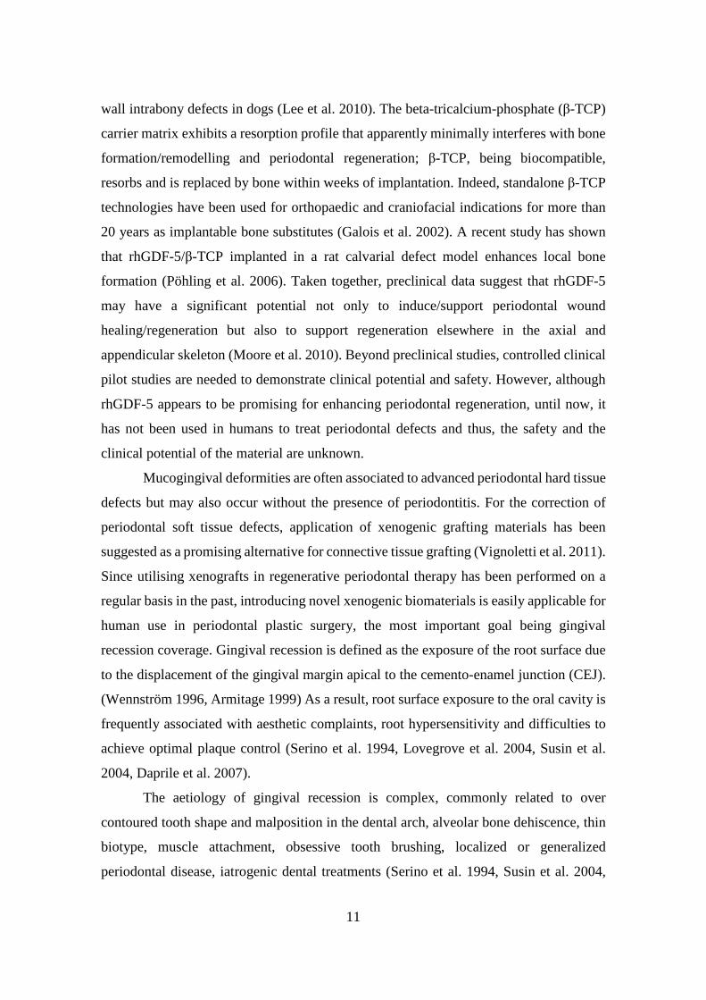
wall intrabony defects in dogs (Lee et al. 2010). The beta-tricalcium-phosphate (β-TCP)
carrier matrix exhibits a resorption profile that apparently minimally interferes with bone
formation/remodelling and periodontal regeneration; β-TCP, being biocompatible,
resorbs and is replaced by bone within weeks of implantation. Indeed, standalone β-TCP
technologies have been used for orthopaedic and craniofacial indications for more than
20 years as implantable bone substitutes (Galois et al. 2002). A recent study has shown
that rhGDF-5/β-TCP implanted in a rat calvarial defect model enhances local bone
formation (Pöhling et al. 2006). Taken together, preclinical data suggest that rhGDF-5
may have a significant potential not only to induce/support periodontal wound
healing/regeneration but also to support regeneration elsewhere in the axial and
appendicular skeleton (Moore et al. 2010). Beyond preclinical studies, controlled clinical
pilot studies are needed to demonstrate clinical potential and safety. However, although
rhGDF-5 appears to be promising for enhancing periodontal regeneration, until now, it
has not been used in humans to treat periodontal defects and thus, the safety and the
clinical potential of the material are unknown.
Mucogingival deformities are often associated to advanced periodontal hard tissue
defects but may also occur without the presence of periodontitis. For the correction of
periodontal soft tissue defects, application of xenogenic grafting materials has been
suggested as a promising alternative for connective tissue grafting (Vignoletti et al. 2011).
Since utilising xenografts in regenerative periodontal therapy has been performed on a
regular basis in the past, introducing novel xenogenic biomaterials is easily applicable for
human use in periodontal plastic surgery, the most important goal being gingival
recession coverage. Gingival recession is defined as the exposure of the root surface due
to the displacement of the gingival margin apical to the cemento-enamel junction (CEJ).
(Wennström 1996, Armitage 1999) As a result, root surface exposure to the oral cavity is
frequently associated with aesthetic complaints, root hypersensitivity and difficulties to
achieve optimal plaque control (Serino et al. 1994, Lovegrove et al. 2004, Susin et al.
2004, Daprile et al. 2007).
The aetiology of gingival recession is complex, commonly related to over
contoured tooth shape and malposition in the dental arch, alveolar bone dehiscence, thin
biotype, muscle attachment, obsessive tooth brushing, localized or generalized
periodontal disease, iatrogenic dental treatments (Serino et al. 1994, Susin et al. 2004,
12
Lovegrove et al. 2004, Daprile et al. 2007). As one of the most significant
predeterminants, a thin gingival biotype is considered to be the most relevant anatomical
factor of gingival recession (Müller et al. 1998), although controversial data have been
published on the minimally sufficient width and thickness of keratinised gingiva, needed
for long-term stability of marginal soft tissue contours (Kennedy et al. 1985, Aguido et
al. 2009). Therefore, most soft tissue augmentation procedures aim not only to obtain
complete root coverage (CRC) and natural tissue blending of the exposed surfaces and
but also to increase gingiva width and thickness to ensure long-term stability.
Results from systematic reviews indicate that at single Miller (Miller 1985) class
I and II gingival recessions CRC can predictably be obtained using different surgical
techniques mainly including coronally advanced flap (CAF) with and without soft tissue
grafting and/or biologic agents such as an enamel matrix derivative (Cairo et al 2008;
Chambrone et al 2010).
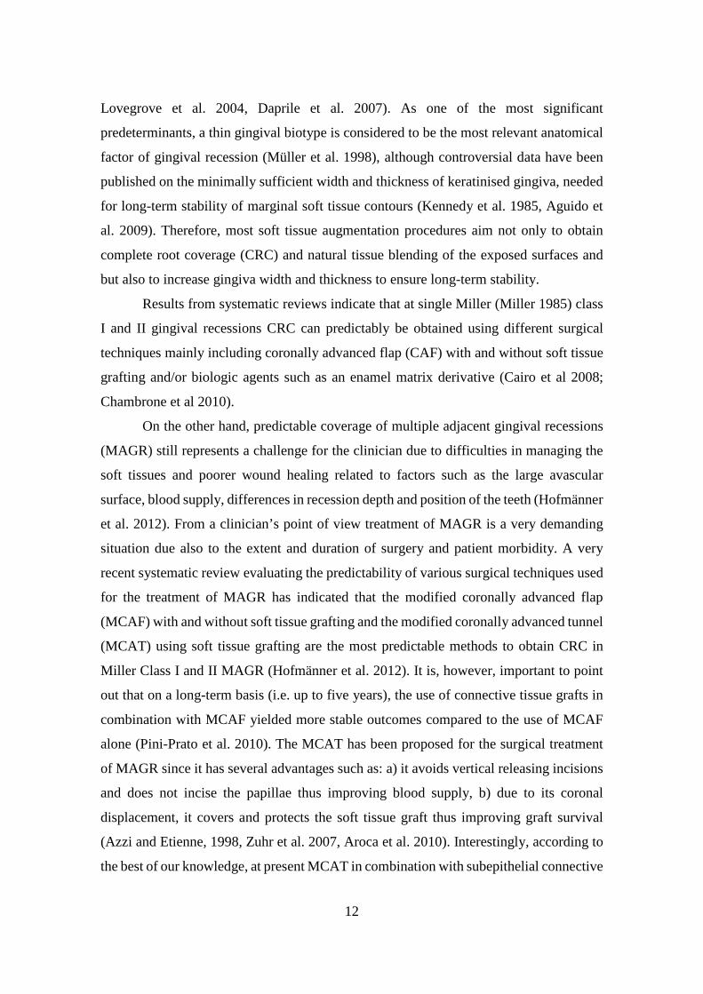
On the other hand, predictable coverage of multiple adjacent gingival recessions
(MAGR) still represents a challenge for the clinician due to difficulties in managing the
soft tissues and poorer wound healing related to factors such as the large avascular
surface, blood supply, differences in recession depth and position of the teeth (Hofmänner
et al. 2012). From a clinician’s point of view treatment of MAGR is a very demanding
situation due also to the extent and duration of surgery and patient morbidity. A very
recent systematic review evaluating the predictability of various surgical techniques used
for the treatment of MAGR has indicated that the modified coronally advanced flap
(MCAF) with and without soft tissue grafting and the modified coronally advanced tunnel
(MCAT) using soft tissue grafting are the most predictable methods to obtain CRC in
Miller Class I and II MAGR (Hofmänner et al. 2012). It is, however, important to point
out that on a long-term basis (i.e. up to five years), the use of connective tissue grafts in
combination with MCAF yielded more stable outcomes compared to the use of MCAF
alone (Pini-Prato et al. 2010). The MCAT has been proposed for the surgical treatment
of MAGR since it has several advantages such as: a) it avoids vertical releasing incisions
and does not incise the papillae thus improving blood supply, b) due to its coronal
displacement, it covers and protects the soft tissue graft thus improving graft survival
(Azzi and Etienne, 1998, Zuhr et al. 2007, Aroca et al. 2010). Interestingly, according to
the best of our knowledge, at present MCAT in combination with subepithelial connective
13
tissue grafting is the only technique which has been shown to result in predictable
improve coverage of Miller Class III MAGR (Aroca et al. 2010; Hofmänner et al.2012).
Connective tissue graft harvesting is often associated with increase patient
morbidity, prolonged surgical time and the possibility of postoperative complications
such as bleeding and numbness in the donor area (Hofmänner et al.2012). In order to
overcome these inconveniences, attempts are made to develop new materials aiming to
replace connective tissue grafts thus, improving patient acceptance and minimizing
morbidity. Both the MCAF and the MCAT techniques have been reported applied in
combination with biological adjuncts, such as EMD (Pilloni et al. 2006), acellular dermal
matrix (ADM) (Modaressi 2009) and platelet rich fibrin (PRF) (Aroca 2009).
Nevertheless, according to a recently published systematic review, none of these
alternative biological factors have reached or surpassed the effecacy and predictability of
connective tissue grafting (Cairo et al. 2008).
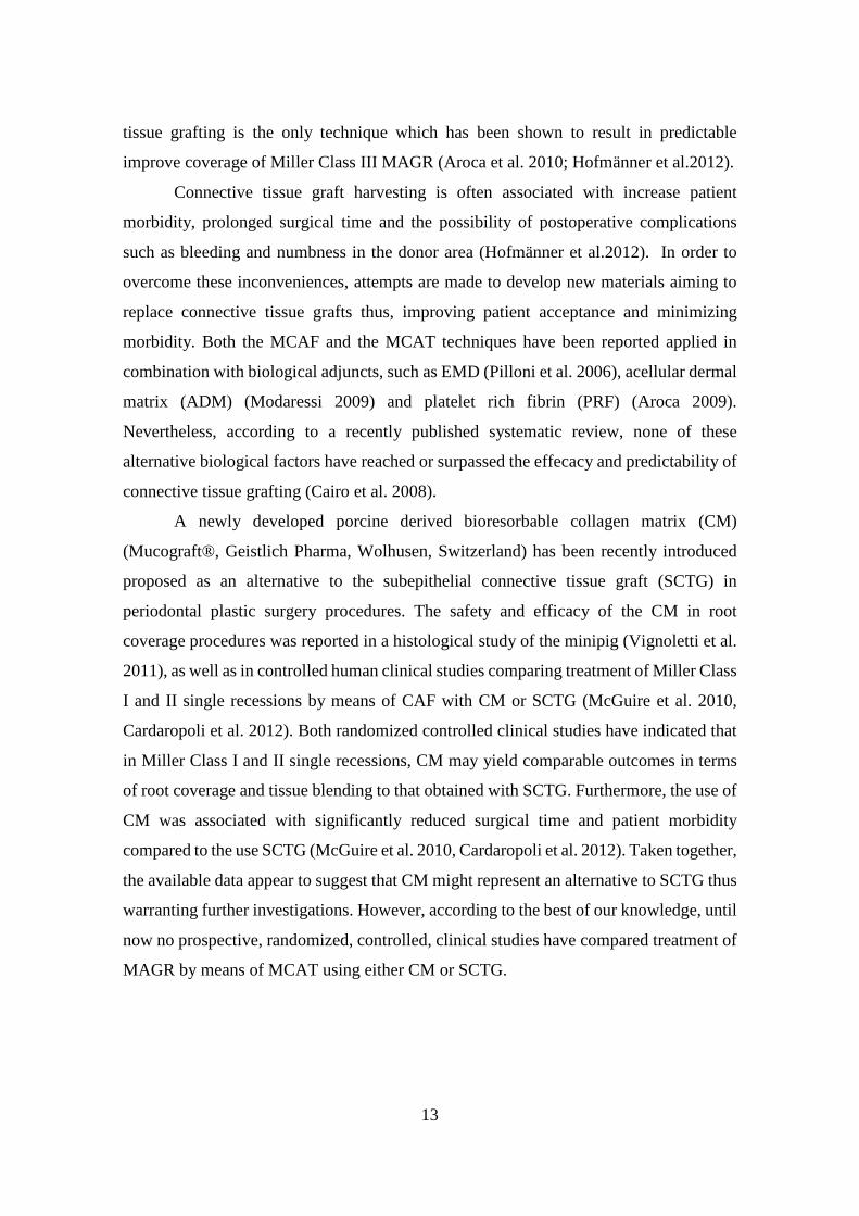
A newly developed porcine derived bioresorbable collagen matrix (CM)
(Mucograft®, Geistlich Pharma, Wolhusen, Switzerland) has been recently introduced
proposed as an alternative to the subepithelial connective tissue graft (SCTG) in
periodontal plastic surgery procedures. The safety and efficacy of the CM in root
coverage procedures was reported in a histological study of the minipig (Vignoletti et al.
2011), as well as in controlled human clinical studies comparing treatment of Miller Class
I and II single recessions by means of CAF with CM or SCTG (McGuire et al. 2010,
Cardaropoli et al. 2012). Both randomized controlled clinical studies have indicated that
in Miller Class I and II single recessions, CM may yield comparable outcomes in terms
of root coverage and tissue blending to that obtained with SCTG. Furthermore, the use of
CM was associated with significantly reduced surgical time and patient morbidity
compared to the use SCTG (McGuire et al. 2010, Cardaropoli et al. 2012). Taken together,
the available data appear to suggest that CM might represent an alternative to SCTG thus
warranting further investigations. However, according to the best of our knowledge, until
now no prospective, randomized, controlled, clinical studies have compared treatment of
MAGR by means of MCAT using either CM or SCTG.
14
5. OBJECTIVES
The goal of my PhD dissertation was to evaluate - based on the existing evidences
available in literature related to adult stem cells, human recombinant growth factors and
novel xenogenic biomaterials - the currently available treatment options and the novel
materials and techniques that might be the future in periodontal hard- and soft tissue
reconstruction.
Available data related to in vitro and clinical research on periodontal regenerative
therapy have raised a number of fundamental questions dealing with possible future
clinical impact of the above mentioned novel regenerative procedures and biomaterials.
These goals focus on establishing the methodological basis to develop future tissue
engineering applications, as well as safety and efficacy of currently available prototype
biomaterials for human periodontal application. During my PhD research in vitro and
clinical studies were conducted to find answers to the main question: how periodontal
wound healing and complete regeneration can be improved beyond current therapeutical
approaches.
The performed in vitro and clinical studies aimed at:
• Establishing cell cultures of periodontal origin, investigating the effect of EMD
on cell proliferation; characterisation of adult stem cells in vitro
• Developing in vitro protocols for osteogenic differentiation of periodontal
ligament stem cells (PDLSCs) for future tissue engineering applications
• Investigating the safety and efficacy of a human recombinant growth factor on a
β-TCP carrier (rhGDF-5) designed for periodontal hard tissue reconstruction in a
pilot clinical study
• Investigating the safety and efficacy of a novel collagen matrix (Mucograft®) for
gingival recession coverage of MAGR in a pilot clinical case series
• Comparing the clinical outcome and patient satisfaction related to the application
of Mucograft® compared to connective tissue grafting in the treatment of MAGR
in a split mouth randomised controlled study

15
6. METHODS
In this section the literature research, experimental, surgical and analytical
methodology will be described. The present thesis reports on a review article, two in vitro
research articles as well as three publications reporting on clinical studies, which are
summarised in Table 1.
In vitro research was carried out at the Department of Oral Biology, Semmelweis
University. All patients included in the clinical studies were referred to the Department
of Periodontology, Semmelweis University for treatment of periodontal soft- or hard
tissue defects.
Table 1: Summary of literature review, clinical- and in vitro studies
I. A literature review was performed to collect relevant informations prior to
initiating further in vitro and clinical periodontal research. To collect valuable
informations, the application of EMD in periodontal regeneration compared
to alternative treatment options (e.g. GTR) was analysed based on currently
available literature data.
Study Description Appendix
I Literature analysis on the application of enamel
matrix proteins in periodontal regenerative therapy
[I]
II Isolation and culturing of PDLSCs, investigating the
effect of EMD on cell proliferation and viability
[II]
III Establishing protocols for in vitro differentiation of
human periodontal and pulpal stem cells
[III]
IV Evaluating the clinical safety and efficacy of a novel
recombinant growth factor for periodontal hard tissue
reconstruction
[IV]
V Assessing a novel bioabsorbable collagen matrix for
soft tissue reconstruction in root coverage procedures
[V]
VI Comparing the novel collagen matrix and connective
tissue grafting for root coverage
[VI]

16
II. In the first in vitro study cell cultures from human periodontal ligament were
established and multipotential adult stem cells (PDLSCs) were identified in
these cultures. The effect of EMD was also analysed with regards to viability
of cells cultures. We established the methodological basis for further in vitro
research.
III. The second in vitro study described the introduction of differentiation
protocols applicable for maintainable cell cultures containing PDLSCs and
dental pulp stem cells (DPSCs). Using optimized pharmacological protocols
the potential of periodontal and pulp derived adult stem cell cultures to form
mineralized tissues and to undergo neuronal differentiation was analysed.
IV. The first clinical exploratory study was specifically designed to evaluate the
clinical and histological outcomes following treatment of intrabony defects
with open flap debridement alone or in combination with rhGDF-5 adsorbed
onto a particulate β-tricalcium phosphate carrier. The publication reported on
the study protocol, safety profile, the early healing phase and the clinical
outcomes at 24 weeks while the histological outcomes were presented and
discussed in great detail in a subsequent paper (Stavropoulos et al. 2011).
V. The second clinical study presented data from a prospective pilot case series,
which was performed to evaluate the safety and efficacy of Mucograft® in the
treatment of Miller class I and II MAGR using the MCAT technique.
VI. The third clinical study reported on a prospective, randomized, controlled,
split-mouth clinical study. This was conducted to clinically evaluate the
treatment of Miller class I and II MAGR using the MCAT technique either in
combination with Mucograft® or SCTG.

17
6.1 Literature review on the application of enamel matrix proteins in periodontal
regenerative therapy
In the literature search a protocol of review was set out with the following
eligibility criteria for study inclusion to collect valuable information on the application of
EMD in periodontal regeneration. A technique or a material must have fitted in the
following categories to be classified as "regeneration-related article"
• In vitro studies, which investigated the cellular and molecular mechanisms of
EMD
• Controlled histological animal studies, which evaluated the formation of new root
cementum, periodontal ligament and alveolar bone.
• Human biopsies, which assessed the formation of root cementum, periodontal
ligament and alveolar bone on a plaque-infected root surface.
• Controlled clinical studies, which measured the magnitude of gain of clinical
attachment and radiographical new bone formation. In the literature overview, the
existing evidence regarding the clinical use of EMD was provided.
6.2 In vitro isolation and differentiation of periodontal ligament stem cells
In our in vitro studies different methodologies were applied to establish and
maintain periodontal and pulpal cell cultures and to achieve differentiation of adult stem
cells into different cell lineages.
6.2.1. Cell isolation and culturing of periodontal ligament stem cells
Our protocol to isolate and culture dental pulp stem cells is based on a procedure
described previously (Gronthos et al. 2000), with some modifications. In brief, normal
human impacted third molars were collected from adults (18-26 years of age) at the
Department of Periodontology, Semmelweis University, under approved ethical
guidelines set by the Ethical Committee of the Hungarian Medical Research Council.
Tooth surfaces were cleaned and the periodontal tissue was removed with a sterile scalpel
and was collected. The tooth was cut around the cemento-enamel junction by sterile
dental fissure burs to expose the pulp chamber, and the pulp tissue was removed from the

18
crown and root. Both pulp tissue and periodontal tissue were then separately digested in
a solution of collagenase type I (3 mg/ml, Sigma-Aldrich, St. Louis, USA) and dispase
(4mg/ml, Roche, Basel, Switzerland) for 1 hour at 37°C. Single-cell suspensions were
obtained by passing the cells through a 70 µm strainer (Falcon). Cells were seeded into
6-well plates (Costar) in alpha modification of Eagle's medium (a-MEM, GIBCO/BRL)
supplemented with 15% Fetal Bovine Serum (FBS, GIBCO/BRL), 100 µM L-ascorbic
acid 2-phosphate (Sigma-Aldrich, St. Louis, USA), 2 mM L-glutamine, 100 units/ml
penicillin, 100 mg/ml streptomycin (GIBCO/BRL), and then incubated at 37°C in 5%
CO2. To assess colony-forming capability, 14 day old cultures were fixed with 4%
paraformaldehyde, and then stained with 2% Giemsa. Aggregates of 50 cells were scored
as colonies
6.2.3. Cell viability studies and treatment with enamel matrix derivative
The influence of FBS and EMD (Emdogain, Straumann, Basel, Switzerland)
containing media as well as osteogenic and neuronal differentiation protocols on primary
cell cultures was assessed by Microculture Tetrazolium (MTT) assay. To test the effect
of these conditions on culture growth, DPSCs and PDLSCs were cultured in 96-well
plates for 24 hours. In each well 3 x 10³ DPSC cells or 5 x 10³ PDLSC cells were grown
in their regular media for 24 hours. Afterwards, cells were serum-starved for another 24
hours. Then, 15% (PDLSC) or 20% (DPSC) FBS containing medium, or serum free
medium (control) was added for 24 hours. Thereafter, 100 µl MTT solution (0.2 mg/ml,
Sigma-Aldrich, St. Louis, USA) diluted in a-MEM was added into each well until
formazane crystal formation occurred. 100 µl DMSO (99.5%, dimethyl-sulfoxid) was
added into wells to dissolve formazane crystals. Then the intensity of staining was
determined by a microplate reader (Model 3550, Biorad, Hercules, USA) at 595 nm
(measurement wavelength) and 650 nm (reference wavelength). Under these
circumstances, the level of optical density is proportional to the number of living cells in
the culture. The proliferative effect was expressed as a ratio between optical density of
treated cells and serum-free cultured control cells and given in percent.

19
6.2.4. Immunocytochemistry
To identify the mesenchymal stem cell marker “stromal cell surface marker-1“
(STRO-1) in our cultures, cells were grown on glass coverslips in 24-well plates (Costar,
Cole-Parmer, Vernon Hills, Illinois, USA) (5x10⁴ cells per well) and fixed with 4% PFA
in phosphate buffered saline (PBS) for 20 min. To block non-specific binding, fixed
cultures were incubated in PBS containing 7.5% FBS for 90 min and incubated with an
anti-STRO-1 primary antibody (1/200, a generous gift from Prof Richard Oreffo,
University of Southampton, Southampton, UK) overnight at 4°C. Subsequently, the cells
were incubated with Alexa 488 conjugated goat anti-mouse IgG (1:1000, Molecular
Probes, Invitrogen, Carlsbad, CA, USA) for 1 hour. Nuclei were counterstained with 10
mg/ml bisbenzimide (Sigma-Aldrich, St. Louis, USA) for 30 minutes.
To evaluate protein expression during differentiation experiments, cells grown on
poly-L-lysine-coated glass coverslips were fixed with 4% PFA in PBS for 20 min at room
temperature (RT), then 0.1% Triton X-100 (in PBS) was added for 8 min to permeabilise
them. Fixed cultures were incubated in PBS containing 4% bovine serum albumin (BSA;
90 min at RT) to block non-specific binding, then reacted with primary antibodies at 4°C
overnight. Antibodies were diluted in 4% BSA as follows: anti-NSE 1/200, anti-NF-M
1/200. IgG anti-mouse and anti-rabbit Alexa Fluor 488 conjugated (Molecular Probes,
Invitrogen, Carlsbad, CA, USA) secondary antibodies were diluted 1/750 and applied for
1 h at RT. Nuclei were counterstained with 10 mg/ml bisbenzimide (Sigma-Aldrich, St.
Louis, USA) for 30 minutes. Labelled preparations were examined by a fluorescent
microscope (Nikon Eclipse E600, Nikon Instruments, Tokyo, Japan), and images were
captured with a cooled CCD camera (SPOT RT Color 2000, Spot Imaging Solutions,
Sterling Heights, Michigan, USA) connected to a PC running an image acquisition
software (SPOT Advanced, Spot Imaging Solutions, Sterling Heights, Michigan, USA.).
Adobe Photoshop® was used to merge the digitized images of bisbenzimide and specific
staining.
6.2.5. FACS analysis
Fluorescence Activated Cell Sorting (FACS) analysis was performed to identify
cells expressing STRO-1, CD34 and c-kit mesenchymal stem cell markers as described

20
previously (Gronthos et al. 1994, Laino et al. 2005). Single cell suspension were prepared
from the cell cultures of 0,2% EDTA content, and subsequently incubated with STRO-
1/CD34/c-kit antibody or with isotype matching negative controls for 1 hour on ice. Cells
were washed with 5% PBS solution of FBS, then fluorescent stain-conjugated secondary
antibodies were added to the samples. Following repeated rinsing with 5% PBS solution
of FBS, cell suspensions were fixated with 4% paraformaldehyde. FACS analysis was
performed subsequently.
6.2.6. Osteogenic induction
Osteogenic differentiation was induced by modifications of a previously reported
protocol (Kemoun et al. 2007). In brief, DPSCs and PDLSCs were cultured with 1% FBS,
100 mg/ml streptomycin, 100 U/ml penicillin, 2 mML-glutamine, 10-8M dexamethazone,
50 mg/ml L-ascorbic acid 2-phosphate, 10 mmol/l b-glycerophosphate in aMEM for 20
days without passaging. The medium was replaced twice a week. After 3 weeks of
treatment calcium accumulation was detected by 2% Alizarin red S (pH 4.2, buffered with
ammonium hydroxide) staining. Similar culture media without dexamethazone and ß-
glycerophosphate was used as control condition.
6.2.7. Neuronal induction
For neuronal differentiation, cultured morphologically homogeneous DPSCs and
PDLSCs, (passage 1-4) were plated (~2×10⁴ cells/well) into a 24 well plate containing
poly-L-lysin coated glass coverslips. After 24 hours, cells were treated with 3 different
protocols:
6.2.7.1 Protocol 1.
Cells were differentiated as previously described by Scintu et al. (Scintu et al.
2006) with 10 ng/ml FGF-1 (R&D, Minneapolis, MN), 200 nM 12-O-
tetradecanoylphorbol-13-acetate (TPA; Sigma-Aldrich, St. Louis, USA), 250 µM IBMX
(Sigma-Aldrich, St. Louis, USA) and 50 µM forskolin (Sigma-Aldrich, St. Louis, USA),
in Dulbecco's modified Eagle's medium/F12 1:1 (DMEM/F12) (Sigma-Aldrich, St.
21
Louis, USA) supplemented with ITS Liquid Media Supplement (Sigma-Aldrich, St.
Louis, USA). The cells were fixed for immunocytochemistry right before and 24 h post-
induction.
6.2.7.2. Protocol 2.
This protocol was also based on a method recently reported by Choi et al. (Choi
et al. 2006). Cells were preinduced for 1 day with DMEM/F12, with 20% FBS, and 10
ng/ml basic fibroblast growth factor (bFGF; Sigma-Aldrich, St. Louis, USA). The
preinduction medium was removed, cells were washed with PBS and then changed to
serum-free induction medium that consisted of DMEM containing 2% DMSO, 200 µM
BHA, 25 mM KCl, 2 mM valporic acid, 10 µM forskolin, 1 µM hydrocortisone and 5
µg/ml insulin (Sigma-Aldrich, St. Louis, USA). The cells were fixed for
immunocytochemistry right before and 24 h post-induction.
6.2.7.3. Protocol 3.
A three-step differentiation method was developed in our own laboratory since
Protocols 1 and 2 did not yield satisfactory results. DPSCs or PDLSCs were seeded onto
poly-L-lysin coated glass coverslips in DMEM/F12, 2.5% FBS, 100 mg/ml streptomycin,
and 100 U/ml penicillin, and cultured for 24 h. Step 1: epigenetic reprogramming was
performed using 10 mM 5-azacytidine in DMEM/F12 containing 2.5% FBS and 10 ng/ml
bFGF for 48 h. Step 2: neural differentiation was induced by exposing the cells to 250
mM IBMX, 50 mM forskolin, 200 nM TPA, 1 mM dbcAMP, 10 ng/ml bFGF, 10 ng/ml
NGF and 30 ng/ml NT-3, supplemented with ITS Liquid Media Supplement in
DMEM/F12 for 3 days. Step 3: at the end of the neural induction treatment, cells were
washed with PBS and then neuronal maturation was performed by maintaining the cells
in Neurobasal A media supplemented with 1 mM dbcAMP, 1% N2, 1% B27, and 30
ng/ml NT-3 for 3-8 days. Solutions 168 were freshly prepared immediately prior to use.
The cells were fixed for immunocytochemistry before treatment, on the first day neuronal
induction (step 2) and on the third day of maturation (step 3).
22
6.2.8. Real-time PCR
Total RNA from DPSCs and PDLSCs was isolated using an RNeasy Plus Micro
Kit (Qiagen) with on-column DNase digestion. The concentration of the RNA was
determined by the Ribogreen method (Invitrogen, Carlsbad, CA, USA). The integrity of
the RNA was verified by electrophoresis on a 1% agarose gel and 200 ng total RNA was
used per sample for cDNA synthesis, using random primers (High-Capacity
cDNAArchive Kit, Applied Biosystems, Invitrogen, Carlsbad, CA, USA) in a total
volume of 50 µl. For quantitative PCR amplification, 5% of the cDNA synthesis reaction
was used with real time PCR primers and a target-specific fluorescence probe (FAM-
labelled MGB probe). The probes and primers were selected from the Applied Biosystem
Assay on Demand database for the specific markers vimentin (VIM) and neurospecific
enolase (NSE) and for the human acidic ribosomal phosphoprotein P0 (RPLP0), which
was used as an internal control. Universal Mastermix (Roche, Basel, Switzerland)
containing AMP-erase was used for amplification in a total volume of 20 µl. For detection
of fluorescence signal during the PCR cycles, a (StepOne® Real-Time PCR System,
Applied Biosystem, Invitrogen, Carlsbad, CA, USA) was used with the default setting
(50°C for 2 min, 95°C for 10 min, 45 cycles: 95°C for 15 s, 60°C for 1 min). Each
treatment was repeated five times and each sample was measured in duplicate. Changes
in gene expression levels were estimated by calculating the relative expression values
normalized to the RPLP0 level from the same sample.
6.2.9. Statistical analysis
Data were presented as means ± S.E.M. For statistical comparisons, analysis of
variance was followed by Bonferroni post-hoc test (Instat, GraphPad Software).
23
6.3 Clinical studies
In our clinical studies hard- and soft tissue regenerative procedures were
investigated using similar preoperative protocol and postsurgical care. Standardised
clinical measurements were taken for evaluation of treatment safety and efficacy. Surgical
protocols varied throughout the studies.
6.3.1 Hard tissue regeneration following treatment with rhGDF-5/β-TCP
This pilot, phase IIa study used a stratified randomized, open, controlled, two-
arm, parallel group design. The overall design and patient treatment allocation is
summarized in Fig. 1. The study was conducted at the Department of Periodontology,
Semmelweis University, Budapest, Hungary between July 2007 and August 2008. The
study protocol was approved by the Hungarian National Institute of Pharmacy and the
Institutional Ethics Committee (application no. 32579/40/06) of the Semmelweis
University, Budapest, Hungary (TUKEB no. 20/2007). All patients received oral and
written explanations of the research protocol. Patients signed a consent form providing
the possibility of withdrawing from the study at any time. The study was planned and
conducted in compliance with the Declaration of Helsinki of 1975 as revised in 2000,
Good Clinical Practice, and relevant local laws.
The total study duration was 175–182 days, in all ten visits/patient. After
screening, selected patients received flap surgery (control) or flap surgery combined with
implantation of rhGDF-5/β-TCP at the qualified defect site (Visit 2). They then returned
for general and oral health evaluations as well as professional tooth cleanings following
a set schedule (Visits 3 through 8). Blood samples were collected at screening (Visit 1),
and at weeks 2 and 24 (Visits 3 and 9) to evaluate routine haematology and clinical
chemistry, rhGDF-5 plasma levels, and antirhGDF-5 antibody formation.
Randomization was performed using a computer-generated randomization list via
block randomization. Ten patients were randomized to each treatment group. A separate
random scheme was generated. The investigators were masked to the block length. The
sponsor retained the randomization scheme for control purposes. The investigator
implemented the predefined randomization by opening a randomization envelope at the
appointment for surgery (Visit 2). The randomization code was opened only after the
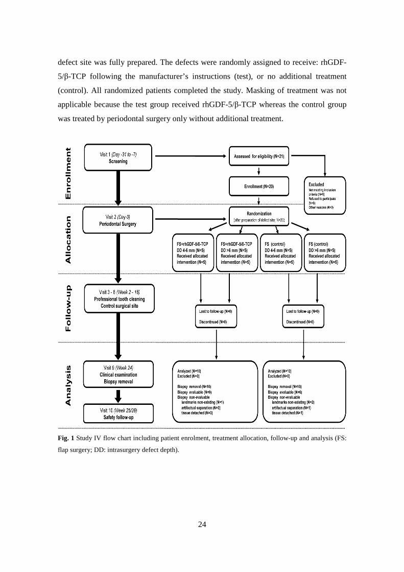
24
defect site was fully prepared. The defects were randomly assigned to receive: rhGDF-
5/β-TCP following the manufacturer’s instructions (test), or no additional treatment
(control). All randomized patients completed the study. Masking of treatment was not
applicable because the test group received rhGDF-5/β-TCP whereas the control group
was treated by periodontal surgery only without additional treatment.
Fig. 1 Study IV flow chart including patient enrolment, treatment allocation, follow-up and analysis (FS:
flap surgery; DD: intrasurgery defect depth).
25
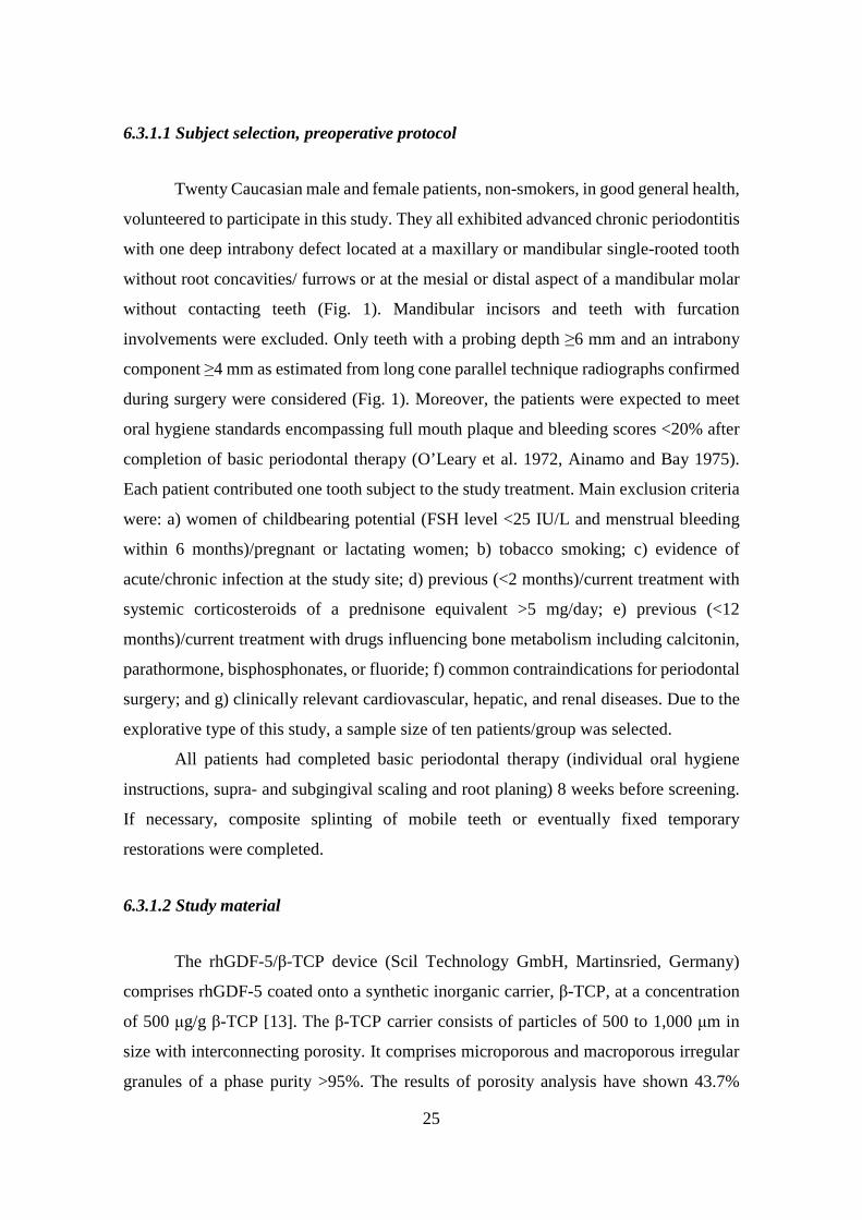
6.3.1.1 Subject selection, preoperative protocol
Twenty Caucasian male and female patients, non-smokers, in good general health,
volunteered to participate in this study. They all exhibited advanced chronic periodontitis
with one deep intrabony defect located at a maxillary or mandibular single-rooted tooth
without root concavities/ furrows or at the mesial or distal aspect of a mandibular molar
without contacting teeth (Fig. 1). Mandibular incisors and teeth with furcation
involvements were excluded. Only teeth with a probing depth ≥6 mm and an intrabony
component ≥4 mm as estimated from long cone parallel technique radiographs confirmed
during surgery were considered (Fig. 1). Moreover, the patients were expected to meet
oral hygiene standards encompassing full mouth plaque and bleeding scores <20% after
completion of basic periodontal therapy (O’Leary et al. 1972, Ainamo and Bay 1975).
Each patient contributed one tooth subject to the study treatment. Main exclusion criteria
were: a) women of childbearing potential (FSH level <25 IU/L and menstrual bleeding
within 6 months)/pregnant or lactating women; b) tobacco smoking; c) evidence of
acute/chronic infection at the study site; d) previous (<2 months)/current treatment with
systemic corticosteroids of a prednisone equivalent >5 mg/day; e) previous (<12
months)/current treatment with drugs influencing bone metabolism including calcitonin,
parathormone, bisphosphonates, or fluoride; f) common contraindications for periodontal
surgery; and g) clinically relevant cardiovascular, hepatic, and renal diseases. Due to the
explorative type of this study, a sample size of ten patients/group was selected.
All patients had completed basic periodontal therapy (individual oral hygiene
instructions, supra- and subgingival scaling and root planing) 8 weeks before screening.
If necessary, composite splinting of mobile teeth or eventually fixed temporary
restorations were completed.
6.3.1.2 Study material
The rhGDF-5/β-TCP device (Scil Technology GmbH, Martinsried, Germany)
comprises rhGDF-5 coated onto a synthetic inorganic carrier, β-TCP, at a concentration
of 500 µg/g β-TCP [13]. The β-TCP carrier consists of particles of 500 to 1,000 µm in
size with interconnecting porosity. It comprises microporous and macroporous irregular
granules of a phase purity >95%. The results of porosity analysis have shown 43.7%
26
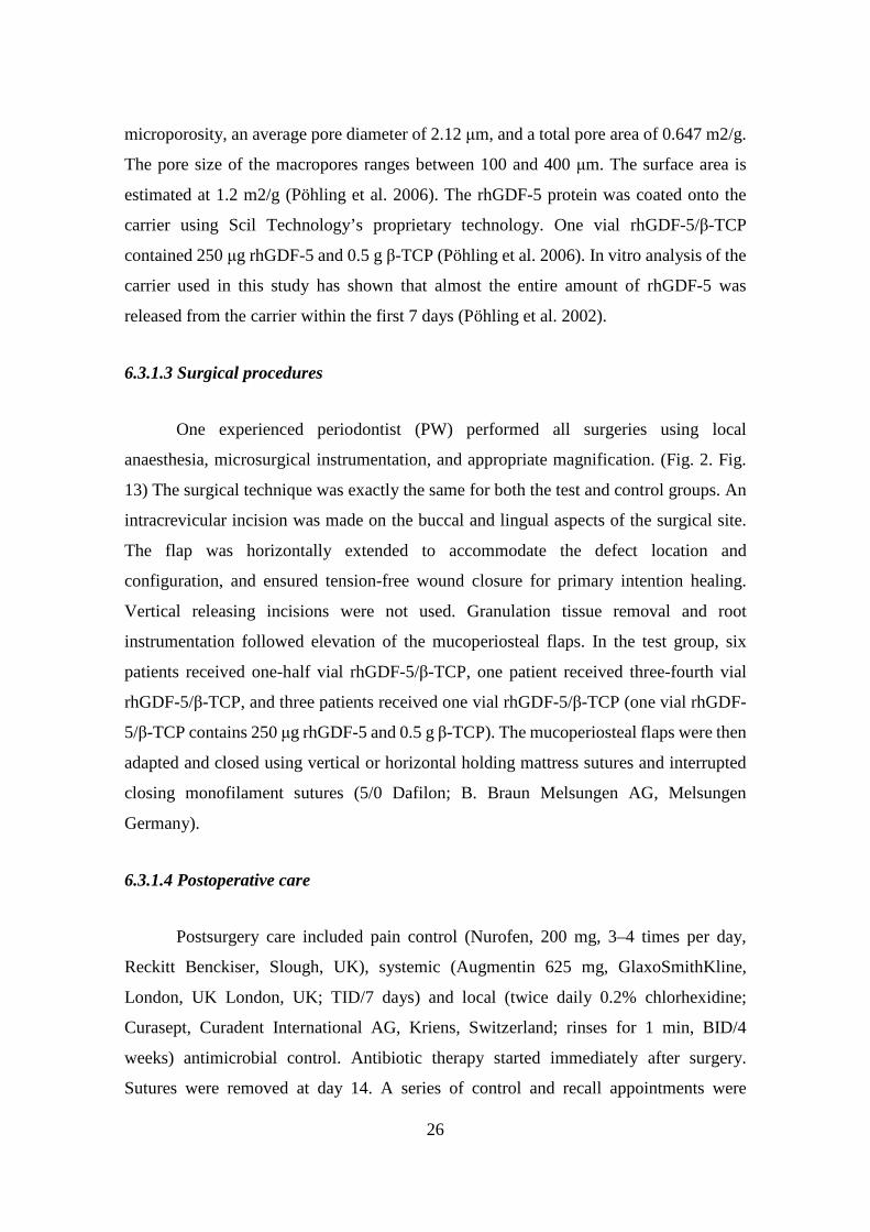
microporosity, an average pore diameter of 2.12 µm, and a total pore area of 0.647 m2/g.
The pore size of the macropores ranges between 100 and 400 µm. The surface area is
estimated at 1.2 m2/g (Pöhling et al. 2006). The rhGDF-5 protein was coated onto the
carrier using Scil Technology’s proprietary technology. One vial rhGDF-5/β-TCP
contained 250 µg rhGDF-5 and 0.5 g β-TCP (Pöhling et al. 2006). In vitro analysis of the
carrier used in this study has shown that almost the entire amount of rhGDF-5 was
released from the carrier within the first 7 days (Pöhling et al. 2002).
6.3.1.3 Surgical procedures
One experienced periodontist (PW) performed all surgeries using local
anaesthesia, microsurgical instrumentation, and appropriate magnification. (Fig. 2. Fig.
13) The surgical technique was exactly the same for both the test and control groups. An
intracrevicular incision was made on the buccal and lingual aspects of the surgical site.
The flap was horizontally extended to accommodate the defect location and
configuration, and ensured tension-free wound closure for primary intention healing.
Vertical releasing incisions were not used. Granulation tissue removal and root
instrumentation followed elevation of the mucoperiosteal flaps. In the test group, six
patients received one-half vial rhGDF-5/β-TCP, one patient received three-fourth vial
rhGDF-5/β-TCP, and three patients received one vial rhGDF-5/β-TCP (one vial rhGDF-
5/β-TCP contains 250 µg rhGDF-5 and 0.5 g β-TCP). The mucoperiosteal flaps were then
adapted and closed using vertical or horizontal holding mattress sutures and interrupted
closing monofilament sutures (5/0 Dafilon; B. Braun Melsungen AG, Melsungen
Germany).
6.3.1.4 Postoperative care
Postsurgery care included pain control (Nurofen, 200 mg, 3–4 times per day,
Reckitt Benckiser, Slough, UK), systemic (Augmentin 625 mg, GlaxoSmithKline,
London, UK London, UK; TID/7 days) and local (twice daily 0.2% chlorhexidine;
Curasept, Curadent International AG, Kriens, Switzerland; rinses for 1 min, BID/4
weeks) antimicrobial control. Antibiotic therapy started immediately after surgery.
Sutures were removed at day 14. A series of control and recall appointments were

27
scheduled (biweekly, the first 6 weeks and then monthly until the end of the study)
including reinforcements of oral hygiene and professional supragingival tooth cleaning.
Fig. 2 Flap surgery (control): Presurgery (top left); intrasurgery defect morphology (top right); the biopsy
event at 24 weeks postsurgery (bottom left); and biopsy including defect site (bottom right). Histological
outcomes were published elsewhere (Stavropoulos et al. 2011).
6.3.1.5 Clinical assessment
Clinical outcomes were evaluated at baseline and at 24 weeks postsurgery.
Probing depth (PD), gingival recession (GR) and clinical attachment level (CAL) were
recorded using a standard periodontal probe (UNC 15, Hu-Friedy, Chicago, IL, USA).
Intraoral radiographs were taken with the long cone parallel technique at baseline and at
24 weeks postsurgery. However, due to the design of the study (i.e. no grafting in the
control group), the radiographs were not evaluated. Full mouth plaque and bleeding
scores were recorded as a percentage of total surfaces (four surfaces/tooth) with the
presence of plaque/bleeding on probing, respectively (O’Leary et al. 1972, Ainamo et al.
1975). One calibrated examiner, masked to the patients’ treatment protocol, performed

28
all clinical recordings. At 24 months postoperatively, biopsy removal was performed.
Histological outcomes were published elsewhere (Stavropoulos et al. 2011).
6.3.1.6 Safety assessment
Adverse events were monitored and recorded throughout the study, as well as
laboratory values, vital signs, and physical status. Adverse events were coded using the
Medical Dictionary of Regulatory Activities (MedDRA)
[http://www.meddramsso.com/index.asp]. Summaries and tabulations by severity and
relationship to therapy were based on the preferred terms and the primary system organ
classes (SOCs). Blood samples were collected at screening (Visit 1), 2 weeks postsurgery
(Visit 3), and prior to conclusion of study (Visit 9) to determine laboratory values (clinical
chemistry, haematology), rhGDF-5 plasma levels, and antirhGDF-5 antibodies.
The determination of rhGDF-5 in human plasma (EDTA) samples was carried out by
Elisa over a quantitation range of 40 pg/ml to 1,250 pg/ml. A monoclonal antibody
specific for rhGDF-5 has been precoated on a 96-well plate. Standards/QCs and samples
were then pipetted into the wells and any rhGDF-5 present was bound by the immobilized
antibody. After washing away any unbound substances, a biotinylated monoclonal
antibody specific for rhGDF-5 was added to the wells. After a second washing step,
PolyHRP Streptavidin was added that bound to the biotinylated antibody. After a third
washing step, peroxidase bound in the complex was visualized by TMB (3,3′,5,5′-
Tetramethylbenzidine) substrate solution. After stopping the enzymatic reaction with
sulphuric acid, the intensity of the resulting colour was determined at 450 nm. The colour
intensity was proportional to the concentration of rhGDF-5 in the sample.
6.3.1.7 Statistical analysis
The statistical analysis was conducted on an intent-to-treat basis. All randomized
patients with periodontal treatment were included in the intent-to-treat population. Paired
sample t-test and Wilcoxon signed-rank test were used to evaluate the impact of surgical
interventions on the various clinical parameters. Mann–Whitney U test (rank sum test)
was used to analyse differences among the various outcome variables between treatment
groups. No formal statistical comparisons were made related to safety data.
29
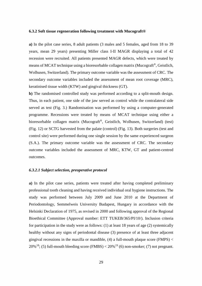
6.3.2 Soft tissue regeneration following treatment with Mucograft®
a) In the pilot case series, 8 adult patients (3 males and 5 females, aged from 18 to 39
years, mean 29 years) presenting Miller class I-II MAGR displaying a total of 42
recession were recruited. All patients presented MAGR defects, which were treated by
means of MCAT technique using a bioresorbable collagen matrix (Mucograft®, Geistlich,
Wolhusen, Switzerland). The primary outcome variable was the assessment of CRC. The
secondary outcome variables included the assessment of mean root coverage (MRC),
keratinised tissue width (KTW) and gingival thickness (GT).
b) The randomised controlled study was performed according to a split-mouth design.
Thus, in each patient, one side of the jaw served as control while the contralateral side
served as test (Fig. 3.) Randomisation was performed by using a computer-generated
programme. Recessions were treated by means of MCAT technique using either a
bioresorbable collagen matrix (Mucograft®, Geistlich, Wolhusen, Switzerland) (test)
(Fig. 12) or SCTG harvested from the palate (control) (Fig. 13). Both surgeries (test and
control site) were performed during one single session by the same experienced surgeon
(S.A.). The primary outcome variable was the assessment of CRC. The secondary
outcome variables included the assessment of MRC, KTW, GT and patient-centred
outcomes.
6.3.2.1 Subject selection, preoperative protocol
a) In the pilot case series, patients were treated after having completed preliminary
professional tooth cleaning and having received individual oral hygiene instructions. The
study was performed between July 2009 and June 2010 at the Department of
Periodontology, Semmelweis University Budapest, Hungary in accordance with the
Helsinki Declaration of 1975, as revised in 2000 and following approval of the Regional
Bioethical Committee (Approval number: ETT TUKEB/365/PI/10/). Inclusion criteria
for participation in the study were as follows: (1) at least 18 years of age (2) systemically
healthy without any signs of periodontal disease (3) presence of at least three adjacent
gingival recessions in the maxilla or mandible, (4) a full-mouth plaque score (FMPS) <
20%18; (5) full-mouth bleeding score (FMBS) < 20%19 (6) non-smoker; (7) not pregnant.

30
Before enrolment, written informed consent forms were obtained from all patients
participating in the study.
b) In the split mouth randomised, controlled study 22 patients with multiple Miller Class
I and II MAGR (Miller 1985) with evidence of CEJ were enrolled in the study after having
signed an informed consent. The study protocol was in accordance with the Helsinki
Declaration of 1975, as revised in 2002 and was submitted to and approved by the ethical
committee of the Semmelweis University Budapest, Hungary (protocol: 5242-0/2010-
101SEKU; 365/PI/10). The study was performed between July 2010 and November 2011
in the Department of Periodontology of the Semmelweis University Budapest. One month
before surgery, individualized oral hygiene instructions were given for each of the
included patients accompanied by full mouth supragingival scaling and polishing. The
following inclusion criteria were applied: 1) Age ≥ 18 years, 2) Absence of relevant
medical conditions, 3) Patients with healthy or treated periodontal conditions. 4) Presence
of ≥ 3 adjacent Miller class 1 and 2 gingival recessions on both sides of the maxillary or
mandibular arch with an apico-coronal extension (i.e. recession depth) > 2 mm, 5), Full-
Mouth Plaque Score (FMPS) ≤ 25% (O`Leary et al. 1972). Patients were excluded on the
basis of the following criteria:1) Pregnant or lactating females, 2), Tobacco smoking, 3)
Uncontrolled medical conditions, 4) Untreated periodontal conditions, 5) Use of systemic
antibiotics in the past 3 months, 6) Use of systemic antibiotics for endocarditis
prophylaxis, 7) Patients treated with any medication known to affect gingival conditions
(e.g. hyperplasia), 8)Infectious diseases such as hepatitis, tuberculosis and HIV, Drug and
alcohol abuse, (9) Failure to sign written informed consent
6.3.2.2 Study material
The CM (Mucograft®, Geistlich Pharma, Wolhusen, Switzerland) has a bilaminar
structure, consisting of two adherent layers: a superficial, compact, cell occlusive
membrane-like layer incorporating collagen fibres, and an underlying three dimensional
spongious collagen matrix designed to serve as scaffold conducing the ingrowth of blood
vessels and cells and to enhance blood clot stability.

31
Fig. 3 Study VI flow chart including patient enrolment, treatment allocation, follow-up and analysis
32
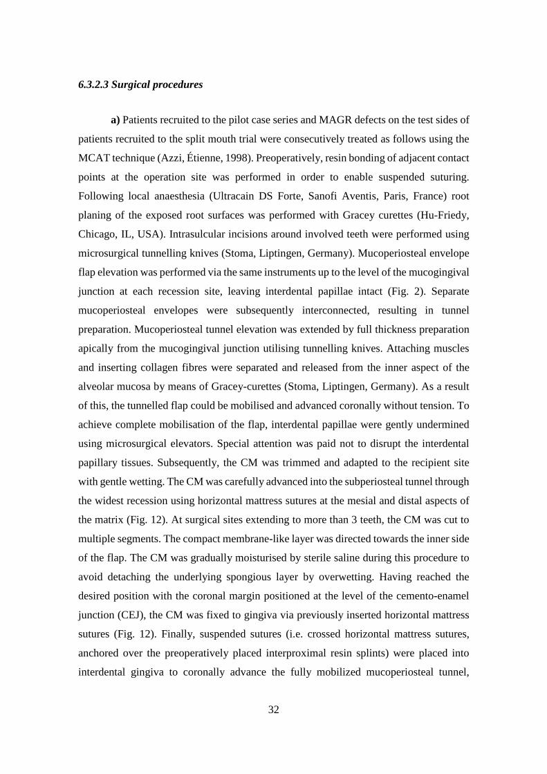
6.3.2.3 Surgical procedures
a) Patients recruited to the pilot case series and MAGR defects on the test sides of
patients recruited to the split mouth trial were consecutively treated as follows using the
MCAT technique (Azzi, Étienne, 1998). Preoperatively, resin bonding of adjacent contact
points at the operation site was performed in order to enable suspended suturing.
Following local anaesthesia (Ultracain DS Forte, Sanofi Aventis, Paris, France) root
planing of the exposed root surfaces was performed with Gracey curettes (Hu-Friedy,
Chicago, IL, USA). Intrasulcular incisions around involved teeth were performed using
microsurgical tunnelling knives (Stoma, Liptingen, Germany). Mucoperiosteal envelope
flap elevation was performed via the same instruments up to the level of the mucogingival
junction at each recession site, leaving interdental papillae intact (Fig. 2). Separate
mucoperiosteal envelopes were subsequently interconnected, resulting in tunnel
preparation. Mucoperiosteal tunnel elevation was extended by full thickness preparation
apically from the mucogingival junction utilising tunnelling knives. Attaching muscles
and inserting collagen fibres were separated and released from the inner aspect of the
alveolar mucosa by means of Gracey-curettes (Stoma, Liptingen, Germany). As a result
of this, the tunnelled flap could be mobilised and advanced coronally without tension. To
achieve complete mobilisation of the flap, interdental papillae were gently undermined
using microsurgical elevators. Special attention was paid not to disrupt the interdental
papillary tissues. Subsequently, the CM was trimmed and adapted to the recipient site
with gentle wetting. The CM was carefully advanced into the subperiosteal tunnel through
the widest recession using horizontal mattress sutures at the mesial and distal aspects of
the matrix (Fig. 12). At surgical sites extending to more than 3 teeth, the CM was cut to
multiple segments. The compact membrane-like layer was directed towards the inner side
of the flap. The CM was gradually moisturised by sterile saline during this procedure to
avoid detaching the underlying spongious layer by overwetting. Having reached the
desired position with the coronal margin positioned at the level of the cemento-enamel
junction (CEJ), the CM was fixed to gingiva via previously inserted horizontal mattress
sutures (Fig. 12). Finally, suspended sutures (i.e. crossed horizontal mattress sutures,
anchored over the preoperatively placed interproximal resin splints) were placed into
interdental gingiva to coronally advance the fully mobilized mucoperiosteal tunnel,
33
resulting in complete coverage of the CM and the recessions (Fig. 12). In cases where
complete CM coverage could not be obtained with the first sutures, additional vertical
mattress sutures were placed interdentally to enable coronal displacement of the tunnel
slightly over the CEJ.
b) MAGR defects on control sides of patients recruited to the split mouth trial
were treated with the MCAT technique as described above, in combination with
connective tissue grafting. A SCTG was immediately harvested after tunnel preparation
by using either a modified distal wedge procedure (Azzi & Etienne 1998) or the single
incision technique (Hürzeler & Weng 1999) depending on anatomical considerations. If
needed, the harvested graft was trimmed using a N°15 blade to achieve an optimal
thickness of 1-1.5 mm. Immediately after SCTG harvesting, the donor site was closed
with either a cross-mattress suture or with a modified mattress suture (Monnet-Corti et
al. 2006) (5-0 polyglactin 910, Vicryl, Ethicon, Johnson & Johnson, USA). SCTG was
always inserted under the tunnelled flap by starting at the deepest recession (Fig 13).
Subsequently, the grafts were pulled laterally towards each end of the tunnel by means of
mattress sutures (Azzi & Etienne 1998) . Finally, the flaps were positioned coronally to
the cemento-enamel junction (CEJ) by means of suspended sutures placed above the
contact point (Azzi & Etienne 1998) (Fig. 4, 14, 15).
6.3.2.4 Postoperative care
Patients attending either of the trials were given postoperative analgesics (3 X 50
mg Cataflam, Budapest, Hungary) for 3 days and antibiotics (3x625 mg Augmentin,
Pfizer KFT, Budapest, Hungary) for 7 days due to university regulation for implantable
biological materials. Patients were instructed to rinse with a 0.2% chlorhexidine solution,
two times a day for one minute for 3 weeks. Patients avoided brushing in the operated
area until suture removal. Patients underwent manual supragingival tooth cleaning twice
a week until suture removal. At suture removal two weeks after surgery, patients were
instructed in mechanical tooth cleaning of the operated areas using a soft tooth brush and
a roll technique. The interproximal resin splints were removed at 21 days. All patients
were recalled after 28 days, 3, 6 and 12 months and received one session of prophylaxis,
including reinforcement of oral hygiene, supragingival debridement, and tooth polishing.
34
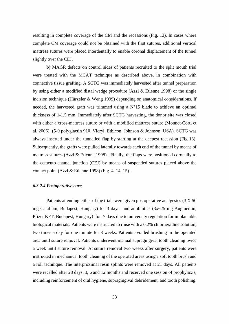
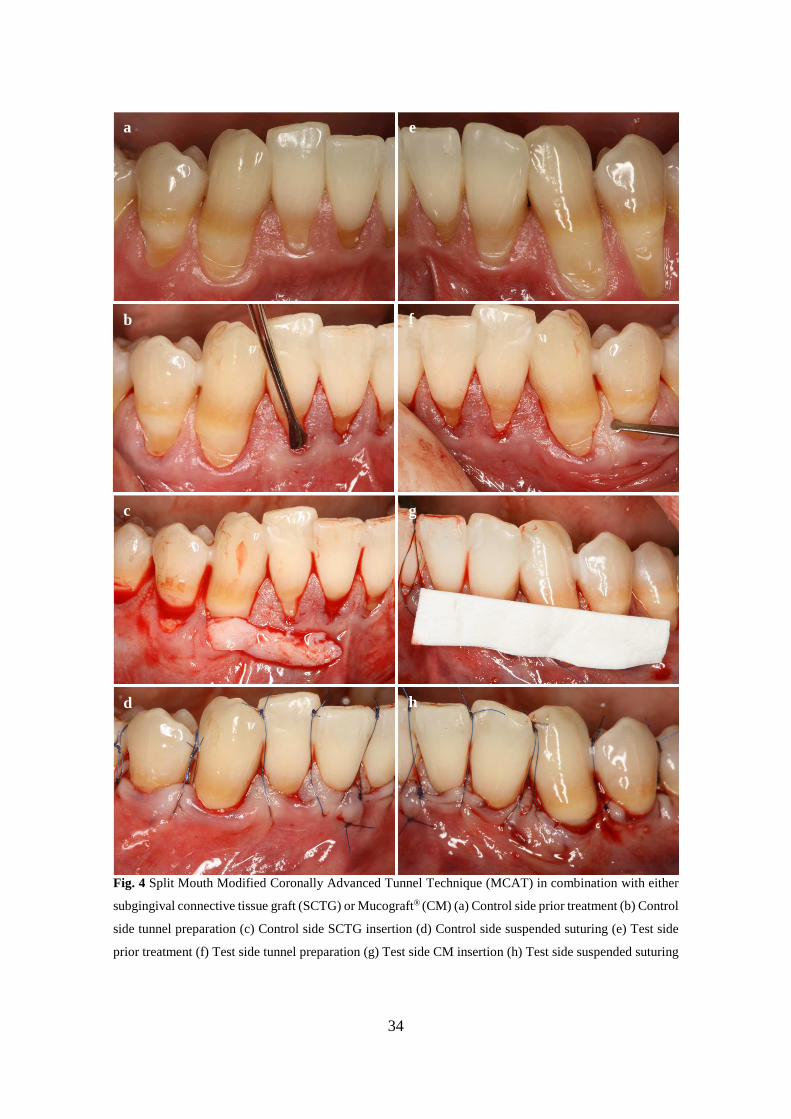
Fig. 4 Split Mouth Modified Coronally Advanced Tunnel Technique (MCAT) in combination with either
subgingival connective tissue graft (SCTG) or Mucograft® (CM) (a) Control side prior treatment (b) Control
side tunnel preparation (c) Control side SCTG insertion (d) Control side suspended suturing (e) Test side
prior treatment (f) Test side tunnel preparation (g) Test side CM insertion (h) Test side suspended suturing
a e
b
c
d
f
g
h
35
6.3.2.5 Clinical assessments
In both studies following measurements were made at the mid-buccal point of the
involved teeth at baseline (prior to surgery) 6, and at 12 months by the same blinded
investigator (B.M.) using the same type of periodontal probe (UNC 15, Hu-Friedy,
Chicago, IL, USA): 1) Gingival Recession Depth (GRD in mm) measured as the distance
from the CEJ to the Gingival Margin, 2) Gingival recession width (GRW in mm)
measured at the CEJ 3), keratinised tissue width (KTW in mm), measured as the distance
from the mucogingival junction (MGJ) to the gingival margin.
To avoid interference with wound healing, the following clinical parameters were
only registered at baseline, 6 and 12 months postoperatively: 4) Gingival thickness (GT
in mm) measured 3 mm apically from the free gingival margin at the mid buccal aspect
of the tooth, 5) pocket probing depth (PPD in mm) at the distobuccal, midbuccal,
mesiobuccal aspects of surgical sites, 6) clinical attachment level (CAL in mm). At
surgery, the length of time of the full procedure was evaluated (in minutes).
Intra-examiner reproducibility: In both trials, the same calibrated investigator
performed all clinical measurements using a standard periodontal probe (PCP-UNC 15,
Hu-Friedy, Chicago, IL, USA). Five patients, not related to the study and each showing
a pair of contralateral single-rooted teeth (with recession depth > 2 mm on the mid-buccal
aspect) were used to calibrate the examiner. The examiner evaluated the patients on two
occasions 24 hours apart. Calibration was accepted if 90% of the recordings could be
reproduced within a difference of 1.0 mm (Pilloni et al. 2006).
6.3.2.6 Evaluation of patients’ satisfaction
In the split mouth trial, at suture removal, both procedures were evaluated by the
patient for discomfort, duration and difficulty on a visual analogue scale (VAS). At 12
months, the aesthetic outcome of both treatments was appreciated by the patient on a VAS
scale.

36
6.3.2.7 Statistical analysis
a) For the evaluation of data obtained in the pilot case series, statistical analysis
was performed using Instats 2000 (Version 3.05, GraphPad Software Inc., San Diego,
CA, USA). The primary outcome variable was CRC. A subject level analysis was
performed for each parameter. Mean values and standard deviations (mean ± SD) for the
clinical variables were calculated for each treatment. The Kolmogorov and Smirnov test
was used to confirm that the data were sampled from a Gaussian distribution. The
significance of the difference within group before and after treatment was evaluated with
the paired samples t-test. Differences were considered statistically significant when the
p-value was <0.05. b) In the randomised controlled trial sample size calculation was
performed based on root coverage outcomes. Using root coverage percentage as the
primary outcome variable and assuming that the standard deviation (SD) of the
differences in the paired measurements would not exceed 30%, the sample size for paired
continuous data were calculated to be 18 subjects per group. This would provide 80%
power to detect a true difference of 20% between test and control (Julious et al. 1999).
To allow for possible dropouts, 22 patients were finally recruited. Our null hypothesis of
the randomised controlled trial was: No statistically significant differences are observed
with respect to the clinical parameters CRC, MRC, KTW and GT and patient-centred
outcomes between the two treatment modalities (i.e. MCAT technique with CM or palatal
SCTG). Statistical analysis was performed using Instats 2000 (Version 3.05, GraphPad
Software Inc., San Diego, CA, USA). A patient-level analysis was performed for each
parameter. Therefore, mean values and SD for the clinical variables were calculated for
each patient per treatment. The method of Kolmogorov-Smirnov analysis was used to
confirm that the data were sampled from a Gaussian distribution. The Kolmogorov–
Smirnov test of baseline data showed a homogeneous distribution of the data as
parameters’ values passed the normality test with p>0.05. Accordingly, the significance
of the difference within each group and between groups before and after treatment was
evaluated with the paired samples t-test regarding all numerical data (GRD, GRW, KTW,
GT, PD, CAL, %root coverage, VAS scale). Finally, Fisher's exact test was applied for
categorical data (frequency of CRC). Differences were considered statistically significant
when the p-value was <0.05.

37
7. RESULTS
7.1 Literature review on the application of enamel matrix proteins in periodontal
regenerative therapy
Based on the presented evidences the following conclusions could have been
drawn:
a) Surgical periodontal treatment of deep intrabony defects with EMD promotes
periodontal regeneration. The application of EMD in the context of non-surgical
periodontal therapy has failed to result in periodontal regeneration.
b) Surgical periodontal therapy of deep intrabony defects with EMD may lead to
significantly higher improvements of the clinical parameters than open flap
debridement alone. The results obtained following treatment with EMD are
comparable to those following treatment with GTR and can be maintained over
a longer period.
c) Treatment of intrabony defects with a combination of EMD + GTR does not
seem to additionally improve the results compared to treatment with EMD alone
or GTR alone.
d) The combination of EMD and some types of bone grafts/bone substitutes may
result in certain improvements in the soft and hard tissue parameters compared
to treatment with EMD alone. However, further studies are needed in order to
definitively clarify the possible advantage of a combination therapy of EMD and
bone grafts/bone substitutes in relation to the single therapies.
e) Treatment of recession-type defects with coronally repositioned flaps and EMD
may promote formation of cementum, periodontal ligament and bone and may
significantly increase the width of the keratinized tissue. Application of EMD
seems to provide better long-term results than coronally repositioned flaps alone.
f) Application of EMD may enhance periodontal regeneration in mandibular class
II furcations. The clinical results are comparable to those obtained following
GTR.

38
7.2 Isolation and in vitro differentiation of periodontal ligament stem cells
7.2.1 Isolation and primary cultures
PDL derived cells from third molars attached to plastic surfaces of culturing dishes
within a couple of hours, cell proliferation was observed under standardised conditions.
As it has been described, osteogenic stem cells can be isolated from aspirates of bone
marrow by their ability to adhere to a plastic surface, and with appropriate stimulation
these cells start to proliferate. Under these circumstances, each colony originates from a
single progenitor cell and displays a wide variation in cell morphology and growth
potential.
We were able to show the ability of both periodontal ligament-derived and pulp-
derived cells to form adherent clonogenic cell clusters of fibroblast-like cells, similar to
those recorded for other mesenchymal stem-cell populations. These colony-forming cell
populations, which we termed PDLSCs and DPSCs had high proliferation rate, as
demonstrated by the doubling of the cell number during culture in about two days. A
mean of 25-30 colony forming units were detectable within 105 cells, this number is well
in line with data in literature referring to mesenchymal stem cells. For this reason, cells
were passaged once a week until they reached confluence. Cells of primary single cell
suspensions and previously attached cells treated with trypsin were rounded. Both
PDLSC (Fig. 5) and DPSC cultures showed typical fibroblast-like morphology and high
clonogenic activity similar to the progeny of human bone marrow colony forming units.
Monolayers were usually formed 2 weeks following tissue preparation and isolation (Fig
3.), at the time point of reaching confluence, proliferation rate of cells in monolayers
decreased due to contact inhibition. We managed to maintain isolated PDL cells within
monolayer cultures, in several cases well over 20 passages.

39
Fig. 5 Morphology of Periodontal Ligament Stem Cells (PDLSCs) following the second passage. (A) Day
1, immediately after trypsin-EDTA passage, single cell suspension (B) 3 hours later, adhering cells (C) Day
3, subconfluent culture (D) Day 5, cells have reached full confluence /200x magnification/
7.2.2 Cell viability studies and treatment with enamel matrix derivative
As mentioned above, both DPSC and PDLSC containing primary cultures of pulp
cells were growing continuously and had to be passaged frequently because of the fast
doubling rate. To test the importance of serum in the test medium, cells were serum-
starved for 24 h and then either received 20% FBS or were left in serum-free a-MEM
medium. To validate our assay system, as an internal control, we plated only half of the
cells into some wells. Our data revealed that FBS stimulated cell proliferation compared
to serum-free controls. When 50% less cells were initially plated, MTT assay also showed
about 40% less optical density than in control after 24 h incubation. DPSC cells showed
a 209%±9% proliferation rate in 20% FBS supplemented medium relative to the control
without FBS. PDLSC cells grown in medium with 15% FBS had a proliferation rate of
142%±8% compared to control.

40
Fig 6. Periodontal Ligament Stem Cells (PDLSCs) following the second passage on an Enamel Matrix
Derivative (EMD) treated surface. (A) Day 2, EMD initiates migration of cells (B) Day 5, a group of cells
formed around an EMD droplet (C) (D) Day 6-7 cell proliferation is continued /200x magnification/
According to our results, effect of EMD compared to serum starving at the
concentration of 25, 50, 100, 200, 400 µg/ml was not linear. EMD yielded a significantly
proliferative effect at concentrations of 200 and 400 µg/ml on the viability of PDL derived
primary cultures (Fig. 7). Migration of PDL cells was observed following the treatment
of plastic culturing surfaces with EMD towards EMD droplets (Fig. 6). This was
confirmed via phase contrast light microscopy. PDL cells with remote localization
seemed to connect with these droplets via cytoplasmic pedicles (Fig. 6).
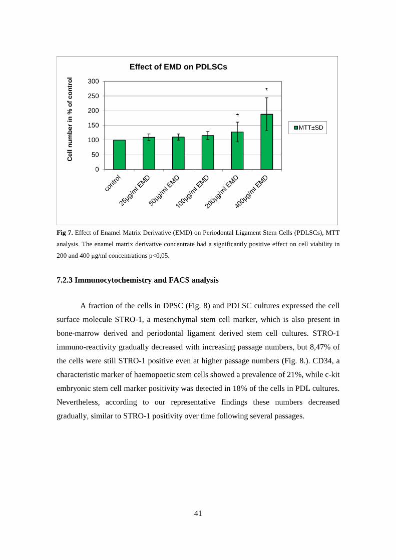
41
Fig 7. Effect of Enamel Matrix Derivative (EMD) on Periodontal Ligament Stem Cells (PDLSCs), MTT
analysis. The enamel matrix derivative concentrate had a significantly positive effect on cell viability in
200 and 400 µg/ml concentrations p<0,05.
7.2.3 Immunocytochemistry and FACS analysis
A fraction of the cells in DPSC (Fig. 8) and PDLSC cultures expressed the cell
surface molecule STRO-1, a mesenchymal stem cell marker, which is also present in
bone-marrow derived and periodontal ligament derived stem cell cultures. STRO-1
immuno-reactivity gradually decreased with increasing passage numbers, but 8,47% of
the cells were still STRO-1 positive even at higher passage numbers (Fig. 8.). CD34, a
characteristic marker of haemopoetic stem cells showed a prevalence of 21%, while c-kit
embryonic stem cell marker positivity was detected in 18% of the cells in PDL cultures.
Nevertheless, according to our representative findings these numbers decreased
gradually, similar to STRO-1 positivity over time following several passages.
0
50
100
150
200
250
300
Cel
l nu
mb
er in
% o
f co
ntr
ol
Effect of EMD on PDLSCs
MTT±SD

42
Fig 8. 8,47 % of periodontal ligament (PDL) cultures showing STRO-1 immuno-positivity following the
second passage
7.2.4 Osteogenic differentiation
In these experiments DPSC and PDLSC (Fig. 9) cultures were grown in the
presence of osteogenic differentiation cocktail consisting of dexamethazone, L-ascorbic
acid 2-phosphate and b-glycerophosphate. Under these conditions cultures uniformly
demonstrated the capacity to form Alizarin red S positive condensed nodules with high
calcium content. The deposits were sparsely scattered throughout the adherent layer as
single mineralized zones. Control cultures formed adherent layers without any sign of
calcium deposition. Thus, this observation confirms the previous findings that DPSCs and
PDLSCs are capable of differentiation to mineralized tissue in vitro in response to
appropriate pharmacological stimulation.

43
Fig. 9 (A) Dental Pulp Stem Cells (DPSCs) produced mineralized deposits under osteogenic conditions as
detected by 2% Alizarin Red S staining. (B) Control staining of non-induced DPSCs. The length of scale
bars indicate 100 µm.
7.2.5 Neuronal differentiation
7.2.5.1 Partial differentiation induced by Protocols 1 and 2
When PDLSC and DPSC cells were treated using Protocol 1 with a mixture
containing substances such as TPA, IBMX and forskolin, the morphology of cells
observed by phase contrast microscopy changed rapidly (Fig. 10, data shown for DPSC).
After 4 hours of treatment initiation, the cytoplasm of the cells retracted toward the
nucleus in many cells, taking a more spherical shape and extending processes. The
percentage of cells with modified morphology decreased within 24 hours. When
differentiation factors were removed from the medium, cell morphology reverted to the
original. Following the application of Protocol 2 (bFGF, KCl, forskolin, DMSO and
BHA) neuronal morphology changes were temporary, followed by an irreversible
distraction of the culture: after 48 hours, the majority of cells were seen to have a rounded
morphology followed by the death of almost the entire culture, except for a few spindle
shaped cells expressing none of the neuronal markers investigated by us (Fig. 10, data
shown for DPSC). These morphological changes were very similar for DPSC and PDLSC
after both treatments.
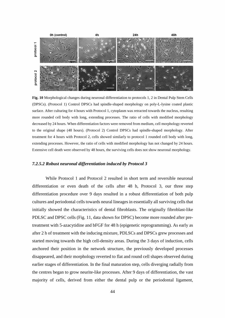
44
Fig. 10 Morphological changes during neuronal differentiation to protocols 1, 2 in Dental Pulp Stem Cells
(DPSCs). (Protocol 1) Control DPSCs had spindle-shaped morphology on poly-L-lysine coated plastic
surface. After culturing for 4 hours with Protocol 1, cytoplasm was retracted towards the nucleus, resulting
more rounded cell body with long, extending processes. The ratio of cells with modified morphology
decreased by 24 hours. When differentiation factors were removed from medium, cell morphology reverted
to the original shape (48 hours). (Protocol 2) Control DPSCs had spindle-shaped morphology. After
treatment for 4 hours with Protocol 2, cells showed similarly to protocol 1 rounded cell body with long,
extending processes. However, the ratio of cells with modified morphology has not changed by 24 hours.
Extensive cell death were observed by 48 hours, the surviving cells does not show neuronal morphology.
7.2.5.2 Robust neuronal differentiation induced by Protocol 3
While Protocol 1 and Protocol 2 resulted in short term and reversible neuronal
differentiation or even death of the cells after 48 h, Protocol 3, our three step
differentiation procedure over 9 days resulted in a robust differentiation of both pulp
cultures and periodontal cells towards neural lineages in essentially all surviving cells that
initially showed the characteristics of dental fibroblasts. The originally fibroblast-like
PDLSC and DPSC cells (Fig. 11, data shown for DPSC) become more rounded after pre-
treatment with 5-azacytidine and bFGF for 48 h (epigenetic reprogramming). As early as
after 2 h of treatment with the inducing mixture, PDLSCs and DPSCs grew processes and
started moving towards the high cell-density areas. During the 3 days of induction, cells
anchored their position in the network structure, the previously developed processes
disappeared, and their morphology reverted to flat and round cell shapes observed during
earlier stages of differentiation. In the final maturation step, cells diverging radially from
the centres began to grow neurite-like processes. After 9 days of differentiation, the vast
majority of cells, derived from either the dental pulp or the periodontal ligament,

45
displayed complex neuronal morphology, expressing both bipolar and stellate forms.
However, a small portion of cells retained their flat shape, and were attached beneath the
processes of the neuronal cells. These elements are presumably committed towards glial
fates, or serve as stanchions for the developing neuronal cells. Therefore, they might be
indispensable for neuronal survival.
Fig. 11 Morphological changes during neuronal differentiation to protocol 3 in Dental Pulp Stem Cells
(DPSCs). (Protocol 3) Control DPSCs had spindle-shaped morphology on poly-L-lysine coated plastic
surface. After 2 days of epigenetic reprogramming, cells formed clusters. After 4 days (2 days of induction),
cells had morphological features typical of neurons, showing complex neuronal processes. After 9 days (4
days of maturation), most of the cells displayed either multi- or bipolar forms. The length of scale bars
indicate 100 µm
The time-dependent changes of neuronal marker gene expression in DPSC and
PDLSC cultures undergoing neural development were evaluated by real time PCR (Fig.
12). Total RNA was harvested at four different time points during this period: at time 0
(non-induced control DPSCs and PDLSCs), on the first day of induction (d3), on the third
day of induction (d5), and after three days of maturation (d8). The expression of each
target gene was normalized to that of the RPLP0 housekeeping gene, and expressed as
fold change relative to the non-induced sample.
DPSCs and PDLSCs showed a very similar expression pattern during neuronal
differentiation treatment. There was a sharp decrease in the expression of the
mesenchymal marker vimentin in response to neurogenic induction. This very striking
decrease became less pronounced during maturation. The expression of the neuronal
marker NSE progressively increased under maturing conditions and remained increased
during maturation. The protein expression pattern investigated by immunocytochemistry
corresponded well to the gene expression data.

46
Fig. 12 mRNA expression pattern of mesenchymal, neuronal markers in Dental Pulp Stem Cells (DPSCs)
and Periodontal Ligament Stem Cells (PDLSCs) during neuronal differentiation with protocol 3. Total RNA
of DPSCs isolated from five independent normal human donors was used. RNA was harvested at four time
points during the differentiation of each sample: from non-induced DPSCs (0 day), after the first day of
induction (3rd day), on the third day of induction (5th day) and on the third day of maturation (8th day).
Target gene expressions were normalized to RPLP0 housekeeping gene expression levels and evaluated as
fold change relative to the non-induced sample. mRNA expression of the mesenchymal vimentin appeared
to be significantly suppressed by the treatment, while the mRNA expression of the neuron specific
glycolytic enolase NSE was significantly elevated by the by the treatment. DPSCs and PDLSCs showed
similar gene expression pattern. Data are reported as mean ± S.E.M.
Non-induced DPSCs and PDLSCs (Fig. 12) showed weak, nonspecific expression
pattern of NFM. The neuronal induction resulted in an increased proportion of cells with
an expression pattern specific to mature neurons in both DPSC and PDLSC (Fig. 12)
cultures. After 8 days cells also showed positive staining for NFM and NSE (Fig. 12),
markers typically expressed by mature neurons.
0,1
1
10
0h (c
ontro
l)
d3 (i
nduc
tion)
d5 (i
nduc
tion)
8d (m
atur
ati o
n)
DPSC VIM
PDLSC VIM
DPSC NSE
PDLSC NSE

47
7.3 Healing following rhGDF-5/β-TCP treatment
7.3.1 Patient demographics and baseline defect distribution
Twenty-one patients were screened, 20 patients fulfilling the inclusion criteria
were randomized into two groups of ten patients each (Fig. 1). One female patient was
not enrolled due to a low FSH level. Sixteen patients were male and four, female. One
female patient was randomized to the control group and three to the rhGDF-5/β-TCP
group. All four female patients were postmenopausal. Patient mean age was 48.8±10.6
years (range 33–74 years). Age did not differ considerably between treatment groups;
median age was 50.0 years for both groups. No dropouts occurred; all randomized patients
completed the study. The depth of the intrabony component measured 6.7±2.8 mm in the
test and 6.4±2.1 mm in the control group, respectively (Tables 2 and 3).
Full mouth plaque scores at Visit 2 averaged 7.3±6.7% for the rhGDF-5/β-TCP
and 8.9±4.8% for the control group; and full mouth bleeding scores 11.2±4.1% for the
rhGDF-5/β-TCP and 12.7±5.4% for the control group. Probing depths averaged 9.1±1.3
mm for the rhGDF-5/β-TCP and 9.2±2.3 mm for the control group. Clinical attachment
level averaged 12.2±1.5 mm for the rhGDF-5/β-TCP and 11.4± 3.5 mm for the control
group. The depth of the intrabony component as measured during surgery measured
6.7±2.8 mm for the rhGDF-5/β- TCP and 6.4±2.1 mm for the control group. The defects
displayed predominantly one- and two-wall configurations (Tables 2 and 3).
7.3.2 Clinical assessments
Healing following surgeries progressed without major complications. Primary
intention healing was observed in nine of ten patients in the rhGDF-5/β-TCP group, and
in all ten patients in the control group. One patient in the rhGDF-5/β-TCP group
experienced a slightly delayed epithelialization of the interdental papilla resulting in
complete wound closure within 3 weeks. At 6 months, both treatments resulted in
significant improvements in terms of PD reduction and CAL gain compared to baseline.
Treatment with rhGDF-5/β-TCP resulted in higher, but statistically not significant, PD
reduction (3.7±1.2 vs. 3.1±1.8 mm; p=0.26) and CAL gain (3.2±1.7 vs. 1.7± 2.2 mm;
p=0.14) compared to the control (Fig. 13.) (Tables 2 and 3).

48
Fig. 13 Flap surgery plus rhGDF-6/ß-TCP: Presurgery (top left); intrasurgery defect morphology (top
right); site following implantation of rhGDF-6/ß-TCP (bottom left); and clinical view at the biopsy event
24 weeks postsurgery (bottom right). Histological outcomes were published elsewhere (Stavropoulos et al.
2011).
Table 2 Patients, experimental teeth and surfaces, predominant defect morphology, intrasurgery defect depth,
initial pocket probing depth (PD), clinical attachment level (CAL) and gingival recession (GR), and changes in
primary outcome variables (mm) for defect sites receiving flap surgery combined with rhGDF-5/ß-TCP.
Patient Tooth Defect
Morphology Defect Depth
PD CAL GR ∆PD ∆CAL ∆GR
2 35d 1-wall 7 8 12 4 5 5 0 3 36d 2-wall 5 9 13 4 4 4 0 6 11m 2-wall 4 9 12 3 3 4 1 10 34d circumferential 5 9 11 2 4 3 -1 13 23p 1-wall 11 11 15 4 6 6 0 14 23d 2-wall 11 11 12 1 4 3 -1 16 21m 1-wall 7 10 12 2 3 2 -1 17 21m 1-wall 4 7 10 3 2 0 -2 18 12d 1-wall 4 8 11 3 3 2 -1 19 45d circumferential 9 9 14 5 3 3 0
Mean±SD 6.7 ± 2.8
9.1 ± 1.3
12.2 ± 1.5
3.1 ± 1.2
3.7 ± 1.2
3.2 ±1.7
0.5 ± 0.8

49
Table 3 Patients, experimental teeth and surfaces, predominant defect morphology, intrasurgery defect depth,
initial pocket probing depth (PD), clinical attachment level (CAL) and gingival recession (GR), and changes in
primary outcome variables (mm) for defect sites receiving flap surgery alone (control).
Patient Tooth Defect
Morphology Defect Depth
PD CAL GR ∆PD ∆CAL ∆GR
1 21m circumferential 4 9 10 1 2 1 -1 4 12m 2-wall 7 9 11 2 4 4 0 7 11m 1-wall 7 12 13 1 6 4 -2 8 23m 1-wall 5 8 9 1 3 1 -2 9 22m 1-wall 5 8 10 2 3 0 -3 11 35d 2-wall 5 7 8 1 1 -1 -2 12 44d 2-wall 9 13 17 4 6 5 -1 15 34d 1-wall 9 8 15 7 3 3 0 20 35d circumferential 9 12 15 3 2 1 -1 21 11m 1-wall 4 6 6 0 1 -1 -2
Mean±SD 6.4 ± 2.1
9.2 ± 2.3
11.4 ± 3.5
2.2 ± 2.0
3.1 ± 1.8
1.7 ± 2.2
1.4 ± 1.0
7.3.3 Safety findings
The safety profile was comparable for patients receiving rhGDF-5/β-TCP and
control treatments. All abnormal haematology and clinical chemistry values were judged
as clinically non-relevant. Most common abnormal values were GGT, followed by
cholesterol, glucose, and white blood cell count. Summary statistics did not show
remarkable trends from Visits 1 to 9 for any of the parameters analysed. All patients
showed negative anti-rhGDF-5 antibody levels at screening and Visits 3 and 9. No
relevant rhGDF-5 plasma levels were detected in any patient. Two patients, both
belonging to the rhGDF-5/β-TCP group showed rhGDF-5 plasma levels at screening
(Visit 1) and postsurgery (Visits 3 and 9). One patient showed a plasma level of 46.7
pg/mL rhGDF-5 at baseline (before implantation of rhGDF-5/β-TCP) vs. 47.3 pg/mL at
Visit 3, and 57.9 pg/mL at Visit 9.The other patient showed a plasma level of 514 pg/mL
rhGDF-5 at baseline vs. 427 pg/mL at Visit 3, and 690 pg/mL at Visit 9. Due to the fact
that the measured rhGDF-5 plasma levels were already positive at the baseline (before
periodontal surgery) and did not increase throughout the study, a causal relationship to
study medication can be excluded. For all other samples, rhGDF-5 plasma levels were
below the limit of quantification (<40.0 pg/mL). It may, thus, be anticipated that these
results were false positive.

50
7.3.4 Adverse events
All adverse events were judged as mild or moderate. Seventy-two events were
reported; 54 in seven patients from the rhGDF-5/β-TCP group (42 of which for one single
patient), and 18 in eight patients from the control. As these were always single isolated
events, one adverse event was recorded for each episode. Most events were related to
back pain (14 events) and headache (26 events). The following two events, in two
different patients, were judged to be possibly related to the regenerative surgery but not
necessarily related to the rhGDF-5: delayed epithelialization and secondary healing of the
interdental papilla. Both were judged as mild and the patients recovered without sequel.
No withdrawals due to an adverse event were recorded. There were also no meaningful
changes in vital signs that would raise a safety concern. Healing following surgeries
progressed without major complications. Primary intention healing was observed in nine
of ten patients in the rhGDF-5/β-TCP group, and in all ten patients in the control group.
One patient in the rhGDF-5/β-TCP group experienced a slightly delayed epithelialization
of the interdental papilla resulting in complete wound closure within 3 weeks.
7.3.5 Protocol deviations
A few patient visits occurred out of schedule. Blood collection Visit 9 was
completed 1–5 days early in five patients. Two patients showed increased liver enzyme
values at screening (ALT=123 U/L, AST=107 U/L, GGT=142 U/L and ALT=101 U/L,
GGT=382U/L) that decreased to normal range within 2weeks (Visit 3) except GGT. No
chronic liver disorder was reported for either patient. One patient had insulin independent
diabetes (Hb1Ac=10.3% at screening), which was medically controlled. None of these
protocol deviations was considered as major violation, and therefore, all patients were
included in the analyses.
51
7.4 Healing following treatment with Mucograft®
7.4.1 Patient demographics and baseline defect distribution
7.4.1.1 Pilot study
Five patients were treated with in the maxilla, three patients in the mandible. Five
patients presented recessions involving only anterior teeth (four in the maxilla and one in
the mandible). One patient presented a recession site on a bicuspid in the maxilla, two
patients in the mandible. Recession sites of two mandibular molars were treated in one of
the cases. The baseline values and the 12 months results are shown in Table 4.
7.4.1.2 Split mouth randomised controlled study
A total of 156 recessions were treated in 22 patients (i.e. 78 received SCTG and
78 CM). Location and distribution of the treated defects is depicted in Table 2. Thirteen
patients had maxillary and nine mandibular recessions. Upper molars were treated in eight
patients while lower molars were included in two patients. None of the treated molars
displayed a furcation involvement. No statistically significant differences (p>0.05) were
observed within and between groups for FMPS values between baseline and 12 months
measurements.
7.4.2 Clinical assessments
7.4.2.1 Pilot study
The postoperative healing was uneventful in all 8 cases. No complications such as
allergic reactions, matrix exfoliations, abscesses or infections were observed throughout
the entire study period. All patients completed the study and no patient was lost during
follow-up. All patients expressed improvement in root sensitivity. At 12 months CRC
was obtained in 2 out of the 8 patients and in 30 out of the 42 recessions (71%). MRC
was 84%. Mean GRD, GRW, GT and KTW improved statistically highly significantly
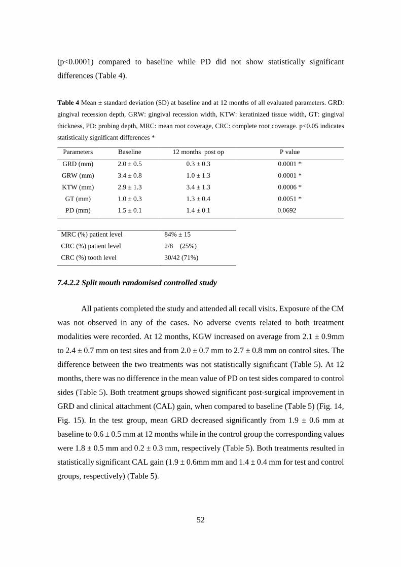
52
(p<0.0001) compared to baseline while PD did not show statistically significant
differences (Table 4).
Table 4 Mean ± standard deviation (SD) at baseline and at 12 months of all evaluated parameters. GRD:
gingival recession depth, GRW: gingival recession width, KTW: keratinized tissue width, GT: gingival
thickness, PD: probing depth, MRC: mean root coverage, CRC: complete root coverage. p<0.05 indicates
statistically significant differences *
7.4.2.2 Split mouth randomised controlled study
All patients completed the study and attended all recall visits. Exposure of the CM
was not observed in any of the cases. No adverse events related to both treatment
modalities were recorded. At 12 months, KGW increased on average from 2.1 ± 0.9mm
to 2.4 ± 0.7 mm on test sites and from 2.0 ± 0.7 mm to 2.7 ± 0.8 mm on control sites. The
difference between the two treatments was not statistically significant (Table 5). At 12
months, there was no difference in the mean value of PD on test sides compared to control
sides (Table 5). Both treatment groups showed significant post-surgical improvement in
GRD and clinical attachment (CAL) gain, when compared to baseline (Table 5) (Fig. 14,
Fig. 15). In the test group, mean GRD decreased significantly from 1.9 ± 0.6 mm at
baseline to 0.6 ± 0.5 mm at 12 months while in the control group the corresponding values
were 1.8 ± 0.5 mm and 0.2 ± 0.3 mm, respectively (Table 5). Both treatments resulted in
statistically significant CAL gain (1.9 ± 0.6mm mm and 1.4 ± 0.4 mm for test and control
groups, respectively) (Table 5).
Parameters Baseline 12 months post op P value
GRD (mm) 2.0 ± 0.5 0.3 ± 0.3 0.0001 *
GRW (mm) 3.4 ± 0.8 1.0 ± 1.3 0.0001 *
KTW (mm) 2.9 ± 1.3 3.4 ± 1.3 0.0006 *
GT (mm) 1.0 ± 0.3 1.3 ± 0.4 0.0051 *
PD (mm) 1.5 ± 0.1 1.4 ± 0.1 0.0692ns
MRC (%) patient level 84% ± 15
CRC (%) patient level 2/8 (25%)
CRC (%) tooth level 30/42 (71%)

53
Table 5 Mean ± standard deviation (SD) at baseline and at 12 months of all evaluated parameters. GRD:
gingival recession depth, GRW: gingival recession width, KTW: keratinised tissue width, GT: gingival
thickness, PD: probing depth, CAL: clinical attachment level. p<0.05 indicates statistically significant
differences *
Baseline (mm)
Mean ± SD
12 months (mm)
Mean ± SD
p-value
Baseline vs. 12 months
GRD Test 1.9 ± 0.6 0.6 ± 0.5 0.0001 *
GRD Control 1.8 ± 0.5 0.2 ± 0.3 0.0001 *
p-value 0.7565 0.0009 * -
GRW Test 3.8 ± 0.8 1.4 ± 1.2 0.0001 *
GRW Control 3.8 ± 0.9 0.5 ± 1.0 0.0001 *
p-value 0.9195 0.0002 * -
KTW Test 2.1 ± 0.9 2.4 ± 0.7 0.0309 *
KTW Control 2.0 ± 0.7 2.7 ± 0.8 0.0001 *
p-value 0.5802 0.0796 -
GT Test 0.8 ± 0.2 1.0 ± 0.3 0.0019 *
GT Control 0.8 ± 0.3 1.3 ± 0.4 0.0001 *
p-value 0.6591 0.0001 * -
PD distal Test 1.8 ± 0.3 1.8 ± 0.2 0.7334 *
PD distal Control 1.8 ± 0.4 1.8 ± 0.2 0.5203 *
p-value 0.2732 0.7344 -
PD midline Test 1.4 ± 0.3 1.4 ± 0.2 0.3744 *
PD midline Control 1.3 ± 0.2 1.3 ± 0.3 0.9453 *
p-value 0.1388 0.3652 -
PD mesial Test 1.9 ± 0.3 1.8 ± 0.2 0.0572 *
PD mesial Control 1.8 ± 0.3 1.8 ± 0.2 0.4223 *
p-value 0.1298 0.5151 -
CAL Test 3.2 ± 0.6 1.9 ± 0.6 0.0001 *
CAL Control 3.1 ± 0.5 1.4 ± 0.4 0.0001 *
p-value 0.3198 0.005 * -

54
When results were expressed as percentage of root coverage at 1 year, both
treatment modalities resulted in a statistically significant percentage of root coverage
amounting to 71% ± 21 % in the test group and 90% ±18 % in the control group,
respectively. The difference between the two groups was statistically significantly greater
for the control treatment (p= 0.0004) (Table 6). At 12 months, CRC was recorded in 5
patients in the test and in 13 patients in the control group, respectively and was
statistically significantly greater for the control treatment (p= 0.0305) (Table 6). In both
groups GRW decreased statistically significantly between baseline and 6 months and 12
months. The differences between the two groups were not statistically significant
(p>0.05) (Fig. 14, Fig. 15) (Table 6). Mean surgery time was significantly lower (p
<0.0001) in the test (i.e. 42.5 ± 4.8 min) compared with control (i.e. 58.6 ± 6.6 min)
(Table 7). Postoperative complaints on the VAS scale were lower for CM. All patients
reported a decrease in root sensitivity. The number of 100% satisfaction was higher in the
test group compared with the control one, but was not statistically significant (p>0.05).
Table 6 Complete- and percentage (%) of root coverage at 12 months. CRC: complete root coverage.
p<0.05 indicates statistically significant differences *
12 months (mm)
CRC Test 5/22
CRC Control 13/22
p-value 0.0305 *
% root coverage Test 71 ± 21 %
% root coverage Control 90 ± 18 %
p-value 0.0004 *
Table 7. Duration of surgery, patients’ complaints and satisfaction at 12 months. VAS: visual analogue
scale p<0.05 indicates statistically significant differences *
Duration of surgery (min)
Patient complaints (VAS)
Patient satisfaction (VAS)
Test 42.5 ± 4.8 7.3 ± 3.4 92.9 ± 8.4 Control 58.6 ± 6.6 12.8 ± 7.5 90.6 ± 7.9 p-value 0.0001 * 0.0020 * 0.203

55
Fig. 14 Clinical results – Mucograft ® (a) Test side prior to treatment (b) intraoperative view (c) immediate
postoperative view (d) twelve months outcome
Fig. 15 Clinical results – SCTG: subepithelial connective tissue graft (a) Control side prior treatment (b)
intraoperative view (c) immediate postoperative view (d) twelve months outcome
c d
b a
a b
c d

56
8. DISCUSSION
The results of our literature review, in vitro and clinical studies allow us to discuss
our findings with respect to the goals formulated in Chapter 2. The conclusions will be
compared with results available in literature. Results from basic research have pointed to
the important role of EMD in periodontal wound healing. Histological results from animal
and human studies have shown that treatment with EMD promotes periodontal
regeneration. Moreover, clinical studies have indicated that treatment with EMD
positively influences periodontal wound healing in humans (World Workshop in
periodontology 1996, Rincon et al. 2003, Donos et al. 2003).
Nevertheless, certain limitations experienced in cases with advanced hard- and
soft tissue defects have raised the demand for safe reconstructive procedures, surpassing
current techniques in terms of efficacy and predictability. Novel approaches should be
ideally characterised by reduced duration of treatment and limited patient morbidity.
Improved efficacy of periodontal regenerative therapy might be achieved by increased
activation of cellular elements contributing to the re-establishment of hard- and soft
tissues. Previous studies have suggested that human bone marrow and dental pulp as well
as periodontal ligament tissue contain postnatal stem cells that are capable of
differentiating into various cell types including osteoblasts, odontoblasts, cementoblasts,
adipocytes (Gronthos et al. 2000, Miura et al. 2003, Seo et al. 2004). In accordance to our
aims, we established, maintained and characterized cell cultures of human PDL. Isolated
cells showed fibroblast morphology in monolayer cultures, we have obtained similar
results to BMSC with regard to cell proliferation patterns (Bianco et Gahron 2000). The
presence of clonogenic cells in the primary cultures was confirmed, the number of colony
forming units was comparable to those data available from literature. These results
confirm the in vivo evidence for PDL cells initiating periodontal regeneration. Using
immunocytochemistry and FACS analysis, we identified PDLSCs, which are similar to
other mesenchymal stem cells in that they are highly clonogenic and show expression of
the STRO-1 mesenchymal progenitor cell marker. This expression could be detected in
8,47% of the whole cell population. We also confirmed the presence of CD34 and c-kit
stem cell marker positivity within the primary cultures. Our results are well in line with
those obtained by investigating DPSC cultures (Laino et al 2005). These findings might

57
allow for establishing more homogenous cell cultures, which contain PDLSCs in higher
numbers thus allowing for establishing future in vitro or in vivo models of periodontal
regeneration.
Our in vitro findings related to the use of EMD indicate that the observed
beneficial clinical effects reported in literature might be related with promotion of cell
proliferation and cell viability (Chong et al. 2006, Rodrigues et al. 2007). The well-known
triggering effect of EMD in periodontal regeneration is supported by the phenomenon
that PDL cells showed increased activity and positive chemotaxis related to EMD droplets
in the cultures. Cell viability was assessed via MTT assay, we investigated the effect of
culturing media containing 5, 10, or 15% FBS. We confirmed the presence of vital cells
in a significantly higher number following FBS treatment compared to serum free
conditions. MTT assay was used to confirm the significantly positive effect of EMD on
vital cells within primary PDL cultures in the concentrations of 200- and 400 µg/ml
compared to serum free cultures. This might indicate the importance of a certain minimal
dosage necessary to achieve regenerative treatment outcomes during clinical application
in specific tissue conditions.
The mechanisms controlling the development of teeth are largely unknown
(Parner et al. 2002), in particular with respect to how craniofacial components including
bone and soft tissues surrounding teeth, participate in the process of tooth development.
DPSCs clearly have the ability to regenerate dentin, at least in experimental animals
(Batouli et al. 2003, Iohara et al. 2004) and therefore have a high potential for tooth
regeneration as odontoblast progenitors. However, based on current information, DPSCs
and PDLSCs may have a broader capacity for differentiation than originally thought
(Gronthos et al. 2002). Growing evidence suggests that they are able to differentiate into
several different cell types (Gronthos et al. 2000, Gronthos et al. 2002, Miura et al. 2003,
Seo et al. 2004). Our data emphasize that optimizing in vitro conditions may soon lead to
successful serum-free culture of DPSCs and PDLSCs, which are potentially applicable
for human transplantation. The osteogenic differentiation cocktail that we used induced
similar and well reproducible mineralization in both DPSC and PDLSC cultures. Several
recent studies reported that DPSCs and PDLSCs were also capable of osteogenic
differentiation (Gronthos et al. 2002, d'Aquino et al. 2007). In our neurodifferentiation
experiments, when we applied Protocol 1 both DPSC and PDLSC cells exhibited a
58
transient neurodifferentiation. Very similar observations were made recently. However,
the expression change was transient, and the cells reverted to the original fibroblastic
BMSCs within 48 hours (Scintu et al. 2006). When we used Protocol 2, based on another
recently described method (Choi et al. 2006), again we observed only transient
neurodifferentiation followed by the death of most of the cells. Our newly developed
neurodifferentiation method, Protocol 3, is based on the elements of previously used
methods in a special temporo-spatial arrangement. The first element aims at
dedifferentiating the cells by the application of 5-azacytidine, a cytidine analogue where
nitrogen replaces a carbon at the 5th position of the pyrimidine ring. 5-azacytidine is
reported to promote the maturation of neurons generated from neural or bone marrow
derived stem cells (Schinstine and Iaconitti 1997, Kohyama et al. 2001). The second step
of our new protocol was a robust, combined activation of the PKC and PKA pathways in
order to activate pathways redirecting the cells to a neuronal fate. Human bone marrow
stromal stem cells (BMSCs) were also shown to differentiate into neural progenitors in
response to increased intracellular cAMP levels (Deng et al. 2001). The final step after
the dedifferentiation and induction steps was the use of a mixture of conventional
neuronal differentiation factors to promote neurodifferentiation. The importance of
neurotrophin-3 (NT-3) and nerve growth factor (NGF) in the induction of advanced
neuronal development has already been described (Tatard et al. 2007). Our three step
differentiation procedure resulted in a robust differentiation of both DPSC and PDLSC
cultures towards neural lineages. At the end of the differentiation, most cells displayed
complex neuronal morphology showing both bipolar and multipolar forms. In both pulp
and periodontal derived cultures morphological changes were accompanied by a similar
increase in the expression of the neuronal marker NSE, and a sharp decrease in the
expression of the mesenchymal marker vimentin. Our immunocytochemical observations
corresponded well with the real time RNA expression data. These clearly suggest that
both DPSC and PDLSC cultures are capable of massive cell differentiation at least as far
as cell morphology, gene expression profile, and molecular marker expression is
concerned.
Culturing of pluripotent tooth derived adult stem cells may lead to promising in
vivo tissue engineering applications in the future in combination with proper carrier
materials. Nevertheless, these techniques are not yet available for human use. This is due
59
to certain safety concerns and lack of information on possible treatment efficacy. On the
other hand, application of recombinant growth- and differentiation factors have been
verified to be safe and efficient in animal experiments, thus allowing pilot human trials.
The presented pilot randomized, controlled study was the first to investigate the influence
of GDF-5 on periodontal wound healing/regeneration in humans. An important finding
was that no safety concerns related to rhGDF-5/β-TCP were encountered. None of the
patients exhibited antiGDF-5 antibody levels while elevated GDF-5 plasma levels were
observed in two patients receiving rhGDF-5/β-TCP. Overall laboratory evaluations
indicated that the rhGDF-5 formulation appears safe. The observation that healing was
uneventful in both groups also indicates that the rhGDF-5/β-TCP treatment was well
tolerated and did not elicit any local adverse reactions. The clinical observations suggest
that the rhGDF-5/β-TCP construct did not appear to exert any detrimental influence on
periodontal wound healing/regeneration. The clinical evaluation has indicated that both
open flap debridement (OFD) combined with rhGDF-5/β-TCP and OFD alone may result
in statistically significant probing depth reductions and clinical attachment gains
compared to baseline. Application of rhGDF-5/β-TCP however, resulted in greater,
although statistically not significant probing depth reduction and clinical attachment gain
compared to the control. These findings are in agreement with those reported in previous
preclinical studies indicating a beneficial effect of GDF-5 on periodontal wound
healing/regeneration (Kim et al. 2009, Lee et al. 2010, Kwon et al. 2010). Nevertheless,
our results failed to demonstrate significant differences in terms of clinical improvements
between test and control groups. The resorption rate of the β-TCP carrier material and the
possibly impaired blood clot stabilisation might have contributed to these observations.
The magnitude of clinical improvements appeared to be in the range of those obtained
with other regenerative materials such as a recombinant platelet derived growth factor
(rhPDGF BB) on a β-TCP carrier, an enamel matrix protein derivative alone or guided
tissue regeneration either alone or combined with grafting materials (Pontoriero et al.
1999, Sculean et al. 2001, Tonetti et al. 2002, Sculean et al. 2003, Tonetti et al. 2004,
Nevins et al. 2005, Kuru et al. 2006, Yilmaz et al. 2010). Furthermore, it is also important
to note that the results observed in the control group compare favourably with previous
studies evaluating treatment of intrabony defects using flap surgery alone. This indicates
that substantial clinical improvements may be achieved with this treatment modality if an
60
optimal level of plaque control is maintained (Pontoriero et al. 1999, Sculean et al. 2001,
Tonetti et al. 2002, Sculean et al. 2003, Tonetti et al. 2004). This might also indicate
limitations related not only to applied biomaterials but surgical techniques, especially flap
designs. On the other hand, it should be kept in mind that the present study was not only
designed to evaluate the safety of rhGDF-5/β-TCP but also the histological outcomes
(Stavropoulos et al. 2011). The histological evaluation has indicated that treatment with
OFD+ rhGDF-5/β-TCP resulted in 2- to 3-fold higher amount of new bone and new
cementum formation compared to OFD alone without differences in frequency of root
resorption and ankylosis between the two groups (Stavropoulos et al. 2011). The amount
of residual β-TCP carrier juxtaposed to the root surface in the present group of biopsies
was generally small (mean 8.4%), suggesting that this carrier would completely degrade
and/or resorb within a relatively short interval (LeGeros RZ 1993).
As far as safety is concerned, xenogenic materials present an even more
established alternative for tissue reconstruction compared to growth- and differentiation
factors. A novel xenogenic CM (Mucograft®) was recently proposed for the correction of
periodontal soft tissue anomalies (Vignoletti et al. 2011). The application of these
biomaterials has gained particular significance since novel minimally invasive surgical
techniques were introduced for the treatment of MAGR (MCAT, Azzi, Etienne 1998).
This technique allows for simultaneous root coverage of MAGR, nevertheless the extent
of donor areas for connective tissue grafting is usually limited. Predictable coverage of
MAGR represents a challenge for the clinician and the data on the literature are still
limited. Until now, the most predictable in terms of CRC and MRC were reported
following the use of either MCAF or MCAT combined with, SCTG. These techniques
appear to yield the most predictable outcomes on both short (6 months to 1 year) and
long-term (up to 5 years) basis (Hofmänner et al. 2012). Since the main drawback in this
approach is related to SCTG harvesting which increases patient morbidity, postoperative
complication rate and surgical time, it is logical that various attempts have been made to
develop new soft tissue replacement materials.
The presented prospective case series was the first to evaluate the possibility to
use the newly developed CM in the treatment of Miller Class I and II MAGR in
combination with the MCAT technique. The used CM was excellently tolerated as
confirmed by other authors (Vignoletti et al. 2011, McGuire and Scheyer 2010,

61
Cardaropoli et al. 2012). The safety and efficacy of the CM in the treatment of single
recessions in conjunction with CAF was evaluated histologically in minipigs (Vignoletti
et al. 2011). The results have shown that both techniques resulted in similar histological
and similar clinical outcomes. In a randomised, controlled, split mouth-study McGuire
and Scheyer (McGuire and Scheyer 2010) have treated single Miller Class I and -II
recessions with CAF + CM (test) or CAF + SCTG (control). At 12 months, the percentage
of root coverage averaged 88.5% in the test group and 99.3% in the control group,
respectively. Both treatments resulted in comparable gains of keratinized tissue width,
while there were no statistically significant differences between subject-reported values
for aesthetic satisfaction, and subject`s assessments of pain and discomfort. Comparable
outcomes were also very recently reported in another study (Cardaropoli et al. 2012). The
results reported in the two aforementioned studies are in line with those from the present
investigation where at 12 months following surgery, CRC was obtained in 2 out of the 8
patients (i.e. in 71% of the total number of recessions) while MRC amounted to 84%.
From a clinicians point of view the present results are even more valuable when one
considers that the present patient population comprised not only anterior teeth located in
the maxillary arch, but also mandibular teeth and molars. This is an important aspect to
be considered since data on treatment of mandibular MAGR are extremely scarce
(Hofmänner et al. 2012). Moreover, the clinical relevance of using CM in the treatment
of MAGR is further substantiated by the fact that in all 8 patients a statistically significant
increase in KTT and KTW was observed.
The subsequently performed randomised controlled study was the first to evaluate
the treatment of Miller Class I and II MAGR by means of MCAT using either CM or
SCTG. The results indicated that compared to baseline, both treatments resulted in
statistically significant root coverage but CM yielded lower CRC compared to SCTG.
The present study comprised a total of 156 recessions (i.e. 78 in the test group and 78 in
the control group, respectively). To analyse the data, a patient level analysis was chosen
since this approach may allow to appreciate the overall outcomes following the surgery,
and thus, increase the clinical relevance of the results (Aroca et al. 2010). The reason to
also include bicuspids and molars was due to the fact that these posterior sites may be of
concern for patients exhibiting root hypersensitivity or for patients with high lip lines and
compromised aesthetics. It has to be pointed out that the inclusion of molars has, most

62
likely, influenced the overall results since recession coverage at molars is still a major
challenge for the clinician. A recent systematic review has evaluated the predictability of
various surgical techniques for obtaining CRC in MAGR (Hofmänner et al. 2012). The
findings indicated that in Miller class I and II MAGR the use of CAF or MCAF with or
without SCTG may lead to predictable CRC while MACT in combination with SCTG
represented a valuable technique in Miller class III MAGR. The fact that the MCAT has
been shown to lead to predictable root coverage even in Miller class III recessions (Aroca
et al. 2010) corroborate the present results and points to the clinical relevance of this
surgical technique. Thus, these surgical principles may be also applied when not only
SCTG but also other soft tissue grafts such as CM are used for the treatment of Miller
class I and II MAGR. One advantage of the applied MCAT is that this technique avoids
the use of vertical releasing incisions, thus maximizing the chance for obtaining complete
defect coverage by enhancing blood supply and decreasing the risk of graft exposure
(Aroca et al. 2010). The postoperative level of the flap, flap tension and complete graft
coverage are also important aspects to be considered for obtaining predictable root
coverage (Pini Prato et al. 2000, 2005). The obtained results with both CM and SCTG in
MAGR may be explained as follows: a) the mucoperiosteal tunnelled flap was released
beyond the muco-gingival line, b) the SCTG or the CM were completely covered which,
in turn, may favour revascularisation of the recipient site (Guiha et al. 2001) since their
stabilization coronal to the gingival margin and the papillae is ensured c) the papillae
were also released interproximally from the underlying bone, and d) the use of suspended
sutures around the contact points allowed for an extended coronal stabilization of the flap.
In the present study, at 12 months, CRC was observed in 5 out of the 22 patients (i.e. in
22%) in the group and are in line with the findings reported by (Molnár et al. 2013). In
the control group, however, CRC was observed in 13 out of the 22 patients (i.e. in 59%).
It needs to be kept in mind that CRC may be affected by factors such as statistical analysis
(evaluation of multiple sites) and surgical technique and may explain the differences
between the present outcomes and those reported by (Zucchelli et al. 2009). In that study,
treatment of MAGR with CAF resulted in 77.7% CRC when releasing incisions were
used, while it amounted to 89.3 % in the group treated without vertical releasing incisions
(Zucchelli et al. 2009). In the present study, the MRC amounted to 71% ± 21% in the test
and 90% ± 18% in the control group, respectively, and is in line with the outcomes of

63
recent reports (Hofmänner 2012, Molnár et al. 2013). These differences might be related
to more favourable clinical handling of the applied SCTG when used in combination with
the MCAT technique. The fact that both treatments yielded comparable improvements in
terms of KT is in agreement with previous studies using CAF for the treatment of MAGR
and may be attributed to the natural tendency of the mucogingival junction to regain its
original, genetically determined position (Zucchelli & De Sanctis 2005, Zucchelli et al.
2009). At 12 months, GT was statistically significantly increased in the control group
compared to the test group. Furthermore, it is interesting to note that CRC was observed
in 9 patients with 0.5 mm GT (test or control sites) and in 5 patients with 1 mm GT (test
and control sites). On the other hand, these findings appear to contradict the outcomes
from other studies reporting a MRC of 64.3% for seven recessions with a flap thickness
of ≤ 0.5mm and a CRC only at sites with a flap thickness > 0.8 mm following the use of
CAF (Baldi et al. 1999). The importance of GT for root coverage with CAF was
emphasized in systematic reviews on single recessions (Hwang & Wang 2006, Cheng et
al. 2007) but limited information is available for MAGR (Paolantonio et al. 2002, Aroca
et al. 2009, 2010) and even less information comparing the influence of soft tissue
thickness of MACF and MCAT in multiple recessions. In a long-term study over five
years (Zuchelli & De Sanctis 2005) reported that the soft tissue margin stability was
correlated to the patient’s individual tendency to develop gingival recession, although
they did not quantify soft tissue thickness. It thus appears that the issue on the importance
of flap thickness and the possibility of obtaining CRC in MAGR is still not completely
elucidated and warrants further studies.
Taken together, our findings stress the clinical significance of possible tissue
engineering applications and application of novel biomaterials such as human
recombinant growth factors and xenogenic materials. A combination of these might be
considered since PDLSCs were shown to initiate periodontal regeneration in vivo on a
proper carrier (Seo et al. 2004). Therefore, biomaterials as rhGDF-5/β-TCP or
Mucograft® might be applied as possible carriers for PDLSCs in future tissue engineering
applications to increase treatment efficacy. On the other hand, current treatment
modalities for periodontal hard- and soft tissue reconstruction seem to be limited among
other factors by the applied surgical techniques. Thus, these novel treatment approaches
should be used in combination with innovative, minimally invasive surgical techniques.

64
9. CONCLUSIONS
In this section the major conclusions from in vitro and clinical investigations will
be drawn. These conclusions refer to the respective impact of publications indicated in
table 1. The following statements are related to a literature review (I), in vitro research
providing the methodological basis for future models of periodontal regeneration (II) as
well as possible in vivo applications (III). Also conclusions of clinical studies on the
human application of rhGDF-5 (IV) as well as Mucograft® (V, VI) will be summarised.
Study I
Surgical periodontal treatment of deep intrabony defects with EMD promotes
periodontal regeneration. The application of EMD in the context of non-surgical
periodontal therapy has failed to result in periodontal regeneration. Surgical periodontal
therapy of deep intrabony defects with EMD may lead to significantly higher
improvements of the clinical parameters than open flap debridement alone. The results
obtained following treatment with EMD are comparable to those following treatment with
GTR and can be maintained over a longer period. Nevertheless, in cases with advanced
attachment loss a different treatment approach may be needed to overcome limitations of
current regenerative techniques.
Study II
Isolation, culturing and characterization of stem cell cultures periodontal ligament
origin was successful. Our in vitro model allows for further in vitro and in vivo
investigations of periodontal regenerative procedures. Our present findings establish the
methodological basis for future research on proliferation and differentiation of PDLSCs.
The identified adult stem cells of periodontal origin might be used in future tissue
engineering applications aiming at periodontal regeneration.

65
Study III
Our data demonstrated that both DPSC and PDLSC cultures may differentiate into
either osteogenic or to a neuronal fate in response to appropriate pharmacological
treatments. Further investigations are still necessary to optimise these procedures,
enabling the utilisation of the differentiation potential of dental human stem cell cultures.
Nevertheless, it is already clear that both the human dental pulp and the periodontal
ligament is a potential source for tissue engineering not only in aspects related to dental
bone regeneration, but also for neuroregenerative applications.
Study IV
The findings of this study indicate that in the tested application the use of rhGDF-
5/β-TCP appeared to be safe and the material possesses a sound biological rationale.
Thus, further, adequately powered, randomised controlled clinical trials are warranted to
confirm the clinical relevance of this new approach in regenerative periodontal therapy.
Study V
Within their limits, our results indicate that treatment of Miller Class I and II
multiple adjacent gingival recessions by means of MCAT and Mucograft® may result in
statistically and clinically significant root coverage. Further studies are thus warranted to
evaluate the performance of Mucograft® compared to connective tissue grafting and
other treatment alternatives.
Study VI
The presented findings indicate that the use of Mucograft® may represent an
alternative to connective tissue grafting by reducing surgical time and patient morbidity
but yielded lower root coverage than connective tissue grafting in the treatment of Miller
Class I and II multiple adjacent gingival recessions when used in conjunction with the
MCAT technique.

66
10. SUMMARY
Several treatment approaches have been suggested in the past for periodontal
hard- and soft tissue reconstruction. Based on our literature review (Study I) surgical
periodontal therapy of intrabony defects with EMD may lead to significantly higher
improvements than open flap debridement alone. These results are comparable to GTR
and can be maintained over a longer period.
The limitations of current techniques for periodontal hard- and soft tissue
reconstruction have raised a demand to introduce novel treatment approaches and
biomaterials aiming at increased treatment efficacy as well as reducing duration of
treatment and patient morbidity. Recently, the possibility of tissue engineering related to
PDLSCs and recombinant growth-factors for periodontal hard tissue reconstruction as
well as xenogenic materials for reconstruction of soft tissue anomalies were suggested.
In vitro (Study II) EMD was shown to promote proliferation of PDL cultures in
vitro, which may be a useful model for further in vitro and in vivo investigations of tissue
engineering applications. We showed both DPSCs’ and PDLSCs’ regenerative potential
confirming in vitro mineralisation induction and neuronal differentiation (Study III).
In an RCT (Study IV) we were the first to clinically and histologically investigate
that rhGDF-5 can be safely used for periodontal regenerative therapy in humans. It was
shown that the tested concentration of rhGDF-5 /β-TCP might be a promising new
approach in regenerative periodontal regeneration.
In a pilot case series (Study V) it was reported that treatment of Miller Class I -
and II MAGR by means of MCAT and Mucograft® may result in statistically significant
root coverage.
In a subsequent split-mouth RCT (Study VI) it was verified that Mucograft® may
be a useful alternative to SCTG by reducing surgical time and patient morbidity for root
coverage procedures. Nevertheless, the use of Mucograft® yielded lower CRC than SCTG
in the treatment of Miller Class I and II MAGR when used in conjunction with MCAT.
Future clinical applications with increased efficacy in periodontal regenerative
therapy might be introduced by combining tissue engineering therapeutical approaches
and growth factors or xenogenic biomaterials utilising improved, minimally invasive
surgical techniques.
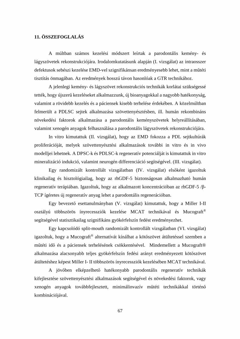
67
11. ÖSSZEFOGLALÁS
A múltban számos kezelési módszert leírtak a parodontális kemény- és
lágyszövetek rekonstrukciójára. Irodalomkutatásunk alapján (I. vizsgálat) az intraosszer
defektusok sebészi kezelése EMD-vel szignifikánsan eredményesebb lehet, mint a műtéti
tisztítás önmagában. Az eredmények hosszú távon hasonlóak a GTR technikához.
A jelenlegi kemény- és lágyszövet rekonstrukciós technikák korlátai szükségessé
tették, hogy újszerű kezeléseket alkalmazzunk, új bioanyagokkal a nagyobb hatékonyság,
valamint a rövidebb kezelés és a páciensek kisebb terhelése érdekében. A közelmúltban
felmerült a PDLSC sejtek alkalmazása szövettenyésztésben, ill. humán rekombináns
növekedési faktorok alkalmazása a parodontális keményszövetek helyreállításában,
valamint xenogén anyagok felhasználása a parodontális lágyszövetek rekonstrukciójára.
In vitro kimutattuk (II. vizsgálat), hogy az EMD fokozza a PDL sejtkultúrák
proliferációját, melyek szövettenyésztési alkalmazások további in vitro és in vivo
modelljei lehetnek. A DPSC-k és PDLSC-k regeneratív potenciálját is kimutattuk in vitro
mineralizáció indukció, valamint neurogén differenciáció segítségével. (III. vizsgálat).
Egy randomizált kontrollált vizsgálatban (IV. vizsgálat) elsőként igazoltuk
klinikailag és hisztológiailag, hogy az rhGDF-5 biztonságosan alkalmazható humán
regeneratív terápiában. Igazoltuk, hogy az alkalmazott koncentrációban az rhGDF-5 /β-
TCP ígéretes új regeneratív anyag lehet a parodontális regenerációban.
Egy bevezető esettanulmányban (V. vizsgálat) kimutattuk, hogy a Miller I-II
osztályú többszörös ínyrecessziók kezelése MCAT technikával és Mucograft®
segítségével statisztikailag szignifikáns gyökérfelszín fedést eredményezhet.
Egy kapcsolódó split-mouth randomizált kontrollált vizsgálatban (VI. vizsgálat)
igazoltuk, hogy a Mucograft® alternatívát kínálhat a kötőszövet átültetéssel szemben a
műtéti idő és a páciensek terhelésének csökkentésével. Mindemellett a Mucograft®
alkalmazása alacsonyabb teljes gyökérfelszín fedési arányt eredményezett kötőszövet
átültetéshez képest Miller I- II többszörös ínyrecessziók kezelésében MCAT technikával.
A jövőben elképzelhető hatékonyabb parodontális regeneratív technikák
kifejlesztése szövettenyésztési alkalmazások segítségével és növekedési faktorok, vagy
xenogén anyagok továbbfejlesztett, minimálinvazív műtéti technikákkal történő
kombinációjával.
68
12. BIBLIOGRAPHY
About I, Bottero MJ, de Denato P, Camps J, Franquin JC, Mitsiadis TA. (2000) Human
dentin production in vitro. Exp Cell Res, 258:33-41.
Agudio G, Nieri M, Rotundo R, Franceschi D, Cortellini P, Pini Prato GP. (2009)
Periodontal conditions of sites treated with gingival-augmentation surgery compared to
untreated contralateral homologous sites: A 10- to 27-year long-term study. J Periodontol,
80: 1399–1405.
Ainamo J, Bay I. (1975) Problems and proposals for recording gingivitis and plaque. Int
Dent J, 25: 229–235.
Armitage GC. (1999) Development of a classification system for periodontal diseases and
conditions. Ann Periodontol, 4: 1-6.
Aroca, S., Keglevich, T., Barbieri, B., Gera, I. & Etienne, D. (2009) Clinical evaluation
of a modified coronally advanced flap alone or in combination with a platelet rich fibrin
membrane for the treatment of adjacent multiple gingival recessions. A 6-month study. J
Periodontol, 80: 244-252.
Aroca, S, Keglevitch, T., Nikolidakis, D, Gera, I, Nagy, K, Azzi, R, Etienne D. (2010)
Treatment of class III multiple gingival recessions: a randomized-clinical trial. J Clin
Periodontol, 1: 88-97.
Arthur A, Rychkov G, Shi S, Koblar SA, Gronthos S. (2008) Adult human dental pulp
stem cells differentiate toward functionally active neurons under appropriate
environmental cues. Stem Cells, 26: 1787-1795.
Azzi, R. & Etienne, D. (1998) Recouvrement radiculaire et reconstruction papillaire par
greffon conjonctif enfoui sous un lambeau vestibulaire tunnélisé et tracté coronairement.
Journal de Parodontologie et d'Implantologie Orale, 17: 71-77.
69
Baldi, C, Pini-Prato, G, Pagliaro, U, Nieri, M, Saletta D, Rotundo R, Cortellini P. (1999)
Coronally advanced flap procedure for root coverage. Is flap thickness a relevant predictor
to achieve root coverage? A 19-case series. J Periodontol, 70: 1077-1784.
Batouli S, Miura M, Brahim J. (2003) Comparison of stem-cell-mediated osteogenesis
and dentinogenesis. J Dent Res, 82: 976-81.
Bianco P, Gehron RP. Marrow stromal stem cells. (2000) J Clin Invest, 105(12): 1663–
1668.
Cairo F, Pagliaro U, Nieri M. (2008) Treatment of gingival recession with coronally
advanced flap procedures: a systematic review. J Clin Periodontol, 35: 136-162.
Cardaropoli D, Tamagnone L, Roffredo A, Gaveglio L. (2012) Treatment of gingival
recession defects using coronally advanced flap with porcine collagen matrix compared
to coronally advanced flap with connective tissue graft: a randimized controlled clinical
trial. J Periodontol, 83: 321-328.
Chambrone L, Sukekava F, Araújo MG, Pustiglioni FE, Chambrone LA, Lima LA.
(2010) Root-coverage procedures for the treatment of localized gingival-type recessions.
J Periodontol, 81: 452–478.
Cheng, Y F, Chen JW, Lin SJ, Lu HK. (2007) Is coronally positioned flap procedure
adjunct with enamel matrix derivative or root conditioning a relevant predictor for
achieving root coverage? A systemic review. J Periodontol, 42: 474-485.
Choi CB, Cho YK, Prakash KV. (2006) Analysis of neuron-like differentiation of human
bone marrow mesenchymal stem cells. Biochem Biophys Res Commun, 350: 138-146.

70
Chong CH, Carnes DL, Moritz AJ , Oates T, Ryu OH, Simmer J, Cochran DL. (2006)
Human periodontal fibroblast response to enamel matrix derivative, amelogenin, and
platelet-derived growth factor-BB. J Periodontol, 77(7): 1242–1252.
Clarke DL. (2003) Neural stem cells. Bone Marrow Transplant, 32 1: 13-17.
Couble ML, Farges JC, Bleicher F, Perrat-Mabillon B, Boudeulle M, Magloire H. (2000)
Odontoblast differentiation of human dental pulp cells in explant cultures. Calcif Tissue
Int, 66: 129-138.
Daprile G, Gatto MR, Checchi L. (2007) The evolution of buccal gingival recessions in
a student population: a 5-year follow-up. J Periodontol, 78: 611-614.
d'Aquino R, Graziano A, Sampaolesi M. (2007) Human postnatal dental pulp cells co-
differentiate into osteoblasts and endotheliocytes: a pivotal synergy leading to adult bone
tissue formation. Cell Death Differ, 14: 1162-1171.
Deng W, Obrocka M, Fischer I, Prockop DJ. (2001) In vitro differentiation of human
marrow stromal cells into early progenitors of neural cells by conditions that increase
intracellular cyclic AMP. Biochem Biophys Res Commun, 282: 148-152.
Donos N, Sculean A, Glavind L, Reich E, Karring T. (2003) Wound healing of degree III
furcation involvements following guided tissue regeneration and/or Emdogain. A
histologic study. J Clin Periodontol, 30: 1061–1068.
Galois L, Mainard D, DeLagoutte JP. (2002) Beta-tricalcium phosphate ceramic as a bone
substitute in orthopaedic surgery. Int Orthop, 26: 109–115.
Gronthos S, Brahim J, Li W. (2002) Stem cell properties of human dental pulp stem cells.
J Dent Res, 81: 531-535.

71
Gronthos S, Graves SE , Ohta S, Simmons PJ. (1994) The STRO-1+ fraction of adult
human bone marrow contains the osteogenic precursors. Blood, 84(12): 4164–4173.
Gronthos S, Mankani M, Brahim J, Robey PG, Shi S. (2000) Postnatal human dental pulp
stem cells (DPSCs) in vitro and in vivo. Proc Natl Acad Sci USA, 97: 13625-30.
Grove JE, Bruscia E, Krause DS. (2004) Plasticity of bone marrow-derived stem cells.
Stem Cells, 22: 487-500.
Guiha, R, el Khodeiry, S, Mota, L, Caffesse R. (2001) Histological evaluation of healing
and revascularization of the subepithelial connective tissue graft. J Periodontol, 72: 470-
478.
Hofmänner P, Alessandri R, Laugisch O, Aroca S, Salvi GE, Stavropoulos A, Sculean,
A. (2012) Predictability of surgical techniques used for coverage of multiple adjacent
gingival recessions. A systematic review. Quint Int, 43: 545-554.
Hürzeler MB, Weng D. (1999) A single-incision technique to harvest subepithelial
connective tissue grafts from the palate. Int J Period Restor Dent, 19(3): 279-87.
Hwang D, Wang HL. (2006) Flap thickness as a predictor of root coverage: a systematic
review. J Periodontol, 77: 1625-1634.
Iohara K, Nakashima M, Ito M, Ishikawa M, Nakasima A, Akamine A. (2004) Dentin
regeneration by dental pulp stem cell therapy with recombinant human bone
morphogenetic protein 2. J Dent Res, 83: 590-595.
Javazon EH, Beggs KJ, Flake AW. (2004) Mesenchymal stem cells: paradoxes of
passaging. Exp Hematol, 32: 414-425.
72
Julious SA, Campbell MJ, Altman DG. (1999) Estimating sample sizes for continuous,
binary, and ordinal outcomes in paired comparisons: practical hints. J Biopharm Stat,
9(2): 241-51.
Kemoun P, Laurencin-Dalicieux S, Rue J. (2007) Human dental follicle cells acquire
cementoblast features under stimulation by BMP-2/-7 and enamel matrix derivatives
(EMD) in vitro. Cell Tissue Res, 329: 283-294.
Kennedy JE, Bird WC, Palcanis KG, Dorfman HS. (1985) A longitudinal evaluation of
varying widths of attached gingiva. J Clin Periodontol, 12: 667–675.
Kim TG, Wikesjö UME, Cho KS, Chai JK, Pippig SD, Siedler M, Kim CK. (2009)
Periodontal wound healing/regeneration following application of recombinant human
growth/differentiation factor-5 (rhGDF-5) in an absorbable collagen sponge carrier into
one-wall intrabony defects in dogs. A dose range study. J Clin Periodontol, 36:589–597.
Kohyama J, Abe H, Shimazaki T. (2001) Brain from bone: efficient "meta-
differentiation" of marrow stroma-derived mature osteoblasts to neurons with Noggin or
a demethylating agent. Differentiation, 68: 235-244.
Koyama N, Okubo Y, Nakao K, Bessho K. (2009) Evaluation of pluripotency in human
dental pulp cells. J Oral Maxillofac Surg, 67: 501-506.
Kuehnle I, Goodell MA. (2002) The therapeutic potential of stem cells from adults. BMJ,
325: 372-376.
Kuru B, Yilmaz S, Argin K, Noyan U. (2006) Enamel matrix derivative alone or in
combination with a bioactive glass in wide intrabony defects. Clin Oral Invest, 10: 227–
234.
73
Kwon DH, Bennett W, Herberg S, Bastone P, Pippig S, Rodriguez NA, Susin C, Wikesjö
UME. (2010) Evaluation of an injectable rhGDF-5/PLGA composite for minimally
invasive periodontal regenerative procedures: a histological study in the dog. J Clin
Periodontol, 37: 390–397.
Laino G, d’Aquino R, Graziano A, Lanza V, Carinci F, Naro F, Pirozzi G, Papaccio G.
(2005) A new population of human adult dental pulp stem cells: a useful source of living
autologous fibrous bone tissue (LAB). J Bone Miner Res, 20(8): 1394–1402.
Le Blanc K, Pittenger M. (2005) Mesenchymal stem cells: progress toward promise.
Cytotherapy, 7: 36-45.
Lee JS, Wikesjö UME, Jung UW, Choi SH, Pippig S, Siedler M, Kim CK. (2010)
Periodontal wound healing/regeneration following implantation of recombinant human
growth/differentiation factor-5 in a beta-tricalcium phosphate carrier into one-wall
intrabony defects in dogs. J Clin Periodontol, 37: 382–389.
LeGeros RZ. (1993) Biodegradation and bioresorption of calcium phosphate ceramics
Clin Mater, 14(1): 65-88.
Lovegrove J, Leichter J. (2004) Exposed root surface: a review of aetiology, management
and evidence-based outcomes of treatment. N Z Dent J, 100: 72-81.
McGuire MK, Scheyer T. (2010) Xenogeneic collagen matrix with coronally advanced
flap compared to connective tissue with coronally advanced flap for the treatment of
dehiscence-type recession defects. J Periodontol, 81: 1108-1117.
Miller PD Jr. (1985) A classification of marginal tissue recession. Int J Period Rest Dent,
5: 8-13.
Miura M, Gronthos S, Zhao M, Lu B, Fisher LW, Robey PG, Shi S. (2003) SHED: stem
cells from human exfoliated deciduous teeth. Proc Natl Acad Sci USA, 100: 5807-12.

74
Modaressi M, Wang HL. (2009) Tunneling procedure for root coverage using acellular
dermal matrix: a case series. Int J Periodontics Restorative Dent, 29(4): 395-403.
Molnár B, Aroca S, Keglevich T, Gera I, Windisch P, Sculean, A. (2013) Treatment of
Miller Class I-II multiple gingival recessions with the modified coronally advanced
tunnel technique by means of a bioresorbable collagen matrix (Mucograft®): A
prospective pilot case series. Quint Int, 44(1): 17-24.
Monnet-Corti V, Santini A, Glise JM, Fouque-Deruelle C, Dillier FL, Liébart MF,
Borghetti A. (2006) Connective tissue graft for gingival recession treatment: assessment
of the maximum graft dimensions at the palatal vault as a donor site. J Periodontol, 77(5):
899-902.
Moore Y, Dickinson DP, Wikesjö UME. (2010) Growth/differentiation factor-5 (GDF-
5): a candidate therapeutic agent for periodontal regeneration? Review of preclinical data.
J Clin Periodontol, 37: 288–298.
Morotome Y, Goseki-Sone M, Ishikawa I, Oida S. (1998) Gene expression of growth and
differentiation factors-5, -6, and -7 in developing bovine tooth at the root forming stage.
Biochem Biophys Res Commun, 244: 85–90.
Müller HP, Eger T, Schorb A. (1998) Gingival dimensions after root coverage with free
connective tissue grafts. J Clin Periodontol, 25: 424–430.
Nakamura T, Yamamoto M, Tamura M, Izumi Y. (2003) Effects of growth/differentiation
factor-5 on human periodontal ligament cells. J Periodont Res, 38: 597–605.
Nevins M, Giannobile WV, McGuire MK, Kao RT, Mellonig JT, Hinrinchs JE,
McAllister BS, Murphy KS, McClain PK, Nevins ML, Paquette DW, Han TJ, Reddy MS,
Lavin PT, Genco RJ, Lynch SE. (2005) Platelet-derived growth factor stimulates bone
fill and rate of attachment level gain: results of a large multicenter randomized controlled
trial. J Periodontol, 76: 2205–2215.

75
Nosrat IV, Smith CA, Mullally P, Olson L, Nosrat CA. (2004) Dental pulp cells provide
neurotrophic support for dopaminergic neurons and differentiate into neurons in vitro;
implications for tissue engineering and repair in the nervous system. Eur J Neurosci, 19:
2388-2398.
O’Leary TJ, Drake RB, Naylor JE. (1972) The plaque control record. J Clin Periodontol,
43: 38.
Palioto DB, Coletta RD, Graner E, Joly JC, de Lima AF. (2004) The influence of enamel
matrix derivative associated with insulin-like growth factor-I on periodontal ligament
fibroblasts. J Periodontol, 75: 498–504.
Paolantonio M, Dolci M, Esposito M., D´Archivio D, Lisanti L, Di Lucco A, Perinetti,
G. (2002) Subpedicle acellular dermal matrix graft and autogenous connective tissue graft
in the treatment of gingival recessions: a comparative 1-year clinical study. J Clin
Periodontol, 73: 1299-1307.
Parner ET, Heidmann JM, Kjaer I, Vaeth M, Poulsen S. (2002) Biological interpretation
of the correlation of emergence times of permanent teeth. J Dent Res, 81: 451-454.
Pilloni A, Paolantonio M, Camargo PM. (2006) Root coverage with a coronally
positioned flap used in combination with enamel matrix derivative: 18-month clinical
evaluation. J Periodontol, 77(12): 2031-9.
Pini Prato G, Pagliaro U, Baldi C, Nieri M, Saletta D, Cairo F, Cortellini P. (2000)
Coronally advanced flap procedure for root coverage. Flap with tension versus flap
without tension: a randomized controlled clinical study. J Periodontol, 71: 188-201.
Pini Prato GP, Baldi C, Nieri M, Franseschi D, Cortellini P, Clauser C, Rotundo R, Muzzi
L. (2005) Coronally advanced flap: the post-surgical position of the gingival margin is an
important factor for achieving complete root coverage. J Periodontol, 76: 713-722.

76
Pini-Prato GP, Cairo F, Nieri M, Franceschi D, Rotundo R, Cortellini P. (2010) Coronally
advanced flap versus connective tissue graft in the treatment of multiple gingival
recessions : a split mouth study with a 5 years follow-up. J Clin Periodontol, 37: 644-
650.
Pontoriero R, Wennström J, Lindhe J. (1999) The use of barrier membranes and enamel
matrix proteins in the treatment of angular bone defects. A prospective controlled clinical
study. J Clin Periodontol, 26: 833–840.
Pöhling S, Jochims K, Happersberger P, Hellerbrand K, Bolz W, Kohnert U.
Enhancement of bone growth by coating of osteoinductive beta-tricalcium phosphate
(beta- TCP) with recombinant human growth factor-5 (rhGDF-5). 2nd European
Conference on Bone Morphogenetic Proteins, May 2002, Zagreb.
Pöhling S, Pippig SD, Hellerbrand K, Siedler M, Schütz A, Dony C. (2006) Superior
effect of MD05, beta-tricalcium phosphate coated with recombinant human
growth/differentiation factor-5, compared to conventional bone substitutes in the rat
calvarial defect model. J Periodontol, 77: 1582–1590.
Rincon JC, Haase HR, Bartold PM. (2003) Effect of Emdogain on human periodontal
fibroblasts in an in vitro wound healing model. J Periodont Res, 38: 290–295.
Rodrigues TL , Marchesan JT , Coletta RD, Novaes AB Jr, Grisi MF, Souza SL , Taba M
Jr, Palioto DB. (2007) Effects of enamel matrix derivative and transforming growth
factor-beta1 on human periodontal ligament fibroblasts. J Clin Periodontol, 34(6): 514–
522.
Sallum EA, Casati MZ, Caffesse RG, Funis LP, Nociti Junior FH, Sallum AW. (2003)
Coronally positioned flap with or without enamel matrix protein derivative for the
treatment of gingival recessions. Am J Dent, 16: 287–291.

77
Schinstine M, Iacovitti L. (1997) 5-Azacytidine and BDNF enhance the maturation of
neurons derived from EGF-generated neural stem cells. Exp Neurol, 144: 315-325.
Scintu F, Reali C, Pillai R. (2006) Differentiation of human bone marrow stem cells into
cells with a neural phenotype: diverse effects of two specific treatments. BMC Neurosci,
7: 14.
Sculean A, Berakdar M, Chiantella GC, Donos N, Arweiler NB, Brecx M. (2003) Healing
of intrabony defects following treatment with a bovine derived xenograft and collagen
membrane. A controlled clinical study. J Clin Periodontol, 30: 73–80.
Sculean A, Windisch P, Chiantella GC, Donos N, Brecx M, Reich E. (2001) Treatment
of intrabony defects with enamel matrix proteins and guided tissue regeneration. A
prospective controlled clinical study. J Clin Periodontol, 28: 397–403.
Sena K, Morotome Y, Baba O, Terashima T, Takano Y, Ishikawa I. (2003) Gene
expression of growth differentiation factors in the developing periodontium of rat molars.
J Dent Res, 82: 166–171.
Seo BM, Miura M, Gronthos S, Bartold PM, Batouli S, Brahim J, Young M, Robey
PG, Wang CY, Shi S. (2004) Investigation of multipotent postnatal stem cells from
human periodontal ligament. Lancet, 364: 149-55.
Serino G, Wennström JL, Lindhe J, Eneroth L. (1994) The prevalence and distribution of
gingival recession in subjects with a high standard of oral hygiene. J Clin Periodontol,
21: 57-63.
Shi S, Bartold PM, Miura M, Seo BM, Robey PG, Gronthos S. (2005) The efficacy of
mesenchymal stem cells to regenerate and repair dental structures. Orthod Craniofac Res,
8: 191-9.

78
Shi S, Gronthos S. (2003) Perivascular niche of postnatal mesenchymal stem cells in
human bone marrow and dental pulp. J Bone Miner Res, 18: 696-704.
Spiro RC, Liu L, Heidaran MA, Thompson AY, Ng CK, Pohl J, Poser JW. (2000)
Inductive activity of recombinant human growth and differentiation factor-5. Biochem
Soc Trans, 28: 362–368.
Stavropoulos A, Windisch P, Gera I, Sculean A, Capsius B, Wikesjö UME. 2011() A
phase IIa randomized controlled clinical and histological pilot study evaluating rhGDF-
5/β-TCP for periodontal regeneration. J Clin Periodontol, 38(11): 1044-54.
Susin C, Haas AN, Oppermann RV, Haugejorden O, Albandar JM. (2004) Gingival
recession: epidemiology and risk indicators in a representative urban Brazilian
population. J Periodontol, 75: 1377-1386.
Tatard VM, D'Ippolito G, Diabira S. (2007) Neurotrophin-directed differentiation of
human adult marrow stromal cells to dopaminergic-like neurons. Bone, 40: 360-373.
Techawattanawisal W, Nakahama K, Komaki M, Abe M, Takagi Y, Morita I. (2007)
Isolation of multipotent stem cells from adult rat periodontal ligament by neurosphere-
forming culture system. Biochem Biophys Res Commun, 357: 917-923.
Tonetti MS, Cortellini P, Lang NP, Suvan JE, Adriaens P, Dubravec D, Fonzar A,
Fourmousis I, Rasperini G, Rossi R, Silvestri M, Topoll H, Wallkamm B, Zybutz M.
(2004) Clinical outcomes following treatment of human intrabony defects with GTR/bone
replacement material or access flap alone. A multicenter randomized controlled clinical
trial. J Clin Periodontol, 31: 770–776.
79
Tonetti MS, Lang NP, Cortellini P, Suvan JE, Adriaens P, Dubravec D, Fonzar A,
Fourmousis I, Mayfield L, Rossi R, Silvestri M, Tiedemann C, Topoll H, Vangsted T,
Wallkamm B. (2002) Enamel matrix proteins in the regenerative therapy of deep
intrabony defects. A multicentre randomized controlled clinical trial. J Clin Periodontol,
29: 317–325.
Tsukamoto Y, Fukutani S, Shin-Ike T. (1992) Mineralized nodule formation by cultures
of human dental pulp-derived fibroblasts. Arch Oral Biol, 37: 1045-1055.
Vignoletti F, Nunez J, Discepoli N, De Sanctis F, Caffesse R, Munoz F, Lopez M, Sanz
M. (2011) Clinical and histological healing of a new collagen matrix in combination with
the coronally advanced flap for the treatment of Miller class-I recession defects: an
experimental study in the minipig. J Clin Periodontol, 38: 847-855.
Wennström JL. (1996) Mucogingival therapy. Ann Periodontol, 1: 671-701.
Widera D, Grimm WD, Moebius JM. (2007) Highly efficient neural differentiation of
human somatic stem cells, isolated by minimally invasive periodontal surgery. Stem Cells
Dev, 16: 447-460.
Wikesjö UME, Sorensen RG, Kinoshita A, Li XJ, Wozney JM. (2004) Periodontal repair
in dogs: effect of recombinant human bone morphogenetic protein-12 (rhBMP-12) on
regeneration of alveolar bone and periodontal attachment. A pilot study. J Clin
Periodontol, 31: 622–670.
World Workshop in periodontology. (1996) The American Academy of Periodontology.
Ann Periodontol, 1: 618–670.
Yilmaz S, Cakar G, Yildirim B, Sculean A. (2010) Healing of two and three wall
intrabony periodontal defects following treatment with an enamel matrix derivative
combined with autoneous bone. J Clin Periodontol, 37: 544–550.
80
Zucchelli G, De Sanctis M. (2005) Long-term outcome following treatment of multiple
Miller class I and II recession defects in esthetic areas of the mouth. J Periodontol, 76:
2286-2292.
Zucchelli G, Mele M, Mazzotti C, Marzadori M, Montebugnoli L, De Sanctis M. (2009)
CAF with and without realising incisions for the treatment of multiple gingival
recessions: A comparative controlled randomized clinical trial. J Periodontol, 80: 1083-
1094.
Zuhr O, Fickl S, Wachtel H, Bolz W, Hurzeler MB. (2007) Covering of gingival
recessions with a modified microsurgical tunnel technique: case report. Int J of Period &
Rest Dent, 27: 457-463.

81
13. BIBILIOGRAPHY OF THE CANDIDATE’S PUBLICATIONS
13.1 Related publications
(I) Sculean A, Windisch P, Döri F, Keglevich T, Molnár B, Gera I. (2007) Emdogain in
regenerative periodontal therapy. A review of the literature. Fogorv Sz, 100(5): 220-32,
211-9.
(II) Molnár B, Kádár K, Király M, Porcsalmy B, Somogyi E, Hermann P, Grimm WD,
Gera I, Varga G. (2008) Isolation, cultivation and characterisation of stem cells in human
periodontal ligament. Fogorv Sz, 101(4): 155-61.
(III) Kádár K, Király M, Porcsalmy B, Molnár B, Rácz GZ, Blazsek J, Kállo K, Szabo
EL, Gera I, Gerber G, Varga G. (2009) Differentiation potential of stem cells from human
dental origin - promise for tissue engineering. J Physiol Pharmacol, 60 Suppl 7: 167-75.
IF: 1.489
(IV) Windisch P, Stavropoulos A, Molnár B, Szendröi-Kiss D, Szilágyi E, Rosta P,
Horváth A, Capsius B, Wikesjö UM, Sculean A. (2012) A phase IIa randomized
controlled pilot study evaluating the safety and clinical outcomes following the use of
rhGDF-5/β-TCP in regenerative periodontal therapy. Clin Oral Investig, 16(4): 1181-9.
IF: 2.364
(V) Molnár B, Aroca S, Keglevich T, Gera I, Windisch P, Stavropoulos A, Sculean A.
(2013) Treatment of multiple adjacent Miller Class I and II gingival recessions with
collagen matrix and the modified coronally advanced tunnel technique. Quintessence Int,
44(1): 17-24. IF: 0.762
(VI) Aroca B, Molnár B, Windisch P, Gera I, Salvi GE, Nikolidakis D, Sculean A. (2013)
Treatment of multiple adjacent Miller class I and II gingival recessions with a Modified
Coronally Advanced Tunnel (MCAT) technique and a collagen matrix or palatal
connective tissue graft. A randomized, controlled clinical trial. J Clin Periodontol, 40(7):
713-20.IF: 2.996
82
13.2 Not related publications
Óvári G, Molnár B, Tarján I, Hermann P, Gera I, Varga G. (2007) Gene polymorphisms
in periodontitis and hypodontia: methodological basis of investigations. Fogorv Sz,
100(5): 266-72, 259-65.
Kádár K, Porcsalmy B, Király M, Molnár B, Jobbágy-Ovári G, Somogyi E, Hermann
P, Gera I, Varga G. (2009) Isolating, culturing and characterizing stem cells of human
dental pulp origin. Fogorv Sz, 102(5): 175-81.
Király M, Porcsalmy B, Pataki A, Kádár K, Jelitai M, Molnár B, Hermann P, Gera I,
Grimm WD, Ganss B, Zsembery A, Varga G. (2009) Simultaneous PKC and cAMP
activation induces differentiation of human dental pulp stem cells into functionally active
neurons. Neurochem Int, 55(5): 323-32. IF: 3.541
Economopoulos TL, Asvestas PA, Matsopoulos GK, Molnár B, Windisch P. (2012)
Volumetric difference evaluation of registered three-dimensional pre-operative and post-
operative CT dental data. Dentomaxillofac Radiol, 41(4): 328-39. IF: 1.081
Molnár B. (2012) Agresszív fogágybetegségben szenvedő páciens komplex
parodontológiai-implantációs protetikai ellátása Astra Tech fogászati implantátumok
segítségével. Magyar Fogorvos, 21(1): 22-26.
83
14. ACKNOWLEDGEMENTS
Study II
The study was founded by Hungarian Scientific Research Fund (OTKA 61543 and
67250).
Study III
The study was supported in part by the Hungarian Scientific Research Fund (OTKA
NI69008, K72385, K75782 and K61543).
Study IV
The study was supported by a grant from Scil Technology GmbH, Martinsried, Germany.
Study V
The study was supported by Geistlich, Wolhusen, Switzerland.
Study VI
The study was funded through a grant from Geistlich, Wolhusen, Switzerland.
This thesis is a result of a collaboration of the author with colleagues of four
universities: Semmelweis University, Budapest (Hungary), University of Bern
(Switzerland), Aarhus University (Denmark), Georgia Health Sciences University
(USA).
I would like to thank for the guidance and help of my tutors, Professor Péter
Windisch and Pofessor Gábor Varga, my mentors Professor István Gera, Professor Anton
Sculean, Professor András Falus, Sofia Aroca, Zsuzsanna Darvas. I would like to express
my sincere gratitude to my co-workers Professor Ulf Wikesjö, Professor Andreas
Stavropoulos, Ferenc Dőri, Tibor Keglevich, as well as Kornélia Barabás, Attila Horváth,
Gabriella Jobbágy-Óvári, Kristóf Kádár, Dea Kovács, Mariann Kun-Király, Éva
Pállinger, Péter Rosta, Péter Stiedl, Dóra Szendrői-Kiss, Emese Szilágyi, Sára Tóth. I
would also like to thank the staff of the Department of Periodontology, the Department
of Oral Biology and the Secretary of the School of PhD studies, especially Tímea Rab.
Last but not least, I would like to thank my loving family, my parents and my
grandmother for their understanding and support during these years.