Influence of heme oxygenase metabolites and...
104
Influence of heme oxygenase metabolites and endocannabinoids on the cerebrocortical blood flow under physiological conditions and during hypoxia and hypercapnia Ph.D. Thesis Mirjam Leszl-Ishiguro, M.D. Semmelweis University Doctoral School of Basic Medicine Supervisor: Zoltán Benyó, M.D., D.Sc. Reviewers: Ferenc Bari, M.D., D.Sc. Violetta Kékesi, M.D., Ph.D. Members of the examination board: Viktor Bérczi, M.D., D.Sc. Rudolf Urbanics, M.D., Ph.D. Ákos Zsembery, M.D., Ph.D. Budapest 2014
Transcript of Influence of heme oxygenase metabolites and...
Influence of heme oxygenase metabolites and
endocannabinoids on the cerebrocortical blood
flow under physiological conditions and
during hypoxia and hypercapnia
Ph.D. Thesis
Mirjam Leszl-Ishiguro, M.D.
Semmelweis University
Doctoral School of Basic Medicine
Supervisor: Zoltán Benyó, M.D., D.Sc.
Reviewers: Ferenc Bari, M.D., D.Sc.
Violetta Kékesi, M.D., Ph.D.
Members of the examination board: Viktor Bérczi, M.D., D.Sc.
Rudolf Urbanics, M.D., Ph.D.
Ákos Zsembery, M.D., Ph.D.
Budapest
2014
2
TABLE OF CONTENTS
1. LIST OF ABBREVIATIONS 5
2. INTRODUCTION 7
2.1. Main regulatory mechanisms of the cerebral circulation 7
2.1.1 Local regulatory mechanisms 7
2.1.1.1 Myogenic autoregulation 7
2.1.1.2 Flow-metabolism coupling 7
2.1.1.2.1 Cellular activity and metabolism 8
2.1.1.2.2 The role of potassium channels in the cell signaling 10
2.1.1.2.3 The role of protein kinase C in the cell signaling 12
2.1.1.3 Neural regulation 13
2.1.2 Adaptation to hypoxia and hypercapnia: Local and global regulatory
mechanisms 15
2.1.2.1 The role of nitric oxide 16
2.1.2.2 The role of prostanoids 21
2.1.2.3 The role of adenosine 22
2.1.2.4 The role of sympathetic control 23
2.2. Influence of the heme oxygenase pathway on the cerebral circulation 25
2.2.1 Heme oxygenase enzymes and the brain 25
2.2.2 The heme oxygenase pathway in the brain and cerebral vasculature 27
2.2.3 The interaction of HO-NO pathways in the brain tissue and cerebral
vasculature 29
2.3. Endocannabinoids and the cerebral circulation 30
2.3.1 N-arachydonylethanolamine (anandamide) 30
2.3.2 2-arachydonylglycerol (2-AG) 33
2.3.3 Cannabinoid receptors 35
2.3.3.1 The CB1 receptor 35
2.3.3.2 The CB2 receptor 37
2.3.3.3 The putative CB3 receptor (GPR55 receptor) 38
3
2.3.3.4 The VR1 receptor 39
2.3.3.5 A putative novel "endothelial anandamide" receptor 39
2.3.3.6 Other putative cannabinoid receptors 41
2.3.4 Cerebrovascular and hemodynamic effects of endocannabinoids 41
3. THE AIMS OF OUR INVESTIGATIONS 46
4. MATERIALS AND METHODS 48
4.1. Experimental animals, anaesthesia and surgical procedures 48
4.2. Routinely recorded physiological variables 48
4.3. Measurement of the cerebral blood flow 48
4.4. Experimental Protocols 49
4.4.1 The influence of heme oxygenase pathway on
cerebrocortical blood flow 49
4.4.1.1 First experimental protocol: The influence of heme oxygenase
pathway on cerebrocortical blood flow under normoxic
normocapnic conditions 49
4.4.1.2 Second experimental protocol: The effect of heme oxygenase
blockade on the cerebrocortical blood flow rise to hypoxia
and hypercapnia 50
4.4.2 The influence of endocannabinoids on cerebral circulation 51
4.4.2.1 Third experimental protocol: The influence of CB1 receptor on the
cerebrocortical blood flow under normoxic normocapnic conditions 51
4.4.2.2 Fourth experimental protocol: The effect of CB1 receptor blockade
on cerebrocortical blood flow rise to hypoxia and hypercapnia 51
4.5. Drugs and chemicals 52
4.6. Statistical analysis 52
5. RESULTS 52
5.1. Effect of heme oxygenase inhibition on the cerebrocortical blood flow
under resting conditions 52
5.2. Effect of heme oxygenase inhibition on the cerebrocortical blood flow
during hypoxia and hypercapnia 56
5.3. Influence of endocannabinoids on the cerebrocortical blood flow under
resting conditions 58

4
5.4. Effects of cannabionoid receptor blockade on the cerebrocortical
circulation during hypoxia and hypercapnia 60
6. DISCUSSION 62
6.1. Role of heme oxygenase metabolites in the regulation of the cerebral
circulation during resting conditions and during hypoxia and hypercapnia 62
6.2. Role of endocannabinoids in the regulation of the cerebral circulation
during resting conditions and during hypoxia and hypercapnia 64
7. CONCLUSIONS 66
8. SUMMARY 67
9. ÖSSZEFOGLALÁS 68
10. LIST OF REFERENCES 69
11. LIST OF PUBLICATIONS 103
12. ACKNOWLEDGEMENTS 104

5
1. LIST OF ABBREVIATIONS
AA arachidonic acid
ABP arterial blood pressure
ACh acetylcholine
2-AG 2-arachydonyl-glycerol
ANA anandamide
AT anandamide transporter
AU arbitrary unit
Ba2+
barium
Ca2+
calcium
cAMP cyclic AMP
CBF cerebral blood flow
CB1 cannabinoid 1 receptors
CB2 cannabinoid 2 receptors
cGMP cyclic GMP
CGRP calcitonin gene-related peptide
CO carbon monoxide
CO2 carbon dioxide
COX cyclooxygenase
CrMP chromium mesoporphyrin
DPCPX 8-cyclopentyl-1,3-dipropylxanthine
EC endocannabinoid
EDHF endothelium-derived hyperpolarizing factor
EDRF endothelium-derived relaxing factor
ET-1 endothelin-1
FAAH fatty-acid amide hydrolase, anandamide amidohydrolase
H/H hypoxia hypercapnia
HO heme oxygenase
ip. intraperitoneally
K+ potassium
KATP ATP-sensitive K+ channels

6
KCa Ca2+
sensitive K+ channels
Kir inwardly-rectifying K+ channels
Kv voltage-gated K+ channels
L-NAME nitro-L-arginine methyl ester
L-NMMA N-mono-methyl-L-arginine
LPI Iysophosphatidylinositol
mACh muscarinic acetylcholine receptor
Mg2+
magnesium
NE norepinephrine
NMDA N-methyl-D-aspartate
NO nitric oxide
NOS NO synthase
PGE2 prostaglandin E2
PGH prostaglandin G/H synthase
PGI2 prostacyclin
PLC phospholipase C
PLA phospholipase A
sGC soluble guanylyl cyclase
SMC smooth muscle cell
SnMPIX tin mesoporphyrin IX
SNP sodium nitroprusside
8-SPT 8-p-sulfophenyl-theophylline
SUR sulphonylurea receptor
TEA tetraethylammonium ion
TxA2 thromboxane A2
VR1 vanilloid 1 receptors
YC-1 3-(5’-hydroxymethyl-2’-furyl)-1-benyilindazol
ZnDPBG zinc deuteroporphyrin 2,4-bis glycol
ZnPPIX zinc protoporphyrin IX
7
2. INTRODUCTION
2.1. Main regulatory mechanisms of the cerebral circulation
2.1.1. Local regulatory mechanisms
Cerebral autoregulation is the ability of cerebral blood flow (CBF) to remain constant in
spite of changes in the systemic arterial blood pressure (ABP) within the range of 60-
150 mmHg, thereby ensuring a steady, optimal level of blood supply to the brain. If
ABP falls outside this range, either at its lower or upper limit, this autoregulation
becomes inadequate (Lavi et al., 2003). Cerebral autoregulation involves the transient
responses of the ABP-CBF relationship that can be observed during spontaneous
fluctuations in ABP or sudden changes in ABP, such as following changes in posture
(Panerai, 2007). Mechanisms that may be involved in the cerebral autoregulation are not
completely understood. Most likely, local regulatory mechanisms of the cerebral blood
flow include myogenic, metabolic, endothelium-mediated, and neuronal regulations.
2.1.1.1. Myogenic autoregulation
The myogenic hypothesis is based on the Bayliss effect. It was shown in rat cerebral
arterioles that at elevated intravascular pressures, vasoconstriction occurred, while at
decreased intravascular pressures, vasodilation occurred (Bohlen and Harper, 1984).
Increased intraluminal arterial pressure results in an increased transmural pressure,
which opens mechanosensitive cation channels (Wu and Davis, 2001). The Na+ influx
into the vascular smooth muscle results in depolarization and as a consequence, voltage-
sensitive Ca2+ channels open and the Ca2+ influx causes cerebral vasoconstriction. The
activation of voltage-sensitive Ca2+
channels is essential for myogenic contraction in rat
cerebral arteries (McCarron et al., 1997).
2.1.1.2. Flow-metabolism coupling
Roy and Sherrington hypothesized in the late 1800s that the brain possesses an intrinsic
mechanism by which its vascular supply can be varied locally via vasodilatory
properties of the chemical products of cerebral metabolism, released in correspondence
to local variations of functional neuronal activity (Roy CS and Sherrington CS, 1890).
The Roy-Sherrington principle suggests that CBF changes are a function of a tight

8
coupling between cellular energy requirements and the supplies of glucose and oxygen.
Increased neuronal activity in the brain is accompanied by a rise in the local CBF,
which serves to satisfy enhanced glucose and oxygen demand (Filosa et al., 2006).
Moment-to-moment regulation of vascular smooth muscle tone involves various
mechanisms that ultimately determine the concentration of free Ca2+
within the vascular
SMCs (Brayden, 2002).
2.1.1.2.1 Cellular activity and metabolism
There is less knowledge of the role of local metabolites in the cerebral autoregulation,
although metabolites released as a result of hypoxia, hypercapnia, pH alterations (such
as adenosine and K+), may have a role. The coupling of increased local CBF to neuronal
activity, the functional hyperaemia, is facilitated by the proximity of sites of neuronal
electrical activity and neurotransmitter release to the cerebral vasculature (Nguyen et
al., 2000). These mechanisms include synaptic, metabolic and ionic signals that interact
together.
Earlier researchers favored the idea of a negative feed-back system, in which decreased
O2-tension or increased CO2-tension in the brain, as a consequence of insufficient CBF,
would initiate the release of vasodilator compounds (adenosine, H+, lactate, K
+) in order
to reset the balance between metabolic demand and energy supply. Recent advances in
functional neuroimaging, called our attention to the pivotal role of neuronal
mechanisms in the coupling of the CBF to the nutrient demand of the brain. Most
notably it has been demonstrated that during neuronal activation the CBF changes
precede the reduction of the O2-tension and the increase of the CO2-tension in the brain
tissue, and a close interplay between neurons, astrocytes and microvessels (i.e. the
“neurovascular unit”) is responsible for the effectiveness of this regulation (Hamel
2006, Schwedt and Dodick 2009, Figley and Stroman 2011). Within this concept,
increased glutamate release during enhanced synaptic activity would activate NMDA
and metabotropic glutamate receptors in postsynaptic neurons and neighboring
astrocytes, and these cells would release arachidonic acid metabolites, NO and K+
leading to relaxation of the cerebrovascular smooth muscle (Attwell et al. 2010, Koehler
et al. 2009).
9
Metabolic regulators of CBF that participate in the flow-metabolism coupling, involve
substances released in the abluminal space during periods of increased metabolic
demand, as a result of immediate changes in tissue energy balance following neuronal
activity and synaptic transmission (Nguyen et al., 2000). An example is the ATP-
derived neurotransmitter and vasodilator adenosine, which may be released by neurons
during metabolic deprivation induced by neuronal firing (Nguyen et al., 2000).
Adenosine matches metabolic activity to CBF in conditions such as neuronal activity,
hypoxia and hypercapnia (see later), with a major role attributed to the adenosine
receptor A2A receptor (Phillis, 2004). The primary effect of receptor signaling is the
activation of KATP channels with consequent smooth muscle relaxation and elevated
CBF (Phillis, 2004). ATP, also an important metabolic regulator, takes part in glial cell
control of cerebral arteriolar diameter (Koehler et al. 2006).
In addition to neurons, cortical astrocytes play a role in the neurovascular coupling. It is
known that astrocytes send processes called ‘endfeet’ to synapses and to arterioles and
are capable of sending signals to pial arterioles, and communicate with other astrocytes
through gap junctions (Dunn and Nelson, 2010). Pial arteries, extrinsically innervated
by sympathetic nerves, run on the surface of the brain, then penetrate into the cortical
parenchyma and become parenchymal arterioles (see Diagram 1, Dunn and Nelson,
2010). In the cortical parenchyma beyond the Virchow-Robin space, extrinsic
innervation is lost and the arteriole becomes surrounded by astrocytic endfeet (Dunn
and Nelson, 2010). Information from active neurons is integrated by astrocytes, which
generate increases in endfoot Ca2+
as a result of astrocytic metabotropic glutamate
receptor activation and inositol 1,4,5-trisphosphate production (Filosa et al., 2006). This
Ca2+
signal in the endfoot may activate neuronal NOS as well as Ca2+
-sensitive
phospholipase A2 (PLA2), thereby releasing arachidonic acid (AA). AA dilates cerebral
arterioles and microvessels through a COX-dependent mechanism, with prostaglandin
E2 (PGE2), a COX metabolite, mediating a significant component of vasodilation (Filosa
et al., 2006). Neuronal NOS-derived NO dilates cerebral vessels via activation of
soluble guanylyl cyclase in the vascular smooth muscle.

10
Figure 1: Cortical parenchymal arterioles and the surrounding astrocyte network
(Dunn and Nelson, 2010)
2.1.1.2.2 The role of potassium channels in the cell signaling
Functional hyperaemia may also be directly linked to electric current flow through
excitable membranes, which involves potassium ions (K+) lost during action potential
repolarization. K+ released by activated neurons to regions of lower K
+ near the
microvasculature act as an important regulator, coupling increases in cerebral
metabolism and blood flow (Nguyen et al., 2000). The elevation of astrocytic endfoot
Ca2+
accompanying neuronal activation activates astrocytic Ca2+
-activated K+ channels
(KCa), which also release K+ into the restricted space between the endfoot and the SMC
of the arteriole (Filosa et al., 2006).
Vascular K+ channels are a diverse group of transmembrane proteins that regulate
membrane potential, thereby modulating Ca2+
entry and cell signalling (Brayden 2002).
The following K+ channels are functionally important in the vasculature, located on
both endothelium and vascular SMCs: Ca2+
-activated K+ channels (KCa), voltage-
dependent K+ channels (KV), ATP-sensitive K
+ channels (KATP) and inwardly rectifying
K+ channels (Kir) (Dunn and Nelson, 2010).
In large cerebral arteries AA causes vasodilation through activation of Ca2+
-activated K+
(KCa) channels, as AA-induced vasodilatation was markedly attenuated by inhibitors of
11
such channels, namely 1 mM tetraethylammonium ion (TEA) and 50 nM iberiotoxin
(Faraci et al., 2001). The basilar artery produced relatively few prostanoids when treated
with AA. COX does not play a major role in the AA-induced relaxation of the basilar
artery (Faraci et al., 2001). It was shown, instead, that AA is metabolized in the basilar
artery largely via the lipoxygenase enzyme. This pathway mediates vasodilatation by
activation of KCa channels in the basilar artery (Faraci et al., 2001).
The Kir channels have been extensively studied in the cerebral circulation, and were
found to be important in coupling cerebral metabolism and CBF (Chrissobolis and
Sobey, 2003). The extracellular concentration of K+ in the brain increases from 3
mmol/L to 7 mmol/L during neuronal activity (Chrissobolis and Sobey, 2004). In this
concentration range, K+ activates SMC Kir channels, elicits marked SMC membrane
potential hyperpolarization, which closes voltage-dependent Ca2+
channels, decreasing
intracellular Ca2+
, and thus leading to dilatations of cerebral arteries and arterioles. In
pial arteries, the disruption of the gene for the Kir channel, abolished K+ induced
vasodilations (Dunn and Nelson, 2010).
In isolated basilar arteries 30 µmol barium (Ba2+
), which, at that particular
concentration, is a selective inhibitor of Kir channels, abolished the hyperpolarization
caused by K+, suggesting that such channels exclusively mediate K
+-induced vascular
hyperpolarization (Chrissobolis et al., 2000). Findings from in vivo studies indicated
that only about one-half of the vasodilator response to K+ was inhibited by the same
concentration of Ba2+
that abolished K+-induced hyperpolarization in vitro. In addition,
this Ba2+
-resistant component of the response to K+ in vivo is insensitive to inhibitors of
KATP channels, KCa channels and Na+-K
+-ATPase, therefore, it is probable that the
entire response is indeed mediated by Kir channels, however, Ba2+
cannot fully access
and block Kir channels when applied topically in vivo (Chrissobolis et al., 2004).
The KATP channels are present in neurons, cerebral vascular SMCs, and brain
microvascular endothelial cells, and are known to play an important role in the
regulation of vascular tone, depending on metabolic need and energy supply (Jansen-
Olesen et al., 2005). KATP channels exist as an octameric complex containing two
distinct types of protein subunits. The channels consist of four inwardly rectifying
potassium channel subunits (Kir6.1 or Kir6.2), with each Kir subunit being associated

12
with a larger regulatory sulphonylurea receptor (SUR) (Brayden 2002). Intracellular
ATP at micromolar concentrations binds to the Kir subunit and inhibits, while following
dissociation of ATP, the ADP, a nucleotide diphosphate, in the presence of magnesium
(Mg2+
) interacts with the SUR subunit and activates KATP channels (Brayden 2002).
This activation of KATP channels causes hyperpolarization of SMCs, which prevents the
opening of depolarization-activated Ca2+
channels, thus blocking Ca2+
entry to the cell,
resulting in vasodilation in cerebral arteries (Jansen-Olesen et al., 2005).
Hyperpolarization of the endothelial cells during impaired energy supply elevates the
concentration of intracellular Ca2+
via the transmembrane Ca2+ influx due to the
membrane potential gradient (Luckhoff and Busse, 1990) and thereby promotes the
Ca2+
-dependent formation of the vasorelaxant NO (Jansen-Olesen et al., 2005).
Via myo-endothelial communication, hyperpolarization may be transmitted electrically
from SMCs to endothelium through gap junctions, which have been described in rat
basilar arteries (Ploug et al., 2006). The significance of SMC hyperpolarization
spreading to the endothelial cells may be that the production of NO by the endothelial
cells may increase the effet of vasodilation.
There is some regional heterogeneity with respect to the apparent distribution of KATP
channels in different sized cerebrovasculatures. Ploug et al. showed that compared to
that measured in the aorta, there is a higher expression level of Kir6.1 and SUR2B
proteins in the basilar and middle cerebral arteries. The differential expression seen at
the protein level suggests a more significant role for the Kir6.1∕SUR2B KATP channel
complex in the basilar and middle cerebral arteries, and thus in the regulation of CBF
(Ploug et al., 2006).
2.1.1.2.3 The role of protein kinase C in the cell signaling
The enzyme protein kinase C (PKC) exerts numerous cellular effects, including
mediation of responses to vasoconstrictor agonists in cerebral arteries (Chrissobolis and
Sobey 2002). The Kir channels and the KATP channels are structurally related, and in the
basilar artery, vasodilator responses mediated by activation of both Kir and KATP
channels are inhibited by the activity of the PKC. Furthuremore, it has been reported
that PKC exerts inhibitory actions on Kv and KCa channels as well, and so PKC-induced
inhibition of K+ channels may normally modulate basal cerebral artery tone and may
contribute to pathophysiological vascular conditions such as cerebral vasospasm

13
(Chrissobolis and Sobey 2002). PKC phosphorylates ryanodine receptors located on the
sarcoplasmic reticulum, which modulates calcium influx into the cell cytoplasm, termed
calcium sparks (Liu QH et al., 2009). Ca2+
sparks can generate hyperpolarizing
spontaneous transient outward currents in cerebral arterial SMCs, which results in the
inhibition of voltage-dependent Ca2+
channels, prevention of Ca2+
influx, and thus cause
cerebral vasorelaxation (Nelson et al., 1995). As PKC reduces the frequency of Ca2+
sparks in cerebral arterial SMCs by phosphorylating ryanodine receptors, it acts in the
direction of cerebral vasoconstriction (Bonev et al., 1997).
2.1.1.3 Neural regulation
CBF may increase out of proportion to metabolic demands, may increase without
significant change in local metabolism, and may increase much faster than the
accumulation of the metabolic end products. Therefore, the 120 year old metabolic
hypothesis of Roy and Sherrington cannot fully explain the increases of CBF during
increased functional activity of the central neurons (Sandor, 1999). Neurogenic stimuli
via perivascular nerve endings may act as rapid initiators, to induce a moment-to-
moment dynamic adjustment of CBF to the metabolic demands, and further
maintenance of these adjusted parameters is ensured by the metabolic and chemical
factors (Sandor, 1999).
Nerve fibers that make up perivascular neuronal networks surround not only
extracortical, pial vessels, but also follow both intracortical arteries and veins into the
cerebral parenchyma (Sandor, 1999). These nerve fibers contain sympathetic adrenergic
nerves of superior cervical ganglionic origin, parasympathetic cholinergic,
trigeminovascular and sensory nerves. The number of nerve fibers in the walls of the
vessels becomes smaller, as the diameter of the vessels decreases (Lee, 2002).
Parenchymal arterioles lack the extrinsic innervation of larger pial arteries (Dunn and
Nelson, 2010). Sympathetic nerve stimulation releases norepinephrine (NE) from
perivascular nerves, although synaptic concentration of NE upon maximum transmural
nerve stimulation is too low to directly affect vascular smooth muscle tone (Lee, 2002).
Furthermore, α-adrenoceptor antagonists were unable to alter pial arteriolar diameter in
species such as cats, piglets and lambs, and transsection of sympathetic nerves in resting
conditions resulted in minimal effect on CBF in several different species (Sandor,

14
1999). Similarly, isolated human pial arteries were shown to have poor innervation and
demonstrated weak responsiveness to adrenoceptor agonists (Lavi et al., 2003). These
results suggest that NE is not the major transmitter for cerebral vasoregulation in large
cerebral arteries. Instead, NE may act on presynaptic receptors of neighboring nerve
terminals and modulate the release of transmitter substances from these nerves. For
example, Lee and colleagues showed that in pig cerebral arteries NE acts on β2
adrenoceptors located on nitrergic nerve terminals, and releases NO, causing
vasodilation (Lee et al., 2000).
Similar to NE, acetylcholine (ACh) is not the main dilatory transmitter at terminal
synapses (Lee, 2002). It is co-released with NO, a potent nonadrenergic, noncholinergic
vasodilator transmitter substance in cerebral blood vessels. Choline acetyltransferase,
which synthesizes and releases ACh, coexists with NOS in the parasympathetic ganglia
and perivascular nerves in cerebral blood vessels.
ACh-induced relaxation depends on the intact functioning of the endothelium (Dong et
al., 2000). In guinea-pig mesenteric and cerebral arteries, ACh induced relaxation,
neither mediated by NO nor mediated by prostanoids, was abolished completely whith
removal of the endothelium as well as when arterial segments were preconstricted with
high K+ concentrations, which is known to cause membrane depolarization. Therefore,
it seems that membrane hyperpolarization is essential in the vasorelaxation process
involving the yet unknown endothelium derived hyperpolarizing factor (EDHF).
The cellular mechanism of EDHF action shows tissue and species variability, and in
guinea pig basilar arteries EDHF-mediated relaxation possibly involves the activation of
Kir and Kv channels (Dong et al., 2000). The toxins apamin and charybdotoxin inhibit
KCa and Kv channels, respectively, both of which inhibit EDHF-mediated relaxation. In
addition, ciclazindol and glibenclamide, inhibitors of Kv and KATP channels, have been
reported to suppress EDHF-mediated relaxation in guinea-pig basilar artery and ACh-
induced smooth muscle hyperpolarization in rabbit middle cerebral artery (Petersson et
al., 1997).
The source of endogenous ACh is presumably the cholinergic neurons that innervate the
basilar artery, while its target M2 mACh receptors are expressed in neurons, endothelial
cells and vascular SMCs (Chrissobolis et al., 2004). Since endothelial denudation has no

15
effect on K+-induced cerebral vasorelaxation in rats, it seems unlikely that endothelial
M2 mACh receptors are involved in K+-induced responses.
Another type of nerve fibers, the sensory nerve fibers arrive either via somatosensory
pathways or from the sensory organs, and the axonterminals form synapses in the
thalamus or in the geniculate bodies (Sandor, 1999). The neurotransmitters involved in
the transmissions of the signals may influence the tone of the cerebral resistance vessels
in the relay nuclei along the sensory pathways and in the appropriate cerebrocortical
regions. Perivascular C-type afferent sensory neurons release substance-P and calcitonin
gene-related peptide (CGRP), both of which cause vasodilation in pial arterioles
(Sandor, 1999). Substance-P acts via an endothelium-derived hyperpolarizing factor
(EDHF) and NO pathway, while CGRP has endothelium-independent actions, by
involving activation of smooth muscle adenylate cyclase, with a subsequent
vasodilation (Edvinsson et al., 1992).
In the guinea-pig basilar artery, ANA induces vasorelaxation via activation of capsaicin-
sensitive sensory nerves (Zygmunt et al., 1999). Moreover, the vasodilatory responses
of isolated arteries exposed to ANA were shown to be mediated through the vanilloid
TRPV1 receptor and involve the release of CGRP from perivascular sensory nerves
(Golech et al., 2004).
2.1.2 Adaptation to hypoxia and hypercapnia: Local and global regulatory mechanisms
It is known that CBF is increased by hypoxia, hypercapnia and concomitant hypoxia
and hypercapnia (Spicuzza et al. 2005). Hypoxic and hypercapnic stimuli can act on
cerebral blood vessels by releasing various local metabolites. Both endothelial and
neural processes take part in the CBF regulation in hypoxia and hypercapnia. The
endothelial cells of the cerebral blood vessels are not passive barriers, but rather play an
important role in the regulation of the cerebral circulation. Both the NO and the
prostanoid pathways are considered to be important endothelium-derived relaxing
factors (EDRFs), with the prostanoids being the dominating EDRF mediating
cerebrovascular responses in the neonatal period, and NO being the dominating EDRF
in the juvenile and adult life (Willis and Leffler, 2001). The mechanisms involved in the
endothelial dependent dilator responses in newborns and in juveniles are not the same.
In cultured endothelial vascular cells from adult pigs, Parfenova et al. found higher

16
endothelial NOS expression and activity, compared to cells from newborn pigs while
finding no differences in the COX expression and activity between the adult and
newborn pigs (Parfenova et al., 2000).
2.1.2.1 The role of nitric oxide
Figure 2: Synthesis of nitric oxide in the endothelium
(Source: http://www.kumc.edu/research/medicine/biochemistry/bioc800/sig02-11.htm)
Nitric oxide (NO) is a highly potent dilator of cerebral arteries and arterioles (Sandor,
1999). The endothelium synthesizes NO via the constitutive eNOS (see Diagram 2).
The shear force acting on the endothelial membrane, such as in response to an increase
in CBF velocity, as well as substances like histamine, bradykinin, epinephrine, NE,
ACh, substance-P, ADP, serotonin and thrombin, stimulate phospholipase C (PLC),
thereby increasing the intracellular calcium (Ca2+
) level, and with it the Ca2+
-calmodulin
complex level, which activates NOS in the endothelium (Faraci et al., 1994). Neurons,

17
besides releasing glutamate, adenosine and other neurotransmitters that may act as
vasoactive signals, also release NO. The neuronal type of the constitutive form of nitric
oxide synthase (nNOS) is related to the endothelial eNOS. The increase in the Ca2+
levels in the neurons activates nNOS, and the NO produced acts not only as a
cotransmitter in different neuronal functions, but also activates soluble guanylyl cyclase
(sGC) in the vascular smooth muscle cells, increasing the cGMP level and causing
vasodilation. This mechanism of vasodilation may play an important role in the flow-
metabolism coupling (Edvinsson et al., 2001).
The variation of the CBF during metabolic perturbations partly involves a
chemoregulatory mechanism, independent of ABP fluctuations. The CBF increases by
50% during hypercapnia and decreases by 35% during hyperventilation, without major
changes in ABP (Lavi et al., 2003). Several studies indicate that NO is necessary to
maintain carbon dioxide (CO2)-mediated CBF. The studies of Sandor et al (1994)
provided clear evidence that NO plays a major role in the mediation of regional CO2-
responsiveness of the cerebral and spinal cord vessels of the cat: flow response of 11
brain and spinal cord regions to CO2 was abolished by 95% after NOS blockade by
NOS inhibitor N-nitro-L-arginine methyl ester (L-NAME). This corresponds well to
Lavi et al.’s investigation in which another NOS inhibitor, N-mono-methyl-L-arginine
(L-NMMA), was shown to attenuate the vasodilatory CBF response to hypercapnia in
rats (Lavi et al., 2003).
There are different results from studies carried out on different species regarding the
importance of NO as a mediator in hypercapnic vasodilation. Goadsby et. al. showed in
cats that NO is not the sole determinant of hypercapnic vasodilation in the
cerebrovascular bed (Goadsby 1994). Lavi et al. showed in dogs that hypocapnia did not
cause a significant change in CBF during infusion of the NO donor sodium
nitroprusside (SNP) (Lavi et al. 2003).
The size of the blood vessel also seems to matter, concerning the contribution of NO
pathway. In larger cerebral arteries as well as arterioles, NO plays a role in cerebral
vasodilation in response to hypercapnia, while in the cerebral parenchymal arteriole and
microvessels, NO derived from endothelial as well as neuronal enzymes does not seem
to mediate vasodilation induced by hypercapnia (Nakahata et al. 2003).

18
Another way to view the varying results is that not the endothelium but perivascular
NO-releasing nerves from extracerebral origin are the main source of basal NO in large
cerebral arteries originating from the anterior portion of the circle of Willis (Iadecola et
al., 1993 and Lindauer et al. 2001). Significantly lower perivascular NO concentrations
may be present at arterioles which are not surrounded by NO-producing tissue and lack
perivascular nerve endings from extracerebral origin. NOS inhibition nearly abolished
the vasodilation response to extraluminal acidic buffer solution of rat MCA, while in cat
pial arterioles, there seemed to be no permissive effect of NO donors under NOS
inhibitor application during hypercapnia (Lindauer et al. 2003).
Several mechanisms might be involved in the molecular relation between CO2 and NO
pathway. NO effect may possibly be mediated by cGMP because cGMP application
during NOS inhibition or during soluble guanylyl cyclise inhibition restored pH-
reactivity (Lindauer et al. 2003). In the vascular smooth muscle cell, hypo- and
hyperpolarizing K+ channel activity may possibly be modulated by NO or cGMP and by
intracellular proton concentration (Lindauer et al. 2003). In the rat MCA, both KATP and
KCa channels were shown to mediate vasodilation to acidosis, and the functions of both
channels were dependent on basal perivascular NO concentration, since NOS inhibition
reduced vasodilation to specific K+ channel openers (Lindauer et al. 2003).
Vasodilation of cerebral arteries during elevation of proton concentrations, producing
pH values of 6.5, is highly dependent on the basal perivascular NO level, whereas at
even lower extraluminal pH (6.0) vasodilation becomes independent of NO and is
probably mediated by different mechanisms (Lindauer et al. 2003). Extracellular
acidosis is associated with increased K+ conductance causing hyperpolarization that, in
turn, leads to closure of voltage-gated Ca2+
channels and smooth muscle relaxation
(Lindauer et al. 2003).
Other authors provided evidence against a permissive action of NO/cGMP on K+
channels involved in vasodilation to acidosis. For instance, it was shown that arginine
analogues inhibit hypercapnic vasodilation by blocking KATP channels independently of
NO or cGMP, and that KATP channels may have an arginine site that influences their
function instead of the proposed specific role of NO or cGMP. In rat pial arterioles,
NOS inhibition had no effect on vasodilation to specific openers of KATP channels
19
(Lindauer et al. 2003). The diminished functional role of basal perivascular NO in pial
and larger penetrating arterioles compared with larger cerebral arteries of the anterior
part of the circle of Willis may possibly explain the observation that in vessel segments
adapted to lower perivascular NO concentrations, KATP channel function may occur less
dependent of NO, whereas in vessels with higher perivascular NO concentration, at least
part of the channel activity may be specifically influenced by NO (Lindauer et al. 2003).
Both hypoxia and hypercapnia may be accompanied by extracellular and/or intracellular
acidosis and acidosis itself produces marked dilatation of the cerebral arteries (Santa et
al. 2003). Also, the fast changes in pH that occur during hypercapnia are important
modulators of NOS (Lavi et al. 2003). It has been proposed that the mechanisms by
which acidosis produces dilataion of the cerebral arteries may be quite different between
hypoxia and hypercapnia, and that extracellular pH rather than intracellular pH may be
the major determinant of hypercapnia-induced, NO-dependent relaxation of the cerebral
arteries in vitro (Santa et al. 2003).
It seems that in the case of the cerebral arteriole, the extracellular change of proton
levels and not the CO2 molecule levels is responsible for vasodilation in hypercapnia,
which is supported by the observation that under the condition of normal pH, applied
CO2 produced no vasodilation of the parenchymal arterioles (Nakahata et al. 2003).
Results concerning contribution of KATP channels in the vasodilation of cerebral arteries
to acidosis are inconclusive, although pharmacological manipulations of KATP channel
activation were found to be possibly involved in the modulation of CO2-NO cerebral
vasomotor reactivity (Faraci et al. 1994). A study of rabbits demonstrated that
glibenclamide, a selective KATP channel antagonist, is partly capable of reducing
dilation of pial arterioles induced by hypercapnia, suggesting that KATP channels may
marginally contribute to cerebral vasodilation during hypercapnia (Faraci et al. 1994).
In addition, study of the canine basilar artery demonstrated that extracellular acidosis
that accompanies hypercapnia causes vasorelaxation partly via KATP channels. In
humans it was shown that in larger cerebral arterioles, mild hypercapnia-induced
vasodilation was only partly mediated by KATP channels. In cerebral parenchymal
arterioles, mild hypercapnia-induced vasodilation was completely abolished by
glibenclamide. These data suggest that KATP channels, expressed in microvessels of

20
cerebral cortex are very sensitive to the mild increase in levels of CO2, and that these
channels are important mediators of hypercapnia-induced vasodilation in the rat cerebral
parenchymal microvessels (Nakahata et al. 2003).
As in the case of hypercapnia, NO may contribute to the increase of CBF evoked by
hypoxia. Endothelial contribution to hypoxic vasodilatation increases throughout early
postnatal life and becomes prominent in adult cerebral arteries (Pearce 2006). A local
role for NO in the fetal cortical vasculature is strongly supported by the finding that the
release of cGMP from the brain increased in response to hypoxia (Hunter et al. 2003).
Hypoxic increases in cortical cGMP production were completely eliminated by NOS
inhibition, strongly suggesting a direct role for NO in hypoxic cortical relaxation. NO
produces vascular relaxation by interacting with the heme group of soluble guanylate
cyclase (sGC), the enzyme that synthesizes the second messenger cGMP, which in turn
promotes vasodilation in many vascular beds, including cerebral arteries. The tonic
release of NO is governed by continuous activation of endothelial cells by stimuli such
as pulsatile flow and sheer stress, and NO directly stimulates cerebrovascular cGC,
which is highly abundant in cerebral arteries, particularly in the fetus, as the
vasorelaxant capacity of the cGMP pathway is attenuated by maturation (Hunter et al.
2003). Pearce et al. have shown that fetal cerebral arteries are more sensitive to the
vasodilator effects of cGMP than adult arteries, suggesting that hypoxia-induced release
of NO from the endothelium and subsequent cGMP formation are an important and age-
dependent component of the cerebral response to acute hypoxia (Pearce 2006).
In the adult rat brain, NOS synthesis seems to be important in the CBF responses to
hypercapnia but not hypoxia (Hunter et al. 2003). In support of this, is that reduced
tissue oxygen levels may drastically reduce NOS activity, as shown by the reduction of
the NO generating capacity in rat brains as a result of reduction of oxygen supply (Bari
et al. 1998). According to studies done on piglets and rats, activation of NMDA
receptors is not an important mechanism involved in promoting arteriolar dilation
during hypoxia; moreover, NMDA-induced cerebral arteriolar dilation was inhibited by
hypoxia and by exogenous adenosine (Bari et al. 1998).
Hypoxia may also promote release of vasodilator opioids, such as methionine
enkephaline, linked to production of NO and cGMP, suggesting important interactions
21
between opioid release and NO during acute hypoxia in the immature brain (Pearce
2006).
As seen in hypercapnia, acute hypoxia also has been reported to activate KCa channels in
neonatal pial arteries through a NO-independent mechanism, while within the adventitia
of the arterial wall, hypoxia may also promote the release of vasodilatory sensory
neuropeptides, while inhibiting the release of NO from perivascular nerves in adult
cerebral arteries (Pearce 2006). Thus one sees the complexity of the cerebrovascular
regulation in hypoxia, which involves the endothelium, the smooth muscle and the
adventitia of the cerebral blood vessel, as well as the cerebral parenchyma.
2.1.2.2 The role of prostanoids
Prostanoids, such as prostacyclin (PGI2) and prostaglandin E2 (PGE2) are important
vasodilator molecules. The endothelium synthesizes prostanoids via the cyclooxygenase
enzyme 1 (COX-1) from arachidonic acid. Both PGI2 and PGE2 act on the same PGI2
receptor, resulting in adenylate cyclase activation (Parfenova et al., 1995). The COX-1
isoform participates in the maintainance of resting CBF and in the vasodilation
produced by hypercapnia or endothelium-dependent vasodilators, and the COX-2
isoform contributes exclusively to vascular responses initiated by neuronal activity, as
COX-2 is constitutively expressed in glutamatergic neurons (Niwa et al., 2001).
Several reports suggested that prostanoids play an important role in the regulation of
basal CBF of adults, newborns, and fetuses; although others suggested that there is no
effect of COX inhibition in adults. In the case of hypoxic regulation of CBF, data
regarding the role of prostanoids are even less clear. Although hypoxia can increase
cerebral PGE2 levels, it has no significant effect on PGI2 or thromboxane A2 (TxA2)
release, suggesting that prostanoids play a modest role in hypoxic cerebral
vasodilatation in the immature brain (Pearce 2006).
Blockade of prostanoid synthesis by diclofenac attenuates the CBF rise in response to
hypoxia, which is related to systemic effects rather than direct effects on the cerebral
vasculature in fetal sheep, because arterial blood pressure was significantly lower due to
diclofenac (Nishida et al. 2006). This indicates that in the brain during hypoxia,
prostanoids do not act locally on resistance vessels directly and instead these mediators

22
and others such as carbon monoxide interact and compensate for one another in a
complex and redundant system to mediate responses to hypoxia (Nishida et al. 2006).
2.1.2.3 The role of adenosine
The cerebral vascular response to hypercapnia seems to be also mediated in part by
adenosine, an endogenously produced purine nucleoside. Adenosine mediates various
physiological processes acting via at least four different adenosine receptor types named
A1, A2A, A2B and A3. The receptors are all coupled to G-proteins and have been
found in a wide range of species and types of tissues (Blood et al. 2003). A1 receptor
expression is notably high in the brain, where activation of the A1 receptor results in
neuronal membrane stabilization and decreased neuronal firing, while A2A and A2B
receptors can be found in cerebral vascular smooth muscle, where both mediate
vasodilation in response to hypoxia and neuronal stimulation (Blood et al. 2003).
Adenosine was shown to depress fetal cerebral oxygen consumption through activation
of neuronal A1 receptors and mediate vasodilation through activation of A2 receptors
on cerebral arteries, suggesting that adenosine is a critically important mediator of
cerebrovascular homeostasis during acute hypoxic insult in the fetus as well as in the
adult (Pearce 2006). In preterm fetal sheep, adenosine A2 receptor was shown to play a
greater role in cerebral vasodilatation than does the A1 receptor, as is supported by the
observations that in adult rats, A2 agonists resulted in greater cerebral vasodilatation
than A1 agonists, and 8-cyclopentyl-1,3-dipropylxanthine (DPCPX), a selective
adenosine A1 receptor antagonist, had no effect on the hypoxic increases in CBF (Blood
et al. 2003). Similarly, in rats selective antagonists of A2A adenosine receptors
significantly attenuated the CO2-evoked increase in cortical arteriole diameter, showing
the involvement of the adenosine receptor during hypercapnia-induced vasodilation as
well, although this antagonism is not complete, suggesting that other mechanisms are
also involved (Phillis et al. 2004).
Adenosine is elevated during hypoxia in the brain of piglets and other animals (Bari et
al. 1998). Plasma and intracerebral adenosine concentrations also increase during
hypoxia in the fetal sheep, newborn lamb and adult rat (Blood et al. 2003). In piglets, it
was shown that blocking adenosine receptors with theophylline, a non-selective
adenosine receptor antagonist, blocked pial arteriolar dilation to hypoxia and N-methyl-
23
D-aspartate-induced dilation (NMDA) during hypoxia (Bari et al. 1998). Adenosine
could reduce Ca2+
entry into nerve cells and activation of NOS by promoting
hyperpolarization or by blocking neural N-type voltage dependent Ca2+
channels, and
also might reduce presynaptic glutamate release, thus suppressing auto-amplification of
glutamate and/or NMDA effects (Bari et al. 1998). It is important to note, however, that
some studies indicate that activation of NMDA receptors is not an important mechanism
involved in promoting arteriolar dilation during arterial hypoxia in piglets or rats (Bari
et al. 1998). The brain parenchyma includes many different cell types, and each of these
can release a different combination of vasoactive factors in response to hypoxia.
Hypoxia initiates a regionally heterogenous change in the interstitial milieu that may not
only promote vasodilatation but may also attenuate vasodilation to some receptor
agonists such as NMDA (Pearce 2006). The reasons for this complexity arise not only
from the mixtures of cerebral cell types that vary from region to region but also from the
arteries and arterioles whose reactivity are labile and varies with age, artery size, and
region (Pearce 2006).
As in the case of piglets, in the case of fetal sheep, intravenous infusion of theophylline,
resulted in a lack of CBF increase during hypoxia (Blood et al. 2003). Theophylline
crosses cell membranes freely, resulting in blockade of adenosine receptors on either
side of the blood brain barrier, while 8-p-sulfophenyl-theophylline (8-SPT), another
non-selective adenosine receptor antagonist, is relatively impermeable to the plasma
membrane and does not to cross the blood brain barrier. 8-SPT infusion resulted in an
inhibition of much of the characteristic increase of CBF provoked by hypoxia,
suggesting that these adenosine receptors are located outside the blood brain barrier
(Blood et al. 2003). In the adult rat, topical application of 8-SPT to the exposed cortex
prevented hypoxic CBF increase, also suggesting that the receptors mediating cerebral
vasodilatation are located outside the blood brain barrier (Blood et al. 2003).
2.1.2.4 The role of sympathetic control
Normocapnic hypoxia increases sympathetic control of the cardiovascular system more
than normoxic hypercapnia, and it is likely that sympathetic stimulation can prevent
excessive cerebral vasodilation in response to combined hypoxia and hypercapnia,

24
which may be important in reducing a possibly dangerous rise in intracranial pressure in
extreme hypoxia and hypercapnia (Spicuzza et al. 2005).
The role of sympathetic activation must not be neglected in the regulation of CBF,
within the autoregulatory range of blood pressure in hypoxia and hypercapnia. This is
interesting also, because although parasympathetic nerves innervate the cerebral vessels,
the parasympathetic fibers of the 7th
cranial nerve, which contains the efferent limb of
the peripheral chemo- and baroreceptor reflex arcs, were found to have no significant
role in the cerebral vasodilation that occurs during hypoxia and hypercapnia. This was
shown on baboon brain, which responded normally to hypoxia and hypercapnia both
before and after unilateral and bilateral transection of the 7th
cranial nerve (Hoff et al.
1977).
Golanov et al., have proposed that at least 50% of hypoxic cerebrovascular vasodilation
is neurogenic, involving a rapid, patterned, sympathetic response originating in the
lower brainstem, with excitation of oxygen detectors, namely sympathoexcitatory
reticulospinal neurons of the rostral ventrolateral medullary nucleus (Golanov et al.
2001). These neurons are directly, selectively, rapidly and reversibly excited in vitro or
in vivo by hypoxia (Golanov et al. 2001). Neurons of the rostral ventrolateral medullary
nucleus do not innervate directly the cerebral cortex but rather synapse in the medullary
vasodilator area, the bilateral lesion of which blocks the cerebrovascular responses to
stimulation of neurons of the rostral ventrolateral medullary nucleus as well as hypoxia-
induced cerebrovasodilation. The vasodilator effect of the excitation of rostral
ventrolateral medullary nucleus is relayed by the subthalamic cerebrovasodilator area to
other brain areas (Golanov et al. 2001).
Cortical cerebrovasodilation evoked by excitation of the above pathway is likely to
depend upon the integrity of cortical neurons (Ilch and Golanov 2004). Selective
excitotoxic lesion of cortical neurons, achieved by ibotenic acid microinjection of the
parietal cortex, blocks CBF increase triggered by stimulation of subthalamic
cerebrovasodilator area and also attenuates hypoxia-induced CBF elevation by 54% in
the cortex (Ilch and Golanov 2004). This supports the view that hypoxic
cerebrovasodilation is partially neurogenic and requires activity of local neurons with
oxygen sensing properties.

25
Hypoxia-induced blood flow increase seen in the brainstem, which is the major site of
cardiorespiratory controls, was shown to be stronger compared to that seen in the
cortex. More precisely, in the ventral respiratory groups of the brainstem, the blood
flow incresase during hypoxia was higher than in the dorsal part of the brainstem,
containing the nucleus tractus solitaries and pontine respiratory groups (Montandon et
al. 2006). These results suggest that blood flow response to hypoxia favours oxygen
delivery in brainstem regions involved in respiratory rhythm generation (Montandon et
al. 2006).
2.2. Influence of the heme oxygenase pathway on the cerebral circulation
2.2.1 Heme oxygenase enzymes and the brain
Heme oxygenase (HO)-mediated heme degradation is the primary cellular mechanism
for production of endogenous carbon monoxide (CO) (Baranano and Snyder, 2001). In
this reaction, heme is degraded into equimolar quantities of iron, biliverdin, and CO
(Tenhunen et al. 1969). Iron is mainly used up for production of new heme, while
biliverdin turns into bilirubin rapidly, via enzymatic action of bilirubin-reductase, which
is at excess (Kutty and Maines 1984). CO in the end, attaches itself to circulating
haemoglobin in the bloodstream, and is transported as carboxyhemoglobin until it is
expired from the lungs. The highest HO expression is in the brain and cerebral
circulation, particularly in endothelium and perivascular astrocytes, and the HO activity
of the brain tissue exceeds that of systemic organs (Maines MD 2000).
Analogous to NOS, the HO enzyme has different constitutive and inducible isoforms,
namely HO-1, HO-2, and HO-3 (Baranano and Snyder, 2001) isoforms. The HO-1
isoform, also known as heat-shock protein 32, is extremely sensitive to heavy metals,
and may be activated by agents that produce oxidative stress and pathological states,
such as heat-shock, GSH-depletion, radiation, hypoxia, and hyperoxia (Maines 1984).
HO-1 induction in vivo occurs in response to hemorrhage, hyperthermia, and ischemia
(Leffler CW et al. 2011). However, HO-1 is not expressed in cerebral vessels or the
brain under physiological conditions, and it does not contribute to acute vascular
responses that involve rapid CO-mediated increases in blood flow. HO-1 inducers
including hydrogen peroxide, arachidonic acid, NO donors, transition metals CoCl2,

26
FeCl2, FeCl3, TNF-α and glutamate failed to acutely induce HO-1 in brain endothelial
cells (Leffler CW et al. 2011).
The HO-2 isoform is abundantly expressed in the brain of various mammalian species,
including mice, rats, pigs, and humans. In newborn and mature animals, HO-2 is
detected in neurons, glial cells, and the cerebral vasculature (Vigne et al. 1995; Zakhary
et al. 1996; Parfenova et al. 2001; Leffler CW et al. 2011). Physiological and
pathophysiological stimulations, including glutamate, seizures, hypoxia, and
hypotension, rapidly increase the in vivo activity of HO-2. A chemical inducer of HO-2
is the adrenal gland-derived glucocorticoid (McCoubrey Jr et al. 1997).
In cerebral vessels and endothelial cells, HO-2 activation by glutamate is mediated by
protein tyrosine kinases and Ca2+
/calmodulin-dependent mechanisms (Leffler CW et al.
2011 and Xi Q et al. 2010). Ca2+
and calmodulin are important regulators of HO-2
activity in freshly isolated cortical astrocytes and in cultured glial cells as well (Xi Q et
al. 2011). In cerebral microvessels, NO may directly inhibit HO-2 catalytic activity but
indirectly stimulate the activity via elevation of cGMP (Leffler CW et al. 2005).
Local heme availability is an important determinant of the rate of CO production.
Factors that increase the heme substrate availability also increase CO production by
cerebral vessels, indicating that HO-2 activity is substrate dependent (Leffler CW et al.
2003). At resting conditions, the concentration of free heme is approximately 0.5-1
µmol/ L, while at excesses of 50 times greater heme concentrations, the plasma bilirubin
and CO production rise only two or three times more (Johnson et al. 1996). The
difference is due to the physical properties of heme. In an aqueous medium, at
physiological pH, free heme remains monomeric until 2 µmol/L concentration, and
above this concentration it forms dimers and polimer chains (Falk 1964). These forms
represent a potential “heme-supply” and are not subtrates of HO enzymes (Tenhunen et
al. 1969). Therefore it may be seen that by addition of heme, the free monomeric heme
concentration increases only by two-three times, and this substrate increase is parallel to
the increase of production of bilirubin or CO (Johnson et al. 1996).
Endogenous CO is not only produced by the degradation of heme. The competitive
inhibition of HO enzyme activity by metalloporphyrins leads to only 30-50% decrease
in CO production (Vreman et al. 1991). While normally heme degradation is the
27
primary route for CO production, under HO inhibition, alternative metabolic pathways
become important for CO production, such as lipid metabolism, although these
associations are scarcely known yet (Vreman et al. 1991; and Wolff 1976). CO
generation is associated with the process of lipid peroxidation in tissues with limited
antioxidant reserves (Vreman et al. 1998).
Several HO inhibitors, such as the zinc deuteroporphyrin 2,4-bis glycol (ZnDPBG), are
able to cross the blood brain barrier, since given intraperitoneally, they are able to
inhibit the HO activity in the brain as well as the peripherial tissues (Vreman et al.
1991; Johnson et al. 1995), while heme does not cross the blood brain barrier (Linden et
al. 1987). However, d-aminolevulinic acid synthase, the rate-determining enzyme for
new heme synthesis, as well as other enzymes acting in the synthesis of heme, were
shown in brain tissue (Moore et al. 1987). The discovery that the brain is not reached by
the circulating heme molecules, but rather locally synthesizes its own heme supply via
the metabolic pathways needed for the synthesis of heme, shows that the central
nervous system is an independent functional HO unit.
2.2.2 The heme oxygenase pathway in the brain and cerebral vasculature
The mechanism of signalling pathways of the HO-CO system under physiological
conditions displays various forms. CO activates the soluble guanylyl cyclase (sGC), its
rate of activity increasing twofold (Makino et al. 1999 and Vogel et al. 1999). There is a
possibility, that CO does not activate sGC directly, because the effect of CO is
dramatically increased by the xenobiotic called 3-(5’-hydroxymethyl-2’-furyl)-1-
benyilindazol (YC-1) (Friebe et al. 1996; Stone and Marletta 1998). YC-1 and CO
together activate sGC as strongly as NO does. There may be a possible endogenous
material, which increases the ability of CO to activate sGC.
CO may also bind to heme-containing proteins, and thereby alters their enzymatic
activities (White and Marletta 1992; Schmidt 1992; Raff and Janowski 1994; Wada et
al. 1985). It follows, that heme proteins are potential signalling messengers for CO.
Enzymes of the cytochrome P450 family are potential mediators of CO actions (White
and Marletta 1992). The binding of CO to these enzymes increases during a rise in
metabolic need and decreased oxygen tensions. The cytochrome P450 enzymes located
on the mitochondrial membrane are involved in the electron transport chain and ATP
28
production, and may be involved in a complex effect of the action of kinases and other
ATP-dependent processes.
Under resting conditions, the HO pathway has a dual influence on the hypothalamic
circulation: a vasodilation mediated by PGE2, and a simultaneous vasoconstriction due
to the reduction of NO synthesis, the two effects being equally potent and neutralizing
each other (Horvath et.al. 2008).
The addition of hemin, a HO inducer and substrate of the HO enzyme increased the
production of PGE2 in the hypothalamus, an action which was blocked by HO
inhibitors, such as zinc protoporphyrin IX (ZnPPIX) and tin mesoporphyrin IX
(SnMPIX) (Mancuso et al. 1997). CO may play a basic role in the hypothalamus PGE2
production. Studies done on piglet cerebral microvessels showed that brain parenchymal
rather than vascular PGE2 release mediates the vasorelaxant effect of constitutive HO
(Kanu et.al. 2006).
In piglets, both exogenous CO and the HO substrate heme-L-lysinate induced pial
arterial dilatation and the latter could be inhibited by the HO blocker chromium
mesoporphyrin (CrMP). Furthermore, hypoxia-induced vasodilation was inhibited by
application of CrMP (Leffler et al. 1999; Leffler et al. 2001; Winestone et al. 2003).
During seizures, the pial arteriolar dilation in piglets and the CBF-increase in adult rats
were reported to be attenuated by HO inhibitors (Montecot et al. 1998; Pourcyrous et al.
2002).
As an in vitro paradigm for ischaemia, cultured astrocytes were exposed to hypoxia,
which triggered marked increase in the expression of 33 kDa stress protein, identified as
HO-1, whose induction was observed within 4 hours of hypoxia and peaked at 12 hours,
accompanied by an accelerated transcription of HO-1 mRNA (Imuta et al. 2007).
Consistent with the induction of HO-1, a platelet bioassay revealed the production of
CO by reoxygenated astrocytes, and the presence of CO in the medium decelerated the
hypoxia-mediated apoptotic type of cell death in cultured cerebral neurons via lowering
the activity of caspase-3, a key enzyme regulating apoptotic cell death. This protection
against apoptosis was likely mediated by CO-mediated increases in intracellular cGMP,
because exposure of hypoxic neurons to CO increased intracellular cGMP levels and
addition of cGMP analogue to hypoxic neuronal cultures suppressed caspase-3 activity

29
and promoted neuronal survival, suggesting that a potentially important paracellular
pathway may exist through which astrocytes may rescue nearby neurons from ischaemic
death (Imuta et al. 2007).
Recently it was demonstrated that endogenous CO dilates cerebral arterioles by
augmenting the coupling of Ca2+
sparks to KCa channels in SMCs (Jaggar et al. 2002).
CO elevates the coupling of Ca2+
sparks to the large-conductance Ca2+
-activated K+
channels (KCa) in cerebral arterial SMCs and elevates KCa channel Ca2+
sensitivity,
leading to more effective KCa channel activation by coupled sparks and KCa channel
activation by previously uncoupled Ca2+
sparks (Leffler CW et al. 2011).
It was observed that an astrocytic signal, notably HO2-derived CO, is used by glutamate
to stimulate arteriole myocyte KCa channels and dilate cerebral arterioles, as glutamate
stimulated CO production by astrocytes with intact HO-2, but did not do that in
genetically deficient HO-2 cells. Glutamate activated transient KCa currents and single
KCa channels in cerebral arteriole myocytes that were in contact with astrocytes, but did
not affect KCa activity in myocytes that were alone (Li et al. 2008).
In vivo, both glutamate and hypoxia dilate newborn pig cerebral arterioles, both
dilations are blocked by inhibition of CO production, and both increase cerebrospinal
fluid CO concentration (Kanu and Leffler 2007). However, although dilation of
newborn pig pial arterioles to glutamate is mediated by activation of KCa channels,
consistent with the intermediary signal being CO, neither KCa channel blockers nor the
guanylyl cyclase inhibitior, 1H-(1,2,4)oxadiazolo(4,3-a)quinoxalin-1-one (ODQ)
blocked dilation to hypoxia, suggesting that the contribution of the HO/CO system to
hypoxia-induced dilation is not due to the stimulation of vascular smooth muscle KCa
channels or guanylyl cyclase (Kanu and Leffler 2007). This needs to be investigated
further.
2.2.3 The interaction of HO-NO pathways in the brain tissue and cerebral vasculature
The physiological roles of NO and CO display notable similarities, as both molecules
appear to be neurotransmitters in the brain and the peripheral autonomic nervous
system. Furthermore, NO and CO both are endothelial-derived relaxing factors for
blood vessels (Baranano and Snyder, 2001). The effect of CO on vascular tone has been
studied frequently in the past, and it was found that CO is able to relax smooth muscle-

30
preparations (Graser et al. 1990). At the same time, CO was shown to bind to NOS and
to inhibit the production of NO (White and Marletta 1992; Matsuoka et al. 1994;
McMillan and Masters 1995), thereby causing vasoconstriction.
This interaction may also be seen the other way around; that is, NO affects the CO
production. For instance, endotoxins and inflammatory cytokines induce HO-1 not
directly, but rather via NO release, since their activity was blocked by NOS inhibition
(Billar et al. 1992). In further experiments, NO-donors were able to induce HO-1
mRNA expression in aorta endothelium (Motterlini et al. 1996), SMCs (Durante et al.
1997) and rat hepatocytes (Kim et al. 1995).
The participation of CO in the regulation of the cerebral circulation has been poorly
studied yet (for review please see: Koehler and Traystman 2002). Freshly isolated
cerebral microvessels produce CO from endogenous heme under basal conditions and
increased CO production was reported after stimulation of ionotropic glutamate
receptors (Parfenova et al. 2003; Leffler et al. 2003a and 2003b). It was observed that in
freshly isolated cerebral microvessels from piglets, glutamate activates NOS, producing
NO that leads to CO synthesis via a cGMP-dependent elevation of HO-2 catalytic
activity, which is consistent with the in vivo findings that either HO or NOS inhibition
blocks cerebrovascular dilation to glutamate in piglets (Leffler et al. 2005).
2.3. Endocannabinoids and the cerebral circulation
Endocannabinoids are lipid mediators, isolated from brain and peripheral tissues, which
include amides, esters and ethers of long chain polyunsaturated fatty acids (Battista et
al. 2004). Endocannabinoids include N-arachidonoylethanolamine (anandamide), 2-
arachidonoylglycerol (2-AG), 2-arachidonoylglyceryl ether (noladin ether) and
virodhamine, also called inverted anandamide, because the arachidonic acid and
ethanolamine is joined together by an ester bond instead of the amide bond of
anandamide (Battista et al. 2004).
2.3.1 N-arachidonoylethanolamine (anandamide)
The first endocannabinoid to be isolated was the lipid soluble eicosanoid derivative
arachidonoylethanolamide also known as anandamide (ANA) from pig brain (Devane et
al. 1992). The sites of ANA synthesis include neurons (Di Marzo et al. 1994 and Cadas

31
et al. 1996), neuroblastoma, leukocytes, retina (Bisogno et al. 1999), circulating and
tumorous macrophages (Di Marzo et al. 1999), vascular tissue (Mechoulam et al. 1998).
The endocannabinoids are synthesized enzymatically in neurons and are released from
them upon stimulation (Bisogno et al. 1997 and Stella et al. 1997). The biosynthesis of
ANA was first thought to involve the enzymatic condensation of arachidonic acid (AA)
and ethanolamine (Deutsch et al. 1993 and Devane et al. 1994). Current opinion on
ANA synthesis is that it is formed from membrane phospholipids via two independent
synthetic pathways.
The first pathway involves the transacylation of the amino group of
phosphatidylethanolamine with arachidonate (Hillard et al. 1997) followed by a D-type
phosphodiesterase activity on the resulting N-arachidonylphosphatidylethanolamide
(NAPE) (Di Marzo et al. 1999 and Schmid et al. 1990). This pathway does not generate
a large amount of ANA, because the tissue level of NAPE is relatively low. The level of
AA esterified at the 1-position of glycerophospholipids is usually very low (Sugiura et
al. 2002).
The second pathway involves the direct N-acylation of ethanolamine catalyzed by the
reverse reaction of ANA amidohydrolase/fatty acid amide hydrolase (FAAH). A
significant amount of ANA is formed via this pathway if concentrations of AA and
ethanolamine reach high levels, co-localized at certain sites within the cell (Sugiura et
al. 2002).
It has been proposed that ANA is synthesized within the cell membrane and acts on the
same cell or neighboring cells as an autocrine or a paracrine mediator (Di Marzo et al.
1999). ANA, unlike other classical neurotransmitters, is not stored in vesicles (Cadas et
al. 1996, 1997), but is produced and immediately released from neurons upon demand
(Di Marzo et al. 1999 and Piomelli et al. 1998). Upon stimulation with depolarizing
agents, an enhanced ANA release was reported (Di Marzo et al. 1994 and Giuffrida et
al. 1999). In the human and rat neocortical tissue, K+ depolarization induced release of
ANA (Steffens et al. 2003).
ANA, with its poor water solubility does not have an extensive free diffusion in the
extracellular space. Once cleaved from NAPE, ANA is immediately expelled out of
the cell membrane with the assistance of a membrane transporter such as P-
glycoprotein (Goutopoulos et al. 2002) or a lipid-binding protein such as lipocalin

32
(Piomelli et al. 1998). Such a lipid-binding protein may also facilitate the diffusion of
ANA through aqueous extracellular medium to its sites of action (Goutopoulos et al.
2002). Thus ANA is paracrine in nature, formed near its site of action, with very low
levels of ANA detected in serum, plasma and cerebrospinal fluid. The tissue levels of
ANA are generally low (in the order of pmol/g tissue) (Sugiura et al. 2002). For
example, ANA is found in the human brain at the following levels: 100 pmol/g in the
hippocampus, 75 pmol/g in the thalamus, 60 pmol/g in the cerebellum, and 55 pmol/g
in the striatum (Martin et al. 1999). The concentration of ANA in cerebellar granule
cells treated in hypoxic conditions was shown to increase (Hillard et al. 1997). Such
concentration increases occur in living tissue under certain conditions, including
hypoxia and brain injury (Goutopoulos et al. 2002).
The degradation of ANA occurs in two steps. First, it is transported into the cell via the
anandamide transporter (AT), a protein with high affinity and specificity for ANA
involving a carrier-mediated process (Beltramo et al. 1997). The facilitated uptake of
ANA into cells is an important part of the inactivation process, as shown by the
observation that the inhibition of the AT protein with selective blockers, for example
linvanil and AM404, produced a more pronounced ANA action (Calignano et al. 1997).
Once inside the cell, ANA is hydrolyzed by FAAH, with the products being AA and
ethanolamine (Maccarrone et al. 2000). FAAH is membrane associated and shows
significant specificity for ANA (Lang et al. 1999). FAAH activity is important in
maintaining the driving force for ANA transmembrane movement and accumulation
inside cells, while transmembrane movement of ANA depends on a concentration
gradient and a significant portion of the intracellular ANA is sequestered by a protein
or lipid compartment, which does not equilibrate with the extracellular pool (Hillard et
al. 2003). Several studies showed that the inhibition of FAAH results in the
enhancement of ANA action. The inhibition of FAAH with methyl-arachidonyl-
fluorophosphonate caused a significant increase in the ANA-induced NO release from
leech and mussel ganglia, which was blocked by CB1 receptor antagonist SR141716A
(Stefano et al. 1998). Likewise, in the presence of methyl-arachidonyl-
fluorophosphonate, ANA caused a greater NO release, demonstrating the presence of
FAAH in human saphenous vein, internal thoracic artery, and right atrium tissue
segments (Bilfinger et al. 1998).

33
ANA, other than being subsrate of FAAH, may also be a substrate of lipoxygenase and
be oxidized to 12-hydroxy-anandamide, which may bind to cannabinoid receptors found
in the brain, acting as a functional cannabinoid agonist (Ueda et al. 2000). The
hydroperoxides generated from ANA by lipoxygenase are potent inhibitors of FAAH,
suggesting that these oxygenated metabolites of ANA may well influence the
endocannabinoid system (Battista et al. 2004).
2.3.2 2-arachidonoylglycerol (2-AG)
Somewhat resembling the chemical structure of ANA, 2-arachidonoylglycerol (2-AG)
is a member of the monoacylglycerol family, having esterified AA at the 2-position of
the glycerol backbone. 2-AG was first isolated from canine gut (Mechoulam et al.
1995), and from rat brain (Sugiura et al. 1995). 2-AG was found to be released in a
Ca2+
-dependent manner and to reach concentrations 170 times higher than those of
ANA in the brain (Stella et al. 1997). The rat brain contains 3.25 nmol/g tissue of 2-AG,
a level about 800 times higher than that of ANA in the same tissue (Sugiura et al. 2002).
2-AG exhibits several cannabimimetic activities similar to those of ANA. 2-AG
activates both CB-1 and CB-2 receptors (Sugiura et al. 2002). However, unlike ANA,
which binds to the CB-2 receptor without activating it, 2-AG binds as well as activates
the CB-2 receptor (Sugiura et al. 1998). Another difference between ANA and 2-AG is
that 2-AG, unlike ANA, does not bind to the vanilloid receptor (Sugiura et al. 2002).
One pathway of 2-AG biosynthesis involves rapid hydrolysis of inositol phopholipids
by PLC and subsequent hydrolysis of the resulting 1,2-diacylglycerol (Prescott et al.
1983) (Figure 3.) After electrical stimulation of Schaffer collaterals, 2-AG was released
from rat hippocampal brain slices (Stella et al. 1997). It was confirmcd by Sugiura et
al. that this pathway is important for the depolarization-induced generation of 2-AG in
rat synaptosomes (Sugiura et al. 2002).

34
Figure 3: The biosynthetic pathways of 2-AG (Ueda et al., 2011)
The second pathway of 2-AG biosynthesis involves hydrolysis of phosphatidylinositol
(PI) by PLA that generates lysophosphatidylinositol (LPI), which in turn is hydrolyzed
to 2-AG by a lysophospholipase C (Piomelli et al. 1998), which is localized in the
synaptosomes (Tsutsumi et al.1995). It is therefore possible that this specific enzyme
may be involved in the generation of 2-AG in synapses.
Besides these two main pathways of 2-AG biosynthesis, there are several possible
routes for the generation of 2-AG, which differ from one another depending on the type
of tissue and cell and the type of stimulus (Sugiura et al. 2002). One route is the rapid
enzymatic conversion of an AA-containing lysophosphatidic acid (2-arachidonoyl-sn-
glycero-3-phosphate) to 2-AG shown in rat brain (Nakane et al. 2002). Another route
involves the biosynthetic precursor 2-arachidonoyl-phosphatid acid in the generation of
2-AG in intact mouse neuroblastoma cells stimulated with ionomycin (Bisogno et al.
1999). Similar to ANA, 2-AG is synthesized within the cell membrane, is not stored in
vesicles (Cadas et al. 1997 and Di Marzo et al. 1999) and is produced and released

35
immediately from neurons upon demand (Di Marzo et al. 1999 and Piomelli et al.
1998).
The re-uptake mechanism of 2-AG is mediated by the same facilitated transport as that
of ANA, via the AT (Di Marzo et al. 1999, Piomelli et al. 1999 and Maccarrone et al.
2000). Based on studies involving radio-labled ANA and 2-AG uptake by rat glioma
cells, it was concluded that once inside the cell, 2-AG undergoes a rapid and efficient
intracellular metabolism, which prevents a sustained accumulation in the cells (Bisogno
et al. 2001). In many tissues, 2-AG is thought to be only an intermediate of a signaling
pathway that generates 1,2-diacylglycerol and AA, both known as signaling molecules
(Goutopoulos et al. 2002). However, in the brain, 2-AG may have regulatory roles, for
it escapes immediate metabolism and accumulates in response to stimuli-generated Ca2+
surges (Stella et al. 1997). These differences may be due to various tissues expressing
and containing different levels of metabolizing isoenzymes. One metabolizing enzyme,
for instance is the FAAH, which recognizes and hydrolizes 2-AG as well (Goparaju et
al. 1999, Di Marzo et al. 1999 and Lang et al. 1999). Another possible metabolizing
enzyme is a specific hydrolase, monoacylglycerol lipase, which breaks down
monoacylglycerols including 2-AG (Goparaju et al. 1999 and Di Marzo et al. 1999). 2-
AG may also undergo direct esterification into membrane phosphoglycerides and
hydrolysis to AA and glycerol. AA produced from 2-AG hydrolysis is also esterified
into phospholipids (Di Marzo et al. 1999).
Recently, the cross-talk of ANA and 2-AG signaling was proposed at striatal synapses,
where ANA acted as inhibitor of 2-AG biosynthesis instead of competing for CB1
receptors (Katona and Freund, 2008). It is known that ANA also inhibits 2-AG uptake
by competing for the same site on the membrane transporter, and intracellular 2-AG
hydrolysis (Bisogno et al. 2001). By contrast, 2-AG is likely to inhibit ANA uptake by
interfering only with the AT (Bisogno et al. 2001).
2.3.3 Cannabinoid receptors
2.3.3.1 The CB1 receptor
Specific cannabinoid binding sites were first described in rat brain by Devane et al. in
1988, using a radioactively labled synthetic cannabinoid compound. Two years later,
Matsuda et al. reported the cloning of a cDNA encoding a cannabinoid receptor from rat
brain cDNA library and named the receptor cannabinoid 1 (CB1) receptor (Matsuda et
36
al. 1990). ANA binds to CB1 receptors in the brain with high affinity (with a binding
coefficient of K=40 nM) and mimics the effects of plant-derived cannabinoids, for
instance Δ9-THC (Devane et al. 1992). However, in CNS synapses, 2-AG may be a
more suitable candidate as an endogenous ligand of CB1 receptors (Katona and Freund
2008). The CB1 receptor has been found elsewhere in the central nervous system as
well as in the periphery, in both neural and non-neural tissues. For instance, CB1
receptors were demonstrated in pial vessels, smooth muscle cells derived from cerebral
microvessels, and peripheral vascular endothelium (Sugiura et al. 2002). In the
periphery, CB1 receptor is found in the retina, adrenal glands, bone marrow, heart,
lungs, prostate, thymus, tonsils, spleen, lymphocytes, phagocytes, smooth muscle,
vascular endothelium, peripheral neurons (e.g. in the gut), kidneys, uterus, and sperm
cells (Goutopoulos et al. 2002).
Various mammalian brain autoradiographic studies have shown that CB1 receptors are
found in the putamen, cerebellum, hippocampus, cerebral cortex, and spinal chord. In
the central nervous system, CB1 receptor density is the highest in the basal ganglia,
substantia nigra pars reticulata, entopeduncular nucleus and the external segment of the
globus pallidus (Herkenham et al. 1990 and Gatley et al. 1998).
The CB1 receptors, with a sequence of 472 amino acids is a member of the G protein
coupled receptor (GPCR) family and contains seven hydrophobic transmembrane
domains (Matsuda et al. 1990 and Gerard et al. 1991). The CB1 receptor is the most
abundant G protein-coupled receptor in the brain, and it is found at GABAergic,
glutamatergic, cholinergic, noradrenergic, and serotonergic CNS synapses (Katona and
Freund 2008). The CB1 receptor seems to consistantly reside on the presynaptic side of
the synapse (Katona and Freund 2008). Retrograde endocannabnoid signaling involves
messengers that are released from the somatodendritic domain of neurons and then
retrograde modify release properties of afferent axon terminals or regulate activity in
nearby glial processes (Katona and Freund 2012). The activation of metabotropic
glutamate receptors cause 2-AG release, which activates presynaptic CB1 receptors,
resulting in the attenuation of neurotransmitter release (Katona and Freund 2008). The
activation of CB1 receptors on hippocampal neurons almost entirely blocked
neurotransmitter release from both glutamatergic and GABAergic axon terminals
(Katona et al. 1999, Katona and Freund 2008).
37
In different brain regions, different G proteins or second messengers that couple to CB1
receptors may mediate different physiological effects (Howlett et al. 1999). The diverse
intracellular effector mechanisms following CB1 receptor activation may explain the
variety of responses to cannabimimetics observed in different types of cells. In bipolar
cells of the retina, for instance, cannabinoid receptor agonists caused a drastic decrease
in the amplitude of the current through voltage-sensitive L type Ca2+
channels (Straiker
et al. 1999).
The G proteins of the Gi/o family are sensitive to the pertussis toxin, that ribosylates and
inhibits G proteins of the Gi and Go subtype (Gebremedhin et al. 1999). The binding of
ANA to the CB1 receptor interacts with the G protein of the Gi/o family, and thereby
inhibits the adenylyl cyclase, causing the reduction of cAMP levels. (Howlett et al.
1998, Vogel et al. 1993 and Holland et al. 1999). This causes inhibition of voltage-gated
N-type Ca2+
channels (Felder et al. 1993) and stimulates voltage-sensitive K+ current
and inwardly rectifying K+
current by activating Kir channels (Deadwyler et al. 1993 and
Mackie et al. 1995). The interaction of the CB1 receptor with the Gi/o protein also
activates the mitogen activated protein kinase (Bouaboula et al. 1995).
Recently, it was shown that the CB1 receptor, like other G-protein-coupled receptors,
can adopt different affinity states, which may represent multiple conformations that can
be stabilized by different ligands, leading to different signaling events (Turu and
Hunyady, 2010).
Endocannabinoids may also have a role in the regulation of the neuroendocrine system.
The mRNA of the CB1 receptor was observed via in situ hybridization in the pituitary
gland (Wenger et al. 1999 and Gonzalez et al. 1999). Δ9-THC and other natural and
synthetic cannabinoid molecules were shown to decrease prolactin and gonadotropin
secretion (Wenger et al. 1995), and to increase the adrenocorticotrop hormone release
in the anterior lobe of the pituitary gland (Gonzalez et al. 1999). Anandamide has
similar actions suggesting that it may also act as a neurotransmittcr and neuromodulator
in the reproductive system (Wenger et al. 1995).
2.3.3.2 The CB2 receptor
The cannabinoid 2 (CB2) receptor is primarily located in the periphery, mainly in
tissues of the immune system (Munro et al. 1993 and Galicque et al. 1995). Molecular
characterization of the CB2 receptor was performed in the macrophages of the marginal

38
zone of the spleen. The amino acid sequence of the CB2 receptor was found to show
44% homology to the CB1 receptor (Munro et al. 1993). The distribution of CB2
receptors include cells associated with the immune system such as leukocytes and mast
cells, in the spleen, thymus and tonsils (Matsuda et al. 1997). Later, the CB2 receptor
was identified in peripheral nerve endings (Griffin et al. 1997). Various immune cells
contain not only CB2 but also CB1 receptors (Cabral et al. 1999). The localization of
CB2 receptors in the immune system suggests that this receptor may have an
immunomodulatory role. It should be mentioned that macrophages have the ability to
synthesize and metabolize endogenous cannabinoid molecules (Di Marzo et al. 1996,
Wagner et al. 1997, and Varga et al. 1998). These data show that cannabinoids may
have significant roles in the regulation of the immune system.
Some of the signal transduction pathways coupled to CB2 receptors are common with
those coupled to CB1 receptors. Examples include the inhibition of adenylyl cyclase
and the stimulation of mitogen activated protein kinase. An important difference
between the signal transductions coupled to CB2 and to CB1 receptors is that until now,
CB2 receptors have not been shown to affect ion channels (Pertwee, 1997). It should be
noted that an alternatively spliced form of the human CB1 receptor, CB1A has been
characterized, although this is thought to be extremely rare, with its shorter mRNA
poorly transcribed in humans and not being expressed in rat or mouse (Howlett, 2000).
2.3.3.3 The putative CB3 receptor (GPR55 receptor)
Recently, the orphan G protein coupled receptor, GPR55, was presented as one of the
missing candidate cannabinoid receptor subtypes (Johns et al. 2007 and Ryberg et al.
2007). The ability of GPR55 to recognize cannabinoids was first described in yeast
expression system in a Glaxo Smith Kline patent, where the CB1 antagonists AM251
and SR141716A acted as agonists in micromolar concentrations (Brown and Wise
2001; Brown and Hiley 2009). However, the classification of the GPR55 as a
cannabinoid receptor is still controversial. Oka et al. reported that GPR55 is not a
typical cannabinoid receptor since numerous endogenous and synthetic cannabinoids
had no effect on GPR55 activity while endogenous lipid lysophosphatidylinositol (LPI)
and its 2-arachidonyl derivatives stimulated the receptor, as a result of their abilities to
phosphorylate the extracellular regulated kinase and induce calcium signaling (Oka et al.
2007 and Oka et al. 2009). In addition, the potent CB1 and CB2 receptor agonist
39
WIN55212-2 does not activate GPR55. Until this controversy is resolved, GPR55 can
be regarded as an atypical cannabinoid receptor (Sharir and Abood 2010).
2.3.3.4 The VR1 receptor
Anandamide, structurally related to capsaicin and olvanil (N-vanillyloleamide)
(compounds that all have an amide bond and an aliphatic side chain), may bind to
vanilloid 1 (VR1) receptors, located on perivascular sensory neurons (Zygmunt et al.
1999). Olvanil is able to bind to the anandamide transporter and to the CB1 receptor,
which suggests that capsaicin-like molecules and endocannabinoids are able to
recognize identical proteins (Di Marzo et al. 1999). Capsaicin and olvanil activate a
subpopulation of primary sensory neurons, which can then become refractory to
subsequent stimuli (desensitization). As such nerves mediate vasodilation, it has been
proposed that anandamide and capsaicin may act as endogenous VR1 receptor
modulators and thereby participate in the regulation of nociception, visceral reflexes,
and local vasodilation. Anandamide induces mesenteric vasodilation by activating VR1
receptor, thereby causing release of CGRP, a known vasodilator neuropeptide (Zygmunt
et al. 1999). The endothelium-independent component of the vasorelaxant effect of
anandamide in rabbit aortic rings is inhibited by the VR1 receptor antagonist
capsazepine (Mukhopadhyay et al. 2002). This also indicates existence of activation
sites of anandamide distinct from CB1 and CB2 receptors, notably in this case, the VR1
receptor.
2.3.3.5 A putative novel "endothelial anandamide" receptor
In 1999, Járai and colleagues demonstrated that anandamide-induced mesenteric
vasodilation persists in mice deficient either in CB1 receptors or in both CB1 and CB2
receptors. In rat mesenteric vessels anandamide caused relaxation partly via an
endothelial anandamide receptor, sensitive to CB1 receptor antagonist SRI417l6A and
to the removal of endothelium (Jarai et al. 1999). Subsequently, they demonstrated that
the endothelial cannabinoid receptor is also activated by the neurobehaviorally inactive
cannabinoid compound "abnormal cannabidiol" (abn cbd) which, similar to
anandamide, causes vasorelaxation that remains unchanged in mice lacking both CB1
and CB2 receptors, and that is inhibited by cannabidiol.
In mesenteric arteries capsazepine did not affect abn cbd-induced vasodilation at a

40
concentration that inhibited the effect of capsaicin, thus ruling out a role for VR1
receptors in the endothelium-dependent effect of abn cbd (Offertaler et al. 2003).
Further characterization of the "endothelial cannabinoid" receptor lead to the finding,
that the synthetic cannabinoid ligands abn cbd and O1918 act as a selective agonist and
a selective silent antagonist of a vascular endothelial receptor, respectively (Offertaler
et al. 2003). As O1918 also inhibits the mesenteric vasorelaxant effect of anandamide,
it was concluded that the same endothelial receptor is the site of anandamide action.
Neither abn cbd, nor O1918 binds to CB1 or CB2 receptors, indicating that this novel
"endothelial anandamide receptor" is distinct from CB1 and CB2 receptors.
This novel receptor is also involved in the relaxation of isolated human pulmonary
arteries elicited by abnormal cannabidiol (Kozlowska et al. 2007) and virodhamine
(Kozlowska et al. 2008). In addition, this O1918-sensitive novel receptor was shown to
be involved in the delayed hypotension induced by anandamide in anesthetized rats
(Zakrzeska et al. 2010).
In the rabbit isolated aortic ring preparation, anandamide was shown to interact with a
nonCBl/CB2 endothelial receptor coupled to Gi/ Go and NOS, supported by inhibition of
the vasorelaxant effect by pertussis toxin and by NOS inhibitor nitro-L-arginine-methyl
ester (L-NAME), respectively (Mukhopadhyay et al. 2002). Pertussis toxin was shown
to inhibit the abn cbd-induced endothelium-dependent vasodilation in rat mesenteric
artery segments, while abn cbd-induced mesenteric vasorelaxation was shown to be
independent of endothelial NO, as L-NAME did not inhibit abn cbd caused vasodilation
(Offertaler et al. 2003). This confirmed the involvement of Gi/Go in the abn cbd induced
vasodilation.
Regarding the signal transduction mechanisms coupled to Gi/Go, it has been shown that
abn cbd induces phosphorylation of p42/44 MAP kinase and protein kinase Akt in
human umbilical vein endothelial cells, which is inhibited by pertussis toxin and the
"endothelial anandamide" receptor antagonist O1918. Another possible effector
mechanism coupled to this receptor is the involvement of endothelial K+ channels, as
the vasorelaxant effect is inhibited by charybdotoxin, an inhibitor of KCa channels and
delayed rectifier K+ channels (Offertaler et al. 2003). This may be possibly related to an
EDHF release (Edwards et al. 1998).
41
2.3.3.6 Other putative cannabinoid receptors
In the central nervous system, other receptors of endocannabinoids may exist, which are
distinct from the ones discussed above. Anandamide and the synthetic cannabinoid
agonist WIN55,212 2 have been shown to stimulate guanosine triphosphate labeling in
brain plasma membrane preparations (Breivogel et al. 2001) and to inhibit
glutamatergic synaptic transmission in hippocampal slices (Hajos et al. 2001) in
preparations obtained from CB1-receptor deficient mice. The possibility that these
receptors are different from the above discussed "endothelial anandamide" receptor is
supported by observations that these neuronal sites seem to be uniquely sensitive to
activation by WIN55,212 (Breivogel et al. 2001 and Hajos et al. 2001), which was
found to be devoid of vasodilator activity in rat mesenteric arteries (Wagner et al. 2001)
and in rat aortic rings (Mukhopadhyay et al. 2002).
In rat hippocampal slices, it was observed that abn cbd does not inhibit glutamatergic
excitatory postsynaptic potentials (Offertaler et al. 2003). In addition, since Δ9-THC
induces a CB1/CB2 receptor independent release of CGRP from sensory nerve
terminals (Zygmunt et al. 2002) and cannot induce mesenteric vasorelaxation (Wagner
et al. 2001), it is likely that this site also differs from the "endothelial anandamide”
receptor. Therefore it is reasonable to predict the existence of yet unidentified
cannabinoid receptors in the central nervous system and in other tissues.
2.3.4 Cerebrovascular and hemodynamic effects of endocannabinoids
The cardiovascular actions of cannabinoids have been studied extensively since the
identification of the biologically active constituents of marijuana (termed later as
phytocannabinoids) in the 1960s, and the discovery of ECs in the early 1990s (Pacher
and Steffens 2009, Kunos et al. 2000, Randall et al. 2002, Montecucco and Di Marzo
2012). Cannabinoids play a role in the control of systemic cardiovascular parameters
such as blood pressure or cardiac output, as well as in the regulation of the regional
vascular resistance and the blood supply to different organs and tissues including the
cerebral circulation. Batkai et al. showed in hypertensive adult rats that
endocannabinoids tonically suppress cardiac contractility and that enhancing the CB1-
mediated effects of endogenous anandamide by blocking its hydrolysis can normalize
blood pressure (Batkai et al., 2004). Upon its intravenous bolus injection into
anesthetized rats and mice, anandamide was found to elicit a triphasic blood pressure
42
response and bradycardia (Pacher et al. 2005). The first vagally mediated phase of the
response consists of a precipitous drop in heart rate and blood pressure that lasts for a
few seconds only. This vagal component is followed by a brief pressor response, which
persists in the presence of α-adrenergic blockade and also in rats in which sympathetic
tone is abolished by pithing, and is thus not sympathetically mediated (Varga et al.
1995). This pressor component is also unaffected by CB1 receptor antagonists and it
persists in CB1 knockout mice (Járai et al. 1999; Pacher et al. 2004), indicating the lack
of involvement of CB1 receptors. The third, and most prominent, phase in the effect of
anandamide is hypotension associated with moderate bradycardia that last about 2–10
minutes, which is absent in conscious normotensive rats (Stein et al. 1996; Lake et al.
1997a), but is present and more prolonged in conscious, spontaneously hypertensive rats
(Lake et al.1997b; Bátkai et al. 2004). Since sympathetic tone is known to be low in
conscious, undisturbed normotensive rats (Carruba et al. 1987), these observations
support that anandamide-induced hypotension and bradycardia involves a sympatho-
inhibitory mechanism.
Literature on the cerebrovascular effects of cannabinoids is controversial. Marijuana
smoking has been reported to elevate CBF in correlation with the increasing plasma
levels of Δ9-THC (Mathew et al. 1992). In accordance, Δ9-THC was able to increase
the cerebral blood perfusion of dogs and humans (Beaconsfield et al. 1972, Mathew et
al. 1999, Mathew et al. 2002). According to previous observations CB1 receptors
mediate vasorelaxation in cerebral vessels (Ellis et al. 1995, Gebremedhin et al. 1999,
Wagner et al. 2001). Both anandamide and the CB1 receptor agonist HU-210 resulted in
enhancement of the CBF in anesthetized rats, effects which could be prevented by the
CB1 receptor antagonist SR141716A (Wagner et al. 2001).
In contrast, conscious rats responded with a reduction of the CBF upon intravenous
administration of Δ9-THC or anandamide (Stein et al. 1998 and Bloom et al. 1997).
These discrepancies are not surprising, if we consider the variety of mechanisms by
which cannabinoids may influence cerebral circulation. One obvious target is the
cerebral vasculature itself, which appears to respond with vasodilation in a CB1-
dependent manner (Ellis et al. 1995, Gebremedhin et al. 1999, Wagner et al. 2001). The
cerebral circulation, however, is tightly regulated by neuronal mechanisms (Sándor
1999, Hamel 2006, Ruisanchez et al. 2012), and in these pathways endocannabinoids
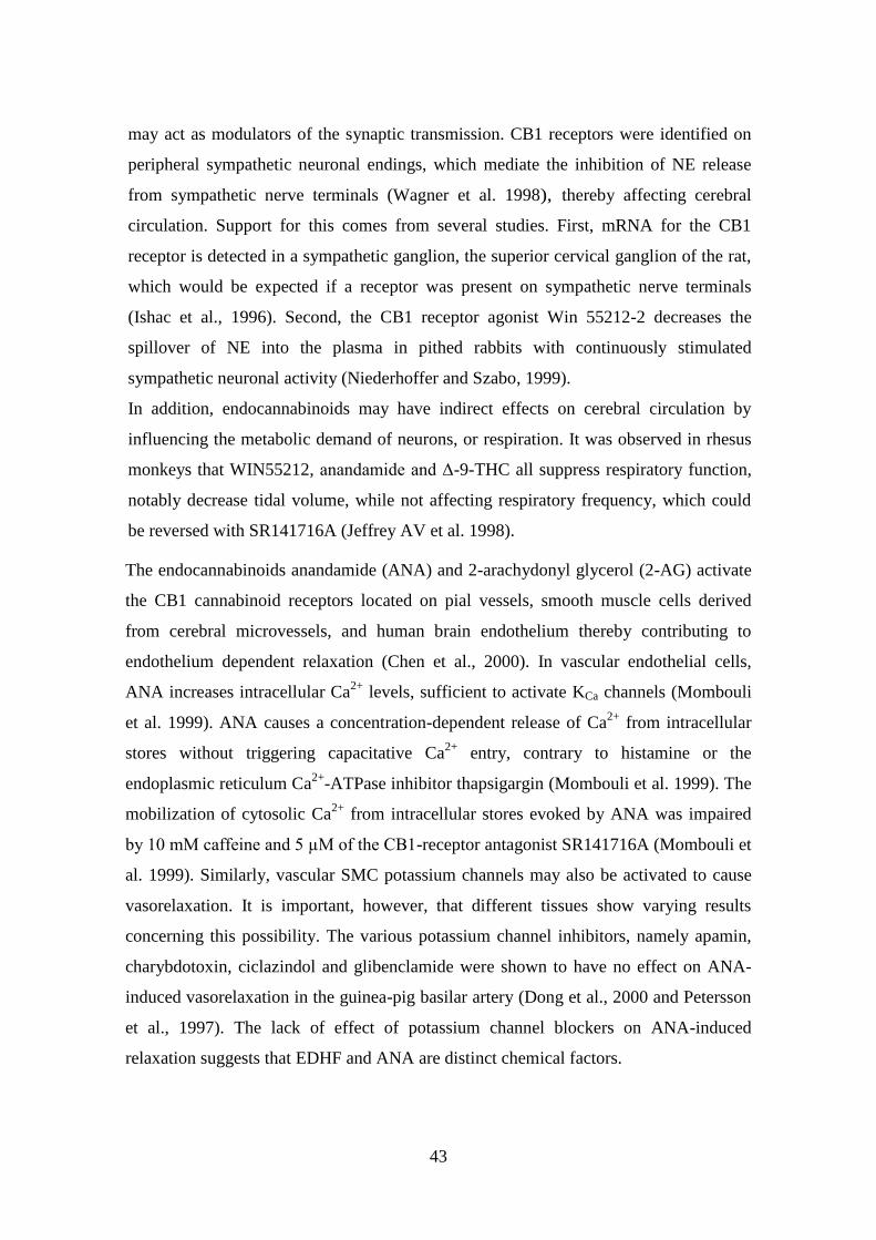
43
may act as modulators of the synaptic transmission. CB1 receptors were identified on
peripheral sympathetic neuronal endings, which mediate the inhibition of NE release
from sympathetic nerve terminals (Wagner et al. 1998), thereby affecting cerebral
circulation. Support for this comes from several studies. First, mRNA for the CB1
receptor is detected in a sympathetic ganglion, the superior cervical ganglion of the rat,
which would be expected if a receptor was present on sympathetic nerve terminals
(Ishac et al., 1996). Second, the CB1 receptor agonist Win 55212-2 decreases the
spillover of NE into the plasma in pithed rabbits with continuously stimulated
sympathetic neuronal activity (Niederhoffer and Szabo, 1999).
In addition, endocannabinoids may have indirect effects on cerebral circulation by
influencing the metabolic demand of neurons, or respiration. It was observed in rhesus
monkeys that WIN55212, anandamide and Δ-9-THC all suppress respiratory function,
notably decrease tidal volume, while not affecting respiratory frequency, which could
be reversed with SR141716A (Jeffrey AV et al. 1998).
The endocannabinoids anandamide (ANA) and 2-arachydonyl glycerol (2-AG) activate
the CB1 cannabinoid receptors located on pial vessels, smooth muscle cells derived
from cerebral microvessels, and human brain endothelium thereby contributing to
endothelium dependent relaxation (Chen et al., 2000). In vascular endothelial cells,
ANA increases intracellular Ca2+
levels, sufficient to activate KCa channels (Mombouli
et al. 1999). ANA causes a concentration-dependent release of Ca2+
from intracellular
stores without triggering capacitative Ca2+
entry, contrary to histamine or the
endoplasmic reticulum Ca2+
-ATPase inhibitor thapsigargin (Mombouli et al. 1999). The
mobilization of cytosolic Ca2+
from intracellular stores evoked by ANA was impaired
by 10 mM caffeine and 5 µM of the CB1-receptor antagonist SR141716A (Mombouli et
al. 1999). Similarly, vascular SMC potassium channels may also be activated to cause
vasorelaxation. It is important, however, that different tissues show varying results
concerning this possibility. The various potassium channel inhibitors, namely apamin,
charybdotoxin, ciclazindol and glibenclamide were shown to have no effect on ANA-
induced vasorelaxation in the guinea-pig basilar artery (Dong et al., 2000 and Petersson
et al., 1997). The lack of effect of potassium channel blockers on ANA-induced
relaxation suggests that EDHF and ANA are distinct chemical factors.
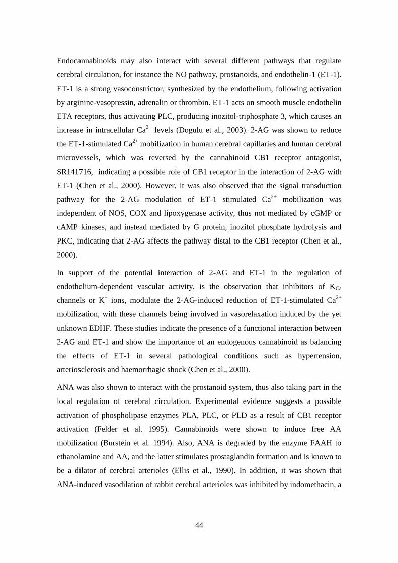
44
Endocannabinoids may also interact with several different pathways that regulate
cerebral circulation, for instance the NO pathway, prostanoids, and endothelin-1 (ET-1).
ET-1 is a strong vasoconstrictor, synthesized by the endothelium, following activation
by arginine-vasopressin, adrenalin or thrombin. ET-1 acts on smooth muscle endothelin
ETA receptors, thus activating PLC, producing inozitol-triphosphate 3, which causes an
increase in intracellular Ca2+
levels (Dogulu et al., 2003). 2-AG was shown to reduce
the ET-1-stimulated Ca2+
mobilization in human cerebral capillaries and human cerebral
microvessels, which was reversed by the cannabinoid CB1 receptor antagonist,
SR141716, indicating a possible role of CB1 receptor in the interaction of 2-AG with
ET-1 (Chen et al., 2000). However, it was also observed that the signal transduction
pathway for the 2-AG modulation of ET-1 stimulated Ca2+
mobilization was
independent of NOS, COX and lipoxygenase activity, thus not mediated by cGMP or
cAMP kinases, and instead mediated by G protein, inozitol phosphate hydrolysis and
PKC, indicating that 2-AG affects the pathway distal to the CB1 receptor (Chen et al.,
2000).
In support of the potential interaction of 2-AG and ET-1 in the regulation of
endothelium-dependent vascular activity, is the observation that inhibitors of KCa
channels or K+ ions, modulate the 2-AG-induced reduction of ET-1-stimulated Ca
2+
mobilization, with these channels being involved in vasorelaxation induced by the yet
unknown EDHF. These studies indicate the presence of a functional interaction between
2-AG and ET-1 and show the importance of an endogenous cannabinoid as balancing
the effects of ET-1 in several pathological conditions such as hypertension,
arteriosclerosis and haemorrhagic shock (Chen et al., 2000).
ANA was also shown to interact with the prostanoid system, thus also taking part in the
local regulation of cerebral circulation. Experimental evidence suggests a possible
activation of phospholipase enzymes PLA, PLC, or PLD as a result of CB1 receptor
activation (Felder et al. 1995). Cannabinoids were shown to induce free AA
mobilization (Burstein et al. 1994). Also, ANA is degraded by the enzyme FAAH to
ethanolamine and AA, and the latter stimulates prostaglandin formation and is known to
be a dilator of cerebral arterioles (Ellis et al., 1990). In addition, it was shown that
ANA-induced vasodilation of rabbit cerebral arterioles was inhibited by indomethacin, a

45
COX inhibitor, which gives the possibility of a COX-dependent mechanism to mediate
ANA-induced vasodilation (Ellis et al., 1995).
In support of its interaction with the prostanoid system, ANA was shown to activate
CB1 receptors located on astrocytes, which are known to play active roles in regulating
both neuronal and cerebrovascular function (Shivachar et al. 1996). Cannabinoid
receptors may be linked to astrocyte signaling, namely to AA release, substrate for
various eicosanoids, that affects cerebrovascular activity. Astrocytes are also capable of
metabolizing exogenous AA via COX, lipoxygenase, and cytochrome P450
epoxygenase pathways (Shivachar et al. 1996). Furthuremore, prostaglandins,
epoxyeicosatrienoic acids, AA, K+ ions, all of which may be released by astrocytes, are
candidate members of communication between astrocyte end-feet and vascular smooth
muscle, taking part in the flow-metabolism coupling (Koehler et al. 2006).
Both anandamide and 2-AG are oxygenated by COX-2 at postsynaptic sites (Katona
and Freund 2012). The resulting prostanoids, for example prostaglandin E2 glycerol
ester, increase neurotransmitter release from axon terminals, which is opposite to the
effect of 2-AG in the retrograde signaling, and thus COX-2 may be an important
molecular switch to change the direction of synaptic retrograde signaling (Katona and
Freund, 2012).
ANA also interacts with the NO system. Previous studies have shown that
endocannabinoids may induce NO release via the constitutive endothelial NOS, by
activating the cannabinoid CB1 receptor (Stefano et al., 1998, and Fimiani et al., 1999).
ANA also stimulates the activity and expression of the inducible endothelial NOS
which is blocked by the NOS inhibitor nitro-L-arginine methyl ester (L-NAME)
(Maccarrone et al., 2000). ANA-induced NO release was sensitive to the selective
antagonist of CB1 receptor, the SR141716A, suggesting the involvement of CB1
receptors in this signaling pathway (Maccarrone et al., 2000). In addition, in vivo
blocking the reuptake of ANA with the AT protein inhibitor AM404 suppressed ex vivo
NO production in sciatic nerve homogenates, indicating that ECs may negatively
regulate NO release from nitroxidergic nerves (Costa et al. 2006).
The interaction of the endocannabinoid system with the NO system also includes the
observation that NO enhances the uptake of 2-AG by cells (Bisogno et al., 2001), and

46
uptake of ANA by human umbilical vein endothelial cells (HUVEC), by potentiating
the activity of the anandamide transporter (AT) (Maccarrone et al., 2000). In general,
the NO system may be part of a physiological regulation of endocannabinoid uptake,
possibly linked to the activation of CB1 receptor (Maccarrone et al., 2000). This may be
a new mechanism, through which ANA and 2-AG can limit their CB1 mediated actions.
In guinea-pig isolated basilar arteries, ANA was shown to evoke a complete relaxation
in endothelium-denuded arterial segments, which failed to respond to ACh, suggesting
that vasodilator response to ANA may also involve endothelium-independent
mechanisms (Zygmunt et al., 1999, Ishioka and Bukoski, 1999). This suggests that
ANA may be involved in the EDHF-mediated relaxation in some tissues, but not in
others, and also shows that EDHF is not a single substance, but rather a common
mechanism that acts through different substances in the various tissues.
3. THE AIMS OF OUR INVESTIGATIONS
As mentioned above, the central nervous system is an independent functional HO unit
which contains the substrate, enzymes and co-factors for the synthesis of CO.
Furthermore, several cell types of the brain are able to produce endocannabinoids and
express their receptors. Since both endogenous CO and endocannabinoids were
previously shown to have vascular effects we hypothesized that they may be involved in
the regulation of the cerebral circulation. Specifically, we aimed to describe the roles of
both the heme oxygenase pathway and the endocannabinoid system on cerebrocortical
blood flow (CBF) under resting conditions as well as during combined hypoxia and
hypercapnia.
1. First, we aimed to examine the effect of the HO inhibitor ZnDPBG on the CBF
under resting conditions in anesthetized adult male rats, by measuring the CBF in
the parietal cortex. We also addressed the potential interaction between the HO and
NOS pathways in the regulation of the cerebrocortical blood flow under resting
conditions.

47
2. Next, we observed the effect of the HO inhibitor ZnDPBG on the cerebrovascular
effects of stepwise hypoxia/hypercapnia (H/H) by measuring the CBF in the parietal
cortex of anesthetized adult male rats.
3. Following this, we aimed to study the effects of the cannabinoid CB1 receptor
antagonist AM251 on the cerebrocortical blood flow under resting conditions using
similar methods.
4. Finally, we examined the effect of CB1-blockade by AM251 on the CBF rise to
stepwise H/H.

48
4. MATERIALS AND METHODS
4.1 Experimental animals, anaesthesia and surgical procedures
The experiments were carried out on adult male Wistar rats (300-400 g) according to the
guidelines of the Hungarian Law of Animal Protection (243/1988), and all procedures
were approved by the Semmelweis University Committee on the Ethical Use of
Experimental Animals (590/99 Rh). The experimental animals were obtained from
Toxi-Coop Kft. (Dunakeszi, Hungary.) During the in vivo experiments, the animals
were anesthetized with urethane (1.3 g/kg ip.), and spontaneously breathing via the
cannulated trachea. Depth of anesthesia was regularly controlled during the experiments
by checking the corneal nociception reflexes, and when necessary, additional urethane
was administered intravenously. Catheters were inserted into the right femoral artery for
measuring systemic arterial blood pressure, into the left femoral artery for blood
sampling, and into the left femoral vein for drug administration.
4.2 Routinely recorded physiological variables
Systemic arterial blood pressure was continuously recorded on a Grass polygraph
(Model 7E, Natus Neurology Incorporated, Warwick, RI, USA). Arterial blood gas
values (aPCO2, aPO2 and O2-saturation) and acid-base parameters (apH and standard
base excess) were measured by an ABL-300 Blood Gas Analyzer (Radiometer,
Brønshøj, Denmark) in femoral arterial samples. Body temperature was kept constant
between 36-38 ºC with a controlled heating pad.
4.3 Measurement of the cerebral blood flow
Cerebrocortical blood flow (CBF), more precisely red blood cell flux, was measured in
the parietal cortex by laser-Doppler (LD) flowmetry (Moor Instruments, Devon, UK).
LD flowmetry is a method of continuous and noninvasive measuring of the tissue blood
flow, utilizing the Doppler shift of laser light as the information carrier (Fabricius et al.,
1997). In experimental medicine, laser-Doppler flowmetry has been used in the study of
spontaneous rhythmical variations as well as in the study of spatial and temporal

49
fluctuations in human skin blood flow. In our experiments, the skull of the rat was fixed
in a stereotaxic head holder with the nose 5 mm down from the interaural line, the
parietal region was exposed and the bone was thinned over the parietal cortex on both
sides with a microdrill, so that the lamina interna of the skull remained intact. LD
probes were placed above both holes of the thinned skull at a 12-angle to the vertical,
to provide an optimal view of the cortex (4 mm caudal from the bregma, 5 mm lateral
from the midline). LD flow (LDF) was measured with a two-channel LDF monitor
(Moor Instruments) and recorded continuously (Lacza et al. 2000). The flowmeter was
calibrated before each individual experiment with a constant movement latex emulsion.
The laser light was in the infrared range (780 nm) and penetrated about 1 mm into the
brain covering approximately 7 mm2 of the parietal region, so that the data acquired
represented the characteristics of the parietal cortex (Haberl et al. 1989). The two LD
flow data sets gained from the parietal region of both hemispheres of the animal were
evaluated and treated as independent measurements.
4.4 Experimental Protocols
4.4.1 The influence of HO pathway on cerebrocortical blood flow
4.4.1.1 First experimental protocol: the influence of HO pathway on CBF under
normoxic/normocapnic conditions
In the first experimental protocol, the effect of HO blockade on the CBF was studied
under physiological normoxic-normocapnic conditions. Animals of the first
experimental group (n=9) served as a vehicle-treated control and CBF was determined
before and after an intraperitoneal injection of 3 mL saline. In the second experimental
group (n=9), 45 µmol/kg zinc deuteroporphyrin 2,4-bis glycol (ZnDPBG, Frontier
Scientific, Logan, UT, USA) was applied in 3 mL saline intraperitoneally for the
inhibition of the HO pathway. CBF was determined before as well as 15, 30, and 45
minutes after the administration of either saline or ZnDPBG. To verify the inhibitory
effect of ZnDPBG on brain HO activity in vivo, matched series of animals were treated
with either ZnDPBG or vehicle as described above. Thirty minutes later, innate cerebral
CO generation was determined using solid-phase gas chromatography (custom built
Peak Performer 1 RCP; Peak Laboratories LLC, Mountain View, CA, USA), according
to Johnson FK et al. 2006. Briefly, isolated midbrain tissues from each animal were

50
individually sonicated in Krebs’ buffer and divided into eight aliquots. For each animal
midbrain sample, head space CO was measured in quadruplicate vials after being
maintained at 2◦C or remaining matched aliquots after being incubated at 37
◦C for 60
min. The differences between the incubated and cold vials were expressed as µmol CO
generated/kg of wet tissue per hour. ZnDPBG induced a reduction of cerebral HO
activity from 4.58 ± 0.87 to 2.47 ± 0.36 µmol/kg per hour (p = 0.025).
In order to investigate the interaction of NO and CO systems, more precisely, the effect
of suppression of NO production on CO-induced CBF-changes, the synthesis of NO
was inhibited. Therefore, animals in the third and fourth experimental groups were
pretreated intravenously with the NO-synthase inhibitor L-NAME (N-nitro-L-arginine
methyl ester) in a dosage of 50 mg/kg, 30 minutes prior to the administration of saline
(n=5) or ZnDPBG (n=6), respectively. In this case, the CBF of the L-NAME treated rats
was determined before as well as 15, 30, and 45 minutes after the administration of
saline or ZnDPBG.
4.4.1.2 Second experimental protocol: the effect of HO blockade on the CBF rise to H/H
In the second experimental protocol, the effect of HO blockade on the CBF increase to
H/H was observed. Stepwise H/H was induced by administration of different gas
mixtures (5% O2-20% CO2-75% N2 for producing moderate H/H and 20% CO2-80%
N2 for producing severe H/H) with a constant flow rate of 3 L/min into a 5 mL chamber
connected to the trachea at atmospheric pressure. Since one side of the chamber was
open, the pressure in the chamber was not different from the atmospheric pressure. CBF
was recorded continuously. Peak CBF values were determined during the two steps of
H/H, each with a duration of 10 minutes. (For the CBF values before and after the H/H,
please see Figure 4). After the first stepwise H/H round, the inhalation of the gas
mixture was stopped, and following a thirty minute recovery period of normal air
breathing, the animals were divided into two experimental groups, thus receiving
intraperitoneally either 3 ml saline or 45 micromol/kg ZnDPBG dissolved in the same
amount of saline. Thirty minutes following intraperitoneal drug treatment, the stepwise
H/H was repeated in both experimental groups with continuous recording of the CBF,
from which peak CBF values were determined again.
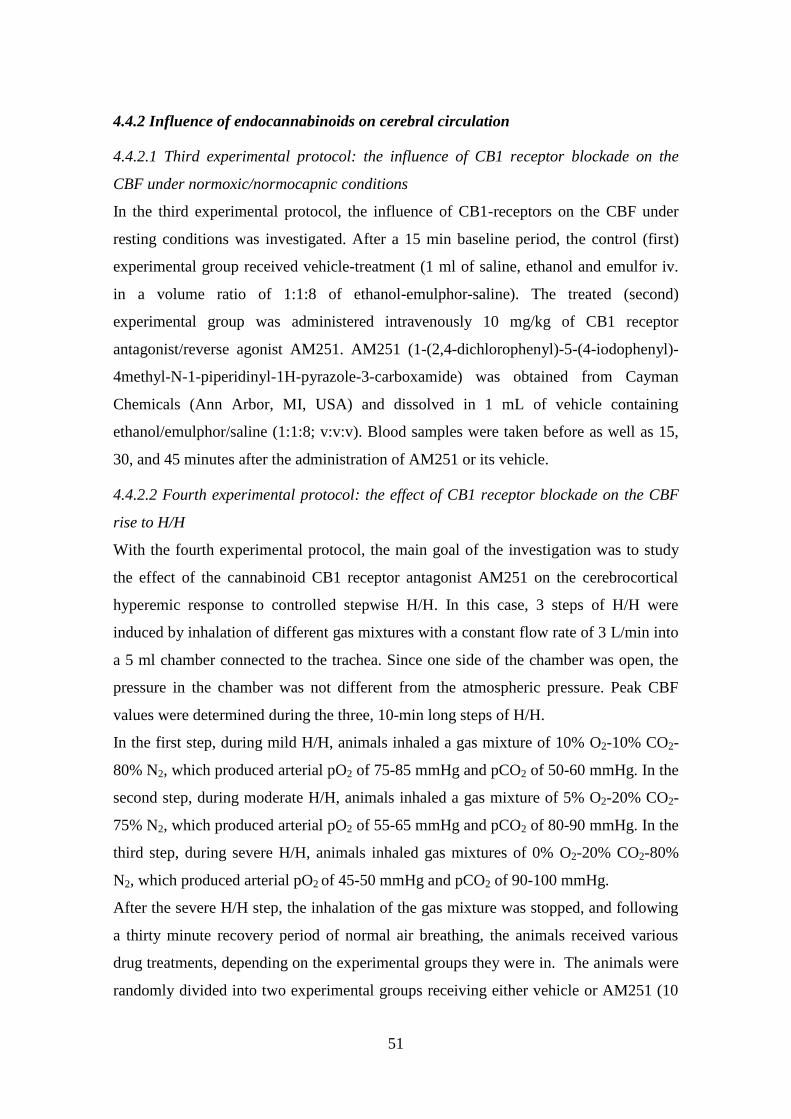
51
4.4.2 Influence of endocannabinoids on cerebral circulation
4.4.2.1 Third experimental protocol: the influence of CB1 receptor blockade on the
CBF under normoxic/normocapnic conditions
In the third experimental protocol, the influence of CB1-receptors on the CBF under
resting conditions was investigated. After a 15 min baseline period, the control (first)
experimental group received vehicle-treatment (1 ml of saline, ethanol and emulfor iv.
in a volume ratio of 1:1:8 of ethanol-emulphor-saline). The treated (second)
experimental group was administered intravenously 10 mg/kg of CB1 receptor
antagonist/reverse agonist AM251. AM251 (1-(2,4-dichlorophenyl)-5-(4-iodophenyl)-
4methyl-N-1-piperidinyl-1H-pyrazole-3-carboxamide) was obtained from Cayman
Chemicals (Ann Arbor, MI, USA) and dissolved in 1 mL of vehicle containing
ethanol/emulphor/saline (1:1:8; v:v:v). Blood samples were taken before as well as 15,
30, and 45 minutes after the administration of AM251 or its vehicle.
4.4.2.2 Fourth experimental protocol: the effect of CB1 receptor blockade on the CBF
rise to H/H
With the fourth experimental protocol, the main goal of the investigation was to study
the effect of the cannabinoid CB1 receptor antagonist AM251 on the cerebrocortical
hyperemic response to controlled stepwise H/H. In this case, 3 steps of H/H were
induced by inhalation of different gas mixtures with a constant flow rate of 3 L/min into
a 5 ml chamber connected to the trachea. Since one side of the chamber was open, the
pressure in the chamber was not different from the atmospheric pressure. Peak CBF
values were determined during the three, 10-min long steps of H/H.
In the first step, during mild H/H, animals inhaled a gas mixture of 10% O2-10% CO2-
80% N2, which produced arterial pO2 of 75-85 mmHg and pCO2 of 50-60 mmHg. In the
second step, during moderate H/H, animals inhaled a gas mixture of 5% O2-20% CO2-
75% N2, which produced arterial pO2 of 55-65 mmHg and pCO2 of 80-90 mmHg. In the
third step, during severe H/H, animals inhaled gas mixtures of 0% O2-20% CO2-80%
N2, which produced arterial pO2 of 45-50 mmHg and pCO2 of 90-100 mmHg.
After the severe H/H step, the inhalation of the gas mixture was stopped, and following
a thirty minute recovery period of normal air breathing, the animals received various
drug treatments, depending on the experimental groups they were in. The animals were
randomly divided into two experimental groups receiving either vehicle or AM251 (10

52
mg/kg i.v.). After another thirty minutes period following drug treatment, the stepwise
H/H was repeated in all experimental groups with continuous recording of the CBF, and
determination of peak CBF values.
4.5 Drugs and chemicals
ZnDPBG (zinc deuteroporphyrin 2,4-bis glycol) was purchased from Frontier Scientific,
Logan, UT, USA, and AM251 (1-(2,4-dichlorophenyl)-5-(4-iodophenyl)-4methyl-N-1-
piperidinyl-1H-pyrazole-3-carboxamide) from Cayman Chemicals, Ann Arbor, MI,
USA. All other chemicals were obtained from Sigma-Aldrich (St. Louis, MO, USA).
4.6 Statistical analysis
Statistical analysis was performed using Student’s t-test for comparision of two values
as well as ANOVA for repeated measurements or one way ANOVA followed by
Tukey’s post-hoc test for three or more values. A p value of less than 0.05 was
considered to be statistically significant.
5. RESULTS
5.1 Effects of heme-oxygenase blockade under resting normoxic/normocapnic
conditions
Baseline arterial blood gas and acid-base parameters were within the physiological
range and showed no significant changes after the administration of ZnDPBG or its
vehicle (saline) (Table I). Neither ZnDPBG nor its vehicle induced any significant
changes in the mean arterial blood pressure (Figure 4.) or in the heart rate (data not
shown). In contrast, administration of ZnDPBG but not its vehicle resulted in a marked
increase of the CBF (Figure 5.).

53
Treatment 0 min 15 min 30 min 45 min
Hemoglobin
conc. (mg/ml) Vehicle 15.1 ± 0.3 14.5 ± 0.3 14.5 ± 0.4 14.5 ± 0.4
ZnDPBG 16.2 ± 0.3 16.0 ± 0,3 15.9 ± 0.3 16.0 ± 0.3
pH Vehicle 7.34 ±
0.01
7.35 ±
0.01
7.34 ±
0.01
7.33 ±
0.01
ZnDPBG 7.36 ±
0.02
7.36 ±
0.02
7.36 ±
0.02
7.35 ±
0.02
PaO2 (mmHg) Vehicle 89.9 ± 2.7 89.6 ± 2.4 89.9 ± 2.7 89.3 ± 2.9
ZnDPBG 94.6 ± 3.3 93.8 ± 3.1 92.5 ± 3.6 89.5 ± 3.5
PaCO2 (mmHg) Vehicle 44.2 ± 1.4 42.6 ± 1.2 41.9 ± 1.2 42.9 ± 0.7
ZnDPBG 42.9 ± 1.4 41.4 ± 1.6 41.6 ± 1.8 42.0 ± 2.0
O2 Saturation
(%) Vehicle 95.8 ± 0.4 96.0 ± 0.3 95.9 ± 0.4 95.8 ± 0.3
ZnDPBG 96.5 ± 0.4 96.5 ± 0.4 96.3 ± 0.5 95.9 ± 0.5
Standard Base
Excess (mmol/l) Vehicle -2.0 ± 0.7 -2.3 ± 0.7 -2.7 ± 0.7 -2.9 ± 0.5
ZnDPBG -1.0 ± 0.8 -1.4 ± 0.9 -1.4 ± 0.9 -1.6 ± 0.8
Table I: Physiological parameters of the arterial blood before (0 min) and at 15, 30 and
45 min after the intraperitoneal administration of 45 μmol/kg zinc deuteroporphyrin 2,4
bis glycol (ZnDPBG) or its vehicle (saline) in anesthetized rats (n=9-9).
There were no significant differences between the vehicle-treated and ZnDPBG-treated
groups or within one of the two experimental groups at different timepoints.

54
T im e
MA
P (
mm
Hg
)
0 m in 1 5 m in 3 0 m in 4 5 m in8 0
1 0 0
1 2 0
1 4 0
1 6 0
S a lin e
Z n D P B G
S a lin e a f te r L -N A M E
Z n D P B G a fte r L -N A M E
* * *
Figure 4: Mean arterial blood pressure (MAP) are shown before (0 min) as well as 15,
30, 45 min after intraperitoneal injection of saline (triangles, n=9) or 45 μmol/kg zinc
deuteroporphyrin 2,4-bis glycol (ZnDPBG) (circles, n=9) in control rats that did not
receive N-nitro-L-arginince methyl ester (L-NAME) (open symbols, n=5 for saline after
L-NAME and n=6 for ZnDPBG after L-NAME) or in L-NAME pre-treated (filled
symbols) urethane-anaesthetized rats. Values are presented as mean ± SEM. At all four
timepoints, there was a significant (***p<0.001) increase of MAP in L-NAME pre-
treated animals, compared to rats with intact NO synthesis.

55
Figure 5: Cerebrocortical blood flow (CBF) before (0 min) as well as 15, 30, 45 min
after intraperitoneal injection of saline (triangles) or 45 micromol/kg zinc
deuteroporphyrin 2,4-bis glycol (ZnDPBG; circles) in control (open symbols) or in N-
nitro-L-arginince methyl ester (L-NAME) pretreated (filled symbols) rats. *p=0.016,
***p< 0.001 vs. ‘0 min.’ with repeated measures analysis of variance and Tukey’s post
hoc test, n= 10-16. AU, arbitrary unit.
In order to evaluate the role of NO in mediating the enhancement of the CBF after HO
blockade, the experiments have been repeated in animals subjected to inhibition of the
NO synthesis. Administration of L-NAME increased mean arterial blood pressure from
102.2 ± 3.0 to 146.3 ± 4.1 mmHg (p< 0.001), decreased heart rate from 424 ± 12 to 373
± 12 beats/min (p= 0.012) and reduced CBF from 359 ± 18 to 258 ± 13 AU (p<0.001)
without influencing arterial blood gas or acid-base parameters (Table II). In L-NAME
pre-treated animals, neither ZnDPBG nor its vehicle induced any significant changes in
Time (min)
Cere
bro
co
rtic
al
Blo
od
Flo
w (
AU
)

56
the mean arterial blood pressure (Figure 4.) or arterial blood gas and acid-base
parameters (Table II.). Most importantly, L-NAME prevented the enhancement of the
CBF after inhibition of the endogenous CO production by administration of ZnDPBG
(Figure 5.).
Treatment 0 min 15 min 30 min 45 min
Hemoglobin
conc. (mg/ml)
L-NAME
+Vehicle 16.2 ± 0.4 16.4 ± 0.4 16.4 ± 0.3 16.2 ± 0.4
L-NAME
+ZnDPBG 16.1 ± 0.4 16.0 ± 0.4 16.0 ± 0.3 15.8 ± 0.4
pH L-NAME
+Vehicle
7.35 ±
0.02
7.36 ±
0.02
7.35 ±
0.02
7.34 ±
0.02
L-NAME
+ZnDPBG
7.36 ±
0.01
7.34 ±
0.01
7.33 ±
0.01
7.35 ±
0.02
PaO2 (mmHg) L-NAME
+Vehicle 99.2 ± 5.1 96.6 ± 3.6 99.9 ± 7.6
102.2 ±
7.2
L-NAME
+ZnDPBG 95.6 ± 4.3 95.9 ± 2.4
100.9 ±
6.3
106.2 ±
6.9
PaCO2 (mmHg) L-NAME
+Vehicle 45.5 ± 1.8 43.9 ± 1.9 44.4 ± 1.6 44.6 ± 2.4
L-NAME
+ZnDPBG 44.8 ± 1.7 44.3 ± 2.8 43.3 ± 2.9 44.7 ± 3.2
O2 Saturation
(%)
L-NAME
+Vehicle 95.5 ± 0.9 95.5 ± 0.9 95.2 ± 0.9 95.6 ± 1.1
L-NAME
+ZnDPBG 96.0 ± 0.7 96.0 ± 0.7 96.0 ± 0.8 96.4 ± 1.1
Standard Base
Excess (mmol/l)
L-NAME
+Vehicle 0.1 ± 0.6 -0.9 ± 0.9 -1.0 ± 0.7 0.2 ± 0.9
L-NAME
+ZnDPBG -0.4 ± 0.7 -1.5 ± 0.9 -1.8 ± 0.9 -0.6 ± 1.3
Table II: Physiological parameters of the arterial blood before (0 min) and after (at 15,
30 and 45 min) administration of zinc deuteroporphyrin 2,4-bis glycol or its vehicle
(saline) in N-nitro-L-arginince methyl ester pre-treated animals. (n=5-6)
5.2 Effects of heme-oxygenase blockade on the hypoxia and hypercapnia induced
increase of cerebrocortical blood flow
Before the initial H/H challenge, baseline cardiovascular, arterial blood gas and acid-
base parameters were within physiological ranges in later ZnDPBG treated or saline
treated experimental groups. During moderate H/H, PaO2 was reduced to 60-65 mmHg,
PaCO2 was increased to 80-85 mmHg (Figure 6.) and pH decreased to 7.10-7.15.

57
Despite no change in MAP (data not shown), CBF increased in both experimental
groups by ~ 45% (Figure 7A.). During severe H/H, PaO2 was reduced to 45-50 mmHg,
PaCO2 was increased to 95-100 mmHg (Figure 6.) and pH decreased to 7.05-7.10; CBF
increased in both experimental groups by ~65% (Figure 7B.), whereas MAP remained
unchanged (data not shown).
The second stepwise H/H challenge, after ZnDPBG or saline, induced similar changes
in the blood-gas tensions and pH as the initial challenge had done (Figure 6.), without
affecting MAP (data not shown). Most importantly, neither ZnDPBG nor saline
treatment had any effect on CBF responses to moderate (Figure 7A.) or severe (Figure
7.B.) H/H, when compared to the pretreatment values.
Figure 6: Arterial blood-gas tensions during mild and severe H/H before (open
symbols) and after (filled symbols) intraperitoneal injection of saline (triangles) or 45
μmol/kg zinc deuteroporphyrin 2,4-bis glycol (ZnDPBG; circles), (n=8-8).
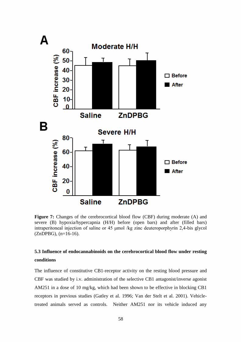
58
Figure 7: Changes of the cerebrocortical blood flow (CBF) during moderate (A) and
severe (B) hypoxia/hypercapnia (H/H) before (open bars) and after (filled bars)
intraperitoneal injection of saline or 45 μmol /kg zinc deuteroporphyrin 2,4-bis glycol
(ZnDPBG), (n=16-16).
5.3 Influence of endocannabinoids on the cerebrocortical blood flow under resting
conditions
The influence of constitutive CB1-receptor activity on the resting blood pressure and
CBF was studied by i.v. administration of the selective CB1 antagonist/inverse agonist
AM251 in a dose of 10 mg/kg, which had been shown to be effective in blocking CB1
receptors in previous studies (Gatley et al. 1996; Van der Stelt et al. 2001). Vehicle-
treated animals served as controls. Neither AM251 nor its vehicle induced any

59
significant changes in the mean aterial blood pressure MAP (Figure 8A.), heart rate HR
(Figure 8B.) or CBF (Figure 8C.) up to 45 minutes after their administration.
Furthermore, arterial blood gas tensions and acid-base parameters remained unchanged
during the observation period (Table III.). These finding indicate that constitutive CB1
activity has no significant influence on the systemic and cerebrocortical circulation
under steady-state resting conditions in healthy normotensive rats.
Figure 8: Mean aterial blood pressure (MAP, panel A), heart rate (HR, panel B) and
cerebrocortical blood flow (CBF, panel C) are shown before (0 minute) as well as 15,
30, 45 minutes after intravenous administration of 10 mg/kg of the CB 1 receptor
antagonist AM251 (open squares, n=7) or its vehicle (open circles, n=5) in urethane-
anaesthetized rats. Values are presented as mean ± SEM. No significant change of the
measured variables was found at any given time points after the administration of
vehicle or AM251. AU, arbitrary unit.

60
Treatment 0 min 15 min 30 min 45 min
pH Vehicle 7.40 ± 0.01 7.36 ± 0.01 7.36 ± 0.01 7.37 ± 0.01
AM251 7.40 ± 0.01 7.37 ± 0.01 7.39 ± 0.01 7.39 ± 0.01
Arterial O2
(PaO2) (mmHg) Vehicle 91.0 ± 2.5 82.2 ± 3.8 83.2 ± 2.4 85.4 ± 3.7
AM251 86.3 ± 1.3 80.3 ± 2.6 86.3 ± 1.6 86.1 ± 1.3
Arterial CO2
pressure (PaCO2)
(mmHg)
Vehicle 40.6 ± 1.4 42.4 ± 1.9 42.2 ± 2.1 41.2 ± 1.7
AM251 40.4 ± 1.7 41.6 ± 0.7 38.4 ± 0.7 37.1 ± 1.1
O2 Saturation
(%) Vehicle 97.5 ± 0.2 95.2 ± 0.2 95.4 ± 0.3 95.8 ± 0.4
AM251 96.6 ± 0.2 95.3 ± 0.4 96.5 ± 0.3 96.5 ± 0.2
Table III: Arterial blood gas and acid-base parameters before (0 min) and after (15, 30,
and 45 min) the intravenous administration of 10 mg/kg AM251 (n=7) or its vehicle
(n=5)
There were no significant differences between the vehicle-treated and AM251 treated
groups or within one of the two experimental groups at different timepoints.
5.4 Effects of endocannabinoid receptor blockade on the cerebrocortical blood flow
during hypoxia and hypercapnia
To test the effects of endocannabinoid receptor blockade on the CBF elevation during
hypoxia and hypercapnia, we produced stepwise H/H (as described in the Methods)
before and after the administration of AM251 or its vehicle, and determined the changes
in CBF. Inhalation of three different gas mixtures containing decreased O2- and
increased CO2-content (as compared to air) induced reproducible levels of hypoxia and
hypercapnia before and after the intravenous administration of 10 mg/kg AM251

61
(Figure 9B.) or its vehicle (Figure 9A.), without significant changes in the BP (data not
shown). The H/H-induced enhancement of CBF was identical before and after the
administration of the vehicle (Figure 10A.). In contrast, AM251 resulted in markedly
increased CBF changes during mild and moderate H/H (by 28.1 ± 8.8% and 39.4 ±
10.0%, respectively) without significantly influencing the peak CBF during severe H/H
(Figure 10B.).
Figure 9: Inhalation of three different gas mixtures containing decreased O2- and
increased CO2-content (as compared to air) induced reproducible levels of hypoxia and
hypercapnia before and after the administration of AM251 (Figure 9B.) or its vehicle
(Figure 9A.).
Figure 10: The hypoxia/hypercapnia (H/H) induced enhancement of the cerebrocortical
blood flow (CBF) was identical before and after the administration of the vehicle of
AM251 (Figure 10A.). In contrast, AM251 resulted in markedly increased CBF changes
during mild and moderate H/H (by 28.1 ± 8.8% and 39.4 ± 10.0%, respectively) without
significantly influencing the peak CBF during severe H/H (Figure 10B.).

62
6. DISCUSSION
6.1 Role of heme oxygenase metabolites in the regulation of the cerebral circulation
during resting conditions and during hypoxia and hypercapnia
Our present study provides evidence for an interaction between the HO and NOS
pathways in the regulation of the cerebrocortical blood flow. We show clearly, that
administration of the HO-inhibitor ZnDPBG causes an increase in the CBF under
physiological (normoxic/normocapnic) resting conditions. ZnDPBG had been
previously shown to effectively inhibit HO activity in the rat brain (Johnson RA et al.
1995). In addition, ZnDPBG was shown to effectively inhibit HO activity in human and
rat brain microsomes (Appleton et al. 1999, Chernick et al. 1989). In our studies
ZnDPBG was preferred to other metalloporphyrins, because it has been shown that its
ip. administration in the dose used in our experiments induced identical cardiovascular
effects to those observed after topical administration of the drug into the nucleus tractus
solitarii (NTS) of rats. Furthermore, microinjection of CO into NTS reversed the effects
of the ip. applied ZnDPBG, indicating that ZnDPBG when ip. injected in this dose,
crosses the blood-brain barrier and inhibits the cerebral HO activity in rats (Johnson et
al. 1997).
The CBF-increase after administration of ZnDPBG and its inhibition with L-NAME
pretreatment indicate that constitutive CO release tonically suppresses NO production
and consequently reduces blood flow in the cerebral cortex. Several recent studies
suggest that its interaction with NOS may modulate significantly the overall vascular
effects of CO (Johnson et al. 2002 and Ndisang et. al. 2004). Furthermore, L-NAME
was shown to augment both the reduction of renal blood flow and the contraction of
isolated renal interlobular arteries in response to the HO inhibitor stannous
mesoporphyrin (Rodriguez et al. 2003). Since endogenous CO was shown to relax
smooth muscle cells via activation of sGC and large-conductance KCa channels
(Johnson RA et al. 1995), it appears that this mediator, depending on the mechanism of
its action, may induce both vasoconstriction and vasodilation.
Our findings should be interpreted in light of the recent, independent study by Ishikawa
et al. This study demonstrated that HO-2 is co-localized with eNOS in the
cerebrovascular endothelium and with nNOS in neurons and arachnoid trabecular cells
63
providing the anatomical basis for interactions between the CO- and NO-generating
pathways (Ishikawa et al. 2005). Furthermore, tricarbonyldichlororuthenium (II)
dimmer, a CO-releasing molecule, reduced NO-release from cultured endothelial cells.
In accordance, HO blockade by zinc protoporphyrin IX (ZnPPIX) induced CO- and L-
NAME-reversible increase of the cerebrovascular and perivascular NO-production by
70-80%, which effect is comparable to the ZnDPBG-induced 67% increase of the NOS
activity in the rat hypothalamus observed in our previous study (Horváth et al. 2003).
Most importantly, ZnPPIX induced a dose-dependent increase of the pial arteriolar
diameter, which could be prevented by co-administration of CO or L-NAME, indicating
the involvement of NO in mediating the vasodilation. Our findings confirm and extend
these observations by providing direct evidence for the significance of the CO-NOS
interaction at the level of cerebrocortical blood perfusion.
In our previous study (Horváth et al. 2003), ZnDPBG failed to relax the isolated middle
cerebral artery while Ishikawa et al. (Ishikawa et al. 2005) reported marked pial
arteriolar dilatation in response to ZnPP in rats. Since in the latter study pial arteriolar
responses were determined in vivo, the most plausible explanation of the discrepancy
between the two findings is that reduction of non-vascular CO release or augmentation
of non-vascular NO release plays an important role in the interaction leading to the NO-
mediated cerebral vasodilation and hyperemia after inhibition of HO.
The second part of our study showed that the HO – CO pathway does not play a
significant role in the regulation of the acute hyperemic response to hypoxia or
hypercapnia in adult rats. However, it was also found that HO blockade significantly
inhibited the pial arteriolar dilation in response to hypoxia or hypercapnia in newborn
piglets (Carratu et al. 2003 and Leffler et al. 1999). Since the regulation of the CBF
during hypoxia or hypercapnia shows marked changes during maturation (Armstead et
al. 2005), the contribution of the HO pathway may likely be different in adult animals as
in our study. Furthermore, we cannot exclude the possibility that endogenous CO by
itself would facilitate the hyperemic response to H/H also in adult rats, but its
simultaneous inhibitory influence on the synthesis of NO, a well established mediator of
cerebral vasodilation during hypoxia and hypercapnia, masks this effect. Further studies
may clarify this hypothesis and investigate the role of endogenous CO in the adaptation

64
of the cerebral circulation to chronic hypoxia where the expression of HO-1 is increased
in the brain (Chang et al. 2005, Mazza et al. 2001).
6.2 Role of endocannabinoids in the regulation of the cerebral circulation during
resting conditions and during hypoxia and hypercapnia
Several studies have described marked changes of the cerebral circulation after
administration of cannabinoid compounds as it has been summarized in the
“Introduction”. The effects of exogenously applied phyto- or endocannabinoids,
however, may hardly resemble the functions of an endogenous control system. We were
able to overcome these limitations, by focusing on the changes elicited by suppressed
activity of the EC system. We used AM251, which works both as an antagonist and as
an inverse agonist at CB1-receptors (Pertwee RG 2005, and Hanlon and Vanderah
2010). In our experiments, we did not observe any significant effect of AM251 on the
systemic or cerebral circulation. It appears that CB1-receptors have no constitutive
influence on the cardiovascular system under steady-state resting conditions, at least in
healthy normotensive rats, which is consistent with reports on the normal hemodynamic
profile of CB1-knockout mice (Mukhopadhyay et al. 2010 and Rajesh et al. 2012).
CB1-receptors have been reported to tonically modulate various physiological functions
either by their constitutive activity or by mediating the effects of constitutively released
ECs (Pertwee 2005, Hanlon and Vanderah 2010). It is well known that both systemic
BP and CBF are vital parameters of homeostasis, and therefore several backup
regulatory mechanisms are involved in their maintenance. For this reason we cannot
exclude the possibility that CB1-mediated pathways do contribute to steady-state BP- or
CBF-regulation and when they are blocked pharmacologically or genetically, other
control mechanisms may take over their function.
The last part of our study was devoted to investigate the role of CB1-receptors in H/H-
induced CBF rise. In spite of the fact that the enhancement of CBF during H/H was the
first well-described reaction of the cerebral circulation, its mechanism is still poorly
understood. In the present study we found that blockade of CB1-receptors enhances
CBF responses to H/H. Since it is well established that both neurons and astrocytes
abundantly express CB1-receptors (Freund et al. 2003, Stella 2010), a CB1-mediated
modulation of neuronal nitric oxide synthase activity may explain our observations,

65
indicating that ECs play an inhibitory role in the CBF response to H/H. In addition, in
previous studies AM251 was reported to inhibit basal G-protein-activity in rat cerebellar
membranes (Savinainen et al. 2003) and to enhance electrically evoked glutamate
release from rat cerebellar neurons (Kreitzer et al. 2001).
CB1-agonists inhibit K+-induced NOS-activation in cerebellar granule neurons without
influencing the basal NO-release from these cells, and the CB1-antagonist rimonabant
both reversed the effect of CB1-activation and produced an increase in NOS activity
that was additive with K+ (Hillard et al. 1999). Furthermore, CB1-receptors reportedly
inhibit both glutamatergic transmission (Freund et al. 2003) and the metabolic activity
of neurons and astrocytes (Duarte et al. 2012), effects that may influence the release of
NO and other vasoactive mediators and consequently alter CBF. Astrocytes are
potential oxygen-sensors of this control system, since it is well established that hypoxia
suppresses glutamate uptake by astrocytes (Vangeison and Rempe 2009), which may
result in activation of the glutamate receptor-mediated release of vasoactive mediators
from neuronal elements of the neurovascular unit. Moreover, it is well documented that
the cerebrovascular responses to H/H are modulated by sympathetic perivascular nerves
(Busija and Heistad 1984, Deshmukh et al. 1972, Harper et al. 1972, Wagerle et al.
1986, Goplerud et al. 1991), a pathway that may be modulated via prejunctional CB1-
receptors. NE, ATP and neuropeptide Y are costored and coreleased from sympathetic
vesicles in sympathetic nerves and prejunctional CB1 receptors inhibit these releases
(Randall et al. 2004, Ralevic and Kendall 2009). Whatever is the exact mechanism by
which CB1-receptors modulate the H/H-induced CBF rise, our results appear to support
the pivotal role of neuronal regulation of CBF during H/H. With regard to the potential
physiological role of CB1 receptor-mediated inhibition of H/H-induced cerebral
hyperemia it is likely that it functions as a negative feed-back mechanism. It is well
established that hypoxia and hypercapnia, especially if they are sustained, may result in
the development of brain edema (Wilson et.al. 2009; Adeva et.al. 2012) . Although the
pathomechanism of H/H-induced brain edema is complex (Yang and Rosenberg 2011),
cerebral vasodilation and the consequent increase of the hydrostatic pressure in brain
capillaries is likely to play a key role in it. Therefore, the CB1 receptor-mediated
attenuation of H/H-induced cerebral vasodilation may represent a protective mechanism
against the development of brain edema during hypoxia and hypercapnia.
66
7. CONCLUSIONS
1. First, we examined the effect of the HO inhibitor ZnDPBG on the CBF under
resting conditions in anesthetized adult male rats, by measuring the CBF in the parietal
cortex. We also examined the interaction between the HO and NOS pathways in the
regulation of the cerebrocortical blood flow under resting conditions. The results of this
study indicate that endogenous CO influences tonically the resting cerebrocortical blood
flow by interacting with the NO synthesis. Inhibition of the HO pathway leads to
cerebrocortical hyperemia by increased neuronal and/or endothelial NO production.
2. We observed the effect of the HO inhibitor ZnDPBG on the cerebrovascular
effects of stepwise hypoxia/hypercapnia by measuring the CBF in the parietal cortex of
anesthetized adult male rats under hypoxia/hypercapnia. We found that the HO – CO
pathway does not appear to influence the CBF increase during hypoxia and hypercapnia
in adult rats.
3. Following this, we studied the effects of the cannabinoid CB1 receptor blockade
by the inverse agonist/antagonist AM251 on the CBF under resting conditions using
similar methods. Our study indicates that under resting physiological conditions CB1-
receptor mediated mechanisms have limited tonic influence on the systemic and
cerebral circulation.
4. Finally, we examined the effect of AM251 on the CBF rise to stepwise H/H.
Most importantly, our data suggest that the endocannabinoid system and CB1-receptors
play an important role in the regulation of the cerebral circulation during hypoxia and
hypercapnia and may potentially be involved in the autoregulation of CBF.

67
8. SUMMARY
The cerebral vasoregulation incorporates various local and global control mechanisms,
involving participation of neuronal and vascular elements. Cerebrovascular regulatory
pathways have been studied extensively for decades, but several important questions,
like the exact mechanism of flow-metabolism coupling or hypercapnia- and hypoxia-
induced vasodilation, are still not clarified completely. In our works we hypothesized
that investigation of recently discovered vasoactive mediators may shed light on
obscure details of the regulation of cerebral circulation. Specifically, we investigated the
involvement of the heme oxygenase (HO) – carbon monoxide (CO) pathway and the
endocannabinoid system in the cerebral vasoregulation under resting
(normoxic/normocapnic) physiological conditions and during hypoxia and hypercapnia
(H/H). Cerebrocortical blood flow (CBF) was measured by laser Doppler flowmetry in
anesthetized adult male Wistar rats. First, the role of the HO pathway and its interaction
with the nitric oxide synthase (NOS) pathway were studied in the regulation of CBF
under normoxic/normocapnic conditions. CBF increased after inhibition of HO, and this
effect was reversed by NOS-blockade, suggesting that constitutive CO release tonically
suppreses NO production and consequently reduces CBF. In order to analyze the role of
the HO – CO pathway in the CBF regulation during H/H we developed an experimental
protocol for reproducible, controlled, stepwise H/H in anesthetized rats by
administration of different gas mixtures, resulting in CBF increase. Surprisingly,
however, HO blockade failed to alter the cerebrocortical hyperemia, indicating that the
HO – CO pathway, albeit influences NO release, does not contribute to the CBF
regulation during H/H. We also investigated the effects of endocannabinoids and CB1
cannabinoid receptors on the CBF under resting physiological conditions and during
H/H. We did not observe any significant effect of CB1 blockade by AM251 on the
cerebrocortical circulation under normoxic/normocapnic conditions. In contrast, AM251
enhanced significantly the CBF rise to H/H, indicating the importance of the CB1
receptors in modulating the response. We propose that the CB1 receptor-mediated
attenuation of cerebral vasodilation may represent a negative feed-back mechanism
against the development of brain edema and increased intracranial pressure during H/H.

68
9. ÖSSZEFOGLALÁS
Az agyi vérkeringés szabályozása igen komplex folyamat, melyben idegi és vaszkuláris
tényezők egyaránt fontos szerepet játszanak. Annak ellenére, hogy vizsgálata évtizedek
óta intenzív kutatások tárgyát képezi, mind a mai napig hiányosak az ismereteink olyan
alapvető szabályozási funkciók tekintetében, mint véráramlás és metabolizmus
kapcsoltsága, ill. a hypoxia és hypercapnia (H/H) hatására kialakuló agyi vazodilatáció
mechanizmusa. Feltételeztük, hogy az újonnan felfedezett vazoaktív mediátorok agyi
vérkeringésre kifejtett hatásainak feltérképezése közelebb vihet a még nyitott kérdések
tisztázásához. Kutatásaink során a hem oxigenáz (HO) – szénmonoxid (CO) rendszer és
az endogén cannabinoid mediátorok hatásait vizsgáltuk az agyi vérkeringésre nyugalmi
(normoxiás/normokapniás) állapotban valamint H/H során. Felnőtt hím Wistar
patkányok agykérgi véráramlását lézer Doppler módszerrel mértük. Először a HO
útvonal szerepét és ennek a nitrogén monoxid szintáz (NOS) rendszerrel való
kölcsönhatását vizsgáltuk normoxiás/normokapniás állapotban. Kimutattuk, hogy a HO
enzim(ek) gátlása agykérgi véráramlás-fokozódást okoz, mely hatást az NO közvetíti.
Ez arra utal, hogy a konstitutív CO felszabadulás tónusosan gátolja az NO szintézisét, és
ezzel csökkenti az agykérgi véráramlást. Mivel a NOS közrejátszik az agyi véráramlás
szabályozásában H/H során, kiváncsiak lettünk a HO rendszer szerepére ebben a
folyamatban. E vizsgálatok kivitelezésére kifejlesztettünk egy olyan kísérleti protokollt,
mely során reprodukálhatóan, kontrolláltan és lépcsőzetes módon H/H-t alakítottunk ki
altatott patkányokban különböző összetételű gázkeverékek belélegeztetésével. Azt
találtuk, hogy a HO rendszer, annak ellenére, hogy hatással van a NOS aktivitására,
nem befolyásolja szignifikánsan a H/H során kialakuló agykérgi véráramlás-fokozódást.
Ezt követően az endocannabinoidok és a CB1 cannabinoid receptorok szerepét
vizsgáltuk az agyi vérkeringés szabályozásában. Azt találtuk, hogy a CB1 antagonista
AM251 nem befolyásolta az agykérgi véráramlást nyugalmi, normoxiás/normocapniás
állapotban. Meglepő módon azonban AM251 hatására fokozódott a H/H során kialakuló
agykérgi véráramlás-fokozódás, jelezve a CB1 receptorok szerepét a folyamatban.
Eredményeink arra engednek következtetni, hogy az endocannabinoidok CB1
receptorok által közvetített módon egy negatív visszacsatolásos szabályozást
valósítanak meg H/H során, melynek célja a túlzott vazodilatáció nyomán kialakuló
ödéma-képződés és intracraniális nyomás-emelkedés megelőzése lehet.

69
10. LIST OF REFERENCES
Adeva MM, Souto G, Donapetry C, Portals M, Rodriguez A and Lamas D. (2012) Brain
edema in diseases of different etiology. Neurochem Int, 61: 166-174.
Appleton SD, Chretien ML, McLaughlin BE, Vreman HJ, Stevenson DK, Brien JF,
Nakatsu K, Maurice DH and Marks GS. (1999) Selective inhibition of heme oxygenase,
without inhibition of nitric oxide synthase or soluble guanylyl cyclase, by
metalloporphyrins at low concentrations. Drug Metab Dispos, 27: 1214-1219.
Armstead WM. (2005) Age and cerebral circulation. Pathophysiology, 12: 5-15.
Attwell D, Buchan AM, Charpak S, Lauritzen M, Macvicar BA and Newman EA.
(2010) Glial and neuronal control of brain blood flow. Nature, 468: 232-243.
Bakalova RA, Matsuura T and Kanno I. (2002) Cyclooxygenase-pathway participates in
the regulation of regional cerebral blood flow in response to neuronal activation under
normo- and hypercapnia. Prostaglandins Leukot Essent Fatty Acids, 67(6): 379-388.
Baranano DE and Snyder SH. (2001) Neural roles for heme oxygenase: contrast to
nitric oxide synthase. Proc Nat Acad Sci USA, 98: 10996-11002.
Bari F, Thore CR, Louis TM and Busija DW. (1998) Inhibitory effects of hypoxia and
adenosine on N-methyl-D-aspartate-induced pial arteriolar dilation in piglets. Brain Res,
780: 237-244.
Bátkai S, Pacher P, Osei-Hyiaman D, Radaeva S, Liu J, Harvey-White J, Offertáler L,
Mackie K, Rudd MA, Bukoski RD and Kunos G. (2004) Endocannabinoids acting at
cannabinoid-1 receptors regulate cardiovascular function in hypertension. Circulation.
110: 1996–2002.
70
Battista N, Fezza F and Maccarrone M. (2004) Endocannabinoids and their involvement
in the neurovascular system. Curr Neurovasc Res 1: 129-140.
Beaconsfield P, Carpi A, Cartoni C, De Basso P and Rainsbury R. (1972) Effect of
delta-9-tetrahydrocannabinol on cerebral circulation and function. Lancet, 300: 1146.
Beltramo M, Stella N, Calignano A, Lin SY, Makriyannis A and Piomelli D. (1997)
Functional role of high-affinity anandamide transport, as revealed by selective
inhibition. Science, 277: 1094-1097.
Bilfinger TV, Salzet M, Fimiani C, Deutsch DG, Tramu G and Stefano GB. (1998)
Pharmacological evidence for anandamide amidase in human cardiac and vascular
tissues. Int J Cardiol, 64: 15-22.
Billar TR, Curran RD, Harbrecht BG, Stadler J, Williams DL, Ochoa JB, Di Silvio M,
Simmons RL, and Murray SA. (1992) Am J Physiol, 262: C 1077-C1082.
Bisogno T, Sepe N, Melck D, Maurelli S, De Petrocellis L and Di Marzo V. (1997)
Biosynthesis, release and degradation of the novel endogenous cannabimimetic
metabolite 2-arachidonoylglycerol in mouse neuroblastoma cells. Biochem J, 322: 671-
677.
Bisogno T, Melck D, De Petrocellis L and Di Marzo V. (1999) Phosphatidic acid as the
biosynthetic precursor of the endocannabinoid 2-arachidonoylglycerol in intact mouse
neuroblastoma cells stimulated with ionomycin. J Neurochem, 72: 2113-2119.
Bisogno T, Maccarrone M, de Petrocellis L, Jarrahian A, Finazzi-Agro A, Hillard C and
Di Marzo V. (2001) The uptake by cells of 2-arachidonoylglycerol, an endogenous
agonist of cannabinoid receptors. Eur J Biochem, 268: 1982-1989.
71
Blood AB, Hunter CJ and Power GG. (2003) Adenosine mediates decreased cerebral
metabolic rate and increased cerebral blood flow during acute moderate hypoxia in the
near-term fetal sheep. J Physiol, 553.3: 935-945.
Bloom AS, Tershner S, Fuller SA, Stein EA. (1997) Cannabinoid-induced alterations in
regional cerebral blood flow in the rat. Pharmacol Biochem Behav, 57: 625-631.
Bohlen HG and Harper SL. (1984) Evidence of myogenic vascular control in the rat
cerebral cortex. Circ Res 55: 554-559.
Bonev AD, Jaggar JH, Rubart M and Nelson MT. (1997) Activators of protein kinase C
decrease Ca spark frequency in smooth muscle cells fromcerebral arteries. Am J Physiol
273: C2090-2095.
Bouaboula M, Poinot-Chazel C, Bourrie B, Canat X, Calandra B, Rinaldi-Carmona M,
Le Fur G and Casellas P. (1995) Activation of mitogen-activated protein kinases by
stimulation of the central cannabinoid receptor CB1. Biochem J, 312: 637-641.
Brayden JE. (2002) Functional roles of KATP channels in vascular smooth muscle. Clin
Exp Pharmacol and Physiol, 29: 312-316.
Breivogel CS, Griffin G, Di Marzo V and Martin BR. (2001) Evidence for a new G
protein-coupled cannabinoid receptor in mouse brain. Mol Pharmacol, 60: 155-163.
Brown A and Wise A. (2001) Identification of modulators of GPR55 activity. USPTO.
WO0186305, GlaxoSmithKline
Brown AJ and Hiley CR. (2009) Is GPR55 an anandamide receptor? Vitam Horm, 81:
111-137.

72
Burstein S, Budrow J, Debatis M, Hunter SA and Subramanian A. (1994)
Phospholipase participation in cannabinoid-induced release of free arachidonic acid.
Biochem Pharmacol, 48: 1253-1264.
Busija DW, Heistad DD. (1984) Effects of activation of sympathetic nerves on cerebral
blood flow during hypercapnia in cats and rabbits. J Physiol, 347: 35-45.
Cabral HR and Novak IT. (1999) Autologous rosette formation by human blood
monocyte-derived macrophages and lymphocytes. Am J Hematol, 60: 285-288.
Cadas H, Gaillet S, Beltramo M, Venance L and Piomelli D. (1996) Biosynthesis of an
endogenous cannabinoid precursor in neurons and its control by calcium and cAMP. J
Neurosci, 16: 3934-3942.
Cadas H, di Tomaso E and Piomelli D. (1997) Occurrence and biosynthesis of
endogenous cannabinoid precursor, N-arachidonoyl phosphatidylethanolamine, in rat
brain. J Neurosci, 17: 1226-1242.
Calignano A, La Rana G, Beltramo M, Makriyannis A and Piomelli D. (1997)
Potentiation of anandamide hypotension by the transport inhibitor, AM404. Eur J
Pharmacol, 337: R1-R2.
Carratu P, Pourcyrous M, Fedinec A, Leffler CW, Parfenova H. (2003) Endogenous
heme oxygenase prevents impairment of cerebral vascular functions caused by seizures.
Am J Physiol Heart Circ Physiol, 285: H1148-H1157.
Carruba MO, Bondiolotti G, Picotti GB, Catteruccia N, Da Prada M. (1987) Effects of
diethyl ether, halothane, ketamine and urethane on sympathetic activity in the rat. Eur J
Pharmacol134:15–24.
73
Chang EF, Claus CP, Vreman HJ, Wong RJ Noble-Haeusslein LJ. (2005) Heme
regulation in traumatic brain injury: relevance to the adult and developing brain. J Cereb
Blood Flow Metab, 25: 1401-1417.
Chen Y, McCarron RM, Ohara Y, Bembry J, Azzam N, Lenz FA, Shohami E,
Mechoulam R and Spatz M. (2000) Human brain capillary endothelium: 2-
arachidonoglycerol (endocannabinoid) interacts with endothelin-1. Circ Res, 87: 323-
327.
Chernick RJ, Martasek P, Levere RD, Margreiter R and Abraham NG. (1989)
Sensitivity of human tissue heme oxygenase to a new synthetic metalloporphyrin.
Hepatology, 10: 365-369.
Cho HG, Shin HK, Shin YW, Lee JH and Hong KW. (2003) Role of nitric oxide in the
CBF autoregulation during acute stage after subarachnoid haemorrhage in rat pial
artery. Fundam Clin Pharmacol, 17(5): 563-573.
Chrissobolis S, Ziogas J, Chu Y, Faraci FM and Sobey CG. (2000) Role of inwardly
rectifying K+
channels in K+-induced cerebral vasodilatation in vivo. Am J Physiol
Heart Circ Physiol, 279: H2704-H2712.
Chrissobolis S and Sobey CG. (2002) Inhibitory effects of protein kinase C on inwardly
rectifying K+ and ATP-sensitive K
+ channel-mediated responses of the basilar artery.
Stroke, 33: 1692-1697.
Chrissobolis S and Sobey CG. (2003) Inwardly rectifying potassium channels in the
regulation of vascular tone. Current Drug Targets, 4: 281-289.
Chrissobolis S and Sobey CG. (2004) Influence of gender on K+-induced cerebral
vasodilatation. Stroke 35: 747-752.

74
Costa B, Siniscalco D, Trovato AE, Comelli F, Sotgiu ML, Colleoni M, Maione S,
Rossi F and Giagnoni G. (2006) AM404, an inhibitor of anandamide uptake, prevents
pain behaviour and modulates cytokine and apoptotic pathways in a rat model of
neuropathic pain. Br J Pharmacol, 148: 1022-1032.
Deadwyler SA, Hampson RE, Bennett BA, Edwards TA, Mu J, Pacheco MA, Ward SJ
and Childers SR. (1993) Cannabinoids modulate potassium current in cultured
hippocampal neurons. Receptors Channels, 1: 121-134.
Deshmukh VD, Harper AM, Rowan JO, Jennett WB. (1972) Experimental studies on
the influence of the sympathetic nervous system on cerebral blood-flow. Br J Surg, 59:
897.
Deutsch D and Chin SA. (1993) Enzymatic synthesis and degradation of anandamide, a
cannabinoid receptor agonist. Biochem Pharmacol, 46: 791-796.
Devane WA, Hanus L, Breuer A, Pertwee RG, Stevenson LA, Griffin G, Gibson D,
Mandelbaum A, Etinger A and Mechoulam R. (1992) Isolation and structure of a brain
constituent that binds to the cannabinoid receptor. Science, 258: 1946-1949.
Devane WA and Axelrod J. (1994) Enzymatic synthesis of anandamide, an endogenous
ligand for the cannabinoid receptor, by brain membranes. Proc Natl Acad Sci, 91: 6698-
6701.
Di Marzo V, Fontana A, Cadas H, Schinelli S, Cimino G, Schwartz JC and Piomelli D.
(1994) Formation and inactivation of endogenous cannabinoid anandamide in central
neurons. Nature, 372: 686-691.
Di Marzo V, De Petrocellis L, Sepe N and Buono A. (1996) Biosynthesis of
anandamide and related acylethanolamides in mouse J774 macrophages and N18
neuroblastoma cells. Biochem J, 316: 977-984.
75
Di Marzo V, Bisogno T, De Petrocellis L, Melck D, Orlando P, Wagner JA and Kunos
G. (1999) Biosynthesis and inactivation of the endocannabinoid 2-arachidonoylglycerol
in circulating and tumoral macrophages. Eur J Biochem, 264: 258-267.
Dogulu FH, Ozogul C, Akpek S, Kurt G, Emmez H, Ercan S and Baykaner MK. (2003)
Intra-arterial simultaneous administration of anandamide attenuates endothelin-1
induced vasospasm in rabbit basilar arteries. Acta Neurochir Wien, 145: 579-582.
Dong H, Jiang Y, Cole WC and Triggle CR. (2000) Comparison of the pharmacological
properties of EDHF-mediated vasorelaxation in guinea-pig cerebral and mesenteric
resistance vessels. Brit J Pharmacol, 130: 1983-1991.
Dunn KM and Nelson MT. (2010) Potassium channels and neurovascular coupling. Circ
J, 74: 608-616.
Duarte JM, Ferreira SG, Carvalho RA, Cunha RA, Köfalvi A. (2012) CB(1) receptor
activation inhibits neuronal and astrocytic intermediary metabolism in the rat
hippocampus. Neurochem Int, 60: 1-8.
Durante W, Kroll MH, Christodoulides N, Peyton KJ and Schafer AI. (1997) Nitric
oxide induces heme oxygenase-1 gene expression and carbon monoxide production in
vascular smooth muscle cells. Circ Res, 80: 557-564.
Edvinsson L, Erlinge D, Ekman R and Thulin T. (1992) Sensory nerve terminal activity
in severe hypertension as reflected by circulating calcitonin gene-related peptide
(CGRP) and substance P. Blood Press, 4: 223-229.
Edvinsson L. (2001) Sensory nerves in man and their role in primary headaches.
Cephalalgia, 21(7): 761-764.
Edwards G, Dora KA, Gardener MJ, Garland CJ and Weston AH. (1998) K+ is an
endothelium-derived hyperpolarizing factor in rat arteries. Nature, 396: 269-272.

76
Ellis EF, Police RJ, Yancey LM, McKinney JS and Amruthesh SC. (1990) Dilation of
cerebral arterioles by cytochrome P-450 metabolites of arachidonic acid. Am J Physiol,
259: 1171-1177.
Ellis EF, Moore SF, Willoughby KA. (1995) Anandamide and delta 9-THC dilation of
cerebral arterioles is blocked by indomethacin. Am J Physiol, 269: H1859-H1864.
Fabricius M, Akgoren N, Dirnagl U and Lauritzen M. (1997) Laminar analysis of
cerebral blood flow in cortex of rats by laser-Doppler flowmetry: a pilot study. Journal
of Cerebral Blood Flow and Metabolism 17:1326-1336.
Falk JE. (1964) Porphyrins and metalloporphyrins. Journal of basic microbiology, 4 (5):
398.
Faraci FM, Breese KR and Heistad DD. (1994) Cerebral vasodilation during
hypercapnia. Role of glibenclamide-sensitive potassium channels and nitric oxide.
Stroke, 25: 1679-1683.
Faraci FM, Sobey CG, Chrissobolis S, Lund DD, Heistad DD and Weintraub NL.
(2001) Arachidonate dilates basilar artery by lipoxygenase-dependent mechanism and
activation of K+ channels. Am J Physiol Regulatory Integrative Comp Physiol, 281:
R246-R253.
Fasia L, Karava V and Siafaka-Kapadi A. (2003) Uptake and metabolism of
[3H]anandamide by rabbit platelets. Lack of transporter? Eur J Biochem, 270: 3498-
3506.
Felder CC, Briley EM, Axelrod J, Simpson JT, Mackie K and Devane WA. (1993)
Anandamide, an endogenous cannabimimetic eicosanoid, binds to the cloned human
cannabinoid receptor and stimulates receptor-mediated signal transduction. Proc Natl
Acad Sci, 90: 7656-7660.
77
Figley CR, Stroman PW. (2011) The role(s) of astrocytes and astrocyte activity in
neurometabolism, neurovascular coupling, and the production of functional
neuroimaging signals. Eur J Neurosci, 33: 577-588
Filosa JA, Bonev AD, Straub SV, Meredith AL, Wilkerson MK, Aldrich RW and
Nelson MT. (2006) Local potassium signalling couples neuronal activity to vasodilation
in the brain. Nature neuroscience, 9(11): 1397-1402.
Fimiani C, Liberty T, Aquirre AJ, Amin I, Ali N and Stefano GB. (1999) Opiate,
cannabinoid, and eicosanoid signaling converges on common intracellular pathways
nitric oxide coupling. Prostaglandins Other Lipid Mediat, 57: 23-34.
Freund TF, Katona I, Piomelli D. (2003) Role of endogenous cannabinoids in synaptic
signaling. Physiol Rev, 83: 1017-1066.
Friebe A, Schultz G and Koesling D. (1996) Sensitizing soluble guanylyl cyclase to
become a highly CO-sensitive enzyme. EMBO Journal 15: 6863-6868.
Galieque S, Mary S, Marchland J, Dussossoy D, Carriere D, Carayon P, Bouaboula M,
Shire D, Le Fur G and Casellas P. (1995) Expression of central and peripheral
cannabinoid receptors in human immune tissues and leukocyte subpopulations. Eur J
Biochem, 232: 54-61.
Gatley SJ, Gifford AN, Volkow ND, Lan R, Makriyannis A. (1996) 123I labeled
AM251: a radioiodinated ligand which binds in vivo to mouse brain cannabinoid CB1
receptors. Eur J Pharmacol, 307: 331-338.
Gatley SJ, Lan R, Volkow ND, Pappas N, King P, Wong CT, Gifford AN, Pyatt B,
Dewey SL and Makriyannis A. (1998) Imaging the brain marijuana receptor:
development of a radioligand that binds to cannabinoid CB1 receptors in vivo. J
Neurochem, 70: 417-423.

78
Gebremedhin D, Lange AR, Campbell WB, Hillard CJ, Harder DR. (1999) Cannabinoid
CB1 receptor of cat cerebral arterial muscle functions to inhibit L-type Ca2+
channel
current. Am J Physiol, 276: H2085-H2093.
Giuffrida A, Parsons LH, Kerr TM, de Fonesca RF, Navarro M and Piomelli D. (1999)
Dopamine activation of endogenous cannabinoid signaling in dorsal striatum. Nat
Neurosci 2: 358-363.
Goadsby PJ. (1994) Nitric oxide is not the sole determinant of hypercapnic or
metabolically driven vasodilation in the cerebral circulation. J Auton Nerv Syst 49: S67-
72.
Golanov EV, Christensen JRC and Reis DJ. (2001) Neurons of a limited subthalamic
area mediate elevations in cortical cerebral blood flow evoked by hypoxia and
excitation of the rostral ventrolateral medulla. J Neuroscience 21(11): 4032-4041.
Golech SA, McCarron RM, Chen Y, Bembry J, Lenz F, Mechoulam R, Shohami E and
Spatz M. (2004) Human brain endothelium: coexpression and function of vanilloid and
endocannabinoid receptors. Molecular Brain Research 132: 87-92.
Gonzalez S, Manzanares J, Berrendero F, Wenger T, Corchero J, Bisogno T, Romero J,
Fuentes JA, Di Marzo V, Ramos JA and Fernandez-Ruiz J. (1999) Identification of
endocannabinoids and cannabinoid CB(1) receptor mRNA in the pituitary gland. J
Neuroendocrinol 70: 137-145.
Goparaju SK, Ueda N, Taniguchi K and Yamamoto S. (1999) Enzymes of porcine brain
hydrolyzing 2-arachidonoylglycerol, an endogenous ligand of cannabinoid receptors.
Biochem Pharmacol 57: 417-423.
Goplerud JM, Wagerle LC, Delivoria-Papadopoulos M. (1991) Sympathetic nerve
modulation of regional cerebral blood flow during asphyxia in newborn piglets. Am J
Physiol 260: H1575-H1580.

79
Goutopoulos A and Makriyannis A. (2002) From cannabis to cannabinergics: new
therapeutic opportunities. Pharmacol Therap 95: 103-117.
Graser T, Vedernikov YP and Li DS. (1990) Study on the mechanism of carbon
monoxide induced endothelium-independent relaxation in porcine coronary artery and
vein. Biomed Biochim Acta 49: 293-296.
Griffin G, Fernando SR, Ross RA, McKay NG, Ashford MLJ, Shire D, Huffman JW,
Yu S, Lainton JA and Pertwee RG. (1997) Evidence for the presence of CB2-like
cannabinoid receptors on peripheral nerve terminals. Eur J Pharmacol 339: 53-61.
Haberl RL, Heizer ML, Marmarou A and Ellis EF. (1989) Laser Doppler assessment of
brain microcirculation: effect of systemic alterations. Am J Physiol 256: H1247- 1254.
Hajos N, Ledent C and Freund TF. (2001) Novel cannabinoid-sensitive receptor
mediates inhibition of glutamatergic synaptic transmission in the hippocampus.
Neuroscience 106: 1-4.
Hamel E. (2006) Perivascular nerves and the regulation of cerebrovascular tone. J Appl
Physiol 100: 1059-1064.
Hanlon KE, Vanderah TW. (2010) Constitutive activity at the cannabinoid CB(1)
receptor and behavioral responses. Methods Enzymol 484: 3-30
Harper AM, Deshmukh VD, Rowan JO, Jennett WB. (1972) The influence of
sympathetic nervous activity on cerebral blood flow. Arch Neurol 27: 1-6.10
Herkenham M, Lynn AB, Little MD, Johnson MR, Melvin LS, De Costa BR and Rice
KC. (1990) Cannabinoid receptor localization in brain. Proc Natl Acad Sci 87: 1932-
1936.

80
Hillard CJ and Campbell WB. (1997) Biochemistry and pharmacology of
arachidonylethanolamide, a putative endogenous cannabinoid. J Lipid Res 38: 2383-
2398.
Hillard CJ, Muthian S, Kearn CS. (1999) Effects of CB1 cannabinoid receptor
activation on cerebellar granule cell nitric oxide synthase activity. FEBS Lett 459: 277-
281
Hillard CJ and Jarrahian A. (2003) Cellular accumulation of anandamide: consensus
and controversy. Br J Pharmacol 140: 802-808.
Hoff JT, MacKenzie ET and Harper AM. (1977) Responses of the cerebral circulation
to hypercapnia and hypoxia after 7th
cranial nerve transaction in baboons. Circ Res
40(3): 258-262. Physiol Neurobiol 150(2-3): 301-306.
Holland M, John Challiss RA, Standen NB, and Boyle JP. (1999) Cannabinoid CB1
receptors fail to cause relaxation, but couple via Gi/Go to the inhibition of adenylyl
cyclase in carotid artery smooth muscle. Br J Pharmacol 128: 597-604.
Horváth B, Hrabák A, Káldi K, Sándor P, Benyó Z. (2003) Contribution of the heme
oxygenase pathway to the maintenance of the hypothalamic blood flow during
diminished nitric oxide synthesis. J Cereb Blood Flow Metab 23: 653-657.
Horváth B, Hortobágyi L, Lenzsér G, Schweer H, Hrabák A, Sándor P and Benyó Z.
(2008) Carbon monoxide-prostaglandin E2 interaction in the hypothalamic circulation.
Neuroreport 19(16): 1601-1604.
Howlett AC. (1998) The CB1 cannabinoid receptor in the brain. Neurobiol Dis 6: 405-
416.
81
Howlett AC, Mukhopadhyay S, Shim JY and Welsh WJ. (1999) Signal transduction of
eicosanoid CB1 receptor ligands. Life Sci 65: 617-625.
Howlett AC. (2000) “Cannabinoid receptors” in: The IUPHAR Compendium of
Receptor Characterization and Classification, 2nd
edition, 129-138.
Hunter CJ, Blood AB, White CR, Pearce WJ and Power GG. (2003) Role of nitric oxide
in hypoxic cerebral vasodilatation in the ovine fetus. J Physiol 549: 625-633.
Iadecola C, Beitz AJ, Renno W, Xu X, Mayer B, Zhang F. (1993) Nitric oxide synthase-
containing neural processes on large cerebral arteries and cerebral microvessels. Brain
Res 606:148-155
Ilch CP and Golanov EV. (2004) Cerebrovasodilation evoked by stimulation of
subthalamic vasodilator area and hypoxia depends upon the integrity of cortical neurons
in the rat. Neurosci Lett 368: 92-95.
Imuta N, Hori O, Kitao Y, Tabata Y, Yoshimoto T, Matsuyama T and Ogawa S. (2007)
Hypoxia-mediated induction of heme oxygenase type I and carbon monoxide release
from astrocytes protects nearby cerebral neurons from hypoxia-mediated apoptosis.
Antioxid Redox Signal 9(5): 543-552.
Ishac EJ, Jiang L, Lake KD, Varga K, Abood ME, Kunos G. (1996) Inhibition of
exocytotic noradrenaline release by presynaptic cannabinoid CB1 receptors on
peripheral sympathetic nerves. Br J Pharmacol 118:2023–2028.
Ishikawa M, Kajimura M, Adachi T, Maruyama K, Makino N, Goda N, Yamaguchi T,
Sekizuka E and Suematsu M. (2005) Carbon monoxide from heme oxygenase-2 is a
tonic regulator against NO-dependent vasodilatation in the adult rat cerebral
microcirculation. Circ Res; 97: e104-e114.

82
Ishioka N and Bukoski RD. (1999) A role for N-arachidonylethanolamine (anandamide)
as the mediator of sensory nerve-dependent Ca2+-induced relaxation. J Pharmacol Exp
Ther 289: 245-250.
Jaggar JH, Leffler CW, Cheranov SY, Tcheranova DES and Cheng X. (2002) Carbon
monoxide dilates cerebral arterioles by enhancing the coupling of Ca2+ sparks to Ca2+-
activated K+ channels. Circ res 91: 610-617.
Jansen-Olesen I, Mortensen CH, El-Bariaki N and Ploug KB. (2005) Characterization
of KATP-channels in rat basilar and middle cerebral arteries: Studies of vasomotor
responses and mRNA expression. Eur J Pharmacol 523: 109-118.
Jarai Z, Wagner JA, Varga K, Lake K, Compton DR, Martin BR, Zimmer AM, Bonner
TI, Buckley NE, Mezey E, Razdan RK, Zimmer A and Kunos G. (1999) Cannabinoid-
induced mesenteric vasodilation through an endothelial site distinct from CB1 or CB2
receptors. Proc Natl Acad Sci 96: 14136-14141.
Johns DG, Behm DJ, Walker DJ, Ao Z, Shapland EM, Daniels DA, Riddick M, Dowell
S, Staton PC, Green P, Shabon U, Bao W, Aiyar N, Yue TL, Brown AJ, Morrison AD
and Douglas SA. (2007) The novel endocannabinoid receptor GPR55 is activated by
atypical cannabinoids but does not mediate their vasodilator effects. Br J Pharmacol
152: 825-831.
Johnson RA, Lavesa M, Deseyn K, Scholer MJ and Nasjletti A. (1996) Heme
oxygenase substrates acutely lower blood pressure in hypertensive rats. Am J Physiol
271: H1132-H1138.
Johnson FK, Teran FJ, Prieto-Carrasquero M, Johnson RA. (2002) Vascular effects of a
heme oxygenase inhibitor are enhanced in the absence of nitric oxide. Am J Hypertens;
15: 1074-1080.

83
Johnson FK and Johnson RA. (2003) Carbon monoxide promotes endothelium-
dependent constriction of isolated gracilis muscle arterioles. Am J Physiol Regul Integr
Comp Physiol 285: R536-R541.
Johnson FK, Johnson RA, Durante W, Jackson KE, Stevenson BK, Peyton KJ. (2006)
Metabolic syndrome increases endogenous carbon monoxide production to promote
hypertension and endothelial dysfunction in obese Zucker rats. Am J Physiol Regul
Integr Comp Physiol 290: R601- R608.
Johnson RA, Lavesa M, Askari B, Abraham NG, Nasjletti A. (1995) A heme oxygenase
product, presumably carbon monoxide, mediates a vasodepressor function in rats.
Hypertension 25: 166-169.
Johnson RA, Colombari E, Colombari DS, Lavesa M, Talman WT and Nasjletti A.
(1997) Role of endogenous carbon monoxide in central regulation of arterial pressure.
Hypertension 30: 962-967.
Kanu A, Gilpin D, Fedinec AL and Leffler CW. (2006) Cyclooxigenase products
stimulate carbon monoxide production by piglet cerebral microvessels. Exp Biol Med
231: 181-185
Kanu A and Leffler CW. (2007) Carbon monoxide and Ca2+-activated K+ channels in
cerebral arteriolar responses to glutamate and hypoxia in newborn pigs. Am J Physiol
Heart Circ Physiol 293(5): H3193-200.
Katona I, Sperlágh B, Sík A, Káfalvi A, Vízi ES, Mackie K and Freund TF. (1999)
Presynaptically located CB1 cannabinoid receptors regulate GABA release from axon
terminals of specific hippocampal interneurons. J Neurosci 19: 4544-4558.
Katona Iand Freund TF. (2008) Endocannabinoid signalling as a synaptic circuit
breaker in neurological disease. Nat Med 14: 923-930.
84
Katona I and Freund TF. (2012) Multiple functions of endocannabinoid signalling in the
brain. Annu Rev Neurosci 35: 529-558.
Kim YM, Bergonia HA, Muller C, Pitt BR, Watkins WD and Lancaster JR. (1995) Loss
and degradation of enzyme-bound heme induced by cellular nitric oxide synthesis. J
Biol Chem 270: 5710-5713.
Koehler RC and Traystman RJ. (2002) Cerebrovascular effects of carbon monoxide.
Antiox redox signal 4: 279-290.
Koehler RC, Gebremedhin D and Harder DR. (2006) Role of astrocytes in
cerebrovascular regulation. J Appl Physiol 100: 307-317.
Koehler RC, Roman RJ, Harder DR (2009) Astrocytes and the regulation of cerebral
blood flow. Trends Neurosci 32: 160-169.
Kozlowska H, Baranowska M, Schlicker E, Kozlowski M, Laudanski J, Malinowska B
(2007). Identification of the vasodilatory endothelial cannabinoid receptor in the human
pulmonary artery. J Hypertens 25: 2240-2248.
Kozlowska H, Baranowska M, Schlicker E, Kozlowski M, Laudanski J, Malinowska B
(2008). Virodhamine relaxes the human pulmonary through the endothelial cannabinoid
receptor and indirectly through a COX product. Br J Pharmacol 155: 1034-1042.
Kreitzer AC, Regehr WG (2001) Retrograde inhibition of presynaptic calcium influx by
endogenous cannabinoids at excitatory synapses onto Purkinje cells. Neuron 29: 717-
727.
Kunos G, Járai Z, Varga K, Liu J, Wang L and Wagner JA. (2000) Cardiovascular
effects of endocannabinoids—the plot thickens. Prostaglandins Other Lipid Mediat 61:
71-84.
85
Kutty RK and Maines MD. (1984) Effects of induction of heme oxygenase by cobalt
and tin on the in vivo degradation of myoglobin. Biochem Pharmacol 18: 2924-2926.
Lacza Z, Erdos B, Gorlach C, Wahl M, Sandor P and Benyo Z. (2000) the
cerebrocortical microcirculatory effect of nitric oxide synthase blockade is dependent
upon baseline red blood cell flow in the rat. Neurosci Lett 291: 65-68.
Lake KD, Compton DR, Varga K, Martin BR, Kunos G. (1997a) Cannabinoid-induced
hypotension and bradycardia in rats mediated by CB1-like cannabinoid receptors. J
Pharmacol Exp Ther 281:1030–1037.
Lake KD, Martin BR, Kunos G, Varga K. Cardiovascular effects of anandamide in
anesthetized and conscious normotensive and hypertensive rats. (1997b) Hypertension
29:1204–1210.
Lang W, Qin C, Lin S, Khanolkar AD, Goutopoulos A, Fan P, Abouzid K, Meng Z,
Biegel D and Makriyannis A. (1999) Substrate specificity and stereoselectivity of rat
brain microsomal anandamide amidohydrolase. J Med Chem 42: 896-902.
Lavi S, Egbarya R, Lavi R and Jacob G. (2003) Role of nitric oxide in the regulation of
cerebral blood flow in humans. Circulation 107: 1901-1905.
Lee TJF, Zhang W and Sarwinski S.(2000) Presynaptic β2 adrenoceptors mediate
nicotine induced NOergic neurogenic vasodilation in the pig cerebral arteries. Am J
Physiol Heart Circ Physiol 279: H808-H816.
Lee TJF. (2002) Sympathetic modulation of nitrergic neurogenic vasodilation in
cerebral arteries. Jpn J Pharmacol 88(1): 26-31.
Leffler CW, Nasjletti A, Yu C, Johnson RA, Ferdinec AL and Walker N. (1999) Carbon
monoxide and cerebral microvascular tone in newborn pigs. Am J Physiol Heart Circ
Physiol 276: H1641-H1646.
86
Leffler CW, Nasjletti A, Johnson RA and Ferdinec AL. (2001) Contributions of
prostacyclin and nitric oxide to carbon monoxide-induced cerebrovascular dilation in
piglets. Am J Physiol Heart Circ Physiol 280: H1490-H1495.
Leffler CW, Balabanova L, Fedinec AL, Waters CM, Parfenova H. (2003a) Mechanism
of glutamate stimulation of CO production in cerebral microvessels. Am J Physiol Heart
Circ Physiol 285: H74-H80.
Leffler CW, Balabanova L, Sullivan CD, Wang X, Fedinec AL, Parfenova H. (2003b)
Regulation of CO production in cerebral microvessels of newborn pigs. Am J Physiol
Heart Circ Physiol 285: H292-H297.
Leffler CW, Balabanova L, Fedinec AL, Parfenova H. (2005) Nitric oxide increases
carbon monoxide production by piglet cerebral microvessels. Am J Physiol Heart Circ
Physiol 289: H1442-H1447.
Leffler CW, Parfenova H and Jaggar JH. (2011) Carbon monoxide as an endogenous
vascular modulator. Am J Physiol Heart Circ Physiol 301: H1-H11.
Li A, Xi Q, Umstot ES, Bellner L, Schwartzman ML, Jaggar JH and Leffler CW. (2008)
Astrocyte-derived CO is a diffusible messenger that mediates glutamate-induced
cerebral arteriolar dilation by activating smooth muscle cell KCa channels. Circ Res
102(2): 234-241.
Lindauer U, Kunz A, Schuh-Hofer S, Vogt J, Dreier JP, Dirnagl U. (2001) Nitric oxide
from perivascular nerves modulates cerebral arterial pH reactivity. Am J Physiol Heart
Circ Physiol 281:H1353–H1363.
Lindauer U, Vogt J, Schuh-Hofer S, Dreier JP and Dirnagl U. (2003) Cerebrovascular
vasodilation to extraluminal acidosis occurs via combined activation of ATP-sensitive
and Ca2+-activated potassium channels. J Cereb Blood Flow Metab 23: 1227-1238.
87
Linden I-B, Tokola O, Karlsson M and Tenhunen R. (1987) Fate of haem after
parenteral administration of haem arginate to rabbits. J Pharmacy Pharmacol 39: 96-
102.
Liu QH, Zheng YM, Korde AS, Li XQ, Ma J, Takeshima H and Wang YX. (2009)
Protein kinase C-ε regulates local calcium signalling in airway smooth muscle cells. Am
J Respir Cell Mol Biol 40: 663-671.
Luckhoff A and Busse R. (1990) Activators of potassium channels enhance calcium
influx into endothelial cells as a consequence of potassium currents. Naunyn
Schmiedeberg’s Arch Pharmacol 342: 94-99.
Maccarrone M, Lorenzon T, Bari M, Melino G, and Finazzi-Agro A. (2000)
Anandamide induces apoptosis in human cells via vanilloid receptors. Evidence for a
protective role of cannabinoid receptors. J Biol Chem 275: 31938-31945.
Mackie K, Lai Y, Westenbroek R and Mitchell R. (1995) Cannabinoids activate an
inwardly rectifying potassium conductance and inhibit Q-type calcium currents in
AtT20 cells transfected with rat brain cannabinoid receptor. J Neurosci 15: 6552-6561.
Maines MD. (1984) New developments in the regulation of heme metabolism and their
implications. Crit Rev Toxicol 12: 241-314.
Maines MD. (2000) The heme oxygenase system and its functions in the brain. Cell
Mol Biol 46: 573-585.
Makino R, Matsuda H, Obayashi E, Shiro Y, Iizuka T and Hori H. (1999) EPR
characterization of axial bond in metal center of native and cobalt-substituted guanylate
cyclase. J Biol Chem 274: 7714-7723.
88
Mancuso C, Pistritto G, Tringali G, Grossman AB, Preziosi P and Navarra P. (1997)
Evidence that carbon monoxide stimulates prostaglandin endoperoxide synthase activity
in rat hypothalamic explants and in primary cultures of rat hypothalamic astrocytes.
Brain Res Mol Brain Res 45: 294-300.
Martin BR, Mechoulam R and Razdan RK. (1999) Discovery and characterization of
endogenous cannabinoids. Life Sci 65: 573-595.
Mathew RJ, Wilson WH, Humphreys DF, Lowe JV, Wiethe KE (1992) Regional
cerebral blood flow after marijuana smoking. J Cereb Blood Flow Metab 12: 750-758.
Mathew RJ, Wilson WH, Chiu NY, Turkington TG, Degrado TR and Coleman RE.
(1999) Regional cerebral blood flow and depersonalization after tetrahydrocannabinol
administration. Acta Psychiatr Scand 100: 67-75.
Mathew RJ, Wilson WH, Turkington TG, Hawk TC, Coleman RE, DeGrado TR and
Provenzale J. (2002) Time course of tetrahydrocannabinol-induced changes in regional
cerebral blood flow measured with positron emission tomography. Psychiatry Res 116:
173-185.
Matsuda LA, Lolait SJ, Browstein MJ, Young AC and Bonner TI. (1990) Structure of a
cannabinoid receptor and functional expression of the cloned cDNA. 346: 561-564.
Matsuda LA. (1997) Molecular aspects of cannabinoid receptors. Crit Rev Neurobiol
11: 143-166.
Matsuoka A, Stuehr DJ, Olson JS, Clark P and Ikeda-Saito M. (1994) L-arginine and
calmodulin regulation of the heme iron reactivity in neuronal nitric oxide synthase.
J Biol Chem 269: 20335-20339.

89
Mazza E, Thakkar-Varia S, Tozzi CA, Neubauer JA. (2001) Expression of heme
oxygenase in the oxygen-sensing regions of the rostral ventrolateral medulla. J Appl
Physiol; 91: 379-385.
McCarron JG, Crichton CA, Langton PD, MacKenzie A and Smith GL. (1997)
Myogenic contraction by modulation of voltage-dependent calcium currents in isolated
rat cerebral arteries. J Physiol 498: 371-379.
McCoubrey Jr WK, Huang TJ and Maines MD. (1997) Isolation and characterization of
a cDNA from the rat brain that encodes hemoprotein heme oxygenase-3. Eur J Biochem
247: 725-732.
McMillan K and Masters BS. (1995) Prokaryotic expression of the heme- and flavin-
binding domains of rat neuronal nitric oxide synthase as distinct polypeptides:
identification of the heme-binding proximal thiolate ligand as cysteine-415. Biochem
34: 3686-3693.
Mechoulam R, Ben- Shabat S, Hanus L, Ligumsky M, Kaminski NE, Schatz AR,
Gopher A, Almog S, Martin BR and Compton DR. (1995) Identification of an
endogenous 2-monoglyceride, present in canine gut, that binds to cannabinoid receptors.
Biochem Pharmacol 50: 83-90.
Mechoulam R, Fride E, Ben-Shabat S, Meiri U and Horowitz M. (1998) Carbachol, an
acetylcholine receptor agonist, enhances production in rat aorta of 2-arachidonoyl
glycerol, a hypotensive endocannabinoid. Eur J Pharmacol 362: R1-R3.
Mombouli JV, Schaeffer G, Holzmann S, Kostner GM and Graier WF. (1999)
Anandamide-induced mobilization of cytosolic Ca2+ in endothelial cells. Br J
Pharmacol 126: 1593-1600.

90
Montandon G, Kinkead R, Lomenech AM, Bairam A and Guenard H. (2006)
Heterogeneity of brainstem blood flow response to hypoxia in the anesthetized rat.
Respir Physiol Neurobiol 150:301-6.
Montecot C, Seylaz J and Pinard E. (1998) Carbon monoxide regulates cerebral blood
flow in epileptic seizures but not in hypercapnia. NeuroReport 9: 2341-2346.
Montecucco F, Di Marzo V (2012) At the heart of the matter: the endocannabinoid
system in cardiovascular function and dysfunction. Trends Pharmacol Sci 33: 331-340
Moore MR, McColl KEL, Rimington C and Goldberg A. (1987) Disorders of porphyrin
metabolism. Plenum Medical Book, 374.
Motterlini R, Hidalgo A, Sammut I, Shah KA, Mohammed S, Srai K and Green CJ.
(1996) A precursor of the nitric oxide donor SIN-1 modulates the stress protein heme
oxygenase-1 in rat liver. Biochem Biophys Res Commun 225: 167-172.
Mukhopadhyay S, Chapnick BM and Howlett AC. (2002) Anandamide-induced
vasorelaxation in rabbit aortic rings has two components: G protein dependent and
independent. Am J Physiol Heart Circ Physiol 282: 2046-2054.
Mukhopadhyay P, Rajesh M, Bátkai S, Patel V, Kashiwaya Y, Liaudet L, Evgenov OV,
Mackie K, Haskó G and Pacher P. (2010) CB1 cannabinoid receptors promote oxidative
stress and cell death in murine models of doxorubicin-induced cardiomyopathy and in
human cardiomyocytes. Cardiovasc Res 85: 773-784.
Munro S, Thomas KL and Abu-Shaar M. (1993) Molecular characterization of a
peripheral receptor for cannabinoids. Nature 365: 61-65.
Nakane S, Oka S, Arai S, Waku K, Ishima Y, Tokumura A and Sugiura T. (2002) 2-
Arachidonoyl-sn-glycero-3-phosphate, an arachidonic acid-containing lysophosphatidic

91
acid: occurrence and rapid enzymatic conversion to 2-arachidonoyl-sn-glycerol, a
cannabinoid receptor ligand, in rat brain. Arch Biochem Biophys 402: 51-58.
Nakahata K, Kinoshita H, Hirano Y, Kimoto Y, Iranami H and Hatano Y. (2003) Mild
hypercapnia induces vasodilation via adenosine triphosphate-sensitive K channels in
parenchymal microvessels of the rat cerebral cortex. Anesthesiol 99: 1333-1339.
Ndisang JF, Tabien HE, Wang R. (2004) Carbon monoxide and hypertension. J
Hypertens 22: 1057-1074.
Nelson MT, Cheng H, Rubart M, Santana LF, Bonev AD, Knot HJ and Lederer WJ.
(1995) Relaxation of arterial sooth muscle by calcium sparks. Science 270: 633-637.
Nguyen T, Winn HR and Janigro D. (2000) ATP-sensitive potassium channels may
participate in the coupling of neuronal activity and cerebrovascular tone. Am J Physiol
Heart Circ Physiol 278: H878- H885.
Niederhoffer N and Szabo B. (1999) Effect of the cannabinoid receptor agonist
WIN55212-2 on sympathetic cardiovascular regulation. Br J Pharmacol 126:457–466.
Nishida N, Blood AB, Hunter CJ, Bragg S, Williams J, Pearce WJ and Power GG.
(2006) Role of prostanoids in the regulation of cerebral blood flow during normoxia and
hypoxia in the fetal sheep. Pediatric Res 60(5): 524-529.
Niwa K, Haensel C, Ross ME and Iadecola C. (2001) Cyclooxygenase-1 participates in
selected vasodilator responses of the cerebral circulation. Circ Res 88: 600-608.
Offertaler L, Mo F, Batkai S, Liu J, Begg M, Razdan RK, Martin BR, Bukoski RD and
Kunos G. (2003) Selective ligands and cellular effectors of a G protein-coupled
endothelial cannabinoid receptor. Mol Pharmacol 63: 699-705.
92
Oka S, Nakajima K, Yamashita A, Kishimoto S and Sugiura T. (2007) Identification of
GPR55 as a lysophosphatidylinositol receptor. Biochem Biophys Res Commun 362:
928-934.
Oka S, Toshida T, Maruyama K, Nakajima K, Yamashita A and Sugiura T. (2009) 2-
Arachidonoyl-sn-glycero-3-phosphoinositol: A possible Natural Ligand for GPR55. J
Biochem 145: 13-20.
Pacher P, Bátkai S, Kunos G. (2004) Haemodynamic profile and responsiveness to
anandamide of TRPV1receptor knock-out mice. J Physiol 558:647–657.
Pacher P, Bátkai S, Kunos G. (2005) Cardiovascular pharmacology of cannabinoids.
Handb Exp Pharmacol 168: 599–625.
Pacher P, Steffens S. (2009) The emerging role of the endocannabinoid system in
cardiovascular disease. Semin Immunopathol 31: 63-77.
Panerai RB. (2008) Cerebral autoregulation: From models to clinical applications.
Cardiovasc Eng 8: 42-59.
Parfenova H, Hsu P and Leffler CW. (1995) Dilator prostanoid-induced cyclic AMP
formation and release by cerebral microvascular smooth muscle cells: inhibition by
indomethacin. J Pharmacol Exp Ther 272: 44-52.
Parfenova H, Massie V and Leffler CW. (2000) Developmental changes in endothelial
derived vasorelaxant factors in cerebral circulation. Am J Physiol 278: H780-H788.
Parfenova H, Neff RA, Alonso JS, Shlopov BV, Jamal CN, Sarkisova SA and Leffler
CW. (2001) Cerebral vascular endothelial heme oxygenase: expression, localization,
and activation by glutamate. Am J Physiol 281: 1954-1963.

93
Parfenova H, Ferdinec A, Leffler CW. (2003) Ionotropic glutamate receptors in cerebral
microvascular endothelium are functionally linked to heme oxygenase. J Cereb Blood
Flow Metab 23: 190-197.
Pearce W. (2006) Hypoxic regulation of the fetal cerebral circulation. J Appl Physiol
100: 731-738.
Pertwee RG. (1997) Pharmacology of cannabinoid CB1 and CB2 receptors. Pharmacol
Ther 74: 129-180.
Pertwee RG (2005) Inverse agonism and neutral antagonism at cannabinoid CB1
receptors. Life Sci 76: 1307-1324
Petersson J, Zygmunt PM and Hogestatt ED. (1997) Characterization of the potassium
channels involved in EDHF-mediated relaxation in cerebral arteries. Br J Pharmacol
120: 1344-1350.
Phillis JW. (2004) Adenosine and adenine nucleotides as regulators of cerebral blood
flow: roles of acidosis, cell swelling, and KATP channels. Crit Rev Neurobiol 16(4):
237-270.
Phillis JW, Lungu CL, Barbu DE and Regan MHO. (2004) Adenosine’s role in
hypercapnia-evoked cerebral vasodilation in the rat. Neurosci Lett 365: 6-9.
Piomelli D, Beltramo M, Giuffrida A and Stella N. (1998) Endogenous cannabinoid
signaling. Neurobiol Dis 5: 462-473.
Piomelli D, Beltramo M, Glasnapp S, Lin SY, Goutopoulos A, Xie XQ and
Makriyannis A. (1999) Structural determinants for recognition and translocation by the
anandamide transporter. Proc Natl Acad Sci 96: 5802-5807.
94
Ploug KB, Edvinsson L, Olesen J and Jansen-Olesen I. (2006) Pharmacological and
molecular comparison of KATP channels in rat basilar and middle cerebral arteries. Eur
J Pharmacol 553: 254-262.
Pourcyrous M, Bada HS, Parfenova H, Daley ML, Korones SB and Leffler CW. (2002)
Cerebrovasodilatory contribution of endogenous carbon monoxide during seizures in
newborn pigs. Pediatr Res 51: 579-585.
Prescott SM and Majerus PW. (1983) Characterization of 1,2-diacylglycerol hydrolysis
in human platelets. Demonstration of an arachidonoyl-monoacylglycerol intermediate. J
Biol Chem 258: 764-769.
Raff H and Jankowski B. (1994) Inhibition of aldosterone release by hypoxia in vitro:
interaction with carbon monoxide. J Appl Physiol 76: 689-693.
Rajesh M, Bátkai S, Kechrid M, Mukhopadhyay P, Lee WS, Horváth B, Holovac E,
Cinar R, Liaudet L, Mackie K, Haskó G and Pacher P. (2012) Cannabinoid 1 receptor
promotes cardiac dysfunction, oxidative stress, inflammation, and fibrosis in diabetic
cardiomyopathy. Diabetes 61: 716-727.
Ralevic V and Kendall DA. (2009) Cannabinoid modulation of perivascular
sympathetic and sensory neurotransmission. Curr Vasc Pharmacol 7: 15-25.
Randall MD, Harris D, Kendall DA, Ralevic V. (2002) Cardiovascular effects of
cannabinoids. Pharmacol Ther 95: 191-202.
Randall MD, Kendall DA, O'Sullivan S. (2004) The complexities of the cardiovascular
actions of cannabinoids. Br J Pharmacol 142: 20-26.
Rodriguez F, Zhang F, Dinocca S, Nasjletti A. (2003) Nitric oxide synthesis influences
the renal vascular response to heme oxygenase inhibition. Am J Physiol Renal Physiol;
284

95
Roy CS and Sherrington CS. (1890) On the regulation of the blood supply of the brain.
J Physiol 11: 85-158.
Ruisanchez E, Cselenyák A, Papp RS, Németh T, Káldi K, Sandor P, Benyó Z. (2012)
Perivascular expression and potent vasoconstrictor effect of dynorphin a in cerebral
arteries. PLoS One 7: e37798
Ryberg E, Larsson N, Sjogren S, Hjorth S, Hermansson NO, Leonova J, Elebring T,
Nilsson K, Drmota T, Greasley PJ. (2007) The orphan receptor GPR55 is a novel
cannabinoid receptor. Br J Pharmacol 152: 1092-1101.
Sandor P, Komjáti K, Reirich M, Nyáry I. (1994) Major role of nitric oxide in the
mediation regional CO2 responsiveness of the cerebral and spinal cord vessels of the
cat. J Cereb Blood Flow Metab 14: 49-58.
Sándor P. (1999) Nervous control of the cerebrovascular system: doubts and facts.
Neurochem Int 35: 237-259.
Santa N, Kitazono T, Ago T, Ooboshi H, Kamouchi M, Wakisaka M, Ibayashi S and
Iida M. (2003) ATP-sensitive potassium channels mediate dilatation of basilar artery in
response to intracellular acidification in vivo. Stroke 34: 1276-1280.
Savinainen JR, Saario SM, Niemi R, Jarvinen T, Laitinen JT. (2003) An optimized
approach to study endocannabinoid signaling: evidence against constitutive activity of
rat brain adenosine A1 and cannabinoid CB1 receptors. Br J Pharmacol 140: 1451-1459
Schmid HH, Schmid PC and Natarajan V. (1990) N-acylated glycerophospholipids and
their derivatives. Prog Lipid Res 29: 1-43
Schmidt HH. (1992) NO., CO and .OH. Endogenous soluble guanylyl cyclase-
activating factors. FEBS Letters 307: 102-107.

96
Schwedt TJ and Dodick DW. (2009) Advanced neuroimaging of migraine. Lancet
Neurol 8: 560-568.
Sharir H and Abood ME. (2010) Pharmacological characterization of GPR55, a putative
cannabinoid receptor. Pharmacol Ther. 126: 301-313.
Shivachar AC, Martin BR and Ellis EF. (1996) Anandamide- and delta9-
tetrahydrocannabinol-evoked arachidonic acid mobilization and blockade by
SR141716A [N-(Piperidin-1-yl)-5-(4-chlorophenyl)-1-(2,4-dichlorophenyl)-4 -methyl-
1H-pyrazole-3-carboximide hydrochloride]. Biochem Pharmacol 51: 669-676.
Spicuzza L, Porta C, Bramanti A, Maffeis M, Casucci G, Casiraghi N and Bernardi L.
(2005) Interaction between central-peripheral chemoreflexes and cerebro-cardiovascular
control. Clin Auton Res 15: 373-381
Stefano GB, Salzet B and Salzet M. (1997) Identification and characterization of the
leech CNS cannabinoid receptor: coupling to nitric oxide release. Brain Res 753: 219-
224.
Stefano GB, Rialas CM, Deutsch DG and Salzet M. (1998) Anandamide amidase
inhibition enhances anandamide-stimulated nitric oxide release in invertebrate neural
tissues. Brain Res 793: 341-345.
Steffens M, Feuerstein TJ, van Velthoven V, Schnierle P and Knorle R. (2003)
Quantitative measurement of depolarization-induced anandamide release in human and
rat neocortex. Naunyn-Schmiedeberg’s Arch Pharmacol 368: 432-436.
Stein EA, Fuller SA, Edgemond WS and Campbell WB. (1996) Physiological and
behavioural effects of the endogenous cannabinoid, arachidonylethanolamide
(anandamide), in the rat. Br J Pharmacol 119:107–114.

97
Stein EA, Fuller SA, Edgemond WS and Campbell WB. (1998) Selective effects of the
endogenous cannabinoid arachidonylethanolamide (anandamide) on regional cerebral
blood flow in the rat. Neuropsychopharmacology 19: 481-491
Stella N, Schweitzer P and Piomelli D. (1997) A second endogenous cannabinoid that
modulates long-term potentiation. Nature 388: 773-778.
Stella N (2010) Cannabinoid and cannabinoid-like receptors in microglia, astrocytes,
and astrocytomas. Glia 58: 1017-1030
Stone and Marletta. (1998) Synergistic activation of soluble guanylate cyclase by YC-1
and carbon monoxide: implications for the role of cleavage of the iron-histidine bond
during activation by nitric oxide. Chem Biol 5: 255-261.
Straiker A, Stella N, Piomelli D, Mackie K, Karten HJ and Maguire G. (1999)
Cannabinoid CB1 receptors and ligands in vertebrate retina: localization and function of
an endogenous signaling system. Proc Natl Acad Sci 96: 14565-14570.
Sugiura T, Kondo S, Sukagawa A, Nakane S, Shinoda A, Itoh K, Yamashita A and
Waku K. (1995) 2-Arachidonoylglycerol: a possible endogenous cannabinoid receptor
ligand in brain. Biochem Biophys Res Commun 215: 89-97.
Sugiura T, Kodaka T, Nakane S, Kishimoto S, Kondo S and Waku K. (1998) Detection
of an endogenous cannabimimetic molecule, 2-arachidonoylglycerol, and cannabinoid
CB1 receptor mRNA in human vascular cells: is 2-arachidonoylglycerol a possible
vasomodulator? Biochem Biophys Res Commun 243: 838-843
Sugiura T Kobayashi Y, Oka S and Waku K. (2002) Biosynthesis and degradation of
anandamide and 2-arachidonoylglycerol and their possible physiological significance.
Prsotaglandins Leukot Essent Fatty Acids 66: 173-192.

98
Tenhunen R, Marver HS, and Schmid R. (1969) Microsomal heme oxygenase.
Characterization of the enzyme. J Biol Chem 244: 6388-6394.
Tsutsumi T, Kobayashi T, Miyashita M, Watanabe S, Homma Y and Okuyama H.
(1995) A lysophosphoinositide-specific phospholipase C distinct from other
phospholipase C families in rat brain. Arch Biochem Biophys 317: 331-336.
Turu G and Hunyady L. (2010) Signal transduction of the CB1 cannabinoid receptor. J
Mol Endocrinol 44: 75-85.
Ueda N, Puffenbarger RA, Yamamoto S and Deutsch DG. (2000) The fatty acid amide
hydrolase (FAAH).Chem Phys Lipids 108: 107-121.
Ueda N, Yamanaka K and Yamamoto S. (2001) Purification and characterization of an
acid amidase selective for N-palmitoylethanolamine, a putative endogenous anti-
inflammatory substance. J Biol Chem 276: 35552-35557.
Van der Stelt M, Veldhuis WB, van Haaften GW, Fezza F, Bisogno T, Bar PR, Veldink
GA, Vliegenthart JF, Di Marzo V and Nicolay K. (2001) Exogenous anandamide
protects rat brain against acute neuronal injury in vivo. J Neurosci 21: 8765-8771.
Vangeison G, Rempe DA. (2009) The Janus-faced effects of hypoxia on astrocyte
function. Neuroscientist 15: 579-588
Varga K, Lake K, Martin BR, Kunos. (1995) G Novel antagonist implicates the CB1
cannabinoid receptor in the hypotensive action of anandamide. Eur J Pharmacol:
278:279–283.
Varga K, Wagner JA, Bridgen DT and Kunos G. (1998) Platelet- and macrophage-
derived endogenous cannabinoids are involved in endotoxin-induced hypotension.
FASEB J 12: 1035-1044.

99
Vigne P, Feolde E, Ladoux A, Duval D and Frelin C. (1995) Contributions of NO
synthase and heme oxygenase to cGMP formation by cytokine and hemin treated brain
capillary endothelial cells. Biochem Biophys Res Commun 214: 1-5.
Vivian JA, Kishioka S, Butelman ER, Broadbear J, Lee KO and Woods JA. (1998)
Analgesic, respiratory and heart rate effects of cannabinoid and opioid agonists in
rhesus monkeys: antagonist effects of SR141716A. J Exp Pharmacol Exp Ther 286:
697-703.
Vogel Z, Barg J, Levy R, Saya D, Helman E and Mechoulam R. (1993) Anandamide, a
brain endogenous compound, interacts specifically with cannabinoid receptors and
inhibits adenylate cyclase. J Neurochem 61: 352-355.
Vogel KM, Hu S, Spiro TG, Dierks EA, Yu AE and Burstyn JN. (1999) Variable forms
of soluble guanylyl cyclase: protein-ligand interactions and the issue of activation by
carbon monoxide. J Biol Inorg Chem 4: 804-813.
Vreman HJ, Lee OK and Stevenson DK. (1991) In vitro and in vivo characteristics of a
heme oxygenase inhibitor: ZnBG. Am J Med Sciences 302: 335-341.
Vreman HJ, Wong RJ, Sanesi CA, Dennery PA and Stevenson DK. (1998)
Simultaneous production of carbon monoxide and thiobarbituric acid reactive
substances in rat tissue preparations by an iron ascorbate system. Canad J Physiol
Pharmacol 76: 1057-1065.
Wada A, Ohnishi T, Nonaka Y, Okamoto M and Yamano T. (1985) Synthesis of
aldosterone by a reconstituted system of cytochrome P-45011 beta from bovine
adrenocortical mitochondria. J Biochem 98: 245-256.
Wagerle LC, Kumar SP, Delivoria-Papadopoulos M. (1986) Effect of sympathetic nerve
stimulation on cerebral blood flow in newborn piglets. Pediatr Res 20: 131-135.

100
Wagner JA, Varga K, Ellis EF, Rzigalinski BA, Martin BR and Kunos G. (1997)
Activation of peripheral CB1 cannabinoid receptors in haemorrhagic shock. Nature 390:
518-521.
Wagner JA, Varga K and Kunos G. (1998) Cardiovascular actions of cannabinoids and
their generation during shock. J Mol Med 76: 824-836.
Wagner JA, Járai Z, Bátkai S, Kunos G. (2001) Hemodynamic effects of cannabinoids:
coronary and cerebral vasodilation mediated by cannabinoid CB1 receptors. Eur J
Pharmacol 423: 203-210.
Wenger T, Toth BE and Martin BR. (1995) Effects of anandamide (endogen
cannabinoid) on anterior pituitary hormone secretion in adult ovariectomized rats. Life
Sci 56: 2057-2063.
Wenger T, Fernandez- Ruiz JJ and Ramos JA. (1999) Immunocytochemical
demonstration of CB1 cannabinoid receptors in the anterior lobe of the pituitary gland.
J Neuroendocrinol 11: 873-878.
White KA and Marletta MA. (1992) Nitric oxide synthase is a cytochrome P-450 type
hemoprotein. Biochemistry 31: 6627-6631.
Willis AP and Leffler CW. (2001) Endothelial NO and prostanoid involvement in
newborn and juvenile pig pial arteriolar vasomotor responses. Am J Physiol Heart Circ
Physiol 281: H2366-H2377.
Wilson MH, Newman S and Imray CH. (2009) The cerebral effects of ascent to high
altitudes. Lancet Neurol 8: 175-191.
Winestone JS, Bonner C and Leffler CW. (2003) Carbon monoxide as an attenuator of
vasoconstriction in piglet cerebral arterioles. Biol Med 228: 46-50.

101
Wolff DG. (1976) The formation of carbon monoxide during peroxidation of
microsomal lipids. Biochem Biophys Communications 73: 850-857.
Wu X and Davis MJ. (2001) Characterization of stretch-activated cation curren in
coronary smooth muscle cells. Am J Physiol Heart Circ Physiol. 280: H1751-61.
Xi Q, Tcheranova D, Parfenova H, Horowitz B, Leffler CW, Jaggar JH. (2004) Carbon
monoxide activates KCa channels in newborn cerebral arteriole smooth muscle cells by
increasing the apparent Ca2+-sensitivity of α-subunits. Am J Physiol Heart Circ Physiol
286: H610-H618.
Xi Q, Umstot E, Zhao G, Narayanan D, Leffler CW and Jaggar JH. (2010) Glutamate
regulates Ca2+ signals in smooth muscle cells of newborn piglet brain slice arterioles
through astrocyte – and heme oxygenase-dependent mechanisms. Am J Physiol Heart
Circ Physiol 298: H562-H569.
Xi Q, Tcheranova D, Basuroy S, Parfenova H, Jaggar JH and Leffler CW. (2011)
Glutamate-stimulated, astrocyte-derived carbon monoxide production is intracellular
calcium concentration –dependent in piglets (Abstract) FASEB J 24: 1b556.
Yang Y and Rosenberg GA. (2011) Blood-brain barrier breakdown in acute and chronic
cerebrovascular disease. Stroke, 42: 3323-3328.
Zakhary R, Gaine SP, Dinerman JL, Ruat M, Flavahan NA and Snyder SH. (1996)
Heme oxygenase 2: Endothelial and neuronal localization and the role in endothelium-
dependent relaxation. Proc Natl Acad Sci USA 93: 795-798.
Zakrzeska A, Schlicker E, Baranowska M, Kozlowska H, Kwolek G. (2010) A
cannabinoid receptor, sensitive to O-1918, is involved in the delaye hypotension
induced by anandamide in anaesthetized rats. Br J Pharmacol 160: 574-584.

102
Zygmunt PM, Petersson J, Andersson DA, Chuang H, Sorgard M, DiMarzo V, Julius D
and Hogestatt ED. (1999) Vanilloid receptors on sensory nerves mediate the vasodilator
action of anandamide. Nature 400: 452-457.
Zygmunt PM, Andersson DA and Hogestatt ED. (2002) Delta 9-tetrahydrocannabinol
and cannabinol activate capsaicin-sensitive sensory nerves via a CB1 and CB2
cannabinoid receptor-independent mechanism. J Neurosci 22: 4720-4727.

103
11. LIST OF PUBLICATIONS
Articles
1. Leszl-Ishiguro M, Horváth B, Johnson RA, Johnson FK, Lenzsér G, Hermán P,
Horváth EM, Benyó Z. (2007) Influence of the heme oxygenase pathway on
cerebrocortical blood flow. NeuroReport 18: 1193-1197. (IF: 2.163)
2. *Iring A, *Ruisanchez É, *Leszl-Ishiguro M, Horváth B, Benkő R, Lacza Z, Járai
Z, Sándor P, Di Marzo V, Pacher P, Benyó Z. (2013) Role of endocannabinoids and
cannabinoid-1 receptors in cerebrocortical blood flow regulation. PLoS One 8(1):
e53390. (IF: 3.730)
*These authors contributed equally to the publication.

104
12. ACKNOWLEDGEMENTS
I would like to thank my tutors Dr. Zoltán Benyó and Dr. Péter Sándor for giving me
the possibility to carry out the whole experimental research in their laboratory, to
complete my PhD studies, and to develop mentally. I thank my colleagues at the
Semmelweis University for their continous support. I thank my coherent family for their
patience, support, and love, and for keeping me intact for all these years.