Composite Poly(methyl methacrylate)/Poly(ethylene glycol) electrospun nanofibrous mats as a novel...
17
Composite Poly(methyl methacrylate)/Poly(ethylene glycol) electrospun nanofibrous mats as a novel wound dressing for controlled release of an anti-scarring agent Malihe-Sadat Poormasjedi-Meibod, PhD candidate Experimental medicine Burn and Wound Healing Lab, Dep. Of Surgery University of British Columbia March 16 th , 2015
-
Upload
preston-carroll -
Category
Documents
-
view
235 -
download
5
Transcript of Composite Poly(methyl methacrylate)/Poly(ethylene glycol) electrospun nanofibrous mats as a novel...

Composite Poly(methyl methacrylate)/Poly(ethylene glycol) electrospun nanofibrous mats as a novel wound dressing for controlled release of an anti-
scarring agent
Malihe-Sadat Poormasjedi-Meibod, PhD candidate
Experimental medicine
Burn and Wound Healing Lab, Dep. Of Surgery
University of British Columbia
March 16th, 2015

Background-Wound healing process

Background-Wound healing spectrumWound healing spectrum
Normal wound healing Non-healing wounds1. Diabetic foot ulcer 2. Pressure ulcer
Post-burnhypertrophic scars
3
Skin fibrosis

Background-HSC current treatments
4
4

Background-KynA as an anti-fobrogeic agentA
C 6.25 12.5 25 50 100 150 (µg/ml)
β-actin
Collagen-I
KynA
MMP1
Normal Control Vehicle
10x
2x
KynA
0 12 240
40
80
120
160
Con
KA
Time: Hours
Nu
mb
er
of
mig
rati
ng
fi
bro
bla
sts
0 36 72 1080
40
80
120
160
Con
KynA
Fib
rob
las
t to
tal c
ell
nu
mb
er
(X1
00
0)
B
C
Poormasjedi-Meibod et al., PLOSone, 2014
**
**

Hypothesis
KynA can be incorporated into nanofibers to develop anti-fibrogenic wound dressings which slowly release the drug and improve the wound healing
outcome.
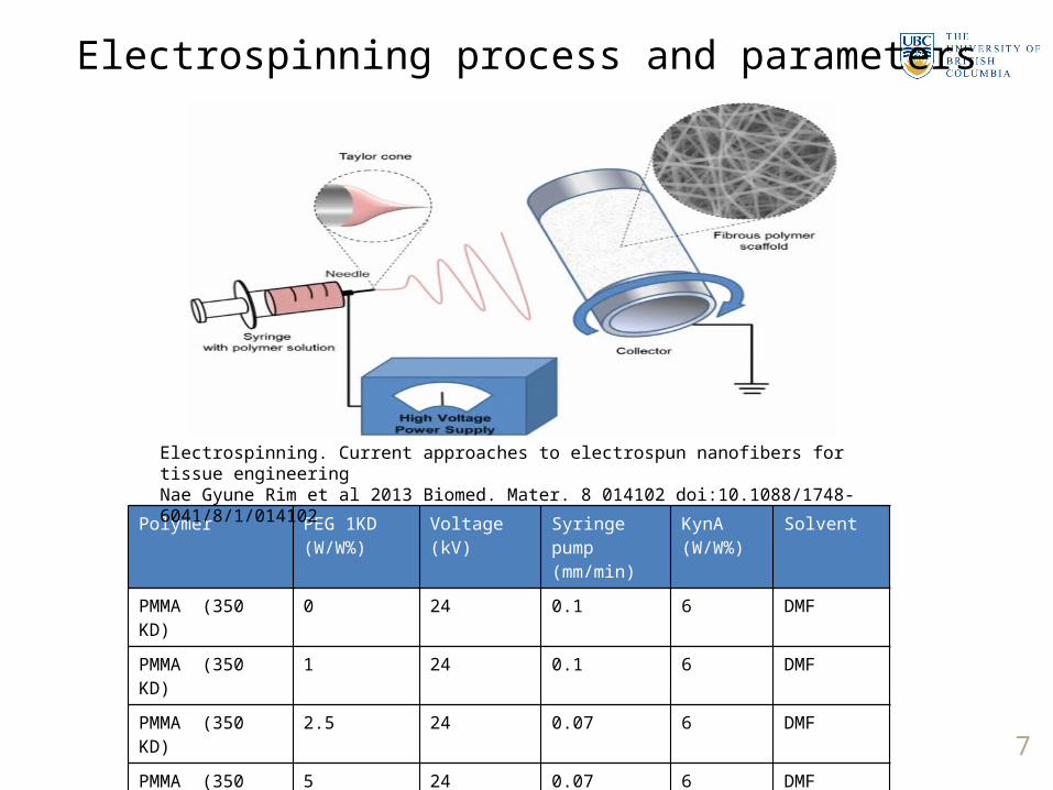
Electrospinning process and parameters
Polymer PEG 1KD(W/W%)
Voltage(kV)
Syringe pump (mm/min)
KynA(W/W%)
Solvent
PMMA (350 KD) 0 24 0.1 6 DMF
PMMA (350 KD) 1 24 0.1 6 DMF
PMMA (350 KD) 2.5 24 0.07 6 DMF
PMMA (350 KD) 5 24 0.07 6 DMF
PMMA (350 KD) 10 24 0.05 6 DMF
PMMA (350 KD) 20 24 0.05 6 DMF 7
Electrospinning. Current approaches to electrospun nanofibers for tissue engineeringNae Gyune Rim et al 2013 Biomed. Mater. 8 014102 doi:10.1088/1748-6041/8/1/014102

SEM images of PMMA-PEG nanofibers
10% 20%1% 2.5% 5%
PEG
1K
5K
10K
PMMA

Dressing’s hydrophobicity and wetting
1 2 5 10 20 300
2
4
6
8
10
12PMMA+10% PEG
PMMA
Time: Minutes
We
igh
t g
ain
e
(% o
f th
e d
res
sin
g w
eig
ht)
9
PMMA PMMA+10%PEGA
B

Release study in PBS, total immersion setting
A
0 20 40 60 80 100 1200
10
20
30
40
50
60
70
80
90
100
PMMA
1% PEG
2.5% PEG
5% PEG
10% PEG
20% PEG
Time: hours
Cu
mu
lati
ve
Ky
nu
ren
ic A
cid
Re
lea
se
(% L
oa
de
d)
10
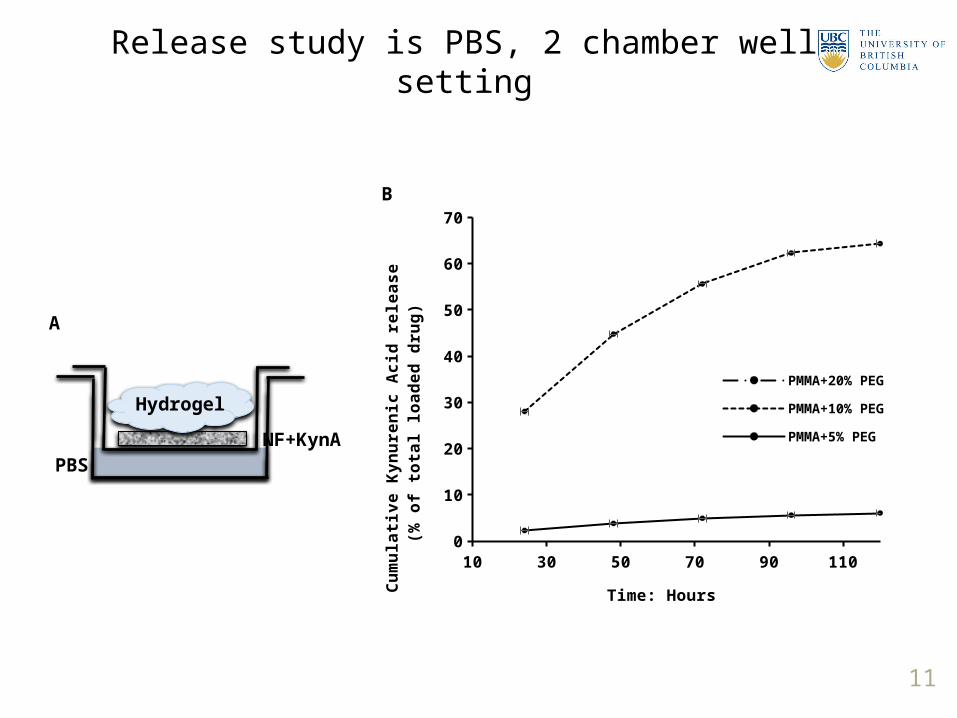
Release study is PBS, 2 chamber well setting
10 30 50 70 90 1100
10
20
30
40
50
60
70
PMMA+20% PEG
PMMA+10% PEG
PMMA+5% PEG
Time: Hours
Cu
mu
lati
ve
Ky
nu
ren
ic A
cid
re
lea
se
(%
of
tota
l lo
ad
ed
dru
g)
B
11
A
PBS
Hydrogel
NF+KynA

B
0 36 72 1080
40
80
120
160
200Control
NF
NF+KynA
KynA
Time:Hours
Fib
rob
las
t n
um
be
r (X
10
00
)
** **
**
**
**
Assessment of medicated mat’s cytocompatibility
12
Control NF NF+ KynA
FibroblastsC
Eth
idiu
m h
om
od
ime
r
Calcein
KynA
Dermal fibroblast
NF+KynA DMEM media
A

Dressing’s biological activity assessment
Con NF NF+KynA KynA0
20
40
60
80
100
120
140
160
180
200
MM
P1
/GA
PD
H(%
of
co
ntr
ol)
* *
Con NF NF+KynA KynA0
20
40
60
80
100
120
Co
llag
en
-I/G
AP
DH
(% o
f c
on
tro
l) ** **
A
Col-I
GAPDH
Con NF NF+KynA KynA
MMP1
13
B C
10x
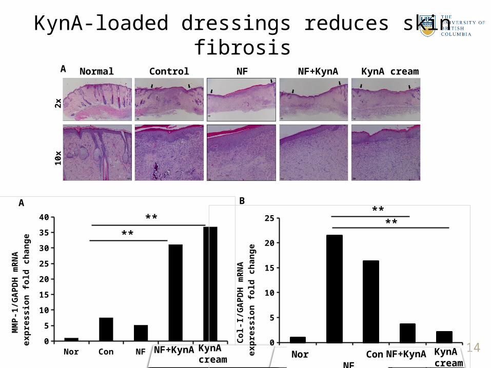
2xA Normal Control NF NF+KynA
KynA cream
14
KynA-loaded dressings reduces skin fibrosis
Nor Con NF NF+Kyn Kyn Cream
0
5
10
15
20
25
30
35
40
MM
P-1
/GA
PD
H m
RN
A
ex
pre
ss
ion
fo
ld c
ha
ng
e
**
**
A
NF+KynA KynA cream Nor
Con NF
NF+Kyn
Kyn C
ream
0
5
10
15
20
25
Co
l-I/G
AP
DH
mR
NA
e
xp
res
sio
n f
old
ch
an
ge
****
B
Nor Con NF NF+KynA KynA cream

Conclusion
PMMA+10% PEG nanofibers can be used as an effective slow releasing drug delivery system for KynA.
Nanofiber-released KynA effectively modulates the expression of ECM components in vitro and in vivo.
Application of KynA-loaded nanofibers can improve the wound healing outcome in patients prone to develop skin fibrosis.

Dr. Aziz GhaharySanam SalimiDr. Layla NabaiRyan HartwellDr. Reza JaliliDr. Yunyuan Li Dr. Ruhi Kilani
Acknowledgements
16
Collaborators:Dr. Hellen Burt
John JacksonDr. Frank Ko
Victor LeungDr. Emma Guns
Dr. Azadeh TabaDr. Yun ZangAli FarokhiDr. Mohsen KhosraviDr. Saman Pakyari

Thank you for your attention!