Complex Imaging for the Oral and Maxillofacial Region€¦ · Complex Imaging for the Oral and...
16
X-Ray Lab & Imaging Currents Conventional two-dimensional radiographic procedures are routinely used for oral and maxillofacial diagnostic tasks. However, these pro- cedures fail to provide information in the third dimension, often ne- cessitating use of advanced imaging modalities. Routine use of com- puted tomography and magnetic resonance imaging is limited by the relative inaccessibility of the imaging modality to the dental sur- geon, the high cost and radiation burden of the examination, the need to immobilize the patient for long periods of time, and the potential for artifact generation, among other things. These imaging modali- ties provide three-dimensional information thereby allowing multiplanar examination of the region of interest. Conventional to- mography is also used for a variety of diagnostic and treatment plan- ning tasks and is often used by dentists due to the lower cost and radiation exposure, and greater accessibility. Conventional tomographic image acquisition employs different con- figurations of motion of the x-ray source. State-of-the-art conven- tional tomographic units such as the OP-100, Scanora and the CommCAT employ a variety of tomographic motions ranging from linear to more complex configurations such as hypocycloidal, tri- spiral or figure-of-eight. The more complex the motion, the better is the blurring of superlying anatomy. Linear configuration of motion involves the generation of linear streak artifacts, which are effec- tively removed by units employing complex motion sequences. The amplitude of travel of the x-ray source determines the slice thick- ness. Implant diagnostic imaging using tomography It is generally recommended by the oral and maxillofacial maxillofa- cial radiologists that some form of cross sectional imaging be em- ployed for evaluation of potential implant sites. Unfortunately, pan- oramic images are used by a majority of surgeons for implant treat- ment planning purposes. Numerous inherent drawbacks of this im- aging modality such as distortions, magnifications, artifacts and ghost images limit the diagnostic value of panoramic images. continued on page 8 Quarterly Publication of the American Association of Dental Maxillofacial Radiographic Techicians Winter 2002 Complex Imaging for the Oral and Maxillofacial Region by: Madhu K. Nair BDS DMD MS Dipl.ABOMR Madhu K. Nair BDS DMD MS Dipl. ABOMR. Director, Oral and Maxillofacial Radiology University of Pittsburgh Inside This Issue Complex Imaging Dr. Madhu................1 President's Message Craig Dial............2 Editor's View Camille Mayorga............5 Tribute to Quint Diane Yamamoto....... 5 News and Trends ..................................6 Lab Promo Beverly Studer ................8 Member’s Recipe Diane Yamamoto........10 Spotlight Lab Dan Halpert ..............11 California Law Matt Kroona..............12 Chemical Phenomenon Gail Finnigan....14 Lab Products............................................15 1
Transcript of Complex Imaging for the Oral and Maxillofacial Region€¦ · Complex Imaging for the Oral and...

X-Ray Lab & Imaging
Currents
Conventional two-dimensional radiographic procedures are routinelyused for oral and maxillofacial diagnostic tasks. However, these pro-cedures fail to provide information in the third dimension, often ne-cessitating use of advanced imaging modalities. Routine use of com-puted tomography and magnetic resonance imaging is limited by therelative inaccessibility of the imaging modality to the dental sur-geon, the high cost and radiation burden of the examination, the needto immobilize the patient for long periods of time, and the potentialfor artifact generation, among other things. These imaging modali-ties provide three-dimensional information thereby allowingmultiplanar examination of the region of interest. Conventional to-mography is also used for a variety of diagnostic and treatment plan-ning tasks and is often used by dentists due to the lower cost andradiation exposure, and greater accessibility.Conventional tomographic image acquisition employs different con-figurations of motion of the x-ray source. State-of-the-art conven-tional tomographic units such as the OP-100, Scanora and theCommCAT employ a variety of tomographic motions ranging fromlinear to more complex configurations such as hypocycloidal, tri-spiral or figure-of-eight. The more complex the motion, the better isthe blurring of superlying anatomy. Linear configuration of motioninvolves the generation of linear streak artifacts, which are effec-tively removed by units employing complex motion sequences. Theamplitude of travel of the x-ray source determines the slice thick-ness.Implant diagnostic imaging using tomographyIt is generally recommended by the oral and maxillofacial maxillofa-cial radiologists that some form of cross sectional imaging be em-ployed for evaluation of potential implant sites. Unfortunately, pan-oramic images are used by a majority of surgeons for implant treat-ment planning purposes. Numerous inherent drawbacks of this im-aging modality such as distortions, magnifications, artifacts and ghostimages limit the diagnostic value of panoramic images.
continued on page 8
Quarterly Publication of the American Association of Dental Maxillofacial Radiographic Techicians Winter 2002
Complex Imaging for the Oral and Maxillofacial Region
by: Madhu K. Nair BDS DMD MS Dipl.ABOMR
Madhu K. Nair BDS DMD MS Dipl. ABOMR.Director, Oral and Maxillofacial RadiologyUniversity of Pittsburgh
Inside This IssueComplex Imaging Dr. Madhu................1
President's Message Craig Dial............2
Editor's View Camille Mayorga............5
Tribute to Quint Diane Yamamoto....... 5
News and Trends ..................................6
Lab Promo Beverly Studer ................8
Member’s Recipe Diane Yamamoto........10
Spotlight Lab Dan Halpert ..............11
California Law Matt Kroona..............12
Chemical Phenomenon Gail Finnigan....14
Lab Products............................................15
1

President's Message
2002 OFFICERSAmerican Association of Dental Maxillofacial Radiographic Technicians
Executive Committee President Craig Dial
Vice President Duane Perry
Secretary Merry Hampton
Treasurer Claudette Buehler
Board Members Membership Gail Finnigan
Recruitment Peggy Weist & Eleanora Prescott
Nominating Donna Lauritzen
Advertising Randy Sailors
Standing Committe Chairs By Laws Jeannie Herriott
Continuing Education Matt Kroona
Staff Photographer Phil Abel
Research Craig Dial & Don Croall
Web Master Matt Kroona
Editor Camille Mayorga
Craig Dial
2

3

4

Editor's View
Putting this issue together was fun and rewarding. I enjoy the process of watching itcome together and take great pleasure in the phone calls and emails with members’comments and suggestions. Summer is a busy time for all the labs, but I hope each ofyou will set aside some time for going somewhere or doing something special. I amlooking forward to seeing everyone in Denver! Camille Mayorga [email protected]
Happy Belated Birthday Mr. Arthur Quint!Born May 28, 19_ _
It should be stated that Mr. Quint would be considered the most senior memberof our organization. Well seasoned and still competent and feisty, he leads ourindustry in formatting radiographic laboratories, supplying all of us with thelatest state-of-the-art equipment, and a service department that has always stoodby the Quint name.
Mr. Quint supported us and knew us when we were just a small group oflaboratory owners known; back then, as CORLA (California Oral Radio-graphic Laboratories Association). He graciously acquiesced to our request of
using his Los Angeles based office as a meeting place for the organization. He even providedus with coffee and doughnuts! Remember that! All you older guys!
Oh-and how many of you have that old Quint Sectograph?Yes, the old Quint “Maytag”, that’s what the Sectograph is. Never any “down time”. That oldmachine is a workhorse. Still required by most labs and still being improved to this day. Everytime I turn around Mr. Quint has added something useful to that old machine of mine. Thank youMr. Quint for NOT putting a motherboard in your Sectograph. Oh yes, there is one thing aboutthat Old Faithful machine—about every two years or so the darn light burns out in the collimatorand I have to change the bulb. But that’s about it!
How is it that you look so great? Someone told me that it was the Gefil Tefish. I asked your wifeand she told me that you don’t even like Gefil Telfish. Must be that bagel you eat every morning.
Well, I hope you had a wonderful birthday and I didn’t want to take the liberty of telling everyoneyour real age. I think that’s up to you. But, your wife spilled it to me the other day. You areamazing!!
Sincerely Your Friend,Diane Yamamoto
5
News and Trends
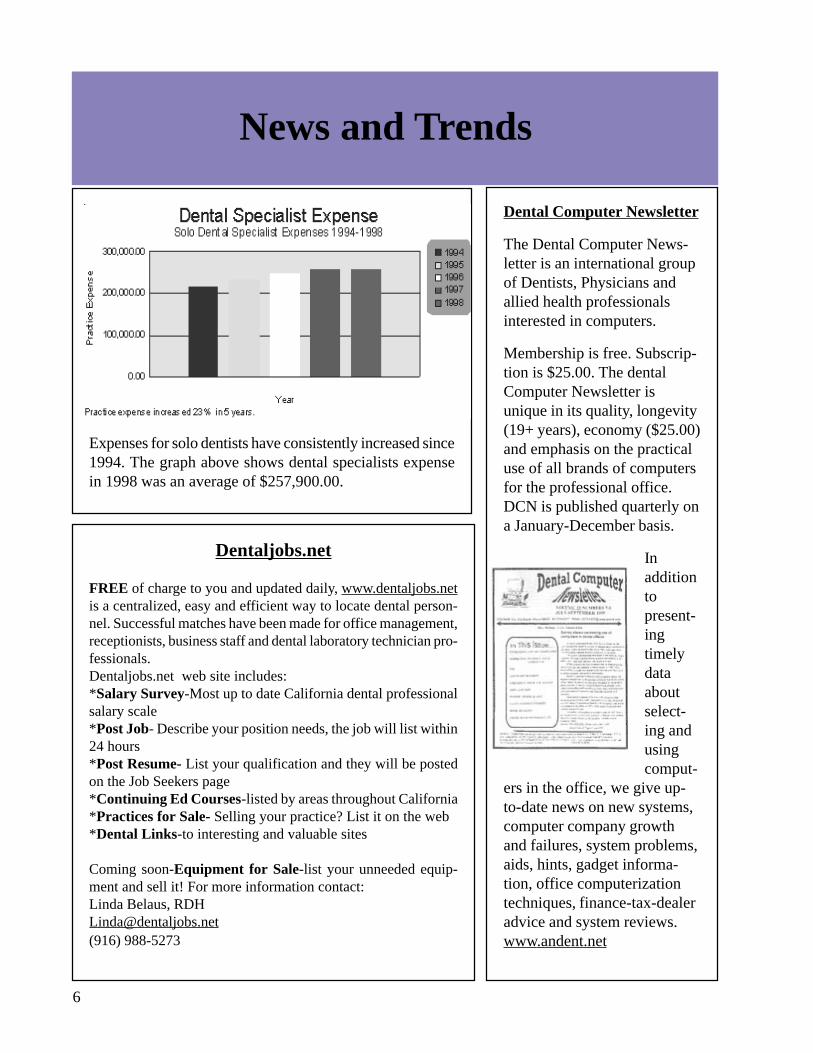
Expenses for solo dentists have consistently increased since1994. The graph above shows dental specialists expensein 1998 was an average of $257,900.00.
Dental Computer Newsletter
The Dental Computer News-letter is an international groupof Dentists, Physicians andallied health professionalsinterested in computers.
Membership is free. Subscrip-tion is $25.00. The dentalComputer Newsletter isunique in its quality, longevity(19+ years), economy ($25.00)and emphasis on the practicaluse of all brands of computersfor the professional office.DCN is published quarterly ona January-December basis.
Inadditiontopresent-ingtimelydataaboutselect-ing andusingcomput-
ers in the office, we give up-to-date news on new systems,computer company growthand failures, system problems,aids, hints, gadget informa-tion, office computerizationtechniques, finance-tax-dealeradvice and system reviews.www.andent.net
Dentaljobs.net
FREE of charge to you and updated daily, www.dentaljobs.netis a centralized, easy and efficient way to locate dental person-nel. Successful matches have been made for office management,receptionists, business staff and dental laboratory technician pro-fessionals.Dentaljobs.net web site includes:*Salary Survey-Most up to date California dental professionalsalary scale*Post Job- Describe your position needs, the job will list within24 hours*Post Resume- List your qualification and they will be postedon the Job Seekers page*Continuing Ed Courses-listed by areas throughout California*Practices for Sale- Selling your practice? List it on the web*Dental Links-to interesting and valuable sites
Coming soon-Equipment for Sale-list your unneeded equip-ment and sell it! For more information contact:Linda Belaus, [email protected](916) 988-5273
6

News and Trends
Group Rate for Liability InsuranceIs there any interest among the membersto look into a group rate for liabilityinsurance? If you have any comments orthoughts on this please contact GaryCason at: [email protected] (650) 348-1012
Kodak Builds Image Quality, ProcessingFlexibility Into New Extraoral Film
ROCHESTER, N.Y., February19—Eastman Kodak Com-pany announced today twonew extraoral dental films thatachieve a new standard for im-age quality while accommo-dating a wider range of pro-cessing conditions.Kodak Ektavision G ExtraoralFilm is a high-contrast film
ideal for visualizing detail of teeth, bony structuresand pathoses such as soft tissue calcifications. KodakEktavision L Extraoral Film is a wide-latitude filmsuitable for imaging soft tissue. “These films repre-sent the next generation in extraoral dental film,” saidDavid Allen, Kodak dental general manager for UnitedStates and Canada.Laurie Carter, DDS, PhD, associate professor and di-rector of oral and maxillofacial radiology at VirginiaCommonwealth University School of Dentistry as-sisted Kodak in a clinical evaluation of Ektavision GFilm. “It was absolutely superior to the comparisonfilms,” reported Dr. Carter. “You could see it fromacross the room.”Dr. Carter used a dry skull test to compare EktavisionG Film with two other extraoral films from Kodak:Kodak Ektavision Imaging System Film and KodakT-Mat G Extraoral Film. She ran a series of four im-ages using each film. “With Ektavision G Film, struc-tures just popped out,” said Dr. Carter. “I could seesubtle, paper-thin septations in the sinuses that werebarely visible using the other films. I was amazed athow many more of these structures I could see.”Dr. Carter now uses the new film to image dental pa-tients when the previous screening has indicated thepresence of pathologies. “I like a contrasty film whenperforming osseous evaluations,” said Dr. Carter.“Ektavision G Film is, in my opinion, an excellentdiagnostic tool.”
AADMRT Welcomes 7 New Membersfrom The Wilson Radiographic Centersof Houston Texas:
Kim Franks Randy SailorsKim Haynes Lori McConathyMandy Ledbetter Bacilia MorenoEleanora Prescott
CommCAT For Sale:
CommCAT X-Ray Unit. Included isDell Computer, HP Scanner, SurgPlanSoftware V3.12, and current CommCATsoftware. $45,000.00 or best offer.Contact: Craig Dial (916) [email protected]
Arizona to Launch Dental School
The Arizona school of health sciencesreceived approval to open a school ofdentistry and oral health in Mesa. Theschool will start its first class of 75students in the fall of 2002.www.ashs.edu
7

Complex Imaging continued:
Negative vertical angulation of projection results in a grossly inaccurate depiction of primarily verticaldimensions.
Use of CT is not indicated in every case owing to the numerous disadvantages listed earlier. Besides, manyimaging centers do not have an understanding of the image acquisition process for purposes of evaluatingimplant sites, so much so that faulty angulations of the patient’s anatomy results in the distortion of refor-matted images. The procedure is very technique sensitive and requires appropriate patient positioning andchoice of scanning protocols. Use of a spiral CT scanning mode employing low mAs, and a minimalnumber of slices help to keep the radiation exposure to a minimum.
Conventional tomography offers a convenient alternative in cases where fewer than 7 or 8 sites are beingevaluated per jaw. Imaging stents are recommended for use with conventional radiography but in in-stances where longitudinal imaging is carried out, stents are not essential. Tooth form lead foil markers areusually recommended over other types of radiopaque markers such as stainless steel balls, cylinders etc.The lead foil from intra-oral film packets are used for this purpose. These are draped over the occlusalcontours of the tooth form and usually have a long buccal arm that helps with patient positioning. In unitssuch as the CommCAT, the model of the patient’s arch with the stent placed on it is scanned in using aflatbed scanner and imported into the software interface for interactive arch plotting. The stent with theradiopaque marker makes it possible to select the slices of interest on the template that is superimposed onthe scanned in image. Alternatively, a cross-sectional occlusal radiographic survey can be used for inputpurposes too. The CommCAT software interface superimposes a slice selection template on the scanned inarch form image. This can be further customized to suit the diagnostic task at hand by changing the sliceangles and projection geometry. Generally a cross-sectional and a longitudinal slice are obtained to corre-late the anatomic landmarks and precisely determine the location of the cut. However, in the presence of astent with a radiopaque marker such as a lead foil, a longitudinal image is not always required. Themagnification factor specified by the manufacturer should be worked into any computations while mea-surements are made from the tomograms. Ideally, a tracing of the tomograms with a written report shouldbe made available to the surgeon. If the imaging stent correlates well with tomographic data, it can bemodified by the surgeon to act as a surgical stent. There are treatment-planning algorithms such as SurgPlanthat enables limited task-based image processing, measurements and simulation of implant placement andbone graft procedures on tomograms.
Slice thickness of 1-3 mm can be selected for cross-sectional tomograms (Figure 1), depending on theimaging modality and unit employed. However, the contrast resolution on a 1mm thick slice acquiredusing conventional techniques is far from satisfactory. Besides, most implants are 3mm or more in crosssectional width. The use of 3-4 mm thick slices is therefore, fully justified in these cases. Conventionaltomography also enables examination of isolated areas of interest unlike CTwhere a set of axial images of the entire jaw of interest is acquired. Otheradvantages of CT include the superior contrast resolution, uniform magnifi-cation, capability to view multiplanar projections and 3-D reconstructions.The use of 3-D reconstructions in pre-surgical implant treatment planninghas not been shown to yield additional data and therefore is probably notwarranted.
Figure 1
9 Figure
Figure 4
10
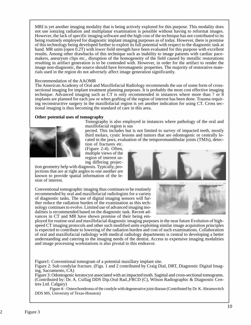
MRI is yet another imaging modality that is being actively explored for this purpose. This modality doesnot use ionizing radiation and multiplanar examination is possible without having to reformat images.However, the lack of specific imaging software and the high cost of the technique has not contributed to itsbeing routinely employed for diagnostic implant imaging purposes as of today. However, there is promiseof this technology being developed further to exploit its full potential with respect to the diagnostic task athand. MR units (open 0.2T) with lower field strength have been evaluated for this purpose with excellentresults. Among other drawbacks of this technique such as inability to image patients with cardiac pace-makers, aneurysm clips etc., disruption of the homogeneity of the field caused by metallic restorationsresulting in artifact generation is to be contended with. However, in order for the artifact to render theimage non-diagnostic, the source should have ferromagnetic properties. The majority of restorative mate-rials used in the region do not adversely affect image generation significantly.
Recommendation of the AAOMRThe American Academy of Oral and Maxillofacial Radiology recommends the use of some form of cross-sectional imaging for implant treatment planning purposes. It is probably the most cost effective imagingtechnique. Advanced imaging such as CT is only recommended in instances where more than 7 or 8implants are planned for each jaw or when grafting of the region of interest has been done. Trauma requir-ing reconstructive surgery in the maxillofacial region is yet another indication for using CT. Cross sec-tional imaging is thus becoming the standard of care in this area.
Other potential uses of tomographyTomography is also employed in instances where pathology of the oral andmaxillofacial region is suspected. This includes but is not limited to survey of impacted teeth, mostlythird molars, cystic lesions and tumors that are odontogenic or centrally lo-cated in the jaws, evaluation of the temporomandibular joints (TMJs), detec-tion of fractures etc.(Figure 2-4). Often,multiple views of theregion of interest us-ing differing projec-
tion geometry help with diagnosis. Typically, pro-jections that are at right angles to one another areknown to provide spatial information of the le-sion of interest.
Conventional tomographic imaging thus continues to be routinelyrecommended by oral and maxillofacial radiologists for a varietyof diagnostic tasks. The use of digital imaging sensors will fur-ther reduce the radiation burden of the examination as this tech-nology continues to evolve. Limited use of advanced imaging mo-dalities is recommended based on the diagnostic task. Recent ad-vances in CT and MR have shown promise of their being em-ployed for routine oral and maxillofacial diagnostic imaging purposes in the near future Evolution of high-speed CT imaging protocols and other such modified units exploiting similar image acquisition principlesis expected to contribute to lowering of the radiation burden and cost of such examinations. Collaborationof oral and maxillofacial radiology with medical radiology departments is central to developing a betterunderstanding and catering to the imaging needs of the dentist. Access to expensive imaging modalitiesand image processing workstations is also pivotal to this endeavor.
Figure1: Conventional tomogram of a potential maxillary implant site.Figure 2: Sub condylar fracture. (Figs. 1 and 2 contributed by Craig Dial, DRT, Diagnostic Digital Imag-ing, Sacramento, CA)Figure 3: Odontogenic keratocyst associated with an impacted tooth. Sagittal and cross-sectional tomograms.(Contributed by: Dr. A. Csillag DDS Dip.Oral Rad.,FRCD (C), Wilson Radiographic & Diagnostic Cen-tres Ltd. Calgary)
Figure 4: : Osteochondroma of the condyle with degenerative joint disease (Contributed by Dr. K. AbramovitchDDS MS, University of Texas-Houston)
2 Figure 3
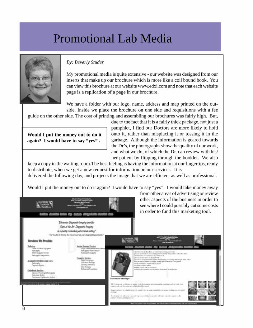
By: Beverly Studer
My promotional media is quite extensive - our website was designed from ourinserts that make up our brochure which is more like a coil bound book. Youcan view this brochure at our website www.edxi.com and note that each websitepage is a replication of a page in our brochure.
We have a folder with our logo, name, address and map printed on the out-side. Inside we place the brochure on one side and requisitions with a fee
guide on the other side. The cost of printing and assembling our brochures was fairly high. But,due to the fact that it is a fairly thick package, not just apamphlet, I find our Doctors are more likely to holdonto it, rather than misplacing it or tossing it in thegarbage. Although the information is geared towardsthe Dr’s, the photographs show the quality of our work,and what we do, of which the Dr. can review with his/her patient by flipping through the booklet. We also
keep a copy in the waiting room.The best feeling is having the information at our fingertips, readyto distribute, when we get a new request for information on our services. It isdelivered the following day, and projects the image that we are efficient as well as professional.
Would I put the money out to do it again? I would have to say “yes”. I would take money awayfrom other areas of advertising or reviewother aspects of the business in order tosee where I could possibly cut some costsin order to fund this marketing tool.
Promotional Lab Media
Would I put the money out to do itagain? I would have to say “yes” .
8

Spotlight Lab
Halpert Dental X-Ray,Northridge, Californiaby: Dan Halpert
HDX opened for business in on April 1, 1976. Aprilfools day. I thought it might be very symbolic. Most ofthe 1970’s were recession years in this area. The aero-space business was in the first of many down turns. Ihad hopes that I was not the fool, having just borrowedsome money and purchased some of the equipment of alocal laboratory that had just closed up due to lack ofwork. Twenty five years later I believe that I can saythis was a good decision.A funny thing happened on my way to my chosen career. A new and different career chose me!With a strong background in electronics and a love of photography I had my heart set on becoming abroadcast engineer. The reality was that I was not earning enough income as a freelance newspaper
photographer to get through the fall semester atcollege. Luckily I found a summer job at a den-tal x-ray laboratory as a darkroom person. Thiswas a very busy lab and all films were hand pro-cessed (1969). I was interested in the imagingas well as the professional relationship betweenthe technicians and the referring doctors. Luck-ily the State of California had just announcedpending legislation that would require all tech-nicians to pass a State Department of Healthexam.The company I worked for had 5 labs and wantedto make sure that all their technicians passed theexam so they started classes for their employ-ees. I was called upon to help them with theelectronics portion of the exam. As a result ofthis legislation, a number of laboratory ownersbanded together and formed CORLA which laterswitched to a technician based membership andeventually became the AADMRT.I find that I am still fascinated by imaging, pho-tography and electronics and it is with great ex-citement that I find that they are merging intothis field with much greater importance. 1993was the first year that we were involved withdigital photography, one of the first 2 labs to usethe Kodak/Nikon DCS series cameras. In 1994
11

Understanding the CaliforniaRulemaking Process
By: Matt Kroona
Many people have expressed concern and ordistress over the uncertainty of the require-ments for Continuing Education for CaliforniaLimited Permits, and the number of years wehave lived with that uncertainty. I recentlydiscover some interesting information regard-ing the California Rulemaking Process that Ithought might be useful. This informationrelates to California but I expect that moststates operate in a similar manner.
In 1979, the California Legislature createdthe Office of Administrative Law (OAL) toensure that state agency regulations are autho-rized by statute, consistent with other law, andwritten in a comprehensible manner. Each year,state agencies propose thousand of regulationswhich, when adopted, affect almost all eco-nomic activities and every segment of theCalifornia public. OAL reviews each proposed regulation and approves a regulation only when therulemaking agency has adequately considered public comments and if the regulation is easily under-stood, necessary, authorized, and consistent with law. When approved and filed with the Secretary ofState, a regulation has the force of law. Those who fail to comply with regulations can be cited, fined or suffer adverse consequences, such aslosing a license or closure. As you can see, this can be a very long and time consuming process. The flow chart below can helpus better understand why we often wait so long for regulations to go into effect.
The California Rulemaking Process by which regulations are proposed, reviewed, and adopted orrejected:
1-State Agency -Starts rulemaking file. -Writes proposed regulations. -Prepares Initial Statement of reasons which sets out factual basis for proposal. -Issues public notice and 45 day, public comment period begins.2-Public Notice -Printed in Notice Register and sent to those on agency’s mailing list.
3-Public -Must write to agency to be put on its mailing list to receive notice. -Is encouraged to submit written comments to agency during this period (specific, factual statementsare more effective). -Has right to see actual text of proposed regulation, Initial Statement of Reasons,material on which agency relied. -Can challenge agency on meeting legal or procedural requirements, data used. -Can propose less burdensome method of compliance.
12

4-State Agency -Prepares Final Statement of reasons. -Summarizes primary points of individual comments from public. -Documents its decisions relative to points public raised. -Adopts final regulations(s). -Submits regulatory record including comments and responses to OAL for review and decision.5-Office of Administrative Law -Has 30 days to review entire contents of regulatory file. -OAL’s review holds agencies accountable to the regulated public and deters unnecessary, unclearand overly burdensome regulations from becoming law. -The same legal and procedural requirements applied to each regulation. -Review is limited to file contents.6-OAL Approval o Disapproval -In most cases, if OAL approves regulations, it is filed with Secretary of State and becomes effectivein 30 days. -If OAL disapproves a regulation, it is returned to agency and does not become law. -OAL issues a detailed disapproval decision that is printed in the California Regulatory NoticeRegister and in the California Code of Regulations Decisions -Agencies may appeal OAL’s disapproval to Governor
For the status of California’s Continuing Education regulation as well as other issues that may affectyou and your lab, check the AADMRT web site at www.aadmrt.com.
Chemical Phenomenon By: Gail Finnigan
This is an interesting chemical phenomenon. I hadmy regular weekly chemical service deliver freshchemicals. I proceeded to clean both my processorsand manually refill them with replenisher chemicalsleft for me. Please note: I have done this the sameway for quite a while and never had a problem. WhenI tried developing duplication film it looked foggedor not completely fixed. X-ray film was developing normally. After a half an hour of trial anderror, (thinking it was the film or the duplication light), every indication pointed to chemicalincompatibility. The chemical serviceman thought it was due to old developer in my large proces-sor replenishing tanks. It wasn’t, because the result was the same in my small processor, whichhad fresh replenisher bottles. If anyone has had this same experience, let me know if it was badfixer or perhaps something else. Finally, I poured a small amount of chemicals from my smallprocessors replenisher bottles into cups and developed a duplication exposed exactly the same,and they came out perfect. My only conclusion is that a different brand fixer was delivered thatwasn’t compatible. Everyone seemed baffled by this, and I never got an answer that satisfied me.
13

Tomography of the IonosphereBy: Marcelle Jones
The other day I took implant tomos on a very in-teresting patient. He told me about how he hadused tomography in his work with the defense de-partment. They were “mapping” the ionosphere.Satellites were used in space and tracking stationson the ground to get “slices” of the ionosphere atvarious locations. The analogy is the tracking sta-tions were the beam and the satellites were the film.The purpose of this was to locate enemy navalships-when the enemy sent radio signals throughthe ionosphere, we could track them and determinetheir location without detection. Because I hardlyunderstand a word of what I just wrote, I was thank-ful I only took implant tomograms and wasn’t deal-ing with enemies, ionospheres, and much morecomplex issues than teeth!This gentleman also told me about mapping thelandmasses from the Antarctica where he spent oneyear –1969. At that time, they were beginning touse satellites to help with more accuracy in map-ping. Apparently, they found the Easter islandswere off by more than one mile and other geo-graphical errors that had been accepted for years.He is now retired and lives in Colorado. He teachesskiing to handicapped adults. This guy made myday…what an inspiration!
14
Lab Products
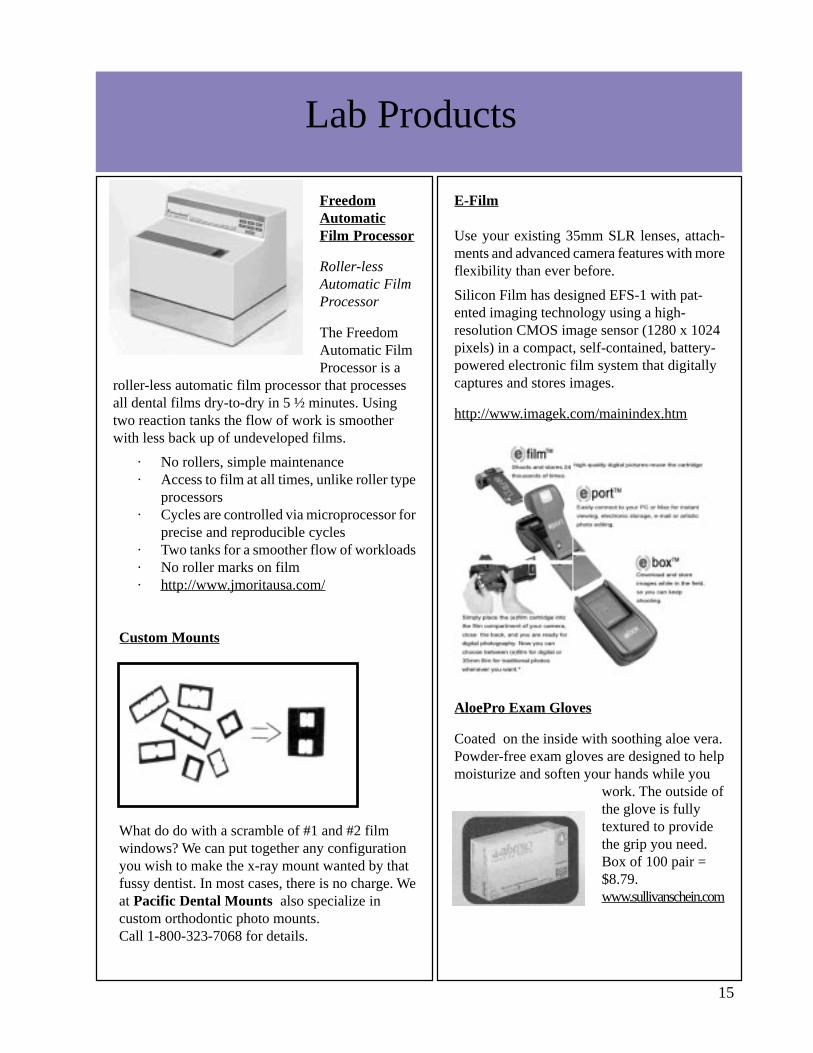
FreedomAutomaticFilm Processor
Roller-lessAutomatic FilmProcessor
The FreedomAutomatic FilmProcessor is a
roller-less automatic film processor that processesall dental films dry-to-dry in 5 ½ minutes. Usingtwo reaction tanks the flow of work is smootherwith less back up of undeveloped films.
· No rollers, simple maintenance· Access to film at all times, unlike roller type
processors· Cycles are controlled via microprocessor for
precise and reproducible cycles· Two tanks for a smoother flow of workloads· No roller marks on film· http://www.jmoritausa.com/
Custom Mounts
What do do with a scramble of #1 and #2 filmwindows? We can put together any configurationyou wish to make the x-ray mount wanted by thatfussy dentist. In most cases, there is no charge. Weat Pacific Dental Mounts also specialize incustom orthodontic photo mounts.Call 1-800-323-7068 for details.
E-Film
Use your existing 35mm SLR lenses, attach-ments and advanced camera features with moreflexibility than ever before.
Silicon Film has designed EFS-1 with pat-ented imaging technology using a high-resolution CMOS image sensor (1280 x 1024pixels) in a compact, self-contained, battery-powered electronic film system that digitallycaptures and stores images.
http://www.imagek.com/mainindex.htm
AloePro Exam Gloves
Coated on the inside with soothing aloe vera.Powder-free exam gloves are designed to helpmoisturize and soften your hands while you
work. The outside ofthe glove is fullytextured to providethe grip you need.Box of 100 pair =$8.79.www.sullivanschein.com
15
Newsletter Deadlines
Winter Issue Deadline, December 1
Spring Issue Deadline, March 1
Summer Issue Deadline, June 1
Fall Issue Deadline, September 1
For Advertising Information : [email protected]
For Article Submission Information: [email protected]
Or Visit our Website: www.aadmrt.org
www.aadmrt.orgX-Ray Lab & Imaging Currents
AADMRT1 Scripps Drive #101
Sacramento, CA 95825
Place Stamp Here
The Organization acknowledges the sponsersof this Newsletter with thanks:
Panoramic Dental Imaging Sciences InternationalDolphin ImagingFlow X-RayDesign TechnologyPacific Dental Mounts