Oral & Maxillofacial Surgery - Open Access...
19
Overview of Myofascial Pain Dysfunction Syndrome Nilima Budhraja*; Shenoi SR; Anurag Choudhary; Pranav Ingole; Vrinda Kolte; Kshitij Bang Department of Oral and Maxillofacial Surgery, VSPM’s DCRC, Nagpur, India. *Correspondensce to: Nilima Budhraja, Department of Oral and Maxillofacial Surgery, VSPM’s DCRC, Nagpur, India. Email: [email protected] Chapter 1 Oral & Maxillofacial Surgery Abstract Myofascial pain dysfunction syndrome (MPDS) is the pain that originates from myofascial trigger points in skeletal muscle. Myofascial pain is the most com- mon muscle pain disorder. There are different treatment modalities available for the management of MPDS. Treatment option ranges from non-surgical to surgi- cal; non-pharmacological to pharmacological and from conservative to invasive. Through this book chapter authors aims to provide widespread information regard- ing the etio-pathogenesis, clinical presentation, diagnostic methods and available treatment options for MPDS. 1. Introduction Temporomandibular joint disorders are most commonly misdiagnosed in medicine. This clinical entity has been identified in the clinical practice since along. It has multiple factor eti- ology resulting in difficulty of pointing out exact cause. Hence, it is important to learn how to rule out this type of temporomandibular joint dysfunction. Different terms used for the various temporomandibular joint [TMJ] symptoms: i) Costen`s syndrome ii) TMJ pain dysfunction syndrome and iii) Myofascial pain dysfunction syndrome (MPDS) Keywords: Myofascial pain dysfunction syndrome; temporomandibular joint disorder; myofascial pain; myofascial trigger points
Transcript of Oral & Maxillofacial Surgery - Open Access...
Overview of Myofascial Pain Dysfunction Syndrome
Nilima Budhraja*; Shenoi SR; Anurag Choudhary; Pranav Ingole; Vrinda Kolte; Kshitij Bang
Department of Oral and Maxillofacial Surgery, VSPM’s DCRC, Nagpur, India.
*Correspondensce to: Nilima Budhraja, Department of Oral and Maxillofacial Surgery, VSPM’s DCRC,
Nagpur, India.
Email: [email protected]
Chapter 1
Oral & Maxillofacial Surgery
Abstract Myofascial pain dysfunction syndrome (MPDS) is the pain that originates from myofascial trigger points in skeletal muscle. Myofascial pain is the most com-mon muscle pain disorder. There are different treatment modalities available for the management of MPDS. Treatment option ranges from non-surgical to surgi-cal; non-pharmacological to pharmacological and from conservative to invasive. Through this book chapter authors aims to provide widespread information regard-ing the etio-pathogenesis, clinical presentation, diagnostic methods and available treatment options for MPDS.
1. Introduction
Temporomandibular joint disorders are most commonly misdiagnosed in medicine. This clinical entity has been identified in the clinical practice since along. It has multiple factor eti-ology resulting in difficulty of pointing out exact cause. Hence, it is important to learn how to rule out this type of temporomandibular joint dysfunction.
Different terms used for the various temporomandibular joint [TMJ] symptoms:
i) Costen`s syndrome ii) TMJ pain dysfunction syndrome and iii) Myofascial pain dysfunction syndrome (MPDS)
Keywords: Myofascial pain dysfunction syndrome; temporomandibular joint disorder; myofascial pain; myofascial trigger points

Oral & Maxillofacial Surgery
2
ww
w.openaccessebooks.comB
udhr
aja
N
Costen (1934) was the first one to indicate an occlusal etiology in TMJ pain. He re-ported that due to loss of posterior teeth with symptoms like ear pain, sinus pain, decreased hearing, tinnitus, dizziness, burning and vertigo and occipital headaches [1].
Schwartz (1956) coined the term TMJ pain dysfunction syndrome and suggested that the spasm of the masticatory and perimasticatory musculature leading towards the symptoms. He also noted that many TMJ sufferers had an altered psychologic make up as well. He advo-cated the used of muscle relaxants, restriction of oral opening for resting the muscles [2].
Travel (1960) through the use of injection of isotonic saline solution intramuscularly showed how pain is transferred from area to another area.’ Trigger points ‘at preauricular area was located by him and concluded that trigger zone leads to referred pain [3].
Laskin (1969) put forward the myofascial pain dysfunction syndrome. He implicated psycho-physiologic theory stating that the psychological stress leads to myospasm [4].
The condition is characterized by unilateral facial pain secondary to the TMJ disorder which is often associated with periarticular muscle spasm and pain with dysfunction of the spastic muscles.
1.1. Definition
MPDS is a pain disorder in which unilateral pain is referred from the trigger point in myofascial structures, to the muscles of the head and neck. Pain is constant dull ache in con-trast to the sudden sharp, shooting intermittent pain of neuralgias (chronic pain) but the pain may range from mild to intolerable.
2. Etiology
The multifactorial origin the can be put into following major groups
1) Psychologic or central etiology 2) Occlusal or peripheral etiology 3) The third group is recently considered is due to intrinsic joint disorder etiology
According to Okeson, etiologic classification is: [5]
2.1. Direct or indirect trauma to the muscle
2.1.1. Direct traumatic
1. Direct blow to the jaw 2. Opening the mouth too wide or for too long a period In some cases

Oral & Maxillofacial Surgery
3
2.1.2. Indirect trauma
1. Whiplash-type 2. Local infection and trauma leading to myositis 3. Occupational and repetitive strain injury 4. Sleep disturbance and nocturnal habits 5. Oral parafunctional muscle tension–producing habits, such as teeth clenching, jaw thrust-ing
2.2. Aetiopathogenesis of MPDS
Repetitive strain hypotheses: [6]
Micro or macro traumatic events or continued muscle contractions
Over activity of Muscles
Muscle fatigue and Decrease level of ATP and energy
Anaerobic muscle environment
Noxious metabolic end products such as lactic acid gets accumulated
Muscle nociceptors gets activated resulting into Muscle nociception
Muscle tone becomes spastic and sore
Resulting into muscle tenderness and pain
3. Clinical Features
MPDS exhibits varied clinical features:

Oral & Maxillofacial Surgery
4
1. Pain
Sites of pain: jaw, facial, temple, frontal, occipital, preauricular, earache and neck pain. Nature of Pain: steady dull ache, can be acute to chronic and duration may vary from hours to days
2. Muscle tenderness
Trigger points (TrPs), is deep, localized, and about 2–5 mm in diameter located in skeletal muscle. They refer a characteristic pain pattern to a distant group of muscles i.e. zone of refer-ence.
3. Limited range of motion of TMJ
4. Clicking of the joint
5. Comorbid conditions - Joint disk displacement, osteoarthritis, malocclusion, connective tis-sue diseases, neuropathic pain disorder, tension-type headaches and gastrointestinal disorders
6. Psychological Symptoms – Bruxism, Clenching Anxiety, Depression, Fear etc
Laskin’s diagnostic creteria for MPDS:
There are four Characteristic sign of MPDS given by Laskin :
1. Unilateral / Bilateral pain in preauricular region that is commonly worse on awaking. 2. Tenderness of one or more muscles of mastication on palpation. 3. Limitation or deviation of mandible on opening. 4. Clicking and popping noise in TMJ
Laskin also emphasized that other than the above positive signs, the following signs must be absent:
1. There should be absence of clinical,radiographic or biochemical evidence of organic changes in TMJ 2. There should be no tenderness on palpation via external auditory meatus
3.1 History of the patient
Intelligent and effective treatment cannot be instituted without an accurate probably the most beneficial aid to diagnosis is a complete and standardized history and examination.
1) Description of Pain: Site, Mode of onset, duration, frequency, type of pain, reference point, aggravating or any relieving factor, and time of the day at which pain is most promi-nent.

Oral & Maxillofacial Surgery
5
2) Habits : Sleeping and Parafunctional (bruxism, cheek or lip biting. )3) History of traumatic injury4) Emotional Disturbance5) Occupation
3.2 Clinical examination
3.2.1. Extra-oral examination:
a. Palpation of TMJ (Fig 1 A and B) – observe for tenderness, synchronization of bilateral TMJ movement and clicking sound. Range of movement of hypomobility or hypermobility and deviation of mandible while opening of mouth.
Normal protrusive and lateral excursive movement range from 8-10mm. Limitation of this movement could be because of joint inflammation and muscle dysfunction.
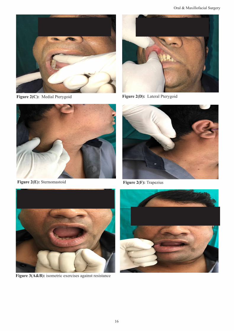
b. Palpation of muscle of mastication and neck muscles – observe for trigger areas [TrP], bilateral or unilateral tenderness, muscle tone, pathology or temperature change (Fig 2A-F).
The muscles are palpated bilaterally and simultaneously with gentle pressure. Observe for Grade of tenderness in various muscles; Grade 0: No pain; Grade 1: Mild pain; Grade 2: Moderate pain; Grade 3: Severe pain [7,8,9]. Palpating the active TrP with sustained deep, single-finger pressure on the taut band of muscle shows intensification, reduction or radiation of pain in the area of pain complaint known as zone of reference.
3.2.2. Intraoral examination
a. Occlusion: Occlusal contacts, Anterior open bite, cross bite, reduced vertical dimension observe b. High points or premature contact if any prosthesis or restoration is there c. Attrition and occlusal facets
4. Investigations
4.1. Radiographic evaluation
The radiographic evaluation is needed to rule out associated pathologies of TMJ
a. OPG- sectional view b. Transpharyngeal view c. Transcranial view d. MRI is the most appropriate investigation to assess the internal soft tissue pathologies of the joint.

Oral & Maxillofacial Surgery
6
It is helpful in diagnosis of the following:
i) Intra articular pathologies ii) Osseous pathologies iii) Soft tissue pathologies
4.2. Recent diagnostic methods
i) TMJ arthrography ii) CT scan and MRI iii) Bone scintigram – nuclear imagine
5. Differential Diagnosis of the Myofascial Pain Dysfunction Syndrome
Various conditions associated with the head and neck region may mimic the pain sec-ondary to MPDS and thus, they warrant consideration for ruling them out.
a. Migraine-more commonly seen in females, pain is severe in the morning, associated with nausea, vomiting and visual abrasions. b. Eagle’s syndrome- Pain during opening of the jaws, deglutition c. Mastoiditis – pain and tenderness over the mastoid process. d. Temporal arthritis – pain and tenderness along the course of temporal arteries. e. Otitis media –chiefly ear pain, hearing impairment. f. Local Muscle Soreness - Muscular soreness is expected result of exercise.
5.1. Treatment of MPDS
Definitive treatment of MPDS involves removal of the underlying etiology. Treatment of MPDS can be done by following modalities:
I. Non surgical management :
i. Counseling and reassurance ii. Stress management and relaxation therapy. iii. Pharmacological iv. Non- Pharmacological a. Injections at Myofascial trigger points b. Physiotherapeutic c. Occlusal splints. v. Intra-articular injections.

Oral & Maxillofacial Surgery
7
II. Surgical management i. Arthrocentesis ii. Arthroscopic iii. Arthrotomy
5.1.1. Non surgical management
i. Counselling and reassurance
Counselling of the patient should be done so that there is acceptance of the treatment modality of the MPDS. Counselling regarding prognosis, modification of lifestyle, daily activ-ity, diet, exercises and incorporation of different ways to reduce stress aids in management of MPDS. Reassurance to the patient motivates to undertake the indicated treatment.
Initially it is advised to take soft diet with smaller bites and to avoid painful move-ments.
ii. Stress Management
Now a day’s stress is increasing in individual’s life. Daily exercises, yoga and deep breathing relaxation therapy is considered to be helpful in relieving stress. Biofeedback device is also useful to relax as it allows patient to observe their increased muscle activity and then relax.
iii. Pharmacological
a. Analgesic
Nonsteroidal anti-inflammatory drugs (NSAIDs) are the most commonly used drugs for MPDS because of their analgesic and anti-inflammatory properties and have a relatively mild side-effect profile. However, long-term use should be avoided due to the side-effects such as gastrointestinal, renal, and antiplatelet [10].
The analgesic-anti inflammatory drugs like Ibuprofen, Diclofenac sodium and CoX 2 inhibitors can be used. Studies using Lidocaine and Diclofenac transdermal patch had shown their significant benefits for management of pain [11-14].
NSAIDs (oral route) can be given for 5-7 days for myositis and TMJ inflammatory dis-orders.
b. Muscle relaxants
Muscle relaxants are administered to reduce skeletal muscle tone and chronic orofacial pain to decrease muscle tone without the impairment of motor function by acting centrally to

Oral & Maxillofacial Surgery
8
depress polysynaptic reflexes.
Studies have suggested that tizanidine should be considered as a first-line agent for the treatment of MPS [15].
Review had shown that use of Cyclobenzaprine and Clonazepam as a muscle relaxant is effective, but should be used cautiously due to associated side effects [10].
A commercially available combination of a muscle relaxant and analgesics consisting of Ibuprofen 400 mg, Paracetamol 325 mg, and Chlorzoxazone 250 mg can administered orally twice a day for 5-7 days initially.
For acute spasm or Myofascial pain, Dypirone 500 mg can be given with a muscle relax-ant, three times daily for two days.
c. Anxiolytic drugs
Alprazolam-0.25,0.5 mg HS or Diazepam- 2mg to 5 mg HS can be used for stress man-agement.
d. Antidepressant
Tricyclic Antidepressant affects central pain pathways [16]. There are limited studies specifically for the treatment of MPS, though they are widely used.
Sumatriptan is a peripheral 5-HT receptor agonist and data suggest that serotonin plays a role in the pathophysiology of myofascial pain [17].
e. Botulinum type A toxin
BoNT-A is a potent neurotoxin that prevents muscle contraction. The literature suggests that BoNT-A injection can be used for management of MPDS. Its side effects of muscle weak-ness and paralysis are mostly local and reversible [18].
iv. Non-Pharmacological
a. Injections at Myofascial trigger points ( MTrPs) – Anesthetic solutions, steroids or dry Needling
Termination of the dysfunctional activity of involved motor endplates due to mechani-cal disruption by the needle results from injections of short or long-acting anesthetic solutions, steroids or dry needling.
In dry needling the needle is placed into MTrPs using an in-and-out technique in mul-tiple directions, in order to inactivate the MTrP but it is an invasive procedure. Literature

Oral & Maxillofacial Surgery
9
has shown that dry needling is an effective treatment and can be used for acute management [19].
Trigger point injections of local anesthetic solutions are similar to dry needling; howev-er it has similar efficacy to dry needling [20]. Steroid injections into MTrPs are controversial.
b. Physiotherapeutic
At acute stage only rest is advised. Physiotherapy plays a role in chronic stage.
Manual Therapy
Manual Therapy such as deep-pressure massage, stretch therapy after vapocoolant spray, application of superficial or deep heat has been considered for management of MPDS [21]. But there have been no controlled studies proving their significant long-term effectiveness.
Massage with vibrator inactivates the MTrPs.
Heat application through moist or dry heating pads or hot moist application of towels can be given for 15 to 20 minutes, 4 times a day. It increases local circulation and lowers muscle ten-sion.
Ethyl chloride vapocoolent spray is applied to painful area for 5 second and then the muscle is stretched.
Ice packs application 4 times a day for 20 minutes raises cutaneous pain threshold by the counter irritation.
Ultrasounds
Ultrasound is a technique that converts electrical energy to sound waves and thus pro-duces heat deep into the tissue and alters the metabolic activity [22]. It is given by the physio-therapist every alternate day.
Transcutaneous electronic nerve stimulator (TENS)
TENS stimulate nerve fibers using an electrical current to provide relief from pain. TENS is beneficial and can be used as an adjuvant therapy.
Infrared diathermy , Short wave diathermy, Microwave diathermy
They use electromagnetic waves that generate heat and provide deeper penetration of the heat. The shorter the wave length, deeper is the heat penetration therefore microwave dia-thermy produces deeper heat penetration.

Oral & Maxillofacial Surgery
10
Electrical twitch obtaining intramuscular stimulation (ETOIMS)
ETOIMS passes an electrical current through a monopolar electromyography needle to engage deep motor endplates. However, there is lack of evidence regarding its utility in man-agement of MPDS [23].
Light amplification by stimulated emission of radiation” (LASER)
The use of low level laser therapy (LLLT) is important conservative management of MPDS. The laser device, a gallium-aluminum-arsenide diode with a wavelength of 810 nm and a continuous 0.5 W peak power output beam with 5 mm spot size can be used. The probe is held perpendicularly with a light pressure on the targeted muscle in continuous mode and in contact with a light pressure on tender points diagnosed at the start of treatment for 60 seconds. 12 such sessions can be given over the period of 4 weeks.
Vi. Exercises
Exercises are important aspect of Non-surgical management of MPDS. Exercises pro-vide stretching to the hypertonic muscles.
Exercises for MPDS can be divided into:
Active jaw exercises Passive jaw exercises Isometric exercises.
Active jaw exercises
They are used to stretch and increase the strength of the muscle mass. It tends to in-crease ADP and other anaerobic metabolites, increasing myospasm, thus not recommended in acute stage of MPDS.
Patient is asked to open the mouth till the point they perceive pain for 5 to 6 seconds, to rest, then to repeat the movement 5 to 6 times. The exercise is repeated 5 to 10 times a day. And then patient stands in front of the mirror and protrudes the mandible forward.
Passive jaw exercises
The thumb is placed on the anterior teeth of one arch and the forefinger is placed on the anterior teeth of the opposite arch. Pushing the fingers apart will allow the patient to manually open the jaw and increase interincisal opening. An effective method for lateral movement is placing a cotton roll between the maxillary and mandibular incisors and then rolling the man-dible from side to side on the roll.
Oral & Maxillofacial Surgery
11
Isometric exercises
Patients with severe pain and trismus can perform these exercises. As one group of the muscle is in total contracture during exercise, the other group of the muscle is in relaxation. This results in vasodilatation and thus the elimination of the anaerobic muscle environment.
Isometric Open Mouth Exercise: [Fig. 3 (A)]
The hand supporting the chin provides resistance and patient tries to open the mouth.
Isometric Lateral: [Fig. 3 (B)]
The patient provides resistance in the lateral movement.
Active Protrusive Stretch: Moving the jaw forward against resistance and holding this posi-tion for 5 to 6 seconds.
Isometric exercise should be done for 3-4 seconds in all directions respectively, initially for 3-4 times and then over the weeks it should be increased for 5-6 seconds for four times a day.
C. Occlusal splints
The occlusal splints are given to serve the following purpose:
-To create balanced joint - tooth relationship. -To reduce spasm, contracture, hyperactivity of musculature. -To improve vertical dimension and prevent the over rotation of the condyle during the clo-sure so as to prevent stretching of the fibres of bilamellar zone.
As the mandible is pushed down by the splint, the condyle also gets pushed inferiorly increasing the space within the joint which facilitates the free movement of the inflamed and oedematous meniscus without getting traumatized and helps in reducing its inflammation.
Occlusal splint is an intraoral appliance which reposition the mandible in centric posi-tion, increasing the space within the joint which aids in the free movement of the inflamed meniscus reducing its inflammation and also reduces spasm as well as hyperactivity of the muscle. It also improve vertical dimension and prevent the over rotation of the condyle. This can be achieved by using hard splint which is made of processed hard acrylic; it is worn on the teeth like retainer or a removable denture. Types of occlusal splints include the stabilization splint, modified Hawley splint, and repositioning splint [24].
Soft splints are more convenient for patients. It is made up of soft resilient material which helps in distributing the heavy load associated with parafunctional habits [25].

Oral & Maxillofacial Surgery
12
Many studies have shown that both soft and hard appliances are equally beneficial in managing masticatory muscle pain in the short term [26].
Preparation of Occlusal Splint:
Master cast of the maxilla was prepared by taking an alginate impression.
Soft Splint: A vacuum pressure molding device and 2-mm-thick rubber sheets measuring 13 × 13 cm. The sheet was properly adapted to the cast in the vacuum device. The sheet was re-moved and trimmed with scissors along the splint edges; palatal portion is not included in the splint. (Figure 4)
Hard splint: A flat anterior bite plane was prepared with self-curing acrylic resin in thickness of 2–3 mm, which separates the posterior teeth while allowing contact between the anterior teeth and splint was retained by Adam’s clasps on the upper first molars.
Occlusal splint therapy is a Non-surgical as well as Non-pharmacological treatment op-tion for management of MPDS. It is beneficial for reducing pain and muscle tenderness and also improves the jaw opening. Splint therapy should be considered for minimum three months to improve temporomandibular disorder symptoms. In case of parafunctional habit, patient is advised to wear splint during night time as in the day time patient can be educate regarding bite relation awareness. Patient should be comfortable with the appliance and revaluated every 3months since there can be change in form of splint due to bruxism. Hard splint should be used for short term and should be monitored regularly to observe for occlusion changes.
v. Intra- articular injections
Initial treatment for MPDS should be non-invasive such as non-steroidal anti-inflam-matory drugs (NSAID), analgesics, physiotherapy, occlusal splints. However, due to struc-tural changes, conservative treatment doesn’t respond much [27]. Thus, procedures like intra-articular injections with corticosteroids, arthrocentesis, arthroscopy or open surgery for TMJ appear as treatment options to manage MPDS in some cases.
Amongst these procedures, intra-articular injections seems to be a non-surgical, low invasive and safe alternative and supported by literature with good results in reducing pain by decreasing the inflammation within the joint [28]. It should be used only once in month as a supportive therapy along with other definitive management.
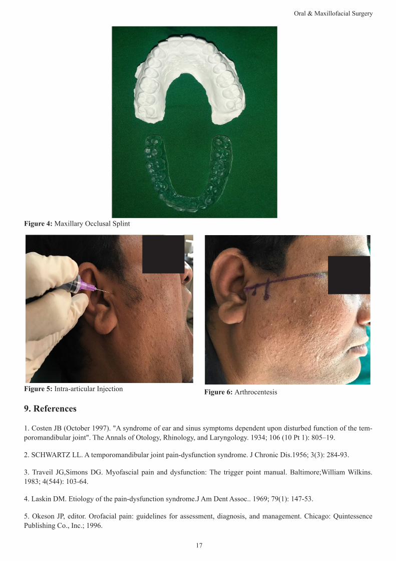
Procedure of TMJ intra- articular injection
Under all aseptic conditions, preauricular area is prepared and mouth prop is placed between posterior teeth to keep mouth open. Now take 2ml luerlok syringe and filled it with 1 ml hydrocortisone solution along with 0.5 ml of 2 per cent lignocaine and use a 25 gauge

Oral & Maxillofacial Surgery
13
needle. Since the mouth is open, the condyle is placed anteriorly and thus, it’s easy to pass the needle (Figure 5). Insert the needle around depth of 2 to 3 cm in inward forward and upward direction till it strikes the roof of the glenoid fossa, then the needle is withdrawn around half a centimetre. This is upper joint cavity, now aspirate and slowly deposit 1 ml of solution. Now change the direction of needle further downward lower joint cavity is reached and deposit rest of the solution. Prescribe routine antibiotic or anti inflammatory regime.
5.2. Surgical management
It includes Arthrocentesis, Arthroscopic and Arthrotomy procedures. When non-surgical therapy has been unsuccessful, then TMJ surgery is indicated.
Arthrocentesis is a conservative procedure. Arthroscopic surgery is closed joint surgical procedure, indicated in patients with moderate to severe pain whereas Arthrotomy is an open TMJ surgery advised in patients with severe pain and on whom other treatment modalities didn’t worked.
i. TMJ Arthrocentesis
Arthrocentesis is a procedure which involves an intra-articular lavage with or without deposit of corticosteroids. It is indicated in patients with intra-articular restrictions to move-ment, as in disc displacement without reduction [29].
Murakami et al had described arthrocentesis procedure in TMJ and referred this proce-dure as manipulation technique after pumping and hydraulic pressure [30].
Traditional method of introducing one needle in upper joint space had been modified to use of two needles instead of only one which allows massive lavage of the TMJ.
Procedure of Arthrocentesis
Under all aseptic precautions this procedure is carried out. Patients head is turned to-wards unaffected side with the patient’s position at 45 degree angle. A line is drawn over the skin from the middle of the tragus of the ear to the outer canthus. Around 10 mm from the middle of the tragus and 2 mm below the canthotragal line, posterior entrance point is located and marked. The anterior point of entry is marked 10 mm farther from the posterior point along the line and 10 mm below it. These markings indicate the location of the articular fossa and the eminence of the TMJ (Figure 6).
A local anesthetic is injected at the marked points, without entering into the joints. Ring-er’s lactate solution is inserted into the superior compartment from posterior entry point with the help of 19- gauge needle and syringe. The solution is injected and aspirated. Then 2 to 3 ml of Ringer’s lactate or local anesthetic solution is injected to distend the upper joint space and

Oral & Maxillofacial Surgery
14
anesthetize the adjacent tissues. A second 19-gauge needle is then inserted into the distended compartment in the anterior point marked which corresponds to area of the articular eminence to enable free flow of the solution through the superior compartment.
Due to lavage, inflammation and pain is reduced since the pain mediators gets washed out and inability of disc to slide can be reversed. During the lavage, the Mandibular movements such as opening of mouth, excursive, and protrusive are done which aids in lysis of adhesions. At the end, one needle is removed and medication can be injected into the joint space.
Disadvantage of Arthrocentesis though they are transient are temporary facial paresis or paralysis due to local anesthetic, or swelling of the surrounding tissues.
Advantages of Arthrocentesis are that it is simple, less invasive, less expensive tech-nique with minimum armamentarium required.
ii. Arthroscopy
It is a closed surgical procedure. It allows direct visualization of joint cavity. It is indi-cated for lavage, lysis of disk adhesion, improvement of hypomobility of the joint due to joint derangement or fibrosis, disk suturing in cases of hypermobility of the joint, articular surface shaving and biopsy [31,32]. TMJ arthroscopy was popularized by Ohinshi in 1975 and had developed 1.7 mm diameter needle type arthroscope.
Depending upon the indication procedure is done under local anesthesia, sedation or general anesthesia. Surgical markings are similar to arthrocentesis. Arthroscope is entered from posterior marking point. Single puncture technique through only posterior marking point is for diagnostic purpose whereas double puncture technique i.e at posterior and anterior mark-ing points are for therapeutic, lavage as well as surgical purpose.
Disadvantages of arthroscopy are damage to the disk, facial paralysis, instrument break-age and costly armamentarium are involved.
iii. Arthrotomy
It is an open surgical procedure that involves condylar shaving or condylectomy .It is indicated in patient who has advanced TMJ disorder that are unmanageable to other treatment modalities [33].
6. Conclusion
It is important to eliminate causative factor of MPDS so that management of MPDS can be easily carried out with conservative management such as exercises, pharmacological occlusal splints. In case where conservative management doesn’t work then procedures intra-

Oral & Maxillofacial Surgery
15
articular injection technique as supportive therapy can be given. If still there is no relief then procedures such as Arthrocentesis or Arthroscopy is indicated. If any of the treatment modal-ity doesn’t provide relief to the patient and patient have severe pain, then Arthrotomy or open joint surgery is lastly indicated. Though there are different treatment modalities available, still there is scope for more research for better management of patients with MPDS.
8. Figures
Figure 1 (A): Palpation of TMJ with index fingers over
the joint
Figure 1(B): Palpation of TMJ with little finger in Exter-
nal auditory meatus
Figure2: Palpation of muscles to identify trigger points: (2A) Masseter
Figure 2(B): Temporalis
Oral & Maxillofacial Surgery
16
Figure 2(C): Medial Pterygoid Figure 2(D): Lateral Pterygoid
Figure 2(E): Sternomastoid Figure 2(F): Trapezius
Figure 3(A&B): isometric exercises against resistance
Oral & Maxillofacial Surgery
17
Figure 4: Maxillary Occlusal Splint
Figure 5: Intra-articular Injection Figure 6: Arthrocentesis
9. References
1. Costen JB (October 1997). "A syndrome of ear and sinus symptoms dependent upon disturbed function of the tem-poromandibular joint". The Annals of Otology, Rhinology, and Laryngology. 1934; 106 (10 Pt 1): 805–19.
2. SCHWARTZ LL. A temporomandibular joint pain-dysfunction syndrome. J Chronic Dis.1956; 3(3): 284-93.
3. Traveil JG,Simons DG. Myofascial pain and dysfunction: The trigger point manual. Baltimore;William Wilkins. 1983; 4(544): 103-64.
4. Laskin DM. Etiology of the pain-dysfunction syndrome.J Am Dent Assoc.. 1969; 79(1): 147-53.
5. Okeson JP, editor. Orofacial pain: guidelines for assessment, diagnosis, and management. Chicago: Quintessence Publishing Co., Inc.; 1996.

18
Oral & Maxillofacial Surgery
6. James Fricton. Myogenous Temporomandibular Disorders: Diagnostic and Management Considerations. Dent Clin N Am. 2007; 51: 61–83.
7. Travell J. Myofascial trigger points: clinical view. In: Bonica JJ, et al, editors. Advances in pain research and therapy. New York: Raven Press.1976; 919–26.
8. Cifala J. Myofascial (trigger point pain) injection: theory and treatment. Osteopath Med.1979; 31–6.
9. Cooper AL. Trigger point injection: its place in physical medicine and rehabilitation. Arch Phys Med 1961; 42: 704–9
10. van Tulder M, Koes B, Bouter L. Conservative treatment of acute and chronic nonspecific low back pain: a sys-tematic review of randomized controlled trials of the most common interventions. Spine (Phila Pa 1976) 1997; 22: 2128–2156.
11. Hsieh L, Hong C, Chern S. Efficacy and side effects of diclofenac patch in treatment of patients with myofascial pain syndrome of the upper trapezius. J Pain Symptom Manag. 2010; 39: 116–125.
12. Affaitati G, Fabrizio A, Savini A, et al. A randomized, controlled study comparing a lidocaine patch, a placebo patch, and anesthetic injection for treatment of trigger points in patients with myofascial pain syndrome: evaluation of pain and somatic pain thresholds. Clin Ther. 2009; 31: 705–720.
13. Dalpiaz A, Lordon S, Lipman A. Topical lidocaine patch therapy for myofascial pain. J Pain Palliat Care Pharmaco-ther. 2004; 18: 15–34.
14. Dalpiaz A, Dodds T. Myofascial pain response to topical lidocaine patch therapy: case report. J Pain Palliat Care Pharmacother. 2002; 16: 99–104.
15. Malanga G, Gwynn M, Smith R, Miller D. Tizanidine is effective in the treatment of myofascial pain syndrome. Pain Physician. 2002; 5: 422–432.
16. Botney M, Fields H. Amitriptyline potentiates morphine analgesia by a direct action on the central nervous system. Ann Neurol. 1983; 13: 160–164.
17. Borg-Stein J. Treatment of fibromyalgia, myofascial pain, and related disorders. Phys Med Rehabil Clin N Am. 2006; 17: 491–510.
18. Sławek J, Madaliński M, Maciag-Tymecka I, Duzynski W. Frequency of side effects after botulinum toxin A injec-tions in neurology, rehabilitation and gastroenterology. Pol Merkur Lekarski. 2005; 18: 298–302.
19. Edwards J, Knowles N. Superficial dry needling and active stretching in the treatment of myofascial pain: a ran-domised control trial. Acupunct Med. 2003; 21: 80–86.
20. Kamanli A, Kaya A, Ardicoglu O, Ozgocmen S, Zengin F, Bayik Y. Comparison of lidocaine injection, botulinum toxin injection, and dry needling. Rheumatol Int. 2005; 25: 604–611.
21. Kostopoulos D, Rizopoulos K. The manual of trigger point and myofascial therapy. New Jersey: SLACK Incorpo-rated; 2001.
22. Gam A, Warming S, Larsen L, et al. Treatment of myofascial trigger-points with ultrasound combined with massage and exercise-a randomised controlled trial. Pain. 1998; 77: 73–79.
23. Chu J, Yuen K, Wang B, Chan R, Schwartz I, Neuhauser D. Electrical twitch-obtaining intramuscular stimulation in lower back pain: a pilot study. Am J Phys Med Rehabil. 2004; 83: 104–111.
24. Wright, E., Anderson, G., Schulte, J. A randomized clinical trial of intraoral soft splints and palliative treatment for masticatory muscle pain. J. Orofac. Pain.1995; 9: 192–199.

19
Oral & Maxillofacial Surgery
25. Okeson, J.P., 2003. Management of Temporomandibular Disorders and Occlusion. 5th ed. Mosby, St. Louis. p. 260. Oral Surg. Oral Med. Oral Pathol. 199; 71: 529-534.
26. Pettengill, Craig A., Growney Jr., Maurice R., Schoff, Robert, Kenworthy, Christian R.,. A pilot study comparing the efficacy of hard and soft stabilizing appliances in treating patients with temporomandibular disorders. J. Prosthet. Dent. 1998; 79: 165–168.
27. Okeson JP. Management of temporomandibular disorders and occlusion. 4th ed. St. Louis: Mosby; 1998.
28. Bjørnland T, Gjaerum AA, Møystad A. Osteoarthritis of the temporomandibular joint: an evaluation of the effects and complications of corticosteroid injection compared with injection with sodium hyaluronate. J Oral Rehabil. 2007; 34(8): 583-9.
29. Nitzan DW, Dolwick MF, Martinez GA. Temporomandibular joint arthrocentesis: a simplified treatment for severe, limited mouth opening. J Oral Maxillofac Surg. 1991; 49(11): 1163–1170.
30. Murakami K, Hosaka H, Moriya Y, et al. Short-term treatment outcome study for the management of temporoman-dibular joint closed lock. A comparison of arthrocentesis to non-surgical therapy and arthroscopic lysis and lavage. Oral Surg Oral Med Oral Pathol Oral Radiol Endodontics. 1995; 80(3): 253-7.
31. Buckley MJ, Merrill RG, Braun TW. Surgical management of internal derangement of the temporomandibular joint. J Oral Maxillofac Surg.1993; 51(1 Suppl 1): 20–27.
32. Blaustein D, Heffez L. Diagnostic arthroscopy of the temporomandibular joint. Part II. Arthroscopic findings of arthrographically diagnosed disk displacements. Oral Surg Oral Med Oral Pathol. 1988; 65(2): 135–141.
33. de Leeuw R. Temporomandibular Disorders. In: de Leeuw R, editor. Orofacial Pain Guidelines for Assesment, Diag-nosis and Management. The American Academy of Orofacial Pain. 4th ed. Hanover Park, IL: Quintessence Publishing Co, Inc; 2008: 158–176.