Comparison of the effects of various periodontal rotary...
8
1 Abstract: The efficacy of scaling and root planing using various periodontal rotary instruments was examined. Eighty extracted human teeth with a history of periodontal disease were divided into four groups of 20 and subjected to one of the following procedures: Use of 1) a Root Burnisher, 2) a Perio Planing Bur (both rotating instruments for contra angle handpieces), 3) a Tooth Planing Bur (rotating instrument for use with an air turbine), or 4) a Gracey Scaler. In each case, the time required for cleaning was measured. Twenty healthy extracted human teeth were used as untreated controls. After treatment, the surface roughness of 10 specimens out of each group were measured using a profilometer and observed by scanning electron microscopy (SEM). Half of the samples were then incubated in dishes with a suspension of fibroblasts. After counting the number of attached cells, the attachment of fibroblasts was observed by SEM. The root surfaces treated with the rotary instruments appeared smooth and there were no significant differences between groups. From the SEM observations, smooth root surfaces with different surface textures were evident and a tight attachment of fibroblasts was observed. The results of this study suggest that use of rotary instruments is superior for periodontal scaling and root planing. (J. Oral Sci. 46, 1-8, 2004) Key words: rotary instrument; roughness; fibroblast; periodontal disease. Introduction Bacterial plaque and calculus are recognized etiological agents in the initiation and progression of periodontal disease (1), and their accumulation and attachment are facilitated by a roughened root surface (2-6). Scaling and root planing to produce smooth root surfaces, together with brushing instructions, are thus an essential component in both treatment and prevention of periodontal disease (7). The instruments currently available for scaling and root planing are hand scalers and ultrasonic scalers. Although hand scalers are frequently used, considerable time and manual dexterity are required for their effective operation (7). Moreover, hand scalers are unable to reach the deeper root surfaces when the periodontal pockets are more than 4 mm deep (7,8). Consequently, ultrasonic scalers have become more widely used in recent years. Although they are simpler to use, it is often difficult to achieve a smooth and calculus-free root surface (9-11), and dental plaque adheres more readily to the roughened root surfaces created by the use of an ultrasonic scaler (12). To overcome these challenges associated with use of ultrasonic scalers and hand scalers, rotary instruments for scaling and root planning have been developed. Previously, a carbide bur and a diamond point were used at high-speed rotation for polishing. However, these rotating instruments have been reported to be associated with an increased risk of damaging the root surface and soft tissues (13). The rotosonic scaler, in which a hexagonal pyramid chip is installed on an air turbine for high-speed rotation, can damage the gingival Journal of Oral Science, Vol. 46, No. 1, 1-8, 2004 Correspondence to Dr. Mamoru Kishida, Nihon University Graduate School of Dentistry, 1-8-13 Kanda-Surugadai, Chiyoda- ku, Tokyo 101-8310, Japan Tel: +81-3-3219-8107 Fax: +81-3-3219-8349 E-mail: [email protected] Comparison of the effects of various periodontal rotary instruments on surface characteristics of root surface Mamoru Kishida § , Shuichi Sato †,‡ and Koichi Ito †,‡ § Nihon University Graduate School of Dentistry, Tokyo, Japan §,† Department of Periodontology, Nihon University School of Dentistry, Tokyo, Japan ‡ Division of Advanced Dental Treatment, Dental Research Center, Nihon University School of Dentistry, Tokyo, Japan (Received 27 September and accepted 20 November 2003) Original
Transcript of Comparison of the effects of various periodontal rotary...
1
Abstract: The efficacy of scaling and root planingusing various periodontal rotary instruments wasexamined. Eighty extracted human teeth with a historyof periodontal disease were divided into four groupsof 20 and subjected to one of the following procedures:Use of 1) a Root Burnisher, 2) a Perio Planing Bur (bothrotating instruments for contra angle handpieces), 3)a Tooth Planing Bur (rotating instrument for use withan air turbine), or 4) a Gracey Scaler. In each case, thetime required for cleaning was measured. Twentyhealthy extracted human teeth were used as untreatedcontrols. After treatment, the surface roughness of 10specimens out of each group were measured using aprofilometer and observed by scanning electronmicroscopy (SEM). Half of the samples were thenincubated in dishes with a suspension of fibroblasts.After counting the number of attached cells, theattachment of fibroblasts was observed by SEM. Theroot surfaces treated with the rotary instrumentsappeared smooth and there were no significantdifferences between groups. From the SEMobservations, smooth root surfaces with differentsurface textures were evident and a tight attachmentof fibroblasts was observed. The results of this studysuggest that use of rotary instruments is superior forperiodontal scaling and root planing. (J. Oral Sci. 46,1-8, 2004)
Key words: rotary instrument; roughness; fibroblast;periodontal disease.
IntroductionBacterial plaque and calculus are recognized etiological
agents in the initiation and progression of periodontaldisease (1), and their accumulation and attachment arefacilitated by a roughened root surface (2-6). Scaling androot planing to produce smooth root surfaces, together withbrushing instructions, are thus an essential component inboth treatment and prevention of periodontal disease (7).The instruments currently available for scaling and rootplaning are hand scalers and ultrasonic scalers. Althoughhand scalers are frequently used, considerable time andmanual dexterity are required for their effective operation(7). Moreover, hand scalers are unable to reach the deeperroot surfaces when the periodontal pockets are more than4 mm deep (7,8). Consequently, ultrasonic scalers havebecome more widely used in recent years. Although theyare simpler to use, it is often difficult to achieve a smoothand calculus-free root surface (9-11), and dental plaqueadheres more readily to the roughened root surfaces createdby the use of an ultrasonic scaler (12). To overcome thesechallenges associated with use of ultrasonic scalers andhand scalers, rotary instruments for scaling and rootplanning have been developed. Previously, a carbide burand a diamond point were used at high-speed rotation forpolishing. However, these rotating instruments have beenreported to be associated with an increased risk of damagingthe root surface and soft tissues (13). The rotosonic scaler,in which a hexagonal pyramid chip is installed on an airturbine for high-speed rotation, can damage the gingival
Journal of Oral Science, Vol. 46, No. 1, 1-8, 2004
Correspondence to Dr. Mamoru Kishida, Nihon UniversityGraduate School of Dentistry, 1-8-13 Kanda-Surugadai, Chiyoda-ku, Tokyo 101-8310, JapanTel: +81-3-3219-8107Fax: +81-3-3219-8349E-mail: [email protected]
Comparison of the effects of various periodontal rotaryinstruments on surface characteristics of root surface
Mamoru Kishida§, Shuichi Sato†,‡ and Koichi Ito†,‡
§Nihon University Graduate School of Dentistry, Tokyo, Japan§,†Department of Periodontology, Nihon University School of Dentistry, Tokyo, Japan
‡Division of Advanced Dental Treatment, Dental Research Center,Nihon University School of Dentistry, Tokyo, Japan
(Received 27 September and accepted 20 November 2003)
Original
2
tissues or the dentin if used incorrectly, and this instrumentis thus no longer in general use (14,15). Recently, otherrotary instruments have been developed for scaling and rootplaning, and their effectiveness in the clinical situationevaluated (16-18). The purpose of this study was to comparethe efficacy of the several rotary instruments in scaling androot planing, and to compare with use of a Gracey Scaler.The hypothesis was that the rotary instruments for scalingand root planing would be more efficacious than a handscaler.
Materials and MethodsRotary instruments
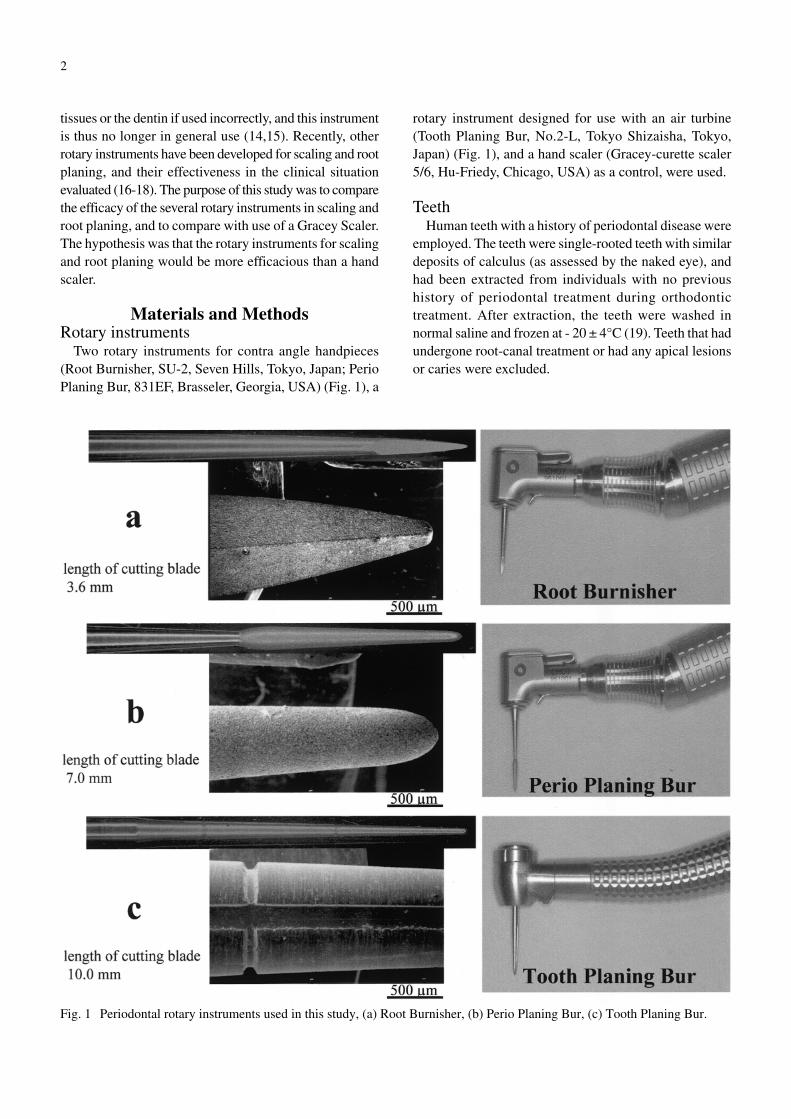
Two rotary instruments for contra angle handpieces(Root Burnisher, SU-2, Seven Hills, Tokyo, Japan; PerioPlaning Bur, 831EF, Brasseler, Georgia, USA) (Fig. 1), a
rotary instrument designed for use with an air turbine(Tooth Planing Bur, No.2-L, Tokyo Shizaisha, Tokyo,Japan) (Fig. 1), and a hand scaler (Gracey-curette scaler5/6, Hu-Friedy, Chicago, USA) as a control, were used.
TeethHuman teeth with a history of periodontal disease were
employed. The teeth were single-rooted teeth with similardeposits of calculus (as assessed by the naked eye), andhad been extracted from individuals with no previoushistory of periodontal treatment during orthodontictreatment. After extraction, the teeth were washed innormal saline and frozen at - 20 ± 4°C (19). Teeth that hadundergone root-canal treatment or had any apical lesionsor caries were excluded.
Fig. 1 Periodontal rotary instruments used in this study, (a) Root Burnisher, (b) Perio Planing Bur, (c) Tooth Planing Bur.
3
Preparation of specimensThe teeth were defrosted at room temperature, and
sections of 5 × 5 × 1 mm were removed from the cemento-enamel junction to the root apex using a water-cooleddental turbine. Calculus attachment to the root surfaces wasevaluated with a 60 × stereomicroscope (SZH-ILLD,Olympus, Tokyo, Japan) using a 10 × 10 micrometerattached to an ocular lens (7). Eighty specimens that were50% covered with calculus were selected. In addition, 20teeth expediently extracted during orthodontic treatmentwere used as healthy control. All specimens were immersedin 2% hypochlorous acid solution and the soft tissue wasremoved (12). The specimens were divided into fourgroups (Root Burnisher, Perio Planing Bur, Tooth PlaningBur, and Gracey Scaler group), each containing 20specimens. Scaling and root planing were performed withwater cooling at 0.98 N (100-gf) pressure for the RootBurnisher and Perio Planing Bur groups, 0.49 N (50-gf)pressure for the Tooth Planing Bur group and 4.90 N (500-gf) pressure for the Gracey Scaler group with the samplesmounted on a force gauge (DPX-5T, Imada, Tokyo, Japan)(20,21). Rotation speed was 20,000 rpm with the use ofthe Root Burnisher or Perio Planing Bur, and 300,000rpm with the use of the Tooth Planing Bur (16). Scalingand root planing were performed until the root surfaceappeared smooth upon visual inspection and examinationwith a periodontal probe. Healthy root specimens wererinsed in 0.2 M PBS (pH 7.2) and classified as untreatedcontrols.
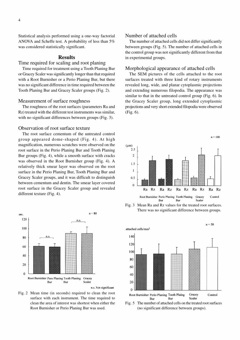
Measurement of the time required for scaling androot planing
The length of time required for scaling and root planingwith each instrument was measured at 5-second intervalsby staff members, and the average time for each group wascalculated.
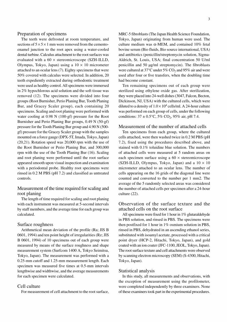
Surface roughnessArithmetical mean deviation of the profile (Ra; JIS B
0601, 1994) and ten point height of irregularities (Rz; JISB 0601, 1994) of 10 specimens out of each group weremeasured by means of the surface roughness and shapemeasurement system (Surfcom 1400 A, Tokyo Seimitsu,Tokyo, Japan). The measurement was performed with a0.25-mm cutoff and 1.25-mm measurement length. Eachspecimen was measured five times at 0.5-mm intervalslengthwise and widthwise, and the average measurementsfor each specimen were calculated.
Cell cultureFor measurement of cell attachment to the root surface,
MRC-5 fibroblasts (The Japan Health Science Foundation,Tokyo, Japan) originating from human were used. Theculture medium was α-MEM, and contained 10% fetalbovine serum (Bio fluids, Bio source international, USA)and antibiotics (penicillin/streptomycin solution, Sigma-Aldrich, St. Louis, USA; final concentration 50 U/mlpenicillin and 50 µg/ml streptomycin). The fibroblastswere cultured at 37°C under 5% CO2 and 95% air and wereused after four or five transfers, when the doubling timehad become constant.
Ten remaining specimens out of each group weresterilized using ethylene oxide gas. After sterilization,they were placed into 24-well dishes (3047, Falcon, Becton,Dickinson, NJ, USA) with the cultured cells, which werediluted to a density of 1.0 × 10⁴ cells/ml. A 24-hour culturewas performed on each group of cells, under the followingconditions: 37 ± 0.5°C, 5% CO2, 95% air, pH 7.4.
Measurement of the number of attached cellsTen specimens from each group, where the cultured
cells attached, were then washed twice in 0.2 M PBS (pH7.2), fixed using the procedures described above, andstained with 0.1% toluidine blue solution. The numbersof attached cells were measured at 5 random areas oneach specimen surface using a 60 × stereomicroscope(SZH-ILLD, Olympus, Tokyo, Japan) and a 10 × 10micrometer attached to an ocular lens. The number ofcells appearing on the 16 grids of the diagonal line werecounted and converted to the number per 1 mm2. Theaverage of the 5 randomly selected areas was consideredthe number of attached cells per specimen after a 24-hourculture (22).
Observation of the surface texture and theattached cells on the root surface
All specimens were fixed for 1 hour in 1% glutaraldehydein PBS solution, and rinsed in PBS. The specimens werethen postfixed for 1 hour in 1% osmium solution in PBS,rinsed in PBS, dehydrated in an ascending ethanol series,substituted with isoamyl acetate, processed with a criticalpoint dryer (HCP-2, Hitachi, Tokyo, Japan), and goldcoated with an ion coater (JFC-1100, JEOL, Tokyo, Japan).The root surface texture and cell attachments were observedby scanning electron microscopy (SEM) (S-4300, Hitachi,Tokyo, Japan).
Statistical analysisIn this study, all measurements and observations, with
the exception of measurement using the profilometer,were completed independently by three examiners. Noneof these examiners took part in the experimental procedures.
4
Statistical analysis performed using a one-way factorialANOVA and Scheffe test. A probability of less than 5%was considered statistically significant.
ResultsTime required for scaling and root planing
Time required for treatment using a Tooth Planing Buror Gracey Scaler was significantly longer than that requiredwith a Root Burnisher or a Perio Planing Bur, but therewas no significant difference in time required between theTooth Planing Bur and Gracey Scaler groups (Fig. 2).
Measurement of surface roughnessThe roughness of the root surfaces (parameters Ra and
Rz) treated with the different test instruments was similar,with no significant differences between groups (Fig. 3).
Observation of root surface textureThe root surface cementum of the untreated control
group appeared dome-shaped (Fig. 4). At highmagnification, numerous scratches were observed on theroot surface in the Perio Planing Bur and Tooth PlaningBur groups (Fig. 4), while a smooth surface with crackswas observed in the Root Burnisher group (Fig. 4). Arelatively thick smear layer was observed on the rootsurface in the Perio Planing Bur, Tooth Planing Bur andGracey Scaler groups, and it was difficult to distinguishbetween cementum and dentin. The smear layer coveredroot surface in the Gracey Scaler group and revealeddifferent texture (Fig. 4).
Number of attached cellsThe number of attached cells did not differ significantly
between groups (Fig. 5). The number of attached cells inthe control group was not significantly different from thatin experimental groups.
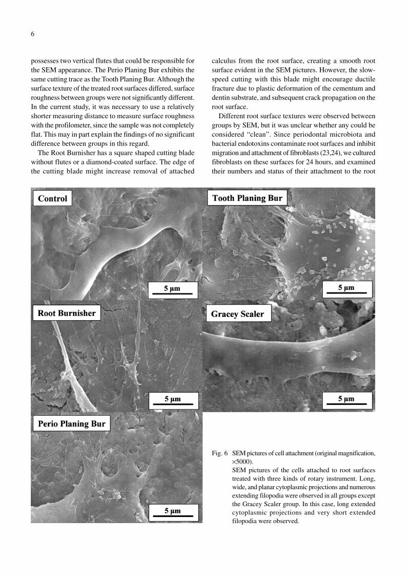
Morphological appearance of attached cellsThe SEM pictures of the cells attached to the root
surfaces treated with three kind of rotary instrumentsrevealed long, wide, and planar cytoplasmic projectionsand extending numerous filopodia. The appearance wassimilar to that in the untreated control group (Fig. 6). Inthe Gracey Scaler group, long extended cytoplasmicprojections and very short extended filopodia were observed(Fig. 6).
Fig. 2 Mean time (in seconds) required to clean the rootsurface with each instrument. The time required toclean the area of interest was shortest when either theRoot Burnisher or Perio Planing Bur was used.
Fig. 3 Mean Ra and Rz values for the treated root surfaces.There was no significant difference between groups.
Fig. 5 The number of attached cells on the treated root surfaces(no significant difference between groups).

5
DiscussionIn the current study, the time required for scaling and
root planing using the rotary instruments and a GraceyScaler were compared and it was apparent that use of aTooth Planing Bur or a Gracey Scaler was more timeconsuming than in the case of a Root Burnisher or PerioPlaning Bur. These results suggest that the cutting efficiencyof the Root Burnisher and Perio Planing Bur are higherthan the other instruments employed.
The surface roughness of the root surfaces treated withthree kind of rotary instruments was similar to that followinguse of a Gracey Scaler. Although similar Ra and Rz valueswere obtained across groups, post-treatment SEM pictures
revealed surface textures differed between groups. Domeshaped cementum was observed in the case of intact rootsurfaces, but the root surfaces treated with a Gracey Scalerexhibited scarry roughness.
The root surface treated with a Gracey Scaler wascovered by a smear layer and a small amount of dentalcalculus remained, in line with previous findings (7). Rootsurfaces treated with a rotary instrument appeared flatand glossy to the naked eye. However, SEM picturesrevealed a roughened and scratched root surface where aPerio Planing Bur or Tooth Planing Bur had been used,but a smoother surface with smaller cracks where a RootBurnisher had been employed. The Tooth Planing Bur
Fig. 4 SEM pictures of the root surface texture (originalmagnification, ×500). Numerous scratches wereobserved on the root surfaces treated with either aPerio Planing Bur or Tooth Planing Bur, while smoothsurfaces with small cracks were observed on the rootsurfaces treated with a Root Burnisher. When a GraceyScaler was used, a distinct root surface texture wasobserved.
6
possesses two vertical flutes that could be responsible forthe SEM appearance. The Perio Planing Bur exhibits thesame cutting trace as the Tooth Planing Bur. Although thesurface texture of the treated root surfaces differed, surfaceroughness between groups were not significantly different.In the current study, it was necessary to use a relativelyshorter measuring distance to measure surface roughnesswith the profilometer, since the sample was not completelyflat. This may in part explain the findings of no significantdifference between groups in this regard.
The Root Burnisher has a square shaped cutting bladewithout flutes or a diamond-coated surface. The edge ofthe cutting blade might increase removal of attached
calculus from the root surface, creating a smooth rootsurface evident in the SEM pictures. However, the slow-speed cutting with this blade might encourage ductilefracture due to plastic deformation of the cementum anddentin substrate, and subsequent crack propagation on theroot surface.
Different root surface textures were observed betweengroups by SEM, but it was unclear whether any could beconsidered “clean”. Since periodontal microbiota andbacterial endotoxins contaminate root surfaces and inhibitmigration and attachment of fibroblasts (23,24), we culturedfibroblasts on these surfaces for 24 hours, and examinedtheir numbers and status of their attachment to the root
Fig. 6 SEM pictures of cell attachment (original magnification,×5000).SEM pictures of the cells attached to root surfacestreated with three kinds of rotary instrument. Long,wide, and planar cytoplasmic projections and numerousextending filopodia were observed in all groups exceptthe Gracey Scaler group. In this case, long extendedcytoplasmic projections and very short extendedfilopodia were observed.

7
surface.There were no significant differences in the number of
attached cells between groups and compared with theuntreated control group. This might be due to the substantialremoval of contaminants, such as endotoxins, from thesurface of the samples. Surface texture, including scratchescreated due to cutting, had no effect on cell attachment.Interestingly, from a morphological perspective, a favorableattachment with the creation of cytoplasmic projectionswith active extensions of filopodia was seen where the rootsurface had been treated with a Root Burnisher, PerioPlaning Bur or a Tooth Planing Bur. The same tendencywas evident in the case of the intact root surface. Wherethe root surface had been treated with a Gracey Scaler,however, poor extensions of filopodia were observed withelongation of the cytoplasmic projections. It has beenreported that an acceptable connective tissue attachmentis created by performing a thorough scaling and rootplaning with a hand instrument (25). Such a difference inattachment might be due to the use of more efficaciousrotary instruments in the current study. As evident fromthe SEM pictures, qualitative and quantitative differencesin the morphology of the smear layer might have resultedin disruption of the connective tissue attachment. Since therewas no significant difference in surface roughness betweengroups, residual contaminants such as endotoxins on theroot surface might be the cause of these morphologicaldifferences.
From the results of the present study, it appears that rotaryinstruments can create as smooth a root surface as foundin the case of a non-diseased root when measured with theprofilometer. When these root surfaces were observed bySEM, surface textures differed with treatment method,and these differences might affect cell attachment to thetreated root surface. Further studies are needed to determinethe cutting efficiency and effectiveness of the rotaryinstruments for root surface scaling, and the effect of thesurface profile after scaling and root planing on cellattachment.
References1. Ash MM, Gitlin BN, Smith WA (1964) Correlation
between plaque and gingivitis. J Periodontol 35,424-429
2. Zander HA (1953) The attachment of calculus to rootsurfaces. J Periodontol 24, 16-19
3. Waerhaug J (1956) Effect of rough surfaces upongingival tissue. J Dent Res 35, 323-325
4. Swartz ML, Phillips RW (1957) Comparison ofbacteria accumulations on rough and smooth enamelsurfaces. J Periodontol 28, 304-307
5. Tureskey S, Renstrup G, Glickman I (1961)Histologic and histochemical observations regardingearly calculus formation in children and adults. JPeriodontol 32, 7-14
6. Stende GW, Schaffer EM (1961) A comparison ofultrasonic and hand scaling. J Periodontol 32, 312-314
7. Rabbani GM, Ash MM Jr, Caffesse RG (1981) Theeffectiveness of subgingival scaling and root planingin calculus removal. J Periodontol 52, 119-123
8. Waerhaug J (1978) Healing of the dento-epithelialjunction following subgingival plaque control. Asobserved on extracted teeth. J Periodontol 49, 119-134
9. Moskow BS, Bressman E (1964) Cemental responseto ultrasonic and hand instrumentation. J Am DentAssoc 68, 698-703
10. Jones SJ, Lozdan J, Boyde A (1972) Tooth surfacestreated in situ with periodontal instruments. BritDent J 132, 57-64.
11. Nishimine D, O’Leary TJ (1979) Hand instru-mentation versus ultrasonic in the removal ofendotoxins from root surfaces. J Periodontol 50, 345-349.
12. Wilkinson RF, Maybury JE (1973) Scanning electronmicroscopy of the root surface followinginstrumentation. J Periodontol 44, 559-563
13. Evian CI, Horowitz RA, Dwayne K, Maltz DO(1998) Evaluation of a surgical debridement burfor use in periodontal surgery. Compend ContinEduc Dent 19, 1124-1132
14. Ellman IA (1962) Rotary ultrasonics for preventiveperiodontics. NY State Dent J 28, 404-410
15. David LC, Kenneth LK, Michel AB (1994) Plaqueand calculus removal. considerations for theprofessional. Quintessence Publishing, Chicago,69-72
16. Daly CG, Seymour GJ, Kieser JB, Corbet EF (1982)Histologic assessment of periodontally involvedcementum. J Clin Periodontol 9, 266-274
17. Schwarz JP, Guggenheim R, Duggelin M, HeftiAF, Ratertschnk-Pluss EM, Rateitschak KH (1989)The effectiveness of root debridement in open flapprocedures by means of a comparison between handinstruments and diamond burs. A SEM study. J ClinPeriodontol 16, 510-518
18. Parashis AO, Anagnow-Vareltzides A, DemetriouN (1993) Calculus removal from multirooted teethwith and without surgical access (I) Efficacy onexternal and furcation surfaces in relation to probingdepth. J Clin Periodontol 20, 63-68
8
19. Moore J, Wilson M, Kieser JB (1986) Thedistribution of bacterial lipopolysaccharide(endotoxin) in relation to periodontally involvedroot surfaces. J Clin Periodontol 13, 748-751
20. Ritz L, Hefti AF, Rateischak KH (1991) An in vitroinvestigation on the loss of root substance in scalingwith various instruments. J Clin Periodontol 18,643-647
21. Schmidlin PR, Beuchat M, Busslinger A, LehmannB, Lutz F (2001) Tooth substance loss resultingfrom mechanical, sonic and ultrasonic rootinstrumentation assessed by liquid scintillation. J Clin Periodontol 28, 1058-1066
22. Tanaka K, Arai N, Hiruma S, Mukai H, Ito K, Murai
S (1987) Cell attachment and growth of the culturedcells to the smooth and rough surfaces of the dentinsurface. Nihon Shisyubyo Gakkai Kaishi 29, 859-869 (in Japanease)
23. Hatfield CG, Baumhammers A (1971) Cytotoxiceffects of periodontally involved surfaces of humanteeth. Arch Oral Biol 16, 465-468
24. Aleo JJ, De Renzis FA, Farber PA, Varvoncoeur AP(1974) The presence and biological activity ofcementum bound endotoxin. J Periodontol 45, 672-675
25. Aleo JJ, De Renzis FA, Farber PA (1975) In vitroattachment of human gingival fibroblasts to rootsurfaces. J Periodontol 46, 639-645