Comparison of anticoagulation strategies for veno-venous ...
11
Seeliger et al. Crit Care (2020) 24:701 https://doi.org/10.1186/s13054-020-03348-w RESEARCH Comparison of anticoagulation strategies for veno-venous ECMO support in acute respiratory failure Benjamin Seeliger 1† , Michael Döbler 2† , Robert Friedrich 1 , Klaus Stahl 3 , Christian Kühn 4 , Johann Bauersachs 5 , Folkert Steinhagen 2 , Stefan F. Ehrentraut 2 , Jens‑Christian Schewe 2 , Christian Putensen 2 , Tobias Welte 1 , Marius M. Hoeper 1 , Andreas Tiede 6 , Sascha David 7,8*† and Christian Bode 2*† Abstract Background: Extracorporeal membrane oxygenation (ECMO) support in acute respiratory failure may be lifesaving, but bleeding and thromboembolic complications are common. The optimal anticoagulation strategy balancing these factors remains to be determined. This retrospective study compared two institutional anticoagulation management strategies focussing on oxygenator changes and both bleeding and thromboembolic events. Methods: We conducted a retrospective observational cohort study between 04/2015 and 02/2020 in two ECMO referral centres in Germany in patients receiving veno‑venous (VV)‑ECMO support for acute respiratory failure for > 24 h. One centre routinely applied low‑dose heparinization aiming for a partial thromboplastin time (PTT ) of 35–40 s and the other routinely used a high‑dose therapeutic heparinization strategy aiming for an activated clot‑ ting time (ACT) of 140–180 s. We assessed number of and time to ECMO oxygenator changes, 15‑day freedom from oxygenator change, major bleeding events, thromboembolic events, 30‑day ICU mortality, activated clotting time and partial thromboplastin time and administration of blood products. Primary outcome was the occurrence of oxygenator changes depending on heparinization strategy; main secondary outcomes were the occurrence of severe bleeding events and occurrence of thromboembolic events. The transfusion strategy was more liberal in the low‑dose centre. Results: Of 375 screened patients receiving VV‑ECMO support, 218 were included in the analysis (117 high‑dose group; 101 low‑dose group). Disease severity measured by SAPS II score was 46 (IQR 36–57) versus 47 (IQR 37–55) and ECMO runtime was 8 (IQR 5–12) versus 11 (IQR 7–17) days (P = 0.003). There were 14 oxygenator changes in the high‑dose group versus 48 in the low‑dose group. Freedom from oxygenator change at 15 days was 73% versus 55% (adjusted HR 3.34 [95% confidence interval 1.2–9.4]; P = 0.023). Severe bleeding events occurred in 23 (19.7%) versus © The Author(s) 2020. Open Access This article is licensed under a Creative Commons Attribution 4.0 International License, which permits use, sharing, adaptation, distribution and reproduction in any medium or format, as long as you give appropriate credit to the original author(s) and the source, provide a link to the Creative Commons licence, and indicate if changes were made. The images or other third party material in this article are included in the article’s Creative Commons licence, unless indicated otherwise in a credit line to the material. If material is not included in the article’s Creative Commons licence and your intended use is not permitted by statutory regulation or exceeds the permitted use, you will need to obtain permission directly from the copyright holder. To view a copy of this licence, visit http://creativecommons.org/licenses/by/4.0/. The Creative Commons Public Domain Dedication waiver (http://creativeco mmons.org/publicdomain/zero/1.0/) applies to the data made available in this article, unless otherwise stated in a credit line to the data. Open Access *Correspondence: David.sascha@mh‑hannover.de; Christian.bode@ukbonn. de † Benjamin Seeliger and Michael Döbler contributed equally and are both considered first authors † Sascha David and Christian Bode contributed equally and are both considered last and corresponding authors 2 Department of Anaesthesiology and Critical Care Medicine, University Hospital Bonn, Bonn, Germany 7 Department of Nephrology and Hypertension, Hannover Medical School, Hannover, Germany Full list of author information is available at the end of the article
Transcript of Comparison of anticoagulation strategies for veno-venous ...

Seeliger et al. Crit Care (2020) 24:701 https://doi.org/10.1186/s13054-020-03348-w
RESEARCH
Comparison of anticoagulation strategies for veno-venous ECMO support in acute respiratory failureBenjamin Seeliger1† , Michael Döbler2†, Robert Friedrich1, Klaus Stahl3, Christian Kühn4, Johann Bauersachs5, Folkert Steinhagen2, Stefan F. Ehrentraut2, Jens‑Christian Schewe2, Christian Putensen2, Tobias Welte1, Marius M. Hoeper1, Andreas Tiede6, Sascha David7,8*† and Christian Bode2*†
Abstract
Background: Extracorporeal membrane oxygenation (ECMO) support in acute respiratory failure may be lifesaving, but bleeding and thromboembolic complications are common. The optimal anticoagulation strategy balancing these factors remains to be determined. This retrospective study compared two institutional anticoagulation management strategies focussing on oxygenator changes and both bleeding and thromboembolic events.
Methods: We conducted a retrospective observational cohort study between 04/2015 and 02/2020 in two ECMO referral centres in Germany in patients receiving veno‑venous (VV)‑ECMO support for acute respiratory failure for > 24 h. One centre routinely applied low‑dose heparinization aiming for a partial thromboplastin time (PTT) of 35–40 s and the other routinely used a high‑dose therapeutic heparinization strategy aiming for an activated clot‑ting time (ACT) of 140–180 s. We assessed number of and time to ECMO oxygenator changes, 15‑day freedom from oxygenator change, major bleeding events, thromboembolic events, 30‑day ICU mortality, activated clotting time and partial thromboplastin time and administration of blood products. Primary outcome was the occurrence of oxygenator changes depending on heparinization strategy; main secondary outcomes were the occurrence of severe bleeding events and occurrence of thromboembolic events. The transfusion strategy was more liberal in the low‑dose centre.
Results: Of 375 screened patients receiving VV‑ECMO support, 218 were included in the analysis (117 high‑dose group; 101 low‑dose group). Disease severity measured by SAPS II score was 46 (IQR 36–57) versus 47 (IQR 37–55) and ECMO runtime was 8 (IQR 5–12) versus 11 (IQR 7–17) days (P = 0.003). There were 14 oxygenator changes in the high‑dose group versus 48 in the low‑dose group. Freedom from oxygenator change at 15 days was 73% versus 55% (adjusted HR 3.34 [95% confidence interval 1.2–9.4]; P = 0.023). Severe bleeding events occurred in 23 (19.7%) versus
© The Author(s) 2020. Open Access This article is licensed under a Creative Commons Attribution 4.0 International License, which permits use, sharing, adaptation, distribution and reproduction in any medium or format, as long as you give appropriate credit to the original author(s) and the source, provide a link to the Creative Commons licence, and indicate if changes were made. The images or other third party material in this article are included in the article’s Creative Commons licence, unless indicated otherwise in a credit line to the material. If material is not included in the article’s Creative Commons licence and your intended use is not permitted by statutory regulation or exceeds the permitted use, you will need to obtain permission directly from the copyright holder. To view a copy of this licence, visit http://creat iveco mmons .org/licen ses/by/4.0/. The Creative Commons Public Domain Dedication waiver (http://creat iveco mmons .org/publi cdoma in/zero/1.0/) applies to the data made available in this article, unless otherwise stated in a credit line to the data.
Open Access
*Correspondence: David.sascha@mh‑hannover.de; [email protected]†Benjamin Seeliger and Michael Döbler contributed equally and are both considered first authors†Sascha David and Christian Bode contributed equally and are both considered last and corresponding authors2 Department of Anaesthesiology and Critical Care Medicine, University Hospital Bonn, Bonn, Germany7 Department of Nephrology and Hypertension, Hannover Medical School, Hannover, GermanyFull list of author information is available at the end of the article
Page 2 of 11Seeliger et al. Crit Care (2020) 24:701
IntroductionIn refractory acute respiratory failure (ARF), implemen-tation of veno-venous extracorporeal membrane oxygen-ation (VV-ECMO) as a rescue strategy may be life-saving and is increasingly applied [1]. However, ECMO sup-port is associated with potentially life-threatening com-plications, mostly related to either bleeding events or thromboembolic complications [2–4]. To minimize such events, most centres use unfractionated heparin (UFH)-based anticoagulation adjusted by partial thromboplastin time (PTT), usually within 40–80 s or by activated clot-ting time (ACT) within 140–180 s [5]. Current guide-lines advice an ACT-guided approach aiming at 1.5 fold increase of normal [6]. With bleeding complications occurring in up to 50% of patients, there is a quest for alternative anticoagulatory strategies without compro-mising integrity of the ECMO circuits and risk of throm-boembolism [2]. Previous studies found lower heparin dosing to be generally safe with regards to thromboem-bolic complications but conclusions are limited by small patient numbers [7–9].
Our study aimed to retrospectively compare oxygen-ator durability, bleeding and thromboembolic events between two experienced ECMO centres with consider-ably different routine anticoagulation strategies but iden-tical oxygenator change management. We hypothesized, that a strategy including low-dose heparin strategy would result in similar oxygenator durability and similar throm-boembolic complications while reducing bleeding events compared to a high-dose strategy.
MethodsDesign, settings and participantsWe conducted a retrospective cohort study including patients with severe ARF receiving VV-ECMO support between April 2015 and February 2020 at two German university hospitals with extensive ECMO experience. Both centres routinely used UFH-based anticoagulation, but with different intensity, thus enabling us to compare a low-dose heparinization strategy aiming for a PTT between 35 and 40 s (measured thrice per day using the actin FS assay by Siemens) with a high-dose hepariniza-tion strategy aiming for an ACT between 140 and 180 s
(measured every 2 h). In the high-dose-group, PTT was measured once daily using the Pathrombin SL assay, with both tests showing excellent correlation [10]. ECMO systems used were Getinge/Maquet RotaFlow or Car-dioHelp with cannulation of the internal jugular and/or femoral veins via 19–25 French cannulas. Standard can-nulation in the high-dose centre was femoral/jugular venous access, while in the low-dose centre a bi-femoral venous access was mostly established. For the RotaFlow device, the permanent-life-support system was used and for the CardioHelp system the HLS Set Advanced was used. Both systems were manufactured by Getinge, were Bioline-coated and possessed equivalent durability [11]. Patients were identified via established ECMO data-bases at both sites. Inclusion criteria were VV-ECMO support ≥ 24 h and provided written informed consent by patients or proxy for analysis of clinical data. Exclu-sion criteria were duration of VV-ECMO support < 24 h; external ECMO support > 24 h before referral; addition of a third (arterial) cannula within 24 h; age < 18 years; acute liver failure with relevant coagulopathy precluding heparin administration; missing informed consent and medical indication for high-dose anticoagulation in the low-dose centre. The study was approved by the institu-tional review boards at both sites.
EndpointsThe primary endpoint was ECMO oxygenator change within the first 15 days. Secondary endpoints were 30-day ICU mortality, severe bleeding complications (defined as need for intervention or ≥ 10 red blood cell (RBC) trans-fusions), symptomatic thromboembolic events, number of platelet and RBC transfusions during ECMO, adminis-tered units of UFH during ECMO and coagulation stud-ies (mean ACT in the high-dose-group, mean PTT in both groups).
Transfusion strategiesAbsent of overt bleeding, the routine threshold for RBC transfusion at the high-dose centre was a haemoglobin level < 8 g/dL versus < 9 g/dL in the low-dose group. Rou-tine threshold for platelet transfusions were < 30.000/µL in the high-dose centre versus 70.000/µL in the low-dose
14 (13.9%) patients (P = 0.256) and thromboembolic events occurred in 8 (6.8%) versus 19 (19%) patients (P = 0.007). Mortality at 30 days was 33.3% versus 30.7% (P = 0.11).
Conclusions: In this retrospective study, ECMO management with high‑dose heparinization was associated with lower rates of oxygenator changes and thromboembolic events when compared to a low‑dose heparinization strat‑egy. Prospective, randomized trials are needed to determine the optimal anticoagulation strategy in patients receiv‑ing ECMO support.
Keywords: ECMO, Heparinization, ARDS, Bleeding, Thromboembolism

Page 3 of 11Seeliger et al. Crit Care (2020) 24:701
centre. With overt bleeding, the routine thresholds for platelet transfusion were 50.000/µL (high-dose centre) versus 100.000/µL (low-dose centre) but could be indi-vidualized depending on the clinical scenario, severity and site of bleeding. Antithrombin III was substituted if antithrombin III levels were < 50% and antifibrino-lytic agents administered in cases of clinical suspicion of hyperfibrinolysis or proof by thromboelastography. Bleeding management regarding administration of pro-thrombin complex concentrates was adjusted by event severity at the discretion of the treating physicians and was not standardized in both centres.
Indication for oxygenator changesOxygenator change was considered in the settings of decreasing post-filter pO2 < 200 mmHg with increasing transmembrane pressure gradient (with the CardioHelp system), overt circuit thrombosis with thrombi > 5 mm, rising D-dimers with progressive thrombocytopenia and hyperfibrinolysis with increasing transmembrane pres-sure gradient, and otherwise unexplained haemolysis with increasing transmembrane pressure gradient.
CovariatesAge, admission and discharge dates to ICU, underlying reason for ARF, body-mass-index, pre-existing antiplate-let therapy and comorbidities were obtained from charts. Simplified acute physiology score (SAPS) II [12] at day of ECMO implantation, Respiratory ECMO Survival Pre-diction (RESP) score [13], sequential organ failure assess-ment score (SOFA) [14], heparin doses, ECMO devices and settings and coagulation studies were extracted from the clinical patient data management system.
Statistical analysisContinuous data was assessed for normal distribution by Shapiro–Wilk-test and group comparison was performed using t-test or rank-sum test, as appropriate. Hazard ratios for freedom from oxygenator change at 15 days was calculated using a multivariable cox regression. Covari-ables were selected if baseline values were significantly unbalanced between the groups or if they were overtly physiologically linked to the outcome. We did not include number of blood product transfusions and ECMO device since they were not independent factors but part of the institutional strategy. The final model included age, sex, BMI, RESP Score, SAPS II score, SOFA renal sub-score, ECMO runtime, sepsis, pre-existing coronary artery dis-ease, prior treatment with aspirin, mean ECMO flow and baseline fibrinogen, d-dimers and antithrombin III levels as covariables. Analyses were performed using STATA V16.0 (STATA Corp LP) and RStudio V1.2.5033 (RStudio Inc).
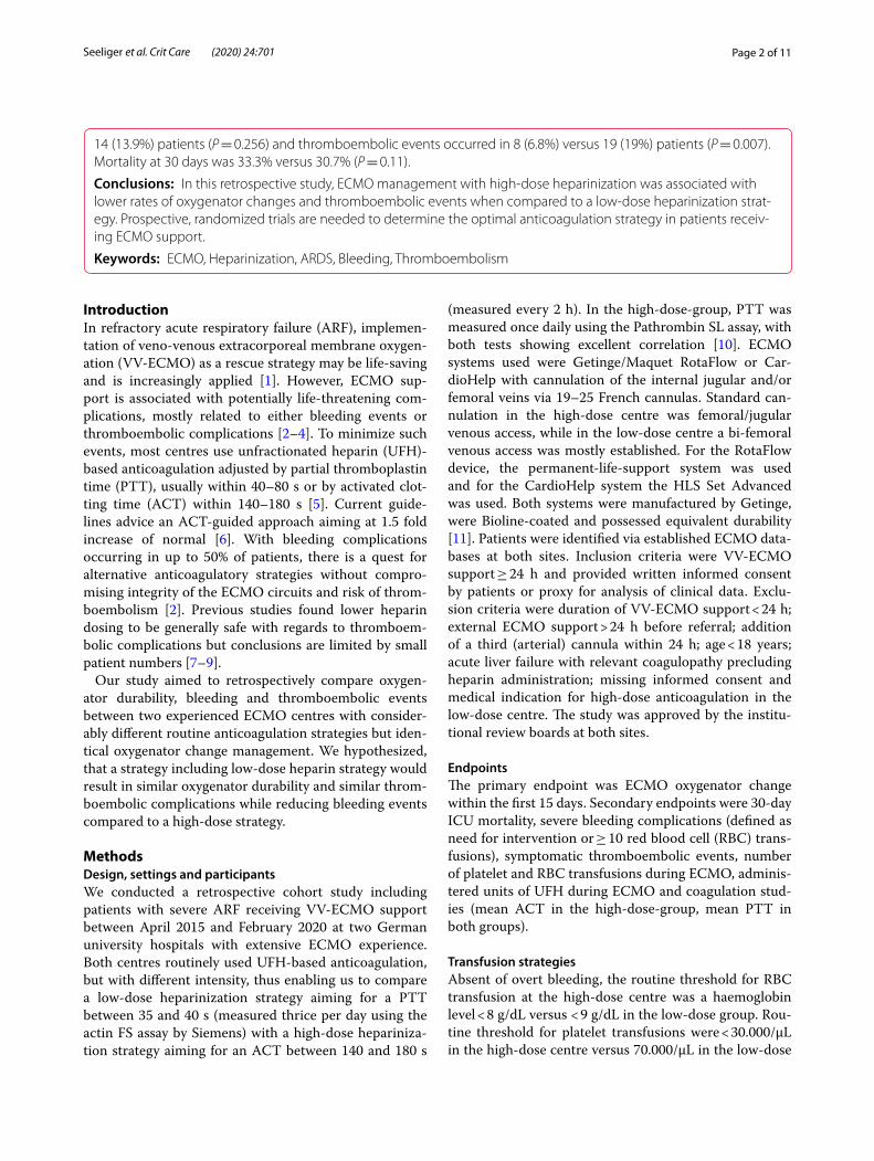
ResultsPatient characteristicsAt screening, 375 patients receiving VV-ECMO sup-port for ARF were identified. A total of 218 patients were included in the analysis (117 high-dose group vs. 101 low-dose group), with details on exclusions provided in Fig. 1. The baseline characteristics are shown in Table 1. ECMO settings and associated laboratory are shown in Table 2. The causes of ARF according the RESP score were viral pneumonia (22.5%), bacterial pneumonia (30.3%), status asthmaticus (1.8%), trauma/burn (1.8%), aspiration pneumonia (7.8%), other acute respiratory causes (28.9%) and non-respiratory or chronic respira-tory causes (6.8%) and were distributed homogenously among the groups (p = 0.359). Patients in the high-dose group were younger (46 years [IQR 29–55] vs. 54 years [interquartile range (IQR) 44–62], P < 0.001) and had a lower BMI (26.2 kg/m2 [IQR 22.5–29.4] vs. 29.1 [IQR 26.0–33.2], P < 0.001. Sepsis was less frequently pre-sent in the high-dose group (75.2 vs. 95.1%, P < 0.001), and primary ARDS was more common in the high-dose group (88.9 vs. 68.3%, P < 0.001). While the SAPS-II score at day of ECMO implantation was comparable between the groups, the RESP-score was higher in the high-dose group (1 [IQR – 1 to 3] vs. −0 [−2 to 3], P = 0.002).
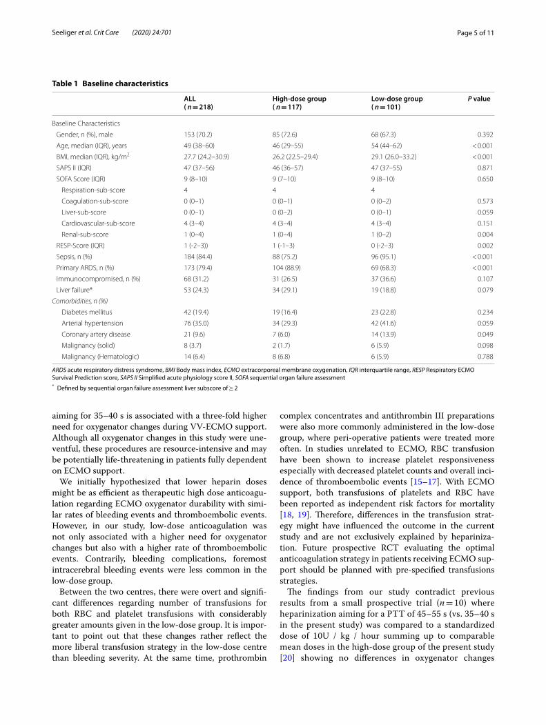
Primary endpointOverall, there were 14 oxygenator changes in the high-dose group versus 48 in the low-dose group in 13 versus 32 patients. Freedom from oxygenator change at 15 days was 73% in the high-dose group versus 55% in the low-dose group (adjusted hazard ratio (HR) 3.34 [95% con-fidence interval 1.2–9.4 with low-dose heparinization], P = 0.023) (Fig. 2), with the entire regression model dis-played in Fig. 3. Reasons for oxygenator changes were decreasing post-filter paO2 (86% and 59%), thrombus formation with increasing D-dimers (14% and 39%), and overt haemolysis (0% and 2%). The results were similar when analysing freedom from oxygenator change with-out censoring data at 15 days (adjusted HR 3.28 [95% confidence interval 1.2–8.6] with low-dose hepariniza-tion, P = 0.016).
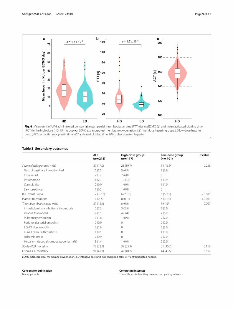
Secondary endpointsThe overall median of the individual mean ACT in the high-dose group was 158 s (IQR 151–165) and the median of the mean PTT in the high-dose group was 48 s (IQR 41–57) versus 38 s (IQR 34–42) in the low-dose group (P < 0.001) (Fig. 4b, c). The corresponding mean units of heparin administered while on ECMO were 17,495 (IQR 10,971–24,327) vs. 11,185 (IQR 4,372–16,750) (P < 0.001) (Fig. 4a). The centre-defined ACT and PTT corridors were well-represented in the groups.

Page 4 of 11Seeliger et al. Crit Care (2020) 24:701
Additional baseline coagulation parameters are shown in Table 2. In the patients who received an oxygenator change, the d-dimer foldchange compared to baseline was 2.97 (IQR 1.5–8.2) compared to 1.5 (IQR 0.4–4.5), in patients who did not undergo oxygenator change (P = 0.002).
Severe bleeding was not different and occurred in 23 (19.7%) of patients in the high-dose group and in 14 (13.9%) in the low-dose group (P = 0.256) (Table 3). Of note, severe intracranial bleeding only occurred in the high-dose group with fatal outcome in 5 of 7 cases, while 3 events of intracranial bleeding in the low-dose group were incidental findings on CT without overt neuro-logical deficit (and where thus not categorized as severe bleeding events).
Applying different in-house standards of transfusion procedures, the number of RBC unit transfusions were significantly lower in the high-dose group compared to the low-dose group (6 [IQR 2–10] vs. 8 [IQR 6–19] P < 0.001), as were units of platelet transfusions (0 [IQR 0–1] vs. 4 [0–10], P < 0.001). More patients in the low-dose group received prothrombin complex concentrates (26 [25.7%] vs. 8 [6.8%], p < 0.001) and antithrombin III substitution (21 [21%] vs. 8 [6.9%], p = 0.002), while administration of tranexamic acid was similar (Table 2). All patients who received prothrombin complex
concentrates had severe coagulopathy in the context of planned intervention with high bleeding risk (high-dose group: 2/8; low-dose group: 15/26) or significant haemo-globin-relevant bleeding (high-dose group: 6/8; low-dose group 11/26). Pre-ECMO use of antiplatelet therapy was not associated with oxygenator change or severe ECMO-related bleeding events.
Fewer thromboembolic events occurred in the high-dose group (8 [6.8%]) than in the low-dose group (19 [19%], P = 0.007). Of note, direct thrombotic events of the ECMO circuit (cannula thrombosis n = 1; coagulation of the oxygenator n = 2) occurred only in the low-dose group (Table 3).
The 30-day ICU mortality was comparable with 33.3% (n = 39) in the high-dose versus 30.7% (n = 31) in the low-dose group (P = 0.11). The main reasons for mortality in the high-dose versus low-dose group were cessation of therapy due to medical futility in 23 (59%) versus 12 (38.7%); refractory multiorgan failure in 11 (28.2%) ver-sus 18 (58.1%), intracranial bleeding in 5 (12.8%) versus 0; abdominal bleeding complications in 0 versus 1 (3.2%).
DiscussionThe key finding of this study is that when compared to an ACT-guided high dose heparinization strategy aiming for 140–180 s, a low dose heparin strategy adjusted by PTT
216 Received VV-ECMOsupport for acute respiratoryfailure on medical ICU
117 Were included in theanalysis and had ACT-guided heparinization with agoal of 140-180s13 had oxygenator changes
77 Excluded (No Consent)6 Excluded (ECMO support for
less than 24h)6 Excluded (acute liver failure with
desolate coagulation function)4 Excluded (ECMO support > 24h
before referral)3 Excluded (triple cannulation
within first 24h)2 Excluded (age < 18 years)1 Excluded (missing data)
159 Received VV-ECMO support foracute respiratory failure onanaesthesiology ICU
101 Were included in theanalysis and had PTTguided heparinization with agoal of 35-40s32 had oxygenator changes
41 Excluded (did not followinstitutional protocol)
9 Excluded (ECMO support forless than 24h)
6 Excluded (missing data)2 Excluded (hereditary
coagulopathy)
High-dose heparin centre Low-dose heparin centre
Fig. 1 Flow chart for patient cohorts by centre / heparinization strategy. The need for written informed consent at the low‑dose centre was waved by the institutional review board due to the retrospective nature of the study
Page 5 of 11Seeliger et al. Crit Care (2020) 24:701
aiming for 35–40 s is associated with a three-fold higher need for oxygenator changes during VV-ECMO support. Although all oxygenator changes in this study were une-ventful, these procedures are resource-intensive and may be potentially life-threatening in patients fully dependent on ECMO support.
We initially hypothesized that lower heparin doses might be as efficient as therapeutic high dose anticoagu-lation regarding ECMO oxygenator durability with simi-lar rates of bleeding events and thromboembolic events. However, in our study, low-dose anticoagulation was not only associated with a higher need for oxygenator changes but also with a higher rate of thromboembolic events. Contrarily, bleeding complications, foremost intracerebral bleeding events were less common in the low-dose group.
Between the two centres, there were overt and signifi-cant differences regarding number of transfusions for both RBC and platelet transfusions with considerably greater amounts given in the low-dose group. It is impor-tant to point out that these changes rather reflect the more liberal transfusion strategy in the low-dose centre than bleeding severity. At the same time, prothrombin
complex concentrates and antithrombin III preparations were also more commonly administered in the low-dose group, where peri-operative patients were treated more often. In studies unrelated to ECMO, RBC transfusion have been shown to increase platelet responsiveness especially with decreased platelet counts and overall inci-dence of thromboembolic events [15–17]. With ECMO support, both transfusions of platelets and RBC have been reported as independent risk factors for mortality [18, 19]. Therefore, differences in the transfusion strat-egy might have influenced the outcome in the current study and are not exclusively explained by hepariniza-tion. Future prospective RCT evaluating the optimal anticoagulation strategy in patients receiving ECMO sup-port should be planned with pre-specified transfusions strategies.
The findings from our study contradict previous results from a small prospective trial (n = 10) where heparinization aiming for a PTT of 45–55 s (vs. 35–40 s in the present study) was compared to a standardized dose of 10U / kg / hour summing up to comparable mean doses in the high-dose group of the present study [20] showing no differences in oxygenator changes
Table 1 Baseline characteristics
ARDS acute respiratory distress syndrome, BMI Body mass index, ECMO extracorporeal membrane oxygenation, IQR interquartile range, RESP Respiratory ECMO Survival Prediction score, SAPS II Simplified acute physiology score II, SOFA sequential organ failure assessment* Defined by sequential organ failure assessment liver subscore of ≥ 2
ALL( n = 218)
High-dose group( n = 117)
Low-dose group( n = 101)
P value
Baseline Characteristics
Gender, n (%), male 153 (70.2) 85 (72.6) 68 (67.3) 0.392
Age, median (IQR), years 49 (38–60) 46 (29–55) 54 (44–62) < 0.001
BMI, median (IQR), kg/m2 27.7 (24.2–30.9) 26.2 (22.5–29.4) 29.1 (26.0–33.2) < 0.001
SAPS II (IQR) 47 (37–56) 46 (36–57) 47 (37–55) 0.871
SOFA Score (IQR) 9 (8–10) 9 (7–10) 9 (8–10) 0.650
Respiration‑sub‑score 4 4 4
Coagulation‑sub‑score 0 (0–1) 0 (0–1) 0 (0–2) 0.573
Liver‑sub‑score 0 (0–1) 0 (0–2) 0 (0–1) 0.059
Cardiovascular‑sub‑score 4 (3–4) 4 (3–4) 4 (3–4) 0.151
Renal‑sub‑score 1 (0–4) 1 (0–4) 1 (0–2) 0.004
RESP‑Score (IQR) 1 (‑2–3)) 1 (‑1–3) 0 (‑2–3) 0.002
Sepsis, n (%) 184 (84.4) 88 (75.2) 96 (95.1) < 0.001
Primary ARDS, n (%) 173 (79.4) 104 (88.9) 69 (68.3) < 0.001
Immunocompromised, n (%) 68 (31.2) 31 (26.5) 37 (36.6) 0.107
Liver failure* 53 (24.3) 34 (29.1) 19 (18.8) 0.079
Comorbidities, n (%)
Diabetes mellitus 42 (19.4) 19 (16.4) 23 (22.8) 0.234
Arterial hypertension 76 (35.0) 34 (29.3) 42 (41.6) 0.059
Coronary artery disease 21 (9.6) 7 (6.0) 14 (13.9) 0.049
Malignancy (solid) 8 (3.7) 2 (1.7) 6 (5.9) 0.098
Malignancy (Hematologic) 14 (6.4) 8 (6.8) 6 (5.9) 0.788

Page 6 of 11Seeliger et al. Crit Care (2020) 24:701
and bleeding events with considerable chances for underpowering. A longitudinal single-centre pre-post designed retrospective trial (n = 40) showed similar survival to decannulation rates, bleeding events and thromboembolic complications [21], but interestingly the ACTs in both groups were rather high (167 s vs. 189 s) compared to our cohort. Another mixed cohort including 22 patients with VV-ECMO compared hep-arinization guided by ACT (140–160 s vs. 180–220) and consistently found fewer bleeding events and simi-lar rates of oxygenator changes [22], also aiming for higher ACT-prolongation than in our cohort. The lack of uniform anticoagulation strategies and outcome
definitions across all studies render comparison of event rates difficult [23].
Beside mere heparin dosage, temporary interrup-tion of heparin (e.g. in response to bleeding) may create a hypercoagulable milieu, thereby increasing the risk of clotting and oxygenator failure [24, 25], which might have influenced the results of the present study. Case series and smaller retrospective studies reported feasibility of heparin-free ECMO support in cases of trauma or severe bleeding [26–29] and intermittent subcutaneous admin-istration of heparin to avoid heparin pauses [25], but the optimal management strategies in these particularly chal-lenging situations needs to be prospectively investigated
Table 2 ECMO settings and relevant coagulation factors
ECMO extracorporeal membrane oxygenation, IQR interquartile range, PCC prothrombin complex centrates
ALL( n = 218)
High-dose group( n = 117)
Low-dose group( n = 101)
P value
ECMO device < 0.001
CardioHelp 31 (26.5) 99 (98)
RotaFlow 86 (73.5) 2 (2)
Canula site out
Jugular vein 3 (1.4) 3 (2.6) 0
Femoral vein 215 (98.6) 114 (97.4) 101 (100)
Canula site in
Jugular vein 135 (61.9) 113 (96.6) 22 (21.8)
Subclavian vein 2 (0.9) 2 (1.7) 0
Femoral vein 81 (37.2) 2 (1.7) 79 (78.2)
Canula size out 23 (23–35) 23 (23–23) 25 (25–25) < 0.001
Canula size in 17 (17–23) 17 (17–17) 23 (21–25) < 0.001
ECMO runtime, median (IQR), days 9 (5–14) 8 (5–12) 11 (7–17) 0.003
ECMO flow, median (IQR), liter per minute 3.8 (3.3–4.4) 3.5 (2.8–3.9) 4.4 (3.8–4.9) < 0.001
Days from mechanical ventilation to ECMO implantation, median (ICR)
1 (0–3) 1 (0–3) 1 (0–4) 0.185
Antiplatelet therapy pre‑ECMO
Aspirin 31 (14) 13 (11) 18 (18) 0.157
P2Y12‑inhibitors 6 (3) 2 (2) 4 (4) 0.311
Antiplatelet therapy during ECMO, n (%)
Aspirin 27 (12) 9 (8) 18 (18) 0.024
P2Y12‑inhibitors 5 (2) 0 5 (5) 0.015
Baseline fibrinogen, g/L (IQR) 4.7 (3.3–6.2) 6.7 (3.3–6.4) 4.8 (3.4–6.1) 0.611
Minimal fibrinogen, g/L (IQR) 2.2 (1.4–3.4) 2.5 (1.6–3.5) 2.0 (1.2–3.4) 0.030
Baseline d‑dimers, mg/L (IQR) 7.6 (3.6–15.4) 5.4 (2.6–12.7) 8.3 (4.4–16.5) 0.010
Maximum d‑dimers, mg/L (IQR) 30 (19.3–35) 28.5 (13–30) 35 (33.4–35) < 0.001
Baseline antithrombin III, % (IQR) 67 (53–86) 79 (60–93) 60 (44–75) < 0.001
Minimum antithrombin III, % (IQR) 59 (45–76) 69 (56–84) 48 (37–62) < 0.001
Antithrombin III substitution, n (%) 29 (13.3) 8 (6.8) 21 (21) 0.002
Baseline thrombocyte count, thousand / µL (IQR) 174 (101–265) 167 (109–269) 183 (97–263) 0.878
Minimum thrombocyte count, thousand / µL (IQR) 62 (36–88) 65 (33–106) 60 (40–81) 0.868
Received PCC, n (%) 36 (16.5) 8 (6.8) 26 (25.7) < 0.001
Received tranexamic acid, n (%) 113 (51.8) 61 (52.1) 52 (51.5) 0.924

Page 7 of 11Seeliger et al. Crit Care (2020) 24:701
and lies beyond the scope of the current retrospective study.
In recent years, more centres integrated anti-Xa lev-els alongside ACT or PTT in their routine coagulation monitoring during ECMO support following promising results mainly in paediatric populations and better reflec-tion of heparin concentrations [30–33]. Since inflamma-tion can influence ACT measurements [34] the ECMO centres of the current study also utilize anti-Xa levels alongside thromboelastography and single clotting factor analysis where coagulation state is uncertain, and PTT or ACT seems out of line with heparin dosing. However, longer turn-around time and varying 24/7 availability of anti-Xa measurements are limiting factors and 97% of ECMO centres were still using ACT for heparin dose adjustments in 2014 [35]. Since current ELSO guidelines recommend ACT and PTT for measuring heparin effects [6], the analysis of different heparinization strategies based on these assays provide valuable information for intensivists caring for ECMO patients. Yet, we acknowl-edge that anti-Xa levels may be a more appropriate meas-urement of heparin effects than ACT or PTT in critically ill patients.
Limitations of this study were inherent to the ret-rospective design and the comparison of two centres, which render the data subject to substantial potential bias, including different bleeding management strategies, different ECMO devices, cannulation sites and patient populations. Our data thus needs prospective validation with uniform strategies for bleeding management, trans-fusion strategies and ECMO configuration to derive solid recommendations.
ConclusionIn this two-centre cohort study, the institutional strategy with a high-dose heparinization during ECMO support was associated with lower rates of oxygenator changes and thromboembolic events, compared to the strategy with low-dose heparinization. Prospective randomized validation with standardized bleeding management and ECMO settings is needed to confirm these findings.
AbbreviationsACT : Activated clotting time; ARDS: Acute respiratory distress syndrome; ARF: Acute respiratory failure; ECMO: Extracorporeal membrane oxygenation; HR: Hazard ratio; ICU: Intensive care unit; IQR: Interquartile range; PTT: Partial
Time [days]
117 107 76 50 31 13101 89 76 59 33 17
No. at riskHDLD
0 3 6 9 12 15
0
25
50
75
100
Adj. HRa 3.34 (95% CI 1.2-9.4)p = 0.023
High-dose heparin group (HD)
Low-dose heparin group (LD)Oxy
gena
tor-
chan
ge fr
ee s
urvi
val [
%]
Fig. 2 Freedom of oxygenator‑change at 15 days between high‑dose and low‑dose heparinization groups. Data is censored for decannulation or mortality. HR hazard ratio, CI confidence interval. aAdjusted for age, gender, body‑mass‑index, Respiratory ECMO Survival Prediction (RESP) score, Simplified acute physiology score II, sequential organ failure assessment renal sub‑score, ECMO runtime, sepsis, coronary artery disease, prior aspirin use, mean ECMO blood flow, baseline fibrinogen, d‑dimer and antithrombin III levels.
Page 8 of 11Seeliger et al. Crit Care (2020) 24:701
thromboplastin time; RBC: Red blood cells; RESP: Respiratory ECMO Survival Prediction; SAPS: Simplified acute physiology score; SOFA: Sequential organ failure assessment; UFH: Unfractionated heparin; VV‑ECMO: Venovenous extracorporeal membrane oxygenation.
AcknowledgementsNone
Authors’ contributionsBS, SD and CB designed this study and data acquisition strategy. BS, MD, RF and CB were responsible for the data collection. BS, MD, AT, SD and CB were responsible for data analysis. BS and SD conducted the manuscript writing. KS, CK, JB, FS, SE, JCS, CP, TW, MMH, AT and CB critically revised the manuscript. BS, MD, SD and CB provided final approval for this version to be published. All authors agreed to be accountable for all aspects of the work in ensuring that questions related to the accuracy or integrity of any part of the work are appropriately investigated and resolved. All authors read and approved the final manuscript.
FundingOpen Access funding enabled and organized by Projekt DEAL. There was no specific funding for this study. BS is supported by PRACTIS – Clinician Scientist Programme of Hannover Medical School, funded by the Deutsche Forschungsgemeinschaft (DFG, ME 3696/3–1) and the German Center for Lung Research (DZL). JB, CK and MMH are supported by the DFG (KFO311, BA 1742/9–1, HO 1599/2–2). The laboratory of SD received funding from the DFG (DA 1209/4–3) and the DZL. The laboratory of CB received funding from the University of Bonn (BONFOR), and the B. Braun Foundation (D‑17‑00098).
Availability of data and materialsThe data used for this research are available from the corresponding author on reasonable request and subject to Institutional Review Board guidelines.
Ethics approval and consent to participateThis study was reviewed and approved by the Institutional Review Board of Hannover Medical School (8715_BO_K_2019) and University of Bonn (398/19).
1 015.0
Characteristic Exposed UnexposedUnadj. HR(95% CI) P
Adj. HR(95% CI) P
Low doseheparinization 320.)4.9-2.1(43.3910.)0.5-2.1(14.2812/711812/101
863.)2.1-7.0(78.0143.)1.1-7.0(09.0y01rep,egA
984.)7.1-3.0(57.0828.)2.2-5.0(80.1812/351812/56elameF
232.)1.1-0.1(30.1605.)0.1-0.1(10.1IMB
775.)1.1-9.0(30.1373.)1.1-0.1(40.1erocS-PSER
420.)0.1-9.0(59.0400.)0.1-9.0(69.0II-SPAS
SOFA RenalSubscore 223.)2.1-6.0(48.0830.)0.1-6.0(57.0
398.)0.1-0.1(00.1373.)0.1-0.1(10.1emitnurOMCE
667.)7.3-4.0(91.1746.)6.3-5.0(82.1812/43812/481sispeS
483.)6.2-1.0(64.0777.)3.3-4.0(61.1812/791812/12DAC
592.)11-5.0(33.2926.)3.3-5.0(62.1812/781812/13SSA
306.)5.1-5.0(78.0294.)6.1-8.0(41.1wolfOMCE
Baselinefibrinogen 344.)3.1-9.0(80.1669.)2.1-8.0(00.1
Baselined-dimers 135.)0.1-9.0(99.0307.)0.1-0.1(00.1
Baselineantithrombin III 379.)0.1-0.1(00.1506.)0.1-0.1(00.1
Unadj. Risk, No./Total
0.20.1 2 5
DecreasedRisk for oxy-
change
IncreasedRisk for oxy-change
Fig. 3 Multivariable cox regression model for oxygenator change. BMI body mass index, CAD coronary artery disease, RESP Respiratory ECMO Survival Prediction score, SAPS simplified acute physiology score, SOFA sequential organ failure assessment
Page 9 of 11Seeliger et al. Crit Care (2020) 24:701
Consent for publicationNot applicable.
Competing interestsThe authors declare they have no competing interests.
DLDLDH DHDH
p = 1.7 x 10⁻⁵ p = 1.7 x 10⁻¹²M
ean
hepa
rin [k
U p
er E
CM
O d
ay]
0
10
20
30
40
50
60
70
20
40
60
80
100
120
140
160
PTT
[s]
100
120
140
160
180
200
AC
T [s
]
cba
Fig. 4 Mean units of UFH administered per day (a); mean partial thromboplastin time (PTT) during ECMO (b) and mean activated clotting time (ACT) in the high‑dose (HD) UFH group (c). ECMO extracorporeal membrane oxygenation, HD high dose heparin groups, LD low dose heparin group, PTT partial thromboplastin time, ACT activated clotting time, UFH unfractionated heparin
Table 3 Secondary outcomes
ECMO extracorporeal membrane oxygenation, ICU intensive care unit, RBC red blood cells, UFH unfractionated heparin
ALL(n = 218)
High-dose group(n = 117)
Low-dose group(n = 101)
P value
Severe bleeding events, n (%) 37 (17.0) 23 (19.7) 14 (13.9) 0.256
Gastrointestinal / intrabdominal 12 (5.5) 5 (4.3) 7 (6.9)
Intracranial 7 (3.2) 7 (6.0) 0
Intrathoracic 16 (7.3) 10 (8.5) 6 (5.9)
Cannula site 2 (0.9) 1 (0.9) 1 (1.0)
Ear‑nose‑throat 1 (0.5) 1 (0.8) 0
RBC transfusions 7 (3–13) 6 (2–10) 8 (6–19) < 0.001
Platelet transfusions 1 (0–5) 0 (0–1) 4 (0–10) < 0.001
Thromboembolic events, n (%) 27 (12.4) 8 (6.8) 19 (19) 0.007
Intraabdominal embolism / thrombosis 5 (2.3) 3 (2.5) 2 (2.0)
Venous thrombosis 12 (5.5) 4 (3.4) 7 (6.9)
Pulmonary embolism 3 (1.8) 1 (0.9) 2 (2.0)
Peripheral arterial embolism 2 (0.9) 0 2 (2.0)
ECMO filter embolism 3 (1.8) 0 3 (3.0)
ECMO cannula thrombosis 1 (0.5) 0 1 (1.0)
Ischemic stroke 2 (0.9) 0 2 (2.0)
Heparin‑induced thrombocytopenia, n (%) 3 (1.4) 1 (0.9) 2 (2.0)
30‑day ICU mortality 70 (32.1) 39 (33.3) 31 (30.7) 0.110
Overall ICU mortality 91 (41.7) 47 (40.2) 44 (43.6) 0.612

Page 10 of 11Seeliger et al. Crit Care (2020) 24:701
Author details1 Department of Respiratory Medicine, Hannover Medical School and Biomed‑ical Research in End‑Stage and Obstructive Lung Disease (BREATH), German Lung Research Center (DZL), Hannover, Germany. 2 Department of Anaesthe‑siology and Critical Care Medicine, University Hospital Bonn, Bonn, Germany. 3 Department of Gastroenterology, Hepatology and Endocrinology, Hannover Medical School, Hannover, Germany. 4 Department of Cardiothoracic, Trans‑plantation and Vascular Surgery, Hannover Medical School, Hannover, Ger‑many. 5 Department of Cardiology and Angiology, Hannover Medical School, Hannover, Germany. 6 Department of Haematology, Haemostasis, Oncology, and Stem Cell Transplantation, Hannover Medical School, Hannover, Germany. 7 Department of Nephrology and Hypertension, Hannover Medical School, Hannover, Germany. 8 Institute for Intensive Care Medicine, University Hospital Zurich, Zurich, Switzerland.
Received: 19 April 2020 Accepted: 14 October 2020
References 1. Combes A, Hajage D, Capellier G, Demoule A, Lavoué S, Guervilly C, et al.
Extracorporeal membrane oxygenation for severe acute respiratory distress syndrome. N Engl J Med. 2018;378:1965–75.
2. Sy E, Sklar MC, Lequier L, Fan E, Kanji HD. Anticoagulation practices and the prevalence of major bleeding, thromboembolic events, and mortality in venoarterial extracorporeal membrane oxygenation: A systematic review and meta‑analysis. J Crit Care. 2017;39:87–96.
3. Kreyer S, Muders T, Theuerkauf N, Spitzhüttl J, Schellhaas T, Schewe J‑C, et al. Hemorrhage under veno‑venous extracorporeal membrane oxy‑genation in acute respiratory distress syndrome patients: a retrospective data analysis. J Thorac Dis. 2017;9:5017–29.
4. Lubnow M, Philipp A, Foltan M, Bull Enger T, Lunz D, Bein T, et al. Technical complications during veno‑venous extracorporeal membrane oxygena‑tion and their relevance predicting a system‑exchange–retrospective analysis of 265 cases. PLoS ONE. 2014;9:e112316.
5. Esper SA, Welsby IJ, Subramaniam K, John Wallisch W, Levy JH, Waters JH, et al. Adult extracorporeal membrane oxygenation: An interna‑tional survey of transfusion and anticoagulation techniques. Vox Sang. 2017;112:443–52.
6. Extracorporeal Life Support Organization. ELSO Guidelines for Cardio‑pulmonary Extracorporeal Life Support Version 1.4. August 2017. https ://www.elso.org/Porta ls/0/ELSO%20Gui delin es%20For %20Adu lt%20Res pirat ory%20Fai lure%201_4.pdf. Accessed 8 Mar 2020.
7. Mazzeffi MA, Tanaka K, Roberts A, Rector R, Menaker J, Kon Z, et al. Bleeding, thrombosis, and transfusion with two heparin anticoagulation protocols in venoarterial ECMO patients. J Cardiothorac Vasc Anesth. 2019;33:1216–20.
8. Raman J, Alimohamed M, Dobrilovic N, Lateef O, Aziz S. A comparison of low and standard anti‑coagulation regimens in extracorporeal mem‑brane oxygenation. J Heart Lung Transpl. 2019;38:433–9.
9. Aubron C, McQuilten Z, Bailey M, Board J, Buhr H, Cartwright B, et al. Low‑dose versus therapeutic anticoagulation in patients on extracor‑poreal membrane oxygenation: a pilot randomized trial. Crit Care Med. 2019;47:e563–71.
10. Derek L, Zivkovic M, Hasperger D, Juricek J, Romic Z. Comparabil‑ity of Pathromtin SL, Dade Actin FS i STA Cephascreen reagens for activated partial thromboplastin time measurement. Biochem Med. 2008;2008:81–7.
11. Philipp A, de Somer F, Foltan M, Bredthauer A, Krenkel L, Zeman F, Lehle K. Life span of different extracorporeal membrane systems for severe respiratory failure in the clinical practice. PLoS ONE. 2018;13:e0198392.
12. Le Gall J‑R. A New Simplified Acute Physiology Score (SAPS II) Based on a European/North American Multicenter Study. JAMA. 1993;270:2957.
13. Schmidt M, Bailey M, Sheldrake J, Hodgson C, Aubron C, Rycus PT, et al. Predicting survival after extracorporeal membrane oxygenation for severe acute respiratory failure the respiratory extracorporeal membrane oxygenation survival prediction (RESP) score. Am J Respir Crit Care Med. 2014;189:1374–82.
14. Vincent JL, Moreno R, Takala J, Willatts S, de Mendonça A, Bruining H, et al. The SOFA (Sepsis‑related Organ Failure Assessment) score to describe organ dysfunction/failure. On behalf of the Working Group on Sepsis‑Related Problems of the European Society of Intensive Care Medicine. Intensive Care Med. 1996;22:707–10.
15. Silvain J, Abtan J, Kerneis M, Martin R, Finzi J, Vignalou J‑B, et al. Impact of red blood cell transfusion on platelet aggregation and inflammatory response in anemic coronary and noncoronary patients: the TRANS‑FUSION‑2 study (impact of transfusion of red blood cell on platelet activation and aggregation studied with flow cytometry use and light transmission aggregometry). J Am Coll Cardiol. 2014;63:1289–96.
16. Valles J, Santos MT, Aznar J, Marcus AJ, Martinez‑Sales V, Portoles M, et al. Erythrocytes metabolically enhance collagen‑induced platelet respon‑siveness via increased thromboxane production, adenosine diphosphate release, and recruitment. Blood. 1991;78:154–62.
17. Goel R, Patel EU, Cushing MM, Frank SM, Ness PM, Takemoto CM, et al. Association of perioperative red blood cell transfusions with venous thromboembolism in a North American Registry. JAMA Surg. 2018;153:826–33.
18. Aubron C, Cheng AC, Pilcher D, Leong T, Magrin G, Cooper DJ, et al. Fac‑tors associated with outcomes of patients on extracorporeal membrane oxygenation support: a 5‑year cohort study. Crit Care. 2013;17:R73.
19. Mazzeffi M, Greenwood J, Tanaka K, Menaker J, Rector R, Herr D, et al. Bleeding, transfusion, and mortality on extracorporeal life support: ECLS working group on thrombosis and hemostasis. Ann Thorac Surg. 2016;101:682–9.
20. Deatrick KB, Galvagno SM, Mazzeffi MA, Kaczoroswki DJ, Herr DL, Rector R, et al. Pilot study evaluating a non‑titrating, weight‑based anticoagulation scheme for patients on veno‑venous extracorporeal membrane oxygena‑tion. Perfusion. 2020;35:13–8.
21. Carter KT, Kutcher ME, Shake JG, Panos AL, Cochran RP, Creswell LL, Cope‑land H. Heparin‑sparing anticoagulation strategies are viable options for patients on veno‑venous ECMO. J Surg Res. 2019;243:399–409.
22. Yeo HJ, Kim DH, Jeon D, Kim YS, Cho WH. Low‑dose heparin during extra‑corporeal membrane oxygenation treatment in adults. Intensive Care Med. 2015;41:2020–1.
23. Oude Lansink‑Hartgring A, de Vries AJ, Droogh JM, van den Bergh WM. Hemorrhagic complications during extracorporeal membrane oxygena‑tion: the role of anticoagulation and platelets. J Crit Care. 2019;54:239–43.
24. Kasirajan V, Smedira NG, McCarthy JF, Casselman F, Boparai N, McCarthy PM. Risk factors for intracranial hemorrhage in adults on extracorporeal membrane oxygenation1. Eur J Cardiothorac Surg. 1999;15:508–14.
25. Kurihara C, Walter JM, Karim A, Thakkar S, Saine M, Odell DD, et al. Feasibil‑ity of venovenous extracorporeal membrane oxygenation without sys‑temic anticoagulation. Ann Thorac Surg. 2020. https ://doi.org/10.1016/j.athor acsur .2020.02.011.
26. Herbert DG, Buscher H, Nair P. Prolonged venovenous extracorporeal membrane oxygenation without anticoagulation: a case of Good‑pasture syndrome‑related pulmonary haemorrhage. Crit Care Resusc. 2014;16:69–72.
27. Seeliger B, Stahl K, Schenk H, Schmidt JJ, Wiesner O, Welte T, et al. Extracorporeal membrane oxygenation for severe ARDS due to immune diffuse alveolar hemorrhage: a retrospective observational study. Chest. 2020;157:744–7.
28. Takagaki M, Yamaguchi H, Ikeda N, Takeda K, Kasai F, Yahagi K, et al. Post‑cardiotomy venovenous extracorporeal membrane oxygenation without heparinization. Gen Thorac Cardiovasc Surg. 2019;67:982–6.
29. Muellenbach RM, Kredel M, Kunze E, Kranke P, Kuestermann J, Brack A, et al. Prolonged heparin‑free extracorporeal membrane oxygenation in multiple injured acute respiratory distress syndrome patients with traumatic brain injury. J Trauma Acute Care Surg. 2012;72:1444–7.
30. Northrop MS, Sidonio RF, Phillips SE, Smith AH, Daphne HC, Pietsch JB, Bridges BC. The use of an extracorporeal membrane oxygenation anticoagulation laboratory protocol is associated with decreased blood product use, decreased hemorrhagic complications, and increased circuit life. Pediatr Crit Care Med. 2015;16:66–74.
31. Liveris A, Bello RA, Friedmann P, Duffy MA, Manwani D, Killinger JS, et al. Anti‑factor Xa assay is a superior correlate of heparin dose than activated partial thromboplastin time or activated clotting time in pediatric extra‑corporeal membrane oxygenation*. Pediatr Crit Care Med. 2014;15:e72–9.

Page 11 of 11Seeliger et al. Crit Care (2020) 24:701
• fast, convenient online submission
•
thorough peer review by experienced researchers in your field
• rapid publication on acceptance
• support for research data, including large and complex data types
•
gold Open Access which fosters wider collaboration and increased citations
maximum visibility for your research: over 100M website views per year •
At BMC, research is always in progress.
Learn more biomedcentral.com/submissions
Ready to submit your researchReady to submit your research ? Choose BMC and benefit from: ? Choose BMC and benefit from:
32. Ranucci M, Cotza M, Isgrò G, Carboni G, Ballotta A, Baryshnikova E. Anti‑factor Xa‑based anticoagulation during extracorporeal membrane oxygenation: potential problems and possible solutions. Semin Thromb Hemost. 2020;46:419–27.
33. Chlebowski MM, Baltagi S, Carlson M, Levy JH, Spinella PC. Clinical con‑troversies in anticoagulation monitoring and antithrombin supplementa‑tion for ECMO. Crit Care. 2020;24:19.
34. Papageorgiou C, Synetos A, Tampakis K, Anninos H, Kontogian‑nis C, Kapelouzou A, et al. Activated clotting time as a marker of inflammation in hospitalized patients. Clin Appl Thromb Hemost. 2020;26:1076029620929090.
35. Bembea MM, Annich G, Rycus P, Oldenburg G, Berkowitz I, Pronovost P. Variability in anticoagulation management of patients on extracorporeal membrane oxygenation: an international survey. Pediatr Crit Care Med. 2013;14:e77‑84.
Publisher’s NoteSpringer Nature remains neutral with regard to jurisdictional claims in pub‑lished maps and institutional affiliations.