Comparative Analysis of Leptospira Strains Isolated from ...
9
Comparative Analysis of Leptospira Strains Isolated from Environmental Soil and Water in the Philippines and Japan Mitsumasa Saito, a Sharon Y. A. M. Villanueva, a Antara Chakraborty, a * Satoshi Miyahara, a Takaya Segawa, a Tatsuma Asoh, a Ryo Ozuru, a Nina G. Gloriani, b Yasutake Yanagihara, a,c Shin-ichi Yoshida a Department of Bacteriology, Graduate School of Medical Sciences, Kyushu University, Higashi-ku, Fukuoka, Japan a ; Department of Medical Microbiology, College of Public Health, University of the Philippines–Manila, Ermita, Manila, Philippines b ; University of Shizuoka, Suruga-ku, Shizuoka, Japan c There have been few reports on the epidemiological analysis of environmental Leptospira isolates. This is probably because the isolation of leptospires from the environment was usually unsuccessful due to the overgrowth of contaminants and the slow growth of Leptospira. In this study, we collected a total of 88 samples of soil and water from three sites: Metro Manila and Nueva Ecija, Philippines (an area where Leptospira is now endemic), and Fukuoka, Japan (an area where Leptospira was once endemic). We succeeded in isolating Leptospira from 37 samples by using the novel combination of five antimicrobial agents reported in 2011. The frequencies of positive isolation of Leptospira in the Philippines and Japan were 40 and 46%, respectively. For Lepto- spira-positive samples, five colonies from each sample were isolated and analyzed by pulsed-field gel electrophoresis (PFGE). The isolates from each area showed their respective characteristics in phylogenetic trees based on the PFGE patterns. Some iso- lates were closely related to each other across borders. Based on 16S rRNA gene-based phylogenetic analysis, four isolates in Fu- kuoka were identified as a pathogenic species, L. alstonii; however, its virulence had been lost. One isolate from Nueva Ecija was identified as the intermediate pathogenic species Leptospira licerasiae. Most of the isolates from the environment belonged to nonpathogenic Leptospira species. We also investigated the strain variation among the isolates in a puddle over 5 months. We demonstrated, using PFGE analysis, that Leptospira survived in the wet soil on dry days and appeared in the surface water on rainy days. These results showed that the soil could be a reservoir of leptospires in the environment. L eptospira species are aerobic, spiral, Gram-negative bacteria. The organisms infect the host renal tubules and are shed into the environment via urination, where they can survive in moist soil and surface water for up to several months (1, 2). Humans and other animals become infected, mainly through their skins and mucous membranes, when they encounter a leptospire-contami- nated environment (3). Due to this correlation between organ- isms in the environment and disease transmission, the isolation of leptospires from the environment is important for epidemiologi- cal studies, as well as for the prevention and control of the disease. However, isolation from environmental samples is challenging due to the slow growth of leptospires and the overgrowth of coex- isting microorganisms. We previously reported a novel combina- tion of antimicrobial agents (sulfamethoxazole, trimethoprim, amphotericin B, fosfomycin, and 5-fluorouracil [STAFF]) for se- lective isolation of leptospires from contaminated samples (4). This cocktail, after being incorporated into Leptospira growth me- dium, inhibited the growth of contaminants and successfully iso- lated leptospires in environmental samples. In 1915, Inada and Ido discovered spirochetes and specific an- tibodies in the blood of Japanese coal miners with infectious jaun- dice (Weil’s disease) in Fukuoka, Japan (5). Now this organism is known as Leptospira interrogans strain Ictero No.1. Until 1960, more than 200 deaths of leptospirosis were reported every year in Japan. Most of them were farmers working in rice fields. After 1960, the mechanization of agriculture was introduced, and farm- ers started using rubber boots while working in the field. At the same time, an inactivated vaccine against pathogenic Leptospira was developed. For these reasons, the number of patients with leptospirosis in Japan decreased dramatically (6). In 2011, only 31 cases were reported. On the other hand, many people have been infected with Leptospira among Asian countries located in tropical areas, such as India, Thailand, Indonesia, Philippines, etc. (6). In the Philippines, the incidence of patients was closely related to rainfall. Outbreaks usually occurred during the rainy season (June to November) and just after the rainy season in flood-prone areas (6). The present study was carried out in order to find out whether the environmental distribution of Leptospira species accounts for the difference of the current status of leptospirosis between Japan and the Philippines. We collected soil and water samples in both countries and succeeded in isolating leptospires from these sam- ples by using the STAFF combination of antimicrobial agents. When the sample was culture positive, five Leptospira colonies from each sample were isolated by using the single colony isola- tion method and were analyzed by pulsed-field gel electrophoresis (PFGE). 16S rRNA gene-based phylogenetic analysis was per- formed for all isolates with the 20 previously described genom- ospecies in the genus Leptospira, and the genomospecies of the isolates were deduced. MATERIALS AND METHODS Isolation procedure of Leptospira species from environmental samples. The collection of environmental samples was performed in three sites, Received 11 September 2012 Accepted 2 November 2012 Published ahead of print 9 November 2012 Address correspondence to Mitsumasa Saito, [email protected]. * Present address: Antara Chakraborty, School of Applied Sciences, Republic Polytechnic, Singapore, Singapore. Copyright © 2013, American Society for Microbiology. All Rights Reserved. doi:10.1128/AEM.02728-12 January 2013 Volume 79 Number 2 Applied and Environmental Microbiology p. 601– 609 aem.asm.org 601 on February 2, 2018 by guest http://aem.asm.org/ Downloaded from
Transcript of Comparative Analysis of Leptospira Strains Isolated from ...

Comparative Analysis of Leptospira Strains Isolated fromEnvironmental Soil and Water in the Philippines and Japan
Mitsumasa Saito,a Sharon Y. A. M. Villanueva,a Antara Chakraborty,a* Satoshi Miyahara,a Takaya Segawa,a Tatsuma Asoh,a
Ryo Ozuru,a Nina G. Gloriani,b Yasutake Yanagihara,a,c Shin-ichi Yoshidaa
Department of Bacteriology, Graduate School of Medical Sciences, Kyushu University, Higashi-ku, Fukuoka, Japana; Department of Medical Microbiology, College of PublicHealth, University of the Philippines–Manila, Ermita, Manila, Philippinesb; University of Shizuoka, Suruga-ku, Shizuoka, Japanc
There have been few reports on the epidemiological analysis of environmental Leptospira isolates. This is probably because theisolation of leptospires from the environment was usually unsuccessful due to the overgrowth of contaminants and the slowgrowth of Leptospira. In this study, we collected a total of 88 samples of soil and water from three sites: Metro Manila and NuevaEcija, Philippines (an area where Leptospira is now endemic), and Fukuoka, Japan (an area where Leptospira was once endemic).We succeeded in isolating Leptospira from 37 samples by using the novel combination of five antimicrobial agents reported in2011. The frequencies of positive isolation of Leptospira in the Philippines and Japan were 40 and 46%, respectively. For Lepto-spira-positive samples, five colonies from each sample were isolated and analyzed by pulsed-field gel electrophoresis (PFGE).The isolates from each area showed their respective characteristics in phylogenetic trees based on the PFGE patterns. Some iso-lates were closely related to each other across borders. Based on 16S rRNA gene-based phylogenetic analysis, four isolates in Fu-kuoka were identified as a pathogenic species, L. alstonii; however, its virulence had been lost. One isolate from Nueva Ecija wasidentified as the intermediate pathogenic species Leptospira licerasiae. Most of the isolates from the environment belonged tononpathogenic Leptospira species. We also investigated the strain variation among the isolates in a puddle over 5 months. Wedemonstrated, using PFGE analysis, that Leptospira survived in the wet soil on dry days and appeared in the surface water onrainy days. These results showed that the soil could be a reservoir of leptospires in the environment.
Leptospira species are aerobic, spiral, Gram-negative bacteria.The organisms infect the host renal tubules and are shed into
the environment via urination, where they can survive in moistsoil and surface water for up to several months (1, 2). Humans andother animals become infected, mainly through their skins andmucous membranes, when they encounter a leptospire-contami-nated environment (3). Due to this correlation between organ-isms in the environment and disease transmission, the isolation ofleptospires from the environment is important for epidemiologi-cal studies, as well as for the prevention and control of the disease.However, isolation from environmental samples is challengingdue to the slow growth of leptospires and the overgrowth of coex-isting microorganisms. We previously reported a novel combina-tion of antimicrobial agents (sulfamethoxazole, trimethoprim,amphotericin B, fosfomycin, and 5-fluorouracil [STAFF]) for se-lective isolation of leptospires from contaminated samples (4).This cocktail, after being incorporated into Leptospira growth me-dium, inhibited the growth of contaminants and successfully iso-lated leptospires in environmental samples.
In 1915, Inada and Ido discovered spirochetes and specific an-tibodies in the blood of Japanese coal miners with infectious jaun-dice (Weil’s disease) in Fukuoka, Japan (5). Now this organism isknown as Leptospira interrogans strain Ictero No.1. Until 1960,more than 200 deaths of leptospirosis were reported every year inJapan. Most of them were farmers working in rice fields. After1960, the mechanization of agriculture was introduced, and farm-ers started using rubber boots while working in the field. At thesame time, an inactivated vaccine against pathogenic Leptospirawas developed. For these reasons, the number of patients withleptospirosis in Japan decreased dramatically (6). In 2011, only 31cases were reported. On the other hand, many people have beeninfected with Leptospira among Asian countries located in
tropical areas, such as India, Thailand, Indonesia, Philippines,etc. (6). In the Philippines, the incidence of patients was closelyrelated to rainfall. Outbreaks usually occurred during the rainyseason (June to November) and just after the rainy season inflood-prone areas (6).
The present study was carried out in order to find out whetherthe environmental distribution of Leptospira species accounts forthe difference of the current status of leptospirosis between Japanand the Philippines. We collected soil and water samples in bothcountries and succeeded in isolating leptospires from these sam-ples by using the STAFF combination of antimicrobial agents.When the sample was culture positive, five Leptospira coloniesfrom each sample were isolated by using the single colony isola-tion method and were analyzed by pulsed-field gel electrophoresis(PFGE). 16S rRNA gene-based phylogenetic analysis was per-formed for all isolates with the 20 previously described genom-ospecies in the genus Leptospira, and the genomospecies of theisolates were deduced.
MATERIALS AND METHODSIsolation procedure of Leptospira species from environmental samples.The collection of environmental samples was performed in three sites,
Received 11 September 2012 Accepted 2 November 2012
Published ahead of print 9 November 2012
Address correspondence to Mitsumasa Saito, [email protected].
* Present address: Antara Chakraborty, School of Applied Sciences, RepublicPolytechnic, Singapore, Singapore.
Copyright © 2013, American Society for Microbiology. All Rights Reserved.
doi:10.1128/AEM.02728-12
January 2013 Volume 79 Number 2 Applied and Environmental Microbiology p. 601–609 aem.asm.org 601
on February 2, 2018 by guest
http://aem.asm
.org/D
ownloaded from

Metro Manila and Nueva Ecija, Philippines, and Fukuoka, Japan, fromJuly 2010 to November 2011. Metro Manila is the most densely populatedarea in the Philippines. Samples were collected from pools of water in themarkets or in the roadsides, uncovered drainage system, canals, and riv-ers. Nueva Ecija, located north of Metro Manila, is a province wheremajority of the population are engaged in agricultural activities. Soil andwater samples were collected from irrigation areas, pools of water wherewater buffaloes bathe, dry canals, ponds, moist soil beside the poolsof water, and an artificial lake. Fukuoka is the prefecture where Inada andIdo discovered leptospires from patients suffering from an acute febrileillness with jaundice in 1915. Soil and water samples were collected in thecampus of Kyushu University, Fukuoka City.
Soil and water samples (approximately 10 g and 10 ml, respectively)were collected in sterile 15-ml screw-cap tubes. The moisture content ofthe 15 soil samples was determined by placing a known quantity of thesample (�5 g) in a dry glass dish, and then the sample was dried at 200°Cfor 2 h until the weight remained constant (7). To the tubes containing therest of the soil samples, 10 ml of sterile water was added, followed bymixing. All of the tubes were kept in a vertical position for 1 h to allow thesediments to settle. Then, 2.0 ml of supernatant from the sample wasadded to 2.5 ml of 2�-concentrated Korthof’s medium (8) supplementedwith 500 �l of 10�-concentrated STAFF (sulfamethoxazole, 400 �g/ml;trimethoprim, 200 �g/ml; amphotericin B, 50 �g/ml; fosfomycin, 4 mg/ml; 5-fluorouracil, 1 mg/ml) (4). These tubes were incubated at 30°C andchecked daily by dark-field microscopy for the presence of Leptospira,which was confirmed by observing their characteristic thin helical struc-tures with prominent hooked ends and motility. Samples were consideredas negative if no leptospires were detected after 28 days of incubation.When Leptospira had been observed microscopically, the cultures werefiltered using a 0.2-�m-pore-size membrane filter, and 0.5 ml of the fil-trate was added to new tubes containing 4.5 ml of fresh Korthof’s mediumwithout STAFF.
For single-colony isolation, solid medium was prepared by the incor-poration of 1% (wt/vol) agar in liquid Korthof’s medium. The bacteria inthe liquid culture medium were counted and diluted to �104 cells/ml. Thesolid medium was inoculated by spreading 0.1 ml of diluted bacterialculture evenly over the surface of the medium with a glass spreader. Theinoculated plate was sealed with tape, followed by incubation at 30°C. Theplates were observed daily for the appearance of subsurface colonies,which took an average of 5 to 14 days. A single colony was picked up withPasteur pipette, transferred into fresh liquid Korthof’s medium, and in-cubated at 30°C.
Enzyme digestion and PFGE. The bacterial suspension was mixedwith a melted 1% SeaKem Gold Agarose (Lonza, Rockland, ME) at aconcentration of 109 cells/ml. The bacteria-agarose mixture was immedi-ately dispensed into the wells of a disposable plug mold (Bio-Rad Labo-ratories, Inc., Hercules, CA) and allowed to solidify. The agarose plugswere incubated in 500 mM EDTA (pH 8.0) containing 1 mg of proteinaseK per ml and 1% N-lauroylsarcosine at 50°C for 1 h by mixing at 15-minintervals. The plugs were subsequently transferred to phenylmethylsulfo-nyl fluoride solution (40 �g/ml in Tris-EDTA [TE] buffer [pH 7.6]) andincubated at 50°C for 30 min twice. The plugs were then washed with TEbuffer (pH 8.0) once. The DNA extracted from each agarose block pre-pared as described above was digested with 40 U of NotI (TaKaRa Bio,Inc., Otsu, Shiga, Japan) in a 37°C incubator overnight. When the DNAwas not digested with NotI, 24 U of SmaI (Toyobo Co., Ltd., Osaka, Japan)or 50 U of PacI (New England BioLabs, Ipswich, MA) was used.
PFGE was performed in 0.5� Tris-borate-EDTA buffer in a contour-clamped homogeneous electric field apparatus (CHEF-DR II apparatus;Bio-Rad Laboratories Inc., Hercules, CA). Portions of the agarose plugscontaining NotI-digested DNA were loaded directly into the wells of a 1%PFGE agarose gel (Bio-Rad). Electrophoresis was performed at 14°C for20 h at 6.0 V/cm with a ramped pulse time of 10 to 60 s. After electropho-resis, the gels were stained for 40 min with 1 mg of ethidium bromide/liter.
DNA bands were visualized on a UV transilluminator and photographedthrough a red filter.
Fingerprint patterns were analyzed using GelCompar II software(Applied Maths, Inc., Austin, TX). Dendrograms were created by UPGMA(unweighted pair-group method with arithmetic averages) cluster analy-ses based on the Dice band-based coefficient. Band comparison settings of1.2% optimization and 1% position tolerance were used.
DNA extraction. For DNA extraction, a confluent culture of isolateswas harvested by centrifugation (16,000 � g, for 3 min) at 4°C. GenomicDNA was extracted using the Illustra Bacteria GenomicPrep mini spin kit(GE Healthcare, Buckinghamshire, United Kingdom) according to theprotocol designated for Gram-negative bacteria.
flaB-PCR. Kawabata et al. (9) reported that the flaB-PCR was capableof detecting pathogenic Leptospira strains. For flaB-PCR, the primers,L-flaB-F1 (5=-CTCACCGTTCTCTAAAGTTCAAC-3=) and L-flaB-R1(5=-TGAATTCGGTTTCATATTTGCC-3=) were used. Each PCR solu-tion (50 �l) consisted of 1� Ex Taq buffer (TaKaRa), 100 �M concentra-tions of each deoxynucleoside triphosphate, 0.25 �M concentrations ofeach universal primer, 100 ng of extracted DNA, and 1.25 U of Ex Taq HSDNA polymerase (TaKaRa). Amplification was carried out in a thermalcycler (Program Temp Control System PC-320; ASTEC Co., Ltd., Fu-kuoka, Japan) under the following conditions: 30 cycles of 94°C for 20 s,54°C for 30 s, and 72°C for 1 min, followed by a final extension at 72°C for6 min. PCR amplification was confirmed by electrophoresis on 1.5% aga-rose gels.
16S rRNA gene sequence determination and phylogenetic analysis.In order to differentiate the species of the leptospiral isolates, 16S rRNAgene sequences were analyzed (10). An internal portion of the 16S rRNAgene (�1,480 bp) was amplified by PCR with a bacterial universal primerset (P16S-8UA [5=-AGAGTTTGATCMTGGCTCAG-3=] and P16S-1485R [5=-TACGGYTACCTTGTTACGACTT-3=]). Amplification wasperformed under the following conditions: 30 cycles of 96°C for 1 min,55°C for 1 min, and 72°C for 1.5 min. After confirmation of the ampliconsof the 16S rRNA gene on 1% agarose gels, the PCR products were purifiedby using the Wizard SV Gel and PCR Clean-Up system (Promega, Madi-son, WI), and the sequence was determined using a 3130 genetic analyzer(Applied Biosystems). The sequences of the other Leptospira species usedfor alignment and for calculating levels of homology were obtained fromGenBank. Multiple sequence alignments of DNA sequences were per-formed using CLUSTAL W software (11). Phylogenetic distances werecalculated using the neighbor-joining method (12). A phylogenetic treewas constructed using TREEVIEW software (13).
Identification of serogroups of isolates. Serogroups of the isolatespresumed to be pathogenic or intermediate pathogenic Leptospira wereidentified by microscopic agglutination test (MAT) using a panel of anti-Leptospira rabbit sera for 23 serogroups, including Icterohaemorrhagiae,Australis (serovar Bratislava), Hebdomadis, Djasiman, Louisiana, Sejroe,Grippotyphosa, Celledoni, Javanica, Pomona, Shermani, Canicola,Sarmin, Manhao, Semaranga (serovar Patoc), Tarassovi, Autumnalis,Panama, Mini, Cynopteri, Sejroe (serovar Hardjo), Pyrogenes (serovarManilae), and Bataviae (serovar Losbanos).
Pathogenicity test of isolates in golden Syrian hamsters. The patho-genicity of the isolates was tested in 4-week-old male golden Syrian ham-sters (Japan SLC, Inc., Shizuoka, Japan). The hamsters were inoculatedintraperitoneally with 107 of Leptospira isolates in 1 ml of phosphate-buffered saline and then observed for 28 days. Hamsters inoculated withKorthof’s medium only were used as negative controls. Blood and kidneysamples from dead or sacrificed hamsters were cultured.
Animal experiments were reviewed and approved by the Ethics Com-mittee on Animal Experiment at the Faculty of Medical Sciences, KyushuUniversity. The experiments were carried out under the conditions stip-ulated by the Guidelines for Animal Experiments of Kyushu University andlaw 105 and notification 6 of the Government of Japan.
Saito et al.
602 aem.asm.org Applied and Environmental Microbiology
on February 2, 2018 by guest
http://aem.asm
.org/D
ownloaded from

RESULTSIsolation of leptospires from water and soil samples. A total of60 samples were collected in the Philippines, and the spirochetes,which showed thin helical structures with prominent hooked endsand characteristic motility, were found by dark-field microscopyin the cultures of 26 samples. Subsequently, 2 of the 26 positivesamples were found to contain only Leptonema species by 16SrRNA gene sequence determination and phylogenetic analysis.Therefore, 24 (40%) were found to be Leptospira positive: 8 inMetro Manila and 16 in Nueva Ecija. In Japan, 28 samples werecollected, and 13 (46%) were found to be positive. The results arepresented in Table 1.
PFGE analysis of Leptospira isolates. We hypothesized that aLeptospira-positive sample might contain more than one Lepto-spira strain. Therefore, single colony isolation was done on the 37positive bacterial cultures using solid medium. When the subsur-face colonies appeared, five single colonies were picked up fromeach plate, and cultured in fresh liquid Korthof’s medium. All ofthe isolates were analyzed by PFGE.
Of the 37 Leptospira-positive samples, 29 showed identical fin-
gerprint patterns among five isolates. However, 3 of the 37 Lepto-spira-positive samples showed two or three kinds of fingerprintpattern among five isolates (Fig. 1). One sample, ES-1, had twoPFGE patterns (Fig. 1A), and two samples, ES-56 (Fig. 1B) andES-96 (Fig. 1C), contained at least three strains showing PFGEpatterns different from each other. Isolates from five samplesshowed no band of NotI-restricted DNA; however, the fingerprintpatterns were seen by using SmaI or PacI (data not shown). Fiveisolates from each sample showed the same SmaI or PacI restric-tion pattern, but the band patterns were different among the fivesamples. Accordingly, a total of 42 isolates was obtained from 37Leptospira-positive samples.
The NotI restriction patterns of the 37 isolates (23 from the Phil-ippines and 14 from Japan) and reference strains of particular Lepto-spira serogroups were compared by phylogenetic tree analysis. In all,35 different PFGE patterns were obtained from the 37 isolates. Thediscrimination power of PFGE with NotI was 95%. The phylogenetictree based on NotI restriction patterns separated the environmentalisolates into seven clusters (clusters A to G) (Fig. 2).
16S rRNA gene sequence determination and phylogeneticanalysis. For the purpose of species differentiation, almost full-length 16S rRNA gene sequences of 42 isolates were determined.The phylogenetic tree was constructed based on 16S rRNA genesequences of isolates and type strains of 20 Leptospira genomospe-cies, deposited in GenBank (Fig. 3). Four isolates, i.e., MS267,MS306, MS311, and MS316, were organized in the clade of patho-genic Leptospira species and showed the highest 16S rRNA genesequence similarity to L. alstonii. One isolate, MS383, was placedwithin the radiation of intermediate pathogenic Leptospira speciesand was mostly related to L. licerasiae. The other 37 isolates weregrouped into the clade of saprophytic Leptospira species. Thirty-
TABLE 1 Summary of isolation data for three areas
Area
No. of positive samples/total no. ofsamples
Water Soil Total
Metro Manila, Philippines 8/39 0/0 8/39Nueva Ecija, Philippines 13/18 3/3 16/21Fukuoka, Japan 10/16 3/12 13/28
Total 31/73 6/15 37/88
FIG 1 PFGE patterns of Leptospira isolated from three environmental water samples: ES-1 (A), ES-56 (B), and ES-96 (C). Chromosomal DNA of isolates wasdigested with the NotI restriction endonuclease. Lanes: M, bacteriophage lambda concatemer molecular size marker; 1 to 5, PFGE patterns of five Leptospirastrains isolated from each sample.
Comparative Analysis of Environmental Leptospira
January 2013 Volume 79 Number 2 aem.asm.org 603
on February 2, 2018 by guest
http://aem.asm
.org/D
ownloaded from

FIG 2 Phylogenetic tree based on PFGE fingerprint patterns showing the positions of 37 isolates (23 from the Philippines and 14 from Japan) and 19 referencestrains. The patterns of isolates from environmental samples were categorized into seven clusters (clusters A to G). Similarity between patterns was calculated byusing the Dice index. The data were sorted by the UPGMA method. Band comparison settings of 1.2% optimization and 1% position tolerance were used.
Saito et al.
604 aem.asm.org Applied and Environmental Microbiology
on February 2, 2018 by guest
http://aem.asm
.org/D
ownloaded from
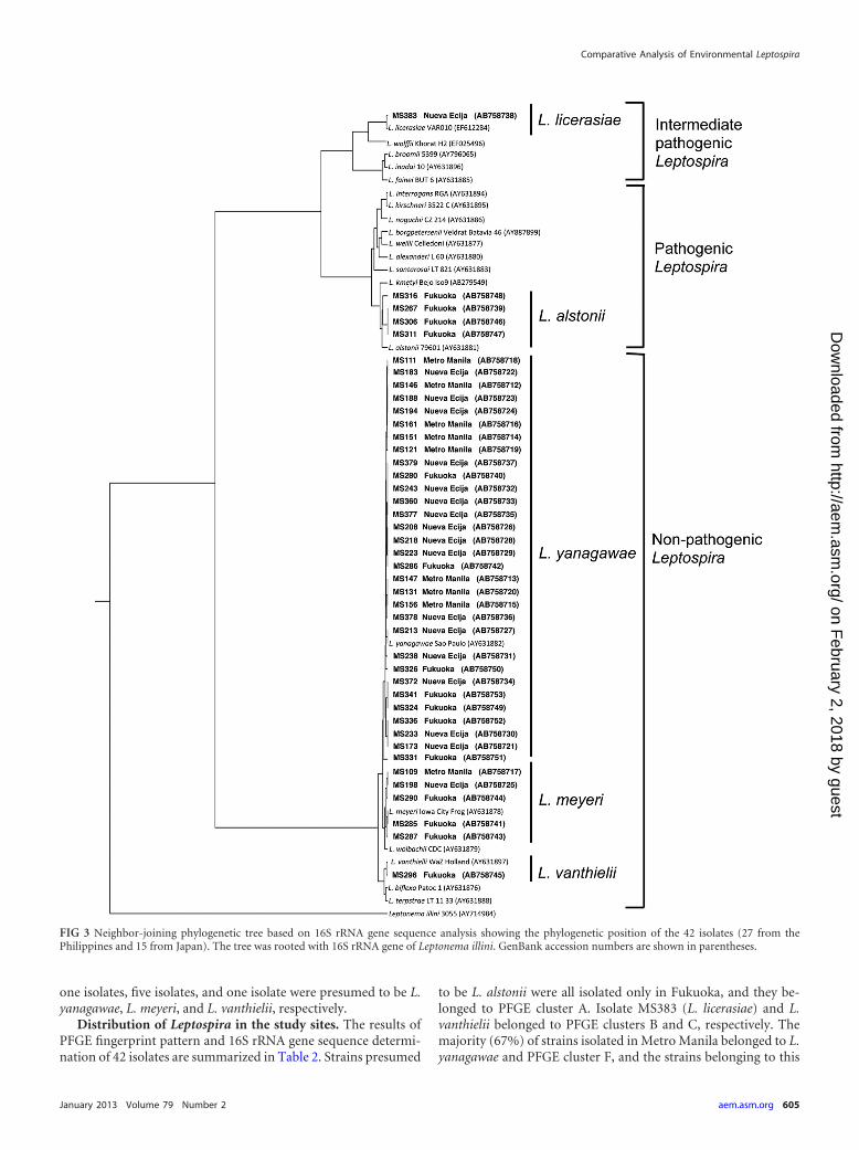
one isolates, five isolates, and one isolate were presumed to be L.yanagawae, L. meyeri, and L. vanthielii, respectively.
Distribution of Leptospira in the study sites. The results ofPFGE fingerprint pattern and 16S rRNA gene sequence determi-nation of 42 isolates are summarized in Table 2. Strains presumed
to be L. alstonii were all isolated only in Fukuoka, and they be-longed to PFGE cluster A. Isolate MS383 (L. licerasiae) and L.vanthielii belonged to PFGE clusters B and C, respectively. Themajority (67%) of strains isolated in Metro Manila belonged to L.yanagawae and PFGE cluster F, and the strains belonging to this
FIG 3 Neighbor-joining phylogenetic tree based on 16S rRNA gene sequence analysis showing the phylogenetic position of the 42 isolates (27 from thePhilippines and 15 from Japan). The tree was rooted with 16S rRNA gene of Leptonema illini. GenBank accession numbers are shown in parentheses.
Comparative Analysis of Environmental Leptospira
January 2013 Volume 79 Number 2 aem.asm.org 605
on February 2, 2018 by guest
http://aem.asm
.org/D
ownloaded from

group were also found to be distributed in Nueva Ecija and Fu-kuoka. In contrast, 16 of 18 strains (89%) isolated in Nueva Ecijawere presumed to be L. yanagawae; however, they had variousPFGE patterns belonging to four clusters (D, E, F, and G). Inparticular, the isolates belonging to PFGE cluster G were obtainedonly in this area. On the other hand, 15 strains isolated in Fukuokawere grouped into various genomospecies. Of these, the strains pre-sumed to be L. alstonii and L. vanthielii were isolated only in Fukuoka.Two strains belonging to L. yanagawae and PFGE cluster D (strainsMS324 and MS336) were closely related to the two strains, MS173and MS372, which were isolated in Nueva Ecija, suggesting the trans-border distribution of this genomospecies (Fig. 2).
Characterization of L. alstonii and L. licerasiae strains. (i)flaB-PCR of the isolates. The flaB-PCR assay produced positivePCR products for the four isolates that showed the highest 16SrRNA gene sequence similarity to L. alstonii (Table 2). The resultsfrom flaB-PCR and 16S rRNA gene sequence determination sug-
gested that isolates MS267, MS306, MS311, and MS316 werepathogenic Leptospira. The other 38 isolates, however, were deter-mined to be negative by flaB-PCR, including MS383, which ispresumed to be an intermediate pathogenic Leptospira (L. licera-siae) (Table 2).
(ii) Serogroups of the isolates. MAT was performed for serolog-ical identification of the five isolates presumed to be pathogenic orintermediate pathogenic Leptospira. Positive agglutination for strainsof L. alstonii was observed; MS267 and MS306 with Bratislava anti-sera, MS311 with Javanica antisera, and MS316 with Grippotyphosaantisera. The PFGE patterns of these four isolates were similar to thatof L. borgpetersenii strain Poi, the reference strain of serogroup Ja-vanica. No agglutination of MS383 (L. licerasiae) was observed withany of the panel of antisera for 23 serogroups.
(iii) Pathogenicity of isolates in golden Syrian hamsters. Toexamine the pathogenicity of the five isolates, groups consisting offive hamsters were inoculated intraperitoneally with 107 of each
TABLE 2 Isolates obtained from environmental samples and their respective genomospecies and PFGE clusters
Isolate Origin Sample Sampling region Sampling mo, yr Genomospecies flaB-PCR result PFGE cluster
MS146 Water ES-1 Metro Manila July, 2010 L. yanagawae – FMS147 Water ES-1 Metro Manila July, 2010 L. yanagawae – FMS151 Water ES-6 Metro Manila July, 2010 L. yanagawae – FMS156 Water ES-16 Metro Manila July, 2010 L. yanagawae – FMS161 Water ES-20 Metro Manila July, 2010 L. yanagawae – FMS109 Water ES-26 Metro Manila December, 2010 L. meyeri – No bandMS111 Water ES-28 Metro Manila December, 2010 L. yanagawae – FMS121 Water ES-29 Metro Manila December, 2010 L. yanagawae – No bandMS131 Water ES-30 Metro Manila December, 2010 L. yanagawae – EMS173 Water ES-40 Nueva Ecija February, 2011 L. yanagawae – DMS183 Water ES-42 Nueva Ecija February, 2011 L. yanagawae – FMS188 Water ES-43 Nueva Ecija February, 2011 L. yanagawae – No bandMS194 Soil ES-44 Nueva Ecija February, 2011 L. yanagawae – FMS198 Water ES-45 Nueva Ecija February, 2011 L. meyeri – No bandMS243 Water ES-46 Nueva Ecija February, 2011 L. yanagawae – EMS208 Water ES-47 Nueva Ecija February, 2011 L. yanagawae – GMS213 Water ES-48 Nueva Ecija February, 2011 L. yanagawae – FMS218 Water ES-49 Nueva Ecija February, 2011 L. yanagawae – EMS223 Soil ES-50 Nueva Ecija February, 2011 L. yanagawae – EMS233 Soil ES-51 Nueva Ecija February, 2011 L. yanagawae – GMS238 Water ES-53 Nueva Ecija February, 2011 L. yanagawae – EMS360 Water ES-92 Nueva Ecija November, 2011 L. yanagawae – FMS372 Water ES-95 Nueva Ecija November, 2011 L. yanagawae – DMS377 Water ES-96 Nueva Ecija November, 2011 L. yanagawae – GMS378 Water ES-96 Nueva Ecija November, 2011 L. yanagawae – FMS379 Water ES-96 Nueva Ecija November, 2011 L. yanagawae – EMS383 Water ES-97 Nueva Ecija November, 2011 L. licerasiae – BMS267 Water ES-54 Fukuoka June, 2011 L. alstonii � AMS280 Water ES-55 Fukuoka June, 2011 L. yanagawae – FMS285 Water ES-56 Fukuoka June, 2011 L. meyeri – FMS286 Water ES-56 Fukuoka June, 2011 L. yanagawae – FMS287 Water ES-56 Fukuoka June, 2011 L. meyeri – EMS290 Water ES-57 Fukuoka June, 2011 L. meyeri – No bandMS296 Water ES-58 Fukuoka July, 2011 L. vanthielii – CMS306 Soil ES-73 Fukuoka July, 2011 L. alstonii � AMS311 Soil ES-75 Fukuoka July, 2011 L. alstonii � AMS316 Soil ES-76 Fukuoka July, 2011 L. alstonii � AMS324 Water ES-79 Fukuoka August, 2011 L. yanagawae – DMS326 Water ES-80 Fukuoka August, 2011 L. yanagawae – FMS331 Water ES-82 Fukuoka August, 2011 L. yanagawae – FMS336 Water ES-83 Fukuoka August, 2011 L. yanagawae – DMS341 Water ES-84 Fukuoka August, 2011 L. yanagawae – F
Saito et al.
606 aem.asm.org Applied and Environmental Microbiology
on February 2, 2018 by guest
http://aem.asm
.org/D
ownloaded from

isolate/ml and observed for 28 days. No hamster showed anysymptoms of leptospirosis or died after infection. Blood and kid-ney samples from hamsters sacrificed on day 28 postinfectionwere cultured. Leptospires were not recovered from any of theinfected hamsters.
(iv) Dynamic change of the pathogenic isolates in four envi-ronmental sites. As already stated, genetically pathogenic lepto-spires, i.e., L. alstonii, were isolated from a water sample (ES-54) orfrom soil samples (ES-73, ES-75, and ES-76) from four sites inFukuoka, Japan. To clarify whether the pathogenic leptospirescolonize and survive for a long period or not, water or soil sampleswere collected from the four sites and cultured again after 5months. We found that the three samples from the three sites,where the samples ES-54, ES-73, and ES-75 were collected, con-tained no Leptospira 5 months after the first isolation. On theother hand, the soil sample from the site where sample ES-76 wascollected contained leptospires. The five isolates from ES-76showed the same PFGE pattern closely related to that of MS316(data not shown). This isolate (MS355) was flaB-PCR positive andshowed 99.9% similarity in the 16S rRNA gene sequence to strainMS316 of L. alstonii serovar Grippotyphosa. However, only 10%of agglutination of MS355 was observed with antisera for serovarGrippotyphosa.
Influence of moisture content and pH of the soil samples.The moisture content of 15 soil samples ranged from 3.5 to 42.8%.A total of 67% of the samples with moisture content of �20%were Leptospira positive, whereas only 23% were found to be pos-itive in the sample with a moisture content of �20% (Fig. 4). ThepH value ranged from 6.2 to 7.2. Three of five samples with a pH of6.2 were found to be positive. This result suggested that a pH of�6.2 (up to 7.2) had no influence on the distribution and survivalof Leptospira in the environmental soil.
Persistence of Leptospira in a certain site. The water sample,ES-55, collected from a rain puddle on 30 June 2011 containedLeptospira strain MS280 (the PFGE pattern of MS280 was referredto as pattern I). This rain puddle usually dried up within a few daysin the absence of rain. We expected that the soil of the puddlecould be a reservoir of Leptospira. Therefore, the soil was collectedfrom both the surface and 3 cm below the surface after 3 days ofdrought on 16 July 2011. The moisture contents of these soil sam-ples were 3.5 and 11.1%, respectively. Only the soil sample from a3-cm depth was found to be Leptospira positive. PFGE analysis ofthe isolates revealed two fingerprinting patterns: I and II (Fig. 5B).
After 5 months, the soil sample from the 3-cm depth (7.8% of themoisture content) was collected from the same site again. Thesample was found to be positive again and had two PFGE patterns,I and III (Fig. 5C). Thus, the isolate referred to as PFGE pattern Iwas found not only in the rain puddle but also in the soil andsurvived for at least 5 months.
DISCUSSION
To understand the dynamic changes in the epidemiology of lep-tospirosis, isolation of Leptospira from not only the human andanimal sources but also the environmental water and soil is essen-tial. Previously, there have been few reports on the epidemiologi-cal analysis of environmental isolates. This is probably because theisolation of leptospires from environmental samples was usuallyunsuccessful due to the overgrowth of contaminants and the slowgrowth of Leptospira. In the present study, the leptospires wereisolated from environmental samples at a high rate by using acombination of five antimicrobial agents (STAFF) (4).
We collected the environmental samples at three sites, MetroManila and Nueva Ecija, Philippines, and Fukuoka, Japan. Thefrequency of isolation from environmental water was highest forNueva Ecija (72%), followed by Fukuoka (62%) and then MetroManila (21%). The water samples from urban area of Metro Ma-nila were cloudy, and some of them were foul-smelling. Lepto-spires were found to be unable to survive in such an environment.The moisture content of soil greatly influenced the frequency ofpositive isolation from the soil. It was suggested that leptospireswould hardly survive in the soil with a moisture content of �20%.On the other hand, the pH values of all soil samples were higherthan 6.2, and such a pH had no influence on the distribution andsurvival of Leptospira in the soil. Henry and Johnson (7) previ-ously reported the percentage of Leptospira-positive soil sampledecreased with increasing distance from the spring, and beyond 25m no positive cultures were obtained from the soil with a moisturecontent of 24% and a pH of 5.4. These findings were consistentwith our results.
The taxonomy of Leptospira has undergone substantial revi-sion with the use of 16S rRNA sequence comparison and DNA-DNA reassociation studies (14). At the time of this writing, thegenus Leptospira consists of 20 genomospecies. The phylogenetictree constructed based on 16S rRNA gene sequences of the typestrains of 20 Leptospira genomospecies gives three clades, i.e.,pathogenic Leptospira, intermediate pathogenic Leptospira andnonpathogenic (or saprophytic) Leptospira. The amplificationand sequencing of the 16S rRNA gene of isolates is capable ofaccurately identifying and differentiating Leptospira species (15).In the present study, only four isolates from Fukuoka were iden-tified as a pathogenic Leptospira species, i.e., L. alstonii. One isolatefrom Nueva Ecija was identified as an intermediate pathogenicLeptospira species, L. licerasiae. Most of the isolates from the en-vironmental soil and water belonged to nonpathogenic Leptospiraspecies. It is thought that the frequency of pathogenic or interme-diate pathogenic Leptospira isolation from the environment is nothigh even in areas of endemicity such as Metro Manila.
We found that our three environmental samples containedmore than one strains by using single colony isolation method andPFGE analysis. This result would provide the first evidence that afew strains of Leptospira occasionally live together in environmen-tal water and soil. Thus, we recommend that at least five strains per
FIG 4 Influence of moisture content and pH of 15 soil samples on Leptospiraisolation. The moisture content of 15 soil samples ranged from 3.5 to 42.8. ThepH values ranged from 6.2 to 7.2. Six samples were Leptospira positive (�),whereas nine samples were Leptospira negative (Œ).
Comparative Analysis of Environmental Leptospira
January 2013 Volume 79 Number 2 aem.asm.org 607
on February 2, 2018 by guest
http://aem.asm
.org/D
ownloaded from

one environmental sample should be subcultured and analyzed byPFGE for accurate epidemiologic study.
The dendrogram based on the PFGE patterns separated the 37environmental isolates into seven clusters. Four strains of L. alsto-nii, one strain of L. licerasiae, and one strain of L. vanthiellii be-longed to three independent clusters. The other 31 strains, includ-ing L. meyeri and L. yanagawae, formed four clusters. Two of theseclusters included both strains of L. meyeri and strains of L. yana-gawae, and these two genomospecies contained a variety of PFGEpatterns (Fig. 2). In the present study, the serogroups of non-pathogenic strains were not determined; however, the classifica-tion of clusters based on the PFGE fingerprint pattern might re-flect serogroup relatedness rather than genetic relatedness, aspreviously reported.
Using NotI as the restriction enzyme is a standard method toperform the PFGE analysis for the isolates of leptospires (8, 16,17). We found that 5 of 42 strains showed no band of NotI-re-stricted DNA; however, the fingerprint patterns were seen usingother enzymes, such as SmaI or PacI. Although there are no re-ports of Leptospira strains without a NotI restriction site, some ofthe isolates that were not restricted by NotI were classified as L.yanagawae and L. meyeri by 16S rRNA gene sequencing. We rec-ommend using SmaI or PacI to digest the DNA when the differ-entiated patterns cannot be obtained when digested with NotI.Incidentally, we also found that two Leptonema isolates showed aladder of small fragments with sizes of �100 kbp, when digestedwith NotI (data not shown).
Unlike pathogenic Leptospira, little is known about the behav-
ior or reservoirs of nonpathogenic (saprophytic) Leptospira in theenvironment. Henry and Johnson (7) reported leptospires thatwere most frequently associated with soils of high moisture, andthe reservoir of the saprophytic Leptospira could be the soil. Weinvestigated the strain changing of the isolates in a puddle during5 months. It is noteworthy that the same strain, which was initiallyisolated from the water of the puddle, was consistently isolatedfrom the soil 3 cm from the depth of the puddle (11.1% moisturecontent) even when the puddle had dried up. No leptospire wasisolated from the surface of the ground (3.5% moisture content),possibly because it could not survive in such a dry condition. Thissuggested that saprophytic Leptospira survives in wet soil on drydays and appears in the surface water on rainy days. This is the firstreport that demonstrated soil as a possible reservoir of saprophyticLeptospira using PFGE analysis. In the Philippines, outbreaks ofleptospirosis usually occur during the rainy season and just afterthe rainy season in flood-prone areas (6). In such areas, pathogenicLeptospira could appear from soil contaminated with the urine ofinfected animals and get mixed with flood water, causing a widespread of the organisms. After a flood, the pathogens might survive inthe soil of the entire flood area for several months, while the virulenceof the pathogens might be lost in time, as described earlier.
The present study was also carried out to clarify the differencesamong isolates between Japan and the Philippines using PFGEanalysis. The isolates of each area showed their own characteris-tics. In Metro Manila, majority (two-thirds) of the isolates were L.yanagawae belonging to PFGE cluster F. The isolates in NuevaEcija showed various PFGE patterns belonging to four clusters.
FIG 5 PFGE patterns of Leptospira strains isolated from a rain puddle on 30 June 2011 (A), 16 July 2011 (B), and 14 December 2011 (C). Chromosomal DNAfrom isolates was digested with NotI restriction endonuclease. Lanes 1 to 5, PFGE patterns of five isolates from each sample. The three PFGE patterns were namedI, II, and III and are indicated below each fingerprint. MC, moisture content.
Saito et al.
608 aem.asm.org Applied and Environmental Microbiology
on February 2, 2018 by guest
http://aem.asm
.org/D
ownloaded from

The PFGE cluster G was unique in this area only. The isolates inFukuoka were classified as a variety of genomospecies, includingthe unique species L. alstonii and L. vanthielii. However, someisolates, for example, four strains in cluster D (Fig. 2), were closelyrelated to each other across borders.
It is well known that mammalian species excrete pathogenicleptospires in their urine and serve as reservoirs for their transmis-sion (18). The pathogens are maintained in sylvatic and domesticenvironments by transmission among rodent species. In these res-ervoirs, infection produces chronic, asymptomatic carriage. Themaintenance of leptospirosis in these populations is due to theircontinuous exposure to rodent reservoirs or to transmissionwithin animal herds (18). In the present study, four strains ofgenetically pathogenic Leptospira were isolated within an area 50m in diameter. We believe these strains are maintained amongsmall animals because no pathogenic isolates were obtained fromthe university campus outside this limited area. To determine theanimal reservoirs, we tried to trap rats and mice but were unsuc-cessful.
Recently, only a small number of cases of leptospirosis havebeen reported in Japan. However, strains of genetically pathogenicLeptospira strains were isolated in Fukuoka. Their virulence hadalready been lost, and three-quarters of the isolates were elimi-nated within 5 months. Trueba et al. (2) reported that a strain of L.interrogans was able to remain motile for 110 days (pH 7.2) indistilled water and survived 347 days when incubated in a semi-solid medium composed of distilled water and 0.5% purified aga-rose. It is hypothesized that pathogenic leptospires may be killedwithin 5 months after excretion into soil and water if some uncer-tain conditions are not satisfied. The ability of pathogenic Lepto-spira to survive long in the environment seems to vary. Adler andcoworkers (19) analyzed the genomes of two pathogenic species,L. borgpetersenii and L. interrogans. These researchers showed thatL. borgpetersenii lost the signal transduction function, and its sur-vival outside a mammalian host was impaired, whereas L. interro-gans retained an environmental sensory function that facilitateddisease transmission through water. We suggest that our L. alstoniiisolates could adapt in the environment only for a short periodand lose their virulent phenotype in the environment outside ofthe body of a mammalian host. Leptospires are known to lose theirvirulent phenotype with prolonged in vitro culture passages, andanimal passage is needed to maintain their virulence. In the sameway, the potential virulence of the pathogenic isolates was possiblylost during the thriving of leptospires in the environment afterbeing excreted in the urine of reservoir animals.
In areas of endemicity, an enormous number of rodent reser-voirs might maintain the virulence of pathogenic Leptospira intheir kidneys and intermittently excrete urine containing thesevirulent leptospires into the environment. It could be said that thecontrol of rodents is one of the best ways to get rid of virulentleptospires in the environment, and this could be a reason for thedramatic decrease in the number of leptospirosis patients after1960 in Japan. There is therefore an urgent need to improve san-itary conditions and control rodents in countries and areas whereleptospirosis is endemic and prevalent.
ACKNOWLEDGMENTS
This study was supported by a grant from the Special Coordination Fundson Science and Technology of the Ministry of Education, Sports, Culture,Science, and Technology (MEXT) of Japan and by a grant of the Science
and Technology Research Partnership for Sustainable Development fromJapan Science and Technology Agency and Japan International Coopera-tion Agency.
We thank Bin Chang for advice regarding the PFGE methods. We alsothank Hiroaki Nakayama and Susumu Shiota for critical discussions andTetsuro Tamura, Yuta Takekawa, Tatsuya Nakayama, Rubelia A. Baterna,Ana Kriselda B. Rivera, Crystal Amiel M. Estrada, Micaella Dato, HidekoKameyama, and Naomi Hidaka for their technical cooperation.
REFERENCES1. Smith DJ, Self HR. 1955. Observations on the survival of Leptospira
australis A in soil and water. J. Hyg. (Lond.) 53:436 – 444.2. Trueba G, Zapata S, Madrid K, Cullen P, Haake D. 2004. Cell aggrega-
tion: a mechanism of pathogenic Leptospira to survive in fresh water. Int.Microbiol. 7:35– 40.
3. Adler B, de la Peña Moctezuma A. 2010. Leptospira and leptospirosis.Vet. Microbiol. 140:287–296.
4. Chakraborty A, Miyahara S, Villanueva SY, Saito M, Gloriani NG,Yoshida S. 2011. A novel combination of selective agents for isolation ofLeptospira species. Microbiol. Immunol. 55:494 –501.
5. Inada R, Ido Y, Hoki R, Kaneko R, Ito H. 1916. The etiology, mode ofinfection, and specific therapy of Weil’s disease (spirochaetosis icterohae-morrhagica). J. Exp. Med. 23:377– 402.
6. Yanagihara Y, Villanueva SY, Yoshida S, Okamoto Y, Masuzawa T.2007. Current status of leptospirosis in Japan and the Philippines. Comp.Immunol. Microbiol. Infect. Dis. 30:399 – 413.
7. Henry RA, Johnson RC. 1978. Distribution of the genus Leptospira in soiland water. Appl. Environ. Microbiol. 35:492– 499.
8. WHO/ILS. 2003. Human leptospirosis: guidance for diagnosis, surveil-lance, and control. World Health Organization, Geneva, Switzerland.
9. Kawabata H, Dancel LA, Villanueva SY, Yanagihara Y, Koizumi N,Watanabe H. 2001. flaB-polymerase chain reaction (flaB-PCR) and itsrestriction fragment length polymorphism (RFLP) analysis are an efficienttool for detection and identification of Leptospira spp. Microbiol. Immu-nol. 45:491– 496.
10. Postic D, Riquelme-Sertour N, Merien F, Perolat P, Baranton G. 2000.Interest of partial 16S rDNA gene sequences to resolve heterogeneitiesbetween Leptospira collections: application to L. meyeri. Res. Microbiol.151:333–341.
11. Thompson JD, Gibson TJ, Plewniak F, Jeanmougin Higgins FDG. 1997.The CLUSTAL X windows interface: flexible strategies for multiple se-quence alignment aided by quality analysis tools. Nucleic. Acids Res. 25:4876 – 4882.
12. Saitou Nei N, M. 1987. The neighbor-joining method: a new method forreconstructing phylogenetic trees. Mol. Biol. Evol. 4:406 – 425.
13. Page RD. 1996. TREEVIEW: an application to display phylogenetic treeson personal computers. Comput. Appl. Biosci. 12:357–358.
14. Zuerner RL. 2011. Genus I: Leptospira. Family IV: Leptospiraceae. PhylumXV. Spirochaetes phyl. nov., p 546 –556. In Krieg NR, Staley JT, Brown DR,Hedlund BP, Paster BJ, Ward NL, Ludwig W, Whitman WB (ed), Bergey’smanual of systematic bacteriology, 2nd ed, vol 4. Springer, New York, NY.
15. Bourhy P, Collet L, Clément S, Huerre M, Ave P, Giry C, Pettinelli F,Picardeau M. 2010. Isolation and characterization of new Leptospira ge-notypes from patients in Mayotte (Indian Ocean). PLoS Negl. Trop. Dis.4:e724. doi:10.1371/journal.pntd.0000724.
16. Galloway RL, Levett PN. 2010. Application and validation of PFGE forserovar identification of Leptospira clinical isolates. PLoS Negl. Trop. Dis.4:e824. doi:10.1371/journal.pntd.0000824.
17. Herrmann JL, Bellenger E, Perolat P, Baranton G, Saint Girons I. 1992.Pulsed-field gel electrophoresis of NotI digests of leptospiral DNA: a newrapid method of serovar identification. J. Clin. Microbiol. 30:1696 –1702.
18. Ko AI, Goarant C, Picardeau M. 2009. Leptospira: the dawn of themolecular genetics era for an emerging zoonotic pathogen. Nat. Rev. Mi-crobiol. 7:736 –747.
19. Picardeau M, Bulach DM, Bouchier C, Zuerner RL, Zidane N, WilsonPJ, Creno S, Kuczek ES, Bommezzadri S, Davis JC, McGrath A, John-son MJ, Boursaux-Eude C, Seemann T, Rouy Z, Coppel RL, Rood JI,Lajus A, Davies JK, Médigue C, Adler B. 2008. Genome sequence of thesaprophyte Leptospira biflexa provides insights into the evolution of Lep-tospira and the pathogenesis of leptospirosis. PLoS One 3:e1607. doi:10.1371/journal.pone.0001607.
Comparative Analysis of Environmental Leptospira
January 2013 Volume 79 Number 2 aem.asm.org 609
on February 2, 2018 by guest
http://aem.asm
.org/D
ownloaded from