COMPARATIVE ANALYSIS OF IN VITRO BIOACTIVITIES AND ...eprints.usm.my/31543/1/Azlinda_Ibrahim.pdf ·...
41
COMPARATIVE ANALYSIS OF IN VITRO BIOACTIVITIES AND PHENOLIC CONTENT OF LEAF EXTRACTS FROM SIX SPECIES OF Aquilaria AZLINDA BINTI IBRAHIM UNIVERSITI SAINS MALAYSIA 2016
Transcript of COMPARATIVE ANALYSIS OF IN VITRO BIOACTIVITIES AND ...eprints.usm.my/31543/1/Azlinda_Ibrahim.pdf ·...
COMPARATIVE ANALYSIS OF IN VITRO
BIOACTIVITIES AND PHENOLIC CONTENT OF
LEAF EXTRACTS FROM SIX SPECIES OF Aquilaria
AZLINDA BINTI IBRAHIM
UNIVERSITI SAINS MALAYSIA
2016
COMPARATIVE ANALYSIS OF IN VITRO
BIOACTIVITIES AND PHENOLIC CONTENT OF
LEAF EXTRACTS FROM SIX SPECIES OF
Aquilaria
by
AZLINDA BINTI IBRAHIM
Thesis submitted in fulfillment of the requirements
for the degree of
Master of Science (Biotechnology)
September 2016
ii
ACKNOWLEDGEMENT
First of all, I would like to express my heartfelt appreciation to my
supervisor, Professor Dr. Shaida Fariza Sulaiman for trusting me and giving me the
great opportunity to learn under her supervision. I deeply appreciate her kindness,
invaluable advices, unceasing support, guidance and knowledge, which inspired me
throughout my study. Without her support, I would never have succeeded in
achieving this milestone. Also, I would like to offer my gratitude to my co-
supervisor, Associate Professor Dr. Hideyuki Nagao for his precious support and
guidance.
Special thanks to my labmates in phytochemical laboratories, Dr. Ooi Kheng
Leong, Ms. Nurul Shafiqah Hashim and Mr. Joshua Jeyentheren Anantham, who had
supported, encouraged and assisted me tremendously during my experimental work.
I am overwhelmed with their kindness and guidance.
Next, I would like to express my gratitude to Professor Baharuddin Salleh
and Mrs. Siti Nurdijati for providing the plant materials in this study. I profusely
thank all the staff of School of Biological Sciences, Universiti Sains Malaysia
especially Mr. Kamarudin and Mrs. Shafawati for their kind help and co-operation
throughout my research project. I am also highly indebted to Ministry of Higher
Education (MyBrain15) for the funding of my study.
iii
I would also like to acknowledge my greatest appreciation and thanks to my
beloved parents, Ayahanda Haji Ibrahim Salleh and Bonda Hajjah Zaleha Daiman
for their trust, love, prayer, patience and moral support. I dedicate my warm thanks
to my dearest sister, Ms. Azreen Ibrahim, who is always there for me through thick
and thin in making this study a success, and also not forgetting my beloved friends,
Ms. Nur Amalina Kamarudin, Mr. Nazirul Mubin Mohamed, Ms. Khairun Dalila
Salihin, Ms. Nurul Hamizah Hamidon, Ms. Nurul Ashiqin Zawawi, and Ms. Nor
Farah Anis Kamaruzaman who always motivated me during this project.
Most of all, thank you to Almighty Allah s.w.t, for giving me His endless
love, blessing, strength and knowledge to complete this study.
Thank you.
Azlinda Ibrahim
iv
TABLE OF CONTENTS
ACKNOWLEDGEMENT ii
TABLE OF CONTENTS iv
LIST OF TABLES viii
LIST OF FIGURES ix
LIST OF SYMBOLS AND ABBREVIATIONS xi
ABSTRAK xiii
ABSTRACT xv
CHAPTER 1: INTRODUCTION 1
1.1 Background of study 1
1.2 Problem statements 4
1.3 Objectives of the study 5
CHAPTER 2: LITERATURE REVIEW 6
2.1 Genus Aquilaria 6
2.1.1 Morphological description of Aquilaria used in this study 7
2.1.2 Uses of Aquilaria leaves 14
2.2 Bioactivities of Aquilaria leaves 15
2.2.1 Antioxidant activity 16
2.2.2 Anti-diabetic activity 17
2.2.3 Anti-inflammatory activity 19
2.2.4 Antibacterial activity 21
v
2.2.4(a) Antibacterial activity of herbal teas from Aquilaria
species
23
2.2.4(b) Gastroenteritis bacteria 25
2.3 Phytochemicals and phenolic compounds in leaves of Aquilaria 32
CHAPTER 3: MATERIALS AND METHODS 38
3.1 Plant materials 38
3.2 Preparation of samples 39
3.3 Extraction 39
3.3.1 Extraction of dried samples into crude extracts 39
3.3.2 Hydrolysis of the crude extracts 40
3.4 Antioxidant tests 41
3.4.1 Preparation of extracts 41
3.4.2 Preparation of DPPH (2,2-diphenyl-1-picrylhydrazyl) solution 42
3.4.2(a) DPPH radical scavenging assay 43
3.4.3 Preparation of ferric reducing antioxidant power (FRAP) reagent 44
3.4.3(a) FRAP assay 44
3.4.4 Preparation of metal chelating solution 45
3.4.4(a) Metal chelating test 45
3.5 Determination of total phenolic content 46
3.5.1 Preparation of extracts and standard 46
3.5.2 Preparation of Folin-Ciocalteu’s (FC) reagent 47
3.5.2(a) Determination of total phenolic content 47
3.6 Enzyme inhibition assays 48
3.6.1 Preparation of extracts 48
vi
3.6.2 Determination of α-glucosidase inhibitory activity 49
3.6.3 Determination of α-amylase inhibitory activity 50
3.6.4 Determination of COX-2 inhibitory activity 51
3.7 Antibacterial activity 52
3.7.1 Bacterial strains 52
3.7.1(a) Preparation and maintenance of stock cultures 53
3.7.2 Culture media 53
3.7.2(a) Nutrient agar (NA) preparation 53
3.7.2(b) Nutrient broth (NB) preparation 54
3.7.2(c) Saline solution preparation 54
3.7.3 Antibacterial test 55
3.7.3(a) Preparation of extracts 55
3.7.3(b) Preparation of inoculums 55
3.7.3(c) Determination of the minimum inhibitory
concentration (MIC)
56
3.8 Ultra Performance Liquid Chromatography (UPLC) 57
CHAPTER 4: RESULTS 61
4.1 Selection of solvent for extraction based on the antioxidant
activities
61
4.1.1 DPPH radical scavenging activity of Aquilaria leaf extracts
using five polar solvents
61
4.1.2 FRAP activity of Aquilaria leaf extracts using five solvents 64
4.1.3 Comparison between the antioxidant activities derived from
the two assays
67
4.2 Yield of Aquilaria leaf extracts 68
4.3 Antioxidant properties of Aquilaria leaf extracts 69
vii
4.3.1 DPPH radical scavenging activity 69
4.3.2 FRAP activity 72
4.3.3 Metal chelating activity 75
4.4 Total phenolic contents of Aquilaria leaf extracts 77
4.5 Enzyme inhibition assays 81
4.5.1 α-Glucosidase inhibitory potentials of Aquilaria leaf extracts 81
4.5.2 α-Amylase inhibitory potentials of Aquilaria leaf extracts 83
4.5.3 Anti-inflammatory properties of Aquilaria leaf extracts 85
4.6 Antibacterial potentials of Aquilaria leaf extracts 87
4.7 UPLC analysis 91
CHAPTER 5: DISCUSSION 113
CHAPTER 6: CONCLUSION 131
REFERENCES 133
viii
LIST OF TABLES
Page
Table 1.1 Aquilaria conservation status in Peninsular Malaysia and
the IUCN Red List of Threatened Species.
3
Table 2.1 Morphological similarities and differences of Aquilaria
species.
9
Table 2.2 Lists of the gastroenteritis bacteria with their diseases
caused
26
Table 2.3 Food poisoning caused by Bacillus cereus. 27
Table 2.4 Gastroenteritis caused by Escherichia coli. 30
Table 2.5 Phenolic compounds and other compounds and their
contents in leaves of Aquilaria species.
34
Table 3.1 Leaf samples of Aquilaria 38
Table 3.2(a) UPLC gradient mode. 59
Table 3.2(b) UPLC gradient mode. 59
Table 4.1 Percentage yields of methanolic and hydrolyzed extracts of
Aquilaria species.
68
Table 4.2 Total phenolic contents of Aquilaria extracts. 78
Table 4.3 α-Glucosidase inhibitory potentials of Aquilaria extracts. 82
Table 4.4 α-Amylase inhibitory potentials of Aquilaria extracts. 84
Table 4.5 Minimum inhibitory concentrations of methanolic and
hydrolysed leaf extracts of six species of Aquilaria against
selected gastroenteritis pathogens.
89
Table 4.6 Contents of phenolic constituents in extracts of six
Aquilaria species.
109
ix
LIST OF FIGURES
Page
Figure 4.1 Graph of free radical scavenging percentage versus five
extracts of each Aquilaria.
62
Figure 4.2 Graph of ferric reducing potentials (FRAP) percentage
versus extracts of each Aquilaria.
65
Figure 4.3 Graph of DPPH radical scavenging percentage versus
methanolic and hydrolyzed leaf extracts of six Aquilaria
species.
70
Figure 4.4 Graph of ferric reducing potentials (FRAP) percentage
versus methanolic and hydrolyzed leaf extracts of six
Aquilaria species.
73
Figure 4.5 Graph of MC assay percentage versus methanolic and
hydrolyzed leaf extracts of six Aquilaria species.
76
Figure 4.6 Linear calibration curve of gallic acid for determination of
total phenolic content.
79
Figure 4.7 Cyclooxygenase inhibiting potentials percentage versus
methanolic and hydrolyzed leaf extracts of six Aquilaria
species.
86
Figure 4.8 UPLC chromatogram and UV spectra of (a) methanolic
extract of Aquilaria hirta at 290 nm (b) methanolic extract
of Aquilaria hirta at 330 nm.
92
Figure 4.9 UPLC chromatogram and UV spectra of (a) hydrolyzed
extract of Aquilaria hirta at 290 nm (b) hydrolyzed extract
of Aquilaria hirta at 330 nm.
93
Figure 4.10 UPLC chromatogram and UV spectra of (a) methanolic
extract of Aquilaria beccariana at 290 nm (b) methanolic
extract of Aquilaria beccariana at 330 nm.
95
x
Figure 4.11 UPLC chromatogram and UV spectra of (a) hydrolyzed
extract of Aquilaria beccariana at 330 nm.
96
Figure 4.12 UPLC chromatogram and UV spectra of (a) methanolic
extract of Aquilaria malaccensis at 290 nm (b) methanolic
extract of Aquilaria malaccensis at 330 nm.
98
Figure 4.13 UPLC chromatogram and UV spectra of (a) hydrolyzed
extract of Aquilaria malaccensis at 290 nm (b) hydrolyzed
extract of Aquilaria malaccensis at 330 nm.
99
Figure 4.14 UPLC chromatogram and UV spectra of (a) methanolic
extract of Aquilaria rostrata at 290 nm (b) methanolic
extract of Aquilaria rostrata at 330 nm.
101
Figure 4.15 UPLC chromatogram and UV spectra of (a) hydrolyzed
extract of Aquilaria rostrata at 290 nm (b) hydrolyzed
extract of Aquilaria rostrata at 330 nm.
102
Figure 4.16 UPLC chromatogram and UV spectra of (a) methanolic
extract of Aquilaria sinensis at 290 nm (b) methanolic
extract of Aquilaria sinensis at 330 nm.
104
Figure 4.17 UPLC chromatogram and UV spectra of (a) hydrolyzed
extract of Aquilaria sinensis at 290 nm (b) hydrolyzed
extract of Aquilaria sinensis at 330 nm.
105
Figure 4.18 UPLC chromatogram and UV spectra of (a) methanolic
extract of Aquilaria subintegra at 290 nm (b) methanolic
extract of Aquilaria subintegra at 330 nm.
107
Figure 4.19 UPLC chromatogram and UV spectra of (a) hydrolyzed
extract of Aquilaria subintegra at 290 nm (b) hydrolyzed
extract of Aquilaria subintegra at 330 nm.
108
Figure 5.1 Summarization of in vitro bioactivities of Aquilaria leaf
extracts.
129
xi
LIST OF ABBREVIATIONS
ANOVA analysis of variance
ATCC American Type Culture Collection
COX Cyclooxygenase
cm Centimeter
DMSO dimethyl sulphoxide
DPPH 2,2-diphenyl picryl-hydrazyl
EDTA ethylene diamine tetracetic acid
EC50 effective concentration at 50% of activity
FRAP ferric reducing antioxidant power
GAE gallic acid equivalent
g Gram
HCl hydrochloride acid
MIC minimum inhibition concentration
M Molar
mg milligram (10-3
g)
ml Milliliter (10-3
litre)
mM millimolar (10-3
M)
NA nutrient agar
NB nutrient broth
nm Nanometer
NSAIDs nonsteroidal anti-inflammatory drugs
PDA photo diode array
PDA potato dextrose agar
PDB potato dextrose broth
PG Postraglandin
Rt retention time
SD standard deviation
TPC total phenolic content
TPTZ 2,4,6-tri (2-pyridyl)-s-triozine
UPLC ultra performance liquid chromatography
xii
UV Ultraviolet
v/v volume to volume
w/v weight to volume
λmax lambda maximum
xiii
ANALISIS PERBANDINGAN BIOAKTIVITI IN VITRO DAN KANDUNGAN
FENOLIK EKSTRAK DAUN DARIPADA ENAM SPESIES Aquilaria
ABSTRAK
Kajian ini dijalankan untuk membandingkan aktiviti antioksida, antibakteria,
anti-diabetes dan anti-radang daripada ekstrak enam spesies daripada genus
Aquilaria iaitu Aquilaria beccariana, Aquilaria hirta, Aquilaria malaccensis,
Aquilaria rostrata, Aquilaria sinensis dan Aquilaria subintegra. Sebatian fenolik
mereka juga telah dikenalpasti dan dikuantifikasi. Daripada penilaian awal
menggunakan lima jenis ekstrak (etil asetat, aseton, etanol, metanol dan air suling)
daripada setiap spesies, ekstrak metanol menunjukkan aktiviti antioksidan yang
terbaik. Pelarut ini kemudiannya dipilih untuk pengekstrakan lanjut dan separuh
daripada ekstrak ini juga dihidrolisiskan. A. sinensis mencatatkan peratusan tertinggi
bagi aktiviti 2,2-difenil-1-pikrilhidrazil (DPPH) (ekstrak terhidrolisis; 94.33±4.89%)
dan aktiviti keupayaan aktioksida penurunan ferik (FRAP) (ekstrak metanol dan
ekstrak terhidrolisis; 96.00±0.55% dan 95.88±0.16%), dan ekstrak metanol A. hirta
memberikan peratusan tertinggi bagi aktiviti pengkelat logam (90.05±3.47%). Hanya
bakteria gastrousus telah dipilih untuk kajian antibakteria. Ekstrak terhidrolisis A.
hirta menunjukkan aktiviti yang baik dengan nilai minimum kepekatan perencatan
(MIC) dalam linkungan 250 to 500 µg/mL. Dalam ujian perencatan α-glukosidase,
2.5 mg/mL ekstrak terhidrolisis A. sinensis mempamerkan peratusan tertinggi
perencatan pada 59.00±8.07%. Dalam ujian perencatan α-amilase, aktiviti
perencatan tertinggi adalah hanya pada 23.35±1.30% dikesan dari 10 mg/mL ekstrak
terhidrolisis A. hirta. Aktiviti perencatan siklooksigenase tertinggi hanyalah pada
25.89±2.59% untuk ekstrak metanol A. subintegra. Ekstrak ini juga mempunyai
xiv
jumlah kandungan fenolik yang tertinggi (127.80±2.57 µg GAE/mg ekstrak).
Daripada analisis kuantitatif menggunakan kromatografi cecair berprestasi ultra
(UPLC), kandungan tertinggi mangiferin dikuantifikasikan daripada ekstrak metanol
A. hirta, A. malaccensis, A. sinensis dan A. subintegra dan ekstrak terhidrolisis A.
hirta. Iriflofenon 2-O-α-rhamnosida didapati merupakan sebatian fenolik utama
dalam A. rostrata dan iriflofenon 3-C-β-glukosida ialah sebatian utama dalam A.
beccariana. 7,4'-di-O-metilapigenin adalah sebatian utama bagi ekstrak terhidrolisis
untuk A. beccariana, A. malaccensis, A. rostrata dan A. subintegra. Manakala 7,3'-
di-O-metilluteolin ialah sebatian utama dalam ekstrak terhidrolisis A. sinensis.
xv
COMPARATIVE ANALYSIS OF IN VITRO BIOACTIVITIES AND
PHENOLIC CONTENT OF LEAF EXTRACTS FROM SIX SPECIES OF
Aquilaria
ABSTRACT
This study was carried out to compare the in vitro antioxidant, antibacterial,
anti-diabetic and anti-inflammatory properties of the leaf extracts of six species from
genus Aquilaria which are; Aquilaria beccariana, Aquilaria hirta, Aquilaria
malaccensis, Aquilaria rostrata, Aquilaria sinensis and Aquilaria subintegra. Their
phenolic compounds were also identified and quantified. From the preliminary
screening using five fresh extracts (ethyl acetate, acetone, ethanol, methanol and
distilled water) from each species, the methanolic extracts exhibited the best
antioxidant activities. This solvent was then selected for further extraction and half
of the extracts were also hydrolyzed. A. sinensis recorded the highest percentage of
2,2-diphenyl-1-picrylhydrazyl (DPPH) activity (hydrolyzed extract; 94.33±4.89%)
and ferric-reducing antioxidant power (FRAP) activity (methanolic extract and
hydrolyzed extract; 96.00±0.55% and 95.88±0.16%), and the methanolic extract of
A. hirta gave the highest percentage of metal chelating activity (90.05±3.47%). Only
gastrointestinal bacteria were selected for antibacterial study. The hydrolyzed extract
of A. hirta performed better activity with minimum inhibitory concentration (MIC)
values ranging from 250 to 500 µg/mL. In the α-glucosidase inhibition test, the 2.5
mg/mL hydrolyzed extract of A. sinensis exhibited the highest percentage of
inhibition at 59.00±8.07%. In α-amylase inhibition test, the highest inhibitory
activity was only 23.35±1.30% detected from the 10 mg/mL hydrolyzed extract of A.

xvi
hirta. The highest cyclooxygenase inhibitory activity was only 25.89±2.59% for the
methanolic extract of A. subintegra. This extract also has the highest total phenolic
content (127.80±2.57 µg GAE/mg extract). From the quantitative analysis using ultra
performance liquid chromatography (UPLC), major content of mangiferin was
quantified from the methanol extracts of A. hirta, A. malaccensis, A. sinensis and A.
subintegra and the hydrolyzed extract of A. hirta. Iriflophenone 2-O-α-rhamnoside
was found to be the major compound of A. rostrata and iriflophenone 3-C-β-
glucoside is the major compound in A. beccariana. 7,4'-di-O-methylapigenin was the
major compound of the hydrolyzed extracts of A. beccariana, A. malaccensis, A.
rostrata and A. subintegra, while 7,3'-di-O-methylluteolin is the major compound in
the hydrolyzed extract of A. sinensis.
1
CHAPTER 1
INTRODUCTION
1.1 Background of study
Aquilaria is an aromatic evergreen tree that belongs to the family of
Thymelaeaceae, which is popularly known as “Gaharu” in the Southeast Asian
countries. Aquilaria is also known under many names all around the globe such as
agarwood, aloeswood, eaglewood, oud, kalambac, chen-xiang, jinkoh, khi-nam and
others depend on their localities (Baharuddin, 2014).
In Malaysia, there are several native species of Aquilaria that are A.
malaccensis, A. hirta, A. microcarpa, A. beccariana and A. rostrata. Three exotic
Aquilaria species, that are also cultivated in Malaysia are A. crassna and A.
subintegra that are native to Thailand and A. sinensis, which originate from China
(Hashim and Ahmad Zuhaidi, 2011).
Agarwood plantations with a total area of 232.8 hectares (ha) had been
established in Malaysia since the beginning of year 2000 (Ismail and Mohd Zin,
2011). In year 2014, the total area for Aquilaria plantations was reported to be 1119
ha (Ismail, 2014). Sabah is the major producing state for agarwood with 311.0 ha of
plantation areas. This is followed by Perak (298.2 ha) and Pahang (144.1 ha).
2
Aquilaria malaccensis that is locally known as karas is randomly scattered
throughout Peninsular Malaysia except in Perlis and Kedah. A. hirta is known by
locals as chandan is mainly distributed in the east coast of the Peninsular Malaysia
especially in the states of Terengganu, Pahang and Johor. Other Aquilaria species
such as A. microcarpa and A. beccariana are confined to Sarawak and Johor
(Whitmore, 1972). Moreover, A. rostrata that is known as chandan gunung was
recently found in Terengganu as well as Gunung Tahan, Pahang (Lee and Mohamed,
2016).
Agarwood that is derived from the resin produced after pathological process
at the injured stem is utilized and practised by mankind since the ancient time for
various purposes. This high-priced agarwood is highly sought-after especially in the
manufacturing of fragrances, medicines and beauty products (Chung and
Purwaningsih, 1999; Baharuddin, 2014). In traditional Malay medicine, it is also
used to treat various disorders such as fatigue, pain in the stomach or chest, edema
and as tonic for men and women as well as a post partum medicine (Gimlett and
Burkill, 1930).
For example, the decoction of the leaves of A. malaccensis is applied
externally to treat swelling and consumed for treating vomiting (Ong, 2004). The
decoction of its root is consumed to heal edema. The bark juice is useful in treating
diarrhea, and to stop vomiting (Quattrocchi, 2012). The incense wood is used to treat
thyroid gland cancer, as a sedative against abdominal complaints, asthma and
diarrhea, and as an aphrodisiac agent. Grated wood is added into various herbal
formulations especially for women during and after childbirth, and to treat
3
rheumatism. Thus, many medicinal and health supplementary products can be
derived from different parts of this agarwood species (Chung and Purwaningsih,
1999; Ong, 2004).
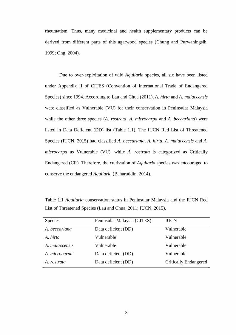
Due to over-exploitation of wild Aquilaria species, all six have been listed
under Appendix II of CITES (Convention of International Trade of Endangered
Species) since 1994. According to Lau and Chua (2011), A. hirta and A. malaccensis
were classified as Vulnerable (VU) for their conservation in Peninsular Malaysia
while the other three species (A. rostrata, A. microcarpa and A. beccariana) were
listed in Data Deficient (DD) list (Table 1.1). The IUCN Red List of Threatened
Species (IUCN, 2015) had classified A. beccariana, A. hirta, A. malaccensis and A.
microcarpa as Vulnerable (VU), while A. rostrata is categorized as Critically
Endangered (CR). Therefore, the cultivation of Aquilaria species was encouraged to
conserve the endangered Aquilaria (Baharuddin, 2014).
Table 1.1 Aquilaria conservation status in Peninsular Malaysia and the IUCN Red
List of Threatened Species (Lau and Chua, 2011; IUCN, 2015).
Species Peninsular Malaysia (CITES) IUCN
A. beccariana Data deficient (DD) Vulnerable
A. hirta Vulnerable Vulnerable
A. malaccensis Vulnerable Vulnerable
A. microcarpa Data deficient (DD) Vulnerable
A. rostrata Data deficient (DD) Critically Endangered
4
Since it takes many years for the wood to produce the aromatic and
valuable resin, the leaves of A. crassna, A. malaccensis and A. sinensis were
harvested for tea production in some Asian countries. The agarwood tea is popular as
daily healthy drink for some communities, especially in China. Besides, the herbal
tea of agarwood is commonly consumed for the treatment of many health disorders
such as diabetes, headache, constipation and high blood pressure (Pranakhon et al.,
2011; Kakino et al., 2012).
Up until now, A. crassna, A. malaccensis and A. sinensis were previously
reported to possess many beneficial biological activities and pharmacological
properties such as antioxidant, anti-diabetic, anti-inflammatory, antibacterial,
antidepressant and antiviral activities (Dash et al., 2008; Zhou et al., 2008; Huda et
al., 2009; Pranakhon et al., 2011; Kamonwannasit et al., 2013). In addition, the
extraction of A. malaccensis leaves using methanol was reported to contain a lot of
chemical constituents such as flavonoids, terpenoids, alkaloids and tannins (Khalil et
al., 2013).
1.2 Problem statements
Recently, instead of focusing only in producing agarwood resin, leaves of
A. malaccensis are used to make health products such as tea (Baharuddin, 2014). To
date, not all leaves of Aquilaria have been studied for their bioactivities. The
identification of phenolic compounds in the leaf extracts was limited to a few
findings from A. crassna, A. malaccensis and A. sinensis (Chen et al., 2012).
5
Besides, there is no comparison of the bioactivities among the Aquilaria leaf
extracts. Therefore, this project was proposed to comparatively evaluate the
bioactivities of the leaf extracts. This study also may provide the scientific evidence
to prove the medicinal values of each Aquilaria species. The composition of
phenolic compounds present in these extracts and their contribution to the
bioactivities were also be evaluated.
1.3 Objectives of the study
The purpose of this study is to identify the leaf extract with the best
bioactivity from six different Aquilaria species and to quantify their phenolic
compounds. For those reasons, the following objectives were drawn for this study:
1. To determine the best solvent to extract antioxidants from the leaf part of
six species of Aquilaria.
2. To screen the in vitro antioxidant activities of the effective extracts using
three different colorimetric assays.
3. To determine anti-diabetic activity using the enzyme inhibitory assays of -
glucosidase and -amylase.
4. To determine anti-inflammatory activity using cyclooxygenase inhibitory
assay.
5. To determine antibacterial activity against gastroenteritis bacteria.
6. To identify and quantify the major phenolic compounds from the
methanolic and hydrolyzed extracts and correlate with the bioactivities.
6
CHAPTER 2
LITERATURE REVIEW
2.1 Genus Aquilaria
Aquilaria is one of the genus of family Thymelaeaceae from the order
Malvales. This genus is native to the Asian continent and widely distributed in India
and from southern China to the countries in Southeast Asia region (Whitmore, 1972;
Allaby, 2012). It is also found mostly in Malaysia, Indonesia, Singapore, Myammar,
Philliphine and Thailand (Huda et al., 2009). The genus Aquilaria comprises of slow
growing trees, medium sized up to 40 meter in height. There are 15 species of
Aquilaria namely A. acuminata, A. baillonii, A. banaensis, A. brachyantha, A.
crassna, A. cumingiana, A. filaria, A. rostrata, A. rugosa, A. sinensis, A. subintegra,
including the Malaysian gaharu which comes from A. hirta, A. malaccensis, A.
beccariana and A. microcarpa (Barden et al., 2000; Hashim and Ahmad Zuhaidi,
2011).
The genus Aquilaria is often recognized with a smooth, stringy hard and
pale grey to dark with dense foliage of outer bark of wood. While the inner bark or
sapwood is white and soft. The leaves of Aquilaria are simple, alternately arranged
with acuminate tips and undulate margins. The flowers are hermaphroditic and
arranged in umbel inflorescences on the short stalks at the terminal twigs where the
fruits are produced. The fruits are woody capsules, drupes or berries and develop
from the calyx lobe (Whitmore, 1972; Hashim and Ahmad Zuhaidi, 2011). This
7
genus is well adapted to grow in various habitats that are sandy or rocky
environments, well-drained slopes and is commonly found in primary and secondary
forest depending on the species, mainly in lowland and hillsides at altitudes up to
850 m with an average daily temperature of 20-22°C (Jantan, 1990, Keller and
Sidiyasa, 1994).
Agarwood is classified as non wood forest product (Lata, 2007). From this
genus, there are four known species that yield high-grade gaharu, which are A.
malaccensis, A. crassna, A. sinensis and A. subintegra (Hashim and Ahmad Zuhaidi,
2011). According to Chua (2011), A. malaccensis is the major source of high-quality
traded gaharu resin in Peninsular Malaysia.
2.1.1 Morphological description of Aquilaria used in this study
Each species of the genus Aquilaria is known under various vernacular
names by the locals. A. hirta, A. beccariana, A. malaccensis, A. rostrata, A. sinensis
and A. subintegra are known by the local people in Malaysia as chandan, gaharu
tanduk, tengkaras or karas, chandan gunung, “Pak Muk Heung” and karas,
respectively. The morphological characteristics of the six Aquilaria species were
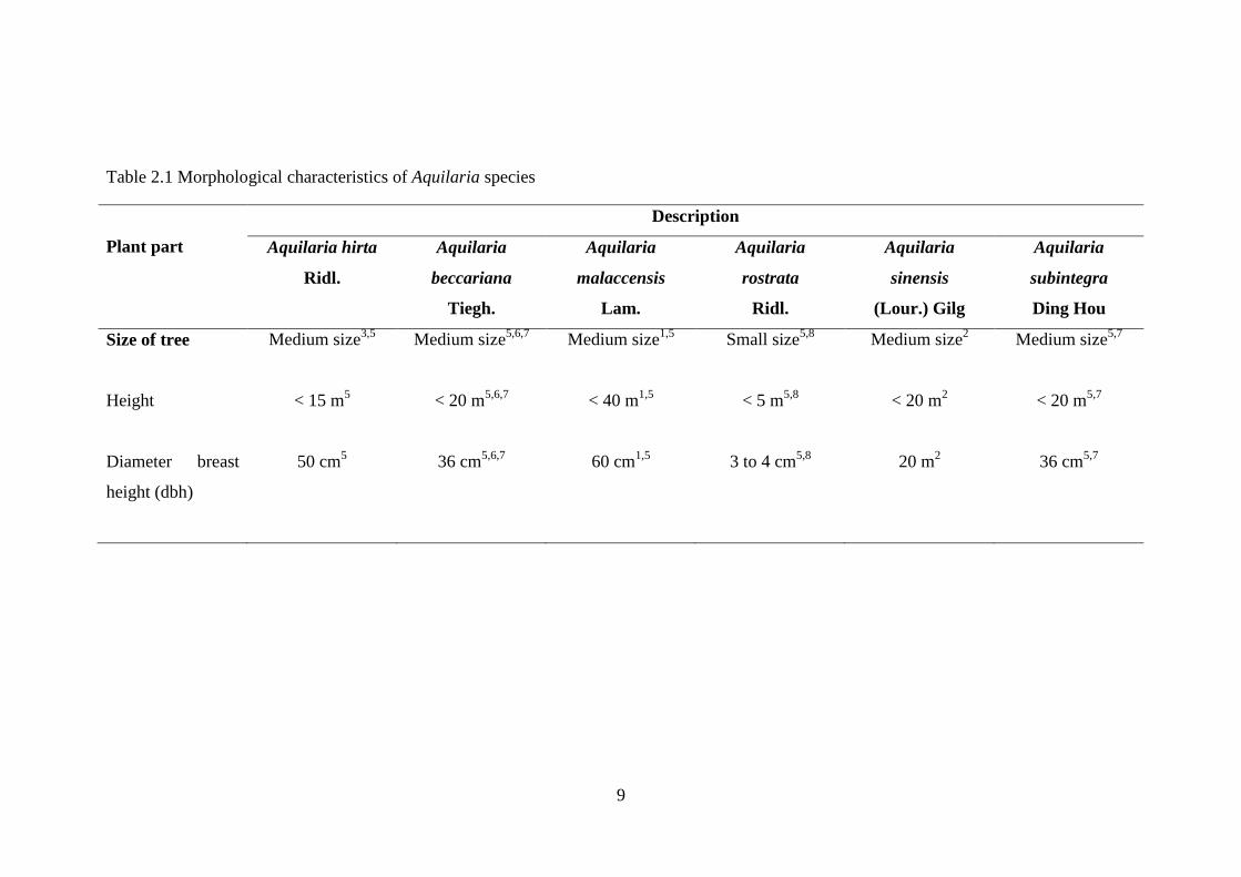
differentiated and compared as shown in Table 2.1.
8
Species in the genus Aquilaria are slow growing trees. The size of the tree of
the genus is different in each species. Most of them are medium in size and can grow
between 5 m to 40 m high. A. rostrata was the shortest with a height of 5 m and has
a relatively the thinnest diameter at breast height (dbh) of 3 to 4 cm. The greatest
size of the species was A. malaccensis which can grow up to 40 m in height with the
widest dbh of 60 cm.
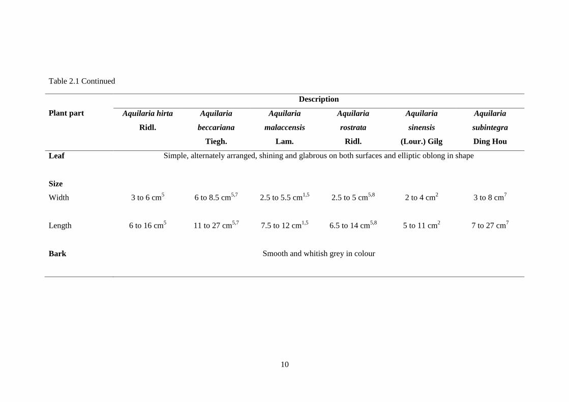
The leaves of all species in the genus Aquilaria comprising similar
morphological features except the size of leaves. A. beccariana has the broadest leaf
area (11-27 cm x 6-8.5 cm), followed by A. subintegra with leaf size (7-27 cm x 3-8
cm) while A. sinensis has the smallest leaf size (5-11 cm x 2-4 cm) with elliptic-
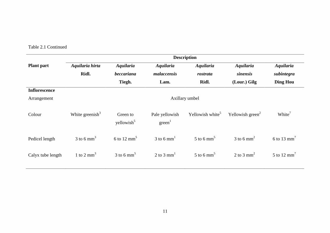
oblong shape. Aquilaria tree barks is smooth, soft and pale in colour. Reproductive
organs of Aquilaria species are hermaphroditic. The flowers are arranged in axillary
umbel inflorescence. Fruits productions from the flowers vary between species.
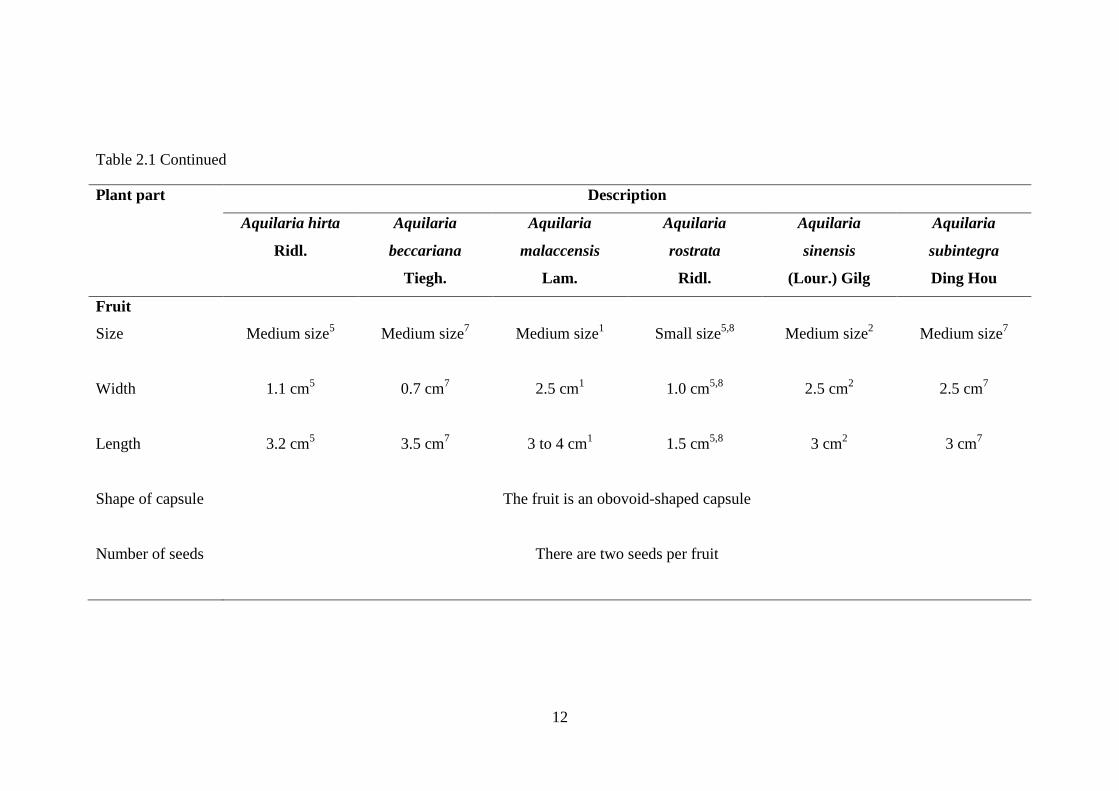
Aquilaria species has a medium size fruit excluding A. rostrata. A.
malaccensis is one of the species that produces the largest fruit measuring about, 3-4
cm x 2.5 cm. Whereas, A. rostrata produces the smallest fruit measuring 1.5 cm x
0.75 cm. Depending on the species of Aquilaria, the shapes of fruit capsules are
different for each species. Some have obovoid-oblong shaped capsule and some have
flattened egg-shaped. However, every fruit for each species in the genus Aquilaria
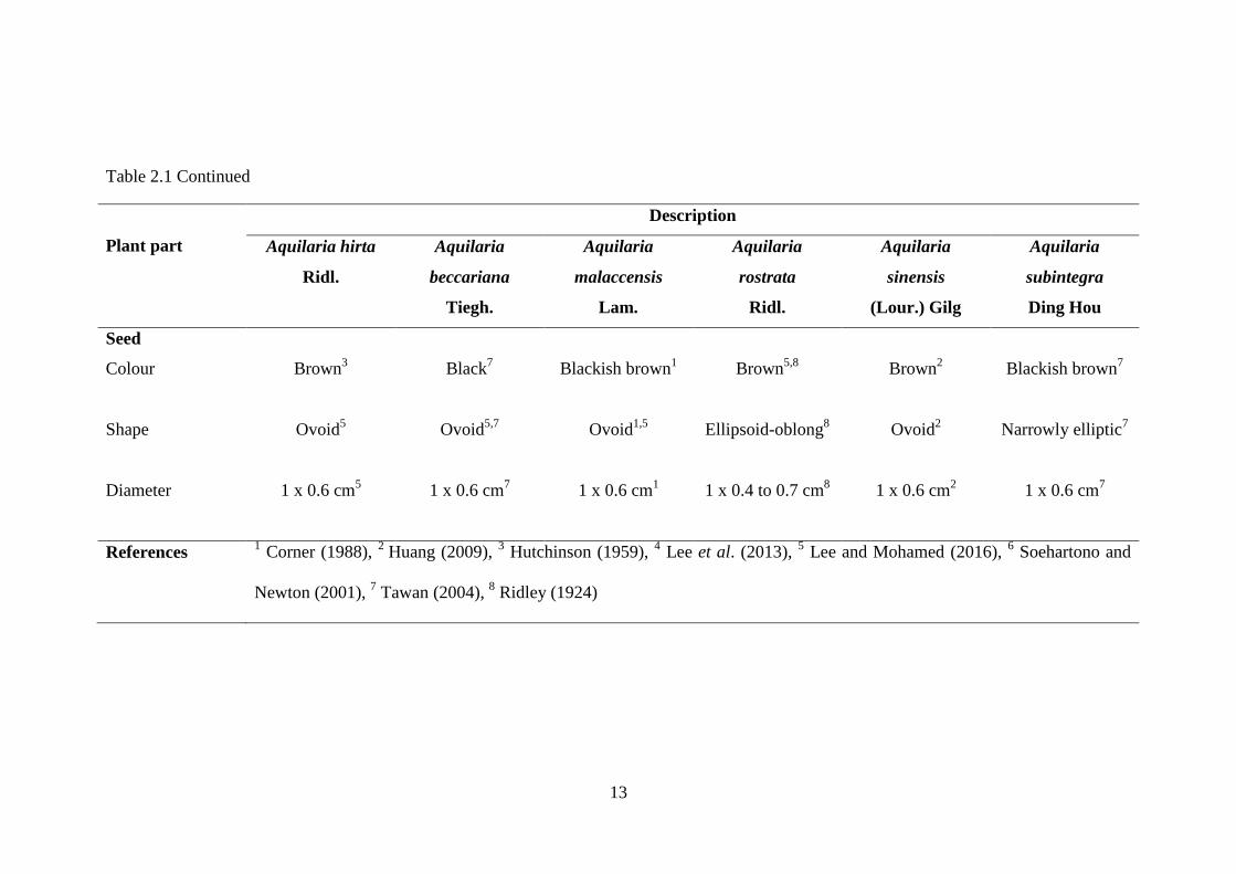
contains two seeds per fruit. In addition, there are various shapes, sizes and colours
of the seeds of the Aquilaria species.
9
Table 2.1 Morphological characteristics of Aquilaria species
Plant part
Description
Aquilaria hirta
Ridl.
Aquilaria
beccariana
Tiegh.
Aquilaria
malaccensis
Lam.
Aquilaria
rostrata
Ridl.
Aquilaria
sinensis
(Lour.) Gilg
Aquilaria
subintegra
Ding Hou
Size of tree
Medium size3,5
Medium size5,6,7
Medium size1,5
Small size5,8
Medium size2
Medium size5,7
Height
< 15 m5
< 20 m5,6,7
< 40 m1,5
< 5 m5,8
< 20 m2
< 20 m5,7
Diameter breast
height (dbh)
50 cm5
36 cm5,6,7
60 cm1,5
3 to 4 cm5,8
20 m2
36 cm5,7
10
Table 2.1 Continued
Plant part
Description
Aquilaria hirta
Ridl.
Aquilaria
beccariana
Tiegh.
Aquilaria
malaccensis
Lam.
Aquilaria
rostrata
Ridl.
Aquilaria
sinensis
(Lour.) Gilg
Aquilaria
subintegra
Ding Hou
Leaf Simple, alternately arranged, shining and glabrous on both surfaces and elliptic oblong in shape
Size
Width 3 to 6 cm5
6 to 8.5 cm5,7
2.5 to 5.5 cm1,5
2.5 to 5 cm5,8
2 to 4 cm2
3 to 8 cm7
Length 6 to 16 cm5
11 to 27 cm5,7
7.5 to 12 cm1,5
6.5 to 14 cm5,8
5 to 11 cm2
7 to 27 cm7
Bark
Smooth and whitish grey in colour
11
Table 2.1 Continued
Plant part
Description
Aquilaria hirta
Ridl.
Aquilaria
beccariana
Tiegh.
Aquilaria
malaccensis
Lam.
Aquilaria
rostrata
Ridl.
Aquilaria
sinensis
(Lour.) Gilg
Aquilaria
subintegra
Ding Hou
Inflorescence
Arrangement Axillary umbel
Colour White greenish3
Green to
yellowish5
Pale yellowish
green1
Yellowish white5
Yellowish green2
White7
Pedicel length 3 to 6 mm3
6 to 12 mm5
3 to 6 mm1
5 to 6 mm5
3 to 6 mm2
6 to 13 mm7
Calyx tube length 1 to 2 mm3
3 to 6 mm5 2 to 3 mm
1
5 to 6 mm5
2 to 3 mm2
5 to 12 mm7
12
Table 2.1 Continued
Plant part Description
Aquilaria hirta
Ridl.
Aquilaria
beccariana
Tiegh.
Aquilaria
malaccensis
Lam.
Aquilaria
rostrata
Ridl.
Aquilaria
sinensis
(Lour.) Gilg
Aquilaria
subintegra
Ding Hou
Fruit
Size Medium size5
Medium size7
Medium size1
Small size5,8
Medium size2
Medium size7
Width 1.1 cm5
0.7 cm7
2.5 cm1
1.0 cm5,8
2.5 cm2
2.5 cm7
Length 3.2 cm5
3.5 cm7
3 to 4 cm1
1.5 cm5,8
3 cm2
3 cm7
Shape of capsule The fruit is an obovoid-shaped capsule
Number of seeds There are two seeds per fruit
13
Table 2.1 Continued
Plant part
Description
Aquilaria hirta
Ridl.
Aquilaria
beccariana
Tiegh.
Aquilaria
malaccensis
Lam.
Aquilaria
rostrata
Ridl.
Aquilaria
sinensis
(Lour.) Gilg
Aquilaria
subintegra
Ding Hou
Seed
Colour Brown3
Black7
Blackish brown1
Brown5,8
Brown2
Blackish brown7
Shape Ovoid5
Ovoid5,7
Ovoid1,5
Ellipsoid-oblong8
Ovoid2
Narrowly elliptic7
Diameter 1 x 0.6 cm5
1 x 0.6 cm7
1 x 0.6 cm1
1 x 0.4 to 0.7 cm8
1 x 0.6 cm2
1 x 0.6 cm7
References 1 Corner (1988),
2 Huang (2009),
3 Hutchinson (1959),
4 Lee et al. (2013),
5 Lee and Mohamed (2016),
6 Soehartono and
Newton (2001), 7 Tawan (2004),
8 Ridley (1924)
14
2.1.2 Uses of Aquilaria leaves
In Malaysia, the decoction of the leaves of A. malaccensis is applied
externally to treat swelling and consumed for treating vomiting (Ong, 2004). In
China, the leaves of A. sinensis are prepared as tea to treat fractures and bruising
(Zhou et al., 2008; Yu et al., 2013). In Thailand and Vietnam, the leaves of A.
crassna have been used as a healthy tea food additive (Sattayasai et al., 2012).
Herbal teas of A. malaccensis, A. sinensis and A. crasnna are found to be
beneficial for health to treat various kind of diseases. The teas can be used as anti-
depressant and anti-aging skin agents. Besides, by consuming agarwood tea from its
leaves also provide energy and soothe sleep disorders (Health benefits, 2015).
The leaves of A. sinensis is also consumed as a laxative agent (Hara et al.,
2008). Preedy (2015) reported that the root and leaves of A. malaccensis were used
as a prescription for dropsy. According to Chung and Parwaningsih (1999), the
leaves of several Aquilaria species were burnt throughout the world for incense
purposes.
15
2.2 Bioactivities of Aquilaria leaves
The term “bioactivity” can be defined as a reaction in or the specific effect on
the living tissues upon exposure to a substance (Carbonell‐Capella et al., 2014).
Scientifically, the term “bioactivity” also refers to an alternative term for “biological
activity” (Cammack, 2006). The concept of bioactivity including events relating to
the transportation and movement of bioactive compounds and reach the target tissue,
their interaction with biomolecules, biotransformation or metabolism they may
undergo, and the generation of biomarkers and the physiological responses they
cause (Fernández-García et al., 2009). Bioactivity is measured primarily based on
events that occurs during the interaction between bioactive components with
biomolecules. This interaction can provide health benefits through the achievement
of systemic physiological responses (such as antioxidant and anti-inflammatory)
(Fernández-García et al., 2009; Carbonell‐Capella et al., 2014).
The evaluation of the bioactivities of an extract or a pure substance from a
living organism can be done through in vivo and in vitro experimental models
(Colegate and Molyneux, 2007; Fernández-García et al., 2009; Carbonell‐Capella et
al., 2014). In vitro methods have been developed to determine bioactivity, including
the screening of various activities such as antioxidant, anti-diabetic, anti-
inflammatory, anti-tumor, and others (Fernández-García et al., 2009). In vitro test is
the most desirable as it is more simple, specific and rapid in data generation.
Meanwhile, in vivo tests in mammals are often variable and highly constrained by
ethical considerations of animal welfare (Colegate and Molyneux, 2007).
16
2.2.1 Antioxidant activity
Antioxidant activity is among the most frequently reported bioactivity
properties from the medicinal plants including Aquilaria species. In addition, there
are many antioxidant studies conducted on leaf samples and other parts of Aquilaria.
Antioxidants can be defined as substances that are present in low concentrations
which could inhibit the oxidation process of substrate and neutralize the action of
free radicals (Li, 1999). From previous studies on the leaf samples of Aquilaria, Ray
et al. (2014) had compared the in vitro antioxidant activity of petroleum ether,
dichloromethane and 95% ethanol extracts of dried leaves of A. subintegra and
found the highest radical scavenging activity from the 95% ethanolic extract. Huda
et al. (2009) had extracted the dried leaves sample of A. malaccensis using ethyl
acetate, dichloromethane, methanol and hexane and found the highest potential of
antioxidant properties from the methanol extract.
Another in vitro study for antioxidant activity by Miniyar et al. (2008), who
had revealed the ethyl acetate extract of A. malaccensis gives a strong antioxidant for
inhibitory effect on nitrite-induced oxidation of haemoglobin in human blood
haemolysate. Han and Li (2012) found the strong antioxidant potential of the
methanol dried leaf extract of A. sinensis after testing it using different in vitro
antioxidant assays; 2,2-diphenyl-1-picrylhydrazyl (DPPH) scavenging activity, 2,2'-
azino-bis(3-ethylbenzothiazoline-6-sulphonic acid) (ABTS) scavenging activity,
ferric ion (Fe3+
) reducing power, cupric ion (Cu2+
) reducing power, superoxide anion

17
(O2 ) scavenging activity, hydroxyl radical (·OH) scavenging activity, metal
chelating assays (Fe2+
and Cu2+
) and lipid peroxidation.
Tay et al. (2014) had extracted the dried leaves of A. crassna using different
percentages of ethanol in water (0% to 100%) and reported that extraction using 60%
ethanol gave the highest yield of polyphenols and extraction using 100% ethanol
gave the highest DPPH radical scavenging activity with low yield of flavonoids. The
essential oil from the stem bark of A. crassna, which was obtained from
hydrodistillation method also exhibited a significant DPPH free radical scavenging
and ferric reducing antioxidant power (FRAP) activities (Dahham et al., 2015).
Rattanama et al. (2014) had compared in vitro antioxidant activity of deionise water
and ethyl acetate extracts of A. subintegra tea leaves and found ethyl acetate extract
gave a better antioxidant activity, which possesed higher extraction yield and total
flavonoid content.
2.2.2 Anti-diabetic activity
Recently, researchers became interested in investigating the plant
polyphenols that are capable in inhibiting carbohydrate-hydrolyzing enzymes such as
α-glucosidase and α-amylase to prevent hyperglycemia. Control of postprandial
hyperglycemia after a meal is the most effective approach for treating Type 2
diabetic patients by preventing the absorption of glucose in the small intestine and
18
enhance glucose disposal in the cells (Yao et al., 2009). Dietary carbohydrates such
as starch are the main source of glucose that is hydrolyzed by α-glucosidase and
pancreatic α-amylase enzymes (Kim et al., 2005). Therefore, inhibiting the activity
of these carbohydrate digesting enzymes are the best way to retard or delay the
absorption of glucose and maintaining the postprandial glucose level in the blood
(Watanabe et al., 1997). The synthetic inhibitors that have been commercialized as
drugs to treat Type 2 diabetic are acarbose, miglitol, and voglibose (Yoshikawa et
al., 1998; Yao et al., 2009).
Feng et al. (2011) used α-glucosidase inhibition assay to isolate the
compounds from 70% aqueous ethanolic extract of A. sinensis leaves. Some potent
phenolic compounds were isolated which are mangiferin, iriflophenone 2-O-α-L-
rhamnopyranoside, iriflophenone 3-C-β-D-glucoside and iriflophenone 3,5-C-β-D-
diglucopyranoside.
Nur Liyana et al. (2013) had compared α-glucosidase and α-amylase
inhibitory activity of A. malaccensis and A. hirta and found the highest α-
glucosidase inhibitory activity of the methanol extract of A. hirta while methanol
extract of A. malaccensis for α-amylase inhibitory activity. Another comparative
study by Yunus et al. (2015) on the drying effects of 70% ethanol extracts of A.
malaccensis and A. subintegra leaves using microwave power intensity (50W-150W)
and found that both of these exhibited the highest α-amylase inhibition and high
yield of polyphenols at low power intensity (50W). The ethanol extract of A.

19
subintegra gave higher α-amylase inhibitory activity than ethanol extract of A.
malaccensis.
The leaves of A. sinensis has been reported to have in vitro and in vivo anti-
diabetic activities. The methanolic extract was found to have a significant effect in
lowering fasting blood glucose level in Steptozotocin-induced (STZ) diabetic rats in
vivo as compared to hexane and ethyl acetate extract. The same extract also was the
most active in enhancing the uptake of glucose into rat adipocytes in vitro
(Pranakhon et al., 2011).
Further in vitro and in vivo anti-diabetic studies by Pranakhon et al. (2015),
found the presence of iriflophenone 3-C-β-glucoside (IPG) in the methanol extract of
A. sinensis, which was active in reducing fasting blood glucose compared to insulin
effectively.
2.2.3 Anti-inflammatory activity
Inflammation can be defined as the physical condition of body that appear
redness, warmth, swollen and pain caused by the body’s response to noxious stimuli,
injury or infection by pathogens (Rahman et al., 2012). Nowadays, non-steroidal
anti-inflammatory drugs (NSAIDs) play an important role as anti-inflammatory
20
drugs to reduce the pain of inflammation in the body by inhibiting prostaglandin
synthesis as well as cyclooxygenase inhibitory activity (Olivier, 2001). However,
long-term consumption of synthetic NSAIDs drugs may cause severe side effects
and carry risk to cardiovascular disorders, gastrointestinal toxicity and others
(Hawkey, 2001; Andreas and Oliver, 2012).
Therefore, in recent time, several studies have been carried out to discover a
novel, effective, less toxic or safe anti-inflammatory drugs from natural sources that
may provide alternative ways to treat the inflammation disorders. Different parts of
several Aquilaria species including leaves have been studied to discover anti-
inflammatory agents within this genus. Kumphune et al. (2011) reported that the
ethyl acetate extract of A. crassna heartwood had anti-inflammatory potential by
inhibiting the tumour necrosis factor-alpha gene expression and secretion in
lipopolysaccharides (LPS)-induced human peripheral blood mononuclear cells. Zhou
et al. (2008) revealed that the ethanolic extract of A. sinensis leaves potentially
inhibited the elevated nitric oxide (NO) level in lipopolysaccharides (LPS)-
stimulated nitric oxide (NO) release from macrophages in vitro.
Wu et al. (2012) reported that the peel extract of A. sinensis had significantly
suppressed inflammation in RAW 264.7 cells by lipopolysaccharides (LPS), which
can be reached by suppressing the protein level of cyclooxygenase (COX-2)
isozymes in vitro. Chen et al. (2014) revealed that the ethyl acetate-soluble fraction
from the pericarp of A. sinensis in the presence bioactive compounds (velutin, pillion
and β-sitostenone) exhibited potent inhibition against lipopolysaccharide (LPS)-
21
induced NF-kB production by macrophages in vitro. Wang et al. (2015) had isolated
twenty-one bioactive compounds including two new flavones (4’-O-geranyltricin
and 3’-O-geranylpolloin) from the stem bark of A. sinensis and stated that these
compounds have potential for the treatment and prevention of inflammatory.
Using animal models to investigate the anti-inflammatory activity in the
genus Aquilaria, Rahman et al. (2012) studied the essential oil of A. malaccensis
wood that was obtained from hydrodistillation method for in vivo and in vitro anti-
inflammatory activity and found this agarwood oil has anti-inflammatory potential
with a significant reduction of edema in carrageenan induced rat paw edema model
in vivo and strong membrane stabilizing on human red blood cell in vitro as
compared with the standard diclofenac. Another in vivo anti-inflammatory study by
Huanze et al. (2013), on A. sinensis leaves, found that the leaves originating from
two different places have similar inhibitory effect when tested on mice induced by
xylene.
2.2.4 Antibacterial activity
Numerous studies of the “gaharu” tree of the genus Aquilaria with
antibacterial properties have been carried out. By definition, antibacterial agent is an
action of substance that kill bacteria and/or inhibits their growth or replication. It will
destroy the pathogen without affecting the infected patient. Some antimicrobial

22
agents are antibiotics and antibiotics can be defined as a substance that is produced
by a microorganism that is effective in killing and suspressing the growth of the
other microorganism. The antibacterial agents usually target pathogen membrane
structure and metabolic processes to accomplish their actions (Paul and Janet, 2008).
Dash et al. (2008) had investigated the inhibitory capacity of aqueous and
methanol extracts from A. malaccensis leaf and bark against Shigella flexneri,
Bacillus brevis, Pseudomonas aeruginosa and Bacillus subtilis and found all of the
extracts had moderate activity against all tested bacteria while the methanol extract
of the leaf gave the highest inhibition against B. subtilis. Kamonwannasit et al.
(2013) reported the effect of aqueous extract of A. crassna leaves that was found to
disrupt the bacterial cell wall and inhibit bacterial biofilm formation against
Staphylococcus epidermis.
Essential oils from different chemical stimulation methods of A. sinensis
were tested against Gram-positive bacterial strains such as Staphylococcus aureus
and Bacillus subtilis and Gram-negative bacterial strain Escherichia coli and found
that all the essential oils, which are containing sesquiterpenes and aromatic
compounds actively inhibited all the tested bacteria except E. coli (Chen et al.,
2011). In agreement with a study by Wen-Jian et al. (2014), their results revealed
that the antibacterial activity of A. sinensis was due to the presence of sesquiterpenes
and 4-hydroxyphenylacetic acid.
23
Remarkable results using the disc diffusion methodology were established
using twenty-eight fungal endophytes of the stem of A. sinensis against
Staphylococcus aureus, Escherichia coli, Bacillus subtilis and Aspergillus fumigatus.
Thirteen endophytic fungi associated with A. sinensis exhibited high antibacterial
activity to at least one of the bacterial tested (Cui et al., 2011). Similarly, the ethyl
acetate extract of endophytic fungus of A. malaccensis had been reported by Shoeb
et al. (2010) to have mild antibacterial activity against Bacillus cereus and
Staphylococcus aureus.
Rahman et al. (2013) studied the antibacterial activity of A. malaccensis oil
and Citrullus lanatus seed oil by agar well diffusion method and compared with
standard ciprofloxacin, against Escherichia coli, Enterococcus faecalis,
Staphylococcus aureus and Pseudomonas aeruginosa. It was found that both oils
have antibacterial activity against the selected bacteria and the Citrullus oil possesed
stronger antibacterial activity than A. malaccensis oil.
2.2.4(a) Antibacterial activity of herbal teas from Aquilaria species
Nowadays, tea from Camelia sinensis has been recognized as a tonic and
remains a kind of medicine particularly in traditional Chinese medicine to treat a
variety of disorders (Chen, 2003). Lee et al. (2006) examined the ethyl acetate and
water polyphenols tea extracts with different strains of intestinal bacteria and found
that pathogenic bacteria such as Clostridium perfringens, Clostridium difficile and

24
Bacteroides spp. were suppressed significantly by tea phenolics and their derivatives
while Clostridium spp. and Bifidobacterium spp. which are anaerobic bacteria and
probiotics such as Lactobacillus sp. were poorly affected by tea phenolics. This
showed that the tea phenolics substantially affected the implementation of the
gastrointestinal tract. The presence of caffeic acid in the tea extracts provided strong
inhibition against E. coli and Salmonella sp. which are usually causing food-borne
diseases such as diarrhea and nausea and also pathogenic bacteria such as
Staphylococcus aureus, Shigella dysenteriae, Pseudomonas aeruginosa, Vibrio
chlorae and others (Chen, 2003; Lee et al., 2006).
In this study, the Aquilaria leaves were tested for their antibacterial activities
against gastrointestinal pathogenic bacteria. Previous studies reported that herbal teas
from bark and stem of A. malaccensis are consumed traditionally to treat
gastrointestinal disorders such as diarrhea and abdominal pain and these teas have
been practised for many years in folk medicine in certain countries (Ong, 2004).
Previous studies by Kamonwannasit et al. (2011) revealed that the aqueous extract of
A. crassna leaves is beneficial for treating diarrhea caused by Staphylococcus aureus
and skin infection associated with Staphylococcus epidermis. The disc-diffusion
methodology was established on aqueous extract of A. malaccensis leaves against
various pathogenic microbes including Bacillus cereus, Candida albicans,
Streptococcus faecalis, Staphylococcus aureus, Pseudomonas aeruginosa,
Escherichia coli and Aspergillus niger. It was found that the extract suppressed the
growth of Staphylococcus aureus and Pseudomonas aeruginosa (Manasi et al.,
2008).