Community acquired respiratory virus infections in cancer ... · patientsdGuideline on diagnosis...
13
Review Community acquired respiratory virus infections in cancer patientsdGuideline on diagnosis and management by the Infectious Diseases Working Party of the German Society for haematology and Medical Oncology Marie von Lilienfeld-Toal a,b,c, *, Annemarie Berger d , Maximilian Christopeit e , Marcus Hentrich f , Claus Peter Heussel g,h,i , Jana Kalkreuth a , Michael Klein j , Matthias Kochanek k , Olaf Penack l , Elke Hauf m , Christina Rieger n , Gerda Silling o , Maria Vehreschild k,p , Thomas Weber q , Hans-Heinrich Wolf q , Nicola Lehners r,y , Enrico Schalk s,y , Karin Mayer t,y a Department of Haematology and Medical Oncology, Jena University Hospital, Jena, Germany b Centre for Sepsis Control and Care (CSCC), University Hospital Jena, Germany c Leibniz Institute for Natural Product Research and Infection Biology, Hans-Kno¨ll-Institut, Jena, Germany d Institute for Medical Virology, University Hospital Frankfurt, Goethe University Frankfurt, Germany e Department of Stem Cell Transplantation, University Hospital Hamburg-Eppendorf, Hamburg, Germany f Department of Haematology and Oncology, Red Cross Hospital, Munich, Germany g Department of Diagnostic and Interventional Radiology, University Hospital, Heidelberg, Germany h Translational Lung Research Center Heidelberg (TLRC), Member of the German Center for Lung Research (DZL), Heidelberg, Germany i Department of Diagnostic and Interventional Radiology with Nuclear Medicine, Thoraxklinik at University of Heidelberg, Heidelberg, Germany j Department I of Internal Medicine, Prosper-Hospital, Recklinghausen, Germany k Department I of Internal Medicine, University Hospital of Cologne, Cologne, Germany l Department of Hematology, Oncology and Tumorimmunology, Charite´Campus Virchow, Berlin, Germany m Department III of Internal Medicine, The University Hospital Klinikum rechts der Isar, Technische Universita¨t Mu¨nchen, Munich, Germany n Lehrpraxis der Ludwig-Maximilians-Universita¨t Mu¨nchen, Germering, Germany o Department of Haematology, Oncology and Stem Cell Transplantation, Faculty of Medicine, RWTH Aachen University, Aachen, Germany p German Centre for Infection Research, Partner Site Bonn-Cologne, Cologne, Germany q Department of Hematology and Oncology, Martin-Luther-University Halle-Wittenberg, Halle, Germany * Corresponding author: Department of Internal Medicine II, Haematology and Medical Oncology, University Hospital Jena, Erlanger Allee 101, 07747 Jena, Germany. Fax: þ49 3641 9324202. E-mail address: [email protected] (M. von Lilienfeld-Toal). y These authors contributed equally. http://dx.doi.org/10.1016/j.ejca.2016.08.015 0959-8049/ª 2016 The Authors. Published by Elsevier Ltd. This is an open access article under the CC BY-NC-ND license (http:// creativecommons.org/licenses/by-nc-nd/4.0/). Available online at www.sciencedirect.com ScienceDirect journal homepage: www.ejcancer.com European Journal of Cancer 67 (2016) 200e212
Transcript of Community acquired respiratory virus infections in cancer ... · patientsdGuideline on diagnosis...

European Journal of Cancer 67 (2016) 200e212
Available online at www.sciencedirect.com
ScienceDirect
journal homepage: www.ejcancer.com
Review
Community acquired respiratory virus infections in cancerpatientsdGuideline on diagnosis and management by theInfectious Diseases Working Party of the German Societyfor haematology and Medical Oncology
Marie von Lilienfeld-Toal a,b,c,*, Annemarie Berger d,Maximilian Christopeit e, Marcus Hentrich f, Claus Peter Heussel g,h,i,Jana Kalkreuth a, Michael Klein j, Matthias Kochanek k, Olaf Penack l,Elke Hauf m, Christina Rieger n, Gerda Silling o, Maria Vehreschild k,p,Thomas Weber q, Hans-Heinrich Wolf q, Nicola Lehners r,y,Enrico Schalk s,y, Karin Mayer t,y
a Department of Haematology and Medical Oncology, Jena University Hospital, Jena, Germanyb Centre for Sepsis Control and Care (CSCC), University Hospital Jena, Germanyc Leibniz Institute for Natural Product Research and Infection Biology, Hans-Knoll-Institut, Jena, Germanyd Institute for Medical Virology, University Hospital Frankfurt, Goethe University Frankfurt, Germanye Department of Stem Cell Transplantation, University Hospital Hamburg-Eppendorf, Hamburg, Germanyf Department of Haematology and Oncology, Red Cross Hospital, Munich, Germanyg Department of Diagnostic and Interventional Radiology, University Hospital, Heidelberg, Germanyh Translational Lung Research Center Heidelberg (TLRC), Member of the German Center for Lung Research (DZL),
Heidelberg, Germanyi Department of Diagnostic and Interventional Radiology with Nuclear Medicine, Thoraxklinik at University of Heidelberg,
Heidelberg, Germanyj Department I of Internal Medicine, Prosper-Hospital, Recklinghausen, Germanyk Department I of Internal Medicine, University Hospital of Cologne, Cologne, Germanyl Department of Hematology, Oncology and Tumorimmunology, Charite Campus Virchow, Berlin, Germanym Department III of Internal Medicine, The University Hospital Klinikum rechts der Isar, Technische Universitat Munchen,
Munich, Germanyn Lehrpraxis der Ludwig-Maximilians-Universitat Munchen, Germering, Germanyo Department of Haematology, Oncology and Stem Cell Transplantation, Faculty of Medicine, RWTH Aachen University,
Aachen, Germanyp German Centre for Infection Research, Partner Site Bonn-Cologne, Cologne, Germanyq Department of Hematology and Oncology, Martin-Luther-University Halle-Wittenberg, Halle, Germany
* Corresponding author: Department of Internal Medicine II, Haematology and Medical Oncology, University Hospital Jena, Erlanger Allee 101,
07747 Jena, Germany. Fax: þ49 3641 9324202.
E-mail address: [email protected] (M. von Lilienfeld-Toal).y These authors contributed equally.
http://dx.doi.org/10.1016/j.ejca.2016.08.015
0959-8049/ª 2016 The Authors. Published by Elsevier Ltd. This is an open access article under the CC BY-NC-ND license (http://
creativecommons.org/licenses/by-nc-nd/4.0/).
M. von Lilienfeld-Toal et al. / European Journal of Cancer 67 (2016) 200e212 201
r Department of Haematology and Oncology, University Hospital of Heidelberg, Heidelberg, Germanys Department ofHaematology andOncology,Medical Centre, Otto-von-GuerickeUniversityMagdeburg,Magdeburg, Germanyt Department of Haematology and Oncology, University Hospital Bonn, Bonn, Germany
Received 5 August 2016; received in revised form 17 August 2016; accepted 17 August 2016
Available online 25 September 2016
KEYWORDS
Upper respiratory
tract infection;
Pneumonia;
Superinfection;
Influenza;
Respiratory syncytial
virus;
Parainfluenza
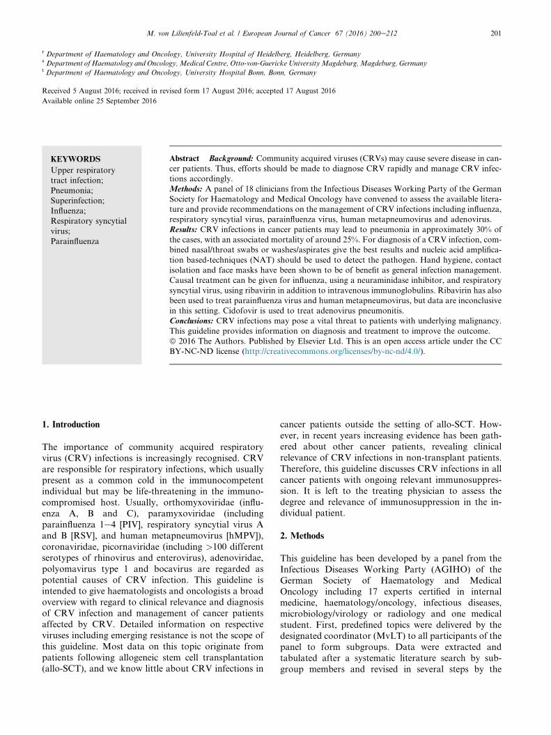
Abstract Background: Community acquired viruses (CRVs) may cause severe disease in can-
cer patients. Thus, efforts should be made to diagnose CRV rapidly and manage CRV infec-
tions accordingly.
Methods: A panel of 18 clinicians from the Infectious Diseases Working Party of the German
Society for Haematology and Medical Oncology have convened to assess the available litera-
ture and provide recommendations on the management of CRV infections including influenza,
respiratory syncytial virus, parainfluenza virus, human metapneumovirus and adenovirus.
Results: CRV infections in cancer patients may lead to pneumonia in approximately 30% of
the cases, with an associated mortality of around 25%. For diagnosis of a CRV infection, com-
bined nasal/throat swabs or washes/aspirates give the best results and nucleic acid amplifica-
tion based-techniques (NAT) should be used to detect the pathogen. Hand hygiene, contact
isolation and face masks have been shown to be of benefit as general infection management.
Causal treatment can be given for influenza, using a neuraminidase inhibitor, and respiratory
syncytial virus, using ribavirin in addition to intravenous immunoglobulins. Ribavirin has also
been used to treat parainfluenza virus and human metapneumovirus, but data are inconclusive
in this setting. Cidofovir is used to treat adenovirus pneumonitis.
Conclusions: CRV infections may pose a vital threat to patients with underlying malignancy.
This guideline provides information on diagnosis and treatment to improve the outcome.
ª 2016 The Authors. Published by Elsevier Ltd. This is an open access article under the CC
BY-NC-ND license (http://creativecommons.org/licenses/by-nc-nd/4.0/).
1. Introduction
The importance of community acquired respiratory
virus (CRV) infections is increasingly recognised. CRV
are responsible for respiratory infections, which usually
present as a common cold in the immunocompetentindividual but may be life-threatening in the immuno-
compromised host. Usually, orthomyxoviridae (influ-
enza A, B and C), paramyxoviridae (including
parainfluenza 1e4 [PIV], respiratory syncytial virus A
and B [RSV], and human metapneumovirus [hMPV]),
coronaviridae, picornaviridae (including >100 different
serotypes of rhinovirus and enterovirus), adenoviridae,
polyomavirus type 1 and bocavirus are regarded aspotential causes of CRV infection. This guideline is
intended to give haematologists and oncologists a broad
overview with regard to clinical relevance and diagnosis
of CRV infection and management of cancer patients
affected by CRV. Detailed information on respective
viruses including emerging resistance is not the scope of
this guideline. Most data on this topic originate from
patients following allogeneic stem cell transplantation(allo-SCT), and we know little about CRV infections in
cancer patients outside the setting of allo-SCT. How-
ever, in recent years increasing evidence has been gath-
ered about other cancer patients, revealing clinical
relevance of CRV infections in non-transplant patients.
Therefore, this guideline discusses CRV infections in all
cancer patients with ongoing relevant immunosuppres-
sion. It is left to the treating physician to assess the
degree and relevance of immunosuppression in the in-dividual patient.
2. Methods
This guideline has been developed by a panel from the
Infectious Diseases Working Party (AGIHO) of the
German Society of Haematology and Medical
Oncology including 17 experts certified in internal
medicine, haematology/oncology, infectious diseases,
microbiology/virology or radiology and one medical
student. First, predefined topics were delivered by thedesignated coordinator (MvLT) to all participants of the
panel to form subgroups. Data were extracted and
tabulated after a systematic literature search by sub-
group members and revised in several steps by the

M. von Lilienfeld-Toal et al. / European Journal of Cancer 67 (2016) 200e212202
members of the panel on the basis of an email-based
discussion process and a face-to-face meeting. Finally,
preliminary recommendations of the panel were dis-
cussed, revised and approved by the AGIHO assembly.
In May 2014, the first literature search was performed
for CRV and immunosuppression using the terms
‘-virus’ and ‘immunocompromised’ (for example:
‘adenovirus immunocompromised’). This search wasperformed for adenovirus, bocavirus, coronavirus,
enterovirus, hMPV, influenza, PIV, parechovirus, RSV
and rhinovirus. The references were then screened by the
subgroup members and relevant articles retrieved as full
papers. Wherever applicable, additional papers were
identified in the reference lists and treated as described.
In February 2016, an update of the literature search was
performed.For grading, the system applied by the European
Society of Clinical Microbiology and Infectious Dis-
eases as proposed by Ullmann et al., in 2012 [1] was
used (Table 1) with one modification: other than Ull-
mann et al., we used the same grading of the strength of
Table 1Grading of evidence as suggested by the ESCMID [1].
Strength of recommendation
A Strongly support a recommendation for use
B Moderately support a recommendation for use
C Marginally support a recommendation for use
D Support a recommendation against use
Quality of evidence for interventionsdlevel
I Evidence from at least one properly designed randomized,
controlled trial
II* Evidence from at least one well-designed clinical trial, without
randomization; from cohort- or case-control analytic studies
(preferably from more than one centre); from multiple time
series; or from dramatic results from uncontrolled experiments.
III Evidence from opinion of respected authorities, based on
clinical experience, descriptive case studies, or report of expert
committees
*Added index
R Meta-analysis or systematic review of randomized controlled
trials
T Transferred evidence, i.e. results from different patients’
cohorts or similar immune status situation
H Comparator group is a historical control
U Uncontrolled trial
A Abstract published at an international meeting
Quality of evidence for diagnostic measuresdlevel
I Evidence from at least one properly designed multicentre cross-
sectional or cohort study
II Evidence from
(1) At least one well-designed prospective singlecentre
cross-sectional or cohort study or
(2) A properly designed retrospective multicentre cross-
sectional or cohort study or
(3) From case-control studiesIII Evidence from opinion of respected authorities, based on
clinical experience, descriptive case studies, or report of expert
committees
recommendation for diagnostic measures as for in-
terventions (Table 1). The results of the literature
search and the following grading process were used to
develop recommendations wherever possible. Recom-
mendations and evidence were then presented at and
approved by the AGIHO assembly during the spring
meeting on 6th March 2015. Following the update of
the literature search in February 2016 no relevantchanges were made.
3. Diseases caused by CRV
CRVs cause respiratory tract infections, which can be
divided into upper respiratory tract infection (URTI),influenza-like illness (ILI) and lower respiratory tract
infection/pneumonia (LRTI). Commonly, URTI can be
assumed, if a patient has a new onset of symptoms
including at least one of cough, coryza, sore throat or
shortness of breath which is deemed to be due to an
infection by the treating physician. LRTI requires clin-
ical or radiological evidence of pneumonia [2]. ILI is
diagnosed in patients with a sudden onset of newsymptoms including at least one of fever or feverishness,
malaise, headache or myalgia and at least one of the
respiratory symptoms cough, sore throat or shortness of
breath [3]. To be certain of the viral origin, the detection
of the virus from respiratory samples like swabs, naso-
pharyngeal aspirates or bronchoalveolar lavage fluid is
required. Of note, surveillance studies showed some
patients to be asymptomatic but still shedding the virus[2,4e7]. For that reason, some authors distinguish be-
tween respiratory infection (detection of virus indepen-
dent of symptoms) and respiratory infection disease
(detection of virus and respective symptoms) [8]. How-
ever, for the purpose of this guideline, we omit this
distinction and define URTI, LRTI and ILI as described
above.
4. Epidemiology and clinical relevance
Some CRV like influenza or RSV have a seasonality
with most infections occurring during the winter months
[2,9,10]. Others like rhinovirus or parainfluenza are in-
dependent of seasonality [11]. Thus, an appropriatediagnostic work-up and clinical management is war-
ranted in any patient presenting with typical symptoms
regardless of the time of the year. As may be expected
considering the nature of the disease, CRV frequently
cause outbreaks in health care settings [7,12e15].
Importantly, outbreaks may also occur in outpatient
settings [16] emphasising the need for awareness during
all periods of cancer treatment.Generally, viral URTI in cancer patients has some
impact on the clinical course because the patients are
symptomatic to a degree that frequently requires post-
ponement of chemotherapy [17]. However, critical

M. von Lilienfeld-Toal et al. / European Journal of Cancer 67 (2016) 200e212 203
illness and mortality due to viral URTI are rare. In
contrast, most patients who died were suffering from
LRTI, which thus poses the biggest threat to cancer
patients. Rates of LRTI and mortality differ amongst
the respective CRV [18] and exact estimation is
hampered by the fact that fatal cases are probably over-
reported. We have tried to deduce reliable information
on LRTI and mortality from the literature for variousCRV: influenza appears to have a high rate of LRTI
with approximately 30% and an associated mortality
rate of approximately 25% [19e22]. RSV appears to be
at least as dangerous with a rate of LRTI of approxi-
mately 33% and an associated mortality rate of 27% [16].
However, it has to be kept in mind, that most data
regarding RSV originate from SCT-recipients and very
little is known regarding patients with other forms ofmalignancy. On the other hand, there are several reports
of outbreaks in general haematology/oncology units,
which showed a significant disease burden even in pa-
tients not undergoing stem cell transplantation [12].
With regard to hMPV and PIV, exact information on
the clinical relevance is even more difficult to obtain.
However, although both viruses may cause asymptom-
atic infection [6,23], the available evidence suggests asimilar overall rate of LRTI and mortality
[7,11,13,24e28] compared with influenza and RSV. In
contrast, despite case reports of fatal outcomes of in-
fections with rhinovirus and coronavirus, these viruses
as well as bocavirus appear to be rarely the cause of
LRTI and dangerous only when patients are coinfected
with other pathogens [2,5,29]. Herpesviridae like herpes
simplex virus, human herpes virus 6, cytomegalovirus,varicella zoster and EpsteineBarr virus as well as pol-
yomaviruses or parechoviruses usually do not cause
CRV infection. Pneumonia due to reactivation of her-
pesviridae in severely immunocompromised patients is
not an infection by CRV and thus not covered by this
guideline.
In CRV infection, coinfection with bacteria, fungi or
even other viruses appears to occur in approximately30% of the patients [10,11,28]. They play a vital role with
regard to outcome of patients since patients with bac-
terial or fungal superinfection have a dramatically
higher mortality rate than those with viral infection only
[11,28]. Therefore, possible co- or superinfection should
be considered when managing cancer patients with
CRV. In addition to LRTI and bacterial or fungal su-
perinfection, other risk factors for adverse outcomeinclude haematological malignancy [22], severe immu-
nosuppression such as steroid use or graft versus host
disease or cytopenias [30e32] or low immunoglobulins
[12].
Epidemiology of adenoviruses is somewhat different
from the other CRV: often, the source of infection is a
childhood infection in children under 5 years [33] and
reactivation as well as new infection have been describedto be the cause of disease [34,35]. Other than CRVs like
RSV or influenza, adenoviruses can cause a variety of
symptoms such as conjunctivitis, haemorrhagic cystitis,
gastroenteritis, and URTI in immunocompetent pa-
tients and hepatitis, colitis, nephritis, meningoencepha-
litis and LRTI in the immunocompromised host. In
adult allo-SCT recipients, DNAemia occurs in up to
20% [36e38], but symptomatic disease by adenovirus is
much less common with T-cell suppression being thepredominant risk factor [36]. Again, coinfection is an
important risk factor for severe illness [39].
5. Diagnosis of CRV infection
5.1. Virologydmaterial
Cancer patients presenting with symptoms consistent
with CRV infection should be diagnosed using material
from the respiratory tract (Table 2). Serology is not
useful to detect ongoing CRV infection and thus notrecommended (D III). Regarding material used for
microbiological diagnosis, a variety of approaches are
used in various centres. As a general rule, a close
collaboration with the local microbiology laboratory is
highly recommended because this may determine which
material should be used preferably since commercially
available test kits are licensed for specific materials. For
example testing for viral antigens usually requires morethorough sampling like combined nasal/throat swabs
than testing for viral nucleic acids which can often be
performed reliably on gargles alone. It is therefore
essential for the clinician to be aware of the tests used in
the respective laboratory. The overall evidence in the
literature is best for combined nasal/throat swabs and
nasopharyngeal aspirates (A IIt, Table 2).
5.2. Virologydtest
The best evidence for reliable detection of a CRV pre-
sent in respiratory samples exists for nucleic acid
amplification based-techniques (NAT) like PCR.
Therefore, the use of NAT is highly recommended (A II,
Table 2) and any methods involving the detection of
antigen appear to be second best in immunosuppressed
cancer patients (C II [40e42]). Also, culture methods arenot commonly used anymore and cannot be recom-
mended for general diagnosis, but they are essential in
individual cases in which no known virus can be detec-
ted or results of PCR-analysis are inconclusive (A II,
[43]). In the era of multiplex-test kits, it is difficult to
make a definite recommendation with regard to which
viruses should be looked for. In the absence of any
reliable data regarding this question, the panel feels thatit is wise to search for influenza, RSV, PIV and viruses
currently prevalent in the local environment in all
immunosuppressed cancer patients presenting with
symptoms. Patients with more severe disease (for

Table 2Recommendations regarding diagnostic approaches in cancer patients with symptoms of CRV infection.
Population Intention Intervention SoR QoE Reference
Symptomatic IS To detect viral pathogen
and diagnose infection
Serology D III [94]
Symptomatic IS To detect viral pathogen Combined nasal/throat
swabs or washes/aspirates
A II [95e103]
Symptomatic IS To detect viral pathogen NAT A II [42,94,104e107],
Symptomatic IS To detect LRTI in patients
with CRV infection
Chest X-ray D II [108]
Symptomatic IS To detect LRTI in patients
with CRV infection
CT scan A II [44,46,47,69,108e115],
SoR, strength of recommendation; QoE, quality of evidence; IS, immunosuppressed cancer patients; NAT, nucleic acid amplification techniques;
LRTI, lower respiratory tract infection; CRV, community acquired respiratory virus; CT, computer tomography.
M. von Lilienfeld-Toal et al. / European Journal of Cancer 67 (2016) 200e212204
example pneumonia or critical illness) may have thepanel broadened to include hMPV and adenovirus and
even viruses that only rarely cause LRTI like rhinovirus
and coronavirus. However, evidence for this approach is
low and it is strongly advisable to define local guidelines
on this topic. It should be kept in mind, that herpes-
viridae are not the cause of CRV infection. Therefore,
there is no rationale to look for cytomegalovirus, Eps-
teineBarr virus, herpes simplex virus or varicella zosterin patients with typical symptoms of CRV infection
only.
5.3. Radiology
In patients with symptoms of LRTI, it is essential to
determine the degree of pulmonary involvement. CRV
can affect the tracheobronchial system or the lung pa-
renchyma [44]. Generally, a chest X-ray has been proven
to be unhelpful to diagnose pathologic changes in this
setting because of lack of sensitivity. It is therefore not
recommended (D II, see Table 2). In contrast, there is
good evidence to recommend a CT scan of the chest todetect LRTI in patients with CRV infection (A II, see
Table 2). Bronchial wall thickening as well as interstitial
infiltrates presenting as ground-glass opacities may be
detected. These are defined as increased lung density,
whereas underlying lung architecture is still detectable.
Ground-glass opacities may be patchy or diffuse [44e47]
and can be well distinguished from consolidations,
which show higher density obscuring e.g. the pulmonaryvessels and which are typical for other differential di-
agnoses including bacterial infection. Affection of the
terminal bronchioles might lead to visibility of those
usually invisible structures at CT as small centrilobular
nodules or ‘tree-in-bud’ sign [45] or evidence of bron-
chiolitis causing air-trapping. To reliably detect air-
trapping while inspiratory CT is normal, a CT scan in
expiration is necessary [44]. In addition to good diag-nostic accuracy with regard to the diagnosis of a viral
LRTI, the CT scan may also reveal evidence for an
outbreak, since specific viruses tend to present with a
typical pattern in the CT scan [45]. For an example see
Fig. 1.
6. Management of CRV infection
6.1. Infection control
In the light of the danger of outbreaks with fatal con-
sequences, the most important measure in the manage-
ment of cancer patients with CRV infection is infection
control (Table 3). Local authorities should give exact
guidance on the necessary precautions in the respectiveinstitutions. The following statements intend to give a
general overview.
There is sufficient evidence to recommend stringent
hand hygiene (A IIt), the use of face masks (B IIt) and
contact isolation (A III, Table 3). Importantly, shedding
of CRV in cancer patients often lasts 2 weeks or longer
[5,10,48]. It is therefore wise to perform follow-up
testing of respiratory material in index patients andstop contact isolation only when they became negative.
Of note, early implementation of infection control ap-
pears to be more effective than late implementation [49]
which gives reason to recommend infection control as
soon as symptoms appear and not only after evidence of
CRV.
6.2. Supportive measures
Almost anybody who catches a cold applies some form
of home remedies convinced that they ease the symp-
toms and positively influence the course of the disease.
In contrast to this widespread use, there is very littleevidence to recommend any such measures. In particular
with regard to cancer patients, evidence is too poor to
give a sound recommendation in favour of the use of
vitamin C [50], echinacea [51], garlic [52], zinc [53], hu-
midified hot air [54] or Chinese herbal medicine [55].
Surprisingly, even painkillers [56] and non-steroidal
anti-inflammatory drugs [57] may ease the pain but
have little influence on severity and duration of the CRVinfection. However, as there is some evidence towards a
considerable placebo effect [58], it can be argued that
patients should be allowed to continue their home
remedies provided there is no reason to assume harm-
fulness as may be the case for some Chinese herbal

Fig. 1. AeB: pneumonia caused by influenza, first CT scan (A) and follow-up scan after 4 d (B). The bilateral diffuse ground-glass
opacities progress over time to cover most parts of the lung. In addition, consolidations with positive bronchopneumogram develop,
indicative of possible bacterial superinfection. CeE: CT scans from three different patients with pneumonia caused by RSV. Again,
ground-glass opacities can be found but are of a more patchy character (Fig. 1C). They are often combined with centrilobular nodules
(tree-in-bud, Fig. 1D). In some cases, only nodules with a ground-glass character are detected (Fig. 1E). RSV, respiratory syncytial virus.
Table 3Recommendations regarding general management of cancer patients with CRV.
Population Intention Intervention SoR QoE Reference
IS, infected persons,
Contact persons
Infection controldprevent transmission Hand hygiene A IIt [49,116]
IS, Infected persons,
Contact persons
Infection controldprevent transmission Face mask B IIt [49,116]
Infected persons Infection controldprevent outbreak Contact isolation A III [117]
allo-SCT and evidence of CRV Prevent disease, improve survival Delay conditioning A II [17]
All other chemotherapy and CRV Prevent disease, improve survival Delay chemotherapy if possible C III [61]
allo-SCT and LRTI due to
adenovirus
Prevent disease, shorten duration Reduce immunosuppression A II [36,62]
allo-SCT and LRTI due to CRV Prevent disease, shorten duration Reduce immunosuppression A IIt [36,62]
allo-SCT and URTI Prevent disease, shorten duration Reduce immunosuppression C III
IS with evidence of CRV Reduce morbidity Steroids >2 mg/kg D III [10]
IS with evidence of RSV Prevent LRTI, improve survival IVIG B III [30,82]
IS with evidence of influenza,
PIV, hMPV
prevent LRTI, improve survival IVIG C III [69,92,118,119]
SoR, strength of recommendation; QoE, quality of evidence; IS, immunosuppressed cancer patients; URTI, upper respiratory tract infection;
LRTI, lower respiratory tract infection; CRV, community acquired respiratory virus; IVIG intravenous immunoglobulins; allo-SCT, allogeneic
stem cell transplantation; RSV, respiratory syncytial virus; PIV, parainfluenza virus; hMPV, human metapneumovirus.
M. von Lilienfeld-Toal et al. / European Journal of Cancer 67 (2016) 200e212 205
medicines contaminated with heavy metals [59] or the
possibility to contract invasive fungal infection from
inhaling contaminated air.
Needless to say, there is little to be gained from
treating a viral infection with antibiotics [60], which is
also true for cancer patients (D IIr,t for treating viral
infections with antibiotics). However, having in mindthe high rate of superinfection, cancer patients with viral
LRTI and suspected or proven bacterial/fungal coin-
fection have to be treated accordingly (A III).
In cancer patients who present with symptoms
consistent with CRV infection prior to initiation of
chemotherapy, delaying treatment should be consid-
ered. Since a retrospective study with 2 groups showed a
benefit, if treatment was delayed in patients undergoing
allo-SCT, we clearly recommend delaying conditioning
in those patients who are scheduled for allo-SCT and
have evidence of CRV infection (A II, see Table 3). The
situation is less clear for patients with less aggressivetreatment, since there have been reports of uneventful
courses of even high-dose chemotherapy in patients
with initial CRV infection [61]. Therefore, the recom-
mendation to delay the chemotherapy if possible to be
on the safe side is merely a weak one (Table 3).
M. von Lilienfeld-Toal et al. / European Journal of Cancer 67 (2016) 200e212206
Similarly, reducing immunosuppression in patients with
LRTI due to adenovirus-infection after allo-SCT is well
founded [36,62] on retrospective cohort studies with 2
groups and is thus clearly recommended (A II, Table 3).
Data can be transferred to the situation of other LRTI
caused by CRV, therefore, reduction of immunosup-
pression is also recommended in allo-SCT recipients
with LRTI caused by other CRV (A IIt, Table 3). Incontrast, the situation is less clear in patients with
URTI and therefore only a very weak recommendation
can be made (C III, Table 3).
With regard to supportive application of systemic
medication other than antivirals, no recommendation
can be made for the use of steroids, since they show no
effect and prolong viral shedding (D III, Table 3). In
contrast, intravenous immunoglobulins (IVIGs) are atherapeutic option in RSV infection (B III) and may also
be beneficial in influenza, PIV and hMPV infection (C
III, Table 3). Because of lack of data, no recommen-
dation can be made regarding the use of IVIG in other
CRV infections.
7. Causal treatment
7.1. Influenza
If causal treatment was deemed necessary, influenza A
was traditionally treated with amantadine or rimanta-
dine. Nowadays, resistance rates are so high that neither
can be recommended (D II [63e66]). In contrast, despite
ongoing discussion regarding the balance of efficacy and
side effects [67,68] the treatment of choice appears to be
a neuraminidase inhibitor, be it oseltamivir, zanamivir
or peramivir. They are recommended as prophylaxis aswell as for treatment (for example http://www.rki.de/
DE/Content/InfAZ/I/Influenza/IPV/IPV_Node.html or
[63,69,70]). However, data regarding the efficacy of
prophylactic use of neuraminidase inhibitors in cancer
patients are very weak and almost exclusively in the
setting of stem cell transplantation [71,72]. Therefore,
the authors believe it is not justified to give any
recommendation in favour of or against their use incancer patients in general. Thus, prevention of influenza
by application of neuraminidase inhibitors remains one
of the unresolved issues requiring further study. Still, we
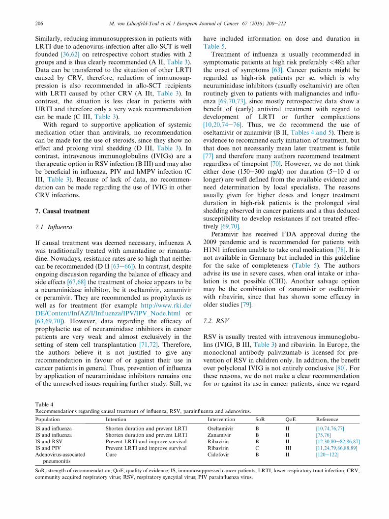
Table 4Recommendations regarding causal treatment of influenza, RSV, parainflu
Population Intention
IS and influenza Shorten duration and prevent LRTI
IS and influenza Shorten duration and prevent LRTI
IS and RSV Prevent LRTI and improve survival
IS and PIV Prevent LRTI and improve survival
Adenovirus-associated
pneumonitis
Cure
SoR, strength of recommendation; QoE, quality of evidence; IS, immunosu
community acquired respiratory virus; RSV, respiratory syncytial virus; PI
have included information on dose and duration in
Table 5.
Treatment of influenza is usually recommended in
symptomatic patients at high risk preferably <48h after
the onset of symptoms [63]. Cancer patients might be
regarded as high-risk patients per se, which is why
neuraminidase inhibitors (usually oseltamivir) are often
routinely given to patients with malignancies and influ-enza [69,70,73], since mostly retrospective data show a
benefit of (early) antiviral treatment with regard to
development of LRTI or further complications
[10,20,74e76]. Thus, we do recommend the use of
oseltamivir or zanamivir (B II, Tables 4 and 5). There is
evidence to recommend early initiation of treatment, but
that does not necessarily mean later treatment is futile
[77] and therefore many authors recommend treatmentregardless of timepoint [70]. However, we do not think
either dose (150e300 mg/d) nor duration (5e10 d or
longer) are well defined from the available evidence and
need determination by local specialists. The reasons
usually given for higher doses and longer treatment
duration in high-risk patients is the prolonged viral
shedding observed in cancer patients and a thus deduced
susceptibility to develop resistances if not treated effec-tively [69,70].
Peramivir has received FDA approval during the
2009 pandemic and is recommended for patients with
H1N1 infection unable to take oral medication [78]. It is
not available in Germany but included in this guideline
for the sake of completeness (Table 5). The authors
advise its use in severe cases, when oral intake or inha-
lation is not possible (CIII). Another salvage optionmay be the combination of zanamivir or oseltamivir
with ribavirin, since that has shown some efficacy in
older studies [79].
7.2. RSV
RSV is usually treated with intravenous immunoglobu-
lins (IVIG, B III, Table 3) and ribavirin. In Europe, the
monoclonal antibody palivizumab is licensed for pre-
vention of RSV in children only. In addition, the benefitover polyclonal IVIG is not entirely conclusive [80]. For
these reasons, we do not make a clear recommendation
for or against its use in cancer patients, since we regard
enza and adenovirus.
Intervention SoR QoE Reference
Oseltamivir B II [10,74,76,77]
Zanamivir B II [75,76]
Ribavirin B II [12,30,80e82,86,87]
Ribavirin C III [11,24,79,86,88,89]
Cidofovir B II [120e122]
ppressed cancer patients; LRTI, lower respiratory tract infection; CRV,
V parainfluenza virus.

Table 5Information on specific drugs.
Name Class Indication Dose Application
mode
Duration Comment Reference
Oseltamivir Neuraminidase
inhibitor
Prophylaxis
influenza
75 mg/d Oral As needed in
seasonal
prophylaxis; 10d in
post-exposure
prophylaxis
Caveat: data too
weak to make a
recommendation,
local strategies
needed
[63,69,71]
Oseltamivir Neuraminidase
inhibitor
Treatment
influenza
2 � 75-
150 mg/d
Oral 5e10 d [10,74,76,77]
Zanamivir Neuraminidase
inhibitor
Prophylaxis
influenza
10 mg/d Inhalation As needed in
seasonal
prophylaxis; 10d in
post-exposure
prophylaxis
Caveat: data too
weak to make a
recommendation,
local strategies
needed
[63,69]
Zanamivir Neuraminidase
inhibitor
Treatment
influenza
2 � 10 mg/d Inhalation Until negativity [75]
Peramivir Neuraminidase
inhibitor
Treatment
influenza
600 mg/d Intravenous Not available in
Germany
[78]
Ribavirin Nucleoside
inhibitor
Treatment RSV,
PIV, hMPV
Daily dose:
2 g for 2 h
every 6 h or
6 g over 18 h
Inhalation 7e10 d Be aware of
potential
teratogenic
effectdspecial
precautions
needed
[82]
Ribavirin Nucleoside
inhibitor
Treatment RSV,
PIV, hMPV
Different
schedulesaOral Be aware of
potential hepatic
and renal
toxicity,
haemolysis
[12,30,80
e82,86,87]
Ribavirin Nucleoside
inhibitor
Treatment RSV,
PIV, hMPV
10e30 mg/kg/
d
Intravenous Be aware of
potential hepatic
and renal
toxicity,
haemolysis
[87]
Cidofovir DNA polymerase
inhibitor
Treatment
adenovirus
Cidofovir 3
e5 mg/kg iv
once weekly
for 2 weeks,
then once
every week
Intravenous To reduce
cidofovir
toxicity, add at
least 2 l of iv
Prehydration and
probenecid 2 g
po 3 h prior and
1 g 2 and 8 h
following
cidofovir
[120e124]
RSV, respiratory syncytial virus; PIV, parainfluenza virus; hMPV, human metapneumovirus.a For example: loading dose: 10 mg/kg, then 3 � 400 mg d2, 3 � 600 mg from d3 [30]; 1800 mg/d [87]; <65 kg body weight: 2 � 400 mg/d;
65e80 kg body weight: 2 � 500 mg/d; >80 kg body weight: 2 � 600 mg/d [12]; <75 kg body weight: 2 � 600 mg/d and �75 kg body weight:
2 � 800 mg/d [81]; 20 mg/kg/d in four divided doses increasing every 24e48 h to 60 mg/kg/d in four divided doses, if tolerated [86].
M. von Lilienfeld-Toal et al. / European Journal of Cancer 67 (2016) 200e212 207
the question whether to use palivizumab instead of
IVIG as an unresolved question requiring further study.
Ribavirin is the agent of choice in the treatment ofRSV infection. Most available data concern allo-SCT
recipients [80], but recent evidence also suggests a benefit
in less severely immunosuppressed cancer patients
[12,81]. It appears to lower the progression rate to LRTI
[82] and is reported to have a positive influence on
survival [12]. However, some authors report favourable
outcome of RSV infections without any causal treat-
ment [61,83,84]. Traditionally, it is used as an aerosol(see Table 5), but this application mode is cumbersome
and may be associated with a teratogeneic effect [85].
Also, patients may not be able to inhale for such a long
time or they may react with bronchospasm. Thus, oralapplication has been used increasingly with a similar
efficacy [12,30,81,86,87] and even intravenous applica-
tion is reported [87]. Despite some reports with a good
outcome without treatment, we believe the available
evidence justifies a recommendation for the use of
ribavirin in cancer patients with RSV infection (B II,
Table 4). Also, at least in high-risk patients the treat-
ment should be given at the stage of URTI, since thishas shown a benefit (B II [82]).

M. von Lilienfeld-Toal et al. / European Journal of Cancer 67 (2016) 200e212208
7.3. Parainfluenza (PIV)
Experience with antiviral therapy (generally ribavirin) inpatients with parainfluenza infection is not very large
and the efficacy is not entirely convincing
[11,24,79,86,88,89]. This may be partly because causal
treatment is started too late in the course of the disease
and partly because the cause of death often is a coin-
fection requiring antibiotic therapy [7,28]. Nonetheless,
it may be reasonable to attempt therapy with ribavirin in
patients with parainfluenza infection (C III, Table 4).
7.4. Adenovirus
Causal therapy with cidofovir is justified in immuno-
suppressed cancer patients with LRTI caused by
adenovirus (B II, Tables 4 and 5). More experimentaltherapies, which are employed in the setting of allo-SCT
include donor-lymphocyte infusions [90] or adaptive
transfer of specific T-cells [91]. However, to date evi-
dence is too weak to justify a recommendation in favour
of or against the use of these treatment modalities.
7.5. Human metapneumovirus (hMPV), rhinovirus,
coronavirus and others
Causal therapy with ribavirin has been attempted in
patients with infections caused by hMPV [92,93], albeit
with unconvincing results. There is not enough evidence
to make a definitive recommendation for or against the
use of any specific antiviral drug or other causal treat-ment approaches like interferon for any of the CRV
other than the ones discussed above.
8. Conclusion and outlook
Early diagnosis and general infection prevention may
improve the outcome of cancer patients with CRV in-
fections. Despite some data regarding some viruses
(influenza, RSV) and patient populations (HSCT-re-
cipients), there is still a lack of information on most
CRV and on other patient populations (for example
those with solid tumours). Also, almost no prospective
randomised trials have been performed for the treatmentof CRV infections in cancer patients. Thus, most rec-
ommendations have to be deduced from other pop-
ulations and further study is urgently needed.
Funding
None. Travel expenses and costs for group meetings
were reimbursed by the German Society for Haematol-
ogy and Medical Oncology.
Conflict of interest statement
MvLT has received honoraria and travel support
from Gilead, MSD, Pfizer, Celgene and Janssen Cilag,
has received travel support from Astellas Pharma and
has received research support from MSD. She is mem-
ber of the advisory board to MSD.MC has received research funding from Deutsche
Forschungsgemeinschaft (DFG) and Erich und Gertrud
Roggenbuck Stiftung, been a speaker for MSD and Basi-
lea, has been a consultant for MSD and Basilea, received
travel grants fromCelgene, Takeda, Gilead andMSD and
is a recipient of the MSD stipend oncology 2013.
MH served on advisory boards of Gilead, Roche
Pharma and Takeda and served on the speakers’ bureaufor Celgene, Novartis, Janssen and Amgen.
CPH is a stock owner of Stada and GSK and has
received consultation fees and/or honoraria from
Schering-Plough, Pfizer, Basilea, Boehringer Ingelheim,
Novartis, Roche, Astellas, Gilead, MSD, Lilly, Inter-
mune, Fresenius, Olympus, Gilead, AstraZeneca,
Bracco, MEDA Pharma, Chiesi, Siemens, Covidien,
Pierre Fabre, Grifols and research funding fromSiemens, Pfizer, MeVis and Boehringer Ingelheim.
MK has received honoraria and travel support and
served on the speakers’ bureau for Gilead, MSD, Pfizer.
OP has received honoraria and travel support from
Astellas, Gilead, Jazz, MSD, Neovii Biotech and Pfizer.
He has received research support from Bio Rad, Gilead,
Jazz, Neovii Biotech, Pierre Fabre, Sanofi and Takeda.
He is member of the advisory board to Alexion, Jazz,Gilead and MSD.
MJGTV is a consultant to: BerlinChemie,MSD/Merck
andAstellasPharma; has servedat the speakers’ bureauof:
Pfizer, Merck/MSD, Gilead Sciences, Organobalance and
Astellas Pharma; received research funding from: 3M,
Astellas Pharma, DaVolterra and Gilead Sciences.
GS has received grant/research support from: MSD
Sharp & Dohme GmbH, Haar, Germany, Pfizer, Berlin,Germany, GILEAD Sciences, Martinsried, Germany
and Astellas Pharma GmbH, Munchen, Germany. She
has served as a consultant to: MSD Sharp & Dohme
GmbH, Haar, Germany and Basilea Pharmaceutical
Internatio Ltd; Switzerland.
All remaining authors have declared no conflicts of
interest.
Acknowledgements
The authors thank Ramona Kraft for technicalassistance with the retrieval of full papers.
References
[1] Ullmann AJ, Cornely OA, Donnelly JP, Akova M,
Arendrup MC, Arikan-Akdagli S, et al. ESCMID* guideline
M. von Lilienfeld-Toal et al. / European Journal of Cancer 67 (2016) 200e212 209
for the diagnosis and management of Candida diseases 2012:
developing European guidelines in clinical microbiology and
infectious diseases. Clin Microbiol Infect 2012;18(Suppl. 7):
1e8.
[2] Mikulska M, Del Bono V, Gandolfo N, Dini S, Dominietto A,
Di Grazia C, et al. Epidemiology of viral respiratory tract in-
fections in an outpatient haematology facility. Ann Hematol
2014;93(4):669e76.[3] European Centre for Disease Prevention and Control. ECDC
Influenza case definitions. http://ecdc.europa.eu/en/healthtopics/
influenza/surveillance/Pages/influenza_case_definitions.aspx.
[Accessed 13 May 2016].
[4] Shachor-Meyouhas Y, Zaidman I, Kra-Oz Z, Arad-Cohen N,
Kassis I. Detection, control, and management of a respiratory
syncytial virus outbreak in a pediatric hematology-oncology
department. J Pediatr Hematol Oncol 2013;35(2):124e8.
[5] Milano F, Campbell AP, Guthrie KA, Kuypers J, Englund JA,
Corey L, et al. Human rhinovirus and coronavirus detection
among allogeneic hematopoietic stem cell transplantation re-
cipients. Blood 2010;115(10):2088e94.
[6] Peck AJ, Englund JA, Kuypers J, Guthrie KA, Corey L,
Morrow R, et al. Respiratory virus infection among hemato-
poietic cell transplant recipients: evidence for asymptomatic
parainfluenza virus infection. Blood 2007;110(5):1681e8.
[7] Harvala H, Gaunt E, McIntyre C, Roddie H, Labonte S,
Curran E, et al. Epidemiology and clinical characteristics of
parainfluenza virus 3 outbreak in a Haemato-oncology unit. J
Infect 2012;65(3):246e54.
[8] Hirsch HH, Martino R, Ward KN, Boeckh M, Einsele H,
Ljungman P. Fourth European Conference on Infections in
Leukaemia (ECIL-4): guidelines for diagnosis and treatment of
human respiratory syncytial virus, parainfluenza virus, meta-
pneumovirus, rhinovirus, and coronavirus. Clin Infect Dis 2013;
56(2):258e66.[9] Hall CB. Respiratory syncytial virus and parainfluenza virus. N
Engl J Med 2001;344(25):1917e28.
[10] Nichols WG, Guthrie KA, Corey L, Boeckh M. Influenza in-
fections after hematopoietic stem cell transplantation: risk fac-
tors, mortality, and the effect of antiviral therapy. Clin Infect Dis
2004;39(9):1300e6.
[11] Nichols WG, Corey L, Gooley T, Davis C, Boeckh M. Para-
influenza virus infections after hematopoietic stem cell trans-
plantation: risk factors, response to antiviral therapy, and effect
on transplant outcome. Blood 2001;98(3):573e8.
[12] Lehners N, Schnitzler P, Geis S, Puthenparambil J, Benz MA,
Alber B, et al. Risk factors and containment of respiratory
syncytial virus outbreak in a hematology and transplant unit.
Bone Marrow Transplant 2013;48(12):1548e53.
[13] Hoellein A, Hecker J, Hoffmann D, Gottle F, Protzer U,
Peschel C, et al. Serious outbreak of human metapneumovirus in
patients with hematologic malignancies. Leuk Lymphoma 2015:
1e5.
[14] Abdallah A, Rowland KE, Schepetiuk SK, To LB, Bardy P. An
outbreak of respiratory syncytial virus infection in a bone
marrow transplant unit: effect on engraftment and outcome of
pneumonia without specific antiviral treatment. Bone Marrow
Transplant 2003;32(2):195e203.
[15] Jones BL, Clark S, Curran ET, McNamee S, Horne G,
Thakker B, et al. Control of an outbreak of respiratory syncytial
virus infection in immunocompromised adults. J Hosp Infect
2000;44(1):53e7.
[16] Chu HY, Englund JA, Podczervinski S, Kuypers J,
Campbell AP, Boeckh M, et al. Nosocomial transmission of
respiratory syncytial virus in an outpatient cancer center. Biol
Blood Marrow Transplant 2014;20(6):844e51.
[17] Peck AJ, Corey L, Boeckh M. Pretransplantation respiratory
syncytial virus infection: impact of a strategy to delay trans-
plantation. Clin Infect Dis 2004;39(5):673e80.
[18] Ljungman P, Ward KN, Crooks BN, Parker A, Martino R,
Shaw PJ, et al. Respiratory virus infections after stem cell
transplantation: a prospective study from the Infectious Dis-
eases Working Party of the European Group for Blood and
Marrow Transplantation. Bone Marrow Transplant 2001;
28(5):479e84.
[19] Ljungman P, de la Camara R, Perez-Bercoff L, Abecasis M,
Nieto Campuzano JB, Cannata-Ortiz MJ, et al. Outcome of
pandemic H1N1 infections in hematopoietic stem cell transplant
recipients. Haematologica 2011;96(8):1231e5.
[20] Chemaly RF, Vigil KJ, Saad M, Vilar-Compte D, Cornejo-
Juarez P, Perez-Jimenez C, et al. A multicenter study of
pandemic influenza A (H1N1) infection in patients with solid
tumors in 3 countries: early therapy improves outcomes. Cancer
2012;118(18):4627e33.
[21] Choi SM, Xie H, Campbell AP, Kuypers J, Leisenring W,
Boudreault AA, et al. Influenza viral RNA detection in blood as
a marker to predict disease severity in hematopoietic cell trans-
plant recipients. J Infect Dis 2012;206(12):1872e7.[22] Schnell D, Mayaux J, de Bazelaire C, Legoff J, Feuillet S,
Scieux C, et al. Risk factors for pneumonia in immunocom-
promised patients with influenza. Respir Med 2010;104(7):
1050e6.[23] Debiaggi M, Canducci F, Sampaolo M, Marinozzi MC,
Parea M, Terulla C, et al. Persistent symptomless human met-
apneumovirus infection in hematopoietic stem cell transplant
recipients. J Infect Dis 2006;194(4):474e8.[24] Marcolini JA, Malik S, Suki D, Whimbey E, Bodey GP.
Respiratory disease due to parainfluenza virus in adult leu-
kemia patients. Eur J Clin Microbiol Infect Dis 2003;22(2):
79e84.
[25] Williams JV, Martino R, Rabella N, Otegui M, Parody R,
Heck JM, et al. A prospective study comparing human meta-
pneumovirus with other respiratory viruses in adults with he-
matologic malignancies and respiratory tract infections. J Infect
Dis 2005;192(6):1061e5.
[26] Renaud C, Xie H, Seo S, Kuypers J, Cent A, Corey L, et al.
Mortality rates of human metapneumovirus and respiratory
syncytial virus lower respiratory tract infections in hematopoietic
cell transplantation recipients. Biol Blood Marrow Transplant
2013;19(8):1220e6.
[27] Debur MC, Vidal LR, Stroparo E, Nogueira MB, Almeida SM,
Takahashi GA, et al. Human metapneumovirus infection in
hematopoietic stem cell transplant recipients. Transpl Infect Dis
2010;12(2):173e9.[28] Ustun C, Slaby J, Shanley RM, Vydra J, Smith AR, Wagner JE,
et al. Human parainfluenza virus infection after hematopoietic
stem cell transplantation: risk factors, management, mortality,
and changes over time. Biol Blood Marrow Transplant 2012;
18(10):1580e8.
[29] Parody R, Rabella N, Martino R, Otegui M, del Cuerpo M,
Coll P, et al. Upper and lower respiratory tract infections by
human enterovirus and rhinovirus in adult patients with
hematological malignancies. Am J Hematol 2007;82(9):
807e11.
[30] Khanna N, Widmer AF, Decker M, Steffen I, Halter J, Heim D,
et al. Respiratory syncytial virus infection in patients with he-
matological diseases: single-center study and review of the
literature. Clin Infect Dis 2008;46(3):402e12.
[31] Shah DP, Ghantoji SS, Ariza-Heredia EJ, Shah JN, El
Taoum KK, Shah PK, et al. Immunodeficiency scoring index to
predict poor outcomes in hematopoietic cell transplant recipients
with RSV infections. Blood 2014;123(21):3263e8.
[32] Seo S, Xie H, Campbell AP, Kuypers JM, Leisenring WM,
Englund JA, et al. Parainfluenza virus lower respiratory tract
disease after hematopoietic cell transplant: viral detection in
the lung predicts outcome. Clin Infect Dis 2014;58(10):
1357e68.

M. von Lilienfeld-Toal et al. / European Journal of Cancer 67 (2016) 200e212210
[33] Cooper RJ, Hallett R, Tullo AB, Klapper PE. The epidemiology
of adenovirus infections in Greater Manchester, UK 1982e96.
Epidemiol Infect 2000;125(2):333e45.
[34] van Tol MJ, Kroes AC, Schinkel J, Dinkelaar W, Claas EC, Jol-
van der Zijde CM, et al. Adenovirus infection in paediatric stem
cell transplant recipients: increased risk in young children with a
delayed immune recovery. Bone Marrow Transplant 2005;36(1):
39e50.[35] Veltrop-Duits LA, van Vreeswijk T, Heemskerk B, Thijssen JC,
El Seady R, Jol-van der Zijde EM, et al. High titers of pre-
existing adenovirus serotype-specific neutralizing antibodies in
the host predict viral reactivation after allogeneic stem cell
transplantation in children. Clin Infect Dis 2011;52(12):1405e13.
[36] Chakrabarti S, Mautner V, Osman H, Collingham KE,
Fegan CD, Klapper PE, et al. Adenovirus infections following
allogeneic stem cell transplantation: incidence and outcome in
relation to graft manipulation, immunosuppression, and im-
mune recovery. Blood 2002;100(5):1619e27.
[37] Munoz-Cobo B, Solano C, Nieto J, de la Camara R,
Remigia MJ, Garcia-Noblejas A, et al. Surveillance for adeno-
virus DNAemia early after transplantation in adult recipients of
unrelated-donor allogeneic stem cell transplants in the absence of
clinically suspected infection. Bone Marrow Transplant 2011;
46(11):1484e6.
[38] Ohrmalm L, Lindblom A, Omar H, Norbeck O, Gustafson I,
Lewensohn-Fuchs I, et al. Evaluation of a surveillance strategy
for early detection of adenovirus by PCR of peripheral blood in
hematopoietic SCT recipients: incidence and outcome. Bone
Marrow Transplant 2011;46(2):267e72.
[39] McCarthy T, Lebeck MG, Capuano AW, Schnurr DP,
Gray GC. Molecular typing of clinical adenovirus specimens by
an algorithm which permits detection of adenovirus coinfections
and intermediate adenovirus strains. J Clin Virol 2009;46(1):
80e4.[40] Drexler JF, Helmer A, Kirberg H, Reber U, Panning M,
Muller M, et al. Poor clinical sensitivity of rapid antigen test for
influenza A pandemic (H1N1) 2009 virus. Emerg Infect Dis
2009;15(10):1662e4.
[41] Ebihara T, Endo R, Ma X, Ishiguro N, Kikuta H. Detection of
human metapneumovirus antigens in nasopharyngeal secretions
by an immunofluorescent-antibody test. J Clin Microbiol 2005;
43(3):1138e41.[42] Camps Serra M, Cervera C, Pumarola T, Moreno A, Perello R,
Torres A, et al. Virological diagnosis in community-acquired
pneumonia in immunocompromised patients. Eur Respir J 2008;
31(3):618e24.
[43] Costa C, Libertucci D, Solidoro P, Sinesi F, Bergallo M,
Margio S, et al. Rapid shell vial culture for the detection of
respiratory viruses from bronchoalveolar lavage in immuno-
compromised patients. Panminerva Med 2007;49(1):1e6.
[44] Kim EA, Lee KS, Primack SL, Yoon HK, Byun HS, Kim TS,
et al. Viral pneumonias in adults: radiologic and pathologic
findings. Radiographics 2002;22. Spec No:S137e49.[45] Mayer JL, Lehners N, Egerer G, Kauczor HU, Heussel CP. CT-
morphological characterization of respiratory syncytial virus
(RSV) pneumonia in immune-compromised adults. Rofo 2014;
186(7):686e92.
[46] Franquet T, Rodriguez S, Martino R, Gimenez A, Salinas T,
Hidalgo A. Thin-section CT findings in hematopoietic stem cell
transplantation recipients with respiratory virus pneumonia.
AJR Am J Roentgenol 2006;187(4):1085e90.
[47] Franquet T, Rodriguez S, Martino R, Salinas T, Gimenez A,
Hidalgo A. Human metapneumovirus infection in hematopoietic
stem cell transplant recipients: high-resolution computed to-
mography findings. J Comput Assist Tomogr 2005;29(2):223e7.
[48] Lehners N, Tabatabai J, Prifert C, Wedde M, Puthenparambil J,
Weissbrich B, et al. Long-term shedding of influenza virus,
parainfluenza virus, respiratory syncytial virus and nosocomial
epidemiology in patients with hematological disorders. PLoS
One 2016;11(2):e0148258.
[49] Cowling BJ, Chan KH, Fang VJ, Cheng CK, Fung RO, Wai W,
et al. Facemasks and hand hygiene to prevent influenza trans-
mission in households: a cluster randomized trial. Ann Intern
Med 2009;151(7):437e46.
[50] Hemila H, Chalker E. Vitamin C for preventing and treating the
common cold. Cochrane Database Syst Rev 2013;1:CD000980.
[51] Karsch-Volk M, Barrett B, Kiefer D, Bauer R, Ardjomand-
Woelkart K, Linde K. Echinacea for preventing and treating the
common cold. Cochrane Database Syst Rev 2014;2:CD000530.
[52] Lissiman E, Bhasale AL, Cohen M. Garlic for the common cold.
Cochrane Database Syst Rev 2014;11:CD006206.
[53] Singh M, Das RR. Zinc for the common cold. Cochrane Data-
base Syst Rev 2013;6:CD001364.
[54] Singh M, Singh M. Heated, humidified air for the common cold.
Cochrane Database Syst Rev 2013;6:CD001728.
[55] Chen W, Lewith G, Wang LQ, Ren J, Xiong WJ, Lu F, et al.
Chinese proprietary herbal medicine listed in ’China national
essential drug list’ for common cold: a systematic literature re-
view. PLoS One 2014;9(10):e110560.
[56] Li S, Yue J, Dong BR, Yang M, Lin X, Wu T. Acetaminophen
(paracetamol) for the common cold in adults. Cochrane Data-
base Syst Rev 2013;7:CD008800.
[57] Kim SY, Chang YJ, Cho HM, Hwang YW, Moon YS. Non-
steroidal anti-inflammatory drugs for the common cold.
Cochrane Database Syst Rev 2013;6:CD006362.
[58] Hayden FG, Diamond L, Wood PB, Korts DC, Wecker MT.
Effectiveness and safety of intranasal ipratropium bromide in
common colds. A randomized, double-blind, placebo-controlled
trial. Ann Intern Med 1996;125(2):89e97.
[59] Genuis SJ, Schwalfenberg G, Siy AK, Rodushkin I. Toxic
element contamination of natural health products and pharma-
ceutical preparations. PLoS One 2012;7(11):e49676.
[60] Kenealy T, Arroll B. Antibiotics for the common cold and acute
purulent rhinitis. Cochrane Database Syst Rev 2013;6:
CD000247.
[61] Aslan T, Fassas AB, Desikan R, Siegel D, Munshi N, Mehta J,
et al. Patients with multiple myeloma may safely undergo
autologous transplantation despite ongoing RSV infection and
no ribavirin therapy. Bone Marrow Transplant 1999;24(5):
505e9.[62] Avivi I, Chakrabarti S, Milligan DW, Waldmann H, Hale G,
Osman H, et al. Incidence and outcome of adenovirus disease in
transplant recipients after reduced-intensity conditioning with
alemtuzumab. Biol Blood Marrow Transplant 2004;10(3):
186e94.
[63] Fiore AE, Fry A, Shay D, Gubareva L, Bresee JS, Uyeki TM.
Antiviral agents for the treatment and chemoprophylaxis of
influenza e recommendations of the Advisory Committee on
Immunization Practices (ACIP). MMWR Recomm Rep 2011;
60(1):1e24.
[64] Bright RA, Shay DK, Shu B, Cox NJ, Klimov AI. Adamantane
resistance among influenza A viruses isolated early during the
2005-2006 influenza season in the United States. JAMA 2006;
295(8):891e4.[65] Englund JA, Champlin RE, Wyde PR, Kantarjian H,
Atmar RL, Tarrand J, et al. Common emergence of amantadine-
and rimantadine-resistant influenza A viruses in symptomatic
immunocompromised adults. Clin Infect Dis 1998;26(6):
1418e24.
[66] Hayden FG, Sperber SJ, Belshe RB, Clover RD, Hay AJ,
Pyke S. Recovery of drug-resistant influenza A virus during
therapeutic use of rimantadine. Antimicrob Agents Chemother
1991;35(9):1741e7.
[67] Dobson J, Whitley RJ, Pocock S, Monto AS. Oseltamivir
treatment for influenza in adults: a meta-analysis of randomised
controlled trials. Lancet 2015;385(9979):1729e37.
M. von Lilienfeld-Toal et al. / European Journal of Cancer 67 (2016) 200e212 211
[68] Jefferson T, Jones MA, Doshi P, Del Mar CB, Hama R,
Thompson MJ, et al. Neuraminidase inhibitors for preventing
and treating influenza in healthy adults and children. Cochrane
Database Syst Rev 2014;4:CD008965.
[69] Casper C, Englund J, Boeckh M. How I treat influenza in pa-
tients with hematologic malignancies. Blood 2010;115(7):
1331e42.
[70] Engelhard D, Mohty B, de la Camara R, Cordonnier C,
Ljungman P. European guidelines for prevention and manage-
ment of influenza in hematopoietic stem cell transplantation and
leukemia patients: summary of ECIL-4 (2011), on behalf of
ECIL, a joint venture of EBMT, EORTC, ICHS, and ELN.
Transpl Infect Dis 2013;15(3):219e32.
[71] Vu D, Peck AJ, Nichols WG, Varley C, Englund JA, Corey L,
et al. Safety and tolerability of oseltamivir prophylaxis in he-
matopoietic stem cell transplant recipients: a retrospective case-
control study. Clin Infect Dis 2007;45(2):187e93.
[72] Tomblyn M, Chiller T, Einsele H, Gress R, Sepkowitz K,
Storek J, et al. Guidelines for preventing infectious complica-
tions among hematopoietic cell transplantation recipients: a
global perspective. Biol Blood Marrow Transplant 2009;15(10):
1143e238.
[73] Chemaly RF, Shah DP, Boeckh MJ. Management of respiratory
viral infections in hematopoietic cell transplant recipients and
patients with hematologic malignancies. Clin Infect Dis 2014;
59(Suppl. 5):S344e51.
[74] Machado CM, Boas LS, Mendes AV, da Rocha IF, Sturaro D,
Dulley FL, et al. Use of oseltamivir to control influenza com-
plications after bone marrow transplantation. Bone Marrow
Transplant 2004;34(2):111e4.[75] Johny AA, Clark A, Price N, Carrington D, Oakhill A,
Marks DI. The use of zanamivir to treat influenza A and B
infection after allogeneic stem cell transplantation. Bone
Marrow Transplant 2002;29(2):113e5.[76] Chemaly RF, Torres HA, Aguilera EA, Mattiuzzi G,
Cabanillas M, Kantarjian H, et al. Neuraminidase inhibitors
improve outcome of patients with leukemia and influenza: an
observational study. Clin Infect Dis 2007;44(7):964e7.
[77] Choi SM, Boudreault AA, Xie H, Englund JA, Corey L,
Boeckh M. Differences in clinical outcomes after 2009 influenza
A/H1N1 and seasonal influenza among hematopoietic cell
transplant recipients. Blood 2011;117(19):5050e6.
[78] Birnkrant D, Cox E. The emergency use authorization of per-
amivir for treatment of 2009 H1N1 influenza. N Engl J Med
2009;361(23):2204e7.[79] Sparrelid E, Ljungman P, Ekelof-Andstrom E, Aschan J,
Ringden O, Winiarski J, et al. Ribavirin therapy in bone marrow
transplant recipients with viral respiratory tract infections. Bone
Marrow Transplant 1997;19(9):905e8.[80] Shah JN, Chemaly RF. Management of RSV infections in adult
recipients of hematopoietic stem cell transplantation. Blood
2011;117(10):2755e63.
[81] Marcelin JR, Wilson JW, Razonable RR. Oral ribavirin therapy
for respiratory syncytial virus infections in moderately to
severely immunocompromised patients. Transpl Infect Dis 2014;
16(2):242e50.[82] Shah DP, Ghantoji SS, Shah JN, El Taoum KK, Jiang Y,
Popat U, et al. Impact of aerosolized ribavirin on mortality in
280 allogeneic haematopoietic stem cell transplant recipients
with respiratory syncytial virus infections. J Antimicrob Che-
mother 2013;68(8):1872e80.
[83] Mendes ET, Ramos J, Peixoto D, Dulley F, Alves T, Vilas
Boas LS, et al. An outbreak of respiratory syncytial virus
infection in hematopoietic stem cell transplantation outpatients:
good outcome without specific antiviral treatment. Transpl
Infect Dis 2013;15(1):42e8.
[84] Anaissie EJ, Mahfouz TH, Aslan T, Pouli A, Desikan R,
Fassas A, et al. The natural history of respiratory syncytial virus
infection in cancer and transplant patients: implications for
management. Blood 2004;103(5):1611e7.
[85] Kilham L, Ferm VH. Congenital anomalies induced in hamster
embryos with ribavirin. Science 1977;195(4276):413e4.
[86] Casey J, Morris K, Narayana M, Nakagaki M, Kennedy GA.
Oral ribavirin for treatment of respiratory syncitial virus and
parainfluenza 3 virus infections post allogeneic haematopoietic
stem cell transplantation. Bone Marrow Transplant 2013;48(12):
1558e61.
[87] Gueller S, Duenzinger U, Wolf T, Ajib S, Mousset S, Berger A,
et al. Successful systemic high-dose ribavirin treatment of res-
piratory syncytial virus-induced infections occurring pre-
engraftment in allogeneic hematopoietic stem cell transplant
recipients. Transpl Infect Dis 2013;15(4):435e40.
[88] Maziarz RT, Sridharan P, Slater S, Meyers G, Post M,
Erdman DD, et al. Control of an outbreak of human para-
influenza virus 3 in hematopoietic stem cell transplant recipients.
Biol Blood Marrow Transplant 2010;16(2):192e8.
[89] Hohenthal U, Nikoskelainen J, Vainionpaa R, Peltonen R,
Routamaa M, Itala M, et al. Parainfluenza virus type 3 in-
fections in a hematology unit. Bone Marrow Transplant 2001;
27(3):295e300.
[90] Hromas R, Cornetta K, Srour E, Blanke C, Broun ER. Donor
leukocyte infusion as therapy of life-threatening adenoviral in-
fections after T-cell-depleted bone marrow transplantation.
Blood 1994;84(5):1689e90.
[91] Geyeregger R, Freimuller C, Stemberger J, Artwohl M, Witt V,
Lion T, et al. First-in-man clinical results with good
manufacturing practice (GMP)-compliant polypeptide-expanded
adenovirus-specific T cells after haploidentical hematopoietic
stem cell transplantation. J Immunother 2014;37(4):245e9.
[92] Egli A, Bucher C, Dumoulin A, Stern M, Buser A, Bubendorf L,
et al. Human metapneumovirus infection after allogeneic he-
matopoietic stem cell transplantation. Infection 2012;40(6):
677e84.
[93] Englund JA, Boeckh M, Kuypers J, Nichols WG, Hackman RC,
Morrow RA, et al. Brief communication: fatal human meta-
pneumovirus infection in stem-cell transplant recipients. Ann
Intern Med 2006;144(5):344e9.
[94] Ison MG, Michaels MG. RNA respiratory viral infections in
solid organ transplant recipients. Am J Transplant 2009;9(Suppl.
4):S166e72.[95] Covalciuc KA, Webb KH, Carlson CA. Comparison of four
clinical specimen types for detection of influenza A and B viruses
by optical immunoassay (FLU OIA test) and cell culture
methods. J Clin Microbiol 1999;37(12):3971e4.
[96] Kaiser L, Briones MS, Hayden FG. Performance of virus
isolation and Directigen Flu A to detect influenza A virus in
experimental human infection. J Clin Virol 1999;14(3):191e7.[97] Debyle C, Bulkow L, Miernyk K, Chikoyak L, Hummel KB,
Hennessy T, et al. Comparison of nasopharyngeal flocked
swabs and nasopharyngeal wash collection methods for res-
piratory virus detection in hospitalized children using real-time
polymerase chain reaction. J Virol Methods 2012;185(1):
89e93.
[98] Heikkinen T, Marttila J, Salmi AA, Ruuskanen O. Nasal swab
versus nasopharyngeal aspirate for isolation of respiratory vi-
ruses. J Clin Microbiol 2002;40(11):4337e9.
[99] Li L, Chen QY, Li YY, Wang YF, Yang ZF, Zhong NS.
Comparison among nasopharyngeal swab, nasal wash, and
oropharyngeal swab for respiratory virus detection in adults with
acute pharyngitis. BMC Infect Dis 2013;13:281.
[100] Macfarlane P, Denham J, Assous J, Hughes C. RSV testing in
bronchiolitis: which nasal sampling method is best? Arch Dis
Child 2005;90(6):634e5.
[101] Meerhoff TJ, Houben ML, Coenjaerts FE, Kimpen JL,
Hofland RW, Schellevis F, et al. Detection of multiple respira-
tory pathogens during primary respiratory infection: nasal swab

M. von Lilienfeld-Toal et al. / European Journal of Cancer 67 (2016) 200e212212
versus nasopharyngeal aspirate using real-time polymerase chain
reaction. Eur J Clin Microbiol Infect Dis 2010;29(4):365e71.
[102] Sung RY, Chan PK, Choi KC, Yeung AC, Li AM, Tang JW,
et al. Comparative study of nasopharyngeal aspirate and nasal
swab specimens for diagnosis of acute viral respiratory infection.
J Clin Microbiol 2008;46(9):3073e6.
[103] Waris M, Osterback R, Lahti E, Vuorinen T, Ruuskanen O,
Peltola V. Comparison of sampling methods for the detection of
human rhinovirus RNA. J Clin Virol 2013;58(1):200e4.
[104] Kuypers J, Campbell AP, Cent A, Corey L, Boeckh M. Com-
parison of conventional and molecular detection of respiratory
viruses in hematopoietic cell transplant recipients. Transpl Infect
Dis 2009;11(4):298e303.
[105] Falsey AR, Formica MA, Treanor JJ, Walsh EE. Comparison of
quantitative reverse transcription-PCR to viral culture for
assessment of respiratory syncytial virus shedding. J Clin
Microbiol 2003;41(9):4160e5.
[106] Hindiyeh M, Hillyard DR, Carroll KC. Evaluation of the Pro-
desse Hexaplex multiplex PCR assay for direct detection of seven
respiratory viruses in clinical specimens. Am J Clin Pathol 2001;
116(2):218e24.
[107] Chen KF, Rothman RE, Ramachandran P, Blyn L, Sampath R,
Ecker DJ, et al. Rapid identification viruses from nasal
pharyngeal aspirates in acute viral respiratory infections by RT-
PCR and electrospray ionization mass spectrometry. J Virol
Methods 2011;173(1):60e6.
[108] Abbo L, Quartin A, Morris MI, Saigal G, Ariza-Heredia E,
Mariani P, et al. Pulmonary imaging of pandemic influenza
H1N1 infection: relationship between clinical presentation and
disease burden on chest radiography and CT. Br J Radiol 2010;
83(992):645e51.
[109] Chandler TM, Leipsic J, Nicolaou S, Quiney B, Romney M,
Muller NL, et al. Confirmed swine-origin influenza A(H1N1)
viral pneumonia: computed tomographic findings in the immu-
nocompetent and the immunocompromised. J Comput Assist
Tomogr 2011;35(5):602e7.
[110] El-Badrawy A, Zeidan A, Ebrahim MA. 64 multidetector CT
findings of influenza A (H1N1) virus in patients with hemato-
logic malignancies. Acta Radiol 2012;53(6):662e7.
[111] Elicker BM, Schwartz BS, Liu C, Chen EC, Miller SA, Chiu CY,
et al. Thoracic CT findings of novel influenza A (H1N1) infec-
tion in immunocompromised patients. Emerg Radiol 2010;17(4):
299e307.
[112] Ko JP, Shepard JA, Sproule MW, Trotman-Dickenson B,
Drucker EA, Ginns LC, et al. CT manifestations of respiratory
syncytial virus infection in lung transplant recipients. J Comput
Assist Tomogr 2000;24(2):235e41.
[113] Laqmani A, Adam G, Regier M. Pulmonary manifestation of
novel swine-origin influenza A (H1N1) virus (S-OIV) infection in
immunocompromised patients: initial findings with multi-
detector computed tomography. Med Princ Pract 2012;21(6):
548e53.
[114] Oikonomou A, Muller NL, Nantel S. Radiographic and high-
resolution CT findings of influenza virus pneumonia in patients
with hematologic malignancies. AJR Am J Roentgenol 2003;
181(2):507e11.
[115] Syha R, Beck R, Hetzel J, Ketelsen D, Grosse U, Springer F,
et al. Human metapneumovirus (HMPV) associated pulmonary
infections in immunocompromised adultseinitial CT findings,
disease course and comparison to respiratory-syncytial-virus
(RSV) induced pulmonary infections. Eur J Radiol 2012;81(12):
4173e8.
[116] Jefferson T, Del Mar CB, Dooley L, Ferroni E, Al-Ansary LA,
Bawazeer GA, et al. Physical interventions to interrupt or reduce
the spread of respiratory viruses. Cochrane Database Syst Rev
2011;(7):CD006207.
[117] RSV Outbreak Investigation Team. Contributing and termi-
nating factors of a large RSV outbreak in an Adult Hematology
and Transplant Unit. PLoS Curr 2014:6.
[118] Falsey AR. Current management of parainfluenza pneumonitis
in immunocompromised patients: a review. Infect Drug Resist
2012;5:121e7.[119] Gokturk B, Pekcan S, Guner SN, Artac H, Keles S, Kirac M,
et al. Efficacy of intravenous immunoglobulin treatment in
immunocompromised children with H1N1 influenza: a clinical
observation. Clin Respir J 2016 Mar;10(2):223e30.[120] Ljungman P, Ribaud P, Eyrich M, Matthes-Martin S, Einsele H,
Bleakley M, et al. Cidofovir for adenovirus infections after
allogeneic hematopoietic stem cell transplantation: a survey by
the Infectious Diseases Working Party of the European Group
for Blood and Marrow Transplantation. Bone Marrow Trans-
plant 2003;31(6):481e6.
[121] Robin M, Marque-Juillet S, Scieux C, Peffault de Latour R,
Ferry C, Rocha V, et al. Disseminated adenovirus infections
after allogeneic hematopoietic stem cell transplantation: inci-
dence, risk factors and outcome. Haematologica 2007;92(9):
1254e7[122] Legrand F, Berrebi D, Houhou N, Freymuth F, Faye A,
Duval M, et al. Early diagnosis of adenovirus infection and
treatment with cidofovir after bone marrow transplantation in
children. Bone Marrow Transplant 2001;27(6):621e6.[123] Leruez-Ville M, Chardin-Ouachee M, Neven B, Picard C, Le
Guinche I, Fischer A, et al. Description of an adenovirus A31
outbreak in a paediatric haematology unit. Bone Marrow
Transplant 2006;38(1):23e8.
[124] Nicolasora NP, Reddy P, Kaul DR. Biopsy-proven adenoviral
diarrhea responding to low-dose cidofovir. Transpl Infect Dis
2008;10(5):346e50.